

Submitted:
17 September 2026
Posted:
28 September 2026
You are already at the latest version
Abstract
Lipedema is a chronic, predominantly female disorder characterized by disproportionate regional expansion of subcutaneous adipose tissue, pain, tenderness and easy bruising. Although adipocyte hypertrophy, altered progenitor-cell behavior, microvascular dysfunction, extracellular-matrix remodeling, immune-cell alterations and variable lymphatic abnormalities have been reported, their causal and temporal relationships remain unresolved. Here, we propose that these findings may represent interacting manifestations of dysregulation within an adipovascular unit (AVU) rather than independent pathological processes.
We define the AVU as a spatially organized regenerative niche comprising adipose stem and progenitor cells, microvascular endothelial and mural cells, immune populations, extracellular matrix and mature adipocytes, linked through reciprocal cellular, paracrine and extracellular-vesicle-mediated communication. We hypothesize that lipedema may arise when physiological, female hormone-responsive adipose remodeling encounters a regionally susceptible AVU in which regenerative activity is inadequately constrained in space, magnitude or duration. Subsequent cross-compartment remodeling could amplify and stabilize the altered tissue state, potentially linking disproportionate adipose expansion with vascular, interstitial, immune, extracellular-matrix and sensory manifestations.
Current evidence is compatible with this framework but does not establish the AVU as the primary site of disease initiation or demonstrate a self-maintaining regenerative state. We therefore derive falsifiable predictions involving paired affected and spared adipose depots, context-dependent endocrine responses, functional cross-talk between AVU compartments, extracellular-vesicle transfer, spatial biological boundaries and persistence after stimulus withdrawal. Testing these predictions using spatially resolved and multicellular experimental systems should determine whether AVU dysregulation represents a causal organizing principle of lipedema biology or a descriptive consequence of disease.
Keywords:
lipedema
; adipovascular unit
; adipose stem and progenitor cells
; adipose regeneration
; estrogen
; microvascular niche
; regional susceptibility
; extracellular vesicles
; regenerative lock-in
1. Introduction
Lipedema is a chronic disorder of subcutaneous adipose tissue characterized by disproportionate, typically symmetrical extremity enlargement, pain and tenderness, easy bruising, and a striking female predominance. Histological and molecular studies report adipocyte hypertrophy, altered adipogenic differentiation, microvascular abnormalities, extracellular-matrix (ECM) remodeling, immune-cell changes, and variable lymphatic dysfunction, but their temporal and causal relationships remain unresolved [1,2,3].
Its association with puberty, pregnancy, and menopause implicates female reproductive physiology without establishing systemic hormone excess as the cause. A more plausible possibility is that physiological endocrine signals act on regionally susceptible adipose tissue. This is biologically credible because subcutaneous adipose tissue is a dynamic regenerative organ in which adipose stem and progenitor cells (ASPCs), endothelial and mural cells, immune populations, ECM, and mature adipocytes interact within spatially organized microenvironments [2,4,5,7,8,9].
Lipedema-specific studies identify abnormalities in stromal/progenitor phenotypes, adipogenic responses, vascular and immune compartments, and extracellular-vesicle (EV) cargo, although results vary with anatomical sampling and experimental context [10,11,12,13,14]. Such heterogeneity argues against a simple cell-autonomous hyperadipogenic disorder and instead suggests dysregulation of a multicellular regenerative system.
We therefore propose the adipovascular unit (AVU) as a spatially organized regenerative niche integrating ASPCs, microvascular endothelial and mural cells, immune populations, ECM, mature adipocytes, and their paracrine and EV-mediated communication. We hypothesize that lipedema may arise when physiological female hormone-responsive remodeling encounters a regionally susceptible AVU in which regenerative activity is inadequately constrained in space, magnitude, or duration. Reciprocal cross-compartment remodeling could then amplify and stabilize the altered tissue state. The following sections develop this model and derive experimentally testable predictions.
2. The Adipovascular Unit as a Regenerative Niche
Adipose tissue remodels throughout adult life through adipocyte hypertrophy and, when required, hyperplasia driven by resident ASPCs. Progenitor identity and differentiation are influenced by sex, anatomical location, and the local microvascular environment [4,5]. Single-cell and spatial studies further show that adipose stem cells are heterogeneous fibroblast-like populations that contribute both adipocyte precursors and ECM-regulating functions, with sexually dimorphic transcriptional states and adipogenic commitment linked to position within the microvascular niche [5].
Recent single-cell mapping of the human subcutaneous adipose vascular niche further demonstrates substantial endothelial heterogeneity and phenotypic plasticity. AlZaim et al. identified canonical arterial, venous, capillary and lymphatic endothelial populations together with a heterogeneous sub-endothelial population exhibiting mixed endothelial, mesenchymal, adipogenic and immune transcriptional features [6]. Computational trajectory analyses and experimental validation suggested that some of these states may emerge through partial endothelial-to-mesenchymal transition (EndMT), highlighting a previously underappreciated plasticity of the adipose vascular compartment. Importantly, these observations were obtained in adipose tissue across metabolic states rather than in lipedema and therefore provide biological plausibility for an integrated adipovascular niche without demonstrating disease-specific EndMT in lipedema.
A key distinction is that ASPCs and vascular mural cells are closely associated but biologically distinguishable populations [5]. The microvasculature is therefore not simply a supply network: endothelial cells, mural cells, progenitors, immune cells, stromal cells, and ECM form a spatially coupled environment in which reciprocal signaling coordinates tissue expansion. Regional differences in progenitor biology and adipose function provide a plausible substrate for the characteristic distribution of lipedema despite uniform systemic endocrine exposure [5,8,9].
2.1. Defining the Adipovascular Unit
We define the adipovascular unit (AVU) as a functional and spatial framework describing the regenerative microenvironment in which adipose progenitor biology and microvascular adaptation are coordinated (Figure 1). Its principal components are ASPCs; microvascular endothelial cells; vascular mural cells, including pericytes and vascular smooth-muscle cells; ECM; resident and recruited immune cells; and mature adipocytes. These components influence one another through direct cellular interactions, soluble mediators, matrix-dependent signaling, and EV-mediated communication.
The AVU is not proposed as a newly discovered anatomical organ but as a systems-level construct for a spatially coupled regenerative niche. Reciprocity is central: adipose expansion requires vascular and matrix adaptation, while vascular, immune, and matrix states can modify progenitor behavior and adipocyte function. Applied to lipedema, this architecture updates our earlier predominantly sequential regenerative-imbalance model toward a network model in which no single compartment is assumed a priori to be the universal initiating lesion [15].
2.2. Physiological Adipose Regeneration and Regional Susceptibility
Hormone-responsive adipose regeneration is physiological. Female subcutaneous adipose tissue has greater capacity for hyperplastic expansion, and estrogen can preserve or recruit committed progenitor populations [7,8,9]. The relevant distinction is therefore not the presence of estrogen-responsive adipogenesis, but whether its regional control, magnitude, duration, or termination is altered.
Three observations motivate this interpretation. First, affected and spared depots coexist within the same systemic endocrine environment, arguing against a purely systemic explanation. Second, normal progenitor biology is sexually dimorphic and estrogen responsive [5,7]. Third, lipedema abnormalities span adipogenic, vascular, immune, matrix, EV, and variably lymphatic compartments [10,11,12,13,14,20,21,22,23,24,25,26]. Together, these findings are compatible with dysregulation of a shared regenerative tissue system rather than independent pathological processes.
2.3. From Physiological to Dysregulated AVU
A useful distinction can therefore be drawn between physiological and dysregulated AVU states. In physiological remodeling, endocrine and metabolic signals induce controlled ASPC recruitment, coordinated microvascular and ECM adaptation, limited adipocyte formation, and restoration of tissue homeostasis. In the proposed lipedema state, the same class of physiological signals encounters a regionally susceptible AVU, producing altered progenitor behavior together with vascular, stromal, immune, and matrix remodeling and disproportionate adipose expansion.
This sequence is a hypothesis, not an experimentally established pathway. It predicts that affected and spared depots should differ in progenitor state, cellular composition, spatial organization, or response to standardized endocrine stimulation; that perturbing one AVU compartment should influence others if cross-talk is causal; and that a self-reinforcing state, if present, should show at least partial persistence after removal of the initiating stimulus.
3. Female Reproductive Physiology and Regional AVU Susceptibility
The female predominance of lipedema and its association with reproductive transitions make reproductive physiology relevant to disease expression, but they do not demonstrate pathological circulating estrogen concentrations. The central question is whether physiological reproductive signals are processed differently by susceptible adipose regions.
3.1. Sexual Dimorphism and Hormone-Responsive Adipose Regeneration
Women preferentially accumulate subcutaneous adipose tissue in gluteofemoral and other peripheral depots, reflecting sex- and depot-dependent adipose biology rather than differences in mature adipocyte size alone [8,9]. Scheidl et al. showed in mice that female subcutaneous depots preserve and recruit committed progenitors more effectively during adipogenic stimulation; ovariectomy reduced this phenotype and 17β-estradiol restored it [7]. Estrogen-responsive hyperplastic expansion is therefore part of normal female adipose physiology.
Uhrbom et al. independently demonstrated sexual dimorphism in adipose stem-cell transcriptional programs and showed that adipogenic commitment is related to anatomical position within the microvascular niche [5]. Together, these observations provide a mechanistic bridge between two defining features of lipedema: female specificity and regional distribution. A systemic reproductive signal could generate different regenerative responses among depots without requiring major differences in circulating hormone concentrations.
3.2. Reproductive Transitions and Altered Estrogen Responsiveness in Lipedema
Clinical descriptions associate lipedema onset or progression with puberty, pregnancy, and menopause, but these transitions represent very different endocrine states [2,16]. Their common feature is not a uniform estrogen concentration but the need for substantial adaptation to changing reproductive, metabolic, and vascular demands. Within the AVU model, reproductive transitions are therefore viewed as physiological challenges to regional regenerative systems rather than episodes of simple estrogen excess.
Experimental data support this distinction. Estrogen regulates the female adipose progenitor compartment in vivo [7], whereas lipedema-derived cells show context-dependent responses to 17β-estradiol in two- and three-dimensional systems, including altered estrogen-receptor and adipogenic signaling [12,13]. The dependence of these responses on dimensionality supports a role for cell-cell, cell-matrix, and niche interactions rather than a purely cell-autonomous hormonal effect.
Lipedema-specific stromal and vascular studies further identify abnormalities across multiple SVF compartments [10,20]. Strohmeier et al. reported regionally increased CYP19A1 expression in affected thigh adipose tissue and increased ZNF423 in endothelial- and pericyte-associated compartments [17,18,19,20]. These findings are compatible with altered local estrogen metabolism and adipogenic signaling within the vascular niche, but they do not demonstrate increased aromatase activity, elevated local estradiol concentrations, or an initiating role for aromatase. The relevant hypothesis is therefore altered local processing and response to physiological endocrine signals, not systemic estrogen excess.
3.3. The Regionality Paradox and Regional AVU Susceptibility
A purely endocrine explanation faces a fundamental regionality problem: puberty, pregnancy, and menopause alter the endocrine environment of the entire organism, whereas lipedema selectively affects characteristic extremity depots while sparing others. If the signal is systemic, why is the pathological response regional?
We propose regional AVU susceptibility as the missing variable. Depot-specific developmental programs, progenitor composition, vascular organization, ECM mechanics, local steroid metabolism, immune tone, and lymphatic/interstitial capacity could create different response thresholds among adipose regions [5,8,9,20]. This does not identify which feature is primary; it predicts that susceptibility should be measurable by comparing affected and spared depots within the same individual.
3.4. From Physiological Adaptation to Pathological Persistence
The transition from physiological adaptation to disease may therefore depend on failure to terminate or spatially constrain a normally adaptive regenerative response. Reproductive or metabolic signals could transiently increase progenitor recruitment, vascular adaptation, and matrix remodeling; in a susceptible AVU, reciprocal interactions among these compartments could prolong or amplify the response after the original stimulus has diminished.
This proposed persistence is termed regenerative lock-in. It is not established by current evidence and should not be interpreted as irreversible disease or as proof that a particular compartment initiates lipedema. Rather, it is a testable systems-level possibility: disease-associated cellular behavior should persist at least partly after withdrawal of candidate stimuli and should depend on multicellular context if reciprocal reinforcement is mechanistically important.
4. From Regional Susceptibility to Regenerative Imbalance and Multicellular Amplification
If regional susceptibility determines where lipedema develops, the next question is how a local regenerative response could become a stable multicellular tissue phenotype. Available evidence is compatible with abnormalities in progenitor, vascular, immune, adipocyte, ECM, and interstitial compartments, but does not establish their temporal hierarchy [10,11,12,13,14,20,21,22,23,24,25,26].
4.1. Regenerative Imbalance and Altered Progenitor Control
Lipedema tissue shows altered stromal/progenitor phenotypes and adipogenic responses, but the direction and magnitude of these changes vary across experimental systems [10,11,12,13]. This variability argues against a universal intrinsic hyperadipogenic defect. Instead, progenitor behavior may depend on disease stage, anatomical site, hormonal context, and the cellular and matrix environment in which ASPCs are studied.
Within the AVU model, regenerative imbalance therefore means loss of coordinated control rather than simply increased adipogenesis: progenitor recruitment, differentiation, and termination become mismatched to vascular, matrix, and tissue demands.
4.2. Multicellular Communication Within the Dysregulated AVU
Evidence for cross-compartment communication is emerging. Lipedema-derived SVF EVs carry characteristic miRNA cargo [14]; endothelial and perivascular abnormalities occur in early-stage tissue [20]; and macrophage-associated signals can influence adipocyte differentiation [23]. These findings demonstrate potential communication routes but do not establish a single causal direction.
The AVU model therefore treats endothelial, mural, progenitor, immune, and matrix compartments as a coupled network. This network interpretation is supported by recent single-cell connectomic analyses of human subcutaneous adipose tissue, in which adipocytes, adipose stem and progenitor cells, and adipose endothelial populations emerged as major participants in metabolic-state-dependent intercellular communication. Altered VEGF, TGF-β and BMP signaling, together with collagen- and laminin-associated communication involving endothelial subpopulations, further illustrates how vascular, stromal and adipocyte compartments can behave as an integrated signaling system. Whether analogous communication networks are altered in lipedema remains to be determined [6]. Functional co-culture, transfer, and perturbation experiments are required to determine which abnormalities are transferable, necessary, or sufficient and whether the strongest disease phenotype emerges only in multicellular context.
4.3. Adipocyte Expansion, Extracellular-Matrix Remodeling, and Interstitial Homeostasis
Established lipedema tissue shows adipocyte hypertrophy, adipose remodeling, and stage-associated fibrosis [22,25,26]. ECM changes may alter tissue mechanics and progenitor behavior, while adipocyte expansion increases demands on perfusion, interstitial clearance, and structural support. These relationships are compatible with reciprocal amplification but do not establish whether ECM remodeling is initiating, adaptive, or secondary.
Interstitial and lymphatic findings are similarly heterogeneous. Increased tissue fluid and functional or morphological lymphatic abnormalities have been reported, but primary universal lymphatic failure has not been demonstrated [28,29,30,31,32,33,34,35]. The AVU model therefore positions interstitial and lymphatic dysfunction as possible modifiers, amplifiers, or consequences whose role may vary by phenotype and disease stage.
4.4. Multicellular Amplification and Regenerative Lock-In
A central hypothesis is that reciprocal remodeling can stabilize an abnormal regional state. Progenitor activation and adipose expansion may increase vascular and matrix demands; vascular or interstitial dysfunction may alter the local signaling environment; immune cells can modify stromal and adipogenic behavior; and ECM remodeling can feed back on cellular phenotype [20,22,23,24,25,26]. No current study demonstrates the complete loop or its temporal sequence.
We use regenerative lock-in to describe the testable possibility that these interactions become sufficiently reinforcing for disease-associated behavior to persist after the initiating or amplifying signal is removed. This concept would be supported by persistence in patient-derived multicellular systems and by reversal after targeted disruption of a causal network node; rapid normalization after stimulus withdrawal would argue against it.
4.5. From Dynamic Dysregulation to a Self-Reinforcing Adipovascular System
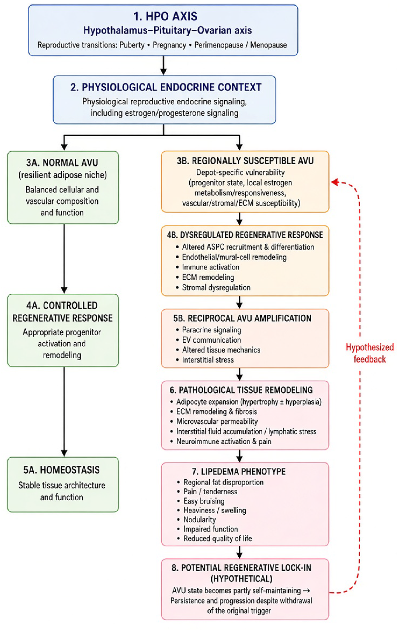
The proposed disease process is therefore better represented as a dynamic network than a linear cascade. Different upstream susceptibilities may enter through progenitor, vascular, immune, matrix, endocrine, or lymphatic pathways and converge on a regional AVU state. The model does not require the same initiating lesion in every patient; its central claim is that multicellular regional interactions may organize and maintain the phenotype (Figure 2).
5. From AVU Dysregulation to the Clinical Lipedema Phenotype
A mechanistic model of lipedema is only meaningful if it can account for the characteristic clinical phenotype. Lipedema is defined by a distinctive combination of regional adipose-tissue expansion and symptoms rather than excess adipose mass alone. Common diagnostic features include pain or pressure sensitivity, bilateral and symmetrical disproportionate enlargement of the extremities, easy bruising, a negative Stemmer sign, and characteristic sparing of the feet [1,2,3,36]. These manifestations may represent different expressions of disturbed regional adipose-tissue homeostasis involving adipogenic, vascular, stromal, matrix, immune, interstitial, and neural compartments.
5.1. Regional Adipose Expansion and Structural Remodeling
The characteristic regional distribution of lipedema, with bilateral and symmetrical enlargement of affected limbs and frequent sparing of the feet [1,2,3], is compatible with depot-specific susceptibility. Differences in progenitor composition, developmental identity, microvascular organization, local steroid metabolism, extracellular matrix, and regenerative thresholds could allow identical systemic signals to generate different tissue responses across anatomical depots.
Histological studies demonstrate adipocyte enlargement in established lipedema, whereas experimental studies indicate altered adipogenic differentiation potential. The AVU model therefore considers adipocyte hypertrophy and potentially increased hyperplastic recruitment as complementary contributors to tissue expansion [10,11,12,13,25,26]. Their relative contributions may vary between patients and disease stages. Increasing interstitial collagen and stage-associated fibrosis further suggest that progressive matrix remodeling may convert a predominantly dynamic cellular abnormality into an increasingly structural tissue state [26].
5.2. Microvascular and Interstitial Manifestations
Easy bruising is characteristic of lipedema, and clinical and experimental studies describe capillary fragility, vascular dilation, increased permeability, and altered endothelial behavior [1,20,25]. Because bruising also depends on vessel-wall integrity, mechanical trauma, perivascular support and coagulation, its precise structural basis remains unresolved. It is nevertheless compatible with altered microvascular and perivascular stability within the affected AVU.
Heaviness and swelling likewise need not imply primary lymphatic failure. Lipedema should not be equated with primary lymphedema, and imaging studies demonstrate heterogeneous lymphatic findings ranging from largely preserved transport to subclinical abnormalities in subsets of patients [28,29,30,31,32,33,34,35]. Disturbed interstitial homeostasis and an emerging load–clearance mismatch could therefore contribute to symptoms before or without severe lymphatic dysfunction.
5.3. Pain and the Dissociation Between Symptoms and Morphology
Pain and pressure sensitivity are defining features of lipedema. Studies support peripheral pain, dermal hypersensitivity and neurogenic inflammatory signaling, while quantitative sensory testing demonstrates regionally altered sensory thresholds [37,38,39]. Potential contributors include altered interstitial mechanics, fibrosis, vascular dysfunction, inflammatory mediators, tissue pressure and peripheral sensory dysfunction, but their causal hierarchy remains unresolved.
Pain may therefore represent an emergent neurovascular–stromal phenotype rather than the endpoint of a simple linear pathway. Morphological stage and symptom severity are not necessarily equivalent [26,39], implying that patients with similar external morphology may occupy different underlying biological states.
5.4. Persistence and Progression as Loss of Tissue Homeostasis
If reproductive transitions contribute to disease manifestation, persistence after the initiating transition has resolved implies durable structural consequences, continuing tissue-level maintenance, or both. The regenerative lock-in hypothesis captures the latter possibility, although clinical persistence alone does not establish it.
Biological progression may involve a shift from altered cellular signaling and regenerative responsiveness toward adipose expansion, multicellular amplification, fibrosis, altered interstitial mechanics and potentially lymphatic impairment [26]. This sequence remains hypothetical and should not replace established clinical staging; rather, it predicts that the dominant biological mechanisms may change during disease progression.
5.5. A Convergent Clinical Phenotype
Regional adipose expansion, bruising, heaviness and swelling, fibrotic remodeling and pain may thus represent different macroscopic manifestations of disturbed regional tissue homeostasis rather than independent pathological processes. The characteristic lipedema phenotype could consequently reflect a regionally susceptible adipose niche in which dysregulated regeneration alters adipose volume, vascular and interstitial behavior, extracellular-matrix architecture and sensory function.
This interpretation generates experimentally testable relationships: affected and spared depots within the same woman should differ biologically; local vascular abnormalities should associate with regional tissue phenotype; and vascular filtration, interstitial-fluid dynamics and lymphatic transport should show phenotype- or stage-dependent relationships. These predictions connect the clinical phenotype directly to experimental validation.
6. Testable Predictions and Experimental Validation of the AVU Hypothesis
The value of the adipovascular unit (AVU) hypothesis depends on whether it generates predictions that distinguish it from alternative explanations of lipedema. If lipedema reflects persistent dysregulation of a regionally susceptible adipose-tissue niche, reproducible biological differences should exist between affected and spared depots, and disease-associated phenotypes should depend at least partly on interactions among AVU compartments. The following predictions provide a framework for experimental validation or falsification.
6.1. Regional and Context-Dependent Predictions
Existing paired-depot, progenitor and hormonal-response studies demonstrate regional and context-dependent differences but do not establish whether these differences define anatomical susceptibility in lipedema [7,13,20,26].
Prediction 1 — Regional AVU dysregulation. Affected adipose tissue should differ from anatomically spared tissue within the same patient in AVU composition, cellular state, spatial organization or intercellular signaling. Paired affected–spared sampling, complemented by anatomically matched controls, provides the most direct test. Repeated biological equivalence between affected and spared depots would substantially weaken the concept of regional AVU susceptibility.
Prediction 2 — Hormonal context dependence. Physiologically relevant reproductive endocrine signals should elicit different responses in affected and spared AVUs from the same patient. Identical responses across relevant hormonal conditions would argue against differential regional endocrine responsiveness as a major determinant of susceptibility.
Prediction 3 — Biological boundary. Spatially resolved sampling across the clinical transition from affected to spared tissue should reveal a corresponding molecular, cellular, structural or functional AVU transition. Failure to identify such a boundary despite precise spatial sampling would challenge the concept of regionally encoded susceptibility.
6.2. Multicellular Communication and Causal Reconstruction
Available functional studies demonstrate selected cross-compartment effects, but their necessity, directionality and sufficiency remain unresolved [14,20,23].
Prediction 4 — Multicellular dependence. Multicellular systems incorporating endothelial, mural, stromal/progenitor, immune, matrix and adipocyte compartments should reproduce disease-associated behavior more completely than isolated monocultures. Robust reproduction of the relevant phenotype by isolated cells alone would reduce the necessity of multicellular AVU dysfunction.
Prediction 5 — Endothelial–stromal cross-talk and vascular plasticity. Reciprocal co-culture of endothelial and stromal/progenitor cells from affected, spared and control tissue should determine whether abnormalities are compartment-intrinsic or emerge through interaction. Single-cell and spatial approaches should additionally test whether affected lipedema tissue contains altered endothelial states, including mesenchymal-, progenitor- or immune-associated transcriptional programs, and whether such states differ between affected and spared depots. Absence of reproducible reciprocal effects or disease-associated endothelial-state differences would weaken a central adipovascular prediction.
Prediction 6 — Extracellular-vesicle transfer. If EVs contribute functionally to disease propagation, EVs from affected tissue or disease-associated AVU populations should transfer measurable phenotypes to recipient cells, with attenuation after EV inhibition or depletion. Failure of such transfer under physiologically relevant conditions would reduce the proposed role of EV-mediated amplification.
Prediction 7 — Network perturbation. Selective perturbation or normalization of one pathological AVU compartment should produce secondary changes in other compartments. Consistent independence of individual compartments would argue against the proposed network architecture.
6.3. AVU States, Clinical Phenotype and Regenerative Lock-In
Disease-associated molecular signatures and stage-dependent remodeling suggest that lipedema may comprise distinct biological states, although longitudinal evidence is lacking [24,26].
Prediction 8 — Changing AVU states. Reproducible AVU-state patterns or trajectories should emerge from combinations of adipogenic activity, vascular dysfunction, immune composition, matrix remodeling, interstitial disturbance and potentially neural involvement [26]. If these abnormalities merely track tissue volume, BMI or conventional stage, AVU-state classification would add limited biological information.
Prediction 9 — Regenerative persistence. In patient-derived multicellular systems, disease-associated features should persist for a measurable period after withdrawal of a candidate initiating or amplifying stimulus if regenerative lock-in exists. Rapid and complete normalization after stimulus withdrawal would argue against a self-maintaining AVU state.
Prediction 10 — Clinical correlation. AVU abnormalities should show reproducible relationships to anatomical distribution, symptoms, progression or other clinically meaningful phenotypes. Biological abnormalities without such relationships would have uncertain relevance to lipedema pathogenesis.
6.4. Experimental Roadmap and Falsification
A staged experimental strategy could progress from spatial definition in paired affected and spared tissue, through multicellular reconstruction and causal perturbation, to clinical correlation and therapeutic response. The AVU hypothesis would be weakened by biological equivalence of affected and spared tissue, predominantly cell-autonomous abnormalities, absence of functional cross-compartment interactions or regional endocrine-response differences, lack of a biological correlate of anatomical distribution, or absence of meaningful clinical associations. Conversely, demonstration that regional reciprocal interactions among AVU compartments are required to reproduce disease-associated phenotypes — and that targeted disruption attenuates or reverses them — would provide strong support for the model.
7. Clinical and Therapeutic Implications of the AVU Model
The AVU hypothesis is a pathogenetic framework rather than a proposal for immediate changes in clinical management. Current treatment remains directed toward symptom reduction, preservation of mobility and function, management of associated obesity and metabolic comorbidity, and, in selected patients, reduction of pathological adipose-tissue burden. Its potential clinical contribution is to distinguish phenotype-directed treatment from future mechanism-directed intervention.
7.1. Phenotype-Directed Treatment
Compression, physical activity, weight management, rehabilitation and other conservative strategies can reduce symptoms or improve function through effects on mechanical load, interstitial fluid handling, mobility or associated metabolic factors [2,3]. Such benefits neither establish nor exclude a specific initiating mechanism and do not remove the affected adipose-tissue compartment itself.
7.2. Reduction Surgery and the Affected Tissue Niche
Lipedema reduction surgery occupies a distinct position because it physically removes substantial amounts of affected subcutaneous adipose tissue rather than primarily modifying downstream manifestations [40,41,42]. Liposuction removes not only adipocytes but also components of the stromal, vascular, immune, extracellular-matrix and interstitial environment.
Within the AVU framework, reduction surgery may therefore reduce pathological tissue mass and mechanical burden while simultaneously disrupting part of a multicellular niche capable of sustaining the altered tissue state. Whether this constitutes true disease modification, or whether residual susceptible tissue can reconstruct the pathological AVU state, remains unknown. Longitudinal characterization before and after surgery could test whether tissue removal produces biological changes extending beyond those attributable to reduced tissue volume alone.
7.3. From Phenotype-Directed to Mechanism-Directed Intervention
If causal interactions maintaining AVU dysregulation are identified, potential intervention points could include progenitor–vascular signaling, immune–stromal communication, extracellular-vesicle-mediated signaling, extracellular-matrix remodeling or local endocrine signal processing. Candidate targets should progress from association to functional transfer, causal perturbation and phenotypic normalization before being considered disease-modifying.
Importantly, physiological female subcutaneous adipose expansion depends on regulated progenitor recruitment and adipogenesis [5,7]; indiscriminate suppression of adipogenesis is therefore not a rational therapeutic objective. Likewise, the AVU model proposes that reproductive signals may reveal or amplify regional susceptibility rather than that systemic estrogen excess universally causes lipedema and provides no rationale for systemic anti-estrogen therapy or hormonal suppression.
If regenerative lock-in exists, therapeutic responsiveness may depend on disease state: dynamically maintained signaling abnormalities might be more reversible than structurally embedded remodeling. The translational value of the AVU framework is therefore not a new treatment algorithm, but a strategy for identifying which abnormalities drive or maintain the pathological tissue state and which interactions must be interrupted to restore regional adipose-tissue homeostasis.
8. Alternative Models, Limitations, and Boundaries of the AVU Hypothesis
The integrative capacity of the AVU hypothesis should not be mistaken for evidence that the AVU is the unique or primary site of disease initiation. Most current studies are cross-sectional, involve relatively small cohorts, and examine established disease; temporal sequence, necessity, and sufficiency therefore remain unresolved.
8.1. Competing Cellular and Tissue-Level Models
Competing models remain plausible. A primary adipocyte/progenitor defect could initiate regional expansion with vascular, immune, matrix, and lymphatic changes occurring secondarily; a stable cell-autonomous defect sufficient to reproduce the phenotype would reduce the need for an AVU-level explanation. Alternatively, microvascular dysfunction, lymphatic impairment, ECM/mechanical abnormalities, or immune-stromal dysregulation could be primary [20,23,27,28,29,30,31,32,33,34,35]. Current evidence does not establish temporal primacy for any of these compartments. The AVU model accommodates them as possible entry points but requires reciprocal regional interactions to add explanatory value beyond a single-compartment model.
8.2. Systemic, Endocrine, and Genetic Susceptibility
Endocrine and genetic mechanisms remain compatible with the model. A purely systemic hormonal explanation faces the regionality problem, whereas familial clustering and genetic association suggest inherited susceptibility without establishing a single causative gene [46,47]. Genetic or developmental variation could influence progenitor biology, vascular stability, ECM, steroid metabolism, immune regulation, or lymphatic capacity. The AVU is therefore best viewed as a candidate level of regional disease expression and maintenance, not necessarily the deepest etiological level.
8.3. Biological Heterogeneity, Obesity, and Diagnostic Uncertainty
Obesity is a major confounder because it frequently coexists with lipedema and shares changes in adipocyte size, inflammation, ECM, vascular biology, and metabolism [2,3,48]. Mechanistic studies should therefore include lipedema with and without obesity, obesity without lipedema, and anatomically matched healthy controls, with rigorous documentation of body composition and metabolic status rather than BMI alone.
Different upstream perturbations may converge on overlapping tissue states, so reproducible AVU patterns or trajectories may ultimately be more informative than a universal biomarker. This possibility requires standardized phenotyping and tissue sampling and should not be used to blur diagnostic boundaries.
8.4. Limitations of Current Evidence
The principal limitation is temporal. Most tissue studies examine established disease, so observed endothelial, immune, progenitor, adipocyte, matrix, and lymphatic abnormalities may be initiators, amplifiers, consequences, or adaptations. No study has demonstrated that an AVU-level pathological state persists autonomously after withdrawal of a candidate stimulus. The AVU itself is a proposed spatial and functional construct, and its precise biological boundaries remain to be defined. Heterogeneous diagnostic criteria, small cohorts, variable anatomical sampling, obesity, disease stage, and ex vivo culture conditions further limit comparison across studies.
8.5. Boundary Conditions and Falsifiability
A systems model risks accommodating almost any finding after the fact. We therefore define explicit boundaries: the hypothesis does not claim that estrogen is the universal cause, endothelium is always primary, lymphatic dysfunction is necessarily secondary, macrophages universally initiate disease, EVs are sufficient, fibrosis is always late, or all patients follow the same trajectory. Its narrower claim is that a regionally susceptible regenerative niche may provide the tissue-level system in which different upstream susceptibilities converge, interact, and potentially become self-maintaining.
8.6. AVU as a Convergence Model
The AVU need not be where lipedema begins. Different upstream routes - genetic/developmental susceptibility, progenitor abnormalities, vascular instability, ECM/mechanical changes, immune dysregulation, endocrine processing, or lymphatic vulnerability - may converge on the same regional tissue system.
The hypothesis should be revised or rejected if regional tissue biology proves irrelevant to disease distribution; affected and spared depots are biologically equivalent; key phenotypes are entirely cell autonomous; cross-compartment interactions are not reproducible; or AVU abnormalities have no relationship to clinical phenotype. It would gain support from reproducible regional multicellular signatures, functional transfer between compartments, persistence after stimulus withdrawal, and reversal after targeted network perturbation.
The contribution of the AVU framework is therefore organizational and experimental: it converts a heterogeneous literature into separable questions about where susceptibility resides, how compartments interact, which abnormalities are causal, and whether a pathological regional state can persist or be reversed.
9. Conclusions
Lipedema is a biologically complex disorder of subcutaneous adipose tissue for which no single cellular abnormality or tissue compartment has been established as the universal initiating lesion. Its female predominance, regional distribution, and reported abnormalities across adipogenic, vascular, immune, matrix, interstitial, and lymphatic compartments argue for a model that can integrate systemic physiology with local tissue susceptibility.
We propose the AVU as such a framework. In this model, physiological reproductive signals act on anatomically distinct adipose regenerative niches composed of ASPCs, endothelial and mural cells, immune populations, ECM, and mature adipocytes. In susceptible regions, dysregulated cross-compartment remodeling may amplify and stabilize disproportionate adipose expansion and associated vascular, interstitial, structural, and sensory manifestations.
Current evidence does not establish the AVU as the primary site of initiation or demonstrate regenerative lock-in. The model is valuable only if its predictions survive testing: affected and spared depots should differ biologically, endocrine responses should be context dependent, multicellular interactions should be functionally demonstrable, regional boundaries should have biological correlates, and disease-associated states should show measurable persistence or reversibility.
The AVU hypothesis therefore reframes lipedema as a testable problem of regional regenerative homeostasis. Paired-depot studies, spatial profiling, multicellular reconstruction, causal perturbation, and longitudinal investigation can determine whether AVU dysregulation is a causal organizing principle of lipedema biology or a descriptive consequence of established disease.
Conflicts of Interest
The authors declare no conflicts of interest.
References
- Wold, L.E.; Hines, E.A., Jr.; Allen, E.V. Lipedema of the legs; a syndrome characterized by fat legs and edema. Ann. Intern Med. 1951, 34, 1243–50. [Google Scholar] [CrossRef] [PubMed]
- Faerber, G.; Cornely, M.; Daubert, C.; Erbacher, G.; Fink, J.; Hirsch, T.; Mendoza, E.; Miller, A.; Rabe, E.; Rapprich, S.; Reich-Schupke, S.; Stücker, M.; Brenner, E. S2k guideline lipedema. J. Dtsch. Dermatol. Ges. 2024, 22, 1303–1315. [Google Scholar] [CrossRef] [PubMed]
- Kruppa, P.; Crescenzi, R.; Faerber, G.; Forner-Cordero, I.; Cornely, M.; Shayan, R.; Karnezis, T.; Simarro, J.L.; de Souza, P.F.; Herbst, K.L.; Ghods, M.; Michelini, S. Lipedema World Alliance Delphi Consensus-Based Position Paper on the Definition and Management of Lipedema: Results from the 2023 Lipedema World Congress in Potsdam. Nat. Commun. 2026, 17, 427. [Google Scholar] [CrossRef] [PubMed] [PubMed Central]
- Spalding, K.L.; Arner, E.; Westermark, P.O.; Bernard, S.; Buchholz, B.A.; Bergmann, O.; Blomqvist, L.; Hoffstedt, J.; Näslund, E.; Britton, T.; Concha, H.; Hassan, M.; Rydén, M.; Frisén, J.; Arner, P. Dynamics of fat cell turnover in humans. Nature 2008, 453, 783–7. [Google Scholar] [CrossRef] [PubMed]
- Uhrbom, M.; Muhl, L.; Genové, G.; Liu, J.; Palmgren, H.; Alexandersson, I.; Karlsson, F.; Zhou, A.X.; Lunnerdal, S.; Gustafsson, S.; Buyandelger, B.; Petkevicius, K.; Ahlstedt, I.; Karlsson, D.; Aasehaug, L.; He, L.; Jeansson, M.; Betsholtz, C.; Peng, X.R. Adipose stem cells are sexually dimorphic cells with dual roles as preadipocytes and resident fibroblasts. Nat. Commun. 2024, 15, 7643. [Google Scholar] [CrossRef] [PubMed] [PubMed Central]
- AlZaim, I.; Hassan, M.N.; Schröter, M.; Mannino, L.; Dragicevic, K.; Sjogaard, M.B.; Festa, J.; Dokshokova, L.; Weinbrenner, S.; Tardajos Ayllon, B.; Hansen, B.; Rasmussen, R.K.; Christensen, J.N.; Wagman, O.; Schipper, R.; Cai, M.; Dheedene, W.; Bohn, A.B.; Farup, J.; Lin, L.; Soraggi, S.; Thorsen, A.D.; Bæk, A.; Thomsen, H.H.; von Heesen, M.; Conradi, L.C.; Evans, P.; Hagberg, C.E.; Heeren, J.; Emont, M.; Rosen, E.D.; Luttun, A.; Etzerodt, A.; Massier, L.; Rydén, M.; Mejhert, N.; Blüher, M.; Khodosevich, K.; Fenton, R.A.; Sheikh, B.N.; Jessen, N.; de Rooij, L.P.M.H.; Kalucka, J. Defining the vascular niche of human adipose tissue across metabolic states. Nat. Metab. 2026, 8, 722–740. [Google Scholar] [CrossRef] [PubMed]
- Scheidl, T.B.; Wager, J.L.; Yonan, S.Z.; Yoon, J.S.; Wellberg, E.A.; Thompson, J.A. Estrogen Mediates Capacity for Hyperplastic Adipose Expansion and Preserves Adipose Progenitor Cell Availability in Subcutaneous Depots of Female Mice. Diabetes 2026, 75, 1162–1175. [Google Scholar] [CrossRef] [PubMed]
- Karastergiou, K.; Smith, S.R.; Greenberg, A.S.; Fried, S.K. Sex differences in human adipose tissues - the biology of pear shape. Biol. Sex. Differ. 2012, 3, 13. [Google Scholar] [CrossRef] [PubMed] [PubMed Central]
- Davis, K.E.; D Neinast, M.; Sun, K.; M Skiles, W.; D Bills, J.; A Zehr, J.; Zeve, D.; D Hahner, L.; W Cox, D.; M Gent, L.; Xu, Y.; V Wang, Z.; A Khan, S.; Clegg, D.J. The sexually dimorphic role of adipose and adipocyte estrogen receptors in modulating adipose tissue expansion, inflammation, and fibrosis. Mol. Metab. 2013, 2, 227–42. [Google Scholar] [CrossRef] [PubMed] [PubMed Central]
- Priglinger, E.; Wurzer, C.; Steffenhagen, C.; Maier, J.; Hofer, V.; Peterbauer, A.; Nuernberger, S.; Redl, H.; Wolbank, S.; Sandhofer, M. The adipose tissue-derived stromal vascular fraction cells from lipedema patients: Are they different? Cytotherapy 2017, 19, 849–860. [Google Scholar] [CrossRef] [PubMed]
- Bauer, A.T.; von Lukowicz, D.; Lossagk, K.; Hopfner, U.; Kirsch, M.; Moog, P.; Bauer, H.; Machens, H.G.; Schmauss, D. Adipose Stem Cells from Lipedema and Control Adipose Tissue Respond Differently to Adipogenic Stimulation In Vitro. Plast. Reconstr. Surg. 2019, 144, 623–632. [Google Scholar] [CrossRef] [PubMed]
- Al-Ghadban, S.; Pursell, I.A.; Diaz, Z.T.; Herbst, K.L.; Bunnell, B.A. 3D Spheroids Derived from Human Lipedema ASCs Demonstrated Similar Adipogenic Differentiation Potential and ECM Remodeling to Non-Lipedema ASCs In Vitro. Int. J. Mol. Sci. 2020, 21, 8350. [Google Scholar] [CrossRef] [PubMed] [PubMed Central]
- Al-Ghadban, S.; Isern, S.U.; Herbst, K.L.; Bunnell, B.A. The Expression of Adipogenic Marker Is Significantly Increased in Estrogen-Treated Lipedema Adipocytes Differentiated from Adipose Stem Cells In Vitro. Biomedicines 2024, 12, 1042. [Google Scholar] [CrossRef] [PubMed] [PubMed Central]
- Priglinger, E.; Strohmeier, K.; Weigl, M.; Lindner, C.; Auer, D.; Gimona, M.; Barsch, M.; Jacak, J.; Redl, H.; Grillari, J.; Sandhofer, M.; Hackl, M.; Wolbank, S. SVF-derived extracellular vesicles carry characteristic miRNAs in lipedema. Sci. Rep. 2020, 10, 7211. [Google Scholar] [CrossRef] [PubMed] [PubMed Central]
- Sandhofer, M.; Hanke, C.W.; Barsch, M.; Faulhaber, J. Pathogenesis of lipedema: a hypothesis-generating model of regenerative imbalance in adipose tissue. J. Aesthetic Med. 2026, 2, 10. [Google Scholar] [CrossRef]
- Katzer, K.; Hill, J.L.; McIver, K.B.; Foster, M.T. Lipedema and the Potential Role of Estrogen in Excessive Adipose Tissue Accumulation. Int. J. Mol. Sci. 2021, 22, 11720. [Google Scholar] [CrossRef] [PubMed] [PubMed Central]
- Simpson, E.R. Sources of estrogen and their importance. J. Steroid Biochem Mol. Biol. 2003, 86, 225–30. [Google Scholar] [CrossRef] [PubMed]
- Cleland, W.H.; Mendelson, C.R.; Simpson, E.R. Effects of aging and obesity on aromatase activity of human adipose cells. J. Clin. Endocrinol. Metab. 1985, 60, 174–7. [Google Scholar] [CrossRef] [PubMed]
- Nelson, L.R.; Bulun, S.E. Estrogen production and action. J. Am. Acad. Dermatol. 2001, 45, S116–24. [Google Scholar] [CrossRef] [PubMed]
- Strohmeier, K.; Hofmann, M.; Jacak, J.; Narzt, M.S.; Wahlmueller, M.; Mairhofer, M.; Schaedl, B.; Holnthoner, W.; Barsch, M.; Sandhofer, M.; Wolbank, S.; Priglinger, E. Multi-Level Analysis of Adipose Tissue Reveals the Relevance of Perivascular Subpopulations and an Increased Endothelial Permeability in Early-Stage Lipedema. Biomedicines 2022, 10, 1163. [Google Scholar] [CrossRef] [PubMed] [PubMed Central]
- Ishaq, M.; Bandara, N.; Morgan, S.; Nowell, C.; Mehdi, A.M.; Lyu, R.; McCarthy, D.; Anderson, D.; Creek, D.J.; Achen, M.G.; Shayan, R.; Karnezis, T. Key signaling networks are dysregulated in patients with the adipose tissue disorder, lipedema. Int. J. Obes. (Lond) 2022, 46, 502–514. [Google Scholar] [CrossRef] [PubMed] [PubMed Central]
- Suga, H.; Araki, J.; Aoi, N.; Kato, H.; Higashino, T.; Yoshimura, K. Adipose tissue remodeling in lipedema: adipocyte death and concurrent regeneration. J. Cutan. Pathol. 2009, 36, 1293–8. [Google Scholar] [CrossRef] [PubMed]
- Wolf, S.; Rannikko, J.H.; Virtakoivu, R.; Cinelli, P.; Felmerer, G.; Burger, A.; Giovanoli, P.; Detmar, M.; Lindenblatt, N.; Hollmén, M.; Gousopoulos, E. A distinct M2 macrophage infiltrate and transcriptomic profile decisively influence adipocyte differentiation in lipedema. Front Immunol. 2022, 13, 1004609. [Google Scholar] [CrossRef] [PubMed] [PubMed Central]
- Straub, L.G.; Funcke, J.B.; Joffin, N.; Joung, C.; Al-Ghadban, S.; Zhao, S.; Zhu, Q.; Kruglikov, I.L.; Zhu, Y.; Langlais, P.R.; Gordillo, R.; Herbst, K.L.; Scherer, P.E. Defining lipedema's molecular hallmarks by multi-omics approach for disease prediction in women. Metabolism 2025, 168, 156191. [Google Scholar] [CrossRef] [PubMed]
- Al-Ghadban, S.; Cromer, W.; Allen, M.; Ussery, C.; Badowski, M.; Harris, D.; Herbst, K.L. Dilated Blood and Lymphatic Microvessels, Angiogenesis, Increased Macrophages, and Adipocyte Hypertrophy in Lipedema Thigh Skin and Fat Tissue. J. Obes. 2019, 2019, 8747461. [Google Scholar] [CrossRef] [PubMed] [PubMed Central]
- Kruppa, P.; Gohlke, S.; Łapiński, K.; Garcia-Carrizo, F.; Soultoukis, G.A.; Infanger, M.; Schulz, T.J.; Ghods, M. Lipedema stage affects adipocyte hypertrophy, subcutaneous adipose tissue inflammation and interstitial fibrosis. Front Immunol. 2023, 14, 1223264. [Google Scholar] [CrossRef] [PubMed] [PubMed Central]
- Stecco, C.; Driscoll, M.; Huijing, P.; Schleip, R. (Eds.) Fascia: The Tensional Network of the Human Body: The Science and Clinical Applications in Manual and Movement Therapy, 2nd ed.; Elsevier, 2021; ISBN 9780702071836. [Google Scholar]
- Felmerer, G.; Stylianaki, A.; Hollmén, M.; Ströbel, P.; Stepniewski, A.; Wang, A.; Frueh, F.S.; Kim, B.S.; Giovanoli, P.; Lindenblatt, N.; Gousopoulos, E. Increased levels of VEGF-C and macrophage infiltration in lipedema patients without changes in lymphatic vascular morphology. Sci. Rep. 2020, 10, 10947. [Google Scholar] [CrossRef] [PubMed] [PubMed Central]
- Allen, M.; Schwartz, M.; Herbst, K.L. Interstitial Fluid in Lipedema and Control Skin. Womens Health Rep. (New Rochelle) 2020, 1, 480–487. [Google Scholar] [CrossRef] [PubMed] [PubMed Central]
- Bilancini, S.; Lucchi, M.; Tucci, S.; Eleuteri, P. Functional lymphatic alterations in patients suffering from lipedema. Angiology 1995, 46, 333–9. [Google Scholar] [CrossRef] [PubMed]
- Amann-Vesti, B.R.; Franzeck, U.K.; Bollinger, A. Microlymphatic aneurysms in patients with lipedema. Lymphology 2001, 34, 170–5. [Google Scholar] [PubMed]
- Stöberl, C.; Partsch, H.; Urbanek, A. Indirekte Lymphographie bei Lipödem. In Ödem; Földi, E., Mannheimer, E., Eds.; Perimed, 1986; pp. 129–132. [Google Scholar]
- Buso, G.; Favre, L.; Maufus, M.; Honorati, M.; Lessert, C.; Depairon, M.; Raffoul, W.; Tomson, D.; Mazzolai, L. Indocyanine green lymphography as novel tool to assess lymphatics in patients with lipedema. Microvasc. Res. 2022, 140, 104298. [Google Scholar] [CrossRef] [PubMed]
- Crescenzi, R.; Donahue, P.M.C.; Petersen, K.J.; Garza, M.; Patel, N.; Lee, C.; Beckman, J.A.; Donahue, M.J. Upper and Lower Extremity Measurement of Tissue Sodium and Fat Content in Patients with Lipedema. Obesity 2020, 28, 907–915. [Google Scholar] [CrossRef] [PubMed] [PubMed Central]
- Crescenzi, R.; Donahue, P.M.C.; Garza, M.; Patel, N.J.; Lee, C.; Guerreso, K.; Hall, G.; Luo, Y.; Chen, S.C.; Herbst, K.L.; Pridmore, M.; Aday, A.W.; Beckman, J.A.; Donahue, M.J. Subcutaneous Adipose Tissue Edema in Lipedema Revealed by Noninvasive 3T MR Lymphangiography. J. Magn. Reson Imaging 2023, 57, 598–608. [Google Scholar] [CrossRef] [PubMed] [PubMed Central]
- Aday, A.W.; Donahue, P.M.; Garza, M.; Crain, V.N.; Patel, N.J.; Beasley, J.A.; Herbst, K.L.; Beckman, J.A.; Taylor, S.L.; Pridmore, M.; Chen, S.C.; Donahue, M.J.; Crescenzi, R. National survey of patient symptoms and therapies among 707 women with a lipedema phenotype in the United States. Vasc. Med. 2024, 29, 36–41. [Google Scholar] [CrossRef] [PubMed] [PubMed Central]
- Hucho, T. Lipödemschmerz – das vernachlässigte Symptom [Lipedema pain-the neglected symptom]. Dermatologie 2023, 74, 575–579. [Google Scholar] [CrossRef] [PubMed]
- Chakraborty, A.; Crescenzi, R.; Usman, T.A.; Reyna, A.J.; Garza, M.E.; Al-Ghadban, S.; Herbst, K.L.; Donahue, P.M.C.; Rutkowski, J.M. Indications of Peripheral Pain, Dermal Hypersensitivity, and Neurogenic Inflammation in Patients with Lipedema. Int. J. Mol. Sci. 2022, 23, 10313. [Google Scholar] [CrossRef] [PubMed] [PubMed Central]
- Dinnendahl, R.; Tschimmel, D.; Löw, V.; Cornely, M.; Hucho, T. Non-obese lipedema patients show a distinctly altered quantitative sensory testing profile with high diagnostic potential. Pain Rep. 2024, 9, e1155. [Google Scholar] [CrossRef] [PubMed] [PubMed Central]
- Wright, T.; Babula, M.; Schwartz, J.; Wright, C.; Danesh, N.; Herbst, K. Lipedema Reduction Surgery Improves Pain, Mobility, Physical Function, and Quality of Life: Case Series Report. Plast. Reconstr. Surg. Glob. Open 2023, 11, e5436. [Google Scholar] [CrossRef] [PubMed] [PubMed Central]
- Sandhofer, M.; Hofer, V.; Sandhofer, M.; Sonani, M.; Moosbauer, W.; Barsch, M. High Volume Liposuction in Tumescence Anesthesia in Lipedema Patients: A Retrospective Analysis. J. Drugs Dermatol. 2021, 20, 326–334. [Google Scholar] [CrossRef] [PubMed]
- Baumgartner, A.; Hueppe, M.; Meier-Vollrath, I.; Schmeller, W. Improvements in patients with lipedema 4, 8 and 12 years after liposuction. Phlebology 2021, 36, 152–159. [Google Scholar] [CrossRef] [PubMed]
- Sandhofer, M.; Hanke, C.W.; Habbema, L.; Podda, M.; Rapprich, S.; Schmeller, W.; Herbst, K.; Anderhuber, F.; Pilsl, U.; Sattler, G.; Sandhofer, M.; Moosbauer, W.; Sattler, S.; Schauer, P.; Faulhaber, J.; Maier, S.; Barsch, M.; Mindt, S.; Halk, A.B. Prevention of Progression of Lipedema With Liposuction Using Tumescent Local Anesthesia: Results of an International Consensus Conference. Dermatol. Surg. 2020, 46, 220–228. [Google Scholar] [CrossRef] [PubMed]
- Sandhofer, M.; Sandhofer, M.; Schauer, P.; Anderhuber, F. Das Lipödem: Anatomische Studie, Diagnostik und perioperatives Umfeld. J. Ästhet Chir. 2017, 10, 61–70. [Google Scholar] [CrossRef]
- Pilsl, U.; Anderhuber, F. Anatomie des Lymphsystems. J. Ästhet Chir. 2019, 12, 51–58. [Google Scholar] [CrossRef]
- Child, A.H.; Gordon, K.D.; Sharpe, P.; Brice, G.; Ostergaard, P.; Jeffery, S.; Mortimer, P.S. Lipedema: an inherited condition. Am. J. Med. Genet A 2010, 152A, 970–6. [Google Scholar] [CrossRef] [PubMed]
- Klimentidis, Y.C.; Chen, Z.; Gonzalez-Garay, M.L.; Grigoriadis, D.; Sackey, E.; Pittman, A.; Ostergaard, P.; Herbst, K.L. Genome-wide association study of a lipedema phenotype among women in the UK Biobank identifies multiple genetic risk factors. Eur. J. Hum. Genet. 2023, 31, 338–344. [Google Scholar] [CrossRef] [PubMed] [PubMed Central]
- Blüher, M. Metabolically Healthy Obesity. Endocr. Rev. 2020, 41, bnaa004. [Google Scholar] [CrossRef] [PubMed] [PubMed Central]
Figure 1.
The adipovascular unit in lipedema: multicellular architecture and reciprocal communication. The adipovascular unit (AVU) is proposed as a spatially organized regenerative niche of subcutaneous adipose tissue comprising adipose stem and progenitor cells (ASPCs), microvascular endothelial cells, mural cells, mature adipocytes, immune populations and extracellular matrix. These compartments interact through direct cell–cell contacts and paracrine communication, including soluble mediators and extracellular vesicles; solid arrows indicate established or directly supported interactions, whereas dashed arrows denote additional context-dependent or incompletely tested relationships. The figure is intended to emphasize the AVU as a reciprocal multicellular network rather than a linear sequence and does not imply that a new anatomically discrete structure has been demonstrated.
Figure 1.
The adipovascular unit in lipedema: multicellular architecture and reciprocal communication. The adipovascular unit (AVU) is proposed as a spatially organized regenerative niche of subcutaneous adipose tissue comprising adipose stem and progenitor cells (ASPCs), microvascular endothelial cells, mural cells, mature adipocytes, immune populations and extracellular matrix. These compartments interact through direct cell–cell contacts and paracrine communication, including soluble mediators and extracellular vesicles; solid arrows indicate established or directly supported interactions, whereas dashed arrows denote additional context-dependent or incompletely tested relationships. The figure is intended to emphasize the AVU as a reciprocal multicellular network rather than a linear sequence and does not imply that a new anatomically discrete structure has been demonstrated.
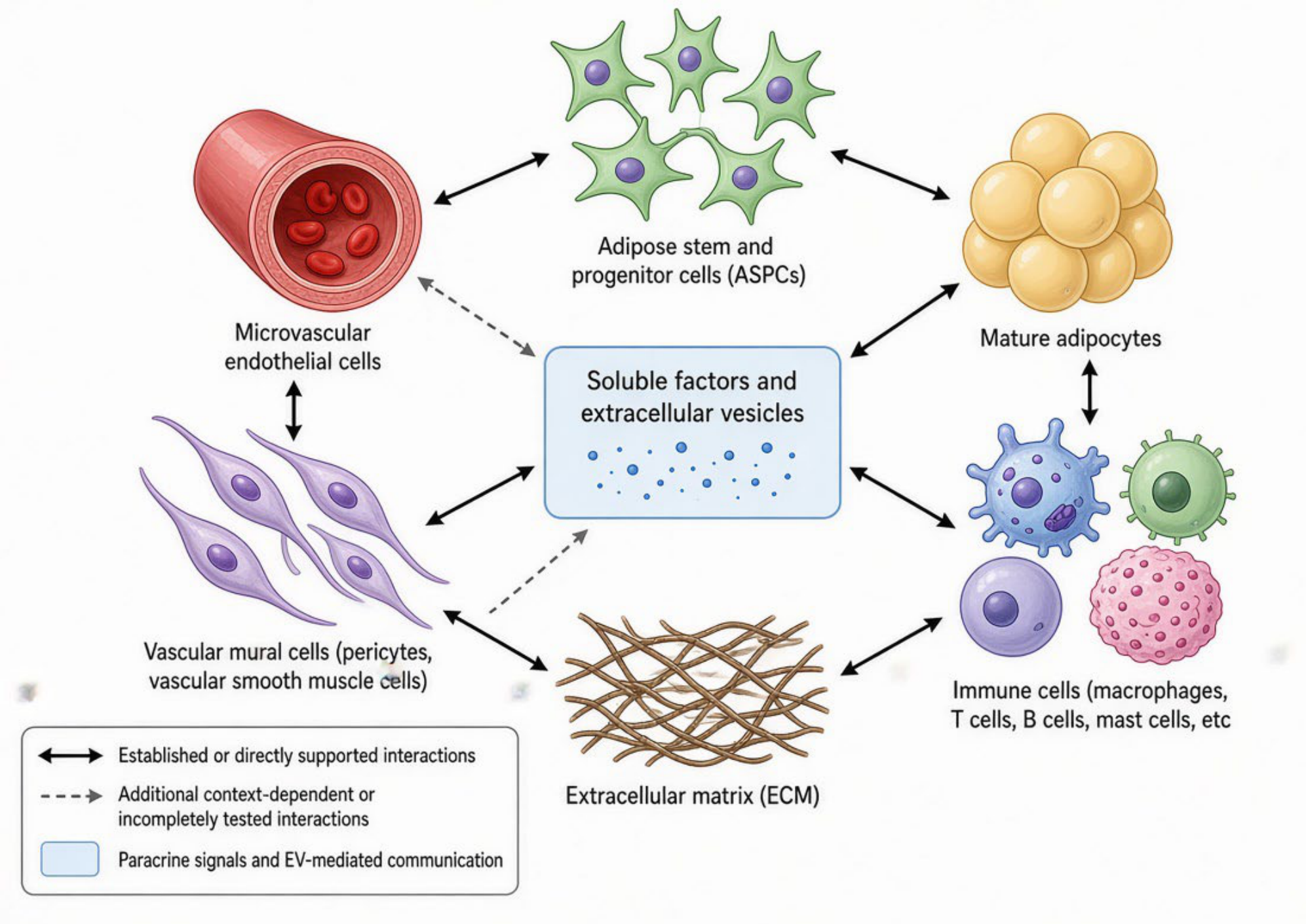
Figure 2.
From physiological endocrine context to regional AVU dysregulation and the lipedema phenotype: a working model. The hypothalamic–pituitary–ovarian axis provides a physiological reproductive endocrine context across puberty, pregnancy, perimenopause and menopause. In a resilient AVU, these signals are accommodated by controlled regenerative responses and tissue homeostasis; in a regionally susceptible AVU, the same physiological context may encounter a lower threshold for regenerative activation, leading to dysregulated progenitor recruitment and differentiation, endothelial–stromal and immune cross-talk, extracellular-matrix remodeling and reciprocal AVU amplification. Progressive tissue remodeling may contribute to the characteristic regional adipose expansion, pain, tenderness, easy bruising, heaviness or swelling and nodularity or fibrosis. Persistence of an altered AVU state after withdrawal of a candidate initiating or amplifying stimulus is shown as a hypothetical regenerative lock-in and remains to be experimentally demonstrated.
Figure 2.
From physiological endocrine context to regional AVU dysregulation and the lipedema phenotype: a working model. The hypothalamic–pituitary–ovarian axis provides a physiological reproductive endocrine context across puberty, pregnancy, perimenopause and menopause. In a resilient AVU, these signals are accommodated by controlled regenerative responses and tissue homeostasis; in a regionally susceptible AVU, the same physiological context may encounter a lower threshold for regenerative activation, leading to dysregulated progenitor recruitment and differentiation, endothelial–stromal and immune cross-talk, extracellular-matrix remodeling and reciprocal AVU amplification. Progressive tissue remodeling may contribute to the characteristic regional adipose expansion, pain, tenderness, easy bruising, heaviness or swelling and nodularity or fibrosis. Persistence of an altered AVU state after withdrawal of a candidate initiating or amplifying stimulus is shown as a hypothetical regenerative lock-in and remains to be experimentally demonstrated.
Disclaimer/Publisher’s Note: The statements, opinions and data contained in all publications are solely those of the individual author(s) and contributor(s) and not of MDPI and/or the editor(s). MDPI and/or the editor(s) disclaim responsibility for any injury to people or property resulting from any ideas, methods, instructions or products referred to in the content. |
© 2026 by the authors. Licensee MDPI, Basel, Switzerland. This article is an open access article distributed under the terms and conditions of the Creative Commons Attribution (CC BY) license (http://creativecommons.org/licenses/by/4.0/).
Copyright: This open access article is published under a Creative Commons CC BY 4.0 license, which permit the free download, distribution, and reuse, provided that the author and preprint are cited in any reuse.