

Submitted:
24 September 2026
Posted:
28 September 2026
You are already at the latest version
Abstract
Podocytes are terminally differentiated components of the glomerular filtration barrier whose structural integrity depends on coordinated signaling between the slit diaphragm, actin cytoskeleton, mitochondria, and stress-response machinery. Evidence from cultured podocytes, experimental kidney disease, human tissue, and selected podocyte-specific genetic models indicates that podocytes can initiate cell-autonomous inflammatory responses rather than functioning solely as passive targets of systemic inflammation. This review examines how NF-κB, p38 MAPK, JAK–STAT, cGAS–STING, NLRP3 inflammasome, and Rho-family GTPase signaling interact with cytoskeletal remodeling, mitochondrial dysfunction, senescence, regulated cell death, and podocyte detachment. Particular attention is given to the distinction between podocyte-specific evidence and findings obtained from whole-kidney analyses, global genetic models, or pharmacological interventions. The available evidence supports a context-dependent network in which inflammatory signaling may initially contribute to adaptation but, when sustained, promotes barrier dysfunction and irreversible podocyte loss. We also assess therapeutic strategies targeting inflammatory sensors, kinases, inflammasome components, cytoskeletal regulators, and metabolic stress pathways. Most remain preclinical, emphasizing the need for podocyte-targeted delivery, rigorous cell-specific validation, and human translational studies.
Keywords:
podocyte
; glomerular filtration barrier
; cell-autonomous inflammation
; NF-κB
; cGAS–STING
; NLRP3 inflammasome
; Rho GTPases
; regulated cell death
; podocyte senescence
; proteinuria
1. Introduction
1.1. Overview of Podocyte Biology and Function
Podocytes are highly specialized, terminally differentiated epithelial cells of the visceral layer of Bowman’s capsule. They form the outermost cellular component of the glomerular filtration barrier and are indispensable for the long-term maintenance of glomerular permselectivity and renal homeostasis (Figure 1) [1,2]. Their unique architecture comprises a cell body, primary processes, and numerous interdigitating foot processes, which are connected by slit diaphragms. The slit diaphragm is a specialized signaling and cell-junction complex containing nephrin, podocin, CD2AP, NEPH1, and ZO-1, which functionally links the filtration barrier to the actin cytoskeleton and intracellular signaling networks [3,4].
Mature podocytes have a highly limited capacity for cell division and replacement. Consequently, podocyte loss is a critical and frequently irreversible event in the progression of chronic glomerular disease. Experimental podocyte-depletion models demonstrate a direct quantitative relationship between the extent of podocyte loss, proteinuria, focal segmental glomerulosclerosis, and declining renal function [5,6,7].
Historically, podocytes were regarded primarily as passive structural elements of the glomerular filter. Over the past decades, this concept has been substantially revised. Contemporary evidence indicates that podocytes are metabolically active and signal-responsive cells capable of sensing a broad spectrum of mechanical, metabolic, and immune stimuli. Podocytes express innate immune receptors, including TLR, and can respond to pathogen- and damage-associated molecular patterns (PAMPs and DAMPs) [8]. In diverse experimental models of glomerular injury, activation of inflammatory and stress-responsive pathways within podocytes has been implicated in altered slit-diaphragm protein expression, actin cytoskeletal remodeling, loss of adhesion to the glomerular basement membrane, and regulated cell-death responses [9,10,11,12]. In addition, microRNA-mediated control of injury signaling is increasingly recognized [13].
Inflammatory signaling in podocytes rarely occurs in isolation. Instead, podocyte injury, local cytokine and chemokine production, complement activation, and immune-cell recruitment can participate in self-sustaining pathological loops that perpetuate glomerular inflammation and structural injury [14]. In diabetic kidney disease and experimental immune glomerular injury, these processes can contribute to progressive podocyte dysfunction and loss, thereby compromising the integrity of the glomerular filtration barrier [9,10,15]. Despite intensive investigation of inflammation in renal pathology, the mechanisms by which inflammatory signaling pathways directly drive podocyte death remain an active area of research. Increasing attention has focused on regulated cell-death modalities, including apoptosis, pyroptosis, and necroptosis, and on their contribution to podocyte injury. Understanding the molecular links between inflammatory signaling and podocyte death is essential for developing therapeutic strategies aimed at preserving podocyte number and limiting disease progression.
1.2. Defining Podocyte-Intrinsic Inflammation
In classical models of glomerular disease, podocytes were largely viewed as passive victims of circulating immune insults and hemodynamic stress. Accumulating evidence supports a fundamentally different paradigm in which podocytes behave as active ‘immune-like’ cells, equipped with an innate immune signaling apparatus capable of sensing danger-associated stimuli and executing cell-autonomous inflammatory programs [8,9]. In this review, we define podocyte-intrinsic inflammatory signaling as the ensemble of stimulus–response pathways that are initiated, integrated, and executed within podocytes themselves even when the initiating stimuli originate from the extracellular environment.
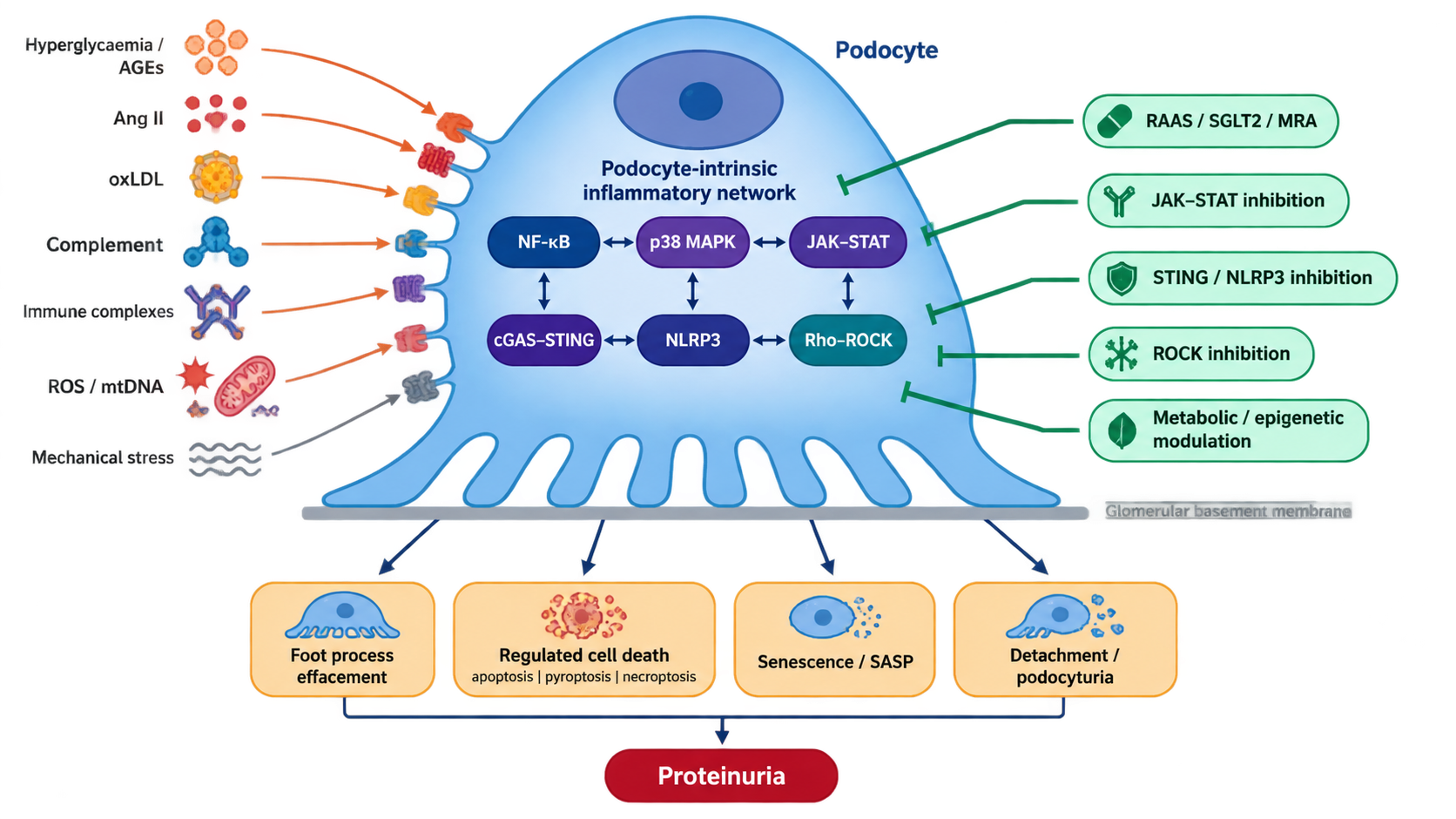
Operationally, podocyte-intrinsic inflammatory signaling arises when endogenous or exogenous stressors, such as hyperglycemia, dyslipidemia, immune complexes, complement activation, mechanical forces, and toxins, engage podocyte-resident receptors and sensors, including cytokine and complement receptors, pattern-recognition receptors, cGAS–STING, NLRP3 inflammasome, and ion channels. These inputs converge on transcriptional and post-transcriptional networks comprising NF-κB, p38 and other MAPKs, JAK–STAT, interferon-regulated factors, and epigenetic and epitranscriptomic regulators, which together reprogram podocyte behavior [9,11,13]. As summarized in Figure 2, podocytes integrate extracellular inflammatory, metabolic, complement-derived, and danger-associated cues through membrane receptors and cytosolic sensors, thereby coupling innate immune signaling to cytoskeletal remodeling, inflammatory output, and filtration-barrier dysfunction.
Conceptually, this intrinsic axis has three defining features. First, the initiation phase is podocyte-centered: danger signals are sensed directly by the podocyte rather than being imposed solely by infiltrating leukocytes or systemic mediators. Second, the integration phase couples inflammatory signaling to podocyte-specific structural and functional modules, including the slit diaphragm, actin cytoskeleton, mitochondrial and autophagy machinery, and regulated cell death programs, thereby translating molecular signals into foot process effacement, detachment, and podocyturia. Third, the propagation phase extends beyond the individual cell: intrinsically activated podocytes secrete cytokines, chemokines, and senescence-associated secretory phenotype (SASP) factors and extracellular vesicles that feed back onto glomerular endothelial, mesangial, tubular, and immune cells, shaping the local microenvironment and influencing disease trajectory.
Within this framework, podocyte-intrinsic inflammation emerges as an important determinant of podocyte cellular fate, modulating the balance between adaptive stress responses and maladaptive injury across proteinuric diseases, including diabetic kidney disease, obesity-related glomerulopathy, focal segmental glomerulosclerosis, lupus nephritis, minimal change disease, and autoimmune glomerular disorders. Transient and appropriately resolved signaling may support short-term adaptation to injury, whereas sustained or dysregulated inflammatory activation can promote senescence, apoptosis, pyroptosis, and other regulated cell-death programs, ultimately contributing to irreversible podocyte loss [13,16,17,18].
Evidence from podocyte-specific genetic models indicates that podocytes can autonomously integrate injury signals and execute inflammatory, cytoskeletal, and cell-death programs without requiring direct effector activity from infiltrating immune cells [9,15,19]. The following sections examine NF-κB, p38 MAPK, JAK–STAT, Rho-family GTPase, cGAS–STING, and NLRP3 inflammasome signaling as interconnected components of this intrinsic inflammatory network.
2. Inflammatory Signaling Pathways in Podocytes
2.1. NF-κB and p38 MAPK Signaling in Podocytes
NF-κB and p38 MAPK function as complementary arms of the podocyte stress response. NF-κB acts primarily as a transcriptional hub, converting inflammatory and metabolic inputs into sustained changes in gene expression. p38 operates in parallel as a stress-responsive kinase, linking cellular stress to rapid cytoskeletal remodeling, inflammatory mediator production, and cell death. Despite their distinct molecular mechanisms, these pathways are co-activated by overlapping stimuli and create coordinated responses that determine podocyte fate.
At the level of the glomerular filtration barrier, NF-κB can be activated in all major glomerular cell populations, including podocytes, mesangial cells, and endothelial cells. However, podocytes, as highly specialized epithelial cells, display a distinctive combination of NF-κB-dependent pro-inflammatory programs and survival pathways. As a result, NF-κB in podocytes exerts a classical dual role: moderate and transient activation may support cellular adaptation and survival, whereas sustained hyperactivation drives podocytopathy, proteinuria, and progression of glomerular disease [19,20,21,22,23,24].
Similarly, p38 MAPK exhibits context-dependent functions in podocytes. While often associated with stress-induced injury and apoptosis, p38 can also restrain pathological pathways and preserve podocyte-endothelial interactions in certain settings. The sections below describe the upstream activators, downstream effectors, and in vivo evidence for both pathways, emphasizing their integration and shared role in driving podocyte dysfunction when chronically activated.
2.1.1. Complementary Functions and Upstream Activation of NF-κB and p38 MAPK in Podocytes
Podocytes express multiple receptor systems that can engage NF-κB signaling, including Toll-like receptors, cytokine receptors such as TNFR1/2 and IL-1R, RAGE, angiotensin II type 1 receptors, and complement receptors (C3aR and C5aR). The expression and functional relevance of individual TLR family members vary across species, disease contexts, and experimental models; therefore, a uniform podocyte TLR repertoire should not be assumed [9,20,22,25,26,27,28]. Loss of nephrin itself activates NF-κB, illustrating how a primary structural defect of the slit diaphragm can be converted into a sustained inflammatory transcriptional program in podocytes [26,29]. This establishes a mechanistic link between slit-diaphragm disruption, inflammatory transcriptional reprogramming, cytoskeletal remodeling, and progressive podocyte injury.
In experimental LPS-induced podocyte injury, activation of the TLR4/MyD88/NF-κB and calcineurin/NFAT pathways increases ANGPTL4 expression. ANGPTL4, in turn, contributes to cytoskeletal remodeling, synaptopodin loss, and proteinuria; pharmacological or siRNA-mediated inhibition of either pathway attenuates ANGPTL4 induction and podocyte injury [30]. During septic acute kidney injury, this NF-κB hyperactivation drives podocyte apoptosis and inflammatory injury. The pathogenic role of NF-κB in this setting is underscored by the protective effect of miR-129-5p, which limits NF-κB activation by targeting the HMGB1/TLR2-TLR4 axis, thereby reducing podocyte apoptosis and improving survival [31].
In cultured mouse podocytes, angiotensin II reduced Akt and p65 NF-κB phosphorylation, decreased cell viability, and increased caspase-9 expression and apoptosis. Pharmacological activation of PI3K partially restored Akt/p65 phosphorylation and attenuated apoptosis, whereas PI3K inhibition aggravated the injurious response, consistent with a protective role of the PI3K/Akt/NF-κB survival axis in this experimental setting [27]. Consistent with these in vitro observations, chronic angiotensin II infusion in rats was associated with reduced nephrin expression and increased podocyte apoptosis in vivo [32]. In a distinct model, angiotensin II-induced podocyte apoptosis was linked to AT1R-dependent endoplasmic reticulum stress and activation of the PKC-δ/p38 MAPK pathway, highlighting that angiotensin II can engage multiple context-dependent injury programs in podocytes [33].
Complement-derived anaphylatoxins can act directly on podocytes. In primary membranous nephropathy, C3a/C3aR signaling has been implicated in podocyte injury, synaptopodin loss, cytoskeletal abnormalities, and impairment of filtration-barrier function. Although C5aR signaling and downstream PI3K/Akt, MAPK, and NF-κB pathways may contribute in selected disease settings, their podocyte-intrinsic roles should be described as context dependent rather than as a single established complement–TLR4 convergence pathway [28,34].
In diabetic nephropathy, hyperglycemia sustains canonical NF-κB signaling in podocytes through several convergent, partly self-amplifying mechanisms. Increased REDD1 expression promotes GSK3β activation, NEMO phosphorylation, and IKK complex assembly, leading to IκBα degradation, NF-κB nuclear translocation, and induction of CCL2, IL-1β, and other pro-inflammatory mediators [35]. In parallel, hyperglycemia increases podocyte expression of receptor activator of NF-κB (RANK), which promotes a p65-dependent oxidative and inflammatory program. This pathogenic role of RANK should be interpreted in a diabetic context, as RANK–RANKL signaling has also been implicated in adaptive pro-survival responses in other models of podocyte injury. In diabetic podocytes, p65 binds to the promoter regions of Nox4 and p22phox, enhancing NADPH oxidase expression, ROS generation, and the production of inflammatory mediators [17]. High glucose-induced ROS can in turn reinforce canonical NF-κB activation, thereby establishing a feed-forward circuit that links metabolic stress to oxidative injury, cytokine expression, and podocyte dysfunction. In parallel, cytokine- and interferon-responsive JAK–STAT pathways may converge with NF-κB-dependent transcriptional programs, further broadening the inflammatory response in diabetic podocytopathies.
NF-κB can also be activated from within podocytes, independent of surface receptors. Intracellular Ca²⁺ imbalance activates calpain, a protease that directly degrades IκBα, releasing NF-κB for nuclear translocation. In GAK-deficient podocytes, enhanced calpain activity causes excessive IκBα degradation, driving NF-κB hyperactivation, podocytopathy, and glomerulosclerosis. Calpain inhibition or podocyte-specific deletion normalizes NF-κB activation and markedly reduces proteinuria and sclerosis [36]. Thus, podocyte NF-κB integrates both receptor-driven signals and intracellular stress, with calpain serving as a key modulator in toxic and metabolic injury.
In podocytes, IKK-dependent NF-κB activation can occur in parallel with stress-activated kinases such as p38, JNK, and ERK in response to selected inflammatory and metabolic stimuli [9,22,35], creating a coordinated transcriptional and post-transcriptional response to TNF, LPS, hyperglycemia, and complement activation. Rather than acting as a universal parallel output of every stimulus, p38 MAPK appears to function as a stress-responsive kinase that couples selected injurious cues to cytoskeletal remodeling, inflammatory gene expression, and apoptotic signaling.
In experimental models of diabetic podocyte injury, high extracellular glucose promotes NADPH oxidase- and mitochondria-derived ROS generation in podocytes, resulting in p38 MAPK and caspase-3 activation within a pro-apoptotic signaling program and contributing to early podocyte depletion in diabetic mouse models [10]. Albumin exposure activates p38–MK2 signaling in podocytes and induces COX-2 expression, whereas pharmacological inhibition of p38 or MK2 attenuates injury-associated cytoskeletal changes in experimental podocyte models [37,38]. Intracellular albumin overload also induces ER stress and activates a PKC-δ–p38–caspase-12 pathway, promoting podocyte apoptosis; this response is reduced by p38 inhibition [39].
TGF-β1 can activate MAPK pathways in cultured podocytes and promote loss of structural proteins and inflammatory responses. Inhibition of p38 attenuates injury-associated changes in selected experimental settings [41]. TGF-β/Smad7-dependent apoptosis provides an additional mechanism of podocyte injury [40]. Angiotensin II activates an AT1R-dependent ER stress/PKC-δ/p38 MAPK cascade in podocytes, leading to increased Bax/Bcl-2 ratio, NHE1 activation, and apoptosis; both AT1R blockade and p38 inhibition are protective in this experimental setting [33].
Hyperglycemia also activates p38 MAPK in podocytes. Activated p38 phosphorylates the nephrin C-terminus, facilitating PKCα recruitment and β-arrestin2-dependent nephrin endocytosis; pharmacological p38 inhibition preserves nephrin surface expression and attenuates albuminuria in experimental hyperglycemia [42]. In adriamycin-induced podocyte injury, loss of plectin activates an integrin α6β4/FAK/p38 MAPK cascade. FAK-dependent p38 activation is associated with podocyte apoptosis and F-actin cytoskeletal disruption, whereas restoration of plectin expression attenuates pathway activation and protects against podocyte injury [43].
By contrast, direct evidence that TLR4/LPS or IL-1β induces p38-dependent podocyte injury remains less extensive than the evidence for TLR–NF-κB signaling. These pathways should therefore be described as plausible contributors to p38 activation rather than as fully established podocyte-intrinsic p38 mechanisms. Together, NF-κB and p38 MAPK function as interconnected components of the podocyte stress response. NF-κB predominantly coordinates inflammatory and pro-survival transcriptional programs, whereas p38 MAPK prominently links cellular stress to cytoskeletal remodeling, apoptotic signaling, and the induction of selected inflammatory mediators. Their concurrent activation by metabolic, hemodynamic, or immune stimuli can integrate transcriptional and structural injury responses; when sustained or insufficiently resolved, this signaling convergence may promote podocyte dysfunction, detachment, and progressive loss.
2.1.2. NF-κB-Dependent Inflammatory Programming in Podocytes
Building on the receptor- and stress-dependent inputs outlined above, canonical NF-κB activation in podocytes converges on the IKK complex, composed of IKKα, IKKβ, and the regulatory subunit NEMO/IKKγ. Although individual stimuli use distinct proximal adaptors and ubiquitin-dependent signaling modules, IKKβ-mediated phosphorylation of IκBα promotes its ubiquitin-dependent degradation and the nuclear translocation of p65/p50 dimers [44,45,46]. In canonical NF-κB signaling, IKKβ phosphorylates IκBα, promoting its ubiquitin-dependent degradation and release of p65/p50 dimers. Nuclear p65/p50 complexes then induce context-dependent transcriptional programs involved in inflammation, cell survival, matrix remodeling, and stress adaptation [44,45,46].
The non-canonical NF-κB pathway is activated by a restricted subset of TNF receptor superfamily members, including RANK, BAFF-R, and CD40. Unlike canonical signaling, it does not rely on MyD88 or TRADD. Instead, receptor activation promotes degradation of the TRAF2–TRAF3–cIAP1/2 complex that constitutively targets NF-κB-inducing kinase (NIK) for proteasomal turnover, thereby allowing NIK stabilization [21,23,47,48,49]. Stabilized NIK activates IKKα, which phosphorylates p100 and promotes its processing to p52, enabling the formation and nuclear accumulation of RelB/p52 dimers [47,48,49]. These dimers regulate a distinct transcriptional program linked to chronic inflammation, fibrosis, and long-term adaptation to persistent stress [21,23]. Although non-canonical NF-κB signaling is well established in immune and stromal cells, its cell-autonomous functions in podocytes and its relationship to cGAS–STING or inflammasome signaling remain incompletely defined. In podocytes, the NF-κB transcriptional response is context dependent. Under basal or transient stress conditions, NF-κB can support adaptive and pro-survival programs, whereas persistent injury shifts target-gene expression towards chemokines, cytokines, matrix-remodeling mediators, and regulators of cytoskeletal dysfunction [50,51].
NF-κB activity in podocytes is further modulated by intracellular kinases such as GSK3β. GSK3β influences p65 phosphorylation and target-gene selection, thereby shifting NF-κB-dependent responses from pro-survival programs, including Bcl-xL, towards pro-inflammatory programs, including Ccl2 and Cxcl1 [22]. In diabetic nephropathy, hyperglycemia-induced REDD1 promotes GSK3β-dependent canonical NF-κB activation in podocytes, contributing to sustained inflammatory mediator expression and podocyte injury. Independently, redox-sensitive GSK3β hyperactivity can impair Nrf2-dependent antioxidant responses and promote oxidative stress and premature podocyte senescence in experimental type 2 diabetic kidney disease [35,52]. Genetic or pharmacological inhibition of GSK3β attenuates injury-associated NF-κB signaling and improves podocyte outcomes in experimental settings; however, pathway-selective and podocyte-targeted therapeutic strategies remain to be established [35,52].
NF-κB signaling is subject to regulation by microRNAs. In immune cells, miR-146a functions as a canonical negative-feedback regulator of innate inflammatory signaling by targeting IRAK1 and TRAF6, two key components of the TLR/IL-1R–NF-κB pathway [53]. Podocyte studies likewise support a protective role for miR-146a in glomerular injury. Global miR-146a deficiency accelerates streptozotocin-induced diabetic glomerulopathy and albuminuria, in association with increased expression of ErbB4 and Notch1 [13]. Podocyte-specific deletion of miR-146a increases susceptibility to low-dose LPS, nephrotoxic serum nephritis, and streptozotocin-induced diabetic kidney disease, resulting in enhanced proteinuria and glomerular injury. These effects are associated with increased ErbB4/EGFR signaling and enhanced TGF-β/Smad3 activity [54]. miR-129-5p has also been reported to limit LPS-associated podocyte injury by attenuating the HMGB1/TLR2/TLR4/NF-κB axis [31]. Conversely, NF-κB can regulate the expression of selected microRNAs and long non-coding RNAs, thereby creating feedback circuits that may sustain chronic inflammatory signaling and promote podocyte injury in glomerular disease [18].
Finally, NF-κB and Notch signaling may converge in injured podocytes, where persistent activation of inflammatory and developmental programs can promote dedifferentiation, apoptosis, and filtration-barrier dysfunction [55,56]. In diabetic settings, glucose-derived advanced glycation end products can activate Notch1 signaling in podocytes and contribute to proteinuria, supporting a role for Notch activation in metabolic podocyte injury [57]. However, the molecular hierarchy and direct causal links between NF-κB and Notch activation remain unresolved across distinct podocytopathies.
2.1.3. p38 MAPK as a Stress and Cytoskeletal Effector
While NF-κB functions as a major transcriptional integrator of podocyte-intrinsic inflammation, p38 MAPK acts as a stress-responsive kinase that links mechanical, metabolic, and protein-overload signals to cytoskeletal regulation, inflammatory gene expression, and context-dependent cell-death responses. In podocytes, activated p38α phosphorylates MK2 (MAPKAPK2), which in turn phosphorylates the small heat-shock protein HSPB1/HSP25, thereby modulating actin filament dynamics. Acute high-glucose exposure activates the p38–HSP25 pathway and supports actin cytoskeletal stability; pharmacological p38 inhibition reduces HSP25 phosphorylation, promotes F-to-G-actin conversion, and disrupts the cytoskeleton in this setting [58].
In contrast, sustained protein-overload injury can engage a maladaptive p38/MK2 program. Albumin exposure activates p38 and MK2 in podocytes and induces COX-2 expression, whereas pharmacological inhibition of p38 or MK2 reduces albumin-induced COX-2 expression and attenuates PAN-associated cytoskeletal injury [37,38]. Thus, the p38–MK2–HSPB1 axis can serve adaptive functions during acute stress but may contribute to inflammatory and structural injury when activation is persistent or combined with nephrotic stressors.
p38 MAPK also contributes to slit-diaphragm dysfunction in diabetic settings. Hyperglycemia-induced p38 activation promotes nephrin phosphorylation and β-arrestin2-dependent nephrin endocytosis, thereby reducing surface nephrin availability and compromising filtration-barrier integrity [42]. The p38 pathway intersects functionally with Rho GTPase networks; in diabetic podocytes, Rac1/PAK1 activation lies upstream of p38 and contributes to β-catenin-associated slit-diaphragm disruption, foot-process effacement, and proteinuria [59].
p38 MAPK also participates in several stress-induced apoptotic programs in podocytes. Intracellular albumin overload induces ER stress, as reflected by increased GRP78 and IRE1α activation, and engages a PKC-δ–p38–caspase-12 cascade that promotes podocyte apoptosis; pharmacological p38 inhibition attenuates this response [39]. Angiotensin II activates an AT1R-dependent ER stress/PKC-δ/p38 MAPK pathway, accompanied by NHE1 activation, an increased Bax/Bcl-2 ratio, and apoptosis, whereas AT1R blockade or p38 inhibition is protective in experimental podocyte models [33]. TGF-β1-induced podocyte apoptosis likewise requires p38 MAPK activation and is associated with increased Bax expression and caspase-3 activity [40]. In adriamycin-exposed podocytes, ROS generation precedes ASK1–p38 activation, and suppression of this pathway reduces oxidative podocyte injury [60]. Collectively, these findings identify p38 MAPK as a context-dependent convergence point through which ER stress, oxidative stress, and profibrotic signaling can promote apoptotic podocyte injury.
2.1.4. NF-κB and p38 MAPK in Glomerular Disease: Evidence from In Vivo Genetic Models
Experimental and clinical studies implicate aberrant NF-κB activation in a broad range of proteinuric and inflammatory glomerular diseases, including diabetic kidney disease, focal segmental glomerulosclerosis, membranous nephropathy, IgA nephropathy, and immune-mediated glomerulonephritides. However, the relevant cell types, upstream triggers, and functional consequences differ substantially among disease settings [9,16,20,21]. The contribution of NF-κB to disease progression operates at two levels: systemic and local inflammation, involving immune cell infiltration and cytokine production in the interstitium and mesangium, and cell-autonomous effects in podocytes that alter the structure and function of the filtration barrier. The available genetic evidence is summarized in Table 1, which distinguishes global from podocyte-specific manipulations and highlights the context-dependent functions of NF-κB, p38 MAPK, and their associated regulatory modules in experimental glomerular disease.
Podocyte-specific genetic models have been essential for defining the cell-autonomous role of NF-κB. In nephrotoxic nephritis, podocyte-specific NEMO/IKKγ-deficient mice display immune-cell infiltration comparable to that of control animals but show accelerated recovery from proteinuria and reduced glomerular expression of NF-κB-dependent pro-inflammatory chemokines. These findings indicate that podocyte NF-κB can sustain glomerular inflammatory signaling and delay functional recovery independently of changes in leukocyte recruitment [9]. Similarly, in adriamycin nephropathy, podocyte-specific inhibition of NF-κB does not impair baseline kidney function but markedly attenuates albuminuria and loss of slit diaphragm proteins such as nephrin and synaptopodin after injury [19]. In a model of nephrotoxic nephritis, podocyte-specific deletion of Myd88 has little effect on the inflammatory transcriptome and disease severity, indicating that podocyte inflammatory responses in this setting are largely independent of canonical MyD88-dependent TLR signaling [61].
In diabetic nephropathy, NF-κB in podocytes integrates signals from RANK, angiotensin II, NLRP3 inflammasome, and NADPH oxidases, amplifying oxidative stress and production of TNF, IL-1β, MCP-1, and other inflammatory mediators [16,18,62,63,64]. Podocyte-specific RANK knockout in HFD/STZ, db/db, and related diabetic models reduces albuminuria, mesangial expansion, and podocyte loss, accompanied by decreased p65-dependent transcription of NOX4 and p22phox and reduced oxidative stress, thereby confirming the importance of RANK-driven NF-κB activation in diabetic podocytopathy [17]. Podocyte-specific REDD1 deletion similarly protects against diabetic injury, as reflected by reduced albuminuria, glomerular hypertrophy, and mesangial matrix deposition, together with preservation of podocyte number and foot process architecture, consistent with REDD1–GSK3β–NF-κB as a major pathogenic axis [35].
In membranous nephropathy and passive Heymann nephritis, local immune deposits and complement activation induce NF-κB-dependent inflammatory responses in podocytes, including TRAF6/NF-κB, TLR/NF-κB, and NLRP3-linked pathways, driving proteinuria and structural remodeling of the filtration barrier [16,28,65,66]. These observations place NF-κB within a broader innate immune network that also involves complement, cGAS–STING, and inflammasome signaling. Astragaloside IV, which has potent anti-inflammatory effects, reduces proteinuria, and improves histology in passive Heymann nephritis and diabetic nephropathy while suppressing NF-κB signaling and lowering TNF, IL-6, and IL-1β expression in podocytes [16,65,66].
The role of podocyte NF-κB is not uniformly pathogenic in every form of renal injury. In renal ischemia–reperfusion injury, podocyte-specific NF-κB blockade has little effect on the severity of acute injury, immune cell infiltration, or functional recovery, whereas systemic NF-κB inhibition reduces inflammation [19,67]. These findings suggest that in purely ischemic acute kidney injury, NF-κB-dependent inflammation is driven predominantly by tubular cells and interstitial macrophages, with a limited contribution from podocytes. Thus, the pathogenic significance of podocyte NF-κB is highly disease- and context-dependent: it is central in metabolic and immune-mediated glomerulopathies but less critical in purely ischemic settings.
Other in vivo models highlight the consequences of primary structural or signaling defects that secondarily hyperactivate NF-κB in podocytes. Nephrin-deficient mice, modeling congenital nephrotic syndrome, demonstrate that nephrin loss triggers NF-κB activation [26,29]. Podocyte-specific GTPase-activating protein (GAK) knockout causes calpain-mediated IκBα degradation and enhanced progressive podocytopathy; additional Capns1 deletion dampens NF-κB activation and significantly improves proteinuria, survival, and glomerular morphology, highlighting the calpain–IκB–NF-κB axis as a driver of podocyte injury [36]. Global and podocyte-specific miR-146a deletion models further illustrate the importance of endogenous post-transcriptional restraint in podocyte injury. Global miR-146a deficiency accelerates streptozotocin-induced diabetic glomerulopathy through dysregulation of the ErbB4–Notch1 axis, whereas podocyte-specific miR-146a deletion increases susceptibility to LPS-, nephrotoxic serum-, and diabetes-induced glomerular injury, with enhanced ErbB4/EGFR and TGF-β-associated signaling [13,54].
Taken together, these in vivo data show that podocyte NF-κB is a dual regulator: it is required for stress-adaptive and survival programs, yet when excessively or improperly activated by diabetes, immune complex nephritis, or structural slit diaphragm defects, it becomes a major driver of proteinuria and progressive podocytopathy [9,16,19,21]. This supports a therapeutic strategy aimed not at indiscriminate global inhibition of NF-κB, but at targeting podocyte-specific fine-tuning modules such as NEMO, REDD1, RANK, miR-146a, and the calpain–IκB axis, thereby selectively attenuating pathological inflammatory signaling while preserving basal podocyte homeostasis.
Like NF-κB, p38 MAPK functions as a context-dependent regulator of podocyte injury. Phospho-p38 is consistently elevated in podocytes and other glomerular cells across proliferative and non-proliferative glomerulonephritides, correlating with morphological damage and renal dysfunction, and is detected in human diabetic nephropathy across mesangial, endothelial, podocyte, and tubulointerstitial compartments. In experimental PAN and adriamycin nephropathy, glomerular p38 activation is transient and precedes overt proteinuria, indicating an early pathogenic role. A complete germline knockout of p38α/MAPK14 is poorly suited to analysis of adult renal injury, because Mapk14−/− mice die embryonically due to severe placental defects [68,69]. Consequently, the contribution of p38α in the kidney, and specifically in podocytes, has been studied primarily in conditional tissue-specific models or via downstream components.
The most informative podocyte-specific model to date is the study by Sugioka et al. (2023), which compared pod-p38 cKO, pod-GC-A cKO, and pod-p38/GC-A double cKO mice [70]. Two-month-old pod-p38 cKO and double cKO animals had no striking baseline phenotype, but under high salt and aldosterone load the most severe injury developed in pod-double cKO mice: serum creatinine and albuminuria increased, macrophage infiltration intensified, foot process structure and nephrin/podocin expression were disrupted, and intracapillary fibrin thrombi formed, indicating secondary endothelial injury [70]. This thrombotic phenotype was driven by elevated PAI-1 (plasminogen activator inhibitor-1, which blocks fibrinolysis and promotes fibrosis). PAI-1 neutralization reduced thrombosis and mitigated damage. Surprisingly, p38α was not purely pro-injurious in this setting: it appeared to restrain PAI-1/TGF-β activation and preserve podocyte-endothelial interactions [70]. Thus, p38α can have protective functions depending on the disease context.
For downstream branches, MK2/MAPKAPK2 and MK3/MAPKAPK3 are the most studied. Systemic MK2 knockout in mice reduces TNF production by ≈90% and confers resistance to LPS-induced endotoxin shock, underscoring the central role of the p38–MK2 axis in inflammatory responses [69]. In an acute proliferative glomerulonephritis model, MK2-, MK3-, and MK2/MK3-deficient mice were compared: MK2 loss alone had modest effects, MK3 deficiency slightly worsened disease, whereas double MK2/MK3 knockout made animals more susceptible to glomerulonephritis [78]. These findings highlight that downstream p38 targets in the kidney act in a non-linear fashion, with disease outcomes depending on kinase combinations and cellular context. Podocyte-specific data for MK2/3 remain less definitive than for the Mapk14/Npr1 double knockout model; protective effects of MK2 and p38 inhibition were shown in podocyte culture rather than in vivo: the MK2 inhibitor C23 and p38 inhibitor SB203580 reduced PAN-induced injury, preserved the actin cytoskeleton, and blocked albumin-induced COX-2 upregulation [37,71].

Table 1.
Genetic models defining context-dependent NF-κB and p38 MAPK functions in podocytes, glomerular disease, and renal inflammatory injury. .
Table 1.
Genetic models defining context-dependent NF-κB and p38 MAPK functions in podocytes, glomerular disease, and renal inflammatory injury. .
| Pathway / mediator | Genetic manipulation | Injury model or context | Principal renal and podocyte phenotype | Reference |
|---|---|---|---|---|
| NF-κB pathway and associated regulators | ||||
| NEMO/IKKγ | Podocyte-specific deletion (Nphs2-Cre) | Nephrotoxic nephritis | Renal leukocyte infiltration was unchanged; proteinuria and podocyte MCP-1 secretion were reduced, supporting a cell-autonomous pathogenic role of podocyte NF-κB | [9] |
| REDD1 | Podocyte-specific deletion (Nphs2-Cre) | STZ-induced diabetes | Reduced albuminuria, podocyte loss, glomerular hypertrophy, and mesangial matrix expansion; attenuated REDD1–GSK3β–NF-κB signaling | [35] |
| miR-146a | Global deletion | STZ-induced diabetes | Increased susceptibility to diabetic kidney injury, with aggravated albuminuria, podocyte injury, and glomerular damage | [13,54] |
| miR-146a | Podocyte-specific deletion (Nphs2-Cre) | Low-dose LPS, nephrotoxic serum nephritis, and STZ-induced diabetes | Increased proteinuria and glomerular injury; enhanced NF-κB-dependent and ErbB4/Notch-associated signaling | [54] |
| NF-κB | Podocyte-specific expression of non-degradable IκBα (IκBΔN) | Adriamycin nephropathy | Reduced proteinuria; preserved nephrin and synaptopodin expression; improved foot-process ultrastructure | [19] |
| NF-κB | Podocyte-specific expression of non-degradable IκBα (IκBΔN) | Renal ischemia– reperfusion injury |
No substantial improvement in acute kidney injury or renal functional indices, indicating limited pathogenic relevance of podocyte NF-κB in this setting | [67] |
| MyD88 | Podocyte-specific deletion | Nephrotoxic nephritis | Normal baseline renal morphology and function; transiently increased proteinuria after injury, indicating that MyD88-dependent signaling may also support adaptive podocyte responses | [61] |
| RANK | Podocyte-specific deletion | STZ-induced diabetes | Reduced albuminuria, mesangial expansion, GBM thickening, NOX4/p22phox expression, oxidative stress, and nuclear p65 accumulation | [17] |
| Nephrin (Nphs1) | Germline deficiency | Spontaneous congenital nephropathy | NF-κB activation in podocytes, severe proteinuria, progressive glomerulosclerosis, and impaired recovery after injury | [26,29] |
| Calpain-1/2 (Capns1) | Podocyte-specific deletion on a Gak-deficient background | Progressive Gak-deficient podocytopathy | Reduced calpain-1/2 activity, IκBα proteolysis, p65-dependent Gadd45β expression, proteinuria, glomerulosclerosis, and mortality | [36] |
| CDK5 | Podocyte-specific deletion | Nephrotoxic nephritis | No developmental phenotype at baseline; increased susceptibility to nephrotoxic nephritis, podocyte apoptosis, and glomerular injury | [72] |
| p38 MAPK pathway and downstream regulators | ||||
| DUSP4 | Global deletion | Diabetic kidney disease | Increased albuminuria, mesangial expansion, glomerular fibrosis, foot-process effacement, and podocyte loss, associated with sustained p38/JNK activation and increased NOX4 expression | [73] |
| MAPK14/p38α | Global deletion | No injury induction | Mid-gestational embryonic lethality is caused predominantly by defective placental development; adult renal phenotyping is not feasible | [74,75] |
| MAPK14/p38α | Podocyte-specific deletion (nephrin-Cre) | Baseline and aldosterone/high-salt challenge | No major baseline phenotype; p38α loss exacerbated injury in the context of podocyte GC-A deficiency | [70] |
| NPR1/GC-A | Podocyte-specific deletion (nephrin-Cre) | Aldosterone/high-salt challenge | Podocyte injury and increased susceptibility to glomerular damage | [70,76] |
| MAPK14/p38α and NPR1/GC-A | Podocyte-specific double deletion (nephrin-Cre) | Aldosterone/high-salt challenge | Severe albuminuria, increased serum creatinine, macrophage recruitment, foot-process injury, reduced nephrin/podocin, intracapillary fibrin thrombi, and increased PAI-1 expression | [70] |
| MAPKAPK2/MK2 | Global deletion | LPS-induced endotoxic shock | Markedly reduced TNF production and resistance to endotoxic shock | [77] |
| MAPKAPK2/MK2 and MAPKAPK3/MK3 | Global single and double deletion | Acute proliferative GN | MK2 deficiency had limited effects; MK3 deficiency modestly aggravated disease; combined MK2/MK3 deficiency increased susceptibility to glomerulonephritis | [78] |
2.2. Cytokine Signaling: JAK–STAT
The JAK–STAT signaling pathway conveys signals from a broad range of extracellular ligands, including cytokines, growth factors, and hormones, and acts as a key mediator of pathological crosstalk between immune cells and podocytes [79]. Whereas NF-κB and p38 MAPK are prominent integrators of innate immune, stress, and metabolic inputs, the JAK–STAT axis primarily translates cytokine, interferon, and selected growth-factor signals into transcriptional programs that reprogram podocyte phenotype [79]. In diabetic and immune-mediated podocytopathies, the JAK2/STAT3 axis has emerged as particularly relevant, with IL-6-driven autocrine loops amplifying and sustaining an inflammatory state [80,81,82].
2.2.1. Activation of JAK–STAT Signaling in Podocytes
The JAK–STAT pathway is a classical cytokine-activated cascade. It is evolutionarily conserved and comprises the receptor-associated tyrosine kinases JAK1, JAK2, JAK3, and TYK2 and the transcription factors STAT1, STAT2, STAT3, STAT4, STAT5A, STAT5B, and STAT6. Ligand binding promotes receptor-associated JAK activation, receptor and STAT phosphorylation, STAT dimerization, nuclear translocation, and context-dependent gene transcription [79]. JAK1 and JAK2 are broadly relevant to cytokine and growth-factor signaling in intrinsic renal cells, whereas JAK3 is expressed predominantly in hematopoietic cells and transduces signals from common γ-chain cytokine receptors. TYK2 contributes to type I interferon and selected cytokine receptor pathways [79].
The prevailing cytokine milieu that shapes JAK–STAT activation in the glomerulus varies markedly between disease entities. In diabetic kidney disease, a pro-inflammatory milieu enriched in IL-6, TNF, and CCL2/MCP-1 contributes to glomerular injury. IL-6 and TNF can be produced by infiltrating monocytes/macrophages and resident kidney cells, including mesangial cells, tubular epithelial cells, and podocytes, whereas CCL2 promotes further monocyte recruitment and chronic inflammation [79,80]. In this context, glomerular JAK–STAT activation can involve IL-6-dependent JAK1/JAK2–STAT3 signaling, whereas TNF reinforces the inflammatory podocyte phenotype, in part through TNFR2-dependent MCP-1/CCL2 induction [81,85,86].
In focal segmental glomerulosclerosis (FSGS), the pattern is more heterogeneous. A subset of patients shows activation of intrinsic TNF signaling, and transcriptomic analyses reveal JAK–STAT pathway activation in kidney tissue and peripheral blood cells [87]. Additionally, recurrent FSGS has been linked to the IL-6-family cytokine CLCF1, which increases glomerular permeability via JAK2/STAT3 [88]. In crescentic glomerulonephritis and anti-GBM nephritis, IL-6/EGFR-associated STAT3 activation dominates: IL-6 is produced by infiltrating lymphocytes and podocytes themselves, while HB-EGF/EGF/TGF-like signals via EGFR sustain and prolong STAT3 activation in podocytes, promoting crescent formation [82,89]. Finally, in FSGS there is also a protective branch of JAK–STAT signaling: IL-15 can activate STAT5 in podocytes, and podocyte-specific Stat5 deletion worsens FSGS, indicating coexistence of pathogenic and adaptive JAK–STAT modules within the same cell [90].
Importantly, under pathologic conditions podocytes not only respond to cytokines but also become sources of inflammatory mediators. Upon TNF stimulation, cultured podocytes sharply increase MCP-1/CCL2 secretion, predominantly via TNFR2 [85]. IL-6 stimulation activates STAT3 and upregulates STAT3 targets in podocytes (ICAM1, CCL2, IL6, and SOCS3), indicating an autocrine amplifying loop [82]. TLR4 activation by LPS or lipid A induces a broad chemokine repertoire, including CCL2, CCL7, CXCL1, and CXCL5, and to a lesser extent CCL3, CCL5, CXCL7, CXCL9, CXCL11, and CXCL13 [8]. Together, these data show that injured podocytes generate their own inflammatory secretome, in which CCL2/MCP-1, IL-6, ICAM-1-associated STAT3 targets, and TLR4-induced chemokines are particularly well documented [8,82,85,86,91].
Growth factors of the EGF family, especially EGF and HB-EGF, further amplify podocyte injury by engaging EGFR and activating JAK–STAT3 signaling. This damage is mediated less by direct cytotoxicity than by the induction of a pro-inflammatory and proliferative podocyte phenotype: EGFR/STAT3 activation drives STAT3 target gene expression (ICAM1, CCL2, IL6, SOCS3), loss of podocyte markers, recruitment, and activation of parietal epithelial cells, and, ultimately, cellular crescent formation with progressive proteinuria and renal dysfunction [82]. In anti-GBM-associated rapidly progressive glomerulonephritis, podocytes exhibit strong phosphorylation of EGFR and STAT3; glucocorticoids suppress this activation, reduce proteinuria and crescent formation, and attenuate parietal epithelial cell activation and proliferation [89]. Thus, pathological EGFR–JAK/STAT3 signaling in podocytes is most convincingly demonstrated in rapidly progressive forms of glomerulonephritis, particularly nephrotoxic serum-induced nephritis and anti-GBM-associated disease [82,89].
2.2.2. Regulation and Functional Outputs of JAK–STAT Signaling in Podocytes
Canonical JAK-STAT signal transduction proceeds through cytokine-induced receptor dimerization, JAK trans-phosphorylation, and STAT recruitment and phosphorylation, followed by STAT dimerization and nuclear translocation to regulate target gene transcription. The set of activated STATs is determined by the receptor complex rather than individual JAKs: type I interferon receptors typically use JAK1/TYK2 to activate STAT1/STAT2; IFN-γ receptors signal via JAK1/JAK2 and primarily activate STAT1; IL-6/gp130-family receptors use JAK1 with JAK2 or TYK2 to mainly activate STAT3. Common γ-chain cytokine receptors, including those for IL-2, IL-7, IL-15, and IL-21, typically signal through JAK1/JAK3 and activate STAT5, with cytokine-specific contributions from other STAT proteins. Functionally, STAT1 is often associated with interferon-driven, pro-apoptotic programs, whereas STAT3 promotes expression of inflammatory, survival, and proliferative genes [79].
Direct data in podocytes most strongly support an IL-6/gp130–JAK2–STAT3 axis. Under high glucose, cultured podocytes show increased JAK2 and STAT3 phosphorylation, and neutralizing IL-6 suppresses this response and reduces cellular hypertrophy [81]. In primary glomerular epithelial cells, IL-6 and EGF induce STAT3 phosphorylation and upregulate STAT3 targets ICAM1, CCL2, IL6, and SOCS3; podocyte-specific Stat3 deletion abolishes this response [82]. Independently, STAT1 has been shown to contribute to high-glucose-induced podocyte injury. Stat1 silencing reduced reactive oxygen species production, BAX abundance, and cleaved caspase-3 levels, while restoring BCL-2, FOXO1, and antioxidant defenses, including catalase, manganese superoxide dismutase, NAD(P)H quinone dehydrogenase 1, and heme oxygenase-1 [83]. Thus, direct podocyte evidence most strongly supports pathogenic IL-6/gp130–JAK2–STAT3 signaling, with an additional context-dependent role for STAT1 in high-glucose-induced oxidative injury; direct evidence for JAK3 function in mature podocytes remains limited [79,81,82,83].
Cytokine-driven JAK–STAT activation is also relevant to APOL1-associated podocytopathy. In primary human podocytes, glomerular endothelial cells, and APOL1 high-risk human kidney micro-organoids, cytokines induced by severe COVID-19 synergistically increased APOL1 expression through JAK/STAT signaling. Pharmacological JAK1/2 inhibition with baricitinib suppressed cytokine-induced APOL1 expression and rescued organoid podocyte viability, providing preclinical evidence that JAK–STAT signaling can couple inflammatory cytokine exposure to APOL1-dependent podocyte injury [84].
JAK–STAT signaling is negatively regulated by several mechanisms, including receptor and JAK dephosphorylation, restriction of STAT nuclear activity, and endogenous inhibitors of the SOCS and PIAS families [79]. For IL-6/gp130-family signaling, SOCS3 is a major inducible negative-feedback inhibitor. SOCS3 binds selected cytokine receptor–JAK complexes, including gp130-associated complexes, and limits sustained JAK–STAT activation [92]. In primary glomerular epithelial cells, IL-6 induces Socs3 together with Icam1, Ccl2, and Il6, consistent with activation of a SOCS3-associated negative-feedback program in podocytes [82]. However, the functional requirement for SOCS3-mediated feedback in podocyte injury remains to be defined by podocyte-specific loss-of-function and rescue studies. PIAS proteins act predominantly in the nucleus and can restrain STAT-dependent transcription by modulating STAT DNA binding, transcriptional-complex assembly, and post-translational regulation [79]. Their functions in podocytes remain substantially less defined than cytokine-induced SOCS3 expression. Overall, IL-6/gp130–JAK2–STAT3 signaling represents the best-defined pathogenic JAK–STAT module in podocytes, particularly during high-glucose exposure and nephrotoxic serum-induced crescentic glomerulonephritis. STAT1 can additionally contribute to high-glucose-associated oxidative injury, whereas cytokine-induced JAK/STAT activation may promote APOL1-dependent podocyte injury in genetically susceptible settings [81,82,83,84]. The functional contributions of JAK3, TYK2, PIAS proteins, and SOCS3-mediated negative feedback require further investigation using podocyte-specific loss-of-function and rescue models [79,82,92].
2.2.3. JAK–STAT Signaling in Glomerular Pathologies and In Vivo Genetic Models
JAK–STAT pathway activation is a prominent feature of diabetic kidney disease. Transcriptomic analyses of kidney biopsies from patients with early diabetic kidney disease, low-grade albuminuria, and preserved renal function identified increased expression of JAK1, JAK2, JAK3, STAT1, and STAT3 predominantly in the glomerular compartment. In progressive diabetic kidney disease with declining renal function, JAK–STAT pathway upregulation becomes more prominent in the tubulointerstitial compartment, indicating a disease-stage-dependent shift in the dominant renal compartment in which this transcriptional program is detected [12,93].
IL-6 is an important inflammatory mediator in diabetic kidney disease. In cultured podocytes, high glucose induces a local autocrine IL-6/gp130/JAK2/STAT3 circuit, increasing IL-6 production, gp130–JAK2 interaction, JAK2 and STAT3 phosphorylation, and cellular hypertrophy; IL-6 neutralization attenuates these responses [81]. In inflammatory glomerular injury, STAT3 activation can additionally induce pro-inflammatory transcriptional programs, including Icam1, Ccl2, and Il6, as demonstrated in nephrotoxic nephritis models [82].
Commonly used diabetic mouse models do not fully reproduce the magnitude and compartmental pattern of JAK–STAT activation observed in progressive human diabetic kidney disease. In particular, db/db C57BLKS mice and STZ-induced diabetes in DBA/2J mice do not recapitulate the pronounced relative shift from glomerular to tubulointerstitial JAK–STAT pathway activation reported in progressive human disease [93]. By contrast, podocyte-specific JAK2 overexpression in diabetic 129S6 mice markedly worsened albuminuria, mesangial expansion, glomerulosclerosis, fibronectin accumulation, GBM thickening, and podocyte loss. Short-term systemic JAK1/2 inhibition partially reversed structural and transcriptional abnormalities, including increased STAT3 phosphorylation. These findings support a causal contribution of enhanced podocyte JAK2 signaling to diabetic glomerular injury, while not establishing a podocyte-exclusive or STAT3-exclusive pharmacological mechanism [94].
In FSGS, kidney biopsies show JAK–STAT activation, with increased phospho-STAT1 and phospho-STAT3 in glomeruli, including podocytes, and JAK–STAT gene signatures that correlate with disease severity [87]. Experimental models likewise support a role for JAK–STAT signaling in podocyte injury in vivo, although the evidence is uneven across pathway branches. In puromycin aminonucleoside nephrosis, glomerular total STAT3 and phospho-STAT3 levels increase during the development of proteinuria, indicating activation of STAT3-associated programs at the stage of active filtration-barrier injury [95].
STAT5 provides an important counterexample to the view that all JAK–STAT signaling is maladaptive in podocytes. In adriamycin-induced FSGS-like nephropathy, podocyte-specific Stat5 deletion worsened albuminuria, podocyte flattening, foot-process effacement, and nephrin loss, whereas IL-15 increased podocyte STAT5 phosphorylation and attenuated glomerular injury through preservation of autophagic flux [90]. Thus, in FSGS and related nephrotic injury models, the best-supported observations are STAT1/3 activation in human biopsies, increased glomerular pSTAT3 in puromycin aminonucleoside nephrosis, and a protective role for podocyte STAT5 in adriamycin nephropathy. Direct podocyte-specific loss-of-function evidence for JAK2 and STAT1 in injured adult podocytes remains limited. Consequently, their cell-autonomous causal roles are less well defined than those of STAT3 and STAT5.
Beyond cytokine receptors, EGF-family ligands can amplify inflammatory podocyte responses through EGFR-associated STAT3 activation. Their pathological role is most clearly demonstrated in rapidly progressive glomerulonephritis rather than in FSGS. In nephrotoxic serum-induced crescentic glomerulonephritis, podocyte-specific Stat3 deletion reduces proteinuria, renal functional decline, crescent formation, glomerular proliferation, loss of podocyte markers, and expression of inflammatory STAT3 target genes [82]. In anti-GBM-associated rapidly progressive glomerulonephritis, increased phospho-EGFR and phospho-STAT3 have been detected in podocytes in human samples and rat models. Glucocorticoid treatment attenuates this activation, proteinuria, crescent formation, and parietal epithelial cell activation and proliferation [89]. In cultured podocytes, HB-EGF activates EGFR/STAT3 signaling, and conditioned medium from stimulated podocytes promotes Notch-dependent proliferation of parietal epithelial cells, supporting a pathogenic podocyte–parietal epithelial cell interaction [89].
In lupus nephritis, direct evidence for podocyte-intrinsic JAK–STAT signaling has historically been more limited than in rapidly progressive glomerulonephritis but has recently expanded. IL-23R expression is increased in glomeruli from patients with lupus nephritis, and IL-23 exposure induces IL-23R expression, actin-cytoskeletal remodeling, increased motility, and STAT3 phosphorylation in podocytes. Importantly, podocyte-specific Il23r deletion in lupus-prone MRL/lpr mice reduces clinical and histological manifestations of lupus nephritis, providing direct genetic evidence that podocyte IL-23R signaling contributes to disease pathogenesis [96]. Systemic STAT3 signaling also contributes to lupus nephritis, as T cell-specific Stat3 deletion in lupus-prone mice markedly attenuates nephritis [97].
Podocyte-specific genetic studies in nephrotoxic serum-induced crescentic glomerulonephritis and HIV-associated nephropathy provide the strongest direct evidence that excessive STAT3 activity can drive podocyte injury in distinct inflammatory and viral disease contexts [82,99]. In Tg26 HIV-transgenic mice, podocyte-specific Stat3 deletion reduces albuminuria, renal functional impairment, glomerular collapse and sclerosis, podocyte proliferation and dedifferentiation, pro-inflammatory STAT3 target-gene expression, and secondary tubulointerstitial injury [99]. Consistent with this genetic evidence, the HIV-1 protein Nef activates Src-dependent STAT3 and MAPK1/2 signaling in cultured podocytes and promotes their proliferation and dedifferentiation [98].
The available genetic models that define JAK–STAT pathway functions in podocytes and glomerular disease are summarized in Table 2. Among these models, STAT3 has been investigated most extensively. Germline Stat3 deletion causes early embryonic lethality at E6.5–E7.5 and is therefore incompatible with the analysis of adult kidney injury [100]. Tissue-specific models are consequently essential for distinguishing cell-autonomous podocyte functions from systemic effects.
Germline Jak2 deletion causes embryonic lethality at approximately E12.5 because of severe anemia and failure of definitive erythropoiesis, precluding the analysis of adult renal phenotypes [101,102]. In diabetic kidney disease, podocyte-specific JAK2 overexpression provides the most persuasive causal genetic evidence for a pathogenic podocyte JAK2 axis, whereas the partial protection afforded by systemic JAK1/2 inhibition does not establish a podocyte-exclusive pharmacological mechanism [12]. In lupus-prone MRL/lpr mice, global Stat1 deletion reduces proteinuria and glomerulonephritis severity but aggravates Th17-associated tubulointerstitial inflammation, accompanied by a shift toward STAT3/STAT4 signaling. Thus, the STAT1 function in the kidney is not uniformly pro-inflammatory but is compartment- and disease-context dependent [103].
Overall, the pathogenic role of podocyte STAT3 is supported most directly by podocyte-specific genetic studies in nephrotoxic nephritis and HIV-associated nephropathy. In diabetic glomerular injury, the available evidence most strongly supports a pathogenic podocyte JAK2–STAT3 axis, but direct podocyte-specific Stat3 loss-of-function studies remain necessary. By contrast, STAT5 preserves podocyte integrity in adriamycin-induced FSGS-like injury, whereas STAT1 exerts compartment- and disease-dependent effects in lupus nephritis. These findings indicate that therapeutic modulation of JAK–STAT signaling should preserve adaptive STAT-dependent survival programs while selectively limiting context-specific pathogenic signaling.
Table 2.
Genetic models defining JAK–STAT pathway functions in podocytes and glomerular disease.
| Mediator | Genetic Manipulation | Experimental Model or Context | Principal Phenotype | Reference |
|---|---|---|---|---|
| JAK2 | Germline deletion | Developmental phenotype | Embryonic lethality at approximately E12.5 caused by severe anemia and failure of definitive erythropoiesis; adult renal phenotypes cannot be assessed in conventional knockout mice. | [101,102] |
| JAK2 | Podocyte-specific overexpression | Diabetic 129S6 mice with Akita diabetes and angiotensin II infusion | Increased albuminuria, mesangial expansion, glomerulosclerosis, fibronectin accumulation, GBM thickening, and podocyte loss; systemic JAK1/2 inhibition partially reverses the phenotype | [94] |
| STAT1 | Germline deletion | MRL/lpr lupus nephritis | Reduced proteinuria and glomerulonephritis severity but aggravated Th17-associated tubulointerstitial inflammation, accompanied by a shift toward STAT3/STAT4 signaling; this model does not establish a podocyte-autonomous STAT1 function. | [103] |
| STAT3 | Podocyte-specific conditional deletion (Nphs2-Cre) | Nephrotoxic serum-induced crescentic glomerulonephritis | Reduced proteinuria, renal functional decline, crescent formation, glomerular proliferation, loss of podocyte markers, and glomerular expression of inflammatory STAT3 target genes. | [82] |
| STAT3 | Germline deletion | Developmental phenotype | Early embryonic lethality at E6.5–E7.5; adult renal phenotypes cannot be assessed. | [100] |
| STAT3 | Podocyte-specific conditional deletion | Tg26 HIV-1 transgenic model of HIV-associated nephropathy | Reduced albuminuria, renal functional impairment, glomerular collapse and sclerosis, podocyte proliferation and dedifferentiation, inflammatory STAT3 target-gene expression, and secondary tubulointerstitial injury. | [99] |
| STAT5 | Podocyte-specific conditional deletion | Adriamycin-induced FSGS-like nephropathy | Increased albuminuria, podocyte flattening, foot-process effacement, and nephrin loss indicate a protective STAT5-dependent podocyte response | [90] |
| IL-23R | Podocyte-specific conditional deletion | MRL/lpr lupus nephritis | Reduced clinical and histological manifestations of lupus nephritis and podocyte injury; IL-23 exposure induced podocyte cytoskeletal remodeling and pSTAT3 accumulation. | [96] |
2.2.4. Epitranscriptomic Regulation of Inflammatory Podocyte Responses
Beyond classical kinase and cytokine signaling, podocyte responses to inflammatory and metabolic stress are increasingly recognized to be shaped by epitranscriptomic mechanisms, including RNA N6-methyladenosine (m6A) modification. Among RNA m6A writer complexes, METTL14 has emerged as a direct regulator of podocyte injury. METTL14 expression and global m6A RNA abundance were increased in adriamycin nephropathy, diabetic nephropathy, and human FSGS or diabetic nephropathy biopsy samples, and METTL14 was increased in injured podocytes. In adriamycin nephropathy, podocyte-specific Mettl14 deletion improved glomerular function and attenuated podocyte injury, with enhanced autophagy and reduced apoptosis and inflammatory responses. In cultured podocytes exposed to adriamycin or advanced glycation end products, METTL14 knockdown similarly promoted autophagy and reduced apoptosis and inflammatory responses. Mechanistically, METTL14 promoted m6A modification and degradation of Sirt1 mRNA, whereas METTL14 suppression restored SIRT1 expression in injured podocytes [104].
A related, but cell-distinct, mechanism has been described in glomerular endothelial cells. In high-glucose-exposed human renal glomerular endothelial cells, METTL14 increased reactive oxygen species, TNF-α, IL-6, and apoptosis while reducing α-Klotho expression through an m6A-dependent mechanism. In db/db mice, systemic rAAV-mediated METTL14 overexpression aggravated renal injury and inflammatory indices, whereas α-Klotho overexpression partially ameliorated these effects. Thus, METTL14-dependent RNA methylation may amplify stress responses in both podocytes and glomerular endothelial cells; however, the endothelial study does not establish a podocyte-autonomous mechanism [105].
Together, these findings identify m6A-dependent post-transcriptional regulation as an additional layer of control over podocyte stress responses and support the investigation of cell-selective epitranscriptomic interventions in proteinuric glomerular disease. Whether METTL14 directly integrates cytokine-induced JAK–STAT or NF-κB signaling with podocyte m6A remodeling remains to be established.
2.3. Cytoskeletal Integration: Rho-GTPases
The Rho family of small GTPases, principally RhoA, Rac1, and Cdc42, functions as molecular switches that cycle between active GTP-bound and inactive GDP-bound states to dynamically regulate the actin cytoskeleton, cell shape, motility, and adhesion [106]. Dysregulation of RhoA/Rac1/Cdc42 activity is increasingly recognized as a common final pathway in hereditary and acquired podocytopathies [107–110). In podocytes, Rho-GTPases serve as central integrators of signals from slit diaphragm proteins, mechanical stretch, and pro-inflammatory pathways, including NF-κB, JAK-STAT, and p38 MAPK (108,111). These inflammatory pathways cause structural dysfunction through Rho-GTPase-dependent actin remodeling, resulting in foot process effacement and barrier loss.
2.3.1. Activation, Regulation, and Function of RhoA/Rac1/Cdc42 in Podocytes
In podocytes, Rho-GTPases are activated in response to a broad range of extracellular stimuli, including mechanical stretch, high glucose, TGF-β, angiotensin II, glucocorticoids, anti-nephrin antibodies, and ligands for slit diaphragm proteins and integrins [112].
Classical studies established that nephrin/podocin and TRPC6 transmit signals to the actin cytoskeleton through Rho-GTPases and their regulatory proteins (guanine nucleotide exchange factors GEFs, GTPase-activating proteins GAPs, and GDP-dissociation inhibitors GDIs), thereby controlling foot process dynamics [111]. An additional level of spatial control is provided by lipid rafts, specialized plasma-membrane microdomains enriched in cholesterol and sphingolipids that organize slit-diaphragm-associated signaling complexes. In podocytes, raft-associated membrane organization supports the localization and function of nephrin, podocin, and TRPC6. Because TRPC6-dependent Ca2+ signaling interfaces with Rho GTPase-controlled actin remodeling, alterations in raft composition may influence cytoskeletal homeostasis and filtration-barrier integrity [113,114,115].
RhoGDIα retains Rho-family GTPases in an inactive cytosolic pool and thereby limits inappropriate signaling. In RhoGDIα-deficient mice, LPS exposure induces more severe proteinuria and podocyte injury than in wild-type animals. Complementary studies in cultured podocytes show that loss of RhoGDIα enhances Rho-family GTPase activity and alters actin-cytoskeletal dynamics, supporting a podocyte-intrinsic buffering role for RhoGDIα while not establishing that the in vivo phenotype is exclusively podocyte autonomous [116].
GEFs promote nucleotide exchange and activation of Rho-family GTPases, whereas GAPs terminate signaling by accelerating GTP hydrolysis. ARHGEF12 has emerged as a candidate regulator of RhoA in podocytes, whereas SRGAP2a and SRGAP1 provide distinct GAP-mediated restraints on cytoskeletal remodeling. In diabetic nephropathy, SRGAP2a is downregulated in podocytes; high glucose and TGF-β suppress its expression, whereas SRGAP2a limits podocyte motility by inactivating RhoA and Cdc42. In parallel, SRGAP1 is a podocyte-enriched RhoGAP that constrains excessive protrusive actin-network activity required for foot-process maintenance. Together, these observations indicate that the fine-tuned spatial and temporal control of RhoA, Rac1, and Cdc42 activity is essential for preserving podocyte architecture and homeostasis [117,118].
A BioID-based proximity proteomic analysis of human podocytes identified 1,927 proximity interactors of RhoA, Rac1, and Cdc42, approximately half of which were unique to podocytes relative to HEK293 and HeLa cells and were enriched in pathways related to cell adhesion and shape organization. This analysis identified KIAA1522 as a novel Cdc42 effector and ARHGEF12 as a key regulator of RhoA, expanding the candidate network through which Rho-family GTPases control podocyte architecture [119].
RhoA, Rac1, and Cdc42 play partially overlapping but functionally divergent roles in podocytes. RhoA, through its effector kinases ROCK1/2 and formins such as mDia, contributes to stress-fiber organization and foot-process structural integrity. Both reduced and sustained excessive RhoA activity can disrupt cytoskeletal homeostasis. In inducible podocyte-specific transgenic mice, constitutively active or dominant-negative RhoA induces albuminuria and foot-process injury. Complementary studies in cultured podocytes show that RhoA depletion disrupts actin stress fibers, reduces YAP expression, and increases apoptosis through a dendrin-dependent mechanism [107,120].
Rac1 and Cdc42 regulate lamellipodia and filopodia formation, podocyte motility, and adaptive cytoskeletal reorganization. Cdc42 is indispensable for the establishment and maintenance of baseline podocyte architecture: podocyte-specific Cdc42 deletion causes congenital nephropathy with severe neonatal proteinuria, diffuse foot-process effacement, altered podocyte polarity, mislocalization of nephrin and podocin, collapsing glomerular lesions, and early lethality. By contrast, podocyte-specific deletion of Rac1 or RhoA causes little overt baseline glomerular injury in the same developmental setting, although both pathways are important in stimulus- and disease-dependent injury responses [121,122].
2.3.2. Cross-Talk with NF-κB, p38 MAPK, and JAK-STAT
Rho-family GTPase signaling is tightly integrated with other intrinsic inflammatory pathways discussed in this review. RhoA/ROCK activity intersects with NF-κB-associated inflammatory signaling, creating bidirectional links between cytoskeletal stress, oxidative injury, and inflammatory gene expression. In mechanically stressed podocytes, Rac1 and RhoA activation promoted a TGF-β-dependent mesenchymal-like phenotypic shift, whereas pharmacological Rac1 or ROCK inhibition attenuated albuminuria and glomerular structural injury in 5/6 nephrectomy models [112]. In diabetic podocytopathy, fasudil reduced albuminuria, podocyte apoptosis, and Jag1/Notch-associated pro-apoptotic signaling, supporting a role for ROCK activity in coupling TGF-β-associated stress to podocyte injury [123]. Conversely, NF-κB-dependent inflammatory programs may reciprocally influence the expression and activity of cytoskeletal regulators, although the precise directionality and cell-autonomous mechanisms of this feedback remain incompletely defined.
Stress-activated MAPKs, particularly p38, provide another interface between inflammatory signaling and the podocyte cytoskeleton. In diabetic podocytes, loss of the phosphatase DUSP4 sustained p38 and JNK activation and was associated with increased NOX4 expression, caspase-3/7 activity, podocyte death, and foot-process injury [73]. Sustained Rac1 hyperactivation also induced podocyte detachment and FSGS-like glomerulosclerosis through a mechanism that was partially dependent on p38 MAPK activity [124]. These observations support a p38–Rho-family GTPase nexus in which stress-activated inflammatory signaling can be translated into cytoskeletal destabilization, loss of slit-diaphragm integrity, and progressive podocyte depletion. However, the consequences of p38 loss are highly context-dependent. In the setting of combined podocyte-specific GC-A and p38 MAPK deletion, aldosterone/high-salt stress aggravated podocyte and endothelial injury and caused glomerular intra-capillary thrombosis [70].
JAK–STAT signaling may also influence podocyte cytoskeletal homeostasis, although direct mechanistic evidence linking this pathway to individual Rho-family GTPases remains less mature. In cultured podocytes lacking the neonatal Fc receptor, impaired IL-6 production and reduced STAT3 phosphorylation in response to inflammatory or immune-complex stimuli were accompanied by abnormal stress fibers, altered motility, and reduced synaptopodin expression. Restoration of IL-6 partially rescued the stress-fibre phenotype, supporting a role for IL-6–STAT3 signaling in actin-cytoskeletal homeostasis [125]. CLCF1 activates STAT3 and induces rapid, concentration-dependent actin-cytoskeletal remodeling in cultured podocytes, characterized by loss of ordered basal stress fibers and expansion of lamellipodia [88]. In high-glucose-exposed podocytes, local IL-6/gp130/JAK2/STAT3 activation promotes cellular hypertrophy; whether this pathway directly regulates Rho-family GTPase activity in diabetic podocytes remains to be resolved [81]. In lupus-prone MRL/lpr mice, fasudil preserved podocyte actin architecture in association with CaMK4/Rho-family GTPase–YAP signaling, although concurrent effects on systemic autoimmunity and immune-complex deposition preclude attribution of this response exclusively to podocyte-autonomous mechanisms [127]. Thus, compared with the p38–Rho-family GTPase and NF-κB–Rho-family GTPase axes, direct JAK–STAT–Rho-family GTPase coupling in podocytes remains incompletely defined.
2.3.3. Rho-GTPase Dysregulation in Proteinuric Kidney Diseases
In diabetic kidney disease, metabolic stress, TGF-β signaling, and oxidative injury can disrupt Rho-family GTPase homeostasis, promoting maladaptive actin remodeling, loss of slit-diaphragm integrity, podocyte apoptosis, and detachment. The contribution of individual pathway components is not uniform: excessive ROCK signaling is pathogenic in several diabetic models, whereas basal RhoA, Rac1, and Cdc42 activity can support cytoskeletal adaptation and survival. Sustained Rac1 activation is likewise injurious in experimental podocytopathies, although its role in diabetic podocyte injury remains context dependent. In cultured podocytes exposed to LPS or adriamycin, siRNA-mediated depletion of RhoA or mDia disrupted actin stress fibers, reduced YAP expression, and promoted dendrin-dependent apoptosis. In db/db mice, systemic Rho-kinase inhibition with fasudil reduced albuminuria, urinary nephrin excretion, and podocyte apoptosis, accompanied by suppression of a TGF-β/ROCK–Jag1/Notch-associated pro-apoptotic program [123]. Genetic evidence further identifies ROCK2 as a pathogenic mediator of diabetic podocytopathy: podocyte-specific Rock2 deletion protected against albuminuria, podocyte loss, glomerular structural injury, and fibrosis while restoring PPARα-dependent fatty-acid oxidation [126].
In lupus nephritis, fasudil preserved nephrin and synaptopodin expression, reduced podocyte apoptosis, and improved actin cytoskeletal integrity in lupus-prone MRL/lpr mice. These effects were associated with altered CaMK4/Rho-family GTPase signaling and restoration of nuclear YAP activity; however, concurrent reductions in systemic autoimmunity and glomerular immune-complex deposition preclude attribution of the in vivo benefit exclusively to podocyte-autonomous ROCK inhibition [127]. In anti-nephrin antibody-induced nephropathy, anti-nephrin antibodies induce TRPC6-dependent Ca2+ influx and phosphorylation of nephrin and ephrin-B1, promoting release of Par6, stabilization of Cdc42, and increased Cdc42 activity. This pathway disrupts podocyte polarity and initiates proteinuria, indicating that Cdc42 hyperactivation can be pathogenic in a disease-specific setting [128]. Conversely, synaptopodin protects the filtration barrier by disrupting Cdc42–IRSp53–Mena signaling complexes and limiting aberrant Cdc42-dependent filopodia formation in podocytes [129].
In progressive chronic kidney disease, mechanical stretch activates Rac1 and RhoA in podocytes and induces a TGF-β-dependent mesenchymal-like phenotypic shift characterized by reduced WT1 and nephrin expression and increased α-SMA and fibronectin expression. In a 5/6 nephrectomy model, systemic Rac1 or ROCK inhibition attenuated albuminuria and structural glomerular injury, supporting a role for Rho-family GTPase signaling in mechanically driven podocyte dysfunction while not establishing a podocyte-exclusive in vivo mechanism [112].
The distinct and context-dependent functions of individual Rho-family GTPases are most clearly demonstrated by podocyte-specific genetic models, which are summarized in Table 3. Podocyte-specific Cdc42 deletion causes congenital nephropathy with severe proteinuria, diffuse foot-process effacement, loss of podocyte polarity, nephrin and podocin mislocalization, collapsing glomerular lesions, and early lethality, whereas podocyte-specific Rac1 and RhoA deletion produces little overt baseline injury [121,130]. These findings identify Cdc42 as indispensable for baseline podocyte polarity and architecture. By contrast, the effects of Rac1 are injury-dependent: podocyte-specific Rac1 deletion reduces acute protamine sulfate-induced foot-process effacement but aggravates chronic hypertensive injury and adriamycin-associated glomerulosclerosis, indicating a role for basal Rac1 signaling in selected repair and adaptive responses [122,130].
Conversely, persistent activation of Rac1 or RhoA is pathogenic. Inducible constitutively active Rac1 expression in podocytes causes a dose-dependent spectrum from proteinuria and foot-process effacement to podocyturia, podocyte detachment, and FSGS-like glomerulosclerosis, with detachment partially dependent on p38 MAPK activity [124]. Similarly, inducible RhoA activation causes albuminuria, synaptopodin loss, and FSGS-like injury, demonstrating that podocyte integrity depends on dynamically regulated rather than constitutively high RhoA activity [131].
Collectively, these findings position Rho-family GTPases and their regulatory networks as central determinants of podocyte polarity, cytoskeletal adaptation, focal-adhesion turnover, and injury responses. Cdc42 is required for basal podocyte architecture, whereas Rac1, RhoA, ARF6, and ROCK2 exert context-, dose-, and time-dependent effects. Regulators including RhoGDIα, SRGAP proteins, and ROCK isoforms further demonstrate that precise spatial and temporal control of Rho-family GTPase activity is required to preserve foot processes and filtration-barrier integrity [118,126,132,133,134]. This signaling module intersects with NF-κB, p38 MAPK, and JAK–STAT pathways and provides a mechanistic bridge between extracellular inflammatory or mechanical stress and the intracellular sensors of mitochondrial and nucleic-acid damage discussed in the following section.
Table 3.
Genetic perturbation models defining Rho-family GTPase and ROCK functions in podocytes.
| Mediator | Genetic Manipulation | Experimental Model or Context | Principal Phenotype | Reference |
|---|---|---|---|---|
| Cdc42 | Podocyte-specific conditional deletion | Development and spontaneous phenotype | Severe congenital proteinuria, diffuse foot-process effacement, loss of podocyte polarity, nephrin/podocin mislocalization, progressive glomerular injury, and early lethality | [121,130] |
| Rac1 | Podocyte-specific conditional deletion | Baseline; protamine sulfate injury; chronic hypertensive injury | Minimal baseline phenotype; reduced acute foot-process effacement after protamine sulfate exposure; increased albuminuria and glomerulosclerosis during chronic hypertensive injury | [130] |
| Rac1 | Podocyte-specific conditional deletion | Adriamycin nephropathy | Increased podocyte detachment and glomerulosclerosis, with reduced mTOR-associated podocyte hypertrophy during repair | [122] |
| Rac1 | Inducible podocyte-specific constitutively active expression | Dose-dependent nephrotic syndrome model | Foot-process effacement and proteinuria at lower Rac1 activation; podocyturia, podocyte detachment, and FSGS-like glomerulosclerosis at higher or sustained activation | [124] |
| RhoA | Podocyte-specific conditional deletion | Baseline / developmental homeostasis | No overt baseline proteinuria or major structural phenotype, indicating that RhoA is less essential than Cdc42 for baseline podocyte homeostasis in this developmental model | [121] |
| RhoA | Inducible podocyte-specific constitutively active expression | Doxycycline-inducible transgenic podocytopathy | Dose-dependent albuminuria and foot-process effacement; low-level activation caused segmental foot-process injury, whereas higher or prolonged activation induced extensive foot-process effacement and FSGS-like glomerulosclerosis | [131] |
| ARF6 | Podocyte-specific conditional deletion | Protamine sulfate-induced acute injury; nephrotoxic serum nephritis | No overt baseline proteinuria or major structural phenotype. Protection from foot-process effacement after protamine sulfate injury but increased proteinuria and more severe glomerular injury in nephrotoxic serum nephritis, supporting a context-dependent role of ARF6 in nephrin-associated podocyte dynamics. | [135] |
| ROCK2 | Podocyte-specific conditional deletion | Adriamycin-induced FSGS-like nephropathy | Reduced albuminuria, glomerulosclerosis, podocyte loss, foot-process widening, GBM thickening, and tubulointerstitial fibrosis | [134] |
| ROCK2 | Podocyte-specific conditional deletion | Diabetic models | Reduced albuminuria, podocyte loss, and glomerular fibrosis; restoration of PPARα-dependent fatty-acid oxidation | [126] |
| RhoGDIα | Global RhoGDIα deficiency; complementary loss-of-function studies in cultured podocytes | Baseline and LPS-associated podocyte injury | Global RhoGDIα deficiency increased susceptibility to LPS-associated proteinuria and podocyte injury. In cultured podocytes, RhoGDIα loss enhanced Rho-family GTPase activity and disrupted actin-cytoskeletal dynamics; the in vivo phenotype cannot be assigned exclusively to podocytes. | [116] |
| RhoGDIα / ARHGDIA | Human homozygous loss-of-function mutation and complementary experimental models | Hereditary nephrotic syndrome | Loss of RhoGDIα disrupts normal regulation of Rac1, RhoA, and Cdc42, leading to impaired cell motility, actin-cytoskeletal dysregulation, and nephrotic syndrome | [132,133] |
2.4. Cytosolic Nucleic-Acid Sensing, Inflammasome Activation, and Regulated Cell Death
Podocytes harbor an intracellular nucleic acid surveillance system capable of initiating inflammatory cascades in response to sterile danger signals. The cGAS–STING pathway, which detects cytosolic double-stranded DNA (dsDNA), represents one of the most interesting intrinsic inflammatory axes in podocytes and exhibits several specific features that distinguish it from innate immune cells [136,137]. ZBP1 is an interferon-inducible Z-nucleic-acid sensor that can engage RIPK3-dependent necroptotic signaling and participate in PANoptosome-mediated inflammatory cell death, including PANoptosis, during sustained innate immune activation. Most mechanistic evidence for these functions derives from viral and other infectious disease models. Whether a cGAS–STING–ZBP1 axis directly operates in podocytes remains to be defined [149,150]. The NLRP3 inflammasome functions as a convergent effector platform that integrates signals from multiple pathways, including NF-κB, cGAS–STING, and metabolic stress, to drive one of the most destructive forms of podocyte death: pyroptosis. In this section, we focus on cytosolic DNA sensing in podocytes, the role of NLRP3 in glomerular diseases, and ZBP1 as an amplifier of podocyte cell death.
2.4.1. Cytosolic DNA Sensing in Podocytes
Activation of the cGAS–STING pathway proceeds through sequential steps involving cytosolic dsDNA recognition by cGAS, synthesis of 2′3′-cGAMP, STING activation and trafficking, and engagement of downstream TBK1/IRF3- and NF-κB-dependent effector programs [137]. Cyclic GMP–AMP synthase (cGAS) recognizes cytosolic dsDNA largely independently of nucleotide sequence but in a DNA-length-dependent manner. Short dsDNA fragments can support cGAS activation, whereas longer DNA molecules more efficiently promote cGAS oligomerization and 2′3′-cGAMP production [138].
cGAS can be activated not only by pathogen-derived DNA from viruses, bacteria, and parasites but also by host-derived nuclear or mitochondrial DNA that reaches the cytosol during genomic instability, mitochondrial dysfunction, cellular senescence, and selected forms of regulated cell death [136,139]. Genome instability can generate micronuclei and other extranuclear chromatin structures. Following micronuclear-envelope rupture, self-DNA may become accessible to cGAS and initiate a cell-intrinsic inflammatory response; however, the magnitude and consequences of this response depend on chromatin context and cell type [141]. cGAS is also present in the nucleus, where its binding to nucleosomes restrains inappropriate activation by chromatinized self-DNA. Nucleosome-associated cGAS engages the acidic patch of histones H2A–H2B and is maintained in an inactive conformation that limits dsDNA-dependent dimerization and cGAMP synthesis [140].
Whether nuclear cGAS sequestration, micronuclear rupture, or chromatin-bridge formation contributes directly to cGAS–STING activation in injured podocytes remains insufficiently defined. In contrast, mitochondrial DNA leakage during metabolic and lipotoxic stress has been directly demonstrated as a source of cytosolic DNA that activates cGAS–STING signaling in podocytes. Other potential sources of cytosolic DNA, including NET-derived extracellular DNA in lupus nephritis and LINE-1-derived reverse-transcribed nucleic acids during cellular senescence, are biologically plausible but have not yet been established as podocyte-autonomous cGAS–STING triggers [136].
2.4.2. Predominance of NF-κB-Associated Output over IRF3 Signaling in Podocytes
Following dsDNA recognition, activated cGAS synthesizes the cyclic dinucleotide 2′3′-cGAMP, which binds to the ER-resident transmembrane adaptor STING and promotes its conformational activation and trafficking from the ER to the Golgi. At the Golgi, STING recruits and activates TBK1. TBK1 phosphorylates STING, generating a phospho-dependent docking site for IRF3; recruited IRF3 is then phosphorylated by TBK1, dimerizes, and translocates to the nucleus to induce type I interferon and interferon-stimulated gene expression [142,143].
The STING–TBK1–IRF3 module induces type I interferons and a context-dependent interferon-stimulated gene program that can include IFIT1, IFIT2, IFIT3, MX1, CXCL10, and IRF7 [146]. ZBP1 is an interferon-inducible Z-nucleic-acid sensor that can couple sustained innate immune activation to inflammatory cell-death programs; however, its direct contribution downstream of cGAS–STING in injured podocytes remains to be established [147,149,150]. In parallel with IRF3 activation, STING can engage TRAF6- and IKK-dependent mechanisms that promote NF-κB activation and context-dependent expression of inflammatory cytokines and chemokines [144]. The contribution of TBK1 to the NF-κB branch is cell-type and stimulus dependent: in myeloid cells, TBK1 and IKKε act redundantly downstream of STING, whereas TBK1 alone is not universally required for STING-induced NF-κB activation [145,148].
In professional innate immune cells, cGAS–STING activation commonly induces a robust type I interferon response and a broad antiviral interferon-stimulated gene program [146]. By contrast, in podocytes, cGAS–STING activation is associated with a relatively limited IRF3/type I interferon response but robust NF-κB-associated inflammatory output, including increased TNF and IL-6 expression together with reduced nephrin and podocin abundance [136]. In cultured podocytes and diabetic or obese mouse models, metabolic and lipotoxic stress induces mitochondrial injury and BAX-dependent mtDNA leakage into the cytosol, activating cGAS–STING signaling and promoting inflammatory podocyte injury [136]. In this experimental setting, NF-κB-associated inflammatory programs and TNF/IL-6 output are more sustained than the comparatively limited IRF3/type I interferon response, supporting NF-κB-associated signaling as a dominant driver of persistent podocyte injury [136]. However, the mechanisms through which NF-κB-associated inflammatory signaling suppresses slit-diaphragm gene expression in podocytes remain incompletely defined.
2.4.3. Priming and Activation of the NLRP3 Inflammasome in Podocytes
Inflammasomes are cytosolic multiprotein platforms that promote caspase-1 activation, maturation of IL-1β and IL-18, and, when gasdermin D is cleaved, pyroptotic cell death. Although several inflammasomes have been described in myeloid cells, including NLRP1, NLRP3, NLRC4, and AIM2, available studies in podocytes most strongly support a role for NLRP3. Canonical NLRP3 activation generally requires two functionally distinct inputs: a priming signal that increases expression of NLRP3 and pro-IL1B, frequently through NF-κB, and a second activation signal that promotes inflammasome assembly and caspase-1 activation [151].
cGAS–STING signaling can intersect with NLRP3 activation through several mechanisms in a cell-type- and disease-dependent manner. In non-podocyte systems, STING can directly bind NLRP3, promote its localization to the endoplasmic reticulum, and facilitate NLRP3 deubiquitination, thereby supporting inflammasome assembly [152]. In podocytes, direct evidence for functional cGAS–STING–NLRP3 coupling has been obtained in APOL1-associated podocytopathy, in which APOL1 risk variants promote mitochondrial dysfunction, cytosolic mtDNA leakage, STING and NLRP3 activation, caspase-1 activation, gasdermin D cleavage, and inflammatory podocyte injury [153].
These data support a model in which cGAS–STING signaling can provide an upstream inflammatory context for NLRP3 activation in podocytes under selected disease conditions. However, the relative contributions of NF-κB-dependent priming, mitochondrial reactive oxygen species, direct STING–NLRP3 interaction, and type I interferon signaling remain to be determined in individual podocytopathies. Thus, cGAS–STING and NLRP3 should be considered interconnected but not universally linear components of podocyte-intrinsic inflammatory signaling.
Canonical NLRP3 inflammasome activation is commonly described as a two-step process. A priming signal, often delivered through pattern-recognition receptors or cytokine receptors, activates NF-κB and increases the expression of NLRP3 and pro-IL1B. A second signal, generated by cellular perturbations such as K+ efflux, mitochondrial dysfunction, reactive oxygen species, lysosomal damage, or extracellular ATP, promotes inflammasome assembly, caspase-1 activation, cytokine maturation, and, when gasdermin D is cleaved, pyroptotic cell death [151,154].
Podocytes express a context-dependent repertoire of innate immune and cytokine receptors that can contribute to inflammatory priming. Cultured podocytes constitutively express several cell-surface Toll-like receptors, including TLR4, and can induce additional receptors under pathological stress. For example, TLR9 is upregulated in podocytes in experimental injury and can respond to endogenous mitochondrial DNA [8,155]. TNF receptors, IL-1 receptors, RAGE, and complement receptors can also provide inflammatory inputs to podocytes, but the relative contribution of each receptor to NLRP3 priming differs among disease models and should not be assumed to be uniform [156].
In podocytes, several disease-associated stimuli may provide activation signals that converge on NLRP3-associated injury pathways. Mitochondrial dysfunction and oxidative stress are particularly relevant because podocytes require tightly regulated mitochondrial function to maintain actin-cytoskeletal organization, foot-process dynamics, and filtration-barrier integrity [157]. In angiotensin II-induced podocyte injury, NLRP3 activation is associated with mitochondrial dysfunction and podocyte damage, whereas genetic Nlrp3 deletion attenuates this phenotype [158]. Complement activation provides another disease-specific context: in membranous nephropathy, C3a and C5a stimulation induces expression of pyroptosis-associated molecules in cultured human podocytes, and inhibition of pyroptosis attenuates complement-induced podocyte injury [159].
Direct genetic evidence further supports a functional role for podocyte NLRP3 in diabetic kidney disease. Podocyte-specific expression of the gain-of-function Nlrp3A350V variant aggravated albuminuria, mesangial expansion, and glomerular basement-membrane thickening in hyperglycemic mice, whereas podocyte-specific deletion of Nlrp3 or Casp1 provided protection. Notably, podocyte-specific Nlrp3 deletion was more protective than Casp1 deletion, suggesting that NLRP3 may exert both canonical inflammasome-dependent and inflammasome-independent functions in diabetic podocytopathy [160].
However, canonical NLRP3 inflammasome activity in podocytes remains controversial. Single-cell transcriptomic analyses, studies in differentiated primary human podocytes, and diabetic mice with podocyte-specific expression of the NLRP3-A350V Muckle–Wells variant found low or undetectable expression of key inflammasome components in kidney epithelial cells and no stress-induced secretion of mature IL-1β or IL-18 from podocytes. These data suggest that, at least in the examined diabetic settings, canonical NLRP3-dependent glomerular inflammation may be driven predominantly by intrarenal immune cells rather than by podocytes [161]. Thus, the contribution of NLRP3 to podocyte injury should be interpreted according to disease context, experimental system, and whether canonical caspase-1-dependent inflammasome activity or inflammasome-independent NLRP3 functions are being assessed.
2.4.4. Consequences of NLRP3 Activation in Podocytes: IL-1β Secretion and Pyroptosis
Following priming and receipt of an appropriate activation signal, NLRP3 undergoes conformational rearrangement and ATP-dependent oligomerization. NLRP3 contains an N-terminal pyrin domain (PYD), a central NACHT domain that mediates nucleotide-dependent oligomerization, and C-terminal leucine-rich repeats that contribute to autoinhibition and signal integration. NEK7 binding is required for licensing of canonical NLRP3 activation after K+ efflux and promotes assembly of the active NLRP3 inflammasome complex [162,163].
Activated NLRP3 recruits the adaptor ASC through PYD–PYD interactions. ASC oligomerization then enables CARD-dependent recruitment and proximity-induced activation of procaspase-1. Active caspase-1 cleaves pro-IL-1β and pro-IL-18 to generate their mature forms and cleaves gasdermin D, releasing its pore-forming N-terminal fragment [162,164]. Oligomerized gasdermin D inserts into the plasma membrane, forming pores that alter ionic homeostasis, permit release of inflammatory mediators, and, when membrane damage is not repaired, culminate in pyroptotic cell death [165,166,167].
In myeloid cells, inflammasome activation commonly couples pathogen sensing to IL-1β/IL-18 release and pyroptotic elimination of infected or damaged cells. In podocytes, by contrast, sustained caspase-1–gasdermin D activation would be expected to be deleterious because these terminally differentiated cells have limited capacity for replacement. Loss of podocytes by pyroptosis or detachment can irreversibly compromise the filtration barrier and promote proteinuria and glomerulosclerosis. In models in which NLRP3-associated signaling is detected in podocytes, inflammasome activation may therefore contribute to cellular injury through cytokine maturation, membrane pore formation, ionic imbalance, mitochondrial stress, and disruption of cytoskeletal or slit-diaphragm homeostasis [153,159,160].
Gasdermin pore formation need not invariably cause immediate lytic cell death. In innate immune cells, sublytic gasdermin D pores can permit regulated cytokine release and alter intracellular ion flux before terminal membrane rupture [Evavold et al., 2018]. Whether an analogous sublytic inflammasome state persistently alters podocyte architecture, cytoskeletal dynamics, or slit-diaphragm stability remains incompletely defined. This possibility is biologically plausible but should not yet be presented as an established podocyte-specific mechanism.
Importantly, the extent to which podocytes support canonical NLRP3–ASC–caspase-1 activity remains controversial. Podocyte-specific NLRP3 gain-of-function and loss-of-function studies have supported a pathogenic role for NLRP3 in diabetic kidney disease, with evidence for both caspase-1-dependent and inflammasome-independent effects [160]. In contrast, single-cell transcriptomic analyses, differentiated primary human podocytes, and diabetic mice expressing podocyte-specific NLRP3-A350V did not support appreciable canonical inflammasome activity or stress-induced secretion of mature IL-1β and IL-18 by podocytes [161]. These divergent findings indicate that canonical NLRP3 inflammasome competence in podocytes is highly dependent on disease context, experimental system, cellular maturation state, and the specific readout used. Consequently, NLRP3 should be considered a potential regulator of podocyte inflammatory stress and cell-death susceptibility rather than a universally dominant local source of mature IL-1β and IL-18 within the glomerulus.
Figure 3.
NLRP3-associated inflammasome signaling and pyroptotic injury in stressed podocytes. Metabolic, oxidative, and inflammatory stressors, including oxidized lipids, hyperhomocysteinemia, reactive oxygen species, ceramide, extracellular ATP, and complement-associated membrane stress, may promote NLRP3-associated signaling in podocytes. Following priming and activation, NLRP3 can recruit ASC and procaspase-1, enabling caspase-1 activation, gasdermin D cleavage, pore formation, inflammatory mediator release, and pyroptotic membrane injury. These processes may impair slit-diaphragm integrity, actin-cytoskeletal organization, and podocyte survival, thereby contributing to filtration-barrier dysfunction and proteinuria. The extent to which podocytes execute bona fide canonical NLRP3–ASC–caspase-1-dependent maturation and secretion of IL-1β and IL-18 remains disease- and context-dependent and should be distinguished from inflammasome-associated signaling in intrarenal immune cells. Experimental genetic and pharmacological studies support NLRP3-associated signaling as a potential therapeutic target, although cell-type specificity and clinical translatability remain unresolved.
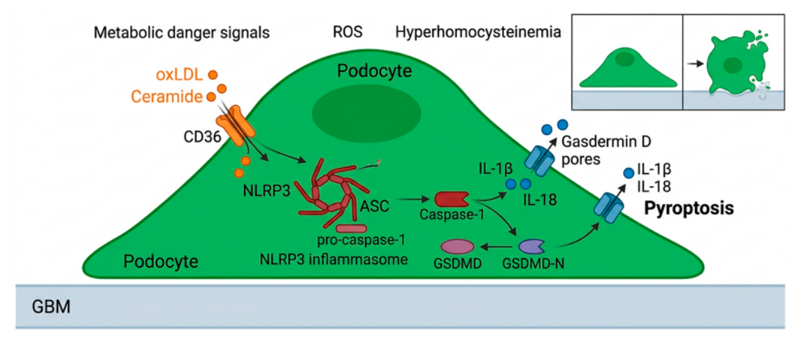
2.4.5. Role of cGAS–STING and NLRP3 Inflammasome Signaling in Kidney Diseases
Direct evidence for a podocyte-intrinsic cGAS–STING axis is currently strongest in diabetic kidney disease. In cultured podocytes and diabetic or obese mouse models, metabolic and lipotoxic stress induces mitochondrial injury and BAX-dependent leakage of mitochondrial DNA into the cytosol. Cytosolic mitochondrial DNA activates cGAS–STING signaling and is associated with NF-κB-dependent inflammatory gene expression, reduced slit-diaphragm protein abundance, and podocyte injury [168]. These findings identify mitochondrial stress as an upstream mechanism linking metabolic injury to intrinsic inflammatory signaling in podocytes.
Genetic studies further support a pathogenic role for podocyte STING signaling in diabetic kidney disease. Podocyte-specific Sting deletion in streptozotocin-induced diabetic mice attenuates podocyte injury, renal dysfunction, proteinuria, and renal inflammation. In this model, podocyte Sting deletion also reduces NLRP3 inflammasome activation and pyroptosis, whereas NLRP3 overexpression offsets the protective effects of STING deficiency [169]. Thus, STING can function upstream of NLRP3-associated inflammatory injury in hyperglycemic podocytes, although the relative contribution of canonical inflammasome activity, pyroptosis, and inflammasome-independent signaling requires further clarification.
Podocyte-specific NLRP3 perturbation models likewise support a context-dependent role for NLRP3 in diabetic glomerular injury. Podocyte-specific expression of the gain-of-function Nlrp3A350V variant aggravates albuminuria, mesangial expansion, and glomerular basement-membrane thickening in hyperglycemic mice. Conversely, podocyte-specific deletion of Nlrp3 or Casp1 protects against diabetic kidney disease. The more complete protection conferred by Nlrp3 deletion than by Casp1 deletion suggests that podocyte NLRP3 can mediate both caspase-1-dependent inflammasome functions and inflammasome-independent effects, including regulation of autophagy [160].
This podocyte-specific evidence should be interpreted alongside the broader contribution of NLRP3 in other renal compartments. Global Nlrp3 deficiency attenuates renal inflammation, fibrosis, albuminuria, glomerular hypertrophy, mesangial expansion, and glomerulosclerosis in diabetic mice [170]. However, global genetic models cannot distinguish direct podocyte effects from altered inflammasome activity in infiltrating immune cells, tubular cells, endothelial cells, mesangial cells, and interstitial compartments. The genetic models summarized in Table 4 provide a framework for distinguishing podocyte-intrinsic cGAS–STING and inflammasome functions from systemic and multicompartment renal effects. In particular, comparison of global, podocyte-specific, and gain-of-function models indicates that the phenotypic consequences of cGAS–STING, NLRP3, caspase-1, and RIPK3 perturbation depend on both the disease context and the cellular compartment in which these pathways are modified.
The functional relevance of canonical NLRP3 inflammasome activity in podocytes remains controversial. In contrast to studies reporting pathogenic podocyte NLRP3 activity in diabetic kidney disease, single-cell transcriptomic analyses, differentiated primary human podocytes, and diabetic mice expressing podocyte-specific Nlrp3A350V did not demonstrate appreciable canonical NLRP3–ASC–caspase-1 activity or stress-induced secretion of mature IL-1β and IL-18 by podocytes [161]. These divergent results indicate that NLRP3 expression, canonical inflammasome competence, caspase-1 activation, cytokine processing, and pyroptotic death should be evaluated as distinct endpoints rather than treated as interchangeable readouts of podocyte injury.
Puromycin aminonucleoside nephropathy provides a widely used experimental model of oxidant-dependent podocyte injury. PAN induces reactive oxygen species-dependent DNA damage in podocytes in vitro and in vivo [171]. In cultured podocytes, PAN also disrupts F-actin organization and induces cytoskeletal injury [172]. In rodent PAN nephropathy, podocyte damage is associated with proteinuria and foot-process abnormalities [173]. However, direct evidence that PAN-induced podocyte injury is mediated by cytosolic mitochondrial DNA sensing through cGAS–STING or by canonical NLRP3 inflammasome activation remains insufficient. PAN nephropathy should therefore be regarded as a relevant oxidative-stress model rather than as direct evidence for a podocyte-intrinsic cGAS–STING–NLRP3 axis.
Beyond diabetic kidney disease, functional cGAS–STING–NLRP3 interactions have been reported in APOL1-associated podocytopathy. APOL1 risk variants promote mitochondrial dysfunction, cytosolic mitochondrial DNA release, and activation of STING and NLRP3-associated inflammatory signaling in podocytes. Genetic and pharmacological pathway interventions attenuate inflammatory podocyte injury in this setting [153]. In membranous nephropathy, complement activation can promote pyroptosis-associated injury in podocytes, supporting an additional disease context in which inflammasome-related signaling and gasdermin-mediated membrane damage may contribute to filtration-barrier dysfunction [159].
Lupus nephritis illustrates the context dependence of cGAS–STING signaling. In TLR-dependent murine lupus models, global deletion of Cgas or Sting did not confer uniform protection and was associated with mildly exacerbated systemic autoimmune disease [145]. These findings do not establish a podocyte-specific role for cGAS–STING in lupus nephritis; rather, they caution that the consequences of systemic pathway inhibition may differ from those of cell-selective intervention in diabetic podocyte injury.
Together, current evidence identifies cGAS–STING and NLRP3 as context-dependent contributors to mitochondrial-stress responses, inflammation, and regulated cell-death susceptibility in podocytes. The most compelling evidence for a podocyte-intrinsic STING–NLRP3 axis is currently available in selected diabetic models. Future studies using inducible, cell-type-specific genetic systems and direct measures of inflammasome assembly, caspase-1 activity, gasdermin cleavage, cytokine maturation, and podocyte survival will be required to define the disease settings in which this pathway is a tractable therapeutic target.
Table 4.
Genetic models defining cGAS–STING, inflammasome, and regulated cell-death functions in glomerular and podocyte injury.
Table 4.
Genetic models defining cGAS–STING, inflammasome, and regulated cell-death functions in glomerular and podocyte injury.
| Mediator | Genetic strategy / experimental approach | Kidney disease or injury model | Principal findings | Reference |
|---|---|---|---|---|
| STING | Global deletion | HFD/STZ-induced diabetic kidney disease | STING deficiency attenuated renal fibrosis and reduced ID1-associated epithelial-to-mesenchymal transition. The protective phenotype was linked primarily to tubular epithelial responses; the direct contribution of podocytes and other renal cell types cannot be resolved in this global model | [174] |
| STING | Podocyte-specific deletion (Nphs2-Cre) | STZ-induced diabetes | Reduced podocyte damage, renal dysfunction, and inflammation, together with attenuation of NLRP3 inflammasome activation and pyroptosis | [169] |
| cGAS | Global deletion | MRL/lpr lupus model | Did not protect against lupus disease and was associated with a mildly exacerbated disease phenotype, demonstrating that global cGAS deficiency is not uniformly protective | [145] |
| RIPK3 | Global deletion | Nephrotoxic nephritis | No significant improvement in urinary albumin-to-creatinine ratio, serum urea, glomerular thrombosis, macrophage infiltration, or glomerular apoptosis | [175] |
| RIPK3 | Podocyte-specific deletion (Podocin-Cre) | Uninephrectomy plus HFD/STZ-induced diabetes | Reduced albuminuria, mesangial matrix expansion, podocyte foot-process injury, and podocyte loss are associated with reduced NF-κB p65-driven inflammation | [176] |
| NLRP3 | Global deletion | STZ-induced diabetes | Reduced albuminuria and renal injury, with attenuation of glomerular hypertrophy, glomerulosclerosis, mesangial expansion, interstitial fibrosis, inflammation, and TGF-β1 expression | [170] |
| NLRP3 | Podocyte-specific gain-of-function Nlrp3A350V expression | Hyperglycemic diabetic mice | Exacerbated albuminuria, mesangial expansion, and glomerular basement-membrane thickening, supporting a pathogenic role for podocyte NLRP3 signaling in diabetic glomerular injury. | [160] |
| NLRP3 | Podocyte-specific deletion (Nphs2-Cre) | STZ-induced diabetes | Reduced albuminuria and diabetic glomerular injury; protection exceeded that observed after podocyte-specific Casp1 deletion, consistent with both caspase-1-dependent and inflammasome-independent NLRP3 functions. | [160] |
| NLRP3 | Podocyte-specific expression of the gain-of-function Nlrp3A350V variant | Diabetic mice | Did not demonstrate appreciable canonical NLRP3 inflammasome activity in podocytes or stress-induced secretion of mature IL-1β and IL-18, indicating context-dependent inflammasome competence in kidney epithelial cells. | [161] |
| Caspase-1 | Podocyte-specific deletion (Nphs2-Cre) | STZ-induced diabetes | Reduced diabetic glomerular injury and inflammasome-associated injury, but with less complete protection than podocyte-specific Nlrp3 deletion. | [160] |
2.4.6. ZBP1 as a Potential Amplifier of Podocyte Cell Death
Z-DNA-binding protein 1 (ZBP1), also known as DAI, is an interferon-inducible cytosolic sensor of nucleic acids in the Z conformation. ZBP1 recognizes Z-form nucleic acids, particularly Z-RNA, through two N-terminal Zα domains and signals through C-terminal RIP homotypic interaction motif (RHIM) domains that engage RIPK1 and RIPK3 [177,178]. In infection and inflammatory models, ZBP1 activation can initiate RIPK3-dependent MLKL phosphorylation and necroptosis and, depending on the stimulus and cellular context, can engage caspase-8- and NLRP3-associated pathways that contribute to PANoptosis [150,179,180].
Endogenous Z-RNAs represent an important potential source of ZBP1 activation. Under homeostatic conditions, ADAR1-mediated RNA editing restrains inappropriate sensing of endogenous double-stranded RNA and Z-form RNA species. Defects in ADAR1 function permit pathological ZBP1 activation and can cause RIPK3-MLKL-dependent necroptosis, inflammation, and PANoptosis [181,182]. ZBP1 is an interferon-inducible sensor; therefore, chronic interferon signaling may increase cellular susceptibility to ZBP1-dependent inflammatory cell death [177,183].
These features make ZBP1 a biologically plausible mediator of podocyte injury in disease settings characterized by interferon signaling, viral nucleic acid sensing, mitochondrial stress, or inflammatory activation. In particular, cGAS–STING activation may increase the expression of interferon-stimulated genes, including ZBP1, and could thereby lower the threshold for RIPK3-dependent death signaling. However, direct evidence that a cGAS–STING–ZBP1 axis drives podocyte death in vivo remains limited. ZBP1 expression, ligand engagement, assembly of a ZBP1-containing PANoptosome, and the relative contribution of apoptosis, necroptosis, and pyroptosis have not yet been systematically resolved in podocyte-specific genetic models.
RIPK3 provides a relevant downstream link between ZBP1 signaling and regulated cell death. In diabetic kidney disease, podocyte-specific Ripk3 deletion reduces albuminuria, podocyte injury, mesangial matrix expansion, and NF-κB p65-driven inflammatory signaling [176]. Notably, high glucose did not increase MLKL phosphorylation in the experimental systems examined, and MLKL knockdown did not rescue podocyte injury, suggesting that RIPK3 can promote inflammatory damage independently of canonical MLKL-mediated necroptosis in diabetic podocytes [176]. These findings support a podocyte-intrinsic role for RIPK3 in diabetic injury but do not establish ZBP1 as its upstream activator in this setting.
Consistent with this evidence gap, podocyte-specific Zbp1 deletion models have not yet been reported. Future studies should combine inducible podocyte-specific Zbp1, Ripk3, and Mlkl deletion with direct assessment of ZBP1 expression, Z-RNA accumulation, RIPK3/MLKL phosphorylation, caspase-8 activation, inflammasome assembly, gasdermin cleavage, and podocyte survival. Such experiments will be necessary to determine whether ZBP1 functions as a bona fide PANoptotic amplifier in podocytes or whether its major renal roles reside primarily in immune or tubular epithelial cells.
3. From Inflammatory Signaling to Podocyte Fate: Regulated Cell Death, Detachment, and Senescence
The inflammatory, metabolic, mitochondrial, and mechanical stress pathways described above do not operate in isolation. Instead, podocytes integrate these inputs into a densely interconnected network that shapes cytoskeletal dynamics, mitochondrial homeostasis, regulated cell death, detachment, and senescence. In proteinuric kidney disease, apoptosis, pyroptosis, necroptosis, and ferroptosis represent the principal regulated cell-death modalities implicated in podocyte loss. These pathways coexist with additional fate-determining processes, including dysregulated autophagy, anoikis-associated detachment, PANoptosis-like signaling, and senescence-associated secretory reprogramming. Figure 4 provides an integrated overview of the major cell-fate programs that can be engaged in stressed podocytes and the metabolic, inflammatory, mechanical, and immune-derived signals that bias the transition between them.
3.1. Apoptosis (Caspase-9/3, Mitochondria)
Apoptosis is a well-established mechanism of podocyte depletion in diabetic kidney disease and other proteinuric glomerular disorders. Oxidative stress, endoplasmic-reticulum stress, and dysregulation of pro- and anti-apoptotic BCL-2 family proteins can promote mitochondrial outer membrane permeabilization, cytochrome c release, apoptosome assembly, and activation of caspase-9 and caspase-3. In diabetic experimental systems, oxidative mitochondrial injury is associated with cytochrome c release, Apaf-1-dependent apoptosome formation, caspase-9 activation, and podocyte apoptosis [184,185].
Podocyte apoptosis may be difficult to quantify in vivo because apoptotic cells can detach from the glomerular basement membrane and be lost into the urinary space before typical nuclear fragmentation is captured in tissue sections. Therefore, apoptosis should be assessed using complementary structural, biochemical, and podocyte-specific loss-of-cell markers rather than a single histological readout.
3.2. Pyroptosis (GSDMD, Caspase-1)
In selected diabetic and complement-associated experimental models, NLRP3–caspase-1–gasdermin D signaling has been associated with podocyte injury, membrane permeabilization, proteinuria, and impairment of the glomerular filtration barrier [159,160]. Podocyte-specific Nlrp3 and Casp1 deletion attenuate diabetic glomerular injury, supporting a contributory role for NLRP3-associated signaling in this disease context [160]. However, canonical inflammasome competence, mature IL-1β/IL-18 secretion, and the extent of bona fide pyroptotic death in podocytes remain disputed and should be distinguished from inflammatory signaling in intrarenal immune cells [161].
Accordingly, evidence of NLRP3, caspase-1, or GSDMD expression alone should not be interpreted as proof of podocyte pyroptosis. Robust attribution requires appropriate cell-specific measurements of caspase activation, GSDMD cleavage, membrane permeabilization, and genetic or pharmacological loss-of-function evidence.
3.3. Necroptosis (RIPK1/RIPK3/MLKL)
Necroptosis is a regulated form of lytic cell death classically mediated by RIPK3-dependent phosphorylation and oligomerization of MLKL. ZBP1 can function upstream of RIPK3 in viral and interferon-rich inflammatory settings, although this mechanism has been established primarily outside the podocyte context [177,178].
In diabetic kidney disease, RIPK3 has a demonstrable podocyte-intrinsic pathogenic role. Podocyte-specific Ripk3 deletion reduces albuminuria, mesangial matrix expansion, foot-process injury, podocyte loss, and NF-κB p65-driven inflammation [176]. However, high-glucose exposure did not increase MLKL phosphorylation in the systems examined, and MLKL knockdown did not rescue podocyte injury, indicating that RIPK3 can drive diabetic podocyte injury independently of canonical MLKL-mediated necroptosis [176]. Direct evidence for ZBP1-dependent necroptosis in podocytes in vivo remains limited.
3.4. Ferroptosis (GPX4 and Lipid Peroxidation)
Ferroptosis is an iron-dependent form of regulated cell death characterized by the accumulation of lipid peroxides to lethal levels and failure of antioxidant defense systems centered on the system Xc−–glutathione–GPX4 axis [186,187]. In podocytes, emerging evidence from experimental focal segmental glomerulosclerosis and lupus nephritis supports an association between impaired GPX4/SLC7A11-related antioxidant defenses, glutathione depletion, iron accumulation, lipid peroxidation, and structural injury [188,189,190]. However, the disease-specific causal contribution of ferroptosis to podocyte loss remains under active investigation.
Preclinical studies in diabetic kidney disease and experimental FSGS support an association between altered GPX4/SLC7A11-related defenses, lipid peroxidation, and podocyte injury. In Adriamycin-associated FSGS models, reduced GPX4 expression in podocytes and pharmacological protection by ferroptosis inhibition have been reported, although the evidence base remains predominantly preclinical [188]. More recently, single-cell and integrated metabolic analyses in diabetic kidneys identified a podocyte-associated MAPK14–SLC7A11–GPX4 axis linked to ferroptosis-related injury [191].
Therapeutically, several preclinical interventions have been reported to attenuate ferroptosis-associated renal and podocyte injury through GPX4-related antioxidant pathways. In experimental diabetic nephropathy, ginkgolide B inhibited GPX4 ubiquitination and restored GPX4 and FTH1 expression, whereas curcumol attenuated podocyte ferroptosis through the xCT/GPX4 axis [192,193]. In lupus nephritis, mesenchymal stromal cells reduced podocyte ferroptosis and improved podocyte structural markers through activation of the Nrf2/HO-1/GPX4 pathway [194]. These findings identify GPX4-related antioxidant defenses as experimentally tractable therapeutic checkpoints. However, the evidence remains preclinical and does not establish ferroptosis as a universally dominant mechanism of podocyte loss across proteinuric kidney diseases.
3.5. PANoptosis and the PANoptosome
PANoptosis is an integrated inflammatory cell-death program in which pyroptotic, apoptotic, and necroptotic mechanisms are coordinated through multiprotein PANoptosome complexes. Depending on the initiating stimulus and cellular context, PANoptosomes can incorporate sensors and signaling molecules such as ZBP1, RIPK1, RIPK3, FADD, caspase-8, inflammasome components, and caspase-1, thereby enabling concurrent or interdependent activation of pyroptosis, apoptosis, and necroptosis [150,179].
Direct evidence that podocytes can undergo PANoptosis has recently been reported in diabetic kidney disease. TRAIL signaling through death receptor 5 induced coordinated activation of apoptotic, pyroptotic, and necroptotic effectors in podocytes, with associated podocyte loss and diabetic glomerular injury [195]. These findings establish that integrated inflammatory cell-death programs can be engaged in podocytes under defined diabetic conditions.
The precise composition of the podocyte PANoptosome and the contribution of individual upstream sensors remain incompletely resolved. In particular, direct evidence that cGAS–STING activation or ZBP1-dependent Z-RNA sensing nucleates a bona fide PANoptosome in podocytes in vivo is currently limited. cGAS–STING, NLRP3, RIPK3, and death-receptor signaling may nevertheless create a cellular environment permissive for overlapping pyroptotic, apoptotic, and necroptotic pathways in diabetic and inflammatory podocytopathies. Future studies should determine whether these pathways converge within a common multiprotein complex or instead represent parallel, partially overlapping injury programs.
3.6. Podocyte Detachment, Anoikis-like Death, and Podocyturia
Podocyte detachment from the glomerular basement membrane is an irreversible mechanism of podocyte depletion and a major source of podocyturia. Podocyte–GBM attachment depends on integrin-containing adhesion complexes, including α3β1 and αVβ3 integrins, and on their connection to the actin cytoskeleton through focal-adhesion proteins such as talin-1, α-actinin-4, Nck adaptors, and α-parvin [196,197,198,199].
Mechanical stress, altered matrix signaling, cytoskeletal disorganization, and disruption of integrin-associated adhesions can reduce podocyte mechanical resilience and promote detachment. Disease-causing ACTN4 variants provide a genetic illustration of this process: mutant α-actinin-4 impairs adaptation of podocytes to repeated mechanical stretch and increases susceptibility to detachment [198]. Similarly, loss of α-parvin compromises integrin adhesome remodeling, reduces adhesion capacity and mechanical resistance, and promotes podocyte detachment in experimental models [199].
Detachment-induced cell death may involve anoikis-like mechanisms, but podocyte detachment should not be treated as a molecularly uniform form of cell death. Viable podocytes can be recovered from urine, indicating that detachment and terminal cell death are not invariably synchronous events [200,201]. The term "podoptosis" has been used in the podocyte literature, but it should be applied cautiously because it has also been used to describe p53-dependent podocyte cell death rather than detachment alone [202].
Complement activation, oxidative stress, and inflammatory mediators can destabilize podocyte architecture by perturbing the actin cytoskeleton, slit-diaphragm components, and plasma-membrane integrity. In membranous nephropathy and other complement-associated glomerular diseases, sublytic C5b-9 injury can induce oxidative and cytoskeletal stress and promote inflammatory or pyroptosis-associated podocyte injury, thereby creating conditions permissive for filtration-barrier dysfunction, proteinuria, and podocyte loss [28,159]. Thus, podocyte detachment is best regarded as a final common structural endpoint of multiple upstream inflammatory, metabolic, mechanical, and cytoskeletal injuries rather than as evidence for one specific regulated cell-death pathway.
3.7. Podocyte Senescence and Senescence-Associated Inflammatory Signaling
Beyond acute cell-death modalities, podocytes can acquire a senescence-like phenotype characterized by persistent cell-cycle arrest, DNA-damage signaling, mitochondrial dysfunction, altered chromatin regulation, and a pro-inflammatory secretory profile. In aging and diabetic experimental settings, podocyte senescence is associated with increased expression of p16, p21, and senescence-associated β-galactosidase activity, together with inflammatory transcriptional programs involving NF-κB-, IL-6-, TNF-, interferon-, and NLRP3-related pathways [176,203].
NLRP3 signaling contributes to inflammaging in podocytes. In middle-aged mice, pharmacological NLRP3 inhibition with MCC950 or genetic Nlrp3 deletion reduced podocyte senescence-associated β-galactosidase activity, p16 expression, and senescence-associated inflammatory transcripts and improved indices of podocyte life span and health span [203]. These findings support a role for NLRP3-associated inflammatory signaling in podocyte aging, although they do not establish that canonical inflammasome-mediated IL-1β/IL-18 secretion is the principal mechanism in podocytes.
Sirtuin- and epitranscriptomic pathways further modulate podocyte stress resilience. Podocyte-specific SIRT1 overexpression and pharmacological SIRT1 activation attenuated diabetes-induced podocyte loss, oxidative stress, albuminuria, and glomerular injury in diabetic mice [204]. Conversely, podocyte-specific Mettl14 deletion mitigated Adriamycin-associated glomerulopathy and podocyte injury, in part through preservation of Sirt1 expression, enhanced autophagy, and reduced apoptosis and inflammatory signaling [104].
These data identify senescence-like reprogramming as a distinct but interconnected outcome of chronic metabolic, oxidative, and inflammatory stress in podocytes. However, the molecular composition and functional consequences of a bona fide podocyte-specific senescence-associated secretory phenotype remain incompletely defined. Future work should distinguish stable senescence from transient cell-cycle arrest, dedifferentiation, autophagic dysfunction, and pre-terminal injury using complementary molecular, structural, and functional criteria.
4. Disease Context and Therapeutic Implications
As summarized in Figure 5, podocyte-intrinsic inflammatory signaling presents therapeutic opportunities at three complementary levels: reduction of upstream hemodynamic, metabolic, cytokine, and complement stress; context-dependent modulation of intracellular inflammatory and cytoskeletal pathways; and limitation of downstream cell-death and senescence-associated programs. The therapeutic goal is not complete suppression of NF-κB, JAK–STAT, STING, NLRP3, or Rho-family GTPase signaling, because these pathways also contribute to stress adaptation, cytoskeletal homeostasis, and host defense. Instead, the available evidence favors partial, disease-stage-specific attenuation of maladaptive pathway activation, ideally combined with cell-selective delivery strategies.
Established kidney-protective therapy should remain the therapeutic foundation. Renin–angiotensin system blockade, SGLT2 inhibitors, and, in appropriate patients with type 2 diabetes and chronic kidney disease, the non-steroidal mineralocorticoid receptor antagonist finerenone reduce albuminuria and improve kidney outcomes [205,206,207]. Their clinical benefit cannot be assigned exclusively to podocytes. Nevertheless, by lowering intraglomerular pressure and improving the metabolic, oxidative, and inflammatory milieu of the kidney, these agents are expected to reduce major upstream drivers of podocyte injury. They should therefore be viewed as systemic kidney-protective therapies that may indirectly constrain podocyte-intrinsic inflammatory signaling, rather than as selective podocyte pathway inhibitors. In experimental diabetic kidney disease, canagliflozin has also been reported to attenuate TXNIP/NLRP3-associated inflammatory signaling and pyroptotic injury in podocytes; this observation requires distinction from the established clinical outcome evidence for the SGLT2 inhibitor class (208).
At the cytokine and JAK–STAT level, anti-cytokine biologics and JAK inhibitors may reduce the inflammatory burden imposed on podocytes in autoimmune or cytokine-rich disease states, but these therapies are not podocyte-specific and can compromise protective immune responses. Baricitinib provides proof of pharmacological and translational plausibility rather than established clinical efficacy in diabetic kidney disease: in a Phase II randomized trial in type 2 diabetes with diabetic kidney disease, baricitinib 4 mg daily reduced the morning urinary albumin-to-creatinine ratio by 41% relative to placebo at Week 24 (209). Whether baricitinib modifies long-term kidney-function decline or kidney-failure risk in diabetic kidney disease remains unknown. Experimental data support a local IL-6–gp130–JAK2–STAT3 circuit in high-glucose-exposed podocytes: high glucose increased IL-6 production, JAK2 activation, nuclear STAT3 phosphorylation, and cellular hypertrophy, whereas IL-6 neutralization attenuated these responses in vitro [81]. Experimental studies further support a pathogenic role for excessive STAT3 activity in diabetic glomerulopathy and inflammatory kidney injury [82]. However, the therapeutic value of direct STAT3 inhibition in diabetic kidney disease remains uncertain because the available evidence is predominantly preclinical, and current small-molecule inhibitors, including Stattic and S3I-201, have unresolved limitations in selectivity, tissue distribution, and long-term safety; no STAT3-directed therapy has been established for glomerular disease [210].
Inhibition of intracellular inflammatory sensors is mechanistically attractive but remains preclinical. In diabetic and obesity-associated experimental models, mitochondrial DNA leakage activates a podocyte cGAS–STING/TBK1/p65 inflammatory axis. Genetic STING suppression and pharmacological inhibition reduced podocyte injury and improved renal phenotypes; C-176 reduced albuminuria and podocyte injury in diet-induced obesity mice [136]. In complementary work, podocyte-specific Sting deletion ameliorated inflammation, renal dysfunction, and podocyte injury in STZ-diabetic mice, whereas H-151 improved renal inflammation and function in db/db mice [169]. However, no cGAS or STING inhibitor has established clinical efficacy in glomerular disease, and species-specific STING pharmacology must be considered, particularly for C-176.
NLRP3 inhibition is supported by substantial mechanistic and preclinical evidence. In db/db mice, MCC950 improved renal function and attenuated albuminuria, mesangial expansion, podocyte loss, and glomerular lipid accumulation, whereas NLRP3 deficiency produced concordant protection in STZ-induced diabetes [214]. In high-glucose-exposed podocytes, NLRP3 silencing or MCC950 reduced apoptosis, cytoskeletal remodeling, mitochondrial ROS generation, NF-κB p65 activation, and lipid accumulation, consistent with an IL-1β/ROS/NF-κB p65-associated mechanism of inflammasome-driven podocyte injury [214]. MCC950 has also been associated with improvements in GBM thickening, foot-process fusion, and podocyte-marker expression in diabetic mice [215]. However, results are not uniform. In a delayed-intervention model of established STZ-induced diabetic kidney disease, MCC950 increased renal inflammation and structural injury, including mesangial expansion and glomerulosclerosis [211]. Treatment timing, disease stage, model selection, pharmacokinetics, and the relative contribution of immune-cell versus intrinsic glomerular inflammasome activity are therefore likely to determine the response to NLRP3 inhibition. MCC950 and related agents should be classified as experimental NLRP3-targeted candidates, not yet clinically validated podocyte-directed therapies. The divergent results obtained with MCC950 emphasize that pharmacological NLRP3 inhibition cannot be interpreted solely as a podocyte-directed intervention, because systemic exposure may modify inflammasome-related responses in infiltrating immune, tubular, endothelial, and mesangial compartments.
The p38 MAPK and Rho–ROCK axes provide complementary therapeutic entry points because they connect inflammatory transcriptional programs to cytoskeletal destabilization, foot-process effacement, and detachment. In human glomerulonephritis, p38 MAPK activation and cellular localization correlate with histological and functional indices of renal injury [212]. Although p38 inhibition has shown benefit in selected experimental renal and glomerular injury models, its effects are model- and cell-context-dependent, and systemic p38 inhibitors have encountered translational constraints, including safety limitations. Accordingly, they should not yet be considered an established class of glomerular therapeutics. By contrast, pharmacological Rho-kinase inhibition with fasudil reduced renal ROCK activity, albuminuria, mesangial matrix expansion, collagen IV accumulation, and glomerular basement membrane thickening in db/db mice without significantly altering blood glucose levels [213]. Genetic data provide a more selective rationale for ROCK2: podocyte-specific ROCK2 deletion protected against albuminuria, renal hypertrophy, mesangial expansion, podocyte loss, foot-process widening, and glomerular basement membrane thickening across STZ-induced, db/db, and high-fat-diet models of diabetes [126]. Mechanistically, ROCK2 deficiency restored PPARα-associated fatty-acid oxidation and improved podocyte energy homeostasis, identifying ROCK2 as a potential metabolic as well as cytoskeletal regulator of diabetic podocytopathy [126]. Fasudil is clinically available in Japan for the management of cerebral vasospasm after aneurysmal subarachnoid hemorrhage, but it is not an approved treatment for diabetic kidney disease or glomerular disease; it should therefore be regarded as a repurposing candidate rather than a validated glomerular therapy [216].
Epigenetic and epitranscriptomic regulators, including the METTL14–m6A–SIRT1 axis, sirtuins, histone-modifying enzymes, and inflammation-associated non-coding RNAs, offer potential means of reprogramming persistent stress and senescence-like phenotypes in podocytes [104,204,217]. These targets remain hypothesis-generating because their functions are strongly cell- and context-dependent, and indiscriminate interference with chromatin or RNA-modifying enzymes may disrupt podocyte homeostasis as well as maladaptive stress responses. Their prioritization requires podocyte-restricted genetic validation, pharmacodynamic evidence of renal target engagement, and targeted-delivery approaches.
Overall, the most immediately translatable approach is to combine guideline-based kidney-protective therapy with carefully selected pathway-directed interventions in biologically stratified disease settings. Progress will require evidence of target engagement in urinary podocytes or kidney biopsy material, separation of podocyte-autonomous from systemic immune effects, and validation in human podocyte, organoid, and glomerulus-on-chip systems. The central challenge is therefore not simply the identification of additional inhibitors but the matching of pathway activity, disease stage, and delivery modality to the inflammatory state of the podocyte.
5. Conclusions and Unresolved Questions
The evidence reviewed here supports a revised view of podocytes as active, context-dependent participants in glomerular inflammation rather than passive structural targets. Podocytes possess cell-autonomous stress-sensing and inflammatory signaling mechanisms that can influence filtration-barrier integrity, cytoskeletal homeostasis, and cellular fate. NF-κB and p38 MAPK function as complementary transcriptional and stress-kinase modules, whereas JAK–STAT relays cytokine inputs, cGAS–STING and NLRP3 respond to intracellular danger signals, and Rho-family GTPases translate inflammatory and metabolic cues into cytoskeletal remodeling. When sustained or dysregulated, this interconnected network can shift from adaptive stress responses toward maladaptive inflammation, senescence-like reprogramming, regulated cell death, detachment, and progressive podocyte depletion. Despite substantial progress, several fundamental questions remain.
First, what determines the transition from adaptive to maladaptive inflammatory signaling? The molecular thresholds that convert transient NF-κB or p38 MAPK activation into persistent injury programs remain incompletely defined. Epigenetic, metabolic, post-transcriptional, and intercellular regulators are likely to contribute, but their hierarchy and disease specificity remain unclear.
Second, which inflammatory pathways are primary drivers and which are secondary amplifiers of podocyte injury? Current evidence often cannot distinguish whether pathway activation initiates structural damage or instead reflects ongoing metabolic, mechanical, or immune-mediated stress. Temporal mapping of pathway activity relative to foot-process remodeling, slit-diaphragm disruption, detachment, and cell death will therefore be essential.
Third, to what extent do podocytes support canonical inflammasome activity in vivo? Single-cell transcriptomic analyses, differentiated primary human podocytes, and diabetic mice expressing podocyte-specific Nlrp3A350V have challenged the concept that podocytes are major sources of mature IL-1β and IL-18 [161]. Resolving whether NLRP3 functions primarily as a regulator of stress adaptation, a cell-death effector, a cytokine-maturation platform, or a combination of these activities will require cell-type-specific reporters, conditional genetic models, and direct measurements of inflammasome assembly, caspase-1 activation, gasdermin cleavage, cytokine maturation, and membrane permeabilization.
Fourth, can inflammatory signaling be therapeutically modulated without compromising podocyte survival? Global inhibition of NF-κB or p38 carries significant risks. Identifying podocyte-specific fine-tuning nodes—such as NEMO, REDD1, miR-146a, or calpain–IκB—may offer safer alternatives, but their therapeutic windows are undefined.
Future research could leverage hiPSC-derived podocytes, kidney organoids, glomerulus-on-chip systems, and CRISPR/Cas9-based isogenic models to dissect podocyte-intrinsic pathways in human genetic and disease contexts. Such platforms can enable controlled interrogation of candidate regulators of inflammation, mitochondrial stress, regulated cell death, and senescence-like reprogramming. Single-cell multi-omics and spatial transcriptomics should further refine the molecular definition of podocyte injury states across diabetic kidney disease, lupus nephritis, and IgA nephropathy, APOL1-associated disease, focal segmental glomerulosclerosis, and hypertensive nephropathy. These approaches, combined with longitudinal analysis of urinary podocytes and blood or urine biomarkers, could help identify disease-stage-specific signatures of pathway activity and inform biologically stratified clinical studies.
Ultimately, the goal is to move beyond broadly immunosuppressive or non-selective pathway inhibition toward more precise, podocyte-informed therapeutic strategies. Such approaches should preserve glomerular filtration-barrier integrity by rebalancing intrinsic inflammatory signaling, stabilizing cytoskeletal and mitochondrial homeostasis, and limiting maladaptive cell death, detachment, and senescence-like programs.
Author Contributions
Conceptualization, V.S.M.; investigation, D.E.B., S.A.K., D.A.O., and V.S.M.; writing—original draft preparation, D.E.B., S.A.K., D.A.O., and V.S.M.; writing—review and editing, D.E.B. and V.S.M.; visualization, D.E.B.; supervision, O.N.D.; funding acquisition, O.N.D. All authors have read and agreed to the published version of the manuscript.
Funding
This work was supported by the grant of the state program of the «Sirius» Federal Territory «Scientific and technological development of the «Sirius» Federal Territory» (Agreement No. 3-03, dated 18 February 2025).
Institutional Review Board Statement
Not applicable.
Informed Consent Statement
Not applicable.
Data Availability Statement
No new data were created or analyzed in this study. Data sharing is not applicable to this article.
Acknowledgments
During the preparation of this manuscript, the authors used Remio (version 3.20.4) for the generation and design of scientific illustrations. The authors reviewed and edited all generated visual content and take full responsibility for the content of this publication.
Conflicts of Interest
The authors declare no conflicts of interest.
Abbreviations
The following abbreviations are used in this manuscript:
| ADR | Adriamycin |
| AGE | Advanced glycation end product |
| AKI | Acute kidney injury |
| APOL1 | Apolipoprotein L1 |
| ASC | Apoptosis-associated speck-like protein containing a caspase recruitment domain |
| AT1R | Angiotensin II type 1 receptor |
| cGAMP | Cyclic guanosine monophosphate–adenosine monophosphate |
| cGAS | Cyclic GMP–AMP synthase |
| C5b-9 | Complement membrane attack complex |
| CARD | Caspase recruitment domain |
| CCL2 | C-C motif chemokine ligand 2 |
| CD2AP | CD2-associated protein |
| CKD | Chronic kidney disease |
| COX-2 | Cyclooxygenase-2 |
| DAMP | Damage-associated molecular pattern |
| DKD | Diabetic kidney disease |
| FSGS | Focal segmental glomerulosclerosis |
| GAP | GTPase-activating protein |
| GBM | Glomerular basement membrane |
| GEF | Guanine nucleotide exchange factor |
| GN | Glomerulonephritis |
| GSDMD | Gasdermin D |
| GSK3β | Glycogen synthase kinase 3 beta |
| GPX4 | Glutathione peroxidase 4 |
| IFN | Interferon |
| IRF3 | Interferon regulatory factor 3 |
| ISG | Interferon-stimulated gene |
| JAK | Janus kinase |
| LRR | Leucine-rich repeat |
| MAPK | Mitogen-activated protein kinase |
| METTL14 | Methyltransferase-like 14 |
| MLKL | Mixed lineage kinase domain-like protein |
| MRL/lpr | Murphy Roths Large/lymphoproliferation mouse strain |
| MyD88 | Myeloid differentiation primary response 88 |
| NACHT | NAIP, CIITA, HET-E and TP1 domain |
| NEK7 | NIMA-related kinase 7 |
| NEMO | NF-κB essential modulator |
| NET | Neutrophil extracellular trap |
| NF-κB | Nuclear factor kappa B |
| NIK | NF-κB-inducing kinase |
| NLRP3 | NOD-like receptor family pyrin domain-containing 3 |
| NPHS1 | Nephrin |
| NPHS2 | Podocin |
| PAMP | Pathogen-associated molecular pattern |
| PANoptosis | Pyroptosis, apoptosis, and necroptosis |
| p65 | NF-κB RelA subunit |
| PI3K | Phosphoinositide 3-kinase |
| pMLKL | Phosphorylated mixed lineage kinase domain-like protein |
| PRR | Pattern-recognition receptor |
| PYD | Pyrin domain |
| RAC1 | Ras-related C3 botulinum toxin substrate 1 |
| RAGE | Receptor for advanced glycation end products |
| RAAS | Renin–angiotensin–aldosterone system |
| RCD | Regulated cell death |
| REDD1 | Regulated in development and DNA damage response 1 |
| RHIM | RIP homotypic interaction motif |
| ROCK | Rho-associated coiled-coil-containing protein kinase |
| ROS | Reactive oxygen species |
| SASP | Senescence-associated secretory phenotype |
| scRNA-seq | Single-cell RNA sequencing |
| SGLT2 | Sodium-glucose cotransporter 2 |
| SIRT1 | Sirtuin 1 |
| SIRT6 | Sirtuin 6 |
| SLC7A11 | Solute carrier family 7 member 11 |
| SOCS3 | Suppressor of cytokine signaling 3 |
| STING | Stimulator of interferon genes |
| STZ | Streptozotocin |
| TBK1 | TANK-binding kinase 1 |
| TGF-β | Transforming growth factor beta |
| TLR | Toll-like receptor |
| TNF | Tumor necrosis factor |
| TRAF | TNF receptor-associated factor |
| ZBP1 | Z-DNA-binding protein 1 |
References
- Pavenstädt, H.; Kriz, W.; Kretzler, M. Cell biology of the glomerular podocyte. Physiol. Rev. 2003, 83, 253–307. [CrossRef]
- Haraldsson, B.; Nyström, J.; Deen, W.M. Properties of the glomerular barrier and mechanisms of proteinuria. Physiol. Rev. 2008, 88, 451–487. [CrossRef]
- Tryggvason, K.; Patrakka, J.; Wartiovaara, J. Hereditary proteinuria syndromes and mechanisms of proteinuria. N. Engl. J. Med. 2006, 354, 1387–1401. [CrossRef]
- Grahammer, F.; Schell, C.; Huber, T.B. The podocyte slit diaphragm—from a thin grey line to a complex signaling hub. Nat. Rev. Nephrol. 2013, 9, 587–598. [CrossRef]
- Kim, Y.H.; Goyal, M.; Kurnit, D.; Wharram, B.; Wiggins, J.; Holzman, L.; Kershaw, D.; Wiggins, R. Podocyte depletion and glomerulosclerosis have a direct relationship in the PAN-treated rat. Kidney Int. 2001, 60, 957–968. [CrossRef]
- Wharram, B.L.; Goyal, M.; Wiggins, J.E.; Sanden, S.K.; Hussain, S.; Filipiak, W.E.; Saunders, T.L.; Dysko, R.C.; Kohno, K.; Holzman, L.B.; Wiggins, R.C. Podocyte depletion causes glomerulosclerosis: Diphtheria toxin-induced podocyte depletion in rats. J. Am. Soc. Nephrol. 2005, 16, 2941–2952. [CrossRef]
- Wiggins, R.C. The spectrum of podocytopathies: A unifying view of glomerular diseases. Kidney Int. 2007, 71, 1205–1214. [CrossRef]
- Banas, M.C.; Banas, B.; Hudkins, K.L.; Wietecha, T.A.; Iyoda, M.; Bock, E.; Hauser, P.; Pippin, J.W.; Shankland, S.J.; Smith, K.D.; Stoelcker, B.; Liu, G.; Gröne, H.-J.; Krämer, B.K.; Alpers, C.E. TLR4 links podocytes with the innate immune system to mediate glomerular injury. J. Am. Soc. Nephrol. 2008, 19, 704–713. [CrossRef]
- Brähler, S.; Ising, C.; Hagmann, H.; Rasmus, M.; Hoehne, M.; Kurschat, C.; Kisner, T.; Goebel, H.; Shankland, S.; Addicks, K.; Thaiss, F.; Schermer, B.; Pasparakis, M.; Benzing, T.; Brinkkoetter, P.T. Intrinsic proinflammatory signaling in podocytes contributes to podocyte damage and prolonged proteinuria. Am. J. Physiol. Renal Physiol. 2012, 303, F1473–F1485. [CrossRef]
- Susztak, K.; Raff, A.C.; Schiffer, M.; Böttinger, E.P. Glucose-induced reactive oxygen species cause apoptosis of podocytes and podocyte depletion at the onset of diabetic nephropathy. Diabetes 2006, 55, 225–233. [CrossRef]
- Zhang, C.; Boini, K.M.; Xia, M.; Abais, J.M.; Li, X.; Liu, Q.; Li, P.L. Activation of Nod-like receptor protein 3 inflammasomes turns on podocyte injury and glomerular sclerosis in hyperhomocysteinemia. Hypertension 2012, 60, 154–162. [CrossRef]
- Zhang, H.; Nair, V.; Saha, J.; Atkins, K.B.; Hodgin, J.B.; Saunders, T.L.; Myers, M.G., Jr.; Werner, T.; Kretzler, M.; Brosius, F.C. Podocyte-specific JAK2 overexpression worsens diabetic kidney disease in mice. Kidney Int. 2017, 92, 909–921. [CrossRef]
- Lee, H.W.; Khan, S.Q.; Khaliqdina, S.; Altintas, M.M.; Grahammer, F.; Zhao, J.L.; et al. Absence of miR-146a in podocytes increases risk of diabetic glomerulopathy via up-regulation of ErbB4 and Notch-1. J. Biol. Chem. 2017, 292, 732–747. [CrossRef]
- Kurts, C.; Panzer, U.; Anders, H.-J.; Rees, A.J. The immune system and kidney disease: Basic concepts and clinical implications. Nat. Rev. Immunol. 2013, 13, 738–753. [CrossRef]
- Sunilkumar, S.; Yerlikaya, E.I.; Toro, A.L.; Chen, H.; Zhou, Y.; Gill, D.L.; Kimball, S.R.; Dennis, M.D. Podocyte-specific expression of the stress response protein REDD1 is necessary for diabetes-induced podocytopenia. Diabetes 2025, 74, 398–408. [CrossRef]
- Barutta, F.; Bellini, S.; Gruden, G. Mechanisms of podocyte injury and implications for diabetic nephropathy. Clin. Sci. 2022, 136, 493–520. [CrossRef]
- Ke, G.; Chen, X.; Liao, R.; Xu, L.; Zhang, L.; Zhang, H.; Kuang, S.; Du, Y.; Hu, J.; Lian, Z.; Dou, C.; Zhang, Q.; Zhao, X.; Zhang, F.; Zhu, S.; Ma, J.; Li, Z.; Li, S.; He, C.; Chen, X.; Wen, Y.; Feng, Z.; Zheng, M.; Lin, T.; Li, R.; Li, B.; Dong, W.; Chen, Y.; Wang, W.; Ye, Z.; Deng, C.; Xiao, H.; Xiao, J.; Liang, X.; Shi, W.; Liu, S. Receptor activator of NF-κB mediates podocyte injury in diabetic nephropathy. Kidney Int. 2021, 100, 377–390. [CrossRef]
- Lang, Y.; Wang, Q.; Sheng, Q.; Lu, S.; Yang, M.; Kong, Z.; et al.; Lv, Z. FTO-mediated m6A modification of serum amyloid A2 mRNA promotes podocyte injury and inflammation by activating the NF-κB signaling pathway. FASEB J. 2024, 38, e23409. [CrossRef]
- Yamashita, M.; Yoshida, T.; Suzuki, S.; Homma, K.; Hayashi, M. Podocyte-specific NF-κB inhibition ameliorates proteinuria in adriamycin-induced nephropathy in mice. Clin. Exp. Nephrol. 2017, 21, 16–26. [CrossRef]
- Sanz, A.B.; Sánchez-Niño, M.D.; Ramos, A.M.; Moreno, J.A.; Santamaría, B.; Ruiz-Ortega, M.; Egido, J.; Ortiz, A. NF-κB in renal inflammation. J. Am. Soc. Nephrol. 2010, 21, 1254–1262. [CrossRef]
- Ren, N.; Wang, W.F.; Zou, L.; Zhao, Y.L.; Miao, H.; Zhao, Y.Y. The nuclear factor kappa B signaling pathway is a master regulator of renal fibrosis. Front. Pharmacol. 2024, 14, 1335094. [CrossRef]
- Bao, H.; Ge, Y.; Peng, A.; Gong, R. Fine-tuning of NFκB by glycogen synthase kinase 3β directs the fate of glomerular podocytes upon injury. Kidney Int. 2015, 87, 1176–1190. [CrossRef]
- Guo, Q.; Jin, Y.; Chen, X.; Ye, X.; Shen, X.; Lin, M.; et al.; Zhang, J. NF-κB in biology and targeted therapy: New insights and translational implications. Signal Transduct. Target. Ther. 2024, 9, 53. [CrossRef]
- Abhirami, B.L.; Krishna, A.A.; Kumaran, A.; Chiu, C.-H. Targeting NF-κB in diabetic nephropathy: Exploring the therapeutic potential of phytoconstituents. Arch. Pharm. Res. 2025, 48, 577–637. [CrossRef]
- Liu, S.; Shi, W.; Xiao, H.; Liang, X.; Deng, C.; Ye, Z.; et al. Receptor activator of NF-kappaB and podocytes: Towards a function of a novel receptor-ligand pair in the survival response of podocyte injury. PLoS ONE 2012, 7, e41331. [CrossRef]
- Hussain, S.; Romio, L.; Saleem, M.; Mathieson, P.; Serrano, M.; Moscat, J.; Diaz-Meco, M.; Scambler, P.; Koziell, A. Nephrin deficiency activates NF-κB and promotes glomerular injury. J. Am. Soc. Nephrol. 2009, 20, 1733–1743. [CrossRef]
- Wang, J.; Fu, D.; Senouthai, S.; You, Y. Critical roles of PI3K/Akt/NF-κB survival axis in angiotensin II-induced podocyte injury. Mol. Med. Rep. 2019, 20, 5134–5144. [CrossRef]
- Bruno, V.; Mühlig, A.K.; Oh, J.; Licht, C. New insights into the immune functions of podocytes: The role of complement. Mol. Cell. Pediatr. 2023, 10, 3. [CrossRef]
- Verma, R.; Venkatareddy, M.; Kalinowski, A.; Li, T.; Kukla, J.; Mollin, A.; Cara-Fuentes, G.; Patel, S.R.; Garg, P. Nephrin is necessary for podocyte recovery following injury in an adult mature glomerulus. PLoS ONE 2018, 13, e0198013. [CrossRef]
- Shen, X.; Weng, C.; Wang, Y.; Wang, C.; Feng, S.; Li, X.; et al. Lipopolysaccharide-induced podocyte injury is regulated by calcineurin/NFAT and TLR4/MyD88/NF-κB signaling pathways through angiopoietin-like protein 4. Genes Dis. 2022, 9, 443–455. [CrossRef]
- Huang, X.; Hou, X.; Chuan, L.; Wei, S.; Wang, J.; Yang, X.; Ru, J. miR-129-5p alleviates LPS-induced acute kidney injury via targeting HMGB1/TLRs/NF-kappaB pathway. Int. Immunopharmacol. 2020, 89, 107016. [CrossRef]
- Jia, J.; Ding, G.; Zhu, J.; Chen, C.; Liang, W.; Franki, N.; Singhal, P.C. Angiotensin II infusion induces nephrin expression changes and podocyte apoptosis. Am. J. Nephrol. 2008, 28, 500–507. [CrossRef]
- Cardoso, V.G.; Gonçalves, G.L.; Costa-Pessoa, J.M.; Thieme, K.; Lins, B.B.; Casare, F.A.M.; et al. Angiotensin II-induced podocyte apoptosis is mediated by endoplasmic reticulum stress/PKC-δ/p38 MAPK pathway activation and trough increased Na+/H+ exchanger isoform 1 activity. BMC Nephrol. 2018, 19, 179. [CrossRef]
- Gao, S.; Cui, Z.; Zhao, M.H. Complement C3a and C3a receptor activation mediates podocyte injuries in the mechanism of primary membranous nephropathy. J. Am. Soc. Nephrol. 2022, 33, 1742–1756. [CrossRef]
- Sunilkumar, S.; Yerlikaya, E.I.; VanCleave, A.; Subrahmanian, S.M.; Toro, A.L.; Kimball, S.R.; Dennis, M.D. REDD1-dependent GSK3β signaling in podocytes promotes canonical NF-κB activation in diabetic nephropathy. J. Biol. Chem. 2025, 301, 108244. [CrossRef]
- Tian, X.; Inoue, K.; Zhang, Y.; Wang, Y.; Sperati, C.J.; Pedigo, C.E.; Zhao, T.; Yan, M.; Groener, M.; Moledina, D.G.; Ebenezer, K.; Li, W.; Zhang, Z.; Liebermann, D.A.; Greene, L.; Greer, P.; Parikh, C.R.; Ishibe, S. Inhibiting calpain 1 and 2 in cyclin G associated kinase-knockout mice mitigates podocyte injury. JCI Insight 2020, 5, e142740. [CrossRef]
- Pengal, R.; Guess, A.J.; Agrawal, S.; Manley, J.; Ransom, R.F.; Mourey, R.J.; Benndorf, R.; Smoyer, W.E. Inhibition of the protein kinase MK-2 protects podocytes from nephrotic syndrome-related injury. Am. J. Physiol. Renal Physiol. 2011, 301, F509–F519. [CrossRef]
- Agrawal, S.; Guess, A.J.; Chanley, M.A.; Smoyer, W.E. Albumin-induced podocyte injury and protection are associated with regulation of COX-2. Kidney Int. 2014, 86, 1150–1160. [CrossRef]
- Gonçalves, G.L.; Costa-Pessoa, J.M.; Thieme, K.; Lins, B.B.; Oliveira-Souza, M. Intracellular albumin overload elicits endoplasmic reticulum stress and PKC-delta/p38 MAPK pathway activation to induce podocyte apoptosis. Sci. Rep. 2018, 8, 18012. [CrossRef]
- Schiffer, M.; Bitzer, M.; Roberts, I.S.D.; Kopp, J.B.; ten Dijke, P.; Mundel, P.; Böttinger, E.P. Apoptosis in podocytes induced by TGF-β and Smad7. J. Clin. Investig. 2001, 108, 807–816. [CrossRef]
- Zheng, X.; Zhong, Q.; Lin, X.; Gu, X.; Ling, X.; Liang, Z.; Qin, Q.; Du, X. Transforming growth factor-β1-induced podocyte injury is associated with increased microRNA-155 expression, enhanced inflammatory responses and MAPK pathway activation. Exp. Ther. Med. 2021, 21, 620. [CrossRef]
- Woznowski, M.P.; Potthoff, S.A.; Königshausen, E.; Haase, R.; Hoch, H.; Meyer-Schwesinger, C.; Wiech, T.; Stegbauer, J.; Rump, L.C.; Sellin, L.; Quack, I. Inhibition of p38 MAPK decreases hyperglycemia-induced nephrin endocytosis and attenuates albuminuria. J. Mol. Med. 2022, 100, 781–795. [CrossRef]
- Ni, Y.; Wang, X.; Yin, X.; Li, Y.; Liu, X.; Wang, H.; et al. Plectin protects podocytes from adriamycin-induced apoptosis and F-actin cytoskeletal disruption through the integrin α6β4/FAK/p38 MAPK pathway. J. Cell. Mol. Med. 2018, 22, 5450–5467. [CrossRef]
- Sun, S.-C.; Ley, S.C. New insights into NF-κB regulation and function. Trends Immunol. 2008, 29, 469–478. [CrossRef]
- Wu, H.; Lo, Y.-C.; Yin, Q. Structural studies of NEMO and TRAF6: Implications in NF-κB activation. In Advances in TNF Family Research; Wallach, D., Kovalenko, A., Eds.; Adv. Exp. Med. Biol.; Springer: New York, NY, USA, 2011; Volume 691, pp. 89–91. [CrossRef]
- Yu, H.; Lin, L.; Zhang, Z.; Zhang, H.; Hu, H. Targeting NF-κB pathway for the therapy of diseases: Mechanism and clinical study. Signal Transduct. Target. Ther. 2020, 5, 209. [CrossRef]
- Li, X.C.; Zhuo, J.L. Nuclear factor-κB as a hormonal intracellular signaling molecule: Focus on angiotensin II-induced cardiovascular and renal injury. Curr. Opin. Nephrol. Hypertens. 2008, 17, 37–43. [CrossRef]
- Sun, S.-C. Non-canonical NF-κB signaling pathway. Cell Res. 2011, 21, 71–85. [CrossRef]
- Sun, S.-C. The non-canonical NF-κB pathway in immunity and inflammation. Nat. Rev. Immunol. 2017, 17, 545–558. [CrossRef]
- Rangan, G.; Wang, Y.; Harris, D. NF-kappaB signaling in chronic kidney disease. Front. Biosci. (Landmark Ed.) 2009, 14, 3496–3522. [CrossRef]
- Zinatizadeh, M.R.; Schock, B.; Chalbatani, G.M.; Zarandi, P.K.; Jalali, S.A.; Miri, S.R. The nuclear factor kappa B (NF-kB) signaling in cancer development and immune diseases. Genes Dis. 2021, 8, 287–297. [CrossRef]
- Chen, M.; Fang, Y.; Ge, Y.; Qiu, S.; Dworkin, L.; Gong, R. The redox-sensitive GSK3β is a key regulator of glomerular podocyte injury in type 2 diabetic kidney disease. Redox Biol. 2024, 72, 103127. [CrossRef]
- Taganov, K.D.; Boldin, M.P.; Chang, K.-J.; Baltimore, D. NF-κB-dependent induction of microRNA miR-146, an inhibitor targeted to signaling proteins of innate immune responses. Proc. Natl. Acad. Sci. USA 2006, 103, 12481–12486. [CrossRef]
- Li, X.; Venkatesh, I.; Villanueva, V.; Wei, H.; Geraghty, T.; Rajagopalan, A.; Helmuth, R.W.; Altintas, M.M.; Faridi, H.M.; Gupta, V. Podocyte-specific deletion of miR-146a increases podocyte injury and diabetic kidney disease. Front. Med. 2022, 9, 897188. [CrossRef]
- Baishya, S.; Sarangi, A.; Somvanshi, P.R.; Pasupulati, A.K. Gene Regulatory Networks Involved in Activation of Notch Signaling by AGEs in the Pathogenesis of Diabetic Kidney Disease. PLoS ONE 2026, 21, e0335768. [CrossRef]
- Niranjan, T.; Bielesz, B.; Gruenwald, A.; Ponda, M.P.; Kopp, J.B.; Thomas, D.B.; Susztak, K. The Notch pathway in podocytes plays a role in the development of glomerular disease. Nat. Med. 2008, 14, 290–298. [CrossRef]
- Nishad, R.; Meshram, P.; Singh, A.K.; Reddy, G.B.; Pasupulati, A.K. Activation of Notch1 signaling in podocytes by glucose-derived AGEs contributes to proteinuria. BMJ Open Diabetes Res. Care 2020, 8, e001203. [CrossRef]
- Dai, T.; Natarajan, R.; Nast, C.C.; LaPage, J.; Chuang, P.; Sim, J.; Tong, L.; Chamberlin, M.; Wang, S.; Adler, S.G. Glucose and diabetes: Effects on podocyte and glomerular p38MAPK, heat shock protein 25, and actin cytoskeleton. Kidney Int. 2006, 69, 806–814. [CrossRef]
- Lv, Z.; Hu, M.; Zhen, J.; Lin, J.; Wang, Q.; Wang, R.; et al. Podocyte-specific Rac1 deficiency ameliorates podocyte damage and proteinuria in STZ-induced diabetic nephropathy in mice. Cell Death Dis. 2018, 9, 342. [CrossRef]
- Gao, K.; Chi, Y.; Sun, W.; Takeda, M.; Yao, J. 5′-AMP-activated protein kinase attenuates adriamycin-induced oxidative podocyte injury through thioredoxin-mediated suppression of the apoptosis signal-regulating kinase 1–p38 signaling pathway. Mol. Pharmacol. 2014, 85, 460–471. [CrossRef]
- Schömig, T.; Diefenhardt, P.; Plagmann, I.; Trinsch, B.; Merz, T.; Crispatzu, G.; Unnersjö-Jess, D.; Nies, J.; Pütz, D.; Sierra Gonzalez, C.; Schermer, B.; Benzing, T.; Brinkkoetter, P.T.; Brähler, S. The podocytes’ inflammatory responses in experimental GN are independent of canonical MYD88-dependent toll-like receptor signaling. Sci. Rep. 2024, 14, 2292. [CrossRef]
- Ke, G.; Chen, X.; Liao, R.; Xu, L.; Zhang, L.; Zhang, H.; Kuang, S.; Du, Y.; Hu, J.; Lian, Z.; Dou, C.; Zhang, Q.; Zhao, X.; Zhang, F.; Zhu, S.; Ma, J.; Li, Z.; Li, S.; He, C.; Chen, X.; Wen, Y.; Feng, Z.; Zheng, M.; Lin, T.; Li, R.; Li, B.; Dong, W.; Chen, Y.; Wang, W.; Ye, Z.; Deng, C.; Xiao, H.; Xiao, J.; Liang, X.; Shi, W.; Liu, S. Receptor activator of NF-κB mediates podocyte injury in diabetic nephropathy. Kidney Int. 2021, 100, 377–390. [CrossRef]
- Wang, J.; Fu, D.; Senouthai, S.; You, Y. Critical roles of PI3K/Akt/NF-κB survival axis in angiotensin II-induced podocyte injury. Mol. Med. Rep. 2019, 20, 5134–5144. [CrossRef]
- Ren, N.; Wang, W.F.; Zou, L.; Zhao, Y.L.; Miao, H.; Zhao, Y.Y. The nuclear factor kappa B signaling pathway is a master regulator of renal fibrosis. Front. Pharmacol. 2024, 14, 1335094. [CrossRef]
- Chen, Y.; Lin, L.; Rao, S.; Tao, X.; Cui, J.; Wan, J. Complement C3 mediates podocyte injury through TLR4/NFκB-p65 signaling during ischemia–reperfusion acute kidney injury and post-injury fibrosis. Eur. J. Med. Res. 2023, 28, 135. [CrossRef]
- Sun, D.L.; Guo, Z.Y.; Liu, W.Y.; Zhang, L.; Zhang, Z.Y.; Hu, Y.L.; et al. Astragaloside IV alleviates podocyte injury in diabetic nephropathy through regulating the IRE-1α/NF-κB/NLRP3 pathway. Chin. J. Integr. Med. 2025, 31, 422–433. [CrossRef]
- Yamashita, M.; Yoshida, T.; Hayashi, M. Podocyte NF-κB is dispensable for the pathogenesis of renal ischemia–reperfusion injury. Physiol. Rep. 2016, 4, e12912. [CrossRef]
- Han, J.; Wu, J.; Silke, J. An overview of mammalian p38 mitogen-activated protein kinases, central regulators of cell stress and receptor signaling. F1000Research 2020, 9, 653. [CrossRef]
- Cánovas, B.; Nebreda, A.R. Diversity and versatility of p38 kinase signaling in health and disease. Nat. Rev. Mol. Cell Biol. 2021, 22, 346–366. [CrossRef]
- Sugioka, S.; Yamada, H.; Ishii, A.; Kato, Y.; Yamada, R.; Mori, K.P.; Ohno, S.; Handa, T.; Ikushima, A.; Ishimura, T.; Osaki, K.; Tokudome, T.; Matsusaka, T.; Nebreda, A.R.; Yanagita, M.; Yokoi, H. Dual deletion of guanylyl cyclase-A and p38 mitogen-activated protein kinase in podocytes with aldosterone administration causes glomerular intra-capillary thrombi. Kidney Int. 2023, 104, 508–525. [CrossRef]
- Stitt-Cavanagh, E.M.; Faour, W.H.; Takami, K.; Carter, A.; Vanderhyden, B.; Guan, Y.; Schneider, A.; Breyer, M.D.; Kennedy, C.R.J. Inhibition of the Protein Kinase MK-2 Protects Podocytes from Nephrotic Syndrome-Related Injury. Am. J. Physiol. Renal Physiol. 2011, 301, F509–F519. [CrossRef]
- Mangold, N.; Pippin, J.; Unnersjoe-Jess, D.; Koehler, S.; Shankland, S.; Brähler, S.; Schermer, B.; Benzing, T.; Brinkkoetter, P.T.; Hagmann, H. The atypical cyclin-dependent kinase 5 (Cdk5) guards podocytes from apoptosis in glomerular disease while being dispensable for podocyte development. Cells 2021, 10, 2464. [CrossRef]
- Denhez, B.; Rousseau, M.; Dancosst, D.-A.; Lizotte, F.; Guay, A.; Auger-Messier, M.; Côté, A.M.; Geraldes, P. Diabetes-induced DUSP4 reduction promotes podocyte dysfunction and progression of diabetic nephropathy. Diabetes 2019, 68, 1026–1039. [CrossRef]
- Adams, R.H.; Porras, A.; Alonso, G.; Jones, M.; Vintersten, K.; Panelli, S.; Valladares, A.; Perez, L.; Klein, R.; Nebreda, A.R. Essential role of p38alpha MAP kinase in placental but not embryonic cardiovascular development. Mol. Cell 2000, 6, 109–116. [CrossRef]
- Mudgett, J.S.; Ding, J.; Guh-Siesel, L.; Chartrain, N.A.; Yang, L.; Gopal, S.; Shen, M.M. Essential role for p38α mitogen-activated protein kinase in placental angiogenesis. Proc. Natl. Acad. Sci. USA 2000, 97, 10454–10459. [CrossRef]
- Kato, Y.; Mori, K.; Kasahara, M.; Osaki, K.; Ishii, A.; Mori, K.P.; Toda, N.; Ohno, S.; Kuwabara, T.; Tokudome, T.; Kishimoto, I.; Saleem, M.A.; Matsusaka, T.; Nakao, K.; Mukoyama, M.; Yanagita, M.; Yokoi, H. Natriuretic peptide receptor guanylyl cyclase-A pathway counteracts glomerular injury evoked by aldosterone through p38 mitogen-activated protein kinase inhibition. Sci. Rep. 2017, 7, 46624. [CrossRef]
- Kotlyarov, A.; Neininger, A.; Schubert, C.; Eckert, R.; Birchmeier, C.; Volk, H.D.; Gaestel, M. MAPKAP kinase 2 is essential for LPS-induced TNF-alpha biosynthesis. Nat. Cell Biol. 1999, 1, 94–97. [CrossRef]
- Guess, A.J.; Ayoob, R.; Chanley, M.; Manley, J.; Cajaiba, M.M.; Agrawal, S.; et al. Crucial roles of the protein kinases MK2 and MK3 in a mouse model of glomerulonephritis. PLoS ONE 2013, 8, e54239. [CrossRef]
- Hu, X.; Li, J.; Fu, M.; Zhao, X.; Wang, W. The JAK/STAT signaling pathway: From bench to clinic. Signal Transduct. Target. Ther. 2021, 6, 402. [CrossRef]
- Feigerlová, E.; Battaglia-Hsu, S.-F. IL-6 signaling in diabetic nephropathy: From pathophysiology to therapeutic perspectives. Cytokine Growth Factor Rev. 2017, 37, 57–65. [CrossRef]
- Jo, H.A.; Kim, J.Y.; Yang, S.H.; Han, S.S.; Joo, K.W.; Kim, Y.S.; Kim, D.K. The role of local IL6/JAK2/STAT3 signaling in high glucose-induced podocyte hypertrophy. Kidney Res. Clin. Pract. 2016, 35, 212–218. [CrossRef]
- Dai, Y.; Gu, L.; Yuan, W.; Yu, Q.; Ni, Z.; Ross, M.J.; Kaufman, L.; Xiong, H.; Salant, D.J.; He, J.C.; Chuang, P.Y. Podocyte-specific deletion of signal transducer and activator of transcription 3 attenuates nephrotoxic serum-induced glomerulonephritis. Kidney Int. 2013, 84, 950–961. [CrossRef]
- Wang, H.; Zhang, Y.; Xia, F.; Zhang, W.; Chen, P.; Yang, G. Protective effect of silencing Stat1 on high glucose-induced podocyte injury via Forkhead transcription factor O1-regulated oxidative stress response. BMC Mol. Cell Biol. 2019, 20, 27. [CrossRef]
- Nystrom, S.E.; Li, G.; Datta, S.; Soldano, K.L.; Silas, D.; Weins, A.; Hall, G.; Thomas, D.B.; Olabisi, O.A. JAK inhibitor blocks COVID-19 cytokine-induced JAK/STAT/APOL1 signaling in glomerular cells and podocytopathy in human kidney organoids. JCI Insight 2022, 7, e157432. [CrossRef]
- Chung, C.H.; Fan, J.; Lee, E.Y.; Kang, J.S.; Lee, S.J.; Pyagay, P.E.; Khoury, C.C.; Yeo, T.K.; Khayat, M.F.; Wang, A.; Chen, S. Effects of tumor necrosis factor-α on podocyte expression of monocyte chemoattractant protein-1 and in diabetic nephropathy. Nephron Extra 2015, 5, 1–18. [CrossRef]
- Jiang, H.; Shen, Z.; Zhuang, J.; Lu, C.; Qu, Y.; Xu, C.; Yang, S.; Tian, X. Understanding the podocyte immune responses in proteinuric kidney diseases: From pathogenesis to therapy. Front. Immunol. 2024, 14, 1335936. [CrossRef]
- Tao, J.; Mariani, L.; Eddy, S.; Maecker, H.; Kambham, N.; Mehta, K.; Hartman, J.; Wang, W.; Kretzler, M.; Lafayette, R.A.; Troost, J.P.; Hsieh, C.; Nair, V. JAK-STAT signaling is activated in the kidney and peripheral blood cells of patients with focal segmental glomerulosclerosis. Kidney Int. 2018, 94, 795–808. [CrossRef]
- Sharma, M.; Zhou, J.; Gauchat, J.-F.; Sharma, R.; McCarthy, E.T.; Srivastava, T.; Savin, V.J. JAK2/STAT3 inhibitors attenuate the effect of cardiotrophin-like cytokine factor-1 and human focal segmental glomerulosclerosis serum on glomerular filtration barrier. Transl. Res. 2015, 166, 384–398. [CrossRef]
- Wu, X.; Ren, L.; Yang, Q.; Song, H.; Tang, Q.; Zhang, M.; Zhang, J.; Tang, Z.; Shi, S. Glucocorticoids inhibit EGFR signaling activation in podocytes in anti-GBM crescentic glomerulonephritis. Front. Med. 2022, 9, 697443. [CrossRef]
- Niasse, A.; Louis, K.; Lenoir, O.; Schwarz, C.; Xu, X.; Couturier, A.; Dobosziewicz, H.; Corchia, A.; Placier, S.; Vandermeersch, S.; Hennighausen, L.; Frère, P.; Galichon, P.; Surin, B.; Ouchelouche, S.; Louedec, L.; Migeon, T.; Verpont, M.C.; Yousfi, N.; Buob, D.; Xu-Dubois, Y.C.; François, H.; Rondeau, E.; Mesnard, L.; Hadchouel, J.; Luque, Y. Protective role of the podocyte IL-15/STAT5 pathway in focal segmental glomerulosclerosis. Kidney Int. Rep. 2024, 9, 1093–1106. [CrossRef]
- Valiño-Rivas, L.; González-Lafuente, L.; Sanz, A.B.; Ruiz-Ortega, M.; Ortiz, A.; Sánchez-Niño, M.D. Non-canonical NFκB activation promotes chemokine expression in podocytes. Sci. Rep. 2016, 6, 28857. [CrossRef]
- White, C.A.; Nicola, N.A. SOCS3: An essential physiological inhibitor of signaling by interleukin-6 and G-CSF family cytokines. JAKSTAT 2013, 2, e25045. [CrossRef]
- Berthier, C.C.; Zhang, H.; Schin, M.; Henger, A.; Nelson, R.G.; Yee, B.; Boucherot, A.; Neusser, M.A.; Cohen, C.D.; Carter-Su, C.; Argetsinger, L.S.; Rastaldi, M.P.; Brosius, F.C.; Kretzler, M. Enhanced expression of Janus kinase–signal transducer and activator of transcription pathway members in human diabetic nephropathy. Diabetes 2009, 58, 469–477. [CrossRef]
- Zhang, H.; Nair, V.; Saha, J.; Atkins, K.B.; Hodgin, J.B.; Saunders, T.L.; Myers, M.G., Jr.; Werner, T.; Kretzler, M.; Brosius, F.C. Podocyte-specific JAK2 overexpression worsens diabetic kidney disease in mice. Kidney Int. 2017, 92, 909–921. [CrossRef]
- Saygı, H.İ.; Güngör, Z.B.; Kalay, F.; Seçkin, İ. In glomerular cells of puromycin aminonucleoside nephrosis rats, both phosphorylated and total STAT3 levels increased during proteinuria. Acta Histochem. 2018, 120, 595–603. [CrossRef]
- Fu, R.; Boulougoura, A.; Yu, S.; Li, H.; Pan, W.; Endo, Y.; Bhargava, R.; Satyam, A.; Kasinath, V.; Jordan, J.; Padmanabha, N.; Tsokos, M.G.; Abdi, R.; Tsokos, G.C. Interleukin-23 receptor deficiency in podocytes averts the development of lupus nephritis. Arthritis Rheumatol. 2025, online ahead of print. [CrossRef]
- Yoshida, N.; He, F.; Kyttaris, V.C. T cell-specific STAT3 deficiency abrogates lupus nephritis. Lupus 2019, 28, 1468–1472. [CrossRef]
- He, J.C.; Husain, M.; Sunamoto, M.; D’Agati, V.D.; Klotman, M.E.; Iyengar, R.; Klotman, P.E. Nef stimulates proliferation of glomerular podocytes through activation of Src-dependent Stat3 and MAPK1,2 pathways. J. Clin. Investig. 2004, 114, 643–651. [CrossRef]
- Gu, L.; Dai, Y.; Xu, J.; Mallipattu, S.; Kaufman, L.; Klotman, P.E.; He, J.C.; Chuang, P.Y. Deletion of podocyte STAT3 mitigates the entire spectrum of HIV-1-associated nephropathy. AIDS 2013, 27, 1091–1098. [CrossRef]
- Takeda, K.; Noguchi, K.; Shi, W.; Tanaka, T.; Matsumoto, M.; Yoshida, N.; Kishimoto, T.; Akira, S. Targeted disruption of the mouse Stat3 gene leads to early embryonic lethality. Proc. Natl. Acad. Sci. U.S.A. 1997, 94, 3801–3804. [CrossRef]
- Neubauer, H.; Cumano, A.; Müller, M.; Wu, H.; Huffstadt, U.; Pfeffer, K. Jak2 Deficiency Defines an Essential Developmental Checkpoint in Definitive Hematopoiesis. Cell 1998, 93, 397–409. [CrossRef]
- Krempler, A.; Qi, Y.; Triplett, A.A.; Zhu, J.; Rui, H.; Wagner, K.-U. Generation of a Conditional Knockout Allele for the Janus Kinase 2 (Jak2) Gene in Mice. Genesis 2004, 40, 52–57. [CrossRef]
- Yiu, G.; Rasmussen, T.K.; Ajami, B.; Haddon, D.J.; Chu, A.D.; Tangsombatvisit, S.; Haynes, W.A.; Diep, V.; Steinman, L.; Faix, J.; Utz, P.J. Development of Th17-Associated Interstitial Kidney Inflammation in Lupus-Prone Mice Lacking the Gene Encoding STAT-1. Arthritis Rheumatol. 2016, 68, 1233–1244. [CrossRef]
- Lu, Z.; Liu, H.; Song, N.; Liang, Y.; Zhu, J.; Chen, J.; Ning, Y.; Hu, J.; Fang, Y.; Teng, J.; Zou, J.; Dai, Y.; Ding, X. METTL14 Aggravates Podocyte Injury and Glomerulopathy Progression through N6-Methyladenosine-Dependent Downregulating of Sirt1. Cell Death Dis. 2021, 12, 881. [CrossRef]
- Li, M.; Deng, L.; Xu, G. METTL14 Promotes Glomerular Endothelial Cell Injury and Diabetic Nephropathy via m6A Modification of α-Klotho. Mol. Med. 2021, 27, 106. [CrossRef]
- Bobkov, D.E.; Lukacheva, A.V.; Gorb, A.I.; Poljanskaya, G.G. Role of Rho Family Small GTPases in the Regulation of Normal and Pathological Processes. Cell Tissue Biol. 2024, 18, 229–243. [CrossRef]
- Wang, L.; Ellis, M.J.; Gomez, J.A.; Eisner, W.; Fennell, W.; Howell, D.N.; Ruiz, P.; Fields, T.A.; Spurney, R.F. Mechanisms of the Proteinuria Induced by Rho GTPases. Kidney Int. 2012, 81, 1075–1085. [CrossRef]
- Saleem, M.A.; Welsh, G.I. Podocyte RhoGTPases: New Therapeutic Targets for Nephrotic Syndrome? F1000Research 2019, 8, 1847. [CrossRef]
- Asano-Matsuda, K.; Ibrahim, S.; Takano, T.; Matsuda, J. Role of Rho GTPase Interacting Proteins in Subcellular Compartments of Podocytes. Int. J. Mol. Sci. 2021, 22, 3656. [CrossRef]
- Steichen, C.; Hervé, C.; Hauet, T.; Bourmeyster, N. Rho GTPases in Kidney Physiology and Diseases. Small GTPases 2022, 13, 141–161. [CrossRef]
- Welsh, G.I.; Saleem, M.A. The Podocyte Cytoskeleton—Key to a Functioning Glomerulus in Health and Disease. Nat. Rev. Nephrol. 2012, 8, 14–21. [CrossRef]
- Babelova, A.; Jansen, F.; Sander, K.; Löhn, M.; Schäfer, L.; Fork, C.; Ruetten, H.; Plettenburg, O.; Stark, H.; Daniel, C.; Amann, K.; Pavenstädt, H.; Jung, O.; Brandes, R.P. Activation of Rac-1 and RhoA Contributes to Podocyte Injury in Chronic Kidney Disease. PLoS ONE 2013, 8, e80328. [CrossRef]
- Bobkov, D.; Semenova, S. Impact of Lipid Rafts on Transient Receptor Potential Channel Activities. J. Cell. Physiol. 2022, 237, 2034–2044. [CrossRef]
- Fornoni, A.; Merscher, S.; Kopp, J.B. Lipid Biology of the Podocyte—New Perspectives Offer New Opportunities. Nat. Rev. Nephrol. 2014, 10, 379–388. [CrossRef]
- Ma, S.; Chang, M.; Zhang, D.; Wang, Y.; Zhang, S.; Wang, H.A.; Liu, Y. The Lipid–Podocyte Axis: Emerging Clues in Membranous Nephropathy Pathogenesis. Front. Med. 2026, 13, 1722758. [CrossRef]
- Robins, R.; Baldwin, C.; Aoudjit, L.; Gupta, I.R.; Takano, T. Loss of Rho-GDIα Sensitizes Podocytes to Lipopolysaccharide-Mediated Injury. Am. J. Physiol. Renal Physiol. 2015, 308, F1207–F1216. [CrossRef]
- Pan, Y.; Jiang, S.; Hou, Q.; Qiu, D.; Shi, J.; Wang, L.; Chen, Z.; Zhang, M.; Duan, A.; Qin, W.; Zen, K.; Liu, Z. Dissection of Glomerular Transcriptional Profile in Patients with Diabetic Nephropathy: SRGAP2a Protects Podocyte Structure and Function. Diabetes 2018, 67, 717–730. [CrossRef]
- Rogg, M.; Maier, J.I.; Dotzauer, R.; Artelt, N.; Kretz, O.; Helmstädter, M.; Abed, A.; Sammarco, A.; Sigle, A.; Sellung, D.; Dinse, P.; Reiche, K.; Yasuda-Yamahara, M.; Biniossek, M.L.; Walz, G.; Werner, M.; Endlich, N.; Schilling, O.; Huber, T.B.; Schell, C. SRGAP1 Controls Small Rho GTPases To Regulate Podocyte Foot Process Maintenance. J. Am. Soc. Nephrol. 2021, 32, 563–579. [CrossRef]
- Ibrahim, S.; Matsuda, J.; Nurcombe, Z.W.; Boulais, J.; Aoudjit, L.; Foxman, E.; Kazan, C.; Suzuki, S.; Leclerc, S.; Shimada, N.; Kitzler, T.; Coté, J.-F.; Takano, T. Proximity-Based Proteomics (BioID) Uncovers the Rho GTPase Interactome in Kidney Podocytes. Front. Cell Dev. Biol. 2025, 13, 1625950. [CrossRef]
- Huang, Z.; Zhang, L.; Chen, Y.; Zhang, H.; Yu, C.; Zhou, F.; Zhang, Z.; Jiang, L.; Li, R.; Ma, J.; Li, Z.; Lai, Y.; Lin, T.; Zhao, X.; Zhang, Q.; Zhang, B.; Ye, Z.; Liu, S.; Wang, W.; Liang, X.; Liao, R.; Shi, W. RhoA Deficiency Disrupts Podocyte Cytoskeleton and Induces Podocyte Apoptosis by Inhibiting YAP/Dendrin Signal. BMC Nephrol. 2016, 17, 66. [CrossRef]
- Scott, R.P.; Hawley, S.P.; Ruston, J.; Du, J.; Brakebusch, C.; Jones, N.; Pawson, T. Podocyte-Specific Loss of Cdc42 Leads to Congenital Nephropathy. J. Am. Soc. Nephrol. 2012, 23, 1149–1154. [CrossRef]
- Asao, R.; Seki, T.; Takagi, M.; Yamada, H.; Kodama, F.; Hosoe-Nagai, Y.; Tanaka, E.; Oliva Trejo, J.A.; Yamamoto-Nonaka, K.; Sasaki, Y.; Hidaka, T.; Ueno, T.; Yanagita, M.; Suzuki, Y.; Tomino, Y.; Asanuma, K. Rac1 in Podocytes Promotes Glomerular Repair and Limits the Formation of Sclerosis. Sci. Rep. 2018, 8, 5061. [CrossRef]
- Matoba, K.; Kawanami, D.; Nagai, Y.; Takeda, Y.; Akamine, T.; Ishizawa, S.; Kanazawa, Y.; Yokota, T.; Utsunomiya, K. Rho-Kinase Blockade Attenuates Podocyte Apoptosis by Inhibiting the Notch Signaling Pathway in Diabetic Nephropathy. Int. J. Mol. Sci. 2017, 18, 1795. [CrossRef]
- Robins, R.; Baldwin, C.; Aoudjit, L.; Côté, J.-F.; Gupta, I.R.; Takano, T. Rac1 activation in podocytes induces the spectrum of nephrotic syndrome. Kidney Int. 2017, 92, 349–364. [CrossRef]
- Tonsawan, P.; Dylewski, J.; Lewis, L.; Blaine, J. Knockout of the Neonatal Fc Receptor in Cultured Podocytes Alters IL-6 Signaling and the Actin Cytoskeleton. Am. J. Physiol. Cell Physiol. 2019, 317, C1048–C1060. [CrossRef]
- Matoba, K.; Takeda, Y.; Nagai, Y.; Sekiguchi, K.; Ukichi, R.; Takahashi, H.; Aizawa, D.; Ikegami, M.; Tachibana, T.; Kawanami, D.; Kanazawa, Y.; Yokota, T.; Utsunomiya, K.; Nishimura, R. ROCK2-induced metabolic rewiring in diabetic podocytopathy. Commun. Biol. 2022, 5, 341. [CrossRef]
- Tian, F.; Huang, S.; Xu, W.; Xie, G.; Gan, Y.; Huang, F.; Fan, Y.; Bao, J. Fasudil compensates podocyte injury via CaMK4/Rho GTPases signal and actin cytoskeleton-dependent activation of YAP in MRL/lpr mice. Int. Immunopharmacol. 2023, 119, 110199. [CrossRef]
- Zhang, Y.; Fukusumi, Y.; Yasuda, H.; Chang, G.; Kayaba, M.; Kawachi, H. Cdc42 activation in antinephrin antibody-induced nephropathy. J. Am. Soc. Nephrol. 2025, 36, 2164–2176. [CrossRef]
- Yanagida-Asanuma, E.; Asanuma, K.; Kim, K.; Donnelly, M.; Choi, H.Y.; Chang, J.H.; Suetsugu, S.; Tomino, Y.; Takenawa, T.; Faul, C.; Mundel, P. Synaptopodin Protects against Proteinuria by Disrupting Cdc42:IRSp53:Mena Signaling Complexes in Kidney Podocytes. Am. J. Pathol. 2007, 171, 415–427. [CrossRef]
- Blattner, S.M.; Hodgin, J.B.; Nishio, M.; Wylie, S.A.; Saha, J.; Soofi, A.A.; Vining, C.; Randolph, A.; Herbach, N.; Wanke, R.; Atkins, K.B.; Kang, H.G.; Henger, A.; Brakebusch, C.; Holzman, L.B.; Kretzler, M. Divergent functions of the Rho GTPases Rac1 and Cdc42 in podocyte injury. Kidney Int. 2013, 84, 920–930. [CrossRef]
- Zhu, L.; Jiang, R.; Aoudjit, L.; Jones, N.; Takano, T. Activation of RhoA in podocytes induces focal segmental glomerulosclerosis. J. Am. Soc. Nephrol. 2011, 22, 1621–1630. [CrossRef]
- Gupta, I.R.; Baldwin, C.; Auguste, D.; Ha, K.C.H.; El Andalousi, J.; Fahiminiya, S.; Bitzan, M.; Bernard, C.; Akbari, M.R.; Narod, S.A.; Rosenblatt, D.S.; Majewski, J.; Takano, T. ARHGDIA: A novel gene implicated in nephrotic syndrome. J. Med. Genet. 2013, 50, 330–338. [CrossRef]
- Gee, H.Y.; Saisawat, P.; Ashraf, S.; Hurd, T.W.; Vega-Warner, V.; Fang, H.; Beck, B.B.; Gribouval, O.; Zhou, W.; Diaz, K.A.; et al. ARHGDIA mutations cause nephrotic syndrome via defective RHO GTPase signaling. J. Clin. Invest. 2013, 123, 3243–3253. [CrossRef]
- Matoba, K.; Nagai, Y.; Sekiguchi, K.; Ohashi, S.; Mitsuyoshi, E.; Shimoda, M.; Tachibana, T.; Kawanami, D.; Yokota, T.; Utsunomiya, K.; Nishimura, R. Deletion of podocyte Rho-associated, coiled-coil-containing protein kinase 2 protects mice from focal segmental glomerulosclerosis. Commun. Biol. 2024, 7, 402. [CrossRef]
- Lin, J.S.; Jeon, J.S.; Fan, Q.; Wong, H.N.; Palmer, M.B.; Holzman, L.B. ARF6 mediates nephrin tyrosine phosphorylation-induced podocyte cellular dynamics. PLoS ONE 2017, 12, e0184575. [CrossRef]
- Zang, N.; Cui, C.; Guo, X.; Song, J.; Hu, H.; Yang, M.; Xu, M.; Wang, L.; Hou, X.; He, Q.; Sun, Z.; Wang, C.; Chen, L. cGAS-STING activation contributes to podocyte injury in diabetic kidney disease. iScience 2022, 25, 105145. [CrossRef]
- Sun, C.; Shi, H.; Zhao, X.; Chang, Y.L.; Wang, X.; Zhu, S.; Sun, S. The Activation of cGAS-STING in Acute Kidney Injury. J. Inflamm. Res. 2023, 16, 4461–4470. [CrossRef]
- Decout, A.; Katz, J.D.; Venkatraman, S.; Ablasser, A. The cGAS–STING Pathway as a Therapeutic Target in Inflammatory Diseases. Nat. Rev. Immunol. 2021, 21, 548–569. [CrossRef]
- Riley, J.S.; Tait, S.W. Mitochondrial DNA in Inflammation and Immunity. EMBO Rep. 2020, 21, e49799. [CrossRef]
- Pathare, G.R.; Decout, A.; Glück, S.; Cavadini, S.; Makasheva, K.; Hovius, R.; Kempf, G.; Weiss, J.; Kozicka, Z.; Guey, B.; Melenec, P.; Fierz, B.; Thomä, N.H.; Ablasser, A. Structural Mechanism of cGAS Inhibition by the Nucleosome. Nature 2020, 587, 668–672. [CrossRef]
- Mackenzie, K.J.; Carroll, P.; Martin, C.-A.; Murina, O.; Fluteau, A.; Simpson, D.J.; Olova, N.; Sutcliffe, H.; Rainger, J.K.; Leitch, A.; et al. cGAS Surveillance of Micronuclei Links Genome Instability to Innate Immunity. Nature 2017, 548, 461–465. [CrossRef]
- Tanaka, Y.; Chen, Z.J. STING Specifies IRF3 Phosphorylation by TBK1 in the Cytosolic DNA Signaling Pathway. Sci. Signal. 2012, 5, ra20. [CrossRef]
- Liu, S.; Cai, X.; Wu, J.; Cong, Q.; Chen, X.; Li, T.; Du, F.; Ren, J.; Wu, Y.-T.; Grishin, N.V.; Chen, Z.J. Phosphorylation of Innate Immune Adaptor Proteins MAVS, STING, and TRIF Induces IRF3 Activation. Science 2015, 347, aaa2630. [CrossRef]
- Abe, T.; Barber, G.N. Cytosolic-DNA-mediated, STING-dependent proinflammatory gene induction necessitates canonical NF-κB activation through TBK1. J. Virol. 2014, 88, 5328–5341. [CrossRef]
- Motwani, M.; McGowan, J.; Antonovitch, J.; Gao, K.M.; Jiang, Z.; Sharma, S.; Baltus, G.A.; Nickerson, K.M.; Marshak-Rothstein, A.; Fitzgerald, K.A. cGAS–STING Pathway Does Not Promote Autoimmunity in Murine Models of SLE. Front. Immunol. 2021, 12, 605930. [CrossRef]
- Schneider, W.M.; Chevillotte, M.D.; Rice, C.M. Interferon-Stimulated Genes: A Complex Web of Host Defenses. Annu. Rev. Immunol. 2014, 32, 513–545. [CrossRef]
- Kuriakose, T.; Kanneganti, T.-D. ZBP1: Innate Sensor Regulating Cell Death and Inflammation. Trends Immunol. 2018, 39, 123–134. [CrossRef]
- Balka, K.R.; Louis, C.; Saunders, T.L.; Smith, A.M.; Calleja, D.J.; D’Silva, D.B.; Moghaddas, F.; Tailler, M.; Lawlor, K.E.; Zhan, Y.; et al. TBK1 and IKKε Act Redundantly to Mediate STING-Induced NF-κB Responses in Myeloid Cells. Cell Rep. 2020, 31, 107492. [CrossRef]
- Koehler, H.; Cotsmire, S.; Zhang, T.; Balachandran, S.; Upton, J.W.; Langland, J.; Kalman, D.; Jacobs, B.L.; Mocarski, E.S. Vaccinia Virus E3 Prevents Sensing of Z-RNA to Block ZBP1-Dependent Necroptosis. Cell Host Microbe 2021, 29, 1266–1276.e5. [CrossRef]
- Karki, R.; Kanneganti, T.-D. PANoptosome Signaling and Therapeutic Implications in Infection: Central Role for ZBP1 to Activate the Inflammasome and PANoptosis. Curr. Opin. Immunol. 2023, 83, 102348. [CrossRef]
- Bauernfeind, F.G.; Horvath, G.; Stutz, A.; Alnemri, E.S.; MacDonald, K.; Speert, D.; Fernandes-Alnemri, T.; Wu, J.; Monks, B.G.; Fitzgerald, K.A. Cutting Edge: NF-κB Activating Pattern Recognition and Cytokine Receptors License NLRP3 Inflammasome Activation by Regulating NLRP3 Expression. J. Immunol. 2009, 183, 787–791. [CrossRef]
- Wang, W.; Hu, D.; Wu, C.; Feng, Y.; Li, A.; Liu, W.; Wang, Y.; Chen, K.; Tian, M.; Xiao, F.; Zhang, Q.; et al. STING Promotes NLRP3 Localization in ER and Facilitates NLRP3 Deubiquitination to Activate the NLRP3 Inflammasome. PLoS Pathog. 2020, 16, e1008335. [CrossRef]
- Wu, J.; Hanna, A.; Moulder, A.; Zhou, W.; Chen, X.; Li, Y.; Wang, Y.; Liu, R.; Liao, J.; Shi, Y.; et al. The Key Role of NLRP3 and STING in APOL1-Associated Podocytopathy. J. Clin. Invest. 2021, 131, e136329. [CrossRef]
- Kelley, N.; Jeltema, D.; Duan, Y.; He, Y. The NLRP3 Inflammasome: An Overview of Mechanisms of Activation and Regulation. Int. J. Mol. Sci. 2019, 20, 3328. [CrossRef]
- Ishimoto, Y.; Inagi, R.; Yoshihara, D.; Kugita, M.; Nagao, S.; Shimizu, A.; Takeda, N.; Wake, M.; Honda, K.; Zhou, J.; et al. Toll-like Receptor 9 Can Be Activated by Endogenous Mitochondrial DNA to Induce Podocyte Apoptosis. J. Am. Soc. Nephrol. 2016, 27, 3629–3642.
- Bruno, V.; Mühlig, A.K.; Oh, J.; et al. New Insights into the Immune Functions of Podocytes: The Role of Complement. Mol. Cell. Pediatr. 2023, 10, 3. [CrossRef]
- Gujarati, N.A.; Vasquez, J.M.; Bogenhagen, D.F.; Mallipattu, S.K. The Complicated Role of Mitochondria in the Podocyte. Am. J. Physiol. Renal Physiol. 2020, 319, F955–F965. [CrossRef]
- Zhang, C.; Boini, K.M.; Xia, M.; Abais, J.M.; Li, X.; Liu, Q.; Li, P.-L. Angiotensin II Stimulates the NLRP3 Inflammasome to Induce Podocyte Injury and Mitochondrial Dysfunction. Kidney Int. 2018, 93, 1049–1061.
- Wang, X.; Yang, X.; Wang, H.; et al. Complement Induces Podocyte Pyroptosis in Membranous Nephropathy. Cell Death Dis. 2022, 13, 297. [CrossRef]
- Shahzad, K.; Fatima, S.; Khawaja, H.; Elwakiel, A.; Gadi, I.; Ambreen, S.; Zimmermann, S.; Mertens, P.R.; Biemann, R.; Isermann, B. Podocyte-Specific Nlrp3 Inflammasome Activation Promotes Diabetic Kidney Disease. Kidney Int. 2022, 102, 766–779. [CrossRef]
- Kunte, S.C.; Marschner, J.A.; Klaus, M.; Honda, T.; Li, C.; Motrapu, M.; Walz, C.; Angelotti, M.L.; Antonelli, G.; Melica, M.E.; De Chiara, L.; Semeraro, R.; Nelson, P.J.; Anders, H.-J. No NLRP3 Inflammasome Activity in Kidney Epithelial Cells, Not Even When the NLRP3-A350V Muckle–Wells Variant Is Expressed in Podocytes of Diabetic Mice. Front. Immunol. 2023, 14, 1230050. [CrossRef]
- Swanson, K.V.; Deng, M.; Ting, J.P.-Y. The NLRP3 Inflammasome: Molecular Activation and Regulation to Therapeutics. Nat. Rev. Immunol. 2019, 19, 477–489. [CrossRef]
- Sharif, H.; Wang, L.; Wang, W.L.; Magupalli, V.G.; Andreeva, L.; Qiao, Q.; Hauenstein, A.V.; Wu, Z.; Núñez, G.; Mao, Y.; Wu, H. Structural Mechanism for NEK7-Licensed Activation of NLRP3 Inflammasome. Nature 2019, 570, 338–343. [CrossRef]
- Broz, P.; Pelegrín, P.; Shao, F. The Gasdermins, a Protein Family Executing Cell Death and Inflammation. Nat. Rev. Immunol. 2020, 20, 143–157. [CrossRef]
- Liu, X.; Zhang, Z.; Ruan, J.; Pan, Y.; Magupalli, V.G.; Wu, H.; Lieberman, J. Inflammasome-Activated Gasdermin D Causes Pyroptosis by Forming Membrane Pores. Nature 2016, 535, 153–158. [CrossRef]
- Sborgi, L.; Rühl, S.; Mulvihill, E.; Pipercevic, J.; Heilig, R.; Stahlberg, H.; Farady, C.J.; Müller, D.J.; Broz, P.; Hiller, S. GSDMD Membrane Pore Formation Constitutes the Mechanism of Pyroptotic Cell Death. EMBO J. 2016, 35, 1766–1778. [CrossRef]
- Evavold, C.L.; Ruan, J.; Tan, Y.; Xia, S.; Wu, H.; Kagan, J.C. The Pore-Forming Protein Gasdermin D Regulates Interleukin-1 Secretion from Living Macrophages. Immunity 2018, 48, 35–44.e6. [CrossRef]
- Zang, N.; Cui, C.; Guo, X.; Song, J.; Hu, H.; Yang, M.; Xu, M.; Wang, L.; Hou, X.; He, Q.; Sun, Z.; Wang, C.; Chen, L. cGAS–STING Activation Contributes to Podocyte Injury in Diabetic Kidney Disease. iScience 2022, 25, 105145. [CrossRef]
- Yang, X.; Chen, Z.; Luo, Z.; Yang, D.; Hao, Y.; Hu, J.; Feng, J.; Zhu, Z.; Luo, Q.; Zhang, Z.; Liang, W.; Ding, G. STING Deletion Alleviates Podocyte Injury through Suppressing Inflammation by Targeting NLRP3 in Diabetic Kidney Disease. Cell. Signal. 2023, 109, 110777. [CrossRef]
- Wu, M.; Han, W.; Song, S.; Du, Y.; Liu, C.; Chen, N.; et al. NLRP3 Deficiency Ameliorates Renal Inflammation and Fibrosis in Diabetic Mice. Mol. Cell. Endocrinol. 2018, 478, 115–125. [CrossRef]
- Marshall, C.B.; Pippin, J.W.; Krofft, R.D.; Shankland, S.J. Puromycin Aminonucleoside Induces Oxidant-Dependent DNA Damage in Podocytes In Vitro and In Vivo. Kidney Int. 2006, 70, 1962–1973. [CrossRef]
- Jiang, L.; Cui, H.; Ding, J.; Yang, A.; Zhang, Y. Puromycin Aminonucleoside-Induced Podocyte Injury Is Ameliorated by the Smad3 Inhibitor SIS3. FEBS Open Bio 2020, 10, 1601–1611. [CrossRef]
- Pippin, J.W.; Brinkkoetter, P.T.; Cormack-Aboud, F.C.; Durvasula, R.V.; Hauser, P.V.; Kowalewska, J.; Krofft, R.D.; Logar, C.M.; Marshall, C.B.; Ohse, T.; Shankland, S.J. Inducible Rodent Models of Acquired Podocyte Diseases. Am. J. Physiol. Renal Physiol. 2009, 296, F213–F229. [CrossRef]
- Wang, H.; Mao, X.; Huang, Y.; Tan, X.; Yang, Y.; Huang, M.; Jiang, Z.; Long, Y.; Fang, X.; Xu, Y. STING Promotes Renal Fibrosis in Diabetic Kidney Disease via ID1-Dependent Epithelial-Mesenchymal Transition. Diabetes Metab. J. 2026, 50, 724–738. [CrossRef]
- Hill, N.R.; Cook, H.T.; Pusey, C.D.; Tarzi, R.M. RIPK3-Deficient Mice Were Not Protected from Nephrotoxic Nephritis. BMC Nephrol. 2018, 19, 61. [CrossRef]
- Li, L.; Li, J.; Li, R.; Zhao, X.; Chen, Y.; Cai, Y.; Yang, Y.; Wang, W.; Zheng, S.; Zhang, L.; Liang, X. Podocyte RIPK3 Deletion Improves Diabetic Kidney Disease by Attenuating NF-κB p65 Driven Inflammation. Adv. Sci. 2025, 12, e03325. [CrossRef]
- Koehler, H.; Cotsmire, S.; Zhang, T.; Balachandran, S.; Upton, J.W.; Langland, J.; Kalman, D.; Jacobs, B.L.; Mocarski, E.S. Vaccinia Virus E3 Prevents Sensing of Z-RNA to Block ZBP1-Dependent Necroptosis. Cell Host Microbe 2021, 29, 1266–1276.e5. [CrossRef]
- Zhang, T.; Yin, C.; Boyd, D.F.; Quarato, G.; Ingram, J.P.; Shubina, M.; Ragan, K.B.; Ishizuka, T.; Crawford, J.C.; Tummers, B.; et al. Z-DNA Binding Protein 1 Mediates Interferon-Induced Necroptosis. Cell 2020, 180, 1115–1129.e13. [CrossRef]
- Malireddi, R.K.S.; Kesavardhana, S.; Kanneganti, T.-D. ZBP1 and TAK1: Master Regulators of NLRP3 Inflammasome/Pyroptosis, Apoptosis, and Necroptosis (PAN-Optosis). Front. Cell. Infect. Microbiol. 2019, 9, 406. [CrossRef]
- Zheng, M.; Kanneganti, T.-D. The regulation of the ZBP1-NLRP3 inflammasome and its implications in pyroptosis, apoptosis, and necroptosis (PANoptosis). Immunol Rev. 2020, 97, 26–38. [CrossRef]
- de Reuver, R.; Verdonck, S.; Dierick, E.; Nemegeer, J.; Hessmann, E.; Ahmad, S.; Jans, M.; Blancke, G.; Van Nieuwerburgh, F.; Van Vlierberghe, P.; et al. ADAR1 Interaction with Z-RNA Promotes Editing of Endogenous Double-Stranded RNA and Prevents MDA5-Dependent Immune Activation. Cell Rep. 2021, 36, 109500. [CrossRef]
- Hubbard, N.W.; Ames, J.M.; Maurano, M.; Chu, L.H.; Somfleth, K.Y.; Gokhale, N.S.; et al. ADAR1 Mutation Causes ZBP1-Dependent Immunopathology. Nature 2022, 607, 769–775. [CrossRef]
- Jiao, H.; Wachsmuth, L.; Kumari, S.; Schwarzer, R.; Lin, J.; Eren, R.O.; Fisher, A.; Lane, R.; Young, G.R.; Kassiotis, G.; Kaiser, W.J.; Pasparakis, M. Z-Nucleic-Acid Sensing Triggers ZBP1-Dependent Necroptosis and Inflammation. Nature 2020, 580, 391–395. [CrossRef]
- Zhao, Y.; et al. A Mitochondrial-Targeted Peptide Ameliorated Podocyte Apoptosis through a HOCl-alb-Enhanced and Mitochondria-Dependent signaling Pathway in Diabetic Rats and In Vitro. J. Enzyme Inhib. Med. Chem. 2019, 34, 394–404. [CrossRef]
- Mallipattu, S.K.; et al. Podocyte-Specific Loss of Krüppel-Like Factor 6 Increases Mitochondrial Injury in Diabetic Kidney Disease. Diabetes 2018, 67, 2420–2430. [CrossRef]
- Dixon, S.J.; Lemberg, K.M.; Lamprecht, M.R.; Skouta, R.; Zaitsev, E.M.; Gleason, C.E.; Patel, D.N.; Bauer, A.J.; Cantley, A.M.; Yang, W.S.; Morrison, B.; Stockwell, B.R. Ferroptosis: An Iron-Dependent Form of Nonapoptotic Cell Death. Cell 2012, 149, 1060–1072. [CrossRef]
- Jiang, X.; Stockwell, B.R.; Conrad, M. Ferroptosis: Mechanisms, Biology and Role in Disease. Nat. Rev. Mol. Cell Biol. 2021, 22, 266–282. [CrossRef]
- He, X.; et al. Targeting Ferroptosis Attenuates Podocyte Injury and Delays Tubulointerstitial Fibrosis in Focal Segmental Glomerulosclerosis. Biochem. Biophys. Res. Commun. 2023, 678, 11–16. [CrossRef]
- Liu, C.; Gan, Y.-H.; Yong, W.-J.; Xu, H.-D.; Li, Y.-C.; Hu, H.-M.; Zhao, Z.-Z.; Qi, Y.-Y. OTUB1 Regulation of Ferroptosis and the Protective Role of Ferrostatin-1 in Lupus Nephritis. Cell Death Dis. 2024, 15, 791. [CrossRef]
- Shi, Y.; Shi, X.; Zhao, M.; Zhang, Y.; Zhang, Q.; Liu, J.; Duan, H.; Yang, B.; Zhang, Y. Ferroptosis Is Involved in Focal Segmental Glomerulosclerosis in Rats. Sci. Rep. 2023, 13, 22250. [CrossRef]
- Qiu, S.; Xie, D.; Guo, S.; Wang, Z.; Cai, Y.; Wang, X.; Hu, Z.; Wang, S.; Lin, C.; Yao, H.; Yang, Q.; Guan, Y.; Zhao, Q.; Tang, S.; Sun, W.; Xie, Y.; Zhang, A. MAPK14/SLC7A11/GPX4 Axis Dysregulation Drives Podocyte Ferroptosis via Mediating Glycerophospholipid Metabolism. Cell Death Discov. 2026, 12, 147. [CrossRef]
- Chen, J.; Ou, Z.; Gao, T.; Yang, Y.; Shu, A.; Xu, H.; Chen, Y.; Lv, Z. Ginkgolide B Alleviates Oxidative Stress and Ferroptosis by Inhibiting GPX4 Ubiquitination to Improve Diabetic Nephropathy. Biomed. Pharmacother. 2022, 156, 113953. [CrossRef]
- Ji, Y.; Wu, Y.; Tang, J.; Li, Y.; Guo, X.; Zhang, J.; Huang, W.; Liu, Y.; Liu, Z.; Liu, W.J. Curcumol Ameliorates Diabetic Nephropathy by Inhibiting Podocyte Ferroptosis Through the xCT/GPX4 Pathway. J. Diabetes Res. 2026, 5577736, 19 pp. [CrossRef]
- Liu, C.; Liu, X.; Wang, Y.; Yu, H.; Li, Q.; Zheng, Y.; Fu, Y.; Yao, G.; Sun, L. Mesenchymal Stromal Cells Reduce Ferroptosis of Podocytes by Activating the Nrf2/HO-1/GPX4 Pathway in Lupus Nephritis. Int. Immunopharmacol. 2025, 153, 114537. [CrossRef]
- Lv, Z.; Hu, J.; Su, H.; Yu, Q.; Lang, Y.; Yang, M.; Fan, X.; Liu, Y.; Liu, B.; Zhao, Y.; Wang, C.; Lu, S.; Shen, N.; Wang, R. TRAIL Induces Podocyte PANoptosis via Death Receptor 5 in Diabetic Kidney Disease. Kidney Int. 2025, 107, 317–331. [CrossRef]
- Nagata, M. Podocyte Injury and Its Consequences. Kidney Int. 2016, 89, 1221–1230. [CrossRef]
- Jones, N.; Blasutig, I.M.; Eremina, V.; Ruston, J.M.; Bladt, F.; Li, H.; Huang, H.; Larose, L.; Li, S.S.-C.; Takano, T.; Quaggin, S.E.; Pawson, T. Nck Adaptor Proteins Link Nephrin to the Actin Cytoskeleton of Kidney Podocytes. Nature 2006, 440, 818–823. [CrossRef]
- Weins, A.; Kenlan, P.; Herbert, S.; Le, T.C.; Villegas, I.; Kaplan, B.S.; Appel, G.B.; Pollak, M.R. Disease-Causing Mutation in α-Actinin-4 Promotes Podocyte Detachment through Maladaptation to Periodic Stretch. Proc. Natl. Acad. Sci. USA 2018, 115, 1275–1280. [CrossRef]
- Hartleben, B.; et al. α-Parvin Defines a Specific Integrin Adhesome to Maintain the Glomerular Filtration Barrier. J. Am. Soc. Nephrol. 2022, 33, 667–684. [CrossRef]
- Kriz, W.; Lemley, K.V. A Potential Role for Mechanical Forces in the Detachment of Podocytes and the Progression of Glomerulosclerosis. J. Am. Soc. Nephrol. 2015, 26, 258–269. [CrossRef]
- Yu, D.; Petermann, A.; Kunter, U.; Rong, S.; Shankland, S.J.; Floege, J. Urinary Podocyte Loss Is a More Specific Marker of Ongoing Glomerular Damage than Proteinuria. J. Am. Soc. Nephrol. 2005, 16, 1733–1741. [CrossRef]
- Yin, L.; Yu, L.; He, J.C.; Chen, A. Controversies in Podocyte Loss: Death or Detachment? Front. Cell Dev. Biol. 2021, 9, 771931. [CrossRef]
- Kaverina, N.; Schweickart, R.A.; Chan, G.C.; Maggiore, J.C.; Eng, D.G.; Zeng, Y.; McKinzie, S.R.; Perry, H.S.; Ali, A.; O’Connor, C.; Pereira, B.M.V.; Theberge, A.B.; Vaughan, J.C.; Loretz, C.J.; Chang, A.; Hukriede, N.A.; Bitzer, M.; Pippin, J.W.; Wessely, O.; Shankland, S.J. Inhibiting NLRP3 Signaling in Aging Podocytes Improves Their Life- and Health-Span. Aging (Albany NY) 2023, 15, 6658–6689. [CrossRef]
- Hong, Q.; Zhang, L.; Das, B.; Li, Z.; Liu, B.; Cai, G.; Chen, X.; Chuang, P.Y.; He, J.C.; Lee, K. Increased Podocyte Sirtuin-1 Function Attenuates Diabetic Kidney Injury. Kidney Int. 2018, 93, 1330–1343. [CrossRef]
- Kidney Disease: Improving Global Outcomes (KDIGO) CKD Work Group. KDIGO 2024 Clinical Practice Guideline for the Evaluation and Management of Chronic Kidney Disease. Kidney Int. 2024, 105, S117–S314. [CrossRef]
- Heerspink, H.J.L.; Stefánsson, B.V.; Correa-Rotter, R.; Chertow, G.M.; Greene, T.; Hou, F.F.; Mann, J.F.E.; McMurray, J.J.V.; Lindberg, M.; Rossing, P.; et al. Dapagliflozin in Patients with Chronic Kidney Disease. N. Engl. J. Med. 2020, 383, 1436–1446. [CrossRef]
- Bakris, G.L.; Agarwal, R.; Anker, S.D.; Pitt, B.; Ruilope, L.M.; Rossing, P.; Kolkhof, P.; Nowack, C.; Schloemer, P.; Joseph, A.; et al. Effect of Finerenone on Chronic Kidney Disease Outcomes in Type 2 Diabetes. N. Engl. J. Med. 2020, 383, 2219–2229. [CrossRef]
- Li, S.; Wang, J.; Chen, Y.; Cheng, Y.; Wang, Y.; Xu, N.; Wang, H.; Wang, L.; Chi, Y.; Ye, X.; Shi, Y.; Fang, J.; Yao, X.; Huang, J.; Xia, Q.; Bai, T.; Zhu, B. Canagliflozin attenuates podocyte inflammatory injury through suppressing the TXNIP/NLRP3 signaling pathway in diabetic kidney disease mice. Inflammation 2025, 48, 3180–3193. [CrossRef]
- Tuttle, K.R.; Brosius, F.C.; Adler, S.G.; Kretzler, M.; Mehta, R.L.; Tumlin, J.A.; Tanaka, Y.; Haneda, M.; Liu, J.; Silk, M.E.; et al. JAK1/JAK2 Inhibition by Baricitinib in Diabetic Kidney Disease: Results from a Phase 2 Randomized Controlled Clinical Trial. Nephrol. Dial. Transplant. 2018, 33, 1950–1959. [CrossRef]
- Pace, J.; Paladugu, P.; Shankland, S.J.; et al. Targeting STAT3 Signaling in Kidney Disease. Am. J. Physiol. Renal Physiol. 2019, 316, F1151–F1161. [CrossRef]
- Østergaard, J.A.; Jha, J.C.; Sharma, A.; Dai, A.; Choi, J.S.Y.; de Haan, J.B.; Cooper, M.E.; Jandeleit-Dahm, K. Adverse renal effects of NLRP3 inflammasome inhibition by MCC950 in an interventional model of diabetic kidney disease. Clin. Sci. 2022, 136, 167–180. [CrossRef]
- Stambe, C.; Nikolic-Paterson, D.J.; Hill, P.A.; Dowling, J.; Atkins, R.C. p38 Mitogen-activated protein kinase activation and cell localization in human glomerulonephritis: Correlation with renal injury. J. Am. Soc. Nephrol. 2004, 15, 326–336. [CrossRef]
- Kolavennu, V.; Zeng, L.; Peng, H.; Wang, Y.; Danesh, F.R. Targeting of RhoA/ROCK signaling ameliorates progression of diabetic nephropathy independent of glucose control. Diabetes 2008, 57, 714–723. [CrossRef]
- Wu, M.; Yang, Z.; Zhang, C.; Shi, Y.; Han, W.; Song, S.; Mu, L.; Du, C.; Shi, Y. Inhibition of NLRP3 inflammasome ameliorates podocyte damage by suppressing lipid accumulation in diabetic nephropathy. Metabolism 2021, 118, 154748. [CrossRef]
- Zhang, C.; Zhu, X.; Li, L.; Ma, T.; Shi, M.; Yang, Y.; Fan, Q. A small molecule inhibitor MCC950 ameliorates kidney injury in diabetic nephropathy by inhibiting NLRP3 inflammasome activation. Diabetes Metab. Syndr. Obes. 2019, 12, 1297–1309. [CrossRef]
- Matoba, K.; Sekiguchi, K.; Nagai, Y.; Takeda, Y.; Takahashi, H.; Yokota, T.; Utsunomiya, K.; Nishimura, R. Renal ROCK activation and its pharmacological inhibition in patients with diabetes. Front. Pharmacol. 2021, 12, 738121. [CrossRef]
- Kato, M.; Natarajan, R. Diabetic Nephropathy—Emerging Epigenetic Mechanisms. Nat. Rev. Nephrol. 2014, 10, 517–530. [CrossRef]
Figure 1.
Anatomy of the podocyte within the glomerular filtration barrier. (A) Schematic representation of the nephron with the glomerulus highlighted, indicating the position of podocytes at the outer aspect of the glomerular capillary tuft. (B) Magnified view of a single glomerular capillary loop in cross-section, showing fenestrated endothelial cells lining the capillary lumen, the glomerular basement membrane (GBM), and a podocyte with its cell body, primary processes, and highly branched foot processes. Foot processes interdigitate and are connected by the slit diaphragm, formed by nephrin, podocin, and associated proteins, which, together with the GBM and endothelium, constitutes the size- and charge-selective glomerular filtration barrier.
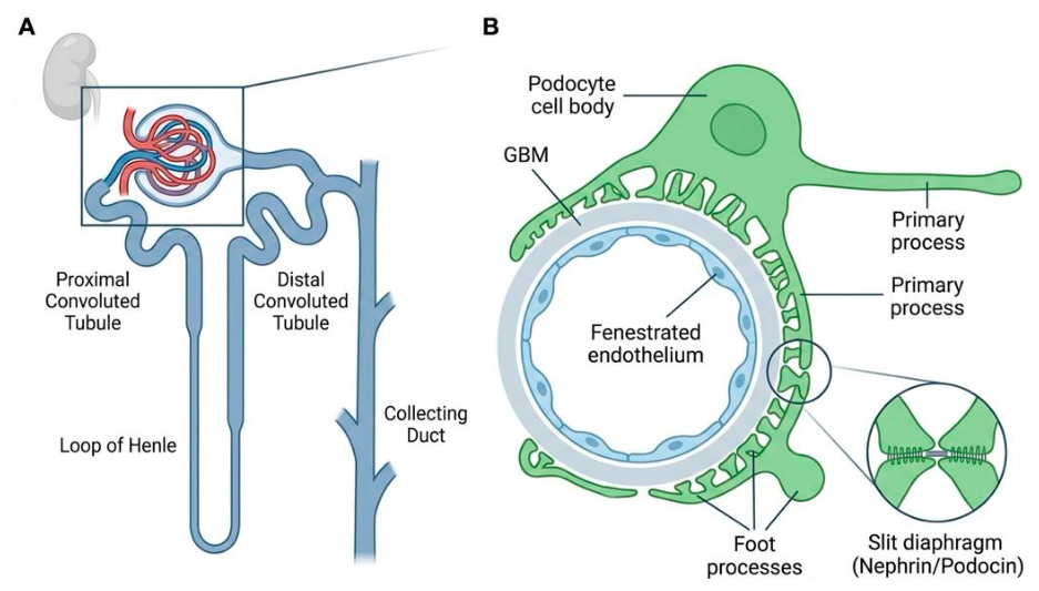
Figure 2.
The immune-like podocyte: membrane receptors, intrinsic innate immune signaling, and effector outputs. A podocyte positioned on the outer surface of the glomerular basement membrane (GBM) is exposed to circulating, glomerular, and local danger-associated signals. The podocyte plasma membrane expresses pattern-recognition receptors, including Toll-like receptors (TLR2 and TLR4); cytokine receptors, including TNFR1, TNFR2, IL-1R, IL-6R, and IFNAR; complement receptors, including C3aR and C5aR; and additional immune-associated receptors, such as CD36, Tim-3, and B7-1. These receptors detect extracellular ligands, including TNF, IL-1β, IL-6, type I interferons, the anaphylatoxins C3a and C5a, oxidized lipids, pathogen-associated molecular patterns, damage-associated molecular patterns, and immune complexes. Receptor-derived and intracellular stress signals converge on innate immune signaling platforms, including the NLRP3 inflammasome, cGAS–STING, MyD88/TRIF-associated pathways, MAVS, and RIPK1/RIPK3/MLKL signaling. These pathways regulate NF-κB-, IRF-, and STAT-dependent transcriptional responses and can promote cytokine and chemokine production, release of extracellular vesicles and reactive oxygen species, actin-cytoskeletal remodeling, foot-process effacement, and disruption of the glomerular filtration barrier.
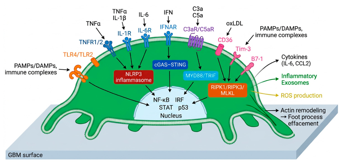
Figure 4.
Podocyte fate under sustained inflammatory and metabolic stress: regulated cell death, detachment, and senescence. A central podocyte is surrounded by major cell-fate programs that may be engaged during glomerular injury. Apoptosis is characterized by mitochondrial outer-membrane permeabilization, caspase activation, and nuclear fragmentation; apoptotic podocytes may detach and be lost into the urinary space before classical apoptotic morphology is fully evident in glomerular sections. Necroptosis involves RIPK1-, RIPK3-, and MLKL-dependent membrane permeabilization and may amplify local inflammation through release of damage-associated molecular patterns. Pyroptosis is mediated by inflammasome-associated caspase-1 activation and gasdermin D pore formation. Ferroptosis is driven by iron-dependent lipid peroxidation, impaired glutathione-dependent antioxidant defense, and reduced GPX4 activity. Autophagy is generally cytoprotective in podocytes; however, persistent autophagic dysregulation can contribute to cellular dysfunction and interact with apoptotic, inflammasome-associated, and metabolic death pathways. Anoikis-associated podocyte detachment represents an irreversible mechanism of filtration-barrier loss, whereas senescence is a persistent non-proliferative state that promotes chronic injury through mitochondrial dysfunction and senescence-associated secretory phenotype production. PANoptosis-like signaling may arise when pyroptotic, apoptotic, and necroptotic components are activated concurrently, although direct evidence for bona fide PANoptosome assembly in podocytes in vivo remains limited. Metabolic, hemodynamic, immunologic, and mechanical stressors, including hyperglycemia, immune complexes, oxidative stress, complement activation, and biomechanical stress, can bias the transition among these cell-fate programs and promote progressive podocyte depletion.
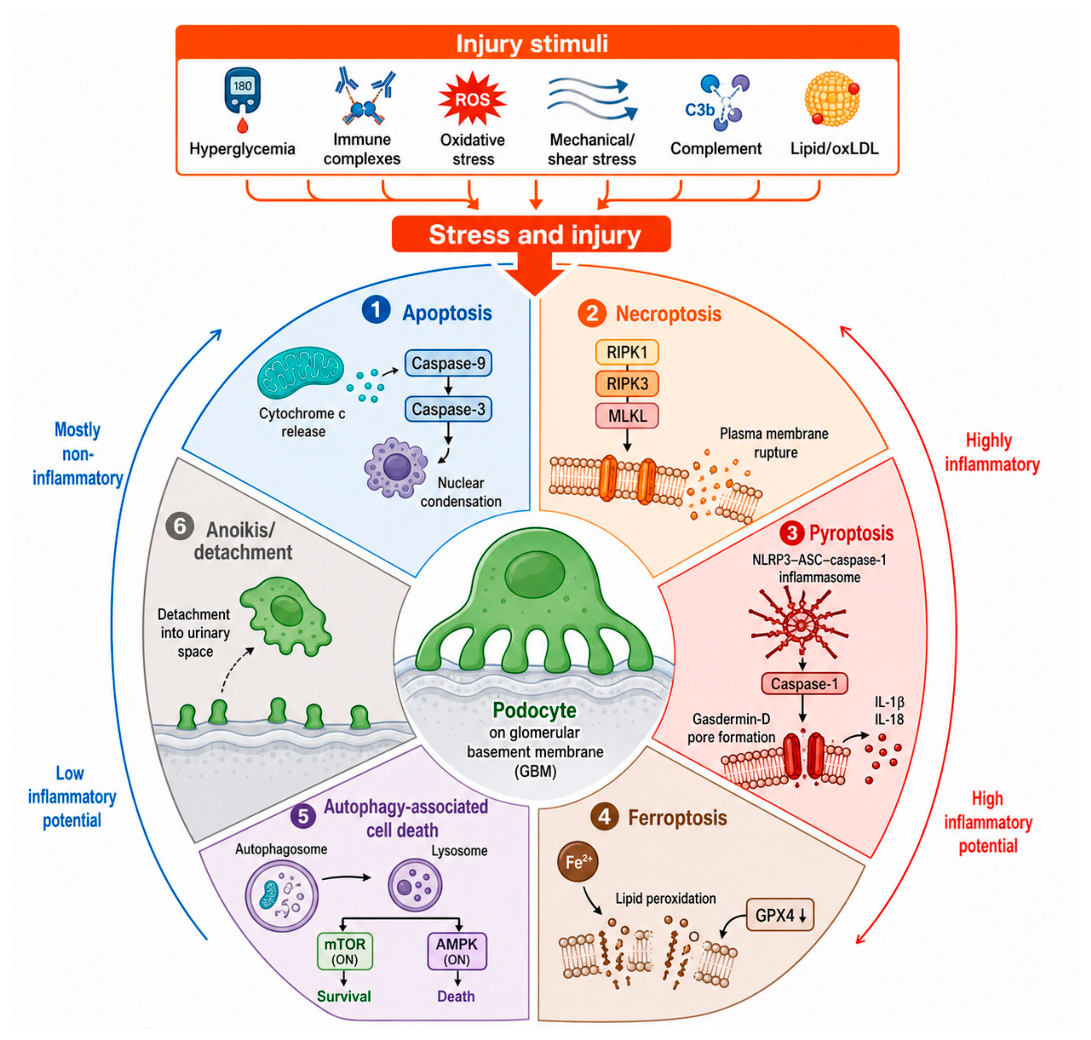
Figure 5.
Therapeutic entry points along the podocyte-intrinsic inflammatory signaling network. Podocyte injury can be targeted at three complementary levels. Upstream interventions reduce hemodynamic, metabolic, cytokine, complement, and lipid-associated stress that activates podocyte receptors and cytosolic danger-sensing pathways. Established kidney-protective therapies, including renin–angiotensin system blockade, SGLT2 inhibitors, and, in appropriate settings, non-steroidal mineralocorticoid receptor antagonists, act predominantly through systemic and intraglomerular mechanisms and may indirectly reduce upstream drivers of podocyte injury. Intracellular inflammatory and cytoskeletal nodes, including NF-κB, JAK–STAT, cGAS–STING, NLRP3, Rho–ROCK, mitochondrial stress, autophagy, and epigenetic regulators, represent potential targets for experimental pathway-directed intervention. Downstream strategies may aim to limit cytoskeletal dysfunction, inflammatory cell death, detachment, and senescence-associated secretory phenotypes. Most intracellular pathway inhibitors remain preclinical, and their therapeutic benefit will depend on disease stage, target engagement, cell-type specificity, pharmacokinetics, and preservation of essential homeostatic and host-defense functions. Human podocyte models, kidney organoids, glomerulus-on-chip systems, and single-cell or spatial profiling can support mechanistic validation, target identification, and biological patient stratification for future podocyte-targeted therapies.
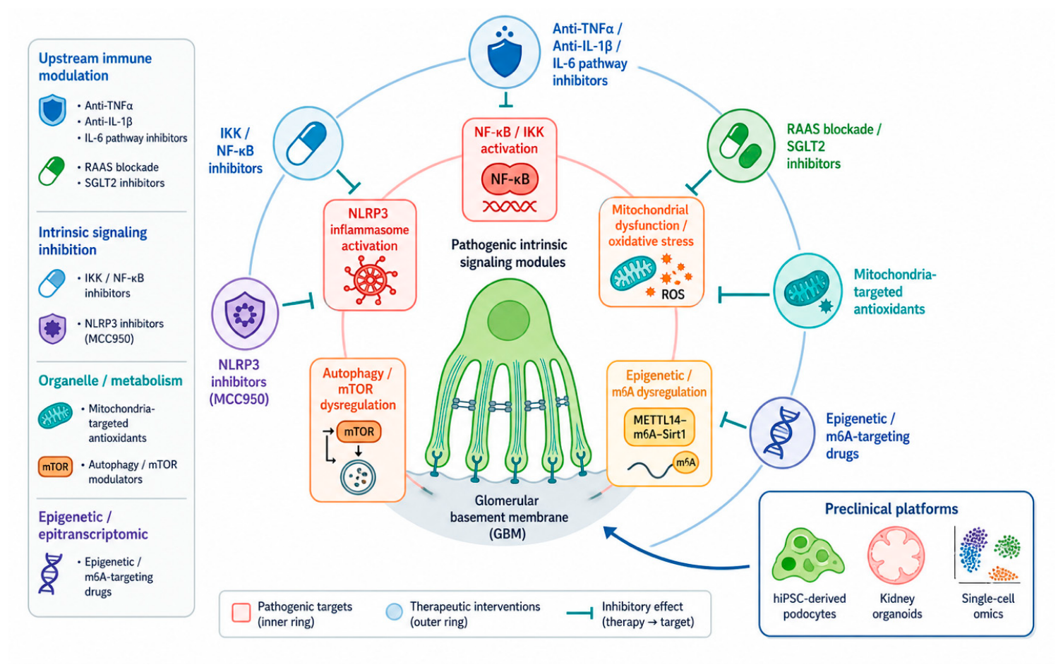
Disclaimer/Publisher’s Note: The statements, opinions and data contained in all publications are solely those of the individual author(s) and contributor(s) and not of MDPI and/or the editor(s). MDPI and/or the editor(s) disclaim responsibility for any injury to people or property resulting from any ideas, methods, instructions or products referred to in the content. |
© 2026 by the authors. Licensee MDPI, Basel, Switzerland. This article is an open access article distributed under the terms and conditions of the Creative Commons Attribution (CC BY) license (http://creativecommons.org/licenses/by/4.0/).
Copyright: This open access article is published under a Creative Commons CC BY 4.0 license, which permit the free download, distribution, and reuse, provided that the author and preprint are cited in any reuse.