

Submitted:
25 September 2026
Posted:
28 September 2026
You are already at the latest version
Abstract
Osmunda japonica sporophyll is a medicinal food source in China and Southeast Asia. However, its functional components are still unknown and its spores are not utilized. This study aims to analyze the main functional compounds of its sporophylls and spores. Selenium-enrichment cultivation of O. japonica was conducted and the main components of sporophylls and spores were comparatively analyzed using ultraviolet-visible spectrum, high performance liquid chromatagraphy and mass spectrum. The common phenolics of spores and sporophylls were chlorogenic acid, caffeic acid and kaempferol-3-rutinoside; sporophylls contained one more component as caffeic acid-3-O-(2’-O-butenyl) rahmnoside; caffeic acid was the main phenolic component in spores (2.15±0.09 g/100g, dried weight); contents of caffeic acid (2.36±0.1 g/100g, dried weight), kaempferol-3-rutinoside (2.69±0.15 g/100g, dried weight), polysaccharides (3.53±0.23 g/100g, dried weight), carotenoids (364±34.9 mg/100g, fresh weight) and chlorophylls (437±40.2 mg/100g, fresh weight) of spores were all significantly higher than those of sporophylls (P<0.05). The organic selenium components of spores were Se-Cys(selenocysteine) and Se-Met (selenomethoine); Se-Cys, Se-Met, chlorogenic acid, carotenoids and chlorophylls were all significantly increased by selenium enrichment (R2>0.9). In conclusion, selenium-enriched japonica spores showed highly nutritional and medicinal values and should be developed as a new medicinal food source and selenium supplementation food source.
Keywords:
Osmunda japonica
; selenium enrichment
; spores
; sporophylls
; functional components
1. Introduction
Medicinal foods, as an emerging food category, had aroused the dramatically increased interest of human beings [1]. Many medicinal plants had been developed as functional foods or medicinal foods, and called “food-medicine homologous medicinal foods” . Most food-medicine homologous plants contain rich functional and medicinal components as polysaccharides [2,3], flavonoids, phenolic acids, saponins, carrotenoids and so on. These functional components are very beneficial for human health protection.
Osmunda japonica Thunb is a medicinal plant species of Osmundaceae (Figure 1). In China and Southeast Asia, young sporophylls of O. japonica are traditionally processed into dried vegetables as delicious medicinal food. O. Japonica rhizomes, as a traditional Chinese medicine, called Ziqi Guanzhong [4], was recorded as heat clearing and detoxifying, hemostasis and killing parasites in the Chinese medicinal monograph “Ben Cao Gang Mu”. There were some reports on O. japonica about alleviating the skin wrinkles [5], arsenic accumulation [6], egg development and ovulation [7], chemical constituents of sterile fronds [3], antibacterial properties of O. japonica flavonoids [8], molecular phylogenetics [9] and mycorrhizal formation [10]. However, there were rare reports on functional component composition of O. japonica sporophylls and no reports on O. japonica spores.
Spores or pollens, the mature germ cells of fungi or plants, rich of bioactive components, are becoming the important medicinal food sources for human health protection [11]. As the typical fungi spores, Ganoderma lucidum spores contained the most commonly used bioactive components as polysaccharides and polyunsaturated fatty acids [spore oils] and had been reported to show the important bioactivities such as anti-Alzheimer [12], anti-aging [13], anti-oxidation [14], anti-obesity and anti-inflammation [15] and anti-tumor [16]. As a kind of plant spores, the pine pollens had been also widely used as medicinal food for its bioactivities as anti-oxidation, anti-inflammation [17], and hepatatic protection [18]. Fungal spores and plant spores [pollens], as the advanced medicinal food sources, are getting increasing interest of human beings [19].
Nowadays, O. japonica are widely cultivated and the sporophylls are harvested as medicinal vegetables, while O. japonica spores which was about 7-16% dried weight of the sporophylls [data not shown] are not separated and utilized. In this study, the main functional components as phenolics, flavonoids, carotenoids, chlorophylls and polysaccharides between sporophylls and spores were comparatively analyzed to show the differences of sporophylls and spores.
Selenium was reported to have physiological functions such as antioxidation [20], anticancer [21], immunity stimulation [22], and HIV inhibition [23]. The bioactivity of selenium depends on its chemical form. Organic selenium compounds such as selenomethionine and methylselenocysteine were better tolerated and exhibited anticarcinogenic activity [24]. Many plants showed the ability to transform inorganic selenium into organic selenium [25] So, selenium-tolerant plants were used for processing organic selenium supplementation. Furthermore, in our previous study, selenium had been found to have promotive effects on the accumulation of some secondary metabolites [26,27]. In this study, selenium-enrichment cultivation of O. japonica was conducted, analysis on composition and quantitation of the functional components such as phenolics, flavonoids, chlorophylls, carotenoids, and organic selenium compounds of selenium-enriched O. japonica spores were performed.
2. Materials and Methods
2.1. Chemicals
Standards of chlorogenic acid (98% purity, CAS: 327-97-9) and caffeic acid [98% purity, CAS:331-39-5] were purchased from Sangon Biotech (Shanghai, China). SeCys, SeMet and proteinase K were from Sigma. Kaempferol-3-rutinoside and caffeic acid-3-O-[2’-O-butenyl] rahmnoside were obtained by the Agilent 1260 Infinity II Preparative. Acetonitrile, sodium citrate and hexanesulfonic acid were of HPLC grade. Acetonitrile, ethanol and acetic acid were of HPLC (high performance liquid chromatography) grade, and the other chemicals were of analytical grade.
2.2. Instruments
Ultrasonic cleaning bath (Shenzhen Jietai Ultrasonic cleaning apparatus Co. Ltd., Shenzhen, China, PS-40, 40kHz, 240 W) were employed for extraction of phenolics, carotenoids, chlorophylls and polysaccharides. A cell wall grinder (SQW-25DI, Yichen Superfine grinding Tech. CO. Ltd., Jinan, Shandong, China) was used for grinding sporophylls and spores. An ultraviolet-visible spectrophotometer (Shanghai Youke instrument and meter co., Ltd, Shanghai, China) was used for analysis of phenolic components, carotenoids, chlorophylls and polysaccharides. HPLC analysis was performed on an Agilent1100 HPLC system consisted of Agilent 1100 ChemStation Rev.A.10.02,G1313A autosampler, G1311A quarternary pump, G1314A variable wavelengthultraviolet (UV) detector and a reverse phase ZORBAX SB-C18 column (5 mm, 4.6×250 mm, Agilent Technologies, USA). LC-MS (liquid chromatography-mass specrtum) analysis was performed on an Agilent1100 series with an LC–MSD trap SL (Agilent Technologies, Berlin, Germany). Preparation of phenolic components was performed on an Agilent 1260 Infinity II Preparative, with a column of Agilent reverse phase Prep 100Å C18 (50×250 mm, 10 µm). Liquid samples were filtered with 0.22µm microporous membranes (EY-H492) for HPLC and UV-visible analysis.
2.3. Sample Preparation
O. japonica samples were collected from the japonica species conservation base of Hubei Minzu University, China The O. japonica species were identified by professor Yi-fa He of Hubei Minzu University, China. Sodium selenite was reported to be good form of selenium fertilizer for plants [26] and used as selenium source in this study. According to our published methods [26,27], selenium-enrichment cultivation of O. japonica was conducted. Namely, sodium selenite was added into MS solution to final Se-concentrations of 10, 20, 30, 40, 50 mg/kg dried quartz sands (called Se-MS solution). Quartz sands were collected by 30 mesh screen and cleaned with pure water. The dried quartz sands were well mixed with the prepared Se-MS solution in the ratio of 0.25 kg Se-MS solution / 1.0 kg dried quartz sands (called Se-soils). O. japonica seedlings were planted in the prepared Se-soils. The cultivation was conducted under glass canopy and irrigated with distilled water to maintain the stable water contents (60-70%), which was monitored with a soil moisture detector (FT-TS600). The cultivation was from May to September, in Hubei Minzu University, Enshi, China, with the average temperature of 25.5℃ and altitude of 531.8m. Sporophylls were harvested before unfolding. The unfolded sporophylls were packaged with filter paper and dried in a silica gel drier. After the sporophylls were dried to constant weight, 180g spores were screened and collected from the sporophylls. Dried spores and sporophylls were ground with a cell wall grinder separately, and the ground powders were used for extraction.
2.4. Separation and Identification of the Phenolic Components
Polyphenols extraction: the ground powders of sporophylls and spores were deoiled with ether in Soxhlet extractor at 80°C water bath for 4 h. Then the deoiled samples were dried at 80°C for 4h, and then extracted with 70% ethanol in ultrasonic cleaning bath for 30min, three cycles, the ratio of liquid to solid was 30 mL/g, and the extracts were filtered through 0.22µm microporous membranes for UV (ultraviolet) and HPLC analysis.
The 70% ethanol extracts prepared above were scanned within 190-1100 nm to primarily detect the chemical compositions. Based on the UV-visible scanning analysis, HPLC separation of phenolics was conducted: acetonitrile as solvent A (1.0% acetic acid as solvent B) 0–18 min, 15–60%, 18–20 min, 60–100%, 20–25 min,100–15%;the column temperature was set at 25°C, the sample volume injected was 10.0 μL, and the flow rate 1.0 mL min-1; the DAD (diode array detector) detector was set at 190–400 nm. Based on the established HPLC separation method, LC[APCI, Atmospheric Pressure Chemical Ionization]-MS were used for qualitative analysis of the phenolic peaks. The MS parameters were set as follows: ion source, APCI; nebulizer, 50psi; dry gas temperature, 350 °C; dry gas flow, 10.0L/min; APCI temperature, 400 °C; HV (High Voltag) capillary, 4000V, and scan range, 500 to 1500 m/z. Nitrogen was used as a drying gas (10.0 L/min ). Molecular structure of each peak in the HPLC spectra were analyzed by MS data and UV data.
2.5. Separation and Identification of the Organic Selenium Components
Preparation of lipid fraction: dried samples (sporophylls and spores, approximately 30g, respectively) was ground, and added with chloroform–methanol (2:1, 60 mL), and then shaken vigorously and filtered. The procedure was repeated three cycles. The extracted samples were vacuum dried at 80 ℃ as lipid-free fraction.
Protein hydrolysis: sodium hydroxide solution (0.1 mol L-1, 10 mL) was added into 1.0 g the lipid-free sample prepared above. After agitation (Vortex) the mixture was centrifuged (10 min, 5000 rpm), the solid residue was again treated with sodium hydroxide solution for three cycles. The combined solutions (20 mL) were neutralized to pH 7.0 with 4 mol L-1 phosphoric acid, and acetone was added to make the final concentration to 80%. The sample was kept at -20℃ for 30 min, and the precipitate (proteins) obtained by centrifugation (10 min, 5000 rpm) was washed with acetone, and vacuum-dried at 60℃. The precipitate was solubilized in acetic acid-ammonium acetate buffer (50 mmol L-1, pH 4.5) containing 5% sodium dodecylsulfate (water bath 60℃, 5 min) and filtered through a hydrophilic low-proteinbinding polysulfone filter (0.45 um). The filtrates were used as protein samples. The proteins obtained above were solubilized in 5 mL phosphate buffer (pH 7.5) and incubated with 1.0 mL of proteinase K (20 mg mL-1, 37℃, overnight), then acetone was added to final concentration of 80%, and the mixture was centrifuged at 5000 rpm for 10 min. The supernatant was vacuum dried at 60℃, and the residue containing selenoamide acids was solubilized in the mobile phase for HPLC–ICP–MS analysis. The mobile phase for analysis of Se-Cys and Se-Met using HPLC–ICP–MS were: 5 mmol/L sodium citrate: 5mmol/L hexanesulfonic acid = 95:5, flow rate of 1.0 mL/min, injection of 20μL.
2.6. Quantitation of the Phenolic Components, Polysaccharides, Carotenoids, Chlorophylls, Se-Cys and Se-Met
Phenolic components: based on the established HPLC separation method above, the 70% ethanol extracts of phenolics were separated using preparative HPLC. The two separated peaks in the HPLC spectra (unknown components, Peak 3, Peak 4) were collected respectively, and vacuum dried at 80°C to constant weight. Calibration curves of the components as caffeic acid, chlorogenic acid, peak 3 and peak 4 were established respectively. Chlorogenic acid: Y=59762X+18057, r = 0.9998, linearity range = 4.87-9.16µg/mL (1); Caffeic acid: Y=32586X+17668, r = 0.9998, linearity range = 6.034-10.11µg/mL (2); Peak 3: Y=32316X+13741, r = 0.9999, linearity range = 3.62-13.72µg/mL (3); Peak 4: Y=43336X+19637, r = 0.9999, linearity range = 5.56-17.15µg/mL (4).
Selenium components: the standards of Se-Cys and Se-Met were used to establish calibration curves respectively. Se-Cys: Y=46773X+15682, r = 0.9998, linearity range = 3.55-8.32µg/mL(5); Se-Met: Y=52537X+17448, r = 0.9998, linearity range = 3.67-9.15µg/mL (6); where, Y is peak area and X is mass concentration.
Polysaccharides: The ground powders of the sporophylls and spores were deoiled as described above, and then refluxed with 80% (v/v) ethanol at 95°C water bath in Soxhlet extractor for 4 h to remove sugars, polyphenols and flavonoids. Finally, the samples were dried at 80°C for 4 h and then extracted with 80°C pure water and ultrasonicated for 30min, three cycles, the ratio of liquid to solid was 30 mL/g; the extracts were filtered through 0.22µm microporous membranes as polysaccharide samples. Phenol-sulfuric method was used for determination of japonica polysaccharides. Polysaccharides concentrations were calculated according to the calibration curve, with glucose as a standard: y = 0.0075x + 0.0056, r = 0.9998, linearity range = 7.5-28.1 μg/mL (7), where y is the absorbance at 490 nm, x is the mass concentration of glucose (μg/mL), r is coefficient correlation.
Carotenoids: estimation of carotenoids was conducted based on our published methods [26] with some modifications. Namely, samples were extracted with absolute ethanol and the extracts were measured at 450 nm, using = 2500 for quantification of carotenoids.
Chlorophylls: concentrations of chlorophyll a, chlorophyll b and chlorophyll a+b were determined using the reported methods [28] with some modifications. Namely, 0.2 g powders were ground with 10 mL absolute ethanol, then placed in a 50 mL centrifuge tube and filled to 30 mL with the same solvent, then kept in a dark place until the sample color changed to white. 1 mL filtered extract was diluted with 3 mL absolute ethanol and the absorbance was measured at 645 nm, 652 nm and 663 nm. The concentrations (mg/mL) of chlorophyll a (Ca), chlorophyll b (Cb), and chlorophyll a + b (Ca+b) were calculated according to the formulae:
Ca= 0.0127D663-0.00269D645 (8)
Cb= 0.0229D645− 0.00468D663 (9)
Ca+b = ((0.0202D645 + 0.00802D663 + D652)/34.5)/2 (10)
Where D is absorbance, C is mass concentration (mg/mL).
3. Results
3.1. The Main Phenolic Compositions: Chlorogenic Acid, Caffeic Acid, Kaempferol-3-Rutinoside and Caffeic Acid-3-O-[2’-O-butenyl] Rahmnoside
The UV-visible spectra of the absolute ethanol extracts of spores and sporophylls indicated the possible compositions of the extracts as phenolics (326nm), carotenoids (440nm) and chlorophylls (660nm) (Figure 2). Based on the MS and UV-visible spectra data of the separated peaks, the three separated peaks of spores were identified as kaempferol-3-rutinoside, chlorogenic acid and caffeic acid (Table 1, Figure 3). The four peaks of sporophylls were identified as chlorogenic acid, caffeic acid, kaempferol-3-rutinoside and caffeic acid-3-O-(2’-O-butenyl) rahmnoside (Table 1, Figure 4). Chlorogenic acid, caffeic acid and kaempferol-3-rutinoside were the common phenolics of sporophylls and spores, while sporophylls contained one more phenolic component: caffeic acid-3-O-(2’-O-butenyl) rahmnoside (Table 1, Figure 4).
In the spores, caffeic acid was the main phenolic compound, with the average content of 2.36±0.1 g/100g dried weight, while in sporophylls, caffeic acid showed the least content of 1.35±0.1 g/100g dried weight (Table 2). There was no significant differences of chlorogenic acid contents between spores and sporophylls (P = 0.765), while the contents of caffeic acid and Kaempferol-3-rutinoside in spores were both significantly higher than those of sporophylls (P = 0.0003, 0.046) (Table 2); caffeic acid-3-O-(2’-O-butenyl) rahmnoside only detected in sporophylls, with the average content of 2.07±0.15 g/100g dried weight (Table 2, Figure 3).
3.2. Other Functional Components: Polysaccharides, Carotenoids and Chlorophylls
Except for phenolic compounds, there were other functional components as polysaccharides, carotenoids and chlorophylls (Figure 2; Table 2). Spores contained the average contents of polysaccharides of 3.53±0.23 g/100gdried weight, significantly higher than those of sporophylls (1.67±0.16 g/100gdried weight) (Table 2). The average contents of carotenoids and chlorophylls in spores were 346±34.9 mg/100g and 437±40.2 mg/100g, respectively, both significantly higher than those of sporophylls (209±41 mg/100g, P=0.012; 261±16.6 mg/100g, 0.002) (Table 2). Except for caffeic acid-3-O-(2’-O-butenyl) rahmnoside and chlorogenic acid, contents of the other components as caffeic acid, kaempfero-3-rutinoside, polysaccharides, carotenoids and chlorophylls in spores were all significantly higher than those of sporophylls (Table 2).
3.3. Selenium Components in Sporophylls and Spores:Selenomethionine and Selenocysteine
The organic selenium components in selenium-enriched O. japonica plants were identified as Se-Met (selenomethionine), and Se-Cys (selenocysteine) (Figure 5 ). The contents of Se-Cys and Se-Met in spores and sporophylls were increased in a selenium-dependent manner (R2>0.97). And furthermore, the contents of Se-Cys and Se-Met in spores were both significantly higher than those of sporophylls (Table 3), which indicated that selenium was crucial for plant development, and so O. japonica spores, as reproductive organs, could absorb and store more organic selenium for propagation. In this sense, selenium-enriched japonica spores could be developed for selenium supplementation food.
3.4. Selenium Significantly Increased the Main Functional Components of Spores: Chlorogenic Acid, Caffeic Acid, Kaempferol-3-glucorhamnoside, Carotenoids and Chlorophylls
The other functional components as chlorogenic acid, caffeic acid, kaempferol-3- glucorhamnoside, carotenoids, and chlorophylls were all increased in a very significantly selenium-dependent manner (R2=0.957/ 0.963/ 0.82/ 0.934/ 0.912), which was mostly in agreement with our previous studies [26,27]. But contents of polysaccharides were not concentration-dependent with selenium, although it had been increased by selenium at 50 mg/kg, which might mean that spores synthesized more polysaccharides to protect cells at the stress of high selenium concentrations. There have been many published researches on structures and functionalities of selenium-polysaccharides, but rare reports on correlation between polysacharrides accumulation with selenium. The reason might be that selenium promotes the biosynthetic pathways of chlorogenic acid, caffeic acid, kaempferol-3- glucorhamnoside, carotenoids, and chlorophylls, but not involve in the synthesis of polysaccharides. The molecular mechanisms deserve further research.
The production of spores was also increased by selenium at relatively low concentrations, but decreased at higher concentration (≥40mg/kg, Table 3). Selenium promotes development of reproduction organs at relatively low concentrations, but inhibits reproduction organ growth at high selenium concentrations, which was partially in agreement with our previous study [27]. The components as polysaccharides, carotenoids and chlorophylls in spores were all significantly higher than those of sporophylls (Table 2), indicating that the spores showed higher functional values than sporophylls.
Different from the traditional fungal spores of Ganodorma lucidum, whose main functional components were polysaccharides [28], japonica spores contained more functional components as phenolics, carotenoids and chlorophylls (Fig.2).
4. Discussion
As described above, O. japonica spores contained the significantly higher bioactive components than those of sporophylls. The reason might be that, as a reproductive organ, spores were the most active metabolic center of O. japonica plants and produced more bioactive components for germination and self-protection against environment stress. Ganodorma lucidum spores were widely used as functional food and a traditional medicine in China and Southeast Asia. The G. lucidum polysaccharides were considered the main functional components in G. lucidum spores with the reported content of 2.98g/100g [29], lower than that of O. japonica spores (3.53g/100g Table 2). In this sense, O. japonica spores could be a better medicinal food. Except for the polysaccharides, the O. japonica spores contained the other bioactive components as phenolics, carotenoids and chlorophylls, compared to G. lucidum spores (Table 2). Phenolics, a large amount of natural products in functional foods, are known for many important medicinal values. Polysaccharides were considered the important active components in foods and traditional medicines and showed many physiological functions as antioxidation, anti-fungal, reducing blood sugar levels, immunological, anti-radiation, anti-blood coagulation, anti-cancer, anti-HIV and hypoglycemic activities [30]. Carotenoids showed activities in antioxidation, anticancer and eyesight enhancing [31]. Chlorophylls as a traditional natural medicine, had been reported to have many important physiological functions such as anti-inflammation, anti-oxidation, metal-chelating and anti-cancer [32]. So in this sense, O. japonica spores might be a more valuable functional food source than G. lucidum spores. Nowadays, fungal spores from G. lucidum were widely used as functional food and traditional medicine, while spores from O. japonica plants are still not separated and utilized. In our cultivation of O. japonica plants, the dried weight of spores proved to be 7-16% of the dried sporophylls (data not shown). In this study, the japonica spores proved to contain many functional components as described above. Hence japonica spores should be developed as a new source for functional food and natural medicine. In order to provide scientific basis for industrial utilization of japonica spores, comparation on the health protective functions and medicinal values between O. japonica spores and G. lucidum spores, cultivation techniques to increase these functional components and related intensive processing techniques deserve further research.
Our selenium-enrichment cultivation of O. japonica plants showed that selenium enrichment significantly increased production of spores and the main functional components of spores. The organic selenium components as Se-Cys and Se-Met were both significantly higher than those of sporophylls. So selenium-enriched japonica spores could be developed as a functional food source and a selenium supplementation food source.
5. Comclusions
Although chlorogenic acid, caffeic acid and kaempferol-3- rutinoside are the common phenolic components of spores and sporophylls and there was no significant differences in chlorogenic acid contents between spores and sporophylls, the contents of caffeic acid, Kaempferol-3- rutinoside, polysaccharides, carotenoids and chlorophylls in spores were all significantly higher than those of sporophylls. Caffeic acid, polysaccharides and, carotenoids and chlorophylls were the main active components in spores. So spores should be separated and utilized as a new food source and natural medicine, and related technics including cultivation, processing and extraction technics deserve further research.
Furthermore, selenium could increase the production of O. japonica spores, and selenium-enriched O. japonica spores contained rich organic selenium components as Se-Cys and Se-Met, and significantly higher components as chlorogenic acid, caffeic acid, kaempferol-3- glucorhamnoside, carotenoids, and chlorophylls than those of sporophylls. So selenium enrichment could be used in the development of O. japonica to increase production and qualities of O. japonica spores and provide a selenium supplementation food source.
Author Contributions
A.Z.: methodology; writing-review and editing; formal analysis; data curation; writing-original draft; visualization. S.A.: conceptualization; methodology; formal analysis; data curation; visualization. Q.Y.: formal analysis; data curation; writing-original draft; visualization. J.D.: conceptualization; methodology; resource; project administration; supervision; writing-original draft; Writing- review & editing. All authors have read and agreed to the published version of the manuscript.
Funding
This research was partially funded by the Doctor Research Project (53210052493),Wuhan Polytechnic University, Wuhan, China.
Data Availability Statement
The data that support the findings of this study are available from the corresponding author upon reasonable request.
Acknowledgments
We are grateful to anonymous reviewers and scientific editors for critical review and valuable suggestions, to Prof. Yi-fa He (Hubei Minzu University, China) for identification of O. japonica, to Ji-Hua Yang (Hubei Minzu University, China) for sampling of O. japonica spores.
Conflicts of Interest
The authors declare no conflicts of interest.
References
- Marilisa, A.; Monica, A. Re-thinking functional food development through a holistic approach. J. Funct. Foods 2021, 81, 104466. [Google Scholar] [CrossRef]
- Liu, X. C.; Xu, R.; Miao, Y.T.; Zang, Y.N.; Huang, Y.X.; Zheng, Y.Y.; Li, F.W.; Wei, M.; Xiao, R.; Yu, X.H. Exploring the immunomodulatory mechanisms of Osmunda japonica Thunb polysaccharides: Activation of MAPK and NF-κB signaling pathways. Food Biosci. 2025, 63. [Google Scholar] [CrossRef]
- Liu, X.; Yu, X.; Lyu, J.; Li, F.; Shang, Y.; Lyu, Y.; Liu, J. Chemical structure characterization of polysaccharide from Osmunda japonica Thunb and its inhibitory activity on uterine fibroids. Arab. J. Chem. 2022, 15, 1–13. [Google Scholar] [CrossRef]
- The State Pharmacopoeia Commission of China. In Pharmacopoeia of China; Chemical Industry Press: Beijing, China, 2020; Volume 321, p. Vol I.
- Kim, T. W.; Hur, N. M.; Jo, K. Y.; Kim, Y. J.; Cho, S. I.; Yun, E. S. Cosmetic composition for skin anti-wrinkle comprising Osmunda japonica extract as effective component. KR20180107616A, 2018. [Google Scholar]
- Srivastava, M.; Santos, J.; Srivastava, P.; Ma, L. Q. Comparison of arsenic accumulation in 18 fern species and four Pteris vittata accessions. Bioresour. Technol. 2010, 101, 2691–2699. [Google Scholar] [CrossRef] [PubMed]
- Cao, J. G.; Dai, X. F.; Wang, Q. X. Cytological features of oogenesis and their evolutionary significance in the fern Osmunda japonica. Sex. Plant Reprod. 2012, 25, 61–69. [Google Scholar] [CrossRef] [PubMed]
- Li, F.; Hu, Y. Antibacterial and Antifungal Properties of Flavonoid Compounds from Osmunda japonica thumb. Asian Joural Chem. 2012, 24, 4964–4966. Available online: https://www.researchgate.net/publication/295626264.
- Tsutsumi, C.; Hirayama, Y.; Kato, M.; Yatabe-Kakugawa, Y.; Zhang, S. Z. Molecular Evidence on the Origin of Osmunda × mildei (Osmundaceae). Am. Fern Journal. 2012, 102, 55–68. [Google Scholar]
- Ogura-Tsujita, Y.; Sakoda, A.; Ebihara, A.; Yukawa, T.; Imaichi, R. Arbuscular mycorrhiza formation in cordate gametophytes of two ferns, Angiopteris lygodiifolia and Osmunda japonica. J. Plant Res. 2013, 126, 41–50. [Google Scholar] [CrossRef] [PubMed]
- Xu, J. H.; Li, P. Researches and application of ganoderma spores powder. Advances in experimental medicine and biology 2019, 157–186. [Google Scholar] [CrossRef] [PubMed]
- Qin, L. H.; Wang, C.; Qin, L.W.; Liang, Y. F.; Wang, G. H. Spore powder of ganoderma lucidum for alzheimer's disease: a protocol for systematic review. Medicine 2019, 98, e14382. [Google Scholar] [CrossRef] [PubMed]
- Song, H.; Kim, M.S. Anti-aging & Skin Hydration Effects of Spore oil Extracted from Ganoderma lucidum. J. Converg. Inf. Technol. 2020, 10, 232–238. [Google Scholar] [CrossRef]
- Senphan, T.; Takeungwongtrakul, S.; Kaewthong, P. Extraction and antioxidant activities of broken Ganoderma lucidum spore. Int. J. Agric. Technol. 2021, 17, 2303–2316. Available online: https://www.cabidigitallibrary.org/doi/full/10.5555/20220051130.
- Sang, T.; Guo, C.; Guo, D.; Wu, J.; Wang, Y.; Wang, X. Suppression of obesity and inflammation by polysaccharide from sporoderm-broken spore of Ganoderma lucidum via gut microbiota regulation. Carbohydr. Polym. 2021, 256, 117594. [Google Scholar] [CrossRef] [PubMed]
- Sun, M.; Ni, L.; Huang, Y.; Wang, B.; Zhang, M.; Ma, C.; Wu, M. Volatile Components and Antitumor Activity of Ganoderma lucidum Spore Oil and Its Molecular Distillation Components. Asian Agric. Res. 2023, 15, 30–35. [Google Scholar] [CrossRef]
- Zhou, C.; Yang, Q.; Chen, L.; Fang, K.; Lu, X.; Song, X.; Sun, L. Antioxidant and anti-inflammatory properties of pine pollen extract promote wound healing. Mater. Express 2023, 13, 327–336. [Google Scholar] [CrossRef]
- Cong, T.; Jin, X.; Zhao, L.; Ma, L.; Guo, C. The protective effects of masson pine pollen aqueous extract on ccl4-induced oxidative damage of human hepaticcells. Int. J. Clin. Exp. 2015, 8, 17773–17780. Available online: https://pubmed.ncbi.nlm.nih.gov/26770368/.
- Rupanar, S. V.; Satpute, R.; Gadhave, P.; Kakade, N. Bee pollen beyond nutrition: An integrated review of its chemical, pharmacological, and biological properties. J. Drug Res. Ayurvedic Sci. 2024, 9, 211–221. [Google Scholar] [CrossRef] [PubMed]
- Wen, S.; Hui, Y.; Chuang, W. Biosynthesis and antioxidation of nano-selenium using lemon juice as a reducing agent. Green Process. Synth. 2021, 10, 178–188. [Google Scholar] [CrossRef]
- Subha, V.; Shanmugam, K.; Sahadevan, R. InVivo Studies and Flow Cytometric Investigation on Anticancer Potential of Selenium Nanoparticles Synthesized via Aqueous Extract of Clerodendronphlomidis. Proc.Antica.Res. 2024, 8, 71–81. [Google Scholar] [CrossRef]
- Debata, N. R.; Sethy, K.; Swain, R. K.; Mishra, S. K.; Panda, N.; Maity, S. Supplementation of nano-selenium [SeNPs] improved growth, immunity, antioxidant enzyme activity, and selenium retention in broiler chicken during summer season. Trop. Anim. Health Prod. 2023, 55[4], 260. [Google Scholar] [CrossRef] [PubMed]
- Pourmoradian, S.; Rezazadeh, L.; Tutunchi, H.; Ostadrahimi, A. Selenium and zinc supplementation in HIV-infected patients. Int. J. Vitam. Nutr. Res. 2023, 94, 153–159. [Google Scholar] [CrossRef] [PubMed]
- Hadrup, N.; Ravn-Haren, G. Toxicity of repeated oral intake of organic selenium, inorganic selenium, and selenium nanoparticles: A review. Journal Trace Elem. Med. Biol. 2023, 79, 127235. [Google Scholar] [CrossRef] [PubMed]
- Collins, J. F. Molecular, Genetic, and Nutritional Aspects of Major and Trace Minerals (2016). Chapter 39: Could Selenium Be a Double-Edged Sword? [CrossRef]
- Dong, J.Z.; Wang, Y.; Wang, S.H.; Yin, L.P.; Xu, G.J.; Zheng, C.; Lei, C. Selenium increases chlorogenic acid, chlorophyll and carotenoids of Lycium chinense leaves. J. Sci. Food Agric. 2013, 93, 310–315. [Google Scholar] [CrossRef] [PubMed]
- Lei, C.; Ma, Q.; Tang, Q.; Ai, X.; Zhou, Z.; Yao, L.; Wang, Y.; Wang, Q.; Dong, J. Sodium selenite regulates phenolica and tuber development of purple potatoes. Sci. Hortic. 2014, 165, 142–147. [Google Scholar] [CrossRef]
- Sanchini, A.; Grosjean, M. Quantification of chlorophyll a, chlorophyll b and pheopigments a in lake sediments through deconvolution of bulk UV–VIS absorption spectra. J. Paleolimnol. 2020, 64, 243–256. [Google Scholar] [CrossRef]
- Fu, Y. J.; Liu, W.; Zu, Y. G.; Shi, X. G.; Liu, Z. G.; Schwarz, G.; Efferth, T. Breaking the spores of the fungus Ganoderma lucidum by supercritical CO2. Food Chem. 2009, 112, 71–76. [Google Scholar] [CrossRef]
- Yang, J.; Sun, H.; Guo, H.; Zhang, X. L.; Zhang, W. T. Research progress on the preparation, structure-activity relationship, and functional mechanism of plant polysaccharides with anti-obesity activity: A review. Int. J. Biol. Macromol. 2025, 331, 148438–148438. [Google Scholar] [CrossRef] [PubMed]
- Tanumihardjo, S.A. Carotenoids: Health effects. Encycl. Hum. Nutr. (Fourth Edition) 2023, 154–161. Available online: https://www.sciencedirect.com/science/article/abs/pii/B9780128218488001852. [CrossRef]
- Sun, D.; Wu, S.; Li, X.; Ge, B.; Zhou, C.; Yan, X.; Cheng, P. The structure, functions and potential medicinal effects of chlorophylls derived from microalgae. Mar. Drugs 2024, 22, 65. [Google Scholar] [CrossRef] [PubMed]
Figure 1.
O. japonica sporophylls.
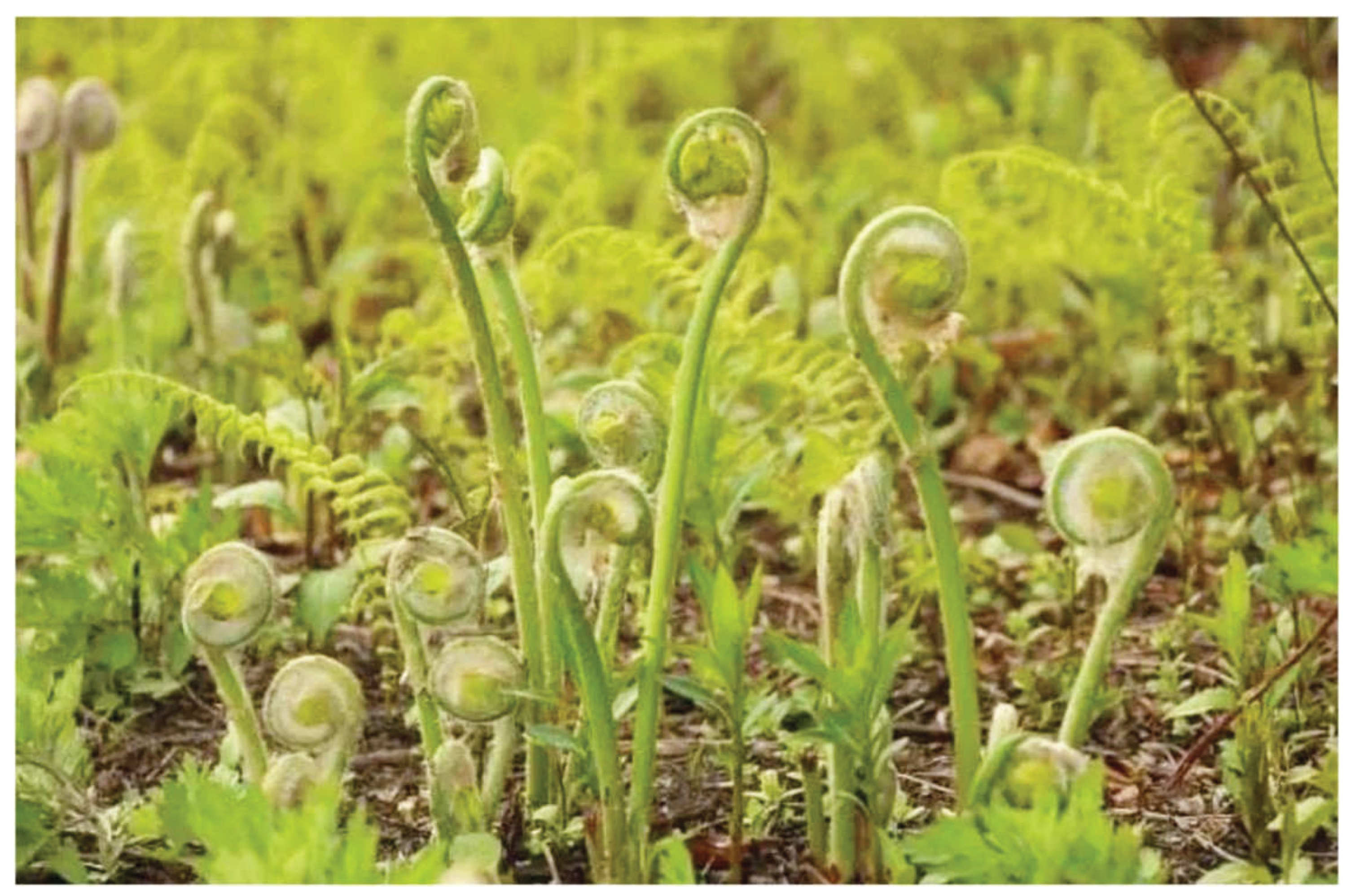
Figure 2.
UV-visible spectra of the absolute ethanol extracts of (a) sporophylls and (b) spores (660nm=chlorophylls; 440=carotenoids; 326/326.5=phenonics).
Figure 2.
UV-visible spectra of the absolute ethanol extracts of (a) sporophylls and (b) spores (660nm=chlorophylls; 440=carotenoids; 326/326.5=phenonics).
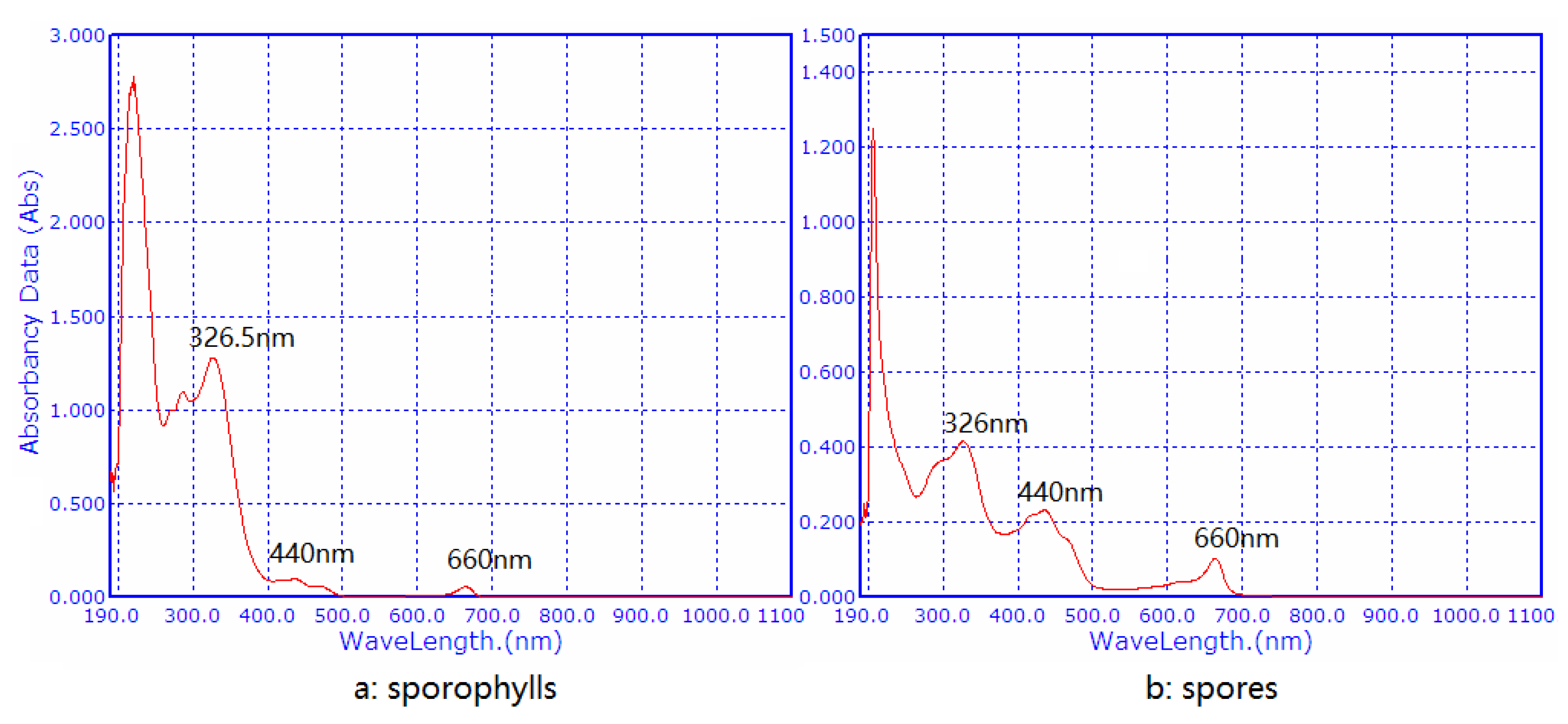
Figure 3.
HPLC separation of the phenolic components of spores. (1= chlorogenic acid; 2= caffeic acid; 3=Kaempferol-3- rutinoside).
Figure 3.
HPLC separation of the phenolic components of spores. (1= chlorogenic acid; 2= caffeic acid; 3=Kaempferol-3- rutinoside).
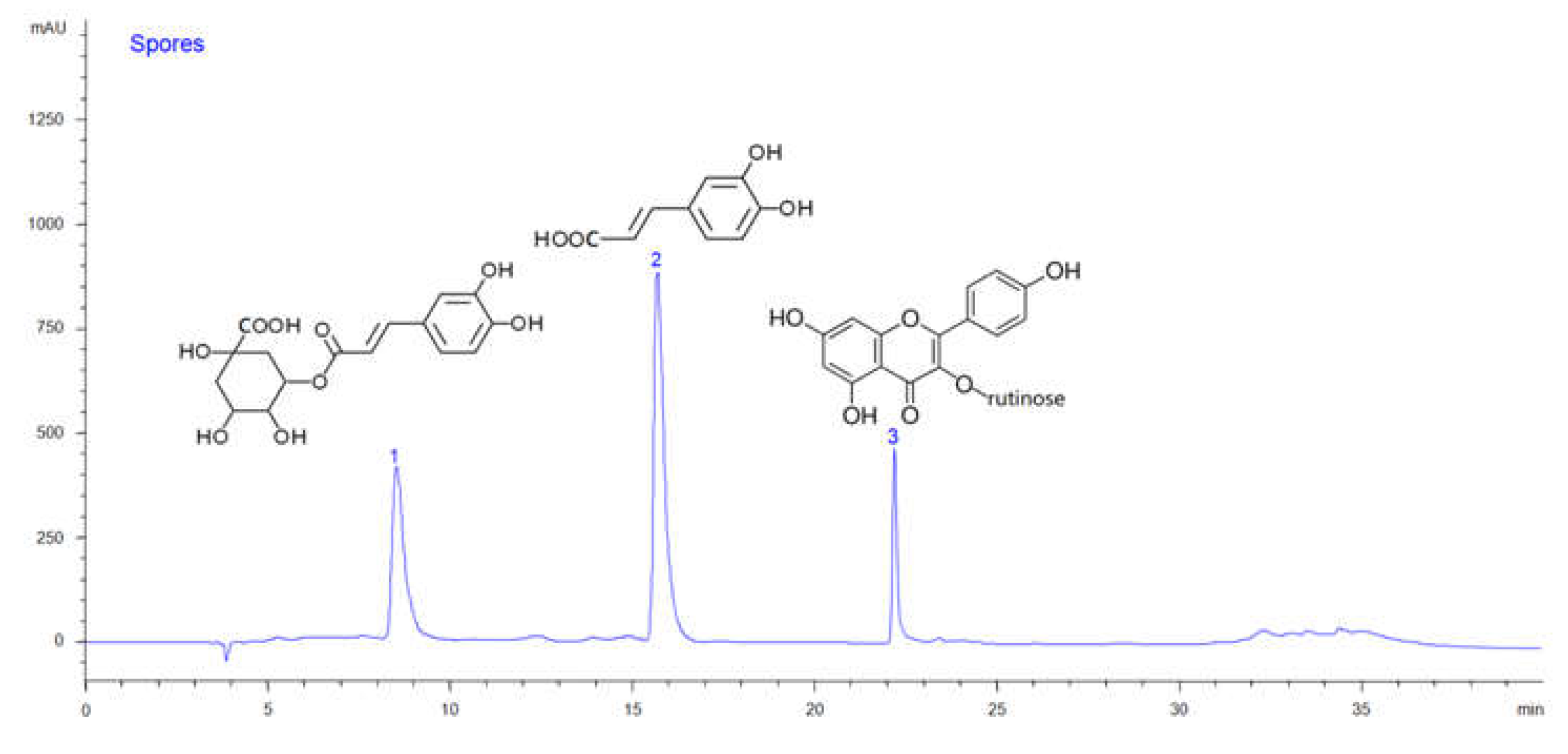
Figure 4.
HPLC separation of the phenolic components of sporophylls. (1= chlorogenic acid; 2= caffeic acid;3=Kaempferol-3- rutinoside; 4= Caffeic acid-3-O-(2’-O-butenyl) rahmnoside).
Figure 4.
HPLC separation of the phenolic components of sporophylls. (1= chlorogenic acid; 2= caffeic acid;3=Kaempferol-3- rutinoside; 4= Caffeic acid-3-O-(2’-O-butenyl) rahmnoside).
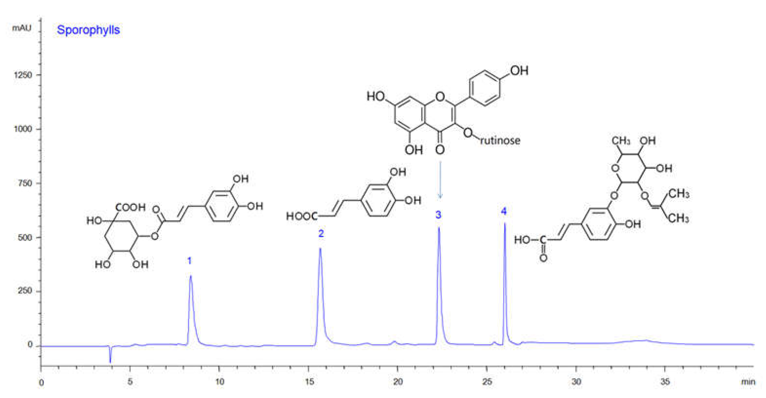
Figure 5.
Composition of organic selenium components from O. japonica spores (Se-Cys=Selenocystein; Se-Met=Selenomethionine).
Figure 5.
Composition of organic selenium components from O. japonica spores (Se-Cys=Selenocystein; Se-Met=Selenomethionine).
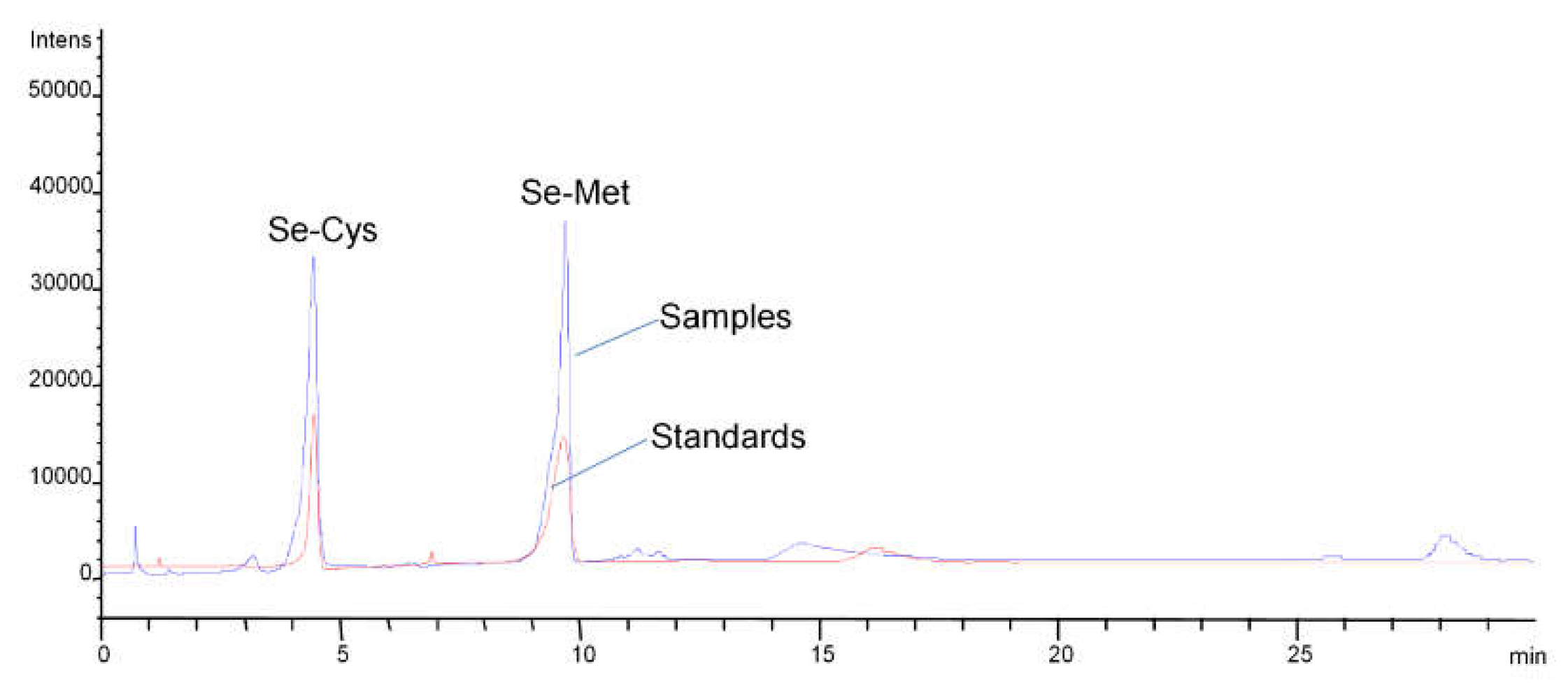

Table 1.
UV-MS data of the main phenolic components.
| Peaks | Components | MW | MS data | UV spectra (nm) |
Retention time (min) | |||
| [M+H] + | [M-H] - | |||||||
| 1 | Chlorogenic acid | 354 | 355.1 / 163.1 /145.2 /117.3 | 353.0 / 190.6 / 172.6 / 110.6 | 297 (sh), 326.5 | 8.565 | ||
| 2 | Caffeic acid | 180 | 181.3 / 163.1 | 178.9 / 134.7 | 295 (sh), 326.5 | 15.873 | ||
| 3 | Kaempferol-3-rutinoside | 594 | 595.0 / 449.2 / 287.0 / 243.1 /157.1 | 593.7 / 447.2 / 284.8 / 255.1 | 265, 301(sh), 351.5 | 22.240 | ||
| 4 | Caffeic acid-3-O- (2’-O-butenyl) rahmnoside |
382 | 383.0 / 327.5 / 181.1 | 381.3 / 178.6 / 134.8 | 251, 341.5 (sh) | 26.113 | ||
UV =ultraviolet spectrum; MS=mass spectrum; MW=molecular weight.
Table 2.
Analysis of the components between spores and sporophylls (N=5).
| Components | Spores | Sporophylls | P-value |
| Chlorogenic acid (g/100g DW) | 2.15±0.09 | 2.17±0.07 | 0.765 |
| Caffeic acid (g/100g DW) | 2.36±0.1 | 1.35±0.1 | 0.0003 |
| Kaempferol-3-glucorhamnoside (g/100g DW) | 2.69±0.15 | 2.13±0.22 | 0.046 |
| Caffeic acid-3-O-(2’-O-butenyl) rahmnoside (g/100g DW) | ND* | 2.07±0.15 | |
| Polysaccharides (g/100g DW) | 3.53±0.23 | 1.67±0.16 | 0.004 |
| Carotenoids (mg/100g FW) | 346±34.9 | 209±41 | 0.012 |
| Chlorophyll a (mg/100g FW) | 217±21.1 | 139±15.5 | 0.008 |
| Chlorophyll b (mg/100g FW) | 195±22.6 | 126±19.5 | 0.008 |
| Chlorophyll a +b (mg/100g FW) | 437±40.2 | 261±16.6 | 0.002 |
* not detected, LOD=0.05 mg/100g; DW=dried weight; FW=fresh weight.
Table 3.
Analysis of the components in Se-spores from different Se-concentration culyivation (N=5).
| Items | Concentrations of Sodium senelite (mg/kg) | R2 | ||||
| 10 | 20 | 30 | 40 | 50 | ||
| Chlorogenic acid (g/100g DW) | 2.23±0.11 | 2.51±0.05 | 2.65±0.21 | 2.77±0.24 | 2.83±0.11 | 0.957 |
| Caffeic acid (g/100g DW) | 2.42±0.18 | 2.57±0.09 | 2.68±0.15 | 2.72±0.18 | 2.82±0.18 | 0.963 |
| Kaempferol-3-glucorhamnoside (g/100g DW) | 2.8±0.15 | 2.97±0.12 | 3.18±0.22 | 3.08±0.25 | 3.58±0.15 | 0.82 |
| Polysaccharides (g/100g DW) | 3.65±0.51 | 3.57±0.63 | 2.95±0.31 | 3.05±0.26 | 3.75±0.51 | 0.02 |
| Carotenoids (mg/100g FW) | 363±27.5 | 375±21.3 | 383±25.7 | 391±22.2 | 413±25.5 | 0.934 |
| Chlorophyll a (mg/100g FW) | 238±23.5 | 244±15.5 | 258±21.5 | 268±23.5 | 271±33.5 | 0.955 |
| Chlorophyll b (mg/100g FW) | 218±19.9 | 235±15.1 | 247±16.5 | 279±19.5 | 284±29.5 | 0.967 |
| Chlorophyll a +b (mg/100g FW) | 455±35.5 | 471±25.3 | 494±15.9 | 525±37.3 | 539±25.1 | 0.912 |
| Se-Cys in spores (ug/100g DW) | 15.68±2.5 | 31.7±5.6 | 52.91±8 | 72.55±7.5 | 75.3±9.1 | 0.975 |
| Se-Met in spores (ug/100g DW) | 21.3±3.5 | 40.22±4.8 | 66.35±7.7 | 89.13±8.2 | 95.11±9 | 0.975 |
| Se-Cys in sporophylls (ug/100g DW) | 6.33±1.5 | 8.1±1.6 | 13.45±2.7 | 15.05±3.5 | 18.6±3.1 | 0.985 |
| Se-Met in sporophylls (ug/100g DW) | 7.25±1.1 | 9.32±1.8 | 12.31±2.7 | 15.51±3.2 | 21.5±3.6 | 0.975 |
| Spores production (%) | 15±3.3 | 16.5±3.8 | 15.5±4.1 | 14±4.4 | 12±6.5 | 0.753 |
Se-Cys=Selenocystein; Se-Met=Selenomethionine; DW-dried weight; FW=fresh weight; Spores production (%)=spores weight/spores weight + sporophylls weight.
Disclaimer/Publisher’s Note: The statements, opinions and data contained in all publications are solely those of the individual author(s) and contributor(s) and not of MDPI and/or the editor(s). MDPI and/or the editor(s) disclaim responsibility for any injury to people or property resulting from any ideas, methods, instructions or products referred to in the content. |
© 2026 by the authors. Licensee MDPI, Basel, Switzerland. This article is an open access article distributed under the terms and conditions of the Creative Commons Attribution (CC BY) license (http://creativecommons.org/licenses/by/4.0/).
Copyright: This open access article is published under a Creative Commons CC BY 4.0 license, which permit the free download, distribution, and reuse, provided that the author and preprint are cited in any reuse.