

Submitted:
24 September 2026
Posted:
25 September 2026
You are already at the latest version
Abstract
Breast cancer remains a leading cause of cancer-related mortality in women, necessitating the identification of robust biomarkers for early detection and prognostic assessment. This study utilizes an integrative transcriptomic approach to evaluate the expression of CCND1 and ITGB1 transcripts in a clinical cohort of 30 patients. Using quantitative Real-Time PCR and 2−∆∆CT analysis, we demonstrate a significant upregulation of both markers in tumor tissues com-pared to normal margins (P < 0.0001). Furthermore, we integrate these clinical findings with large-scale transcriptomic repository data, revealing a statistically detectable, though modest, association (R = 0.068, P = 0.019). These results suggest that the coordinated dysregulation of cell cycle control and cellular adhesion serves as a coordinated molecular axis for breast cancer progression and prognostic assessment.
Keywords:
molecular oncology
; breast carcinoma
; CCND1
; ITGB1
; qPCR
; systems biology
; prognostic biomarkers
1. Introduction
Breast cancer remains a major global health challenge, representing the most frequently diagnosed malignancy and a of the leading causes of cancer-related mortality in women (Sung et al. 2021; Loibl et al. 2021; Islami et al. 2024). Understanding the complex genomic landscapes driving this disease is essential for developing targeted therapies (Vogelstein et al. 2013; Perou et al. 2000). The development and progression of this disease are characterized by an abnormal and uncontrolled proliferation of somatic cells, a process fundamentally driven by a complex sequence of genetic and epigenetic alterations following a multi-step stochastic model of carcinogenesis (Nordling 1953; Armitage and Doll 1954; Knudson 1971; Moolgavkar and Knudson 1981). As established by modern genomic landscapes, the transformation of a normal cell into a malignant one requires the sequential acquisition of driver mutations and the dysregulation of critical molecular signaling hubs (Yousafzai et al. 2024; Lebedeva et al. 2025). In the mammary gland, this process involves a variety of signaling pathways that govern cell cycle entry, survival, and interaction with the extracellular matrix (ECM) (Hunter 1995). The heterogeneity of breast cancer, categorized into molecular subtypes such as Luminal A, Luminal B, HER2-enriched, and Triple-Negative Breast Cancer (TNBC), requires the discovery of robust molecular biomarkers that can bridge the gap (Prat et al. 2015; Perou et al. 2000; Harbeck et al. 2019; Palacios et al. 2024).
The transition of normal mammary epithelial cells into invasive carcinomas is a latent evolutionary process. Recent studies using deep DNA sequencing have demonstrated the prevalence of somatic clonal expansion in healthy tissues, implying that this expansion often serves as a precursor to tumorigenesis (Martincorena et al. 2015; Maeda and Kakiuchi 2024; Mitchell et al. 2022). Furthermore, longitudinal tracking of these clones reveals that specific selective pressures within the mammary microenvironment dictate the transition from benign expansion to invasive malignancy (Fabre et al. 2022; Moore et al. 2020; Baird and Caldas 2013). In the mammary ductal system, as shown in Figure 1, the histological progression involves significant structural and molecular changes. Central to this transition is the dysregulation of the cell cycle at the G1/S phase checkpoint. Cyclin D1, encoded by CCND1, is a critical rate-limiting factor; by forming an active kinase complex with cyclin-dependent kinases (CDK4/6), it facilitates the phosphorylation of the retinoblastoma (Rb) protein, triggering the synthesis of genes required for DNA replication (Musgrove et al. 2011; Wang et al. 2024). Cyclin D1 overexpression is observed in more than 50% of primary breast cancers and is now recognized as a driver of epigenetic remodeling and therapeutic resistance (Musaogullari and Chai 2020; Montalto and De Amicis 2020). Beyond its role in the cell cycle, Cyclin D1-CDK4/6 signaling has been shown to modulate anti-tumour immunity and endocrine sensitivity (Goel et al. 2017; Dimitrakopoulos et al. 2021).
In parallel with proliferation, the ability of a tumor to invade surrounding tissues depends on the alteration of the cellular adhesion mechanisms. Integrins are a family of transmembrane receptors that mediate cell-ECM interactions, facilitating signaling pathways that influence survival and migration (Sun et al. 2016; Desgrosellier and Cheresh 2010). Integrin 1, encoded by the ITGB1 gene, is particularly vital in the mammary epithelium, where it regulates "inside-out" and "outside-in" signaling (Hamidi and Ivaska 2018; Vishnoi et al. 2022; Feng et al. 2024). Abnormal levels of Integrin 1 have been identified as a hallmark of aggressive breast cancer, promoting the interaction between tumor cells and components of the metastatic niche (Yousafzai et al. 2024; Adorno-Cruz et al. 2021). In TNBC, Integrin 1 has emerged as a potential prognostic biomarker, as its signaling often bypasses traditional growth requirements and promotes plastic cellular states (Song et al. 2022; Aydin et al. 2024).
This study integrates primary clinical quantification with large-scale in silico systems biology to define this synergy. Our cohort consisted of 30 patients, with samples taken from tumor tissues and adjacent normal margins. The quantification of expression was performed using the method. Our experimental findings revealed a significant increase in the expression of both transcripts in malignant tissues (). To provide a higher level of scientific rigor, we conducted an in silico correlation analysis using the breast invasive cancer (BRCA) data set. The synergy between proliferation (Cyclin D1) and adhesion (Integrin 1) suggests a coordinated molecular axis. As illustrated in Figure 2, signaling through Integrin 1 can trigger mechanotransduction pathways that enhance the stability and transcription of Cyclin D1 (Dai et al. 2013; Schwartz and Assoian 2001; Li and Feng 2011).
The relationship between clonal expansion and tumorigenesis serves as a paradigmatic framework for understanding solid tumors. Similarly, since somatic clonal dynamics implies a precursor stage in other organs (Fabre et al. 2022; Moore et al. 2020), coordinated upregulation of Cyclin D1 and Integrin 1 in normal-appearing margins may indicate a "field effect" of pre-malignant expansion (Maeda and Kakiuchi 2024; Mitchell et al. 2022). Our study considers these implications by correlating molecular data with clinical parameters, including patient age (20–70 years) and disease stage (stage 0–III). Using Real-Time PCR and in silico datasets, this study supports the use of a dual-marker signature for improved diagnostic precision in breast cancer management, aligned with modern systems biology and high-confidence protein network analysis (Colaprico et al. 2016; Szklarczyk et al. 2025; Tang et al. 2019).
2. Materials and Methods
2.1. Clinical Cohort and Tissue Procurement
The present study involved 30 female patients diagnosed with primary breast carcinoma. Tissue samples and clinical data were processed and analyzed at the Gholhak Clinical Laboratory after surgical resection by qualified clinical staff. The cohort included patients who covered clinical stages 0 through III, with the following distribution: Stage 0 (3.3%), Stage I (13%), Stage II (53%) and Stage III (30%). Demographic analysis revealed an age distribution of 20–30 years (3.3%), 30–40 years (20%), 40–50 years (30%), 50–60 years (26.7%) and 60–70 years (20%). Given the limited representation of Stage 0 cases (n=1), this group was included in the total cohort analysis to demonstrate the expression spectrum, although its individual statistical power is noted as a limitation. Following surgical resection by a specialist surgeon and subsequent pathological confirmation of the tumor and tumor-adjacent margins (histologically normal tissue), samples were processed. To maintain transcriptomic stability, samples (50–100 mg) were immediately transferred to sterile cryotubes free of RNAse and DNAse and immediately frozen at until further analysis. The study protocol was reviewed and approved by the Institutional Ethics Committee (Protocol Code: IR.IAU.VARAMIN.REC.1400.023), and the research was conducted in accordance with the Declaration of Helsinki.
2.2. Isolation of Total RNA and Quality Assessment
Total RNA was isolated using the phenol-chloroform extraction method (Trizol reagent, Invitrogen). Briefly, 50–100 mg of tissue was homogenized using a sterile scalpel and lysed in 1000 L of Trizol reagent through multiple vortexing cycles. To ensure complete phase separation, 200 L of chloroform was added, followed by centrifugation at 12,000 rpm for 15 minutes at . The aqueous phase containing the RNA was carefully retrieved and precipitated overnight with chilled isopropanol at .
The resulting RNA pellet was washed twice with 70% cold ethanol, air-dried, and resuspended in 30 L of DEPC-treated water. To ensure high-quality downstream applications, concentration and purity were assessed using spectrophotometry. The ratio was required to be for optimal RNA purity, and were monitored to exclude chemical contaminants (e.g., phenol or salts). To facilitate complete dissolution, the samples were briefly incubated in a Hot Block at for 5 minutes before storage at .
2.3. cDNA Synthesis and Quality Control
Total RNA was extracted using Trizol reagent (Invitrogren). The purity of the RNA was confirmed by spectrophotometry (A260/A280 ratio 1.8). cDNA was synthesized from 1000 ng of standardized total RNA using the SMOBIO kit protocol. The specific thermal profile for reverse transcription is detailed in Table 1.
2.4. Quantitative Real-Time PCR (qRT-PCR) and Primer Design
Target gene quantification was performed using SYBR Green chemistry on a StepOnePlus Real-Time PCR System (Applied Biosystems). GAPDH served as internal housekeeping control. For ITGB1, a 15 L reaction was utilized containing 3 pmol of primers, while the CCND1 protocol was optimized for high-sensitivity detection. The thermal cycling consisted of an initial denaturation at for 15 minutes, followed by 42 cycles of amplification. Relative changes in gene expression were calculated using the method. The finalized primer sequences and optimized PCR thermal profiles are presented in Table 2.
2.5. Statistical Analysis and Relative Gene Expression
To determine the relative fold change of the CCND1 and ITGB1 mRNA in tumor tissues compared to normal margins, the method was used as established in previous protocols (Livak and Schmittgen 2001). The normalization process began with the calculation of the for each sample as shown in Eq. [eq:delta_ct]:
Subsequently, the difference between the tumor and normal tissue margins was determined using Eq. [eq:ddct], which provides the value:
To facilitate visualization of high-magnitude expression shifts, the final relative quantification was expressed on a scale :
The experimental data was analyzed using GraphPad Prism 8.0 and Gnuplot 6.0. The statistical significance of differential expression between tumor and adjacent normal tissues was evaluated using the paired Student’s t-test. The results were considered statistically significant at .
2.6. Bioinformatic Workflow and Integrative Analysis
To extend clinical findings (), a multi-platform bioinformatic approach was implemented:
- TCGA Validation: In silico Large-scale in silico validation was conducted using the GEPIA2 platform (Tang et al. 2019) and the TCGAbiolinks framework (Colaprico et al. 2016), analyzing the TCGA-BRCA dataset (N = 1058).
- Interactome Mapping: The STRING database v12.0 (Szklarczyk et al. 2025) was utilized to map functional protein–protein interaction (PPI) networks with high confidence (0.700).
- Survival Analysis: Kaplan-Meier Plotter was used to assess the impact of the CCND1-ITGB1 signature on overall survival (OS) in established breast cancer patient cohorts.
3. Results
3.1. Standardization and Analytical Validation of qRT-PCR
The precision of transcriptomic quantification is highly dependent on the thermodynamic stability of the primer-template duplex and the linearity of the amplification. As a foundational step, we validated the reference gene GAPDH alongside our target oncogenes to ensure that normalization remained invariant across all biological replicates.
The standard curves for GAPDH exhibited an exceptional correlation coefficient (), ensuring that the normalization factor remained constant in all clinical samples. The dissociation analysis (melting curve) for GAPDH confirmed a single discrete peak at according to Figure 3, demonstrating the absence of primer dimers or genomic DNA interference. This high degree of linearity across serial dilutions indicates that internal control provides a stable baseline for comparative analysis .
Validation for CCND1 and ITGB1 targets included log-amplification plots to confirm exponential phase entry and standard curves for efficiency calibration. Both assays exhibited efficiencies within the optimal 95–105% range, a prerequisite for accurate application of the method. The discrete melting temperatures (; ) verified the specificity of our Exon-Exon junction primers, ensuring that no pseudogenes or non-specific isoforms were amplified, as shown in Figure 4. The tight clustering of replicates in the log-phase suggests high technical reproducibility across the clinical cohort.
3.2. Clinical Quantification of the CCND1 - ITGB1 Axis
Analysis of the 30-patient cohort revealed a robust transcriptional shift in both the cell-cycle regulator CCND1 and the adhesion marker ITGB1. To manage the high magnitude of differential expression, the values were transformed to for relative comparison.
Our data indicate that upregulation of CCND1 and ITGB1 was a universal event in all tumor samples (). As shown in Figure 5, the abundance of CCND1 mRNA exhibited a highly significant increase in tumor tissues compared to normal paired margins (). Specifically, the mean relative fold-change () for CCND1 was 4.62, corresponding to a significant shift on the relative scale compared to normal margins.
Similarly, ITGB1 quantification (Figure 6) demonstrated a clear separation between normal and neoplastic tissues. The mean relative upregulation of ITGB1 was calculated at 4.02-fold ().Coordinated overexpression of ITGB1 suggests that it is not a stochastic event, but a targeted remodeling of the tumor microenvironment. Consistent with our in silico findings, these results highlight that as tumors progress (stages I–III), cells couple mitotic potential via CCND1 with the migratory capacity provided by ITGB1 dysregulation.
3.3. Correlation Analysis and Prognostic Significance
To determine whether the observed upregulation translates into biological dependency, we conducted an in silico correlation analysis using large-scale clinical datasets. As demonstrated in Figure 7, we identified a statistically significant positive correlation between CCND1 and ITGB1. Although the correlation coefficient () reflects complexity and noise (Tang et al. 2019), the significant -value () supports a model in which integrin-mediated adhesion signaling acts as a scaffold for transcriptional regulation of cell cycle machinery (Dai et al. 2013; Schwartz and Assoian 2001).
In addition, Kaplan-Meier survival mapping was used to assess the long-term clinical impact of this molecular axis. Integration of both markers into a signature of the "mean probe" (averaging normalized expression) yielded a cumulative high-risk profile (, consistent with system-level prognostic datasets (Tang et al. 2019)). As seen in Figure 8, patients with high expression of both markers showed significantly reduced overall survival (OS) compared to low-expression cohorts. This synergistic effect suggests that the dual-marker signature is a more potent predictor of a poor prognosis than either marker used in isolation.
3.4. Systems Interactome and Functional Enrichment Analysis
To bridge the gap between clinical expression and biological mechanism, we performed an integrative systems biology analysis. The Protein-Protein Interaction (PPI) network (Figure 9) identified the CCND1 - ITGB1 axis as a central mediator within a larger center of kinases and ECM receptors. The network architecture analyzed through STRING v12.0 (Szklarczyk et al. 2025) suggests that these proteins do not operate in isolation, but are integrated into a feedback loop involving focal adhesion kinases (FAK) and mitogen-activated protein kinases (MAPK).
Functional enrichment analysis (Szklarczyk et al. 2025) revealed that these genes are predominantly involved in the assembly of the cell-substrate junction and "focal adhesion" with astronomical statistical significance (). As visualized in Figure 10, this pathway analysis provides the "missing link" for our clinical data: co-upregulation creates a cellular state where the cell is simultaneously pushed to divide (cell cycle) and equipped to move (adhesion remodeling), a hallmark of aggressive metastatic potential (Hanahan 2022; Sun et al. 2016).
Finally, large-scale expression validation using the TCGA-BRCA dataset () through TCGA biolinks (Colaprico et al. 2016) confirmed that the trends observed in it our clinical cohort () are globally consistent between larger populations. As shown in Figure 11, both targets exhibit significant over-expression in malignant tissues in various molecular subtypes. This validation reinforces the clinical utility of CCND1 and ITGB1 as consistent diagnostic biomarkers (Lebedeva et al. 2025; Palacios et al. 2024).
4. Discussion
The molecular orchestration of breast cancer progression involves a sophisticated synergy between the internal division machinery of the cell and its external structural relationship with the tumor microenvironment. In this study, we utilized a robust dual-methodology—combining primary clinical qRT-PCR data from the patient-derived cohort with high-throughput in silico validation—to characterize the CCND1 / ITGB1 axis. Our findings demonstrate that the synchronized upregulation of these two transcripts is not a stochastic event, but a coordinated molecular program that drives malignancy and dictates poor patient outcomes.
4.1. The Proliferative Command: CCND1 and Genomic Instability in Neoplastic Progression
The observation that CCND1 is significantly overexpressed in our clinical samples () aligns with the work of Wang et al. (2024), who characterized the Cyclin D1-CDK4/6 axis as the main engine of endocrine resistance. In our cohort, the nearly universal elevation of CCND1 supports its status as a critical rate-limiting factor. This is further corroborated by genomic data showing that Cyclin D1 acts as a driver of epigenetic remodeling in breast cancer (Montalto and De Amicis 2020; Musaogullari and Chai 2020). The prevalence of CCND1 elevation suggests a breakdown in traditional cell cycle checkpoints, allowing unchecked entry into the S-phase (Goel et al. 2017; Glaviano et al. 2025). This progression follows established multi-stage models of carcinogenesis, where sequential mutations accumulate to drive malignancy (Fearon and Vogelstein 1990; Nordling 1953; Armitage and Doll 1954). Historical clinical evaluations have long identified Cyclin D1 expression as a significant prognostic indicator in breast cancer patients receiving endocrine therapy (Mohammadizadeh et al. 2013) and the study of environmental and genetic risk factors has shaped our understanding of these mutation rates (Fisher 1958; Moolgavkar and Knudson 1981; Knudson 1971).Recent insights suggest that this axis is not only a mitotic trigger but also a modulator of antitumor immunity (Goel et al. 2017; Turner et al. 2024).
Beyond mere proliferation, our results align with emerging evidence that CCND1 acts as a "pioneer" oncogene. The stage-dependent increase observed in our clinical data suggests that while Cyclin D1 initiates early-stage hyperplasia, its continued presence likely contributes to chromosomal instability and therapeutic evasion (Dimitrakopoulos et al. 2021; Musgrove et al. 2011). This correlation confirms that CCND1 levels serve as a robust indicator of disease progression, validated by our analysis of the GEPIA2 dataset () following the methods of Tang et al. (2019).
4.2. The Invasive Switch: ITGB1 and Mechanical Remodeling
While CCND1 governs the temporal control of the cell-cycle, ITGB1 dictates the spatial dynamics of survival and migration. Our clinical results revealed a robust elevation of ITGB1 mRNA (P < 0.0001), a finding central to the transition to a pro-metastatic niche (Adorno-Cruz et al. 2021). Integrins act as mechanotransducers that "sense" matrix stiffness to modulate cellular plasticity (Aydin et al. 2024). High levels of Integrin allow cancer cells to respond to the stiffened stroma, triggering intracellular signaling cascades. Computational modeling of ITGB1-associated networks has further elucidated how these mechanical signals are integrated into the decision-making processes of the cell (Gu et al. 2025). Our functional enrichment analysis using STRING v12.0 (Szklarczyk et al. 2025) confirmed this, showing enrichment in the "cell-substrate junction assembly" (). This mechanical primer is likely the catalyst that moves a tumor from a localized state to invasive carcinoma (Vishnoi et al. 2022; Hamidi and Ivaska 2018). In TNBC specifically, ITGB1 has emerged as a key regulator of TGF- oncogenic activities, further driving progression (Yousafzai et al. 2024).
4.3. Molecular Synergy: The CCND1 - ITGB1 Cross-Talk
A primary novelty of this research is the identification of a positive correlation () between CCND1 and ITGB1. This interaction is likely mechanically driven; ITGB1-mediated adhesion to the ECM is a prerequisite for CCND1 induction through the synergy of adhesion and mitotic signaling (Li and Feng 2011). Specifically, the transcriptional regulation of the CCND1 promoter is highly sensitive to signals derived from matrix stiffness and adhesion, creating a direct bridge between the ECM and the mitotic center (Aydin et al. 2024; Schwartz and Assoian 2001). In clinical cohorts, matrix stiffness has been shown to directly regulate CCND1 expression, confirming the importance of the physical microenvironment in cell cycle control (Lim et al. 2024).
In breast cancer, simultaneous over-expression suggests that the tumor has hijacked this safety mechanism. High ITGB1 provides a constant "pseudo-attachment" signal that maintains CCND1 levels, while CCND1 ensures that the cell remains in a proliferative state even during migration. The maPPIng of the PPI network (Szklarczyk et al. 2025) identifies a "feedback" loop in which increased adhesion signaling increases proliferative drive, a hallmark of aggressive metastatic potential (Hanahan 2022).
4.4. Prognostic Implications and Combinatorial Inhibition
The Kaplan-Meier survival analysis provides a compelling argument for the clinical utility of this dual-marker signature. Integration of both markers into a signature of the "mean probe" yielded a high-risk profile (), indicating that this synergy is a more robust predictor of mortality than single markers. This is consistent with broader transcriptomic landscapes that define survival outcomes in breast cancer through the integration of multiple gene signatures (Fox et al. 2019). Furthermore, the interaction between tumor cells and the spatiotemporal dynamics of the immune landscape plays a critical role in determining long-term patient OS (Bindea et al. 2013).
From a clinical perspective, this study supports "combinatorial inhibition." Targeting the CDK4/6 complex is now a clinical standard (Turner et al. 2024; Freedman et al. 2024). Our integration of clinical data with the TCGA-BRCA repositories (Colaprico et al. 2016; Gao et al. 2013) allows a nuanced understanding of these markers. Since resistance often emerges by bypassing cell-cycle checkpoints through ECM-mediated signaling (Feng et al. 2024), simultaneously targeting the ITGB1 node could prevent this escape. By disrupting both the "motor" (CCND1) and the "anchor" (ITGB1), a more durable therapeutic response can be achieved (Lebedeva et al. 2025; Palacios et al. 2024).
5. Conclusion
The present study provides a comprehensive molecular characterization of the CCND1 / ITGB1 axis. By integrating primary qPCR data with large-scale in silico validation (), we established that simultaneous upregulation of these markers is a pervasive characteristic of mammary oncogenesis.
The clinical importance of this research lies in the demonstrated correlation () between a master proliferative regulator and a primary invasive mediator. This synergy suggests a coordinated "command and control" center where the neoplastic cell synchronizes its division rate with its invasive capacity. Our survival modeling identifies a high-risk subgroup, highlighting the necessity of multi-gene panels in modern precision oncology. Ultimately, the CCND1 / ITGB1 axis represents a critical node for future combinatorial therapies, providing a framework for overcoming resistance to current CDK4/6 inhibition strategies (Turner et al. 2024; Wang et al. 2024).
Ethics approval and consent to participate
This study was conducted in accordance with the Declaration of Helsinki. Ethical approval was provided by the Institutional Ethics Committee of Islamic Azad University (Protocol Code: IR.IAU.VARAMIN.REC.1400.023, date of approval 15-02-2021). Informed consent was obtained from all individual participants included in the study.
Consent for publication
The author confirms that informed consent was obtained from all participants regarding the publication of molecular data and associated clinical findings derived from their samples.
Availability of data and materials
The datasets generated and analyzed during the current study are available from the corresponding author on reasonable request. In silico data analyzed are available through the repositories GEPIA2, STRING v12.0, and TCGA-BRCA.
Competing interests
The author declares that he has no competing interests.
Authors’ contributions
F.G.M. was responsible for the conception, experimental design, material preparation, clinical data collection, molecular analysis, bioinformatic validation, and manuscript drafting. The author read and approved the final manuscript.
Authors’ information
Farnam Gholipour Maralan is a researcher at the Faculty of Medicine, Complutense University of Madrid, Spain, specializing in molecular oncology and integrative transcriptomics.
List of Abbreviations
| Cyclin D1 |
| Integrin Beta-1 |
| Quantitative Real-Time Polymerase Chain Reaction |
| The Cancer Genome Atlas |
| Breast Invasive Carcinoma |
| Extracellular Matrix |
| Overall Survival |
| Hazard Ratio |
| Protein-Protein Interaction |
| Gene Expression Profiling Interactive Analysis 2 |
| Triple-Negative Breast Cancer |
| Cyclin-Dependent Kinase 4 and 6 |
| Focal Adhesion Kinase |
Funding
No funds, grants, or other support were received during the preparation of this manuscript.
Acknowledgments
Not applicable.
References
- Adorno-Cruz, Valery; et al. ITGA2 Promotes Expression of ACLY and CCND1 in Enhancing Breast Cancer Stemness and Metastasis. Genes Dis. 2021, 8(2), 180–90. [Google Scholar] [CrossRef]
- Armitage, Peter; Doll, Richard. The Age Distribution of Cancer and a Multi-Stage Theory of Carcinogenesis. Br. J. Cancer 1954, 8, 1–12. [Google Scholar] [CrossRef]
- Aydin, H. B.; Ozcelikkale, A.; Acar, A. Exploiting Matrix Stiffness to Overcome Drug Resistance. ACS Biomater. Sci. Eng. 2024, 10(7), 4154–69. [Google Scholar] [CrossRef]
- Baird, Richard D.; Caldas, Carlos. Genetic Heterogeneity in Breast Cancer: The Road to Personalized Medicine? BMC Med. 2013, 11, 151. [Google Scholar] [CrossRef]
- Bindea, Gabriela; et al. Spatiotemporal Dynamics of Intratumoral Immune Cells Reveal the Immune Landscape in Human Cancer. Immunity 2013, 39, 782–95. [Google Scholar] [CrossRef]
- Colaprico, Antonio; et al. TCGAbiolinks: An R/Bioconductor Package for Integrative Analysis of TCGA Data. Nucleic Acids Res. 2016, 44, e71. [Google Scholar] [CrossRef]
- Dai, Meiou; et al. Cyclin D1 Cooperates with P21 to Regulate TGF-Mediated Breast Cancer Cell Migration. Breast Cancer Res. 2013, 15, R49. [Google Scholar] [CrossRef]
- Desgrosellier, Jay S.; Cheresh, David A. Integrins in Cancer: Biological Implications and Therapeutic Opportunities. Nat. Rev. Cancer 2010, 10, 9–22. [Google Scholar] [CrossRef]
- Dimitrakopoulos, F. I.; et al. Endocrine Resistance and Epigenetic Reprogramming in Breast Cancer. Cancer Lett. 2021, 517, 55–65. [Google Scholar] [CrossRef]
- Fabre, Margot A.; et al. The Longitudinal Dynamics and Natural History of Clonal Haematopoiesis. Nature 2022, 606, 335–42. [Google Scholar] [CrossRef]
- Fearon, Eric R.; Vogelstein, Bert. A Genetic Model for Colorectal Tumorigenesis. Cell 1990, 61, 759–67. [Google Scholar] [CrossRef]
- Feng, Xiuqin; et al. Targeting Extracellular Matrix Stiffness for Cancer Therapy. Front. Immunol. 2024, 15, 1467602. [Google Scholar] [CrossRef]
- Fisher, Ronald A. Cancer and Smoking. Nature 1958, 182, 596. [Google Scholar] [CrossRef]
- Fox, Natalie S.; et al. Landscape of Transcriptomic Interactions Between Breast Cancer and Its Microenvironment. Nat. Commun. 2019, 10, 3106. [Google Scholar] [CrossRef]
- Freedman, Rachel A.; Caswell-Jin, Jennifer L.; Hassett, Michael; Somerfield, Mark R.; Giordano, Sharon H. Optimal Adjuvant Chemotherapy and Targeted Therapy for Early Breast Cancer—Cyclin-Dependent Kinase 4 and 6 Inhibitors: ASCO Guideline Rapid Recommendation Update. J. Clin. Oncol. 2024, 42(18), 2233–35. [Google Scholar] [CrossRef]
- Gao, Jianjiong; et al. Integrative Analysis of Complex Cancer Genomics and Clinical Profiles Using the cBioPortal. Sci. Signal. 2013, 6(269), pl1. [Google Scholar] [CrossRef]
- Glaviano, Antonino; et al. Cell Cycle Dysregulation in Cancer. Pharmacol. Rev. 2025, 77(2), 100030. [Google Scholar] [CrossRef]
- Goel, Shom; et al. CDK4/6 Inhibition Triggers Anti-Tumour Immunity. Nature 2017, 548, 471–75. [Google Scholar] [CrossRef]
- Gu, Xuerong; et al. Identification of Dynamic Network Biomarker ITGB1 for Erlotinib Pre-Resistance. Mol. Ther.-Oncolytics ahead of print. 2025. [Google Scholar] [CrossRef]
- Hamidi, Heini; Ivaska, Johanna. Every Step of the Way: Integrins in Cancer Progression and Metastasis. Nat. Rev. Cancer 2018, 18(10), 599–614. [Google Scholar] [CrossRef]
- Hanahan, Douglas. Hallmarks of Cancer: New Dimensions. Cancer Discov. 2022, 12(1), 31–46. [Google Scholar] [CrossRef]
- Harbeck, Nadia; et al. Breast Cancer. Nat. Rev. Dis. Primers 2019, 5, 66. [Google Scholar] [CrossRef]
- Hunter, Tony. Protein Kinases and Phosphatases: The Yin and Yang of Protein Phosphorylation and Signaling. Cell 1995, 80, 225–36. [Google Scholar] [CrossRef]
- Islami, Farhad; Marlow, Emily C.; Thomson, Blake; et al. Proportion and Number of Cancer Cases and Deaths Attributable to Potentially Modifiable Risk Factors in the United States, 2019. CA A Cancer J. Clin. ahead of print. 2024. [Google Scholar] [CrossRef]
- Knudson, Alfred G. Mutation and Cancer: Statistical Study of Retinoblastoma. Proc. Natl. Acad. Sci. USA 1971, 68, 820–23. [Google Scholar] [CrossRef]
- Lebedeva, Valeriia; et al. Triple-Negative Breast Cancer Unveiled: Bridging Science, Treatment Strategy, and Economic Aspects. Int. J. Mol. Sci. 2025, 26(19), 9714. [Google Scholar] [CrossRef]
- Li, Dong-Mei; Feng, Yu-Mei. Signaling Mechanism of Cell Adhesion Molecules in Breast Cancer Metastasis. Breast Cancer Res. Treat. 2011, 128, 7–21. [Google Scholar] [CrossRef]
- Lim, Justin J.; et al. Matrix Stiffness-Dependent Regulation of Immunomodulatory Genes in Human MSCs. Proc. Natl. Acad. Sci. USA 2024, 121(32). [Google Scholar] [CrossRef]
- Livak, Kenneth J.; Schmittgen, Thomas D. Analysis of Relative Gene Expression Data Using Real-Time Quantitative PCR and the 2−ΔΔCT Method. Methods 2001, 25, 402–8. [Google Scholar] [CrossRef]
- Loibl, Sibylle; et al. Breast Cancer. The Lancet 2021, 397, 1750–69. [Google Scholar] [CrossRef]
- Maeda, Hirona; Kakiuchi, Nobuyuki. Clonal Expansion in Normal Tissues. Cancer Sci. 2024, 115(7), 2117–24. [Google Scholar] [CrossRef]
- Martincorena, Inigo; et al. High Burden and Pervasive Positive Selection of Somatic Mutations in Normal Human Skin. Science 2015, 348, 880–86. [Google Scholar] [CrossRef]
- Mitchell, Emily; et al. Clonal Dynamics of Haematopoiesis Across the Human Lifespan. Nature 2022, 606, 343–50. [Google Scholar] [CrossRef]
- Mohammadizadeh, Fereshteh; et al. Role of Cyclin D1 in Breast Carcinoma. J. Res. Med. Sci. 2013, 18(12), 1021–25. [Google Scholar]
- Montalto, Francesca Ida; De Amicis, Francesca. Cyclin D1 in Cancer: A Molecular Connection for Cell Cycle Control. Cells 2020, 9(12), 2648. [Google Scholar] [CrossRef]
- Moolgavkar, Suresh H.; Knudson, Alfred G. Mutation and Cancer: A Model for Human Carcinogenesis. J. Natl. Cancer Inst. 1981, 66, 1037–52. [Google Scholar] [CrossRef]
- Moore, Luiza; et al. The Mutational Landscape of Normal Human Endometrial Epithelium. Nature 2020, 580, 640–46. [Google Scholar] [CrossRef]
- Musaogullari, Aysenur; Chai, Yuh-Cherng. Redox Regulation by Protein s-Glutathionylation: From Molecular Mechanisms to Implications in Health and Disease. Int. J. Mol. Sci. 2020, 21(21), 8113. [Google Scholar] [CrossRef]
- Musgrove, Elizabeth A.; et al. Cyclin D as a Therapeutic Target in Cancer. Nat. Rev. Cancer 2011, 11(8), 558–72. [Google Scholar] [CrossRef]
- Nordling, C. O. A New Theory on the Cancer-Inducing Mechanism. Br. J. Cancer 1953, 7, 68–72. [Google Scholar] [CrossRef]
- Palacios, Jose; et al. Biomarkers in Breast Cancer 2024: An Updated Consensus Statement by the Spanish Society of Medical Oncology. Clin. Transl. Oncol. 2024, 26(11), 2543–60. [Google Scholar] [CrossRef]
- Perou, Charles M.; et al. Molecular Portraits of Human Breast Tumours. Nature 2000, 406, 747–52. [Google Scholar] [CrossRef]
- Prat, Aleix; et al. Clinical Implications of the Intrinsic Molecular Subtypes of Breast Cancer. The Breast 2015, 24, S26–35. [Google Scholar] [CrossRef]
- Schwartz, M. A.; Assoian, R. K. Integrins and Cell Proliferation: Regulation of Cyclin-Dependent Kinases by Adhesion. J. Cell Sci. 2001, 114, 2553–60. [Google Scholar] [CrossRef]
- Song, Hao; et al. Single-Cell Transcriptome Analysis Reveals Changes of Tumor Immune Microenvironment in Breast Cancer. Front. Cell Dev. Biol. 2022, 10, 914120. [Google Scholar] [CrossRef]
- Sun, Zhiqi; Guo, Shengzhen S.; Fässler, Reinhard. Integrin-Mediated Mechanotransduction. J. Cell Biol. 2016, 215(4), 445–56. [Google Scholar] [CrossRef]
- Sung, Hyuna; et al. Global Cancer Statistics 2020: GLOBOCAN Estimates of Incidence and Mortality Worldwide for 36 Cancers in 185 Countries. CA A Cancer J. Clin. 2021, 71, 209–49. [Google Scholar] [CrossRef]
- Szklarczyk, Damian; et al. The STRING Database in 2025: Protein Networks with Directionality of Regulation. Nucleic Acids Res. 2025, 53(D1), D730–37. [Google Scholar] [CrossRef]
- Tang, Zefeng; et al. GEPIA2: Enhanced Web Server for Expression Profiling and Interactive Analysis. Nucleic Acids Res. 2019, 47, W556–60. [Google Scholar] [CrossRef]
- Turner, Nicholas C.; et al. The CDK4/6 Inhibitor Revolution—a Game-Changing Era for Breast Cancer Treatment. Nat. Rev. Clin. Oncol. 2024, 21(2), 89–105. [Google Scholar] [CrossRef]
- Vishnoi, M.; Upadhyay, R.; Cho, W. C. Editorial: The Role of the Extracellular Matrix in Tumor Progression. Front. Mol. Biosci. 2022, 9, 994506. [Google Scholar] [CrossRef]
- Vogelstein, Bert; et al. Cancer Genome Landscapes. Science 2013, 339, 1546–58. [Google Scholar] [CrossRef]
- Wang, Xueqing; Zhao, Shanshan; Xin, Qinghan; Zhang, Yunkun; Wang, Kainan; Li, Man. Recent Progress of CDK4/6 Inhibitors’ Current Practice in Breast Cancer. Cancer Gene Ther. 2024, 31, 1283–91. [Google Scholar] [CrossRef]
- Yousafzai, Neelum Aziz; et al. Kindlin-2 Regulates the Oncogenic Activities of Integrins and TGFβ in Triple-Negative Breast Cancer. Oncogene 2024, 43, 3291–305. [Google Scholar] [CrossRef]
Figure 1.
Molecular anatomy and histological progression of breast carcinoma. The diagram illustrates the transition from healthy ductal epithelium to invasive carcinoma, highlighting the role of cellular over-proliferation.
Figure 1.
Molecular anatomy and histological progression of breast carcinoma. The diagram illustrates the transition from healthy ductal epithelium to invasive carcinoma, highlighting the role of cellular over-proliferation.
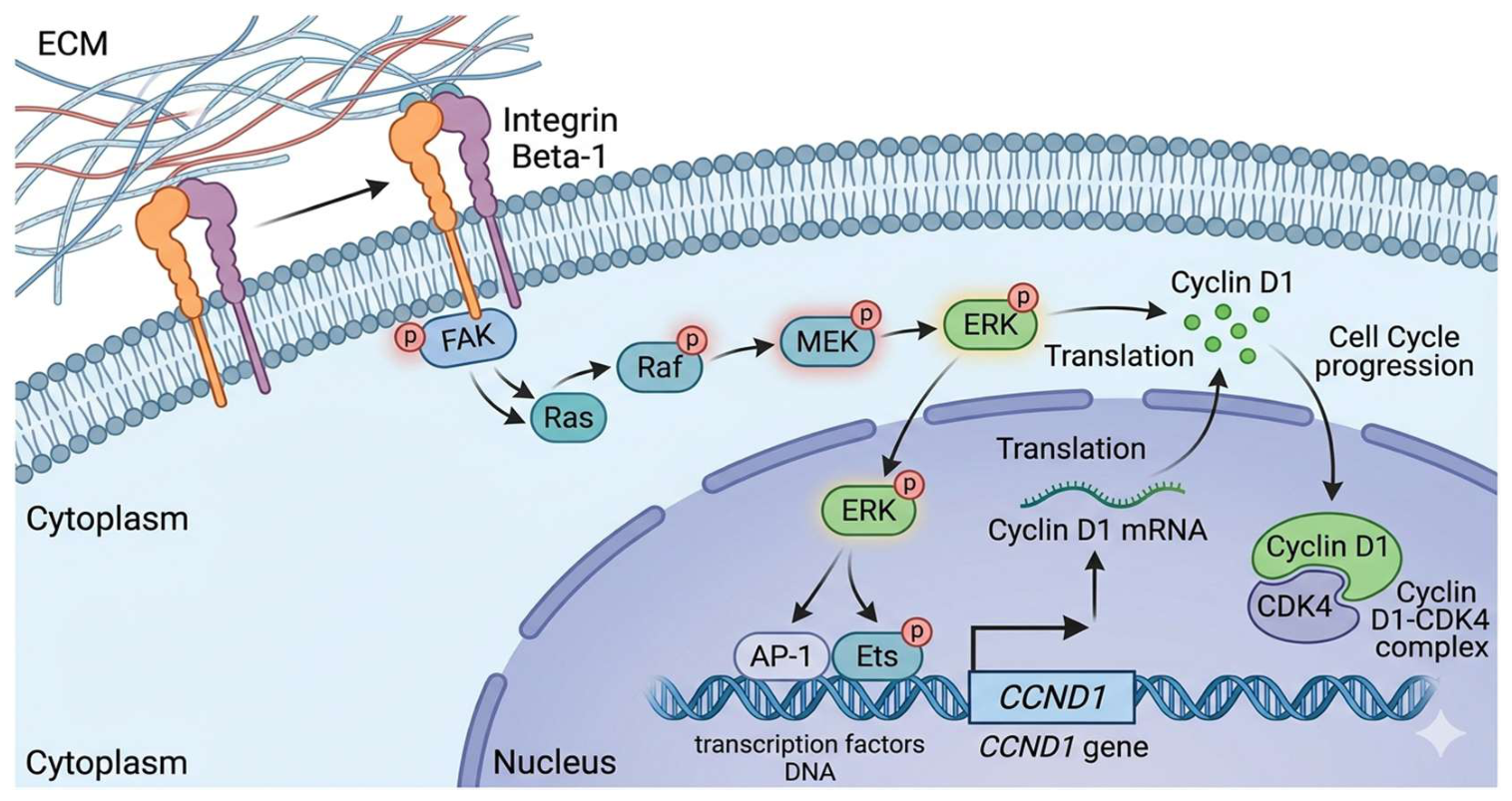
Figure 2.
Mechanistic pathway of the CCND1 - ITGB1 axis. Integrin 1-mediated adhesion to the extracellular matrix triggers signaling cascades that drive the expression of Cyclin D1, facilitating G1/S phase transition.
Figure 2.
Mechanistic pathway of the CCND1 - ITGB1 axis. Integrin 1-mediated adhesion to the extracellular matrix triggers signaling cascades that drive the expression of Cyclin D1, facilitating G1/S phase transition.
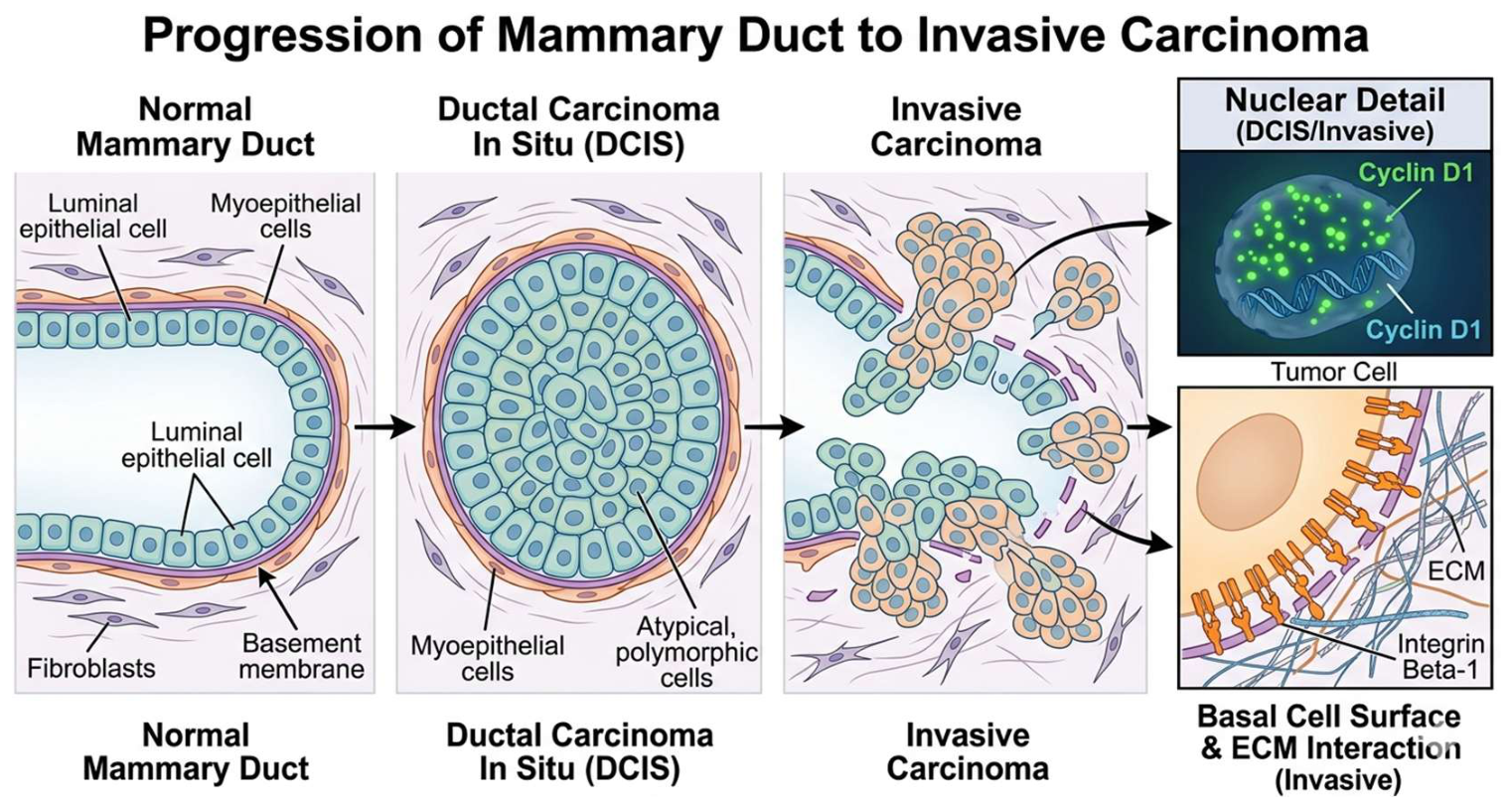
Figure 3.
Analytical validation of the reference gene GAPDH showing high linearity and thermal specificity.
Figure 3.
Analytical validation of the reference gene GAPDH showing high linearity and thermal specificity.
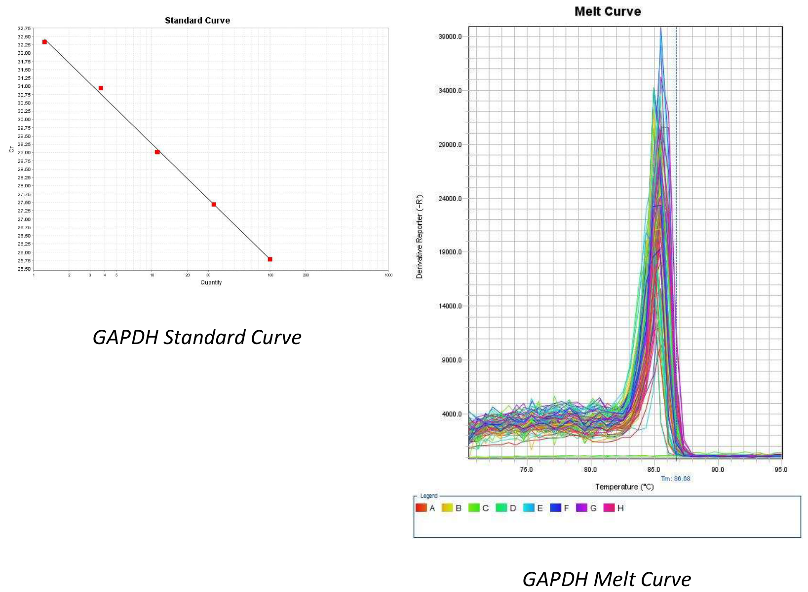
Figure 4.
Thermodynamic and kinetic validation of CCND1 and ITGB1 qPCR assays.
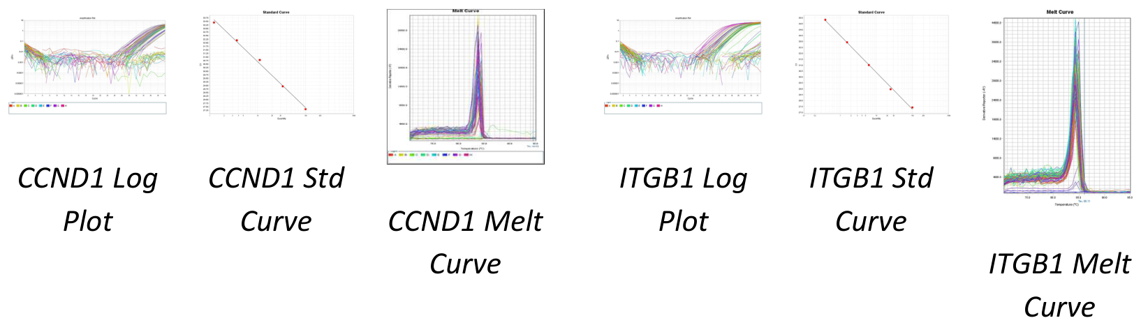
Figure 5.
Comparative expression changes of CCND1 in tumor versus normal tissues. The distribution shows a strong bias toward significant upregulation in neoplastic transcriptomes.
Figure 5.
Comparative expression changes of CCND1 in tumor versus normal tissues. The distribution shows a strong bias toward significant upregulation in neoplastic transcriptomes.
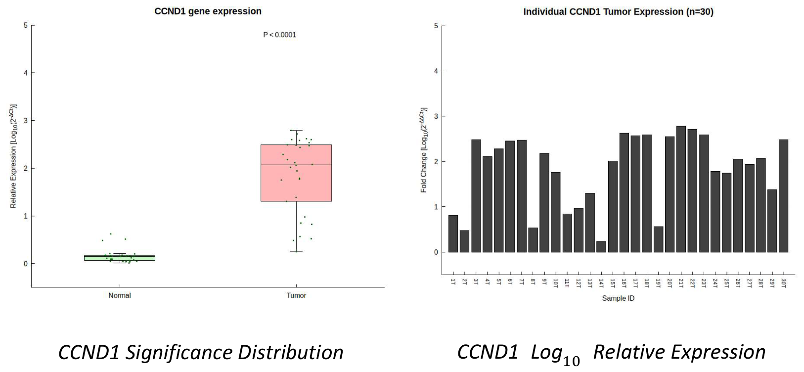
Figure 6.
Clinical quantification of ITGB1 expression levels. Data represent the significant clustering of high-expression values in tumor tissues compared to normal controls (P < 0.0001).
Figure 6.
Clinical quantification of ITGB1 expression levels. Data represent the significant clustering of high-expression values in tumor tissues compared to normal controls (P < 0.0001).
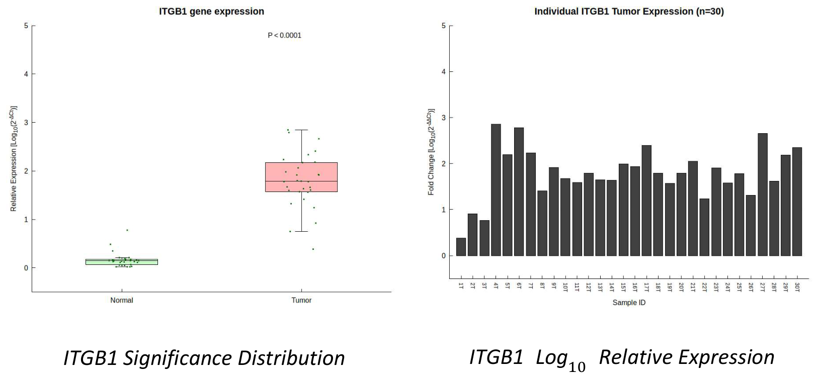
Figure 7.
In silico correlation analysis demonstrating the positive relationship between CCND1 and ITGB1 transcript levels (R = 0.068, P = 0.019).
Figure 7.
In silico correlation analysis demonstrating the positive relationship between CCND1 and ITGB1 transcript levels (R = 0.068, P = 0.019).
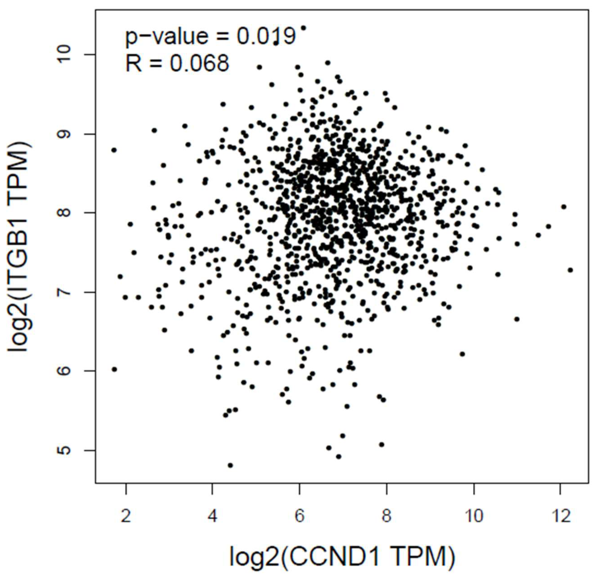
Figure 8.
Kaplan-Meier survival analysis demonstrating the prognostic utility of the dual-gene signature.
Figure 8.
Kaplan-Meier survival analysis demonstrating the prognostic utility of the dual-gene signature.
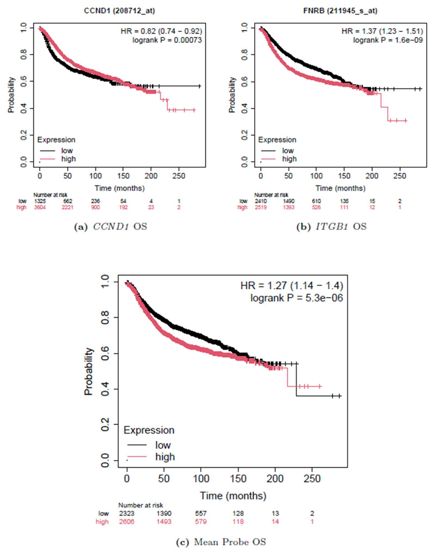
Figure 9.
Integrative Protein-Protein Interaction (PPI) network identifying the CCND1 - ITGB1 axis as a central mediator within a larger hub of kinases and ECM receptors.
Figure 9.
Integrative Protein-Protein Interaction (PPI) network identifying the CCND1 - ITGB1 axis as a central mediator within a larger hub of kinases and ECM receptors.
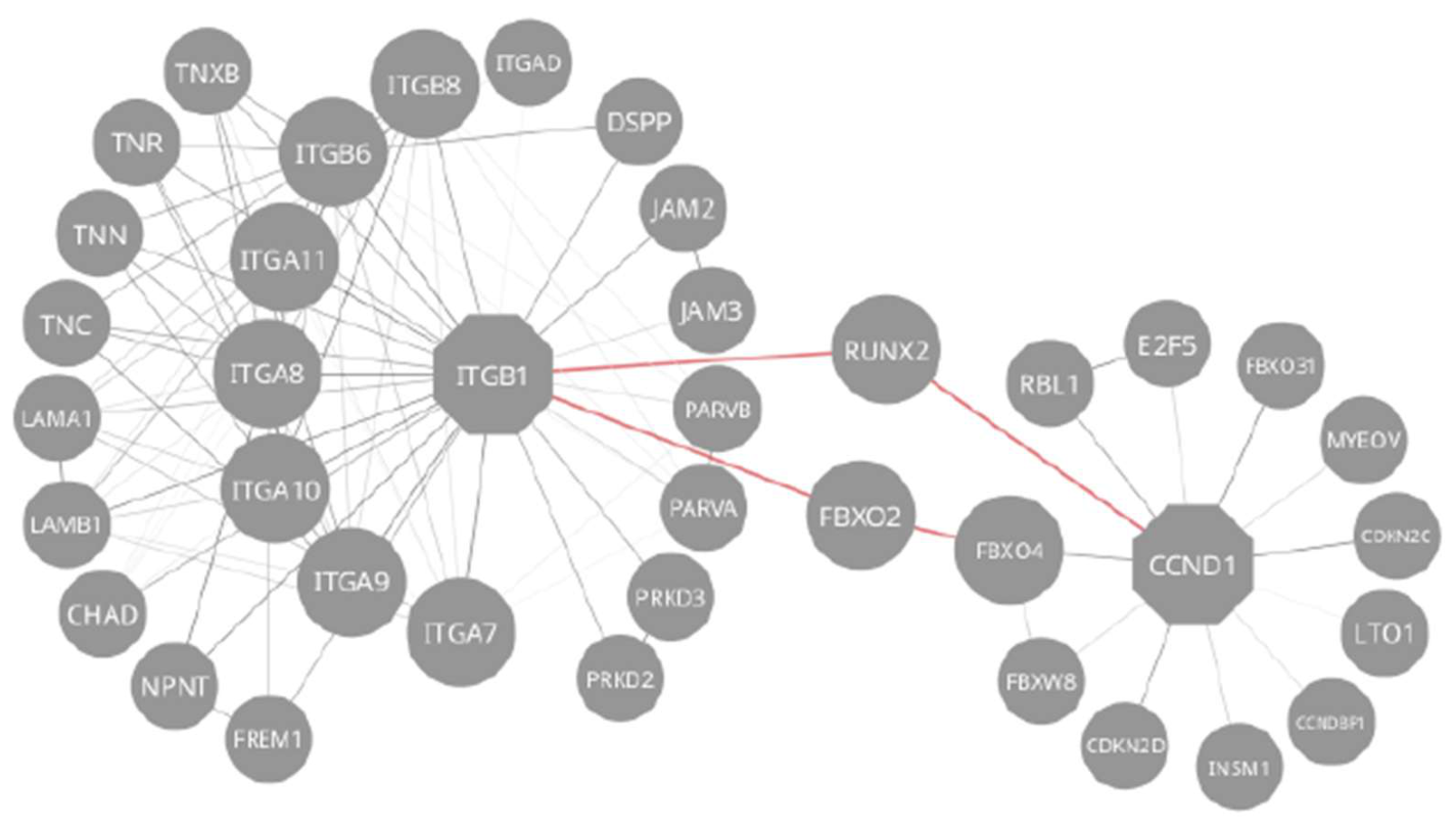
Figure 10.
Functional enrichment analysis demonstrating that the target genes are predominantly involved in cell-substrate junction assembly and focal adhesion ().
Figure 10.
Functional enrichment analysis demonstrating that the target genes are predominantly involved in cell-substrate junction assembly and focal adhesion ().
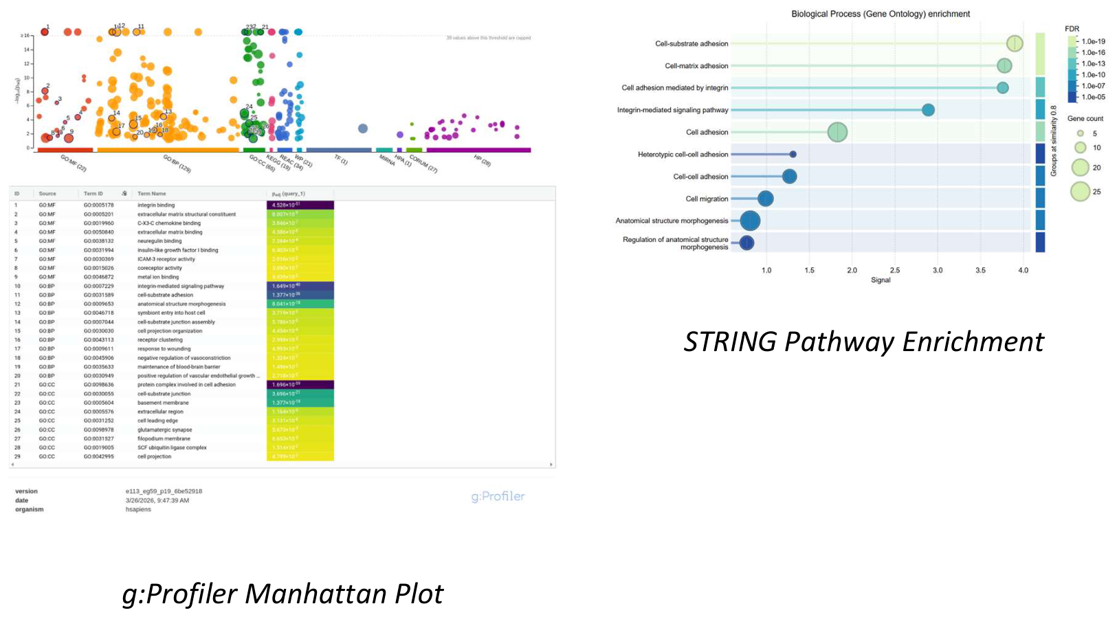
Figure 11.
In silico validation of differential expression across the TCGA-BRCA cohort, confirming significant over-expression of both markers in malignant tissues.
Figure 11.
In silico validation of differential expression across the TCGA-BRCA cohort, confirming significant over-expression of both markers in malignant tissues.
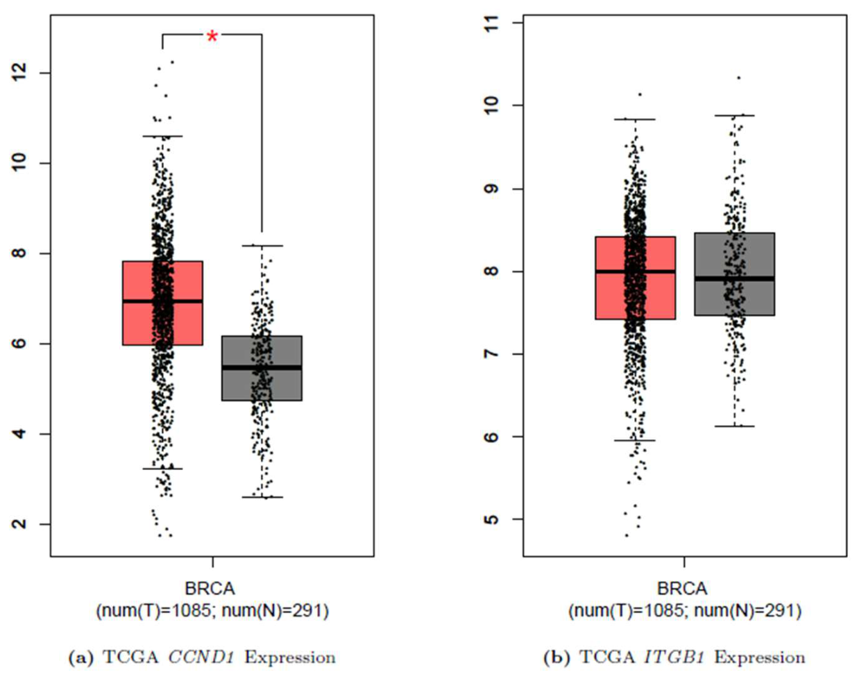

Table 1.
Consolidated cDNA Synthesis components and thermal conditions.
| Component | Volume | Step | Conditions |
|---|---|---|---|
| Standardized Total RNA (1000 ng) | 4 L | Primer Annealing | – 2 min |
| dNTP Mix | 0.5 L | Denaturation | – 5 min |
| Oligo dT & Random Hexamers | 0.5 L | Reverse Transcription | – 55 min |
| 5X Buffer & RNAse Inhibitor | 2.5 L | Enzyme Inactivation | – 5 min |
| Reverse Transcriptase | 0.5 L | Storage |
Table 2.
Primer sequences and optimized qRT-PCR thermal cycling conditions.
| Gene | Forward (5’ 3’) | Reverse (5’ 3’) |
|---|---|---|
| CCND1 | CCAAGACGACTGTTACAA | GAAGCCCTCCATGATAAC |
| ITGB1 | CTGTGAATGTGAATGCCAAAGC | GACGCACTCTCCATTGTTACTG |
| GAPDH | ACACCCACTCCTCCACCTTTG | TCCACCACCCTGTTGCTGTAG |
| PCR Step | Temp/Time (CCND1) | Temp/Time (ITGB1) |
| Activation | / 15 min | / 15 min |
| Denaturation | / 15 sec | / 15 sec |
| Annealing | / 20 sec | / 30 sec |
| Extension | / 30 sec | / 30 sec |
Disclaimer/Publisher’s Note: The statements, opinions and data contained in all publications are solely those of the individual author(s) and contributor(s) and not of MDPI and/or the editor(s). MDPI and/or the editor(s) disclaim responsibility for any injury to people or property resulting from any ideas, methods, instructions or products referred to in the content. |
© 2026 by the authors. Licensee MDPI, Basel, Switzerland. This article is an open access article distributed under the terms and conditions of the Creative Commons Attribution (CC BY) license (http://creativecommons.org/licenses/by/4.0/).
Copyright: This open access article is published under a Creative Commons CC BY 4.0 license, which permit the free download, distribution, and reuse, provided that the author and preprint are cited in any reuse.