

Submitted:
24 September 2026
Posted:
24 September 2026
You are already at the latest version
Abstract
Serum protein electrophoresis (SPE) remains a central laboratory method for the detection and characterization of monoclonal gammopathies and other dysproteinemic patterns. Despite substantial analytical automation, interpretation still relies largely on graphical representation and expert visual assessment, making low-amplitude or borderline abnormalities particularly challenging. Artificial intelligence (AI), including machine-learning (ML) and deep-learning (DL) approaches, has increasingly been applied to SPE, with high performance reported for automated recognition of established abnormalities, particularly M-proteins, i.e., monoclonal immunoglobulins or immunoglobulin fragments produced by clonal plasma cells. However, most models have been developed against predefined diagnostic targets and evaluated using expert interpretation, immunofixation, or immunotyping as reference standards. Improved recognition of known abnormalities should therefore not be equated with discovery of additional clinically meaningful information. Modern electrophoretic platforms generate machine-readable numerical signals that may preserve features not explicitly considered during routine graphical interpretation. This creates the possibility of extending AI-assisted SPE from conventional pattern recognition toward detection of low-amplitude, distributed, or sub-visual signal characteristics. Yet current literature has focused predominantly on algorithmic performance, while the validation pathway required to determine whether additional AI-detected features are biologically meaningful and clinically actionable remains insufficiently defined. Evidence is strongest for automated M-protein recognition, including low-amplitude abnormalities, whereas non-canonical systemic patterns, longitudinal signal analysis, and AI-assisted decomposition of therapeutic monoclonal antibody interference remain exploratory. This critical review addresses this translational gap by evaluating AI applications according to the nature of detected information, strength of supporting evidence, and proximity to clinical implementation. We propose a translational framework in which additional AI-detected electrophoretic features progress through analytical validity, biological validity, clinical validity, and clinical utility before being considered actionable laboratory information.
Keywords:
artificial intelligence
; serum protein electrophoresis
; monoclonal gammopathy
; M-protein
; machine learning
; high-dimensional signal analysis
; clinical validation
; laboratory medicine
1. Introduction
Serum protein electrophoresis (SPE) remains a core method in laboratory medicine for the detection and characterization of dysproteinemias, particularly monoclonal gammopathies, while also providing information on broader inflammatory, immune-mediated, hepatic, and neoplastic processes [1,2,3,4]. Despite substantial automation of the analytical phase, interpretation still relies largely on graphical representation of the electrophoretic profile and expert visual assessment. This approach is generally effective for well-defined abnormalities but becomes less robust when monoclonal components are small, poorly demarcated, or embedded within heterogeneous polyclonal backgrounds, where subtle morphological changes may be inconsistently recognized [1,2,3,4].
Artificial intelligence (AI) has emerged as a potential means of reducing this interpretative variability. Machine-learning (ML) and deep-learning (DL) approaches have been applied to electrophoretic profiles, extracted features, and numerical SPE data, with several large retrospective studies reporting high performance for automated recognition of M-proteins and other predefined electrophoretic abnormalities [7,8,9,10,11,12,13,14]. Collectively, these studies establish an increasingly convincing role for AI in automating and extending conventional pattern recognition, including the detection of low-concentration monoclonal components that may be difficult to identify visually.
However, this progress also defines the current boundary of the field. Most available models have been developed using diagnostic categories whose clinical meaning is already established and have been evaluated against expert interpretation, immunofixation, immunotyping, or related laboratory reference procedures [7,9,10,11,12,13,14]. Consequently, improved classification of an established target demonstrates enhanced recognition of known abnormalities, but does not establish that AI has uncovered previously inaccessible biological information. Improved detection of a known abnormality, however, should not be interpreted as evidence that AI has identified a new clinically meaningful signal. This distinction becomes particularly relevant when SPE is considered not only as a graphical pattern, but as a machine-readable quantitative signal. Modern electrophoretic platforms generate sequential numerical measurements that can be analysed computationally before, or independently of, their conventional graphical representation [8,9,13]. Such data potentially preserve low-amplitude, distributed, and local signal characteristics that are not explicitly considered during routine visual interpretation. AI may therefore expand the portion of the electrophoretic signal that is computationally accessible. Yet greater access to signal complexity does not necessarily translate into greater diagnostic information. A computationally detectable feature may reflect disease biology, but it may equally represent physiological heterogeneity, transient inflammation, pre-analytical variation, analytical noise, preprocessing effects, or platform-specific characteristics.
From an analytical perspective, this distinction also reflects the way electrophoretic information is generated. Regardless of whether SPE is performed by agarose gel densitometry or capillary electrophoresis, modern electrophoretic systems generate a digitally sampled numerical representation of the electrophoretic signal, consisting of sequential measurements of optical density or absorbance. The graphical electrophoretic profile routinely inspected by laboratory professionals therefore constitutes only a visual rendering of a much richer numerical representation. AI algorithms can analyse these raw signal sequences directly, potentially capturing subtle local variations in slope, intensity distribution, and signal continuity that may be attenuated or overlooked after graphical rendering. Whether these additional computationally accessible features represent biologically meaningful information, however, remains an independent question requiring dedicated validation.
This represents the central translational gap in the current AI-SPE literature. Previous research has predominantly addressed whether algorithms can recognize predefined electrophoretic abnormalities accurately, whereas considerably less attention has been given to the validation pathway required to determine whether additional AI-detected features are analytically valid, biologically meaningful, clinically valid, and ultimately useful for patient management. This issue becomes particularly important as AI applications move toward low-amplitude abnormalities, non-canonical electrophoretic patterns, longitudinal signal analysis, and other features lying beyond conventional visual classification.
Accordingly, this review does not primarily examine which AI architecture performs best in SPE. Instead, it critically evaluates what type of electrophoretic information current AI approaches actually detect, how strong the supporting evidence is, and what evidence is still required before additional signal features can be considered clinically actionable. We distinguish established applications from emerging and conceptual uses and propose a translational framework progressing from analytical validity to biological validity, clinical validity, and clinical utility. Within this framework, the relevant objective is not simply to detect more electrophoretic signal, but to establish which additional information provides reproducible and clinically meaningful value beyond existing laboratory interpretation.
2. Literature Search and Critical Appraisal
A structured narrative literature search was conducted to identify studies investigating the application of artificial intelligence (AI), machine learning (ML), and deep learning (DL) to serum protein electrophoresis (SPE). PubMed, Scopus, and Web of Science were searched for English-language publications addressing serum protein electrophoresis, capillary electrophoresis, artificial intelligence, machine learning, deep learning, M-protein detection, monoclonal gammopathy, and automated interpretation. The search was performed in August 2026. Additional relevant publications were identified through reference-list screening and citation tracking of key studies and methodological reviews.
Particular attention was given to original studies evaluating automated SPE interpretation, M-protein detection, low-amplitude electrophoretic abnormalities, and applications based on numerical or high-dimensional signal representations. Studies addressing therapeutic monoclonal antibody interference were also considered where relevant to the conceptual discussion of signal separation and computational deconvolution.
Studies were considered relevant when they evaluated computational methods applied directly or indirectly to SPE data and reported their analytical or clinical target, data representation, validation strategy, and performance. Methodological reviews and established laboratory literature were included to provide analytical and clinical context. Studies were not excluded solely on the basis of model architecture or reported performance; rather, their contribution was considered in relation to the maturity of the intended clinical application.
Given the heterogeneity of study designs, analytical platforms, AI methodologies, reference standards, and reported outcomes, no meta-analysis was performed. Instead, representative studies were critically appraised according to the strength and translational maturity of the supporting evidence. The assessment considered dataset size and representativeness, definition of the intended clinical target, quality of the reference standard, analytical platform and input representation, independence of development and validation cohorts, external validation, potential for overfitting, and proximity to prospective clinical implementation. Particular attention was given to whether reported performance reflected improved recognition of a predefined electrophoretic abnormality or evidence that additional AI-detected signal features provided information beyond conventional interpretation.
The qualitative appraisal was informed by principles derived from TRIPOD+AI and PROBAST+AI [23,24]. Because this was a structured narrative review and reporting was heterogeneous across studies, no numerical risk-of-bias score or formal overall quality classification was assigned. Evidence was instead considered along a translational continuum ranging from proof-of-concept and internal validation to external or multicenter validation, prospective workflow evaluation, and routine clinical implementation. Translational maturity was therefore assessed according to the robustness and clinical relevance of the evidence rather than diagnostic performance alone.
3. From Electrophoretic Profiles to Machine-Readable Signals: Analytical Foundations and Current Evidence
The application of AI to SPE depends not only on model architecture but also on how electrophoretic information is represented. SPE data can be provided to computational models as rendered curves or images, conventional fraction measurements, engineered features, or numerical sequences derived directly from the electrophoretic signal. These representations are not equivalent: each preserves different aspects of the analytical output and therefore determines which features a model can access and learn [7,8,9,10].
From a computational perspective, different AI architectures interact with these representations in distinct ways. Convolutional neural networks (CNNs) are particularly effective when electrophoretic information is represented as curves or images, allowing hierarchical extraction of local morphological features without predefined descriptors. Conversely, numerical electrophoretic sequences can be analysed directly by one-dimensional CNNs (1D-CNNs), transformer-based architectures, or conventional supervised machine-learning algorithms. While feature-based methods such as random forests or support vector machines can process high-dimensional numerical vectors, they generally treat individual variables independently and do not explicitly model the sequential structure of the electrophoretic signal. Architectures designed for sequential data are therefore theoretically better suited to capture spatial dependencies and local signal continuity within raw electrophoretic profiles.
Most AI applications developed to date have treated SPE primarily as a pattern-recognition problem. Deep-learning models can learn morphological characteristics directly from electrophoretic profiles, while conventional machine-learning approaches can operate on extracted features or numerical representations. Large studies such as SPECTR and AIRSPE have demonstrated high performance in recognizing established abnormalities, including M-spikes and low-concentration monoclonal components [7,12]. Similarly, numerical capillary electrophoresis data have been successfully analysed using ensemble and feature-based ML approaches for M-protein detection [9,10]. Collectively, this evidence establishes that clinically relevant classification does not require exclusive reliance on the graphical representation traditionally inspected by laboratory professionals.
The implications of numerical signal analysis, however, extend beyond replacing visual interpretation with automated classification. Wang et al. showed that feature representation itself influences model performance and demonstrated expert-level M-protein classification and localization using a U-Net–Transformer architecture [13]. This observation highlights a broader methodological principle: the optimal representation depends on the clinical question being asked. Detecting a predefined M-spike and searching for subtle distributed changes within an otherwise non-canonical profile are fundamentally different tasks and may require different representations of the same underlying electrophoretic information.
This distinction becomes particularly important when AI is proposed as a means of accessing information beyond established diagnostic categories. Models trained on conventional labels may achieve excellent discrimination while remaining constrained by those same labels. More granular numerical representations may preserve local relationships, low-amplitude variations, or distributed signal characteristics that are less evident after graphical rendering. Yet increased signal resolution does not establish biological relevance. A computationally detectable feature may reflect disease-related information, physiological variability, or analytical structure. Detecting additional features is only the first step; their reproducibility and clinical relevance must be demonstrated separately. Preprocessing further complicates this transition. Electrophoretic signals are influenced by analytical platform, signal acquisition, baseline behaviour, migration alignment, instrument-specific characteristics, detector ageing, reagent lot differences, and other technical sources of variation. Baseline correction, normalization, noise management, and profile alignment can improve comparability, but may also suppress, amplify, or reshape precisely the low-amplitude features that computational models are intended to detect. Consequently, preprocessing, input standardization, and cross-platform evaluation should be regarded as integral components of analytical validation rather than preliminary technical procedures [10,11]. In multicenter applications, insufficient control of these effects creates the risk that models learn laboratory- or platform-specific signatures rather than transferable biological patterns. The independent retrospective validation reported by Lahtiharju et al. illustrates the importance of testing AI-assisted SPE interpretation outside the original development environment [11].
Finally, conventional performance metrics address discrimination against a predefined reference standard, not the biological or clinical significance of the detected information. Current high-performing SPE models have predominantly been evaluated against immunofixation, immunotyping, or expert-defined electrophoretic categories [7,9,10,11,12,13]. High sensitivity, specificity, accuracy, or area under the receiver operating characteristic curve therefore demonstrates successful recognition of a known target but cannot, by itself, establish the existence of a clinically meaningful sub-visual signature. Once AI moves beyond automated recognition of established abnormalities, the principal problem is no longer classification performance but validation of the signal itself.
4. Beyond Pattern Recognition: What Does the Evidence Support?
The clinical relevance of AI in SPE depends primarily on what the model detects and how convincingly that information has been validated, rather than on the complexity of the algorithm. Current applications occupy markedly different positions along this continuum. At one end, AI-assisted recognition of established electrophoretic abnormalities, particularly monoclonal proteins, is supported by large retrospective datasets and, in selected studies, independent validation [7,9,10,11,12,13,14]. At the other, proposed applications involving non-canonical systemic patterns, longitudinal sub-visual changes, or computational separation of overlapping therapeutic and endogenous monoclonal components remain exploratory or conceptual.
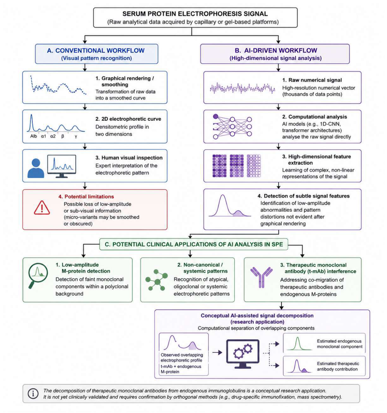
This distinction is essential because three different claims are often conflated: improved recognition of a known abnormality, detection of additional signal features, and discovery of clinically meaningful information. The first can be established against existing laboratory reference procedures. The second requires evidence that the detected feature is analytically reproducible. The third requires substantially stronger biological and clinical validation. The available SPE literature predominantly supports the first claim, provides emerging evidence for the second, and remains limited for the third. The following applications are therefore considered according to this hierarchy of evidence rather than according to model architecture or reported diagnostic performance alone. Figure 1 summarizes this transition from conventional visual interpretation to AI-driven signal analysis and places the main established, emerging, and conceptual applications within a single laboratory-oriented framework.
4.1. Established and Emerging Evidence: From M-Protein Recognition to Low-Amplitude Abnormalities
Recognition of monoclonal proteins represents the most mature application of AI-assisted SPE. Multiple studies using deep learning, feature-based machine learning, and numerical electrophoretic data have demonstrated high performance for M-protein detection [7,9,10,11,12,13,14]. Importantly, the clinical target in these studies is already biologically and diagnostically defined, allowing algorithmic predictions to be compared with expert interpretation, immunofixation, immunotyping, or other established laboratory procedures. AI therefore operates within a diagnostic framework in which the abnormality of interest and its clinical meaning are already known.
Within this established application, low-amplitude monoclonal abnormalities represent a particularly relevant extension of conventional pattern recognition. Small M-proteins may be difficult to distinguish from polyclonal background when their concentration approaches the analytical or interpretative limits of routine SPE. AI-assisted analysis can improve recognition at this boundary, and recent evidence, particularly from AIRSPE, supports increased sensitivity for low-concentration M-spikes [12]. This represents a clinically plausible advantage because equivocal AI findings can still be evaluated against independent confirmatory methods.
The interpretation of this improvement nevertheless requires precision. A monoclonal component that escapes routine visual recognition but is subsequently confirmed by immunofixation is sub-visual in an operational sense, but not biologically novel. AI has improved access to an established diagnostic signal; it has not demonstrated the existence of a previously unknown disease signature. This distinction matters because increasingly sensitive computational detection is sometimes implicitly interpreted as evidence of earlier disease recognition. Current studies do not establish that an AI-detected electrophoretic alteration precedes conventional biochemical or clinical evidence of monoclonal gammopathy, nor that detecting it earlier improves patient outcomes.
The next evidentiary step should therefore move beyond retrospective agreement with established reference procedures. Prospective studies should specifically evaluate AI-positive but conventionally equivocal profiles, determining whether they predict subsequent confirmation of monoclonality, clinically relevant progression, organ involvement, or changes in patient management beyond existing diagnostic pathways. Only such evidence can establish whether increased computational sensitivity provides incremental clinical value rather than simply increasing the number of abnormalities referred for confirmatory testing.
4.2. Beyond Monoclonal Gammopathies: The Ground-Truth Problem
Extending AI-assisted SPE analysis beyond monoclonal gammopathies is scientifically attractive but supported by substantially weaker evidence. Inflammatory, autoimmune, infectious, hepatic, and neoplastic conditions can produce distributed changes across albumin and globulin fractions rather than a single discrete electrophoretic abnormality [1,2,3]. Conventional interpretation generally describes these profiles using broad morphological categories, such as polyclonal hypergammaglobulinemia or beta–gamma bridging, which may encompass considerable biological heterogeneity.
From a computational perspective, this application is considerably more challenging than monoclonal peak detection. Whereas monoclonal gammopathies are characterized by relatively localized abnormalities, inflammatory and autoimmune conditions often produce diffuse and multidimensional changes distributed across several electrophoretic fractions. AI models designed to analyse continuous numerical signals may therefore be particularly advantageous because they can evaluate multiple regions of the electrophoretic profile simultaneously rather than focusing on isolated morphological abnormalities. Nevertheless, whether these distributed computational signatures correspond to biologically meaningful disease-specific processes remains unknown.
High-dimensional analysis could, in principle, characterize these distributed patterns more comprehensively than visual inspection by integrating local morphology, relative intensity, inter-fraction relationships, and subtle changes across the continuous electrophoretic signal. This raises the possibility that profiles currently regarded as nonspecific may contain reproducible information associated with particular biological states or disease phenotypes [8]. At present, however, this possibility should be regarded as a hypothesis rather than an established diagnostic application.
The principal limitation is not simply the availability of sufficiently sophisticated algorithms, but the definition of an appropriate ground truth. In monoclonal gammopathies, an AI-detected abnormality can be evaluated against independent procedures such as immunofixation or immunotyping. For systemic inflammatory or autoimmune conditions, no equivalent electrophoretic reference standard exists. Clinical diagnostic labels are themselves heterogeneous and may incorporate differences in disease activity, treatment exposure, organ involvement, comorbidities, and inflammatory burden. A model trained on such labels may therefore achieve apparently high discrimination while learning nonspecific correlates of disease rather than a reproducible disease-related electrophoretic signature.
This distinction has direct implications for study design. Demonstrating statistical separation between disease groups is insufficient to establish a novel SPE biomarker. Candidate computational signatures should first be shown to be analytically reproducible and independent of major technical and biological confounders, and subsequently replicated in independently phenotyped populations. Their incremental value should then be evaluated against routinely available laboratory and clinical variables. Without these steps, increasing model complexity risks producing more precise classification of heterogeneous labels rather than more specific biological information.
Accordingly, AI-derived systemic or autoimmune SPE signatures should currently be considered exploratory and hypothesis-generating. Their potential value lies not yet in diagnostic classification, but in determining whether apparently nonspecific electrophoretic variation contains reproducible biological structure worthy of subsequent validation.
4.3. Therapeutic Monoclonal Antibody Interference: A Conceptual Use Case
Therapeutic monoclonal antibodies (t-mAbs) represent a distinct challenge for SPE interpretation because circulating drug-derived immunoglobulins may generate electrophoretic abnormalities that overlap with endogenous monoclonal proteins. This interference is particularly relevant in multiple myeloma, where agents such as daratumumab can complicate assessment of residual disease and treatment response by SPE and immunofixation [15,16,17]. Drug-specific immunofixation assays and mass-spectrometry-based approaches have therefore been developed to distinguish therapeutic antibodies from endogenous M-proteins [16,18,19,20,21,22].
Conceptually, this application differs from conventional pattern recognition because it represents a signal separation problem rather than a classification task. Therapeutic antibodies are manufactured molecules with reproducible physicochemical properties and may therefore generate relatively consistent electrophoretic signal characteristics under standardized analytical conditions. If such characteristics prove sufficiently stable, computational models could potentially learn the contribution of the therapeutic antibody within an overlapping electrophoretic profile and estimate the residual endogenous monoclonal component through signal decomposition. The objective would thus be to separate two analytically superimposed sources of signal rather than simply classify a profile as abnormal. This application remains hypothetical and requires rigorous experimental validation before clinical use can be considered.
However, current evidence does not establish that AI can reliably perform this decomposition in clinical SPE data. The electrophoretic behaviour of a therapeutic antibody may vary with drug concentration, analytical platform, surrounding serum proteins, and other technical or biological factors. A model could therefore learn recurrent platform- or treatment-associated patterns without achieving valid quantitative separation of the endogenous monoclonal component. The availability of an apparently stable computational “digital footprint” should not be assumed without experimental demonstration.
AI-assisted deconvolution should consequently be regarded as a research hypothesis rather than a clinically validated application. A credible validation strategy would require paired samples with documented therapeutic exposure, independent characterization of the endogenous monoclonal component, and orthogonal confirmation using drug-specific immunofixation or mass spectrometry [16,18,19,20,21,22]. External evaluation across analytical platforms and laboratories would then be necessary to determine whether the learned signal characteristics are transferable. This use case illustrates the broader principle underlying AI-assisted SPE: computational separability of a signal must be demonstrated before biological attribution or clinical interpretation can be justified.
4.4. From Algorithmic Performance to Translational Readiness
The high diagnostic performance reported by several AI-assisted SPE studies provides convincing evidence of technical feasibility, particularly for M-protein detection [7,9,10,11,12,13,14]. However, sensitivity, specificity, accuracy, F1 score, and area under the receiver operating characteristic curve describe performance against a defined reference standard; they do not, by themselves, establish generalizability, clinical validity, or clinical utility. Direct comparison across studies is further complicated by differences in cohort composition, prevalence and spectrum of electrophoretic abnormalities, analytical platforms, input representations, reference standards, and validation strategies.
Dataset size should likewise not be interpreted as a surrogate for translational maturity. Large retrospective cohorts provide valuable conditions for model development, but a model trained and tested within a single analytical environment may still exploit laboratory-, instrument-, or population-specific characteristics. Independent validation across populations and analytical settings therefore provides qualitatively stronger evidence than increasingly high performance within the development environment. The retrospective evaluation reported by Lahtiharju et al. in an independent Finnish clinical setting is relevant in this respect because it examined AI-assisted SPE interpretation outside the original development context [11].
A further distinction concerns what is being validated. For established targets such as M-proteins, external validation primarily addresses whether model performance is transferable. For additional or sub-visual signal features, however, external reproducibility is only the first requirement. Even a highly reproducible computational feature cannot be considered clinically informative until its biological attribution, association with relevant clinical states or outcomes, and incremental value over existing diagnostic pathways have been demonstrated.

Table 1.
Critical appraisal and evidence maturity of representative AI studies in serum protein electrophoresis.
Table 1.
Critical appraisal and evidence maturity of representative AI studies in serum protein electrophoresis.
| Study | Primary target | AI/data approach | Validation setting | Main contribution | Critical limitation | Evidence level |
| Ognibene et al. [5] | Monoclonal component detection | Computer-assisted SPE analysis (CASPER) | Multicenter evaluation | Early demonstration of computer-assisted recognition in routine SPE | Predates current AI architectures; limited evidence regarding modern external generalizability | Early multicenter evidence |
| Chabrun et al. [7] | Multiple predefined SPE abnormalities | Deep learning (SPECTR) | Large development cohort and independent validation cohort | Demonstrated expert-level automated interpretation across several established SPE patterns | Validation remains based on predefined expert-annotated categories | Independent retrospective validation |
| Sopasakis et al. [9] | M-protein detection | Numerical capillary SPE; multiple ML algorithms | Retrospective development/evaluation | Demonstrated feasibility of classification directly from numerical electrophoretic data | Limited evidence of cross-platform and prospective transportability | Internal retrospective validation |
| Elfert et al. [10] | M-protein detection | Feature-based ML and CNN | Large retrospective cohort | Demonstrated high performance using both feature-based and DL approaches | Focused on an established diagnostic target; clinical impact not prospectively evaluated | Internal retrospective validation |
| Lahtiharju et al. [11] | AI-assisted SPE interpretation | AI analysis of clinical SPE profiles | Independent Finnish clinical setting | Provided evidence of performance outside the original development environment | Retrospective design; impact on workflow and patient outcomes remains uncertain | Independent retrospective validation |
| Shen et al. [12] | M-spike detection, including low-concentration components | AIRSPE | Internal and external retrospective evaluation | Extended AI-assisted recognition toward low-amplitude M-spikes | Demonstrates enhanced detection of a known abnormality rather than validation of a novel biological signal | Advanced retrospective validation |
| Wang et al. [13] | M-protein classification and localization | Multiple representations; U-Net–Transformer | Retrospective evaluation | Demonstrated the importance of input representation and combined classification/localization | External generalizability and incremental clinical value remain to be established | Internal retrospective validation |
| Cheung et al. [14] | Automated SPE interpretation | Supervised ML | Retrospective clinical dataset | Supports feasibility of automated interpretation in a large clinical dataset | Prospective workflow and outcome-based utility not established | Internal retrospective validation |
Abbreviations: AI, artificial intelligence; CNN, convolutional neural network; DL, deep learning; ML, machine learning; SPE, serum protein electrophoresis. Table note: Translational maturity was qualitatively appraised according to the intended clinical target, independence of validation, analytical setting, generalizability, and proximity to prospective clinical implementation. The categories are descriptive rather than formally validated evidence levels, and reported diagnostic performance alone was not considered sufficient to establish translational maturity [23,24].
Taken together, the current literature supports a progressive evidence hierarchy. Automated recognition of established electrophoretic abnormalities has reached internal and, for selected models, independent retrospective validation [7,9,10,11,12,13,14]. Evidence becomes substantially less mature as the intended use moves toward additional signal detection, biological attribution, longitudinal prediction, or clinically actionable decision support. Prospective workflow studies and outcome-based evaluations remain comparatively limited.
For clinical translation, model performance must be demonstrated across analytical environments and shown to provide information beyond that already available from routine laboratory testing. This distinction provides the rationale for a validation framework specifically addressing the transition from computational signal detection to clinically meaningful laboratory information.
5. From Signal Detection to Clinical Meaning: A Validation Framework
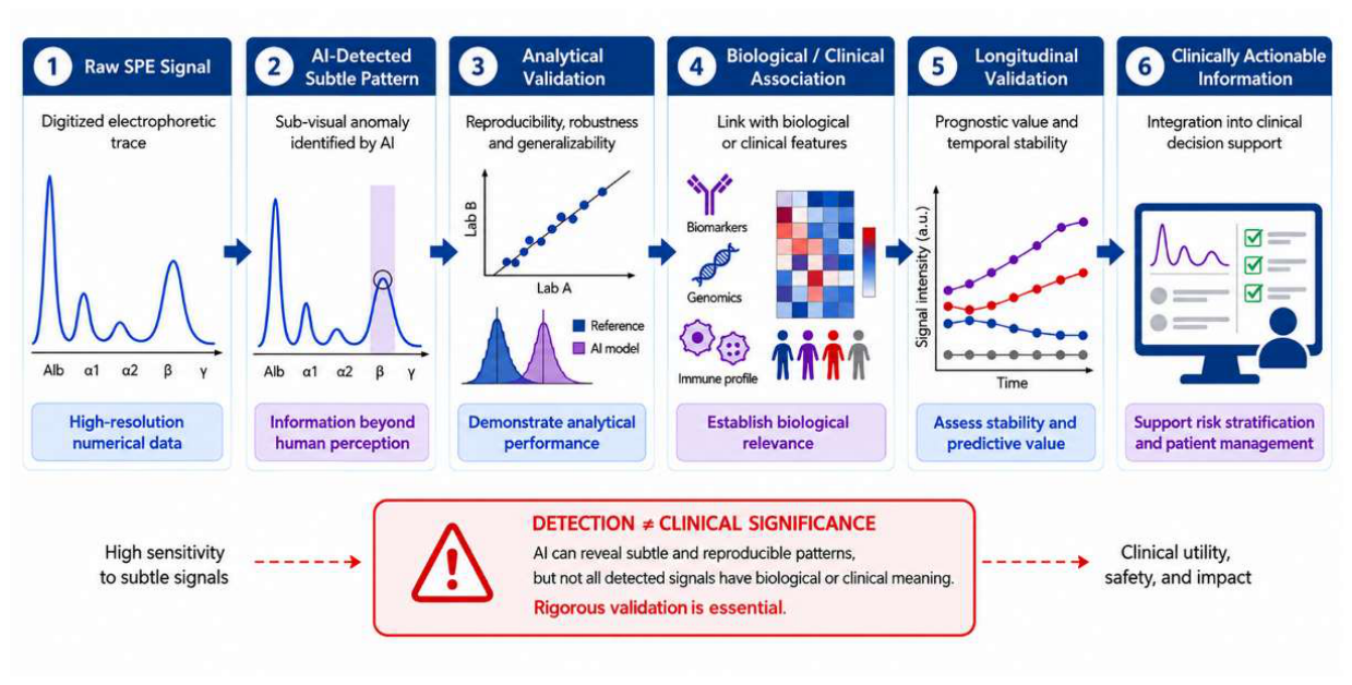
As AI increases the amount of electrophoretic information accessible to computational analysis, the central challenge shifts from detecting abnormalities to determining which detected features represent clinically meaningful information. This distinction is particularly relevant for low-amplitude, distributed, or non-canonical signal changes that do not correspond directly to established electrophoretic categories. Such features may reflect early or previously unrecognized biological processes, but they may equally arise from physiological variation, pre-analytical conditions, analytical noise, preprocessing, or platform-specific effects. Computational detectability should therefore be regarded as the beginning of validation rather than evidence of biomarker status.
We propose that additional AI-detected SPE features should progress through four distinct but sequential levels of validation: analytical validity, biological validity, clinical validity, and clinical utility. This framework separates the ability of an algorithm to identify a reproducible signal from the substantially stronger claim that the signal represents clinically actionable information.
5.1. Analytical Validity
The first requirement is to establish that an AI-detected feature is a reproducible property of the electrophoretic signal rather than a consequence of the analytical process. This includes assessment of within-run and between-run reproducibility, sample stability, preprocessing effects, migration alignment, instrument variability, and robustness across analytical platforms and laboratories. Particular attention is required for low-amplitude features because baseline correction, smoothing, normalization, and noise reduction may modify precisely the signal characteristics that computational models are intended to exploit.
Importantly, analytical reproducibility should be demonstrated not only for model predictions but also for the underlying computational features themselves. A feature that appears consistently informative within one analytical environment may disappear after changes in preprocessing, platform, or signal acquisition. Consequently, validation should address both algorithmic reproducibility and feature reproducibility before biological interpretation is attempted.
Analytical validation should therefore extend beyond conventional model discrimination. A feature that consistently contributes to an algorithmic prediction within one dataset may still represent an instrument-specific or preprocessing-dependent signature. Cross-platform and multicenter reproducibility are consequently essential before biological interpretation is attempted. In this context, model reproducibility and signal reproducibility are related but distinct requirements: a reproducible prediction does not necessarily imply that the underlying feature represents a stable biological property.
5.2. Biological Validity
Once analytical robustness has been demonstrated, the next question is whether the detected feature reflects a defined biological process. This step is particularly challenging for AI-derived SPE features because electrophoretic profiles integrate multiple protein populations and may change in response to inflammation, immune activation, hepatic function, nutritional status, treatment, and other physiological or pathological conditions.
Biological validation should therefore seek independent evidence linking the computational feature to its proposed biological origin. Depending on the application, this may involve immunofixation, immunotyping, quantitative immunoglobulins, serum free light chains, mass spectrometry, proteomic characterization, inflammatory biomarkers, or other orthogonal laboratory measurements. The objective is not simply to demonstrate statistical association with a diagnostic label, but to determine what biological phenomenon the detected signal actually represents.
This distinction is essential for proposed non-canonical or systemic SPE signatures. A model may distinguish two patient groups with high accuracy without identifying a disease-specific biological feature. Correlation with diagnosis alone should therefore not be considered evidence of biological validity. Similar methodological challenges have also been described in other emerging AI applications in laboratory medicine, where computationally identified patterns require independent analytical and biological validation before they can be interpreted as clinically meaningful biomarkers [25].
5.3. Clinical Validity
A biologically interpretable feature must subsequently demonstrate a reproducible relationship with a clinically relevant state or outcome. Clinical validity requires testing in independent and representative populations, with appropriate consideration of disease spectrum, comorbidities, treatment exposure, demographic variability, and relevant laboratory confounders.
Importantly, clinical validity should not be reduced to discrimination between predefined case and control groups. For an AI-derived SPE feature to provide meaningful diagnostic information, it should demonstrate incremental value beyond existing laboratory and clinical variables. Depending on the intended use, relevant endpoints may include confirmation of monoclonality, disease progression, development of organ involvement, treatment response, or another clinically meaningful outcome. Calibration and performance across clinically relevant subgroups should also be evaluated rather than relying exclusively on global discrimination metrics.
Longitudinal evidence may be particularly informative at this stage. If a putative sub-visual feature is proposed as an early disease signal, its value should be demonstrated by showing that its presence or evolution precedes and predicts a clinically relevant event rather than merely correlating with established disease at a single time point.
5.4. Clinical Utility
The final requirement is evidence that using the AI-derived information improves clinical or laboratory decision-making. A feature may be analytically robust, biologically plausible, and associated with disease while still providing little practical benefit if it does not alter diagnosis, follow-up, confirmatory testing, treatment, or patient outcomes.
Clinical utility should therefore be evaluated prospectively within the intended workflow. Relevant questions include whether AI-assisted interpretation reduces missed clinically relevant abnormalities, improves appropriate selection of confirmatory tests, shortens diagnostic pathways, reduces unnecessary investigations, or provides prognostic information that changes patient management. Potential harms should be considered equally carefully. Increased computational sensitivity may generate additional equivocal findings, unnecessary immunofixation or other confirmatory testing, prolonged surveillance, and patient anxiety if the clinical meaning of detected abnormalities is uncertain.
The objective should consequently not be maximal signal detection, but net clinical benefit. Only when an additional AI-derived feature improves decision-making beyond current laboratory practice should it be considered clinically actionable.
Figure 2.
Translational framework from AI-detected electrophoretic signals to clinically actionable information. Computational detection alone does not establish biomarker status. Additional SPE signal features identified by AI should progress through sequential assessment of analytical validity, biological validity, clinical validity, and clinical utility. Failure at any level limits interpretation at subsequent stages. Longitudinal assessment and external validation operate across this pathway and can strengthen evidence of reproducibility, temporal relevance, and generalizability.
Figure 2.
Translational framework from AI-detected electrophoretic signals to clinically actionable information. Computational detection alone does not establish biomarker status. Additional SPE signal features identified by AI should progress through sequential assessment of analytical validity, biological validity, clinical validity, and clinical utility. Failure at any level limits interpretation at subsequent stages. Longitudinal assessment and external validation operate across this pathway and can strengthen evidence of reproducibility, temporal relevance, and generalizability.
6. Research Priorities: Longitudinal and Multimodal SPE
The next phase of AI-assisted SPE research should focus less on increasing model complexity and more on generating the evidence required to determine whether additional electrophoretic information has clinical value. Two directions appear particularly relevant: longitudinal signal analysis and multimodal integration. Both may extend SPE beyond cross-sectional pattern recognition, but neither should be considered clinically established.
Longitudinal analysis offers a conceptually important shift from population-based classification toward patient-specific signal trajectories. Serial numerical SPE profiles could allow computational models to establish an individual electrophoretic baseline and identify small but reproducible deviations over time. This approach may be particularly relevant in monoclonal gammopathies, where within-patient changes could potentially provide information that is attenuated in single cross-sectional measurements. However, detecting change earlier is not itself sufficient. If longitudinal AI-derived features are proposed as early indicators of disease evolution, they should demonstrate temporal stability, precede clinically relevant events, and provide predictive information beyond established markers and current monitoring strategies.
Such patient-specific trajectories may ultimately prove more informative than population-based reference intervals for detecting early biological deviations. Rather than relying exclusively on cross-sectional comparison with normal populations, AI could identify progressive departures from an individual's own electrophoretic baseline, potentially improving sensitivity for slowly evolving dysproteinemias or chronic inflammatory conditions. Whether these subtle temporal changes precede clinically relevant outcomes remains to be established prospectively.
Multimodal integration represents a complementary direction. SPE-derived features could be combined with routinely available laboratory information, including quantitative immunoglobulins, serum free light chains and their ratios, complete blood count parameters, renal function, calcium, inflammatory biomarkers, and other biochemical measurements. Such integration may help determine whether an electrophoretic feature retains independent information after established laboratory variables are considered or merely reflects biological processes already captured by conventional assays.
More complex integration with proteomic, metabolomic, genomic, or other omics data may eventually help characterize the biological basis of AI-detected electrophoretic features. At present, however, technological complexity should not precede demonstration of incremental value using simpler and clinically accessible data. The relevant question is not whether SPE can be incorporated into increasingly complex multimodal models, but whether doing so adds information that changes interpretation, prediction, or patient management.
Accordingly, near-term research priorities should include standardized access to numerical SPE signals, harmonized preprocessing procedures, multicenter and cross-platform datasets, external validation, longitudinal sampling, appropriate calibration, and prospective evaluation within routine laboratory workflows. Studies should also report the intended clinical use explicitly and distinguish models designed for automated recognition of established abnormalities from those intended to discover or validate additional electrophoretic information [23,24].
The field should therefore progress from model-centered development to question-driven validation. Longitudinal and multimodal approaches are valuable not because they necessarily increase algorithmic performance, but because they may help determine whether additional AI-accessible SPE features are reproducible, biologically interpretable, and incrementally informative.
7. Conclusions
AI is reshaping the interpretation of serum protein electrophoresis, but the maturity of its clinical applications remains uneven. The strongest evidence concerns automated recognition of established abnormalities, particularly monoclonal proteins, for which ML and DL models have achieved high diagnostic performance in large retrospective datasets and, in selected studies, independent validation [7,8,9,10,11,12,13,14]. These findings establish the value of AI for improving the consistency, scalability, and potentially sensitivity of conventional SPE interpretation. However, they predominantly demonstrate better recognition of known diagnostic targets rather than discovery of new clinically meaningful electrophoretic information.
This distinction defines an important gap in the current literature. While previous research has largely focused on algorithm development and performance for predefined SPE abnormalities, considerably less attention has been given to what should happen after an AI model detects information beyond conventional visual interpretation. Modern electrophoretic platforms provide machine-readable numerical signals that may expose low-amplitude, distributed, or longitudinal features not explicitly considered during routine interpretation. Yet the biological meaning and clinical relevance of these additional features remain largely unresolved. A computationally detectable signal may represent early disease-related information, but it may equally reflect physiological variability, analytical effects, or platform-specific characteristics.
This review therefore shifts the focus from algorithmic performance to signal validation. We propose that additional AI-detected electrophoretic features should not be regarded as candidate biomarkers solely because they are detectable or reproducible, but should progress through a structured translational pathway encompassing analytical validity, biological validity, clinical validity, and ultimately clinical utility. This framework also provides a basis for distinguishing established applications, such as M-protein recognition, from emerging or conceptual uses in which biological attribution and clinical relevance have yet to be demonstrated.
The next phase of AI-assisted SPE research should therefore be driven less by increasing model complexity than by determining whether computationally accessible information provides incremental value beyond existing laboratory and clinical pathways. AI should be viewed neither simply as an automation layer nor as a replacement for expert interpretation, but as a tool for determining whether underused electrophoretic signal features can be transformed into analytically reproducible, biologically meaningful, and clinically actionable laboratory information.
Ultimately, the future value of AI in SPE will depend not on how much additional signal algorithms can detect, but on whether that signal can be shown to matter for laboratory diagnosis and patient care.
Author Contributions
Conceptualization, E.R..; Methodology, T.B., LL, E.R., FR, VB.; Investigation, T.B., L.L., F.R., V.B. S.C.M; Writing – Original Draft Preparation, T.B., L.L., F.R., V.B..; Writing – Review & Editing, T.B., L.L., E.R., F.R., V.B., A.M. G.G, A.C., A.F.; Visualization, E.A.; Supervision, T.B., L.L., F.R., V.B., A.M., G.G., A.C., A.F.
Funding
This research received no external funding.
Institutional Review Board Statement
Not applicable.
Informed Consent Statement
Not applicable.
Data Availability Statement
No new data were generated in this study. All information discussed in this review was derived from previously published studies, which are appropriately cited in the manuscript.
Conflicts of Interest
The authors declare no conflicts of interest.
Abbreviations
The following abbreviations are used in this manuscript:
| AI | Artificial intelligence |
| AIRSPE | Artificial Intelligence-based Recognition of Serum Protein Electrophoresis |
| AUROC | Area under the receiver operating characteristic curve |
| CNN | Convolutional neural network |
| DL | Deep learning |
| 1D-CNN | One-dimensional convolutional neural network |
| ML | Machine learning |
| M-protein | Monoclonal protein |
| PROBAST+AI | Prediction Model Risk of Bias Assessment Tool plus Artificial Intelligence |
| SPE | Serum protein electrophoresis |
| t-mAb | Therapeutic monoclonal antibody |
| TRIPOD+AI | Transparent Reporting of a Multivariable Prediction Model for Individual Prognosis or Diagnosis plus Artificial Intelligence |
References
- Bossuyt, X. Advances in serum protein electrophoresis. Adv. Clin. Chem. 2006, 42, 43–80. [Google Scholar] [CrossRef] [PubMed]
- O’Connell, T.X.; Horita, T.J.; Kasravi, B. Understanding and interpreting serum protein electrophoresis. Am. Fam. Physician 2005, 71, 105–112. [Google Scholar] [PubMed]
- Lee, A.Y.; Cassar, P.M.; Johnston, A.M.; Adelstein, S. Clinical use and interpretation of serum protein electrophoresis and adjunct assays. Br. J. Hosp. Med. (Lond) 2017, 78, C18–C20. [Google Scholar] [CrossRef] [PubMed]
- Lim, S.M.; Wijeratne, N.; Choy, K.W.; Nguyen, T.T.H.; Setiawan, L.; Loh, T.P. A review of clinical guidelines, laboratory recommendations and external quality assurance programs for monoclonal gammopathy testing. Crit. Rev. Clin. Lab Sci. 2024, 61, 107–126. [Google Scholar] [CrossRef] [PubMed]
- Ognibene, A.; Graziani, M.S.; Caldini, A.; Terreni, A.; Righetti, G.; Varagnolo, M.C.; et al. Computer-assisted detection of monoclonal components: results from the multicenter study for the evaluation of CASPER algorithm. Clin. Chem. Lab Med. 2008, 46, 1183–1188. [Google Scholar] [CrossRef] [PubMed]
- Altinier, S.; Sarti, L.; Varagnolo, M.; Zaninotto, M.; Maggini, M.; Plebani, M. An expert system for the classification of serum protein electrophoresis patterns. Clin. Chem. Lab Med. 2008, 46, 1458–1463. [Google Scholar] [CrossRef] [PubMed]
- Chabrun, F.; Dieu, X.; Ferre, M.; Gaillard, O.; Mery, A.; Chao de la Barca, J.M.; et al. Achieving expert-level interpretation of serum protein electrophoresis through deep learning driven by human reasoning. Clin. Chem. 2021, 67, 1406–1414. [Google Scholar] [CrossRef] [PubMed]
- He, H.; Wang, L.; Wang, X.; Zhang, M. Artificial intelligence in serum protein electrophoresis: history, state of the art, and perspective. Crit. Rev. Clin. Lab Sci. 2024, 61, 226–240. [Google Scholar] [CrossRef] [PubMed]
- Sopasakis, A.; Nilsson, M.; Askenmo, M.; Nyholm, F.; Mattsson Hultén, L.; Rotter Sopasakis, V. Machine learning evaluation for identification of M-proteins in human serum. PLoS ONE 2024, 19, e0299600. [Google Scholar] [CrossRef] [PubMed]
- Elfert, E.; Kaminski, W.E.; Matek, C.; Hoermann, G.; Axelsen, E.W.; Marr, C.; Piehler, A.P. Expert-level detection of M-proteins in serum protein electrophoresis using machine learning. Clin. Chem. Lab Med. 2024, 62, 2498–2506. [Google Scholar] [CrossRef] [PubMed]
- Lahtiharju, T.; Paavolainen, L.; Suvisaari, J.; Nokelainen, P.; Rotgers, E.; Anttonen, M.; Itkonen, O. Artificial intelligence aided serum protein electrophoresis analysis of Finnish patient samples: retrospective validation. Clin. Chim. Acta 2025, 567, 120086. [Google Scholar] [CrossRef] [PubMed]
- Shen, R.; Hu, Y.; Wu, Q.; et al. Achieving a new artificial intelligence system for serum protein electrophoresis to recognize M-spikes. ACS Omega 2025, 10, 5770–5777. [Google Scholar] [CrossRef] [PubMed]
- Wang, X.; Zhang, M.; Li, C.; Jia, C.; Yu, X.; He, H. Performance and efficiency of machine learning models in analyzing capillary serum protein electrophoresis. Clin. Chim. Acta 2025, 569, 120165. [Google Scholar] [CrossRef] [PubMed]
- Cheung, Y.T.; Leung, H.S.; Tseung, J.S.B.; et al. Supervised machine learning model for serum protein electrophoresis data interpretation. Pathology 2025, 57, 912–916. [Google Scholar] [CrossRef] [PubMed]
- Tang, F.; Malek, E.; Math, S.; Schmotzer, C.L.; Beck, R.C. Interference of therapeutic monoclonal antibodies with routine serum protein electrophoresis and immunofixation in patients with myeloma: frequency and duration of detection of daratumumab and elotuzumab. Am. J. Clin. Pathol. 2018, 150, 121–129. [Google Scholar] [CrossRef] [PubMed]
- van de Donk, N.W.C.J.; Otten, H.G.; El Haddad, O.; et al. Interference of daratumumab in monitoring multiple myeloma patients using serum immunofixation electrophoresis can be abrogated using the daratumumab IFE reflex assay. Clin. Chem. Lab Med. 2016, 54, 1105–1109. [Google Scholar] [CrossRef] [PubMed]
- McCudden, C.R.; Axel, A.E.; Slaets, D.; Dejoie, T.; Clemens, P.L.; Frans, S.; et al. Monitoring multiple myeloma patients treated with daratumumab: teasing out monoclonal antibody interference. Clin. Chem. Lab Med. 2016, 54, 1095–1104. [Google Scholar] [CrossRef] [PubMed]
- Kirchhoff, D.C.; Murata, K.; Thoren, K.L. Use of a daratumumab-specific immunofixation assay to assess possible immunotherapy interference at a major cancer center: our experience and recommendations. J. Appl. Lab Med. 2021, 6, 1476–1483. [Google Scholar] [CrossRef] [PubMed]
- Kohlhagen, M.C.; Mills, J.R.; Willrich, M.A.V.; Dasari, S.; Dispenzieri, A.; Murray, D.L. Clearing drug interferences in myeloma treatment using mass spectrometry. Clin. Biochem. 2021, 92, 61–66. [Google Scholar] [CrossRef] [PubMed]
- Noori, S.; Verkleij, C.P.M.; Zajec, M.; Langerhorst, P.; Bosman, P.W.C.; de Rijke, Y.B.; et al. Monitoring the M-protein of multiple myeloma patients treated with a combination of monoclonal antibodies: the laboratory solution to eliminate interference. Clin. Chem. Lab Med. 2021, 59(12), 1963–1971. [Google Scholar] [CrossRef] [PubMed]
- Barnidge, D.; Sakrikar, D.; Kubicki, T.; Derman, B.A.; Jakubowiak, A.J.; Lakos, G. Distinguishing daratumumab from endogenous monoclonal proteins in serum from multiple myeloma patients using an automated mass spectrometry system. J. Appl. Lab Med. 2025, 10, 235–249. [Google Scholar] [CrossRef] [PubMed]
- Milandri, J.; Jaillard, M.; Laffineur, M.; Dechomet, M.; Kolopp-Sarda, M.N. Decoding the interference: How therapeutic monoclonal antibodies challenge serum protein electrophoresis and immunofixation. Clin. Biochem. 2025, 140, 111038. [Google Scholar] [CrossRef] [PubMed]
- Collins, G.S.; Moons, K.G.M.; Dhiman, P.; et al. TRIPOD+AI statement: updated guidance for reporting clinical prediction models that use regression or machine learning methods. BMJ 2024, 385, e078378. [Google Scholar] [CrossRef] [PubMed]
- Moons, K.G.M.; Damen, J.A.A.; Kaul, T.; Hooft, L.; Andaur Navarro, C.; Dhiman, P.; et al. PROBAST+AI: an updated quality, risk of bias, and applicability assessment tool for prediction models using regression or artificial intelligence methods. BMJ 2025, 388, e082505. [Google Scholar] [CrossRef] [PubMed]
- Becherucci, V.; Romano, F.; Russo, E. Artificial Intelligence for Integrated Analysis of Non-Blood Biological Fluids: From Biomarker Discovery to Clinical Decision-Support Systems. Diagnostics 2026, 16, 2478. [Google Scholar] [CrossRef] [PubMed]
Figure 1.
Integrated overview of conventional and AI-driven serum protein electrophoresis (SPE) workflows and emerging computational applications. Conventional SPE interpretation relies on graphical rendering of the analytical signal and expert visual assessment, in which low-amplitude or sub-visual features may be difficult to recognize. AI-driven approaches can instead interrogate the numerical electrophoretic signal directly using architectures such as one-dimensional convolutional neural networks (1D-CNNs) or transformers, enabling high-dimensional feature extraction and the detection of subtle signal abnormalities. Potential applications include improved recognition of low-amplitude M-proteins, exploration of non-canonical or systemic electrophoretic patterns, and analysis of therapeutic monoclonal antibody (t-mAb) interference. For t-mAb interference, computational signal decomposition is shown as a conceptual research framework in which overlapping drug-related and endogenous monoclonal components could theoretically be estimated separately. This application is not yet clinically validated and would require confirmation against_orthogonal methods, including drug-specific immunofixation or mass spectrometry, before clinical implementation.
Figure 1.
Integrated overview of conventional and AI-driven serum protein electrophoresis (SPE) workflows and emerging computational applications. Conventional SPE interpretation relies on graphical rendering of the analytical signal and expert visual assessment, in which low-amplitude or sub-visual features may be difficult to recognize. AI-driven approaches can instead interrogate the numerical electrophoretic signal directly using architectures such as one-dimensional convolutional neural networks (1D-CNNs) or transformers, enabling high-dimensional feature extraction and the detection of subtle signal abnormalities. Potential applications include improved recognition of low-amplitude M-proteins, exploration of non-canonical or systemic electrophoretic patterns, and analysis of therapeutic monoclonal antibody (t-mAb) interference. For t-mAb interference, computational signal decomposition is shown as a conceptual research framework in which overlapping drug-related and endogenous monoclonal components could theoretically be estimated separately. This application is not yet clinically validated and would require confirmation against_orthogonal methods, including drug-specific immunofixation or mass spectrometry, before clinical implementation.
Disclaimer/Publisher’s Note: The statements, opinions and data contained in all publications are solely those of the individual author(s) and contributor(s) and not of MDPI and/or the editor(s). MDPI and/or the editor(s) disclaim responsibility for any injury to people or property resulting from any ideas, methods, instructions or products referred to in the content. |
© 2026 by the authors. Licensee MDPI, Basel, Switzerland. This article is an open access article distributed under the terms and conditions of the Creative Commons Attribution (CC BY) license (http://creativecommons.org/licenses/by/4.0/).
Copyright: This open access article is published under a Creative Commons CC BY 4.0 license, which permit the free download, distribution, and reuse, provided that the author and preprint are cited in any reuse.