

Submitted:
23 September 2026
Posted:
24 September 2026
You are already at the latest version
Abstract
Pathogens present significant risks to public health and typically require detection in a hospital or clinic setting. The advancement of lateral flow assay (LFA) cassettes has made detection accessible to all, allowing individuals to perform tests from nearly any location. The extensive use of antigen rapid test (ART) kits for self-screening of Covid-19 has reinforced the critical role of LFAs in accelerating diagnostic processes and subsequent disease prevention measures. This study delves into the realm of lateral flow assays, encompassing their fundamental design principles, interpretation of results, and the cutting-edge research focused on enhancing reporter materials and signal amplification techniques. Additionally, it emphasizes recent progress in employing LFAs for the effective identification of a diverse array of pathogenic microorganisms. This comprehensive overview has the potential to serve as a valuable reference for researchers in related disciplines.
Keywords:
lateral-flow assay (LFA)
; reporting markers
; pathogen detection
1. Introduction
Test strips have become a widely utilized method for diagnosing COVID-19 throughout the pandemic [1,2]. With a single scoop of body fluid, Antigen Rapid Test (ART) kits can provide accurate results within a timeframe of 5 to 10 minutes. These kits, categorized as a type of Point-of-Care Testing (POCT), necessitate minimal training, resulting in decreased manpower requirements in comparison to Polymerase Chain Reaction (PCR) tests. Moreover, they are associated with considerably lower costs and offer a much quicker turnaround than PCR methodologies, while also being less constrained by the testing environment. Test strips enhance convenience for both healthcare providers and patients. This tool serves as a valuable supplement in situations where precise testing methods, such as PCR instruments, are unavailable. Additionally, it has the potential to minimize nosocomial transmission by facilitating triage while awaiting PCR test results.
The ART operates on the principles of lateral flow assays (LFAs), which are paper-based biosensing platforms that analyze liquid samples as they move through the test via capillary action. The results are visually depicted with high sensitivity, allowing for straightforward interpretation through intuitive outcomes [3,4]. Initial iterations of LFAs primarily provide qualitative or semi-quantitative outcomes, which are adequate for detecting analytes present in significantly higher concentrations than ordinary levels. Conventional test strips utilize AuNPs as indicators, with the human chorionic gonadotropin (HCG) test for pregnancy serving as a prominent example, which was established in 1980s. Subsequently, commercial lateral flow tests (LFTs) have been developed to screen various infectious diseases, including human immunodeficiency virus (HIV), influenza A/B virus, group A Streptococcus and malaria. These LFA are crucial for the prevention of transmissible diseases and contribute significantly to public health efforts globally.
However, as the need for testing low-concentration analytes has increased, particularly in complex human bodily fluids, the efficacy of AuNPs has proven inadequate for yielding reliable results. Consequently, numerous advancements have been made to enhance the sensitivity of LFAs, including modifications to traditional reporters, develop new reporters and the implementation of signal amplification technologies. Novel labeling agents have been developed as alternatives to AuNPs to improve the detection sensitivity of analytes present in lower concentrations, thereby providing additional options for analysis. Prominent examples of these agents include magnetic nanoparticles (MNPs) [5,6], colored particles [7,8], upconversion nanoparticles (UCNPs) [9]- [10], quantum dots (QDs) [11,12,13,14], and various other fluorescent nanoparticles [15,16].
LFAs can be engineered to detect various analytes, contingent upon the identification of specific recognition molecules. A variety of pathogen recognition molecules has been utilized in LFAs, encompassing nucleic acids [17], aptamers [18], proteins [19,20]- [21], and traditional antibody used in the immunoassays. In this context, numerous signal amplification methods have been explored alongside LFAs, such as surface-enhanced Raman scattering (SERS) [5,22,23], PCR [24,25], Loop-mediated isothermal amplification (LAMP) [26], thermal contrast amplification [27], among others. As a result, LFAs can be applied to a diverse array of complex samples, including serum [28], whole blood [29], urine [30], saliva [31], sweat [32], and other body fluid [33]. Nevertheless, the intricate nature of these samples poses significant challenges for the labeling agents and detection methodologies used.
In this Review, we provide an overview of the principles and outcome evaluation methods of LFTs. We have examined key parameters essential for assessing the accuracy, repeatability, and overall quality of these tests. The evolution of reporting techniques and the role of signal enhancement have also been covered, with illustrative examples provided throughout the discussion. The review emphasizes the detection of a variety of pathogens, offering key insights and lessons from current practices. It also elaborates on the detection of bacteria with specific examples, highlighting the practical applications of LFTs in diagnostic processes. A particular focus has been placed on the most recent innovations that have emerged since 2020, reflecting the rapid progress and adaptation of LFT technology in response to emerging health challenges, such as the COVID-19 pandemic. By concentrating on recent developments, this review aims to provide a snapshot of the current state and future potential of LFTs, particularly in the context of POC testing, where rapid and accurate diagnostic capabilities are crucial.
2. Lateral Flow Assays (LFAs)
LFA Test strips are specifically designed to identify characteristic receptors of pathogens, thereby facilitating the detection of diseases. When antibodies are employed as the recognition elements, this approach is referred to as lateral flow immunoassays (LFIAs). Additionally, nucleic acids and proteins can also serve as recognition sites, though they typically require integration with amplification techniques such as PCR or Loop-Mediated Isothermal Amplification (LAMP) to enhance sensitivity. The fundamental principle of a lateral flow assay (LFA) involves the capillary-driven flow of a liquid sample along a test strip. A pretreated liquid sample is applied to the test strip, which is composed of a polymeric porous cellulose substrate. As the sample flows through the strip, it passes over immobilized recognition molecules. Upon binding to these molecules, a visible or detectable signal is generated, indicating the presence or concentration of the target analyte in the sample.
2.1. Structure and Test Principal
The lateral flow test strip typically consists of five crucial components: sample pads composed of glass fiber, a nitrocellulose membrane with a fixed test line (T line) and control line (C line), a conjugate release pad containing bioreceptor (antibodies or aptamers) specific to the pathogen, an absorption pad, and a support pad to facilitate the assembly of all the components (Figure 1). The sample pad is infused with buffer salts and surfactants, which facilitate the sample’s compatibility with the detection system. The sample pad guarantees that the target analyte within the sample can effectively bind to the bioreceptor on the membrane. The conjugate release pad is equipped with a colored or fluorescent reporter-labeled specific bioreceptor designed to target analytes. The analyte, bound to the recognition agent and reporter, migrate with the liquid and will ultimately be captured by the test line, provided that the target analyte is present. Thus, the emergence of the reporter signal at the test line indicates a positive result. The control line typically comprises secondary receptors specific to the marker labeled receptor used in the conjugate pad. The appearance of a signal at the control line confirms proper fluid flow and validates the assay’s operation. Consistent signal appearance in both test line and control line is imperative. If the signal is observed only at the test line, it indicates a potential issue with the assay’s performance, such as improper liquid flow. In such cases, it is recommended to repeat the test to ensure accurate results.
The majority of LFAs function based on a comparable fundamental mechanism. In this section, we elucidate the principles of testing associated with LFIAs to provide a comprehensive understanding of the testing process. Most commercially available assay utilizes two distinct types of antibodies or other recognition molecules, resulting in the formation of a sandwich structure, wherein both antibodies engage with the analyte. The primary antibody is fixed onto the test line in test strip, while the secondary antibody is labelled with a detectable marker and stored in the conjugate release pad. Upon loading a sample to the strip, any present antigen will initially be captured by the secondary antibody on the release pad. During the movement of the liquid sample, the antigen-antibody-marker complex will be captured by the immobilized primary antibody, resulting in the formation of a sandwich structure at the test line. The antigen can subsequently be quantified by assessing the signal emitted by the markers. This setup is ideal for large size analytes that contain multiple antigenic sites. For instance, the human chorionic gonadotropin (HCG) used in pregnancy tests features multiple antigenic sites, allowing two specific antibodies simultaneously targeting HCG [34]. Similarly, the p24 antigen from the HIV, which is a large size viral core protein [35], can also be analyzed with this direct assay method. The use of two antibodies guarantees the recognition of only the appropriate antigen, thereby improving the specificity of the signal and minimizing the chances of non-specific interactions. Nevertheless, the employment of two antibodies raises the overall expense of the test strip.
Molecules that are small or possess only a single antigenic determinant cannot engage in simultaneous binding with two antibodies. To overcome this limitation, competitive assays are utilized. These assays involve the competition between the analyte and a labeled analog for binding to a limited number of antibodies binding sites, allowing for the detection and quantification of small molecules or single-epitope antigens. Initially, binding molecules specific to the analyte are labeled with markers formed marker-receptor complex, which are temporarily stored on the conjugate pad. When sample moves through the conjugate pad together with the complex and reach to the test zone, the target analyte (if present) will compete with the labeled complex for binding to a limited number of immobilized capture antibodies specific to the analyte. If the analyte is present in high concentrations, it will outcompete the marker-receptor complexes for the binding sites, resulting in fewer complexes binding to the capture antibodies and a weaker signal in the test line. Consequently, the concentration of the analyte antigen can be inferred by calculating the difference in the labeled antigen’s signal before and after the introduction of the sample. Similar to the sandwich assay, a control line contains antibodies that bind to the marker-receptor complex ensure that integrity and functionality of the labeled complex used in the assay. The competitive assay method simplifies the process by utilizing just one set of antibodies and a single labeled antigen, thereby reducing costs. However, it may struggle with the detection of antigens present in very low concentrations, which result in reduced sensitivity compared to sandwich assays. Furthermore, the possibility of non-specific binding of the labeled antigen can compromise the specificity of the assay, rendering it less accurate than the sandwich method.
2.2. Test Result Assessment
Diagnostic tests can present results in diverse formats, making it essential to have a standardized measure to compare their capabilities. The “Limit of Detection” (LOD) is a key concept that characterizes a test’s ability to reliably identify the presence of an analyte. It represents the lowest concentration of the analyte that can be distinguished from the background noise with a specified level of confidence, typically 95%. The LOD is a critical indicator of a test’s sensitivity and helps in comparing the performance of different diagnostic methods.
Furthermore, the quality of diagnostic assays is commonly assessed using two key metrics: sensitivity and specificity. Sensitivity refers to the test’s ability to correctly identify true positive cases among those with the condition, while specificity is the ability to correctly identify the fraction of non-diseased people tested a negative test. A test with high sensitivity and specificity is considered to be of high quality, as it minimizes false negatives and false positives, respectively.
The determination utilizes a collection of test results depicted in the supplied table (Figure 2). The results obtained with this approach is regarded as the most accurate and dependable assessment, commonly known as the “gold standard.”
Sensitivity = number of true positives/(number of true positives + number of false negatives)
Specificity = number of true negatives/(number of true negatives + number of false positives)
In cases where the sample size is insufficient, it becomes necessary to have a reference standard in place. This is crucial when comparing the outcomes derived from the rapid test in order to assess its diagnostic accuracy. A perfect test strip would ideally exhibit both 100% sensitivity and specificity, ensuring that it accurately detects all true positive cases while also correctly ruling out all true negative cases.
A range of strategies can be utilized to enhance detection sensitivity. One approach involves signal amplification, which may be accomplished by broadening the test line and employing alternative, more luminous reporters than AuNPs. Additionally, the adoption of suitable quantitative systems, such as thermal contrast, lasers, or light-emitting diodes (LEDs), the concurrent detection of two proteins, and the incorporation of computational binary logic gate techniques represent other viable methods [36]. Assay sensitivity is significantly influenced by the dissociation constant of the receptor-analyte complex. A lower dissociation constant correlates with a higher binding affinity, which can improve sensitivity. Moreover, the capillary force-driven flow may not provide adequate interaction time between the bioreceptor and the target analyte, potentially affecting the assay’s sensitivity. Furthermore, the colorimetric signal of markers is crucial for establishing assay sensitivity, as the binding or reaction of the antigen is represented by the signal.
2.3. Innovation and Alternation of Lateral Flow in Test Strips
The fundamental principles of lateral flow test strips are well established. However, traditional fluid flow theories and models still exhibit significant limitations when applied in practice. Firstly, current LFT design often hinge on simplified assumptions in fluid flow [37], depicting flow as one-dimensional and uniform, while overlooking the intricate multi-dimensional flow dynamics and intricate interactions between materials and fluids. These oversimplifications can lead to significant deviations between the predicted results and the actual flow patterns, as well as the material-fluid interactions observed in real detection processes. For instance, under real conditions, fluid flow is influenced by the surface properties of materials such as sample pads, conjugate pads, and nitrocellulose (NC) membranes, and may exhibit non-uniform distribution or localized retention of the fluid, posing challenges in achieving precise control over its movement. Secondly, traditional flow models fail to fully account for the dynamic changes in fluid flow, such as variations in sample concentration gradients, changes in fluid viscosity, and the impact of external environmental factors (such as temperature and humidity) on flow behavior [38]. These factors can lead to instability in fluid flow during actual detection, thereby affecting the repeatability and accuracy of the detection results. Moreover, conventional models fall short in comprehensively capturing the intricacies of fluid-material interactions, thereby failing to precisely depict fluid dynamics within porous media. Consequently, the intricate processes of permeation and diffusion of fluids through such media remain insufficiently explored.
Therefore, the behavior of fluid flow plays a pivotal role in determining the performance of LFTS. For instance, variations in fluid flow velocities can lead to insufficient mixing of the sample and reagent, potentially impacting the reliability of the test results. Moreover, the intricate interaction between a fluid and a material surface, influenced by factors such as surface hydrophilicity, hydrophobicity, and charge distribution, significantly influences the dynamics of their interplay. By optimizing the structural design of microchannels with specific sizes and shapes, the efficiency of fluid mixing can be enhanced, and non-specific binding can be reduced. Consequently, this improves the accuracy and stability of detection in LFTS, ensuring more reliable test results. For instance, the Unisart StructSure® has implemented a wave-like structure flow pattern in LFA [39], which is defined by two notable characteristics: 1. Multiple, narrower flow channels rather than a single channel (Figure 3a), and 2. A wave-like flow pathway (Figure 3b). This wave-structured channel prolongs the flow distance, facilitating an extended incubation period between the analyte and the detector, which in turn enhances sensitivity (Figure 3c). The curved flow profiles induce turbulence at the curves, where variations in velocity modify fluid dynamics, thus allowing for controlled mixing of the sample with the assay reagents. By replacing a single 14 mm-wide channel with a set of six 2 mm channels, the wave-like configuration improves laminar flow, as evidenced by the low Reynolds number (Re). The decreased channel width amplifies capillary forces, which is beneficial for managing more viscous samples. Furthermore, this arrangement necessitates a smaller sample volume, making it particularly suitable for scenarios where sample availability is limited.
Another structurally innovative design involves integrating the assay channel with a rotary device [40]. In this configuration, the test strip remains stationary while the top portion of the device rotates, aligning the reagent and absorbent pads with each end of the paper strip and initiating fluid flow (Figure 4). Through further incremental rotation, an adjacent pair of pads aligns simultaneously, facilitating the flow of subsequent reagents that were preloaded in the reagent pad. This device empowers users to regulate the sequence and duration of fluid flow, thereby streamlining the process of dispensing multiple reagents onto the strip. This design facilitates the effortless delivery of multiple reagents to a paper strip, thereby supporting complex multistep assays.
Later on, more advanced rotary designs have emerged in the literature. For instance, Ji et al. introduced innovative rotational paper-based microfluidic chips capable of detecting phenolic pollutants [41]. The chip is able to perform both qualitative and quantitative analyses of 4-nitrophenol (4-NP) and 2,4,6-trinitrophenol (TNP) concurrently, thus demonstrating their efficacy in simultaneous detection of these compounds. Pattarapon et al. created an automated rotary lateral flow immunoassay platform that incorporates a servo motor [42]. This multistep immunoassay is capable of quantifying the colorimetric detection of E. coli O157:H7 bacteria. A three-dimensional (3D) microfluidic analytical device was developed to conduct multistep diagnostic assays in limited space. This device enables dual colorimetric and electrochemical detections, exhibiting a wide dynamic range for detection, with a remarkable detection limit of 0.35 ng/mL for cardiac Troponin I [43].
These innovative structural designs clearly demonstrate significant benefits. Nevertheless, the processes involved in preparation and manufacturing can be rather complex, which may result in increased costs and longer lead times. This complexity might also influence the yield of qualified products. Consequently, the primary challenge is to uphold cost-effectiveness while guaranteeing production feasibility, without sacrificing either element.
3. Markers/Labels Used in LFA
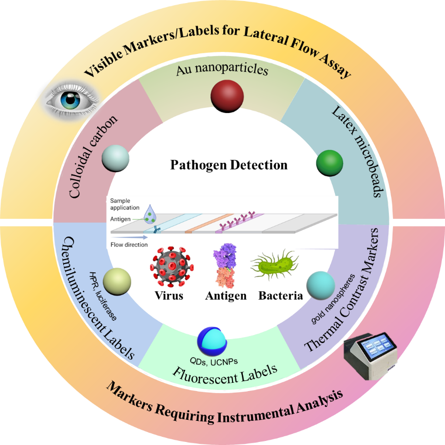
Lateral flow strip tests rely on markers to indicate the presence or absence of a target pathogen. These markers fall into two main categories based on how they convey their signals: visual labels [44], which provide a direct and readily observable color change can be read by the naked eye, and labels that require specialized instruments for detection [45]. These visual labels are commonly employed in rapid diagnostic tests due to their convenience and immediate results, whereas the labels requiring instrumental detection often offer higher sensitivity and specificity but involve a more complex analytical process.
When a material demonstrates observable changes, such as variations in color, fluorescence, or precipitation, upon interacting with the analyte, it can function as a visual marker in LFA. Nonetheless, the efficacy of these visual markers is generally limited to scenarios where significant concentrations of analytes are present in the samples. The process of visual assessment has inherent constraints. To overcome these limitations, the implementation of advanced LFT labels is essential for enhancing both sensitivity and specificity. In instances where low concentrations of analytes are analyzed or quantified, it is customary to use labels in combination with specialized instruments to ensure accurate detection. Instruments such as fluorescence microscopes [46], flow cytometers, spectrophotometers, chemiluminescence detectors [47,48], autoradiography instruments, or electrochemical analyzers [49,50] are typically utilized for effective detection and quantification. These instrumental labels offer higher sensitivity and accuracy compared to visual labels, making them ideal for precise quantification and high-throughput pathogen detection in laboratory settings.
3.1. Visible Markers
Visual markers are now seamlessly integrated into LFIA strip technology in pathogen detection [51]. This integration enables rapid, cost-effective, and user-friendly identification of diverse pathogens [52], a crucial feature in applications ranging from medical diagnostics53, 54 to food safety [53,54] and environmental monitoring [55,56]. With this visual labeling technology, on-site testing and preliminary screening become highly accessible, providing an efficient and dependable solution for scenarios demanding immediate and accurate results.
3.1.1. Gold Nanoparticle (AuNPs)
Gold nanoparticles (AuNPs) serve as one of the most prevalent visual markers in LFTs [57] owing to their distinctive optical attributes and straightforward synthesis process. AuNPs exhibit vibrant color as a result of surface plasmon resonance (SPR), a phenomenon that occurs when the conduction electrons in the metal nanoparticles collectively oscillate in response to the incoming light. The SPR property enables the AuNP to exhibit size-dependent colors (such as red, blue, and purple) by manipulating the particle size or aggregation state [58]. Typically, AuNPs in 13 nm diameter turns out wine-red, while an increase in size results in a red shift. When AuNPs aggregate at distances shorter than their diameter, they exhibit a blue color due to interparticle plasmon coupling. The robust plasmonic signal of AuNPs allows for visualization with the naked eye, making them a compelling option for markers in LFT. Additionally, AuNCs can be easily conjugated with biomolecules, including antibodies and aptamers, enhancing their versatility for various pathogen diagnostics. Their high chemical stability renders them resistant to degradation in complex environment, w thereby prolonging the shelf life of test strips. AuNPs used in LFT demonstrate significant stability against temperature and humidity, making them ideal for rapid point-of-care testing (POCT) applications. The synthesis of colloidal AuNPs is relatively straightforward, accommodating large-scale production needs and consequently reducing the costs associated with LFA.
Despite these advantages, the use of AuNPs in LFA is limited by their low sensitivity, due to the fact that naked eye detection is only reliable when pathogens are present at high concentrations [59]. Ongoing efforts aim to enhance and optimize AuNP tags to overcome these limitations. For example, red and blue dual-color AuNPs were used in one LFA to enhance the sensitivity of tests [59]. Spherical red AuNPs was used to label control line and durian-shaped blue AuNPs was used to label the test line respectively. The utilization of distinct colors for the two lines significantly improved both the diagnostic sensitivity and specificity, achieving 100% when detecting human Brucella-positive serum, even at a dilution factor of 10-5. The LOD was recorded at 0.04 IU/mL, which is two orders of magnitude lower than traditional LFIA strips. A study was conducted by Byzova et al. to investigate the impact of AuNP shape on the LOD [60]. In their study, two types of AuNPs was compared as labels in LFIA for the detection of the cardiomarker troponin I. One type was quasispherical AuNPs with average diameters ranging from 18.6 to 47.5 nm, while the other type was superspherical AuNPs with average sizes of 20.2 to 90.4 nm. Their findings demonstrated that the superspherical AuNPs achieved an 8-fold reduction in LOD when contrasted with the quasispherical AuNPs. Due to the minute interparticle gaps, which facilitate profound LSPR hybridization, nanoclusters exhibit a remarkable augmentation in plasmonic scattering, stemming from the intricate plasmon coupling processes. In the context of LFA, AuNPs dimers have been employed with the intent of elevating its operational efficacy [61]. The detection of porcine epidemic diarrhea virus in clinical samples achieved 50 times greater sensitivity than that of a test utilizing spherical colloidal gold. Another study showed the AuNP aggregates, induced by polyamidoamine (PAMAM) dendrimers, improved the sensitivity up to five times for the aptamer-based LFA in rapid detection of ochratoxin A [62]. In addition to designing novel AuNPs, integrating them with other labels represents a strategic approach to enhance the signals in LFTs.
Due to surface plasmonic resonance, Au and Ag nanoparticles could produce significantly amplified electromagnetic fields near their surface with an enhancement factor up to 1014 [63]. This resonance effect significantly amplifies molecular vibration in the vicinity of the Au or Ag nanoparticles, resulting in inelastic light scattering [64]. Employing SERS spectroscopy enables the identification of inelastic light scattering from the molecules, characterized by a spectrum with a narrow full width at half maximum, thereby generating highly sensitive signals [65]. Consequently, the integration of SERS technology with LFA presents a novel approach for highly sensitive detection. In recent years, SERS-based LFAs have been effectively implemented for precise and quantitative analysis of diverse targets. However, the low chemical stability of Au or Ag nanotags necessitates engineering modifications before their practical application.
3.1.2. Colloidal Carbon
Colloidal carbon is also a widely employed markers in LFT. This is attributable to several key advantages, including its exceptional stability, minimal cost, and the simplicity with which it can be produced on a large scale [66]. Colloidal carbon particles form black or gray visible signals on the strip, serving as an effective visual indicator. Shi et al. [67] employed colloidal carbon as a marker in a test strip designed to detect antibodies against Mycoplasma bovis, a pathogen could cause severe respiratory diseases, otitis media and mastitis in cattle and tuberculous in human as well. The strips demonstrated remarkable performance with 100% specificity and 97.67% sensitivity, which was 99.49% concordance to ELISA test. The high specificity and sensitivity of the LFT offer veterinarians and farmers a user-friendly tool to promptly identify and control outbreaks of M. bovis, thereby enhancing better disease management in livestock. Colloidal carbon was employed in the competitive LIFA test strip designed to detect the fungal pathogen Botrytis cinereal [68], notorious for causing gray mold disease in herbal medicine plants. This test strip showcased remarkable sensitivity, achieving a limit of detection (LOD) of 6.25 μg/mL specifically for identifying IgG3 and IgG2a antibodies. The significance of this lies in its ability to facilitate early detection, preventing the rapid progression of the disease by up to four days, thereby averting substantial economic losses in the herbal medicine industry.
3.1.3. Polymer Microbeads and Nanoparticles
Polymer nanoparticles and microspheres exhibit distinct advantages in lateral flow assays owing to their ability to be tailored and endowed with unique physicochemical properties. For instance, polymer nanoparticles possessing excellent biocompatibility, such as those made from polystyrene and polylactic acid, provide stability and functionality in intricate biological environments. Moreover, polymer beads can be intricately designed to incorporate a core-shell structure or a porous architecture, with the goal of enhancing their dye loading capacities and signal amplification efficiency. Surface properties can be tailored to boost interaction efficiency with the target analyte, thereby improving their uniformity and stability in lateral flow tests. Latex microbeads are versatile colloidal particles that have been widely used in lateral flow tests owing to their user-friendliness, cost-effectiveness, and capacity to deliver rapid, visible results. These microbeads were among the pioneering materials employed as labels in lateral flow diagnostic tests for qualitative assay or semi-quantification [69]. Traffic light colored red, green, and blue latex microspheres were fabricated in a LFT to identify aflatoxin B1 (AFB1), T-2 toxins (T-2), and zearalenone (ZEN) in cereals, respectively (Figure 5) [70]. The LOD and limit of quantitation (LOQ) of the LFIA with these latex labels could achieve as low as 0.04/0.40/1.21, and 0.09/0.99/1.56 μg kg-1 for AFB1/T-2/ZEN, respectively. As long as the color contrast allows, it is feasible to utilize rainbow color tracers for the simultaneous detection of multiple analytes.
Cellulose nanoparticles coated with polydopamine were loaded with dyes to facilitate the competitive detection of multiple mycotoxins and to conduct a sandwich analysis of combined inflammatory biomarkers, thereby identifying the types of infections present. The colorimetric signal generated by these particles was notably more sensitive compared to AuNPs, owing to their larger size and brighter color, which enhanced the visual readout. [71] Visual labels greatly simplified the operation of LFT and results interpretation. The signal readout does not require additional instruments or equipment but only visual inspection. Thus, the assay cost is reduced in terms of manpower and time, and the efficiency are largely improved.
By employing a variety of labels including gold nanoparticles [57,72], colloidal carbon [66,68], and colored latex microspheres [73,74], LFT can achieve quick and straightforward result interpretation, making it suitable for a range of on-site test scenarios with limited resources [75]. However, despite the benefit of vibrant and high-contrast coloring, the human naked eye has its own visual limitations, which can impede the use of visual labels in the diagnosis of low concentration analytes. In comparison to more recent technological advancements, the sensitivity and specificity of lateral flow tests based on latex microbeads may be moderately restricted. With ongoing progress and efforts, these markers will become more applicable for pathogen detection in LFT under more extensive and diverse conditions.
3.2. Markers Requiring Instrumental Analysis
The reliance on visual labels that can only be discerned by the naked eye significantly restricts the detection capabilities for analytes present in low concentrations. Therefore, there is a need for alternative labeling methods that can provide highly sensitive and precise test outcomes. There are several types of emerging markers used in LFT to indicate the capturing analytes, including fluorescent labels, chemiluminescent labels and thermal contrast labels.
3.2.1. Fluorescent Labels
Fluorescent markers require illumination with specific wavelengths of light, typically provided by a laser source, to produce fluorescence signals [76]. These signals can be observed with the naked eye or detected using specialized equipment like fluorescence microscopes [46] or other imaging tools [77,78] designed for signal analysis. Fluorescent labels present a viable solution to visible markers, as they exhibit a high signal-to-noise ratio. Additionally, fluorescent molecules are considerably smaller than colored beads, making them more comparable in size to the analytes in LFT. This size compatibility results in enhanced sensitivity of the signal, thereby enabling highly accurate quantitative assessments [79,80,81]. Various types of fluorescent labels, such as fluorescent dyes [76,82,83], fluorescent proteins [84], quantum dots [56,85,86], UCNPs [87,88,89], and polymer nanoparticles [90,91], have been implemented in LFT. Each label presents unique benefits and drawbacks. The careful selection of labels based on particular applications and experimental circumstances can significantly improve the sensitivity and accuracy of the tests.
In a study by Zhang et al. in 2022 [92], a lateral flow immunoassay chip using a second near-infrared (NIR-II) fluorescent dye encapsulated in polystyrene nanoparticles achieved a low detection limit for influenza A and influenza B viruses. This assay achieved a detection limit of 0.015 ng/mL for influenza A nucleoprotein and 102.08 TCID50/mL (TCID50 refers to tissue culture infection dose at 50% end point) for influenza A virus, as well as 0.037 ng/mL and 100.43 TCID50/mL for influenza B virus. In contrast, the visual inspected LFT with microbeads only achieve a LOD of 5.8 × 10³ plaque-forming units (PFU)/mL for influenza A and 8.2 × 10² PFU/mL (1 TCID50 ≈ 0.7 PFU) for influenza B virus, respectively [93].
Fluorescent inorganic nanomaterials, like quantum dots and UCNPs, offer advantages of high photostability and high fluorescence efficiency, making them suitable for broad-spectrum and ultra-sensitive virus/pathogen detection. Wang et al. [94] developed a multiplex LFIA platform could achieve multiple virus test on one strip with detection limits as low as pg/mL levels (Figure 6a). The design of tracer for the LFIA contained two main parts: adsorption of Fe3O4 magnetic nanoparticle and fluorescent CdSe/ZnS-MPA quantum dots on a graphene oxide (GO) nanofilm (GF@DQD) as a versatile probe, conjugation of 4-aminophenylboronic acid (APBA) was used as a broad-spectrum recognition molecule. The test LODs for SARS-CoV-2 SP, MPXV A29, and EBOV GP antigens were 0.93, 1.03, and 0.89 pg/mL, which was 200 times lower than ELISA and 500 times lower than AuNP-based LFIA. The multiple tests in one strip could greatly reduce the assay time and cost. Hu et al. [95] developed a highly luminous CdSe/ZnS quantum dots with hydrazide groups (QBs-NH-NH2) modified as the signal amplifier for LFA. The hydrazide-mediated oriented coupling facilitated antibody erecting on the particle surface, thereby enhances the bio-activity of the probes on luminescent nanomaterials. This strip exhibited a broad dynamic linearity, accommodating HBsAg concentrations ranging from 0.05 ng/mL to 3200 ng/mL with a LOD of 50 pg/mL.
Unlike quantum dots and other fluorescent molecules, upconversion nanoparticles (UCNP) produce visible fluorescence when irradiated by NIR light. This distinctive characteristic allows UCNP to potentially eliminate autofluorescence from biological samples and cross-talk fluorescence from other fluorophores, resulting in enhanced detection specificity. Additionally, emission spectrum of UCNP can be precisely adjusted within a specific range by incorporating selective doping ions. Moreover, it is feasible to engineer a single nanoparticle to emit two different colors of light under distinct excitation wavelength. Leveraging this dual-emissive characteristic of UCNP as a signaling mechanism, Guo et al. [96] have incorporated both test and control functionalities into a single line on a LFT strip (Figure 6b). The UCNPs are capable of emitting red light when excited at 980 nm and green light when excited at 800 nm, allowing one emission to act as a test signal and the other as a control or calibration signal. The test line has been innovatively designed in circular configuration to enable multiple detection capabilities. By integrating several circular test lines onto a single strip, this design allows for high-throughput analysis, a feature that is not attainable with the conventional ladder-shaped strips. This represents a significant evolution from traditional LFT designs, enhancing diagnostic potential while also reducing the physical size and cost of the strips. With this innovative LFT, the LOD for aflatoxin B1 has been reported to reach as low as 25 ng/mL, demonstrating the effectiveness of this approach in sensitive detection.
Chen and colleagues [88] developed a dual-flux immuno-chromatographic assay (dICA) for the simultaneous quantification of two distinct mycotoxins OTA and DON. In this study, upconversion nanoparticles (UCNP) were utilized for fluorescence labeling of the antigen, while AuNPs acted as fluorescence quenchers to label monoclonal antibodies. The green fluorescence emitted by UCNPs at 540 nm was used as the analytical signal to indicate the formation of the antigen-antibody complex, confirming the presence or absence of the target analytes. Additionally, the red fluorescence emitted by UCNPs at 660 nm served as a quality control signal. By employing a dual-flux bidirectional single-line labeling mode, this assay enabled the simultaneous detection of two distinct mycotoxins on separate test lines. The study highlights the sensitivity, speed, and reliability of dICA for on-site detection of multiple mycotoxins.
Jiang et al. [69] compared two commercially available test strips that one using colored microspheres as tracers while another using fluorescent microspheres (Figure 7a,b). The study focused on evaluating the performance and efficiency of these test strips for the detection of rotavirus and adenovirus in the stool specimens. Phenylethene luminative monomeric copolymers and blister were used as tracers in the two types of strips. Through analysis of 350 patients’ specimens, they found the sensitivity of fluorescent was significantly higher than that of color beads, but false positives occur in the fluorescent test strips. The same research group [93] further compared two commercial test strips with fluorescent microspheres or colored microspheres as tracers for simultaneous detection influenza A and B viruses (Figure 7c,d). Both test strips demonstrated excellent performance in terms of sensitivity and specificity, highlighting their potential for rapid influenza virus detection. In both of these research studies, the performance of fluorescent microsphere-based test strips surpassed that of colored microsphere-based test strips.
3.2.2. Chemiluminescent Labels
Chemiluminescent molecules could emit light as a result of a chemical reaction. This distinct feature makes them exceptionally sensitive, ideal for the detection of even trace amounts of target molecules. When applied in lateral flow tests (LFTs), they offer a significant advantage over fluorescent labels by removing the need for external light sources, thus simplifying the detection process and enhancing its reliability. Typically, these labels comprise a substrate that interacts with an enzyme or catalyst to produce light. Frequently utilized enzymes in conjunction with these labels include horseradish peroxidase (HRP), alkaline phosphatase (AP), and luciferase. The enzyme facilitates the oxidation of the chemiluminescent substrate, resulting in the release of energy in the form of light. The intensity of the emitted light correlates directly with the quantity of the label present, enabling precise quantification of the target molecule. The signal generated at the molecular level facilitates improved precision in quantitative outcomes across various analytical and diagnostic assays.
In a lateral flow test, chemiluminescent molecules may serve as labels for antibodies or probes, producing light signals when they bind to the target pathogen. These signals are subsequently detected and quantified using photomultiplier tubes or charge-coupled devices (CCDs), which provide high sensitivity in measurement [97]. Han [98] reported a chemiluminescence (CL)-based LFA that involved a conjugation scheme using AuNPs, aldehyde-activated peroxidase and antibody, to detect Cardiac Troponin I in human serum. HRP (horseradish peroxidase) catalyzed luminol-H2O2 chemiluminescence reaction facilitates quantitative detection, achieving a LOD of 5.6 pg·mL-1, which is 110-fold increase in sensitivity compared to AuNP-Ab conjugate LFAs. g-C3N4/BiFeO3 nanocomposites were also utilized as chemiluminescent markers in LFIA for detection of endosulfan and imidacloprid, with both colorimetric and chemiluminescent signal dual-readout [99].
Chemiluminescent labels reduce interference and produce minimal background noise because the light emission is a result of a chemical reaction. These labels do not require external light sources, which is superior to fluorescent labels. Additionally, they could be used to perform quantitative analysis across a broad dynamic range. However, their use is limited by the need for rapid signal detection, as the emitted light decays quickly. Furthermore, the application of chemiluminescent labels involves more complex procedures compared to colorimetric or fluorescent labels, necessitating precise control over reaction conditions. These complexities have somewhat hindered their widespread adoption.
3.2.3. Thermal Contrast Markers
In lateral flow assays, the binding of analytes to receptors can result in exothermic (heat-releasing) or endothermic (heat-absorbing) reactions, causing localized temperature changes. The thermal signals from specific and nonspecific bindings are distinct, enabling the sensitive and specific identification and quantification of the target analyte. Thermal Contrast Amplification (TCA) is an innovative method that enhances the sensitivity and detection capabilities of LFAs. It uses thermal imaging with a near-infrared (NIR) camera as an alternative to traditional colorimetric interpretation of LFA results. NIR camera can detect temperature variations as subtle as 0.1 °C, facilitating potentially quantitative analysis with a lower LOD. This approach harnesses the thermal responses to provide a sensitive and precise diagnostic tool.
For example, gold nanospheres (30 nm and 100 nm), and 150 nm gold nanoshells with silica cores were employed as TCA agent in the visual detection of HIV p24 protein [27]. The limits are reported to be 250, 62, and 62 pg/ml for 30 nm spheres, 100 nm spheres, and 150 nm shells, respectively. Furthermore, the thermal LOD for spiked p24 protein in human serum is determined to be at 8 pg/ml, showcasing the potential of TCA in achieving highly sensitive and quantitative results in diagnostic applications.
TCA presents a compelling approach to boost the capabilities of lateral flow assays. However, it may necessitate the use of specialized temperature detection equipment. Thermal contrast materials are designed to exhibit property changes in response to temperature fluctuations, such as alterations in color, variations in emission intensity, shifts in wavelength, or modifications in temperature-dependent reflectivity and absorbance. These changes are correlated with the concentration of markers. Converting thermal detection into optical signals can be an effective alternative for enhancing the versatility and applicability of thermal-based assays. However, the temperature changes detected could be susceptible to interference from external temperature variations, potentially affecting the assay’s sensitivity.
Incorporating instrumentally analyzed labels in LFT significantly boosts the sensitivity and accuracy of pathogen detection. By employing fluorescent and chemiluminescent labels, LFT can provide rapid qualitative results and precise quantitative analysis. Application scenarios indeed impose specific requirements for diagnostic tests, such as the need for sensitivity and specificity when analyzing various sample types like blood, serum, or other body fluids. Additionally, the speed at which detection must be accomplished and the instrumentation available for use are critical factors. These requirements guide the selection and design of appropriate markers and detection methods to ensure test efficiency and reliability for each particular context. Ongoing advancements in labeling technologies are continually refining the precision, velocity, and ease of pathogen detection in LFT. These improvements are pivotal for more accurate disease diagnosis, effective monitoring, and enhanced control measures.
4. Applications of Lateral Flow Assays in Pathogen Diagnostic
4.1. LFAs Applied on Virus Detection
Viruses are ubiquitous in our environment, with their pathogenic nature leading to a variety of human diseases such as COVID-19, influenza, Ebola, HIV/AIDS, HBV, HPV [18,100,101,102,103], and others. The rapid replication and transmission of viruses have significant implications for public health, often resulting in fatalities and societal alarm [104]. Detecting virus infections is essential for individual isolation and for governments to implement effective control measures. LFAs offer a cost-effective and convenient method for early screening of infectious diseases.
4.1.1. Detection of COVID-19
COVID-19, an acute infectious respiratory disease caused by the novel coronavirus SARS-CoV-2, emerged as a global pandemic in December 2019 [102]. Unlike previous outbreaks of severe acute respiratory syndrome (SARS) and Middle East respiratory syndrome (MERS) (outbreak in 2002 and 2012, respectively [105]) caused by other coronaviruses, COVID-19 has proven to be more widespread and transmissible. The infected people and area by COVID-19 are overwhelming exceed SARS and MERS. One single COVID-19 patient can infect more than three healthy people [106], as the virus primarily spreads through respiratory droplets. The clinical manifestations of COVID-19 can range from mild to severe, with prevalent symptoms including fever, cough, and respiratory distress. Populations at heightened risk, particularly the elderly and those with pre-existing health conditions, are more susceptible to severe outcomes from the infection. Consequently, the creation of affordable and rapid diagnostic methods is essential for effectively managing the transmission of COVID-19.
The development of lateral flow tests (LFTs) for various infectious diseases typically required several years; however, the antigen LFTs for SARS-CoV-2 were created and implemented within a few months. The Antigen Rapid Diagnostic Test was officially launched in the second quarter of 2021. The companies involved in the production of ART include Biomeme from the United States, Bioneer and SD Biosensor from South Korea, and Qlife from Sweden. By March 2022, more than 3 billion self-tests using LFA kits for SARS-CoV-2 had been conducted globally [107]. Given the rapid and extensive spread of the virus, the promotion of testing and immediate quarantine measures has become fundamental to our pandemic surveillance efforts, significantly aiding in the reduction of virus transmission.
Coronaviruses (CoVs) are enveloped, positive-stranded RNA viruses. Within the SARS-CoV-2 virus, there are four major structural proteins: E (envelope protein), M (membrane protein), N (nucleocapsid protein), and S (spike protein) [108]. In the context of COVID-19 detection, the S and N protein antigens are commonly used as biomarkers [109]. Kim et al. developed the first LFA [110] to identify SARS-CoV-2 spike S protein using commercial antibodies (S1-mAb) specific to angiotensin-converting enzyme 2 (ACE2), without cross-reactivity to MERS-CoV Spike 1 protein (Figure 8a). Red cellulose nanobeads conjugated with ACE2 were used as the detection probe. With a semi-quantified portable analyzer, the LOD was determined to be 1.86 ×10⁵ copies/mL, equivalent to less than 5 ng of recombinant proteins in nasal-swab specimens. The detection process took 20 minutes. To increase specificity and sensitivity, colorimetric and fluorescent bimodal LFA was developed for detection of SARS-CoV-2 S1 antibodies (subunit of the spike protein, with a LOD of 185 pg ml−1) and SARS-CoV-2 antigen (nucleocapsid (N)) protein (with a LOD of 212 pg ml−1) in nasopharyngeal swab samples [111]. In this study, polymer-coated gold nanorods were utilized as a versatile bimodal colorimetric and fluorescent reporter platform in LFAs. The colorimetric signals facilitated qualitative visual detection, enabling straightforward identification of targets through naked-eye observation. Complementarily, the fluorescence signals permitted precise quantitative analysis, particularly at low concentration limits, thereby enhancing the sensitivity and reliability of the detection method.
Besides protein, nucleic acid is also used Zou et al. [112] developed a LFA strip for detecting SARS-CoV-2 RNA sequences in oropharyngeal swab samples without RNA isolation (Figure 8b). The assay involves the conditional catalysis of two single-stranded hairpin DNA molecules, H1 and H2, by the target RNA sequence. The presence of the target RNA is confirmed by the detection of H1-H2 hybrid duplexes. Initially, the RNA is amplified through a catalytic hairpin assembly (CHA) reaction. Streptavidin-conjugated Polyethylene (PE) nanoparticles labeled with the fluorophore Alexa Fluor 647 are immobilized on the conjugation pad. Mouse anti-digoxin/digoxigenin monoclonal antibodies were fixed on the test line, and biotin on the control line. In a positive sample, the H1-H2 hybridization occurs and is captured by the test line through biotin-streptavidin interactions, resulting in a fluorescence signal from Alexa Fluor 647. The LOD for SARS-CoV-2 RNA was determined to be 2,000 copies/mL.
Viral RNAs exhibit lower stability compared to proteins like antigens and antibodies [113], making them more suitable for transport and storage. Zhang and colleagues introduced a sandwich test strip for Covid-19 antigen detection utilizing Aggregation-Induced Emission (AIE) luminogens as a reporter [114]. The test line trapped the binary complexes of antigen and AIE-labeled antibody, and the aggregation-induced fluorescence signal was then read upon illumination of a 365-nm UV lamp. This assay demonstrated a LOD of 5.5 ng/mL for C-reactive protein (CRP), 6.9 ng/mL for the receptor binding domain (RBD) protein, and 7.2 ng/mL for the N protein (Figure 9a,b). Another work utilizing AIE nanoparticles as the fluorescent marker for the early detection of covid-19 was detecting antibody, immunoglobulin M (IgM) and immunoglobulin G (IgG) against SARS-CoV-2 in human serum samples (Figure 9c) [115]. The AIE nanoparticles exhibit fluorescence in the NIR region, effectively reducing background and noise from autofluorescence. The detection limits for IgM and IgG were determined to 0.236 and 0.125 μg mL–1, respectively. Following the analysis of 172 serum samples, the test strip exhibited a sensitivity of 78% for IgM and 95% for IgG, surpassing the performance of ELISA and colloidal AuNP-based test strips. The sensitivity levels achieved by this lateral immunoassay were comparable to those of ELISA and superior to the colloidal AuNP-based test strip.
4.1.2. Detection of Influenza Virus
Influenza virus can disseminate rapidly through the air or by contact with contaminated surfaces, thus emerging as a significant public health issue. Symptoms exhibited by patients often mirror those of Covid-19, including fever, cough, sore throat, body aches, and fatigue. The emergence of a drug-resistant pandemic H1N1 (pH1N1) virus in 2009 triggered a global outbreak that claimed the lives of around 20,000 individuals [116]. Early detection and adherence to preventive measures are key in managing the spread of influenza and preventing serious complications.
Influenza infections are caused by negative-strand RNA viruses, including Influenza A viruses (IAVs), Influenza B viruses (IBVs) and Influenza C viruses(ICVs) [117]. There are two types of influenza globally: sporadic pandemics caused by IAVs and epidemic (seasonal or interpreting) influenza caused by IAVs and IBVs [118]. IAVs are zoonotic pathogens that can spread and mutate in various animal hosts, such as birds, pigs, and humans. IAVs are classified based on the presence of two viral surface proteins, hemagglutinin (HA) and neuraminidase (NA), with16 HA and 9 NA subtypes. Unlike IBV, IAV has the capacity to cause a pandemic by undergoing reassortment between avian and human IAVs [8]. Among the IAV subtypes, H1N1 and H3N2 are the most common subtypes causing disease in humans.
In 2017, Wiriyachaiporn. et al. reported an LFA for the detection of influenza A virus H1N1 and H3N2 strains from cell lysates using an antibody-antigen-carbon nanotag antibody complex [119], achieving a LOD of 350 TCID50.mL−1 (Figure 10). Carbon nanoparticles in the form of nanostrings were employed as signal reporters in the LFA, allowing for the direct visualization of the analytical signal.
The researchers observed that the incorporation of SERS tags led to a significant improvement in the LOD, reducing it by nearly 40 times compared to traditional visual detection methods. Wang et al. [120] conducted a study on a quantitative LFIA using SERS for the detection of influenza A H1N1 virus and human adenovirus (HAdV), where Fe3O4@Ag nanoparticles were utilized as magnetic SERS nanotags. The test results were visible to the naked eye at concentrations of 105 PFU/mL for H1N1 and 104 PFU/mL for HAdV. In 2024, Zhu et al. [121] developed gold-shell silica-core nanoparticles for use as SERS substrate, which was subsequently integrated into a lateral flow test strip for the purpose of detecting IAV. In conjunction with enzyme Recombinase Polymerase Amplification (RPA), the LFT demonstrated an impressive sensitivity of 105 copies/mL for the DNA standard of the IAV matrix gene. This enhancement in sensitivity allowed for the quantitative analysis of the virus within a linear range spanning from 2.63 × 10³ copies/mL to 109 copies/mL, providing a reliable and accurate means of virus detection and quantification. Other methods, such as entropy-driven hairpin-free amplification approach, has also been integrated in LFT for H1N1 detection [122]. This entropy-driven mechanism facilitates isothermal nucleic acid amplification. With DNA coupled to fluorescent nanospheres (FNs) as detection markers, the lateral flow assay exhibited high sensitivity in detecting H1N1-RNA, achieving a remarkable low detection limit of 2.02 pM.
Combing an aptamer and an antibody in one single lateral flow assay has also been explored to enhance the test specificity. Le et al. [123] demonstrated this by directly detecting whole influenza H3N2 virus particles, achieving a detection limit of 2 × 10⁶ virus particles. This approach integrates the specificity of aptamers with the rapid binding kinetics of streptavidin-biotin to address issues like indistinct control lines.
Due to the similarity in symptoms, research has been conducted to differentiate SARS-CoV-2 infection from influenza. For instance, Lee et al. [124] reported a multiplex lateral flow detection for detecting SARS-CoV-2, as well as influenza A and B viruses in human saliva. The assay features a colorimetric signal amplification process to improve sensitivity. This method demonstrated a remarkable ability to detect SARS-CoV-2, influenza A, and influenza B viruses, achieving a sensitivity that is approximately 25 times higher compared to traditional commercial lateral flow assays.
4.1.3. Detection of Hepatitis
Hepatitis is primarily caused by viruses such as hepatitis B and C, which invade the body via bloodborne infections, mainly damaging the liver. Chronic hepatitis B infection poses a significant risk of severe liver damage and an increased likelihood of developing liver cancer. Early detection is crucial for prompt treatment and preventing transmission. The primary diagnostic method for hepatitis B and C involves blood tests for antibodies, followed by confirmatory tests to assess the viral load in the liver and bloodstream. The presence of hepatitis B surface antigen (HBsAg) in the serum is a key diagnostic marker for HBV infection [125]. For example, Liang et al. [126] developed a competitive LFA for the quantitative detection of hepatitis B core antigens, which used
Eu (III) chelate microparticles as a reporter (Figure 11). The assay displayed a LOD of 0.31 IU/mL and a linear range from 0.63 to 640 IU/mL for the quantification of anti-hepatitis B core antigen (anti-HBc) levels.
In contrast to HBV, Hepatitis C virus (HCV) is an enveloped single positive-stranded RNA virus belonging to the family Flaviviridae [127]. This family also include the yellow fever virus, dengue virus, West Nile virus , and Zika [128]. The HCV protein system consists of non-structural proteins (NS2, NS3, NS4A, NS4B, NS5A and NS5B) as well as structural proteins (core, E1, E2 and p7) [129]. It is widely acknowledged that core antigen can be identified within 1 to 2 days following HCV RNA detection, typically during the pre-seroconversion stage. For RNA detection, amplification techniques, particularly LAMP (Loop-Mediated Isothermal Amplification), have been usually integrated into LFA to enhance the test’s sensitivity. Therefore, antigen detection could serve as an alternative to HCV RNA detection in resource-limited settings [130]. However, HCV core antigen becomes undetectable when anti-HCV antibodies present in the blood. Patel et al. [131] addressed the challenge with antigen-antibody combination based LFA, enabling the simultaneous detection of HCV core antigen and anti-HCV antibodies (Figure 12). The assay is capable of identifying HCV infection approximately 7 to 12 days earlier than traditional anti-HCV antibody detection methods, thereby significantly enhancing the early diagnosis of HCV. Notably, the assay demonstrated no false-negative results when tested with 27 samples that were strongly positive for anti-HCV antibodies, as well as serum samples that were spiked with recombinant HCV core antigen at concentrations ranging from 1.37 to 1000 ng/mL.
Notably, the commercialization of LFA for HBV detection has made significant progress, with companies such as Abbott leading the way. The diagnostic sensitivity and specificity of HBsAg (Hepatitis B Surface Antigen) LFA assays have achieved remarkable levels, exceeding 99% and 98%, respectively, thereby ensuring highly accurate results.
4.1.4. Detection of HIV
The human immunodeficiency virus type 1 (HIV-1) specifically targets white blood cells, leading to a progressive deterioration of the body’s immune system. This infection renders individuals more vulnerable to a myriad of diseases and infections, with symptoms ranging from flu-like illness in the early stages to the development of Acquired Immunodeficiency Syndrome (AIDS) in the most advanced stage [132]. Patients infected with HIV typically produce antibodies against the virus as part of the immune response, usually within a 28-day window period following infection. The timely and precise diagnosis of HIV is of paramount importance in preventing the spread of infections, enabling the commencement of appropriate treatment, and significantly reducing mortality rates linked to the virus. Key viral markers such as anti-HIV antibodies [133], HIV-1 capsid protein (p24 antigen) [134,135]and HIV-1 RNA [136] are commonly utilized in the detection of HIV infection.
The primary methods for detecting HIV involve identifying antibodies in either blood or saliva samples, where techniques such as enzyme immunoassays (EIAs) and Western blot analysis are typically employed to ascertain the presence of the virus. It is essential to stress that following potential high-risk exposure to HIV, a confirmatory test should be performed by healthcare professionals, especially if the initial test results are negative. This is due to the possibility of low antibody levels during the window period, which could lead to an inaccurate result. Additionally, rapid antibody tests are insufficient for diagnosing HIV in infants under 18 months old. In such cases, nucleic acid tests (NATs) are recommended to directly identify the viral genetic material in the blood, ensuring a more accurate diagnosis.
Deng and colleagues created LFA strips that utilize CdTe-H2 quantum dots to detect HIV-DNA [11]. The assay was able to achieve a LOD as minimal as 0.76 pM (S/N = 3) and a detection range spanning from 1 pM to 10 nM. Choo et al. [137] improved the quantitative detection of HIV-1 DNA by incorporating SERS in LFA. As an exceptional optical enhancing agent in surface-enhanced Raman spectroscopy (SERS), AuNPs significantly augment the SERS signals of the Raman reporter molecule, 4-(dimethylamino)azobenzene-4’-thiol (MGITC). This enhancement effect remarkably boosts the detection sensitivity, enabling more precise and efficient analysis. This study successfully achieved a remarkable detection limit of 0.24 pg/mL. In comparison with a commercially available fluorescence assay kit designed for HIV-1 DNA detection, the minimum detectable concentration in this study was determined to be 80 pg/mL.
Integrating a test strip with a mobile device not only holds the potential to significantly reduce errors in interpretation and transcription but also has the potential to minimize the need for comprehensive workforce training, thereby enhancing efficiency and streamlining processes. In a study conducted by I. Martiskainene et al. [138], the double-antigen bridge assay format LFA was created for the identification of anti-HIV-1 and HIV-2 antibodies, where upconversion nanoparticle (UCNP) was employed as reporter [139]. The test outcomes could be conveniently accessed through a mobile phone application, and the platform facilitates the seamless integration and transfer of data to electronic health record systems, enhancing the overall management and accessibility of patient information. This integration not only streamlines clinical workflows but also contributes to more informed decision-making in healthcare settings.
4.1.5. Detection of HPV
Human papillomavirus (HPV) is a non-enveloped, double-stranded DNA virus that has been implicated in over 5% of all cancer cases worldwide. These include various forms of cancer such as head and neck cancer, oropharynx cancer, vaginal cancer, penis cancer, anus cancer, and most significantly, cervix cancer [140]. Of the 15 high-risk oncogenic HPV genotypes identified, HPV16 and HPV18 are responsible for approximately 70% of cervical cancer cases. Consequently, current strategies for early detection and intervention primarily concentrate on these two distinct HPV types. For instance, Chen et al. [141] developed an LFA capable of simultaneously detecting HPV16 and HPV18 in clinical specimens. The AuNP-FITC antibody conjugate was employed to bind the target DNA, thereby facilitating the transmission of colorimetric signals. In conjunction with PCR, this biosensor demonstrated the capability to detect HPV16 and HPV18 DNA with remarkable sensitivity, with a minimum concentration threshold of 700 copies. The PCR can complicate detection and delay results. In a study using CuO NPs as markers, PCR or other amplification procedure was omitted, yet HPV16 was still quantitatively detected (Figure 13) [142]. The LFA allowed visual qualitative detection and quantitative analysis through measuring the peak area of the test line on the Kinbio strip reader. The CuO-based LFA has a detecting range of 5 nM to 100 nM, with a LOD of 1.0 nM.
4.1.6. Detection of Ebola Virus
On May 15th, 2026, the Democratic Republic of the Congo (DRC) and Uganda jointly announced a new outbreak of Ebola, marking the 17th occurrence of the disease in the Congo since its initial discovery in 1976. As of Jul 18th, 2026, official reports from DRC indicate a total of 2344 cases confirmed through laboratory testing, and tragically, 930 fatalities [143].
The Ebola virus is an extremely transmissible and frequently fatal virus that leads to Ebola virus disease (EVD) in humans and other primates. The virus spreads primarily through contact with the bodily fluids of infected individuals or animals. Due to its rapid transmission and high fatality rate among humans, Ebola virus disease poses a substantial health threat for affected areas, particularly in West Africa. This alarming mortality rate significantly complicated and impeded ongoing research endeavors. Tragically, a genomic sequencing study resulted in the deaths of six authors prior to its publication in the journal Science in 2014 [144]. In response to the urgent need for diagnostic tools, the World Health Organization (WHO) granted emergency authorization in March 2015 for the first lateral flow assay (LFA) for EVD, designating it as a valuable supplementary resource to enhance PCR testing capabilities.
Magnetic nanoparticle (MNPs) are capable of catalyzing peroxidase substrates and leading to an obvious color reaction, reminiscent of peroxidase-like activity, thus earning them the classification as nanozymes. Drawing on this property, Duan and colleagues created LFA strip with magnetic nanoparticle (MNPs) of Fe3O4 as reporter to detect the glycoprotein (GP) of the Ebola virus (EBOV-GP) (Figure 14). This approach demonstrates a sensitivity 100 times greater than that of gold nanoparticles (Au NPs), enabling the detection of EBOV-GP at an impressively low concentration of 1 ng/mL through visual observation alone [145].
Nanosphere composed of CdSe/ZnS QD and AuNP was also used as the reporting mechanism in LFA for detection of Ebola virus (EBOV) glycoprotein [146]. This reporter integrates hundreds of quantum dots and dozens of AuNP (RNs@Au) into a poly(styrene/acrylamide) copolymer nanosphere, enabling a dual-signal readout that combines fluorescence for quantitative analysis with a colorimetric signal for visual identification (Figure 15). The analyte capture is achieved through the interaction of biotin-avidin pairs, with antibodies and streptavidins conjugated to the RNs@Au. The lateral flow assay demonstrates the capability of naked-eye detection of glycoprotein at a concentration of 2 ng/mL within a 20-minute timeframe, while the quantitative detection limit was established at 0.18 ng/mL, showcasing its high sensitivity and efficiency.
Recovery from EVD in human survivors triggers a rapid development of both cell-mediated and humoral immune responses. In light of this, Polina et al. developed a Serological LFIA (Figure 16) for the Detection of IgG Antibodies against Ebola Virus in Human Survivors [147]. This innovative diagnostic tool utilizes the body’s immune response to detect the presence of IgG antibodies specifically targeting the Ebola virus. in conjunction with a smartphone reader, it is capable of detecting and semiquantifying Ebola-specific antibodies in survivors. The performance of the strip validated a remarkable 100% sensitivity and 98% specificity with freshly collected patient samples in Uganda.
Given the rapid transmission and high mortality rate associated with the Ebola virus, the swift screening and timely diagnosis made possible by lateral flow tests (LFTs) become especially invaluable in the context of an outbreak. In a race against time to save lives, the potential limitations of LFTs are often outweighed by their utility in facilitating effective patient management and implementing containment strategies.
4.1.7. Detection of Dengue Virus
Dengue, a debilitating infectious disease spread by mosquitoes, is caused by one of four serotypes of the dengue virus (DENV1-DENV4) [148]. The disease predominates in tropical and subtropical climates, and its symptoms closely resemble those of influenza [149]. The clinical symptoms manifestations of dengue include a characteristic fever, persistent headaches, intense muscle and joint pain, as well as skin rashes. Treatment for dengue does not involve a specific therapeutic regimen; instead, it emphasizes supportive care aimed at pain relief and the restoration of fluids and electrolytes. Furthermore, there are currently no fully approved vaccines for dengue, and prevention relies solely on vector control measures to reduce the likelihood of transmission.
Dengue IgM antibodies are a reliable marker of recent infection but not necessarily acute infection. The quantity of nonstructural protein 1 (NS1) antigen released has been recognized as a potential marker for assessing the severity of the disease [150]. Panbio, Biorad, and SD are among the companies that have developed LFA strips to detect dengue virus NS1 antigen and IgM, IgG, and IgA antibodies. These strips have been in use since the 1990s and have advanced to the third generation [151]. These LFAs consist of nitrocellulose strips with colloidal gold-labeled monoclonal antibodies serving as reporters, exhibiting consistent specificity ranging from 92.5–100%.
The aggregation of AuNP on the conjugate release pad often leads to elevated background signals, thereby reducing sensitivity. To address this issue, Chen and his colleagues [7] developed a lateral flow assay (LFA) strip that employed the near-infrared (NIR) fluorescent dye DyLight-800, enhancing the detection of anti-DENV1 IgG antibodies. Through the analysis of 19 primary clinical samples, they discovered that the sensitivity of the NIR-LFA significantly exceeded that of the Panbio IgG ELISA and the Dengue Duo IgM/IgG Cassette, achieving a remarkable twofold improvement in the lower limit of detection. This innovation holds great promise for more accurate and efficient diagnostic applications.
Additionally, the use of different labeling techniques, such as TEMPO, Au-rGO, and FND, demonstrates the versatility and potential of lateral flow assays in improving diagnostic capabilities. Sinawang et al. [152] developed an LFA with hydrophilic redox label TEMPO, capable of detecting dengue NS1 protein at concentrations as low as 50 ng/mL. Another study by Kumar et al. [153] introduced a lateral flow assay for the detection of dengue NS1, employing gold-decorated graphene oxide (Au-rGO) sheets as labels. This innovative method achieved a remarkable detection limit of 4.9 ng/mL, showcasing its efficacy in identifying dengue NS1. Le et al. [154] developed an LFIA using Fluorescent Nanodiamonds (FND) as reporting agents. The negatively charged nitrogen-vacancy centers in FND can alter the electromagnetic field, triggering fluorescence. This fluorescence is used as the reporter in LFA, allowing for the detection of NS1 serotypes (DV1, DV2, DV3, and DV4) with a limit of 0.1 to 1.3 ng/mL. RT-RPA was also employed for rapid DENV detection in lateral flow strips [155]. These advancements in diagnostic techniques have significant implications for the early detection and management of dengue fever. The ability to detect the virus at such low concentrations can lead to faster and more accurate diagnoses, ultimately reducing the spread of the disease.
4.1.8. Detection of Zika Virus
Zika virus (ZIKV) is primarily spread by Aedes mosquitoes and can also be transmitted sexually and from mother to fetus during pregnancy. Since identified in 1947, Zika virus has emerged as a significant global public health concern [156]. The 2015 outbreak in Brazil led to 4,000 cases of microcephaly and 1.5 million infections. Many infected individuals are asymptomatic, but symptoms can include fever, rash, joint pain, and conjunctivitis. Zika infection in pregnancy is associated with severe birth defects like microcephaly. There is no specific treatment, so management focuses on supportive care and minimizing mosquito exposure.
Similar to other members of the Flaviviruses genus, ZIKV is an enveloped virus characterized by a single-stranded positive-polarity RNA. The conventional analysis of Zika virus (ZIKV) relied on the detection of specific DNA sequences following reverse transcription-polymerase chain reaction (RT-PCR). However, recent research has delved into the application of the loop-mediated isothermal amplification (LAMP) platform for the detection of amplicons, offering a potentially more efficient and rapid diagnostic approach. Lee et al. [157] utilized LFA to identify the amplicons of RNA products following treatment of the sample with Bst 3.0 polymerase and optimized Reverse Transcription Loop-Mediated Isothermal Amplification (RT-LAMP). This amplification technique specifically targets Zika virus (ZIKV) RNA in pure water and human whole blood, thereby eliminating the need for exogenous reverse transcriptase. In a similar vein, Gna Ahn et al. [158] conducted a study utilizing LFA to identify RT-LAMP amplicons of ZIKV DNA, employing the fluorescent indicator SYBR Green I. They utilized six primers designed to target specific regions on the DNA strands for detection. The specificity of the ZIKV RT-LAMP test in human samples reached 100% at a concentration of 1 × 10³ PFU.
In addition to diagnosing nucleic acid amplicons, LFA is also designed to identify specific proteins associated with ZIKV. ZIKV RNA genome encodes a singular polyprotein. This polyprotein is associated with multiple copies of the capsid protein, all encapsulated within an icosahedral structure [159]. ZIKV consists of three structural proteins-capsid, and seven nonstructural proteins, namely NS1, NS2A, NS2B, NS3, NS4A, NS4B and NS5 [160,161]. Among these, nonstructural protein 1 (NS1) is secreted by cells infected with the virus and interact with the host [162]. Within the cellular environment, NS1 forms homodimers and associates with the adipocyte membrane system, which plays a role in viral replication [163]. As a key antigen, NS1 can induce the production of antibodies, making it a valuable target for the early diagnosis of viral infections [164]. Nevertheless, the antigenic resemblance between Zika virus (ZIKV) and dengue virus leads to serological cross-reactivity, which hinders the ability of antibody-based tests to accurately differentiate between infections caused by ZIKV and dengue virus. Rong. et al. [165] introduced an LFA platform for identification of the Zika virus nonstructural protein 1 (ZIKV NS1) with CdSe/ZnS quantum dot microspheres as fluorescent probes (Figure 17). The intensity of the test line of ZIKV NS1 demonstrated a sigmoidal relationship within a concentration range of 0.01 to 1000 ng/mL, achieving LODs of 0.045 ng/mL in buffer and 0.15 ng/mL in serum. This system incorporated external optical and electrical elements into a smartphone readout platform to enhance user convenience. Li. et al. [166] created four monoclonal antibodies (mAbs), designated A1, B1, C1, and 9E-1, that specifically target the envelope protein of ZIKV. Among these, mAbs B1 and 9E-1 were selected for incorporation into an LFIA aimed at detecting the ZIKV envelope protein and virions, utilizing AuNPs as the reporter in a traditional LFA design.

Table 1.
Virus detection with LFAs.
| Viruses | Subtypes | Labels or the signal enhancement methods | Detection object | Recognition element | Read-out way | Reference |
|---|---|---|---|---|---|---|
| Influenza Virus | H1N1 and H3N2 | Carbon nanoparticles | Influenza A nucleprotein | Antibody | Naked eyes | [118] |
| H1N1/HAdV | SERS(Fe3O4@Ag) | Virus | Antibody | Naked eyes/device | [119] | |
| H1N1 | Entropy-driven amplification /fluorescent nanospheres | RNA | DNA | Device | [122] | |
| pH1N1 | SERS (MGITC) | pH1N1 H275Y neuraminidase | 6E3 antibody | Naked eyes/device | [167] | |
| H3N2 | SERS/magnetic (Au NPs@4MBA and Fe3O4/Au NPs) | Antigen | Antibody | Device | [5] | |
| H3N2 | Gold Nanoparticles | RNA | Aptamer | Naked eyes/device | [123] | |
| HIV | HIV-1 | SERS (MGITC) | DNA | DNA | Naked eyes /device | [137] |
| Quantum dots | DNA | Hairpin DNA | Device | [11] | ||
| HIV-1/-2 | UCNP | Anti-HIV-1/2 antibodies | r-HIV-1env and r-HIV-2 Env antigens | Device | [138] | |
| Viruses | Subtypes | Labels or the signal enhancement methods | Detection object | Recognition element | Read-out way | Reference |
| Coronavirus | Catalytic hairpin assembly reaction | RNA | DNA probes | Device | [112] | |
| UCNPs@mSiO2 | Spike protein (SP) and nucleocapsid protein (NP) | Antibodies | Device | [168] | ||
| Red cellulose nanobeads | SARS-CoV-2 spike 1 (S1) protein | ACE2 and antibody | Device | [110] | ||
| Ebola | Fe3O4 magnetic nanoparticle | Glycoprotein | Antibody | Naked eyes | [145] | |
| Colloidal gold | Glycoprotein and nucleoprotein | Antibody | Naked eyes/device | [169] | ||
| Quantum dots (RNs@Au) | Glycoprotein | Antibody | Naked eyes/device | [146] | ||
| Dengue virus | Near-infrared fluorescent dye (DyLight-800) | Anti-DENV1 IgG antibodies | Recombinant dengue type 1 envelope protein | Device | [7] | |
| PEG-stabilized and TEMPO-tagged AuNPs | Dengue NS1 protein | Antibody | Device | [152] | ||
| Reverse transcription RPA | RNA | / | Naked eyes | [155] | ||
| Gold decorated graphene oxide (Au-rGO) | Dengue NS1 protein | Antibody | Device | [153] | ||
| HPV | Recombinase polymerase amplification (RPA) | Circulating cell-free DNA | DNA | Device | [170] | |
| CuO NPs | DNA | Capture DNA | Naked eyes/device | [142] | ||
| Hepatitis B virus | Quantum dot-beads | Hepatitis B virus surface antigen (HBsAg) | Antibody | Device | [12,95] | |
| Europium nanoparticle | Hepatitis B core antigen | Antibody | Device | [126] | ||
| Viruses | Subtypes | Labels or the signal enhancement methods | Detection object | Recognition element | Read-out way | Reference |
| Hepatitis C Virus | Ag-Ab Combo | HCV core antigen and anti-HCV antibody | Anti-HCV core antibody and combined HCV antigen | Device | [131] | |
| Zika virus | Quantum dots | ZIKV nonstructural protein 1(NS1) | Antibody | Device | [165] | |
| Colloidal gold | ZIKV envelope (E) protein | mAbs (A1, B1, C1, and 9E-1) | Device | [166] | ||
| Reverse transcription loop-mediated isothermal amplification (RT-LAMP) | RNA | AuNP: polyadenylated (polyA10) ZIKV probe | Naked eyes/device | [158] |
4.2. Detection of Bacteria
Bacteria are unicellular prokaryotic organisms that are ubiquitous on Earth. They are known for their simple cellular structure, which does not include a nucleus or membrane-bound organelles, distinguishing them from eukaryotic cells. Bacteria exhibit an extraordinary degree of diversity, and they can be found in nearly every habitat, from deep-sea vents to the Arctic ice, and even reside within living organisms. Bacteria’s impact on our world is profound, with both beneficial and detrimental effects. Their influence spans across various aspects of life, with both positive and negative outcomes. For example, bacteria play pivotal roles in ecosystems by breaking down organic matter and facilitating nutrient cycles. However, excessive bacterial growth, such as the pollution in the Rein River that delayed Olympic events, can have adverse effects. Meanwhile, the beneficial bacteria residing in our colons are essential for our health and bodily functions. Conversely, certain bacteria can cause serious illnesses, including tuberculosis, streptococcal infections, and Lyme disease, or lead to foodborne poisoning. The detection of bacteria is crucial for protecting our health. For accurate identification, tests may assess bacterial morphology, antibiotic resistance, and genetic material. Given the microscopic size and the fact that sophisticated detection equipment may not always be accessible, LFTs provide a convenient and rapid alternative for preliminary bacterial detection. A brief overview is presented in Table 2.
4.2.1. Detection of Treponema pallidum
Treponema pallidum is a bacterium causing syphilis, a sexually transmitted infection that can also be passed from an infected mother to her child during gestation. Within the genus Treponema, four subspecies are known to infect humans: T. pallidum subsp. pallidum, T. pallidum subsp. pertenue (responsible for yaws), T. pallidum subsp. endemicum (associated with bejel or endemic syphilis), and T. carateum (which causes pinta) [171]. All four pathogenic spirochetes exhibit similar morphological characteristics and can be identified through serological tests that detect antibodies for syphilis.
Among these pathogens, T. pallidum is regarded as the most virulent due to its unique ability to breach both the blood-brain barrier and the maternal-fetal barrier. Recent studies have indicated that polyelectrolyte-coated gold magnetic nanoparticles (PGMNs) can facilitate the rapid and accurate detection of antibodies against Treponema pallidum [172]. Rong et al. developed a comprehensive LFA platform capable of simultaneous multiplex detection for four biomarkers of infectious diseases from clinical serum samples [173]. These targets include three human antibodies specific to HIV, syphilis (TP), and HCV, as well as one antigen, HBsAg (Figure 18). Utilizing quantum dot beads as markers, the assay demonstrated low limits of detection: 0.11 NCU/mL for the HIV antibody, 0.62 IU/L for the TP antibody, 0.14 NCU/mL for the HCV antibody, and 0.22 IU/mL for HBsAg. The entire assay process was completed within a 20-minute timeframe.
4.2.2. Detection of Escherichia coli O157: H7
Escherichia coli, a Gram-negative bacillus, is a common resident in the lower intestines of warm-blooded animals, where it typically exists symbiotically. The majority of E. coli strains are benign and even beneficial to human digestion. However, certain strains, such as O157:H7, produce Shiga toxins, which can lead to serious foodborne diseases [174]. These include gastrointestinal illness, Hemolytic Uremic Syndrome (HUS), and Thrombotic Thrombocytopenic Purpura (TTP). The ‘H7’ in its name indicates the specific flagellar antigen, which is associated with the bacterium’s mobility. The strain E. coli O157:H7 poses a significant threat to public health due to its propensity to trigger severe diseases and its regular involvement in foodborne illness outbreaks. Therefore, prompt and precise detection of E. coli O157:H7 is essential in preventing widespread disease.
Ye and colleagues created a combined test strip designed for the specific detection of E. coli O157:H7 by concurrently identifying both the O157 antigen and the H7 antigen [175]. The assay utilized monoclonal antibodies that were generated using classical hybridoma technology by the research team, achieving a detection sensitivity of 1 × 10^4 colony-forming units (CFU) per milliliter. Research by Shi et al. [176] demonstrated that a SERS-based LFIA could significantly enhance the sensitivity for identifying E. coli O157:H7 by 2000-fold, achieving a LOD as low as 50 cells per milliliter in a PBS sample. In this study, gold-shell silica-core (SiO2/Au) nanospheres served as SERS tags to enhance the system’s sensitivity.
In another advancement in 2021, Hasan and colleagues [177] developed a method that combines magnetic enrichment of bacteria from real samples with rapid SERS detection, enabling selective isolation and enrichment of E. coli. The researchers used the rennet enzyme to detach casein-modified Fe3O4/Au-PEI nanoparticles from the primary antibody-bound bacteria, preventing nanoparticle aggregation. They established a linear correlation between the concentration of E. coli and the SERS signal in the range of 101 to 107 CFU/mL (with a coefficient of determination, R2, of 0.984), and they determined the LOD to be 0.52 CFU/mL.
A label-free LFA has been developed for rapid detection of E. coli O157: H7, eliminating the need for paired antibodies and traditional labeled probes in strip-based sensors [178]. In this assay, nanozyme-mannose modified Prussian blue (man-PB) nanoparticle serve dual roles as both the recognition agent and the signal indicator. The man-PB nanoparticles facilitate three signal readout formats in the strip: qualitative detection through naked eye readout, quantitative detection by analysis the signal intensity on T line, and the colorimetric signal after catalytic reaction. This LFA demonstrates superior performance in detecting target pathogens, with a detection range spanning from 102–108 CFU·mL−1 and a low limit of detection at 102 CFU/mL, adaptable to various readout methods.
Alternative labeling techniques have been utilized in the detection of E. coli. For instance, UCNPs functioned as reporters in a sandwich LFIA for identifying E. coli O157:H7 [179]. The use of Cucurbit [7]uril-modified UCNPs has shown potential to enhance sensitivity and reduce costs. Additionally, 2,3-Bis(4-(bis(4-(tert-butyl)phenyl)amino)phenyl)fumaronitrile (BAPF), an aggregation-induced emission luminogen, has been employed for both qualitative and quantitative analysis of E. coli [180], offering a sensitive and cost-effective detection approach.
4.2.3. Detection of Listeria monocytogenes
Listeria monocytogenes is a rod-shaped, Gram-positive bacterium that can cause the serious foodborne illness known as listeriosis. This infection has the potential to escalate into severe health issues, including meningitis, sepsis, and adverse pregnancy outcomes such as miscarriages or stillbirths [181]. This bacterium is particularly noteworthy for its ability to multiply and establish biofilms at temperatures below 0 °C, a condition commonly found in household refrigerators. Moreover, it exhibits resistance to various disinfectants and has the potential to contaminate a wide array of food products, such as unpasteurized dairy products, fresh fruits and vegetables, and various types of meat [182]. Therefore, the identification of L. monocytogenes is crucial to mitigate the risk of infection.
Du et al. developed an LFIA for L. monocytogenes detection, using AuNPs as labelling agent [183]. The assay employed 60 μg/mL of L. monocytogenes antibody, achieving detection limits of 2.5 × 10⁵ CFU/mL in pure cultures of L. monocytogenes and 2.85 × 10⁵ CFU/mL in samples of pork tenderloin (Figure 19a). The incorporation of SERS technique markedly improves the sensitivity of LFIA. The enhancement could result in detection limits as low as 75 cfu/mL, with linear responses observed within the concentration ranges of 102–107 cfu/mL with for L. monocytogenes [184] (Figure 19b).
Aptamer-gated LFA also demonstrated notable specificity in the detection of L. monocytogenes. B. Busra described a label-free Aptamer-gated LFA, which employing the release of 3, 3′, 5, 5′-Tetramethylbenzidine (TMB) as reporter molecule [185]. The release of TMB occurs upon bio-affinity interactions, subsequently reacting with horseradish peroxidase immobilized in the test region, resulting in the formation of blue particles through enzymatic conversion (Figure 19c). The identification of pretreated bacteria was facilitated by biotin-avidin specific binding. Avidin-coated gold slides served as the substrate of test region. One end of the aptamer, which was conjugated to biotin, was utilized to capture the target bacteria, while the opposite end of the aptamer sequence included an additional six nucleotides at the 3’ terminus to specifically bind to L. monocytogenes. The assay achieved a LOD of 53 cells/mL within a timeframe of less than five minutes.
Table 3.
Other detection details and complements are classified according to reporter molecules of detections.
Table 3.
Other detection details and complements are classified according to reporter molecules of detections.
| Reporters & Signal enhancement techniques | Applications | Recognition element | Detection format | Liner Range | Limit of Detection (LOD) | Reference | |
|---|---|---|---|---|---|---|---|
| Nucleic acid | Entropy-driven amplification | H1N1 | DNA | Sandwich | NM | 2.02 pM | 48 |
| SELEX strategies | H3N2 | Aptamer | Sandwich | NM | 2 × 10⁶ virus particles | 52 | |
| Reverse transcription RPA | Dengue fever | / | Sandwich | / | 10 copies/μL | 79 | |
| Recombinase polymerase amplification | HPV | DNA | Sandwich | 80 copies/mL HPV16 E7 DNA, 86 copies/mL HPV18 E7 DNA | 85 | ||
| RT-LAMP | ZIKV | / | Competitive | 3 - 3×10⁵ copies | Lower than 3 copies | 103 | |
| Catalytic hairpin assembly reaction | SARS-CoV-2 | DNA | Sandwich | 2,000 copies/mL | 68 | ||
| Aptamer-gated silica Nanoparticles | Listeria monocytogenes | Aptamer | / | 53 cells/mL | 116 | ||
| Near infrared dyes | DyLight-800 | Dengue fever | Antibody | Sandwich | 17 | ||
| Gold particles | Au-rGO reduced graphene oxide | Dengue fever | Antibody | Sandwich | 3 to 25 ng/mL | 4.9 ng/mL | 82 |
| Dengue fever | Antibody | Sandwich | 100 to 1000 ng/mL | 50 ng/mL | 78 | ||
| Colloidal gold | ZIKV | Antibody | Sandwich | 6.3×10⁶ pfu/mL | 102 | ||
| Colloidal gold | Escherichia coli O157: H7 | Antibody | Sandwich | O157: 103 CFU/mL and H7: 104 CFU/mL | 108 | ||
| AEDR-AuNPs | Listeria monocytogenes | Antibody | Sandwich | 2.5×10⁵ CFU/mL | 117 | ||
| Magnetic particles | MnFe2O4 | H1N1 | Antibody | Sandwich | 0.007 HAU | 39 | |
| Fe3O4 magnetic nanoparticle | Ebola virus | Antibody | Sandwich | 1 ng/mL | 72 | ||
| Fe3O4/Au-PEI(SERS) | Escherichia coli O157: H7 | Antibody | Sandwich | 101 - 107 CFU/mL | 0.52 CFU/mL | 111 | |
| Signal enhancement techniques and reporter molecules | analyte | Recognition element | Detection format | Liner Range | LOD | Reference | |
| Quantum dots | HIV-1 | Hairpin DNA | Sandwich | 1 pM to 10 nM | 0.76 pM | 59 | |
| Ebola virus | Antibody | Sandwich | 2 - 1000 ng/mL | 0.18 ng/mL | 74 | ||
| HBV | Antibody | Sandwich | 0.05 - 3200 ng/mL | 50.0 pg/m | 90 | ||
| HBV | Antibody | Sandwich | 75 pg/mL to 75 ng/mL | 75 pg/mL | 23 | ||
| Carboxyl functionalized CdSe/ZnS | ZIKV | Antibody | Sandwich | 0.045 ng mL | 101 | ||
| Treponema pallidum | Antibody | Sandwich | 0.62 IU/mL | 106 | |||
| SERS | Fe3O4@Ag | H1N1/HAdV | Antibody | Sandwich | 10 - 107pfu /mL | 50 and 10 pfu/mL | 46 |
| H3N2 | Antibody | Sandwich | 102 to 5×10³ TCID50/mL | 102 TCID50/mL | 14 | ||
| pH1N1 | Antibody | Sandwich | 102 PFU/mL | 51 | |||
| HIV-1 | DNA | Sandwich | 8 pg/mL to 64 ng/mL | 0.24 pg/mL | 58 | ||
| SiO2/Au | Escherichia coli O157: H7 | Antibody | Sandwich | 50 cells/mL | 109 | ||
| 4-MBA-conjugated AuNPs | Listeria monocytogenes | Antibody | Sandwich | 102–107 CFU /mL | 75 CFU/mL | 115 | |
| Carbon nanoparticles | H1N1, H3N2 | Antibody | Sandwich | 350 TCID50/mL | 45 | ||
| Upconversion nanoparticles | HIV-1/-2 | Antibody | Sandwich | 60 | |||
| SARS-CoV-2 | Antibody | Sandwich | 1.6 ng/mL/ 2.2 ng/mL | 70 | |||
| UCNPs-PAA-Ab | Escherichia coli O157: H7 | Antibody | Sandwich | 5×10⁴ - 1×10⁷ CFU/mL | 2.8×10⁴ CFU/mL | 110 | |
| CuO | HPV | DNA | Sandwich | 5 nM to 100 nM | 1.0 nM | 86 | |
| Sliver enhancement | Ag-Ab Combo | HCV | Anti-HCV core antibody and combined HCV antigen | Sandwich | 94 | ||
| Europium nanoparticle | HBV | Antibody | Sandwich | 0.63 - 640 IU/mL | 0.31 IU/mL | 91 | |
Abbreviations: EID50: 50% Egg Infective Dose, HAU: hemagglutinin units, CFU: colony forming units, IU: international units, PFU: plaque forming units, SERS: surface enhanced Raman scattering, TCID50: tissue culture infection dose at 50% end point.Conclusion and outlook.
This review provides an in-depth analysis of the use of LFA for detecting pathogens. It covers the foundational concepts of LFAs and highlights the notable progress that has been made to optimize and simplify the technology in recent years. The evolution of reporters for target labeling has been thoroughly discussed. The review emphasizes how modifications in reporter molecules and the incorporation of signal enhancement techniques have led to greater flexibility and diversity in LFAs. These advancements have not only improved sensitivity and reproducibility but have also helped to lower the costs associated with production. Furthermore, the review delves into the practical applications of LFAs in detecting a range of viruses and bacteria, using specific examples to illustrate their utility. The continuous refinement of LFA technology is a testament to the persistent efforts of researchers in the field. Ongoing research is not only sustaining the progress of LFAs but also expanding their potential in diagnostic testing.
The widespread use of rapid antigen tests for COVID-19 has demonstrated the potential of LFA technology to deliver both high sensitivity and specificity. The simplicity and efficiency of these tests, providing rapid and accurate results, have been crucial for point-of-care testing by the general public during the pandemic, underscoring their vital role in curbing disease spread. As we look to the future, the integration of digital technologies, such as smartphones and wireless communication platforms, is expected to further enhance diagnostic capabilities. This fusion of lateral flow assays with digital innovations presents promising opportunities for disease control and health management. By harnessing the power of digital connectivity, LFAs can become even more accessible and informative, revolutionizing the way we approach diagnostics and healthcare.
Acknowledgments
This study is financially supported by Innovative Research Team of High-Level Local Universities in Shanghai.
Conflicts of Interest
The authors declare no competing financial interest.
References
- Ramirez-Chavarria, R.G., et al., Loop-mediated isothermal amplification-based electrochemical sensor for detecting SARS-CoV-2 in wastewater samples. J Environ Chem Eng, 2022. 10(3): p. 107488. [CrossRef]
- Zhang, G.Q., et al., A wearable AIEgen-based lateral flow test strip for rapid detection of SARS-CoV-2 RBD protein and N protein. Cell Rep Phys Sci, 2022. 3(2): p. 100740. [CrossRef]
- Bahadır, E.B. and M.K. Sezgintürk, Lateral flow assays: Principles, designs and labels. TrAC Trends in Analytical Chemistry, 2016. 82: p. 286-306. [CrossRef]
- Sena-Torralba, A., et al., Toward Next Generation Lateral Flow Assays: Integration of Nanomaterials. Chemical Reviews, 2022. 122(18): p. 14881-14910. [CrossRef]
- Sun, Y., et al., A promising magnetic SERS immunosensor for sensitive detection of avian influenza virus. Biosens Bioelectron, 2017. 89(Pt 2): p. 906-912. [CrossRef]
- Zhang, M., et al., Fe3O4@CuS-based immunochromatographic test strips and their application to label-free and dual-readout detection of Escherichia coli O157:H7 in food. Food Chem, 2020. 332: p. 127398. [CrossRef]
- Chen, L., et al., A rapid point-of-care test for dengue virus-1 based on a lateral flow assay with a near-infrared fluorescent dye. J Immunol Methods, 2018. 456: p. 23-27. [CrossRef]
- Sakurai, A., et al., Multi-colored immunochromatography using nanobeads for rapid and sensitive typing of seasonal influenza viruses. J Virol Methods, 2014. 209: p. 62-8. [CrossRef]
- Liang, Z., et al., Upconversion Nanocrystals Mediated Lateral-Flow Nanoplatform for in Vitro Detection. ACS Appl Mater Interfaces, 2017. 9(4): p. 3497-3504. [CrossRef]
- He, H., et al., Quantitative Lateral Flow Strip Sensor Using Highly Doped Upconversion Nanoparticles. Anal Chem, 2018. 90(21): p. 12356-12360. [CrossRef]
- Deng, X., et al., Applying strand displacement amplification to quantum dots-based fluorescent lateral flow assay strips for HIV-DNA detection. Biosens Bioelectron, 2018. 105: p. 211-217. [CrossRef]
- Shen, J., et al., Immunochromatographic assay for quantitative and sensitive detection of hepatitis B virus surface antigen using highly luminescent quantum dot-beads. Talanta, 2015. 142: p. 145-9. [CrossRef]
- Liu, J., et al., Quantitative ciprofloxacin on-site rapid detections using quantum dot microsphere based immunochromatographic test strips. Food Chem, 2021. 335: p. 127596. [CrossRef]
- Garcia-Fernandez, J., et al., A quantum dot-based immunoassay for screening of tetracyclines in bovine muscle. J Agric Food Chem, 2014. 62(7): p. 1733-40. [CrossRef]
- Juntunen, E., et al., Performance of fluorescent europium(III) nanoparticles and colloidal gold reporters in lateral flow bioaffinity assay. Anal Biochem, 2012. 428(1): p. 31-8. [CrossRef]
- Qiu, W., et al., Carbon nanotube-based lateral flow biosensor for sensitive and rapid detection of DNA sequence. Biosens Bioelectron, 2015. 64: p. 367-72. [CrossRef]
- Cassedy, A., A. Parle-McDermott, and R. O’Kennedy, Virus Detection: A Review of the Current and Emerging Molecular and Immunological Methods. Front Mol Biosci, 2021. 8: p. 637559. [CrossRef]
- Reid, R., et al., Application of aptamers as molecular recognition elements in lateral flow assays. Anal Biochem, 2020. 593: p. 113574. [CrossRef]
- Choi, S., et al., Quantitative analysis of thyroid-stimulating hormone (TSH) using SERS-based lateral flow immunoassay. Sensors and Actuators B: Chemical, 2017. 240: p. 358-364. [CrossRef]
- Frohnmeyer, E., et al., Aptamer lateral flow assays for rapid and sensitive detection of cholera toxin. The Analyst, 2019. 144(5): p. 1840-1849. [CrossRef]
- Quesada-Gonzalez, D. and A. Merkoci, Nanoparticle-based lateral flow biosensors. Biosens Bioelectron, 2015. 73: p. 47-63. [CrossRef]
- Xiao, M., et al., Ultrasensitive detection of avian influenza A (H7N9) virus using surface-enhanced Raman scattering-based lateral flow immunoassay strips. Anal Chim Acta, 2019. 1053: p. 139-147. [CrossRef]
- Shen, H., et al., A novel SERS-based lateral flow assay for differential diagnosis of wild-type pseudorabies virus and gE-deleted vaccine. Sensors and Actuators B: Chemical, 2019. 282: p. 152-157. [CrossRef]
- Warden, A.R., et al., Portable Infrared Isothermal PCR Platform for Multiple Sexually Transmitted Diseases Strand Detection. Anal Chem, 2018. 90(20): p. 11760-11763. [CrossRef]
- Dong, X., et al., Rapid PCR powered by microfluidics: A quick review under the background of COVID-19 pandemic. Trends Analyt Chem, 2021. 143: p. 116377. [CrossRef]
- Becherer, L., et al., Loop-mediated isothermal amplification (LAMP) – review and classification of methods for sequence-specific detection. Analytical Methods, 2020. 12(6): p. 717-746. [CrossRef]
- Zhan, L., et al., Development and optimization of thermal contrast amplification lateral flow immunoassays for ultrasensitive HIV p24 protein detection. Microsystems & Nanoengineering, 2020. 6(1): p. 54. [CrossRef]
- Nicol, T., et al., Assessment of SARS-CoV-2 serological tests for the diagnosis of COVID-19 through the evaluation of three immunoassays: Two automated immunoassays (Euroimmun and Abbott) and one rapid lateral flow immunoassay (NG Biotech). JOURNAL OF CLINICAL VIROLOGY, 2020. 129. [CrossRef]
- Drain, P.K., et al., Validation of clinic-based cryptococcal antigen lateral flow assay screening in HIV-infected adults in South Africa. SCIENTIFIC REPORTS, 2019. 9. [CrossRef]
- Bjerrum, S., et al., Lateral flow urine lipoarabinomannan assay for detecting active tuberculosis in people living with HIV. COCHRANE DATABASE OF SYSTEMATIC REVIEWS, 2019(10). [CrossRef]
- Tao, D.Y., et al., A saliva-based rapid test to quantify the infectious subclinical malaria parasite reservoir. SCIENCE TRANSLATIONAL MEDICINE, 2019. 11(473). [CrossRef]
- Dalirirad, S. and A.J. Steckl, Aptamer-based lateral flow assay for point of care cortisol detection in sweat. SENSORS AND ACTUATORS B-CHEMICAL, 2019. 283: p. 79-86. [CrossRef]
- Naik, L., et al., Rapid screening test for detection of oxytetracycline residues in milk using lateral flow assay. FOOD CHEMISTRY, 2017. 219: p. 85-92. [CrossRef]
- Butler, S.A., S.A. Khanlian, and L.A. Cole, Detection of early pregnancy forms of human chorionic gonadotropin by home pregnancy test devices. Clin Chem, 2001. 47(12): p. 2131-6. [CrossRef]
- Workman, S., et al., Rapid detection of HIV-1 p24 antigen using magnetic immuno-chromatography (MICT). Journal of Virological Methods, 2009. 160(1): p. 14-21. [CrossRef]
- Chen, J., et al., Computational Lateral Flow Biosensor for Proteins and Small Molecules: A New Class of Strip Logic Gates. Analytical Chemistry, 2012. 84(15): p. 6321-6325. [CrossRef]
- Stasheuski, S., et al., Impact of modelling assumptions in cavitating flow of simplified injector. International Journal of Multiphase Flow, 2024. 177: p. 104847. [CrossRef]
- Olanrewaju, A., et al., Capillary microfluidics in microchannels: from microfluidic networks to capillaric circuits. Lab on a Chip, 2018. 18(16): p. 2323-2347. [CrossRef]
- Jarling, T., The Next Generation of LFA - Multiplexing on Unisart StructSure® Membranes. 2024, SCIENION GmbH, Berlin, Germany: https://www.scienion.com › wp-content.
- Shin, J.H. and J.-K. Park, Functional Packaging of Lateral Flow Strip Allows Simple Delivery of Multiple Reagents for Multistep Assays. Analytical Chemistry, 2016. 88(21): p. 10374-10378. [CrossRef]
- Qi, J., et al., Rotational Paper-Based Microfluidic-Chip Device for Multiplexed and Simultaneous Fluorescence Detection of Phenolic Pollutants Based on a Molecular-Imprinting Technique. Analytical Chemistry, 2018. 90(20): p. 11827-11834. [CrossRef]
- Phangwipas, P., B. Thangavel, and J.H. Shin, Automated Multistep Lateral Flow Immunoassay Using a Smartphone for the Quantification of Foodborne Bacteria from Fresh Lettuce. Chemosensors, 2023. 11(1): p. 36. [CrossRef]
- Wu, Y., et al., Three-dimensional paper based platform for automatically running multiple assays in a single step. Talanta, 2019. 200: p. 177-185. [CrossRef]
- Wu, P., et al., A Universal Bacterial Catcher Au–PMBA-Nanocrab-Based Lateral Flow Immunoassay for Rapid Pathogens Detection. Analytical Chemistry, 2022. 94(10): p. 4277-4285. [CrossRef]
- Hu, J., et al., Colorimetric-Fluorescent-Magnetic Nanosphere-Based Multimodal Assay Platform for Salmonella Detection. Analytical Chemistry, 2019. 91(1): p. 1178-1184. [CrossRef]
- Lin, W.-Z., et al., Highly sensitive β-galactosidase detection using streptavidin-display E. coli and lateral flow immunoassay. Sensors and Actuators A: Physical, 2023. 350: p. 114114. [CrossRef]
- Zangheri, M., et al., Smartphone biosensor for point-of-need chemiluminescence detection of ochratoxin A in wine and coffee. Analytica Chimica Acta, 2021. 1163: p. 338515. [CrossRef]
- Song, M., et al., Pomegranate-Bionic Encapsulating Horseradish Peroxidase Using Dopamine Flexible Scaffold-Coated Multishell Porous ZIF-8 To Enhance Immunochromatographic Diagnosis. ACS Nano, 2023. 17(11): p. 10748-10759. [CrossRef]
- Ying, X., et al., Electrochemical Lateral Flow Immunoassay with Built-In Electrodes for Ultrasensitive and Wireless Detection of Inflammatory Biomarkers. Analytical Chemistry, 2024. 96(26): p. 10630-10638. [CrossRef]
- Miglione, A., et al., Merging Lateral Flow Immunoassay with Electroanalysis as a Novel Sensing Platform: Prostate Specific Antigen Detection as Case of Study. Analytical Chemistry, 2024. 96(6): p. 2297-2302. [CrossRef]
- Selvarajan, R., et al., A rapid and sensitive lateral flow immunoassay (LFIA) test for the on-site detection of banana bract mosaic virus in banana plants. Journal of Virological Methods, 2020. 284: p. 113929. [CrossRef]
- Tang, Y., et al., Nanocellulose aerogel inserts for quantitative lateral flow immunoassays. Biosensors and Bioelectronics, 2021. 192: p. 113491. [CrossRef]
- Wei, C., et al., Recent progress on lateral flow immunoassays in foodborne pathogen detection. Food Bioscience, 2023. 52: p. 102475. [CrossRef]
- Wang, P., et al., The Developments on Lateral Flow Immunochromatographic Assay for Food Safety in Recent 10 Years: A Review. Chemosensors, 2024. 12(6): p. 88. [CrossRef]
- Gao, F., et al., A nanoparticle-assisted signal-enhancement technique for lateral flow immunoassays. Journal of Materials Chemistry B, 2024. [CrossRef]
- Fang, B., et al., Tailored quantum dots for enhancing sensing performance of lateral flow immunoassay. TrAC Trends in Analytical Chemistry, 2022. 157: p. 116754. [CrossRef]
- Li, X., et al., The Development of Aptamer-Based Gold Nanoparticle Lateral Flow Test Strips for the Detection of SARS-CoV-2 S Proteins on the Surface of Cold-Chain Food Packaging. Molecules, 2024. 29(8): p. 1776. [CrossRef]
- Qin, L., et al., “Gold rush” in modern science: Fabrication strategies and typical advanced applications of gold nanoparticles in sensing. Coordination Chemistry Reviews, 2018. 359: p. 1-31. [CrossRef]
- Zhu, M., et al., Ultrasensitive dual-color rapid lateral flow immunoassay via gold nanoparticles with two different morphologies for the serodiagnosis of human brucellosis. Analytical and Bioanalytical Chemistry, 2019. 411(30): p. 8033-8042. [CrossRef]
- Byzova, N.A., et al., Advantages of Highly Spherical Gold Nanoparticles as Labels for Lateral Flow Immunoassay. Sensors, 2020. 20(12): p. 3608. [CrossRef]
- Shi, X., et al., Gold nanoparticle dimer–based immunochromatography for in situ ultrasensitive detection of porcine epidemic diarrhea virus. Microchimica Acta, 2023. 190(11): p. 430. [CrossRef]
- Liu, Y., et al., Design of a Signal-Amplified Aptamer-Based Lateral Flow Test Strip for the Rapid Detection of Ochratoxin A in Red Wine. Foods, 2022. 11(11): p. 1598. [CrossRef]
- Langer, J., et al., Present and Future of Surface-Enhanced Raman Scattering. ACS Nano, 2020. 14(1): p. 28-117. [CrossRef]
- Chen, R., et al., Au@SiO2 SERS nanotags based lateral flow immunoassay for simultaneous detection of aflatoxin B1 and ochratoxin A. Talanta, 2023. 258: p. 124401. [CrossRef]
- Alvarez-Puebla, R.A. and L.M. Liz-Marzan, SERS-based diagnosis and biodetection. Small, 2010. 6(5): p. 604-10. [CrossRef]
- Liu, B., et al., Development and comparison of immunochromatographic strips with three nanomaterial labels: Colloidal gold, nanogold-polyaniline-nanogold microspheres (GPGs) and colloidal carbon for visual detection of salbutamol. Biosensors and Bioelectronics, 2016. 85: p. 337-342. [CrossRef]
- Shi, F., et al., Development and application of a colloidal carbon test strip for the detection of antibodies against Mycoplasma bovis. World Journal of Microbiology and Biotechnology, 2020. 36(10): p. 157. [CrossRef]
- Wen, S., et al., Preparation of monoclonal antibody and development of test strips enabling on-field detection of Botrytis cinerea infection on herbal medicinal plant Fritillaria thunbergii Miq. Journal of Plant Diseases and Protection, 2023. 130(5): p. 1027-1040. [CrossRef]
- Jiang, N., et al., Comparison of two different combined test strips with fluorescent microspheres or colored microspheres as tracers for rotavirus and adenovirus detection. Virology Journal, 2018. 15(1): p. 44. [CrossRef]
- Chen, J., et al., Rainbow latex microspheres lateral flow immunoassay with smartphone-based device for simultaneous detection of three mycotoxins in cereals. Analytica Chimica Acta, 2022. 1221: p. 340138. [CrossRef]
- Tian, Y., et al., Colored Cellulose Nanoparticles with High Stability and Easily Modified Surface for Accurate and Sensitive Multiplex Lateral Flow Assay. ACS Nano, 2025. 19(4): p. 4704-4717. [CrossRef]
- Dykman, L.A. and N.G. Khlebtsov, Immunological properties of gold nanoparticles. Chemical Science, 2017. 8(3): p. 1719-1735. [CrossRef]
- Zhu, Z., et al., Comparative analysis of three immunochromatographic test strips for rapid detection of cadmium ion in Asparagus. International Journal of Food Science & Technology, 2023. 58(12): p. 6598-6608. [CrossRef]
- Huang, Y., et al., Sensitive and rapid detection of tetrodotoxin based on gold nanoflower-and latex microsphere-labeled monoclonal antibodies. Frontiers in Bioengineering and Biotechnology, 2023. 11. [CrossRef]
- Ma, Z., et al., Lateral flow immunoassay (LFIA) for dengue diagnosis: Recent progress and prospect. Talanta, 2024. 267: p. 125268. [CrossRef]
- Ou, L., et al., Development of a lateral flow immunochromatographic assay for rapid detection of Mycoplasma pneumoniae-specific IgM in human serum specimens. Journal of Microbiological Methods, 2016. 124: p. 35-40. [CrossRef]
- Willemsen, L., et al., Biosensing Chlorpyrifos in Environmental Water Samples by a Newly Developed Carbon Nanoparticle-Based Indirect Lateral Flow Assay. Biosensors, 2022. 12(9): p. 735. [CrossRef]
- Masello, M., et al., A lateral flow-based portable platform for determination of reproductive status of cattle. Journal of Dairy Science, 2020. 103(5): p. 4743-4753. [CrossRef]
- Kim, S.K., et al., Bio-conjugated nanoarchitectonics with dual-labeled nanoparticles for a colorimetric and fluorescent dual-mode serological lateral flow immunoassay sensor in detection of SARS-CoV-2 in clinical samples. RSC Advances, 2023. 13(39): p. 27225-27232. [CrossRef]
- Wang, Y., et al., High-Performance Fluorescent Microspheres Based on Fluorescence Resonance Energy Transfer Mode for Lateral Flow Immunoassays. Analytical Chemistry, 2023. 95(48): p. 17860-17867. [CrossRef]
- Hu, J.-X. and S.-N. Ding, In Situ Synthesis of Highly Fluorescent, Phosphorus-Doping Carbon-Dot-Functionalized, Dendritic Silica Nanoparticles Applied for Multi-Component Lateral Flow Immunoassay. Sensors, 2024. 24(1): p. 19. [CrossRef]
- Cai, Y., et al., Rapid and Sensitive Detection of Cardiac Troponin I for Point-of-Care Tests Based on Red Fluorescent Microspheres. Molecules, 2018. 23(5): p. 1102. [CrossRef]
- Chowdhury, K.B., J. Joseph, and M. Sivaprakasam. Image based quantitative reader for Lateral flow immunofluorescence assay. in 2015 37th Annual International Conference of the IEEE Engineering in Medicine and Biology Society (EMBC). 2015. [CrossRef]
- Zhang, F., et al., Microfabrication of engineered Lactococcus lactis biocarriers with genetically programmed immunorecognition probes for sensitive lateral flow immunoassay of antibiotic in milk and lake water. Biosensors and Bioelectronics, 2024. 252: p. 116139. [CrossRef]
- Goryacheva, O.A., et al., Express test for NT-proBNP competitive detection based on lateral flow immunoassay using silanized fluorescent quantum dots. Talanta Open, 2023. 7: p. 100186. [CrossRef]
- Li, Y., et al., Rapid quantitative detection for multiple antibiotics in honey using a quantum dot microsphere immunochromatographic strip. Food Control, 2021. 130: p. 108256. [CrossRef]
- Zhao, X., et al., Self-ratiometric fluorescent platform based on upconversion nanoparticles for on-site detection of chlorpyrifos. Food Chemistry, 2024. 439: p. 138100. [CrossRef]
- Chen, C., et al., A novel dual-flux immunochromatographic test strip based on luminescence resonance energy transfer for simultaneous detection of ochratoxin A and deoxynivalenol. Microchimica Acta, 2022. 189(12): p. 466. [CrossRef]
- Ding, H., et al., A semi-quantitative upconversion nanoparticle-based immunochromatographic assay for SARS-CoV-2 antigen detection. Frontiers in Microbiology, 2023. 14. [CrossRef]
- Wang, G., et al., Novel strategy to prepare fluorescent polymeric nanoparticles based on aggregation-induced emission via precipitation polymerization for fluorescent lateral flow assay. Materials Chemistry Frontiers, 2021. 5(5): p. 2452-2458. [CrossRef]
- Yang, Y.-C., et al., Bimodal Multiplexed Detection of Tumor Markers in Non-Small Cell Lung Cancer with Polymer Dot-Based Immunoassay. ACS Sensors, 2021. 6(11): p. 4255-4264. [CrossRef]
- Zhang, R., et al., Second near-infrared fluorescent dye for lateral flow immunoassays rapid detection of influenza A/B virus. Analytical Biochemistry, 2022. 655: p. 114847. [CrossRef]
- Pan, Q., et al., Comparison of the detection performance of two different one-step-combined test strips with fluorescent microspheres or colored microspheres as tracers for influenza A and B viruses. Virology Journal, 2019. 16(1): p. 91. [CrossRef]
- Wang, C., et al., Phenylboronic Acid-Modified Membrane-Like Magnetic Quantum Dots Enable the Ultrasensitive and Broad-Spectrum Detection of Viruses by Lateral Flow Immunoassay. ACS Nano, 2024. 18(26): p. 16752-16765. [CrossRef]
- Hu, J., et al., Hydrazide mediated oriented coupling of antibodies on quantum dot beads for enhancing detection performance of immunochromatographic assay. Talanta, 2021. 223(Pt 1): p. 121723. [CrossRef]
- Guo, X., et al., Single-Line Flow Assay Platform Based on Orthogonal Emissive Upconversion Nanoparticles. Analytical Chemistry, 2021. 93(5): p. 3010-3017. [CrossRef]
- Zhang, Q., et al., Optical lateral flow test strip biosensors for pesticides: Recent advances and future trends. TrAC Trends in Analytical Chemistry, 2021. 144: p. 116427. [CrossRef]
- Han, G.R. and M.G. Kim, Highly Sensitive Chemiluminescence-Based Lateral Flow Immunoassay for Cardiac Troponin I Detection in Human Serum. Sensors (Basel), 2020. 20(9). [CrossRef]
- Ouyang, H., et al., Colorimetric and chemiluminescent dual-readout immunochromatographic assay for detection of pesticide residues utilizing g-C3N4/BiFeO3 nanocomposites. Biosensors and Bioelectronics, 2018. 106: p. 43-49. [CrossRef]
- Nguyen, V.T., et al., Recent advances in high-sensitivity detection methods for paper-based lateral-flow assay. Biosens Bioelectron, 2020. 152: p. 112015. [CrossRef]
- Hung, L.Y., et al., Magnetic nanoparticle-based immunoassay for rapid detection of influenza infections by using an integrated microfluidic system. Nanomedicine, 2014. 10(4): p. 819-29. [CrossRef]
- Zhou, P., et al., A pneumonia outbreak associated with a new coronavirus of probable bat origin. Nature, 2020. 579(7798): p. 270-273. [CrossRef]
- Wagstaffe, H.R., et al., Antibody-Dependent Natural Killer Cell Activation After Ebola Vaccination. The Journal of Infectious Diseases, 2021. 223(7): p. 1171-1182. [CrossRef]
- Zhou, B., et al., SARS-CoV-2 spike D614G change enhances replication and transmission. Nature, 2021. 592(7852): p. 122-127. [CrossRef]
- de Wit, E., et al., SARS and MERS: recent insights into emerging coronaviruses. Nat Rev Microbiol, 2016. 14(8): p. 523-34. [CrossRef]
- Li, X., et al., Molecular immune pathogenesis and diagnosis of COVID-19. J Pharm Anal, 2020. 10(2): p. 102-108. [CrossRef]
- The ACT-Accelerator: two years of impact. , W.H.O. (WHO), Editor. 2022.
- Satarker, S. and M. Nampoothiri, Structural Proteins in Severe Acute Respiratory Syndrome Coronavirus-2. Arch Med Res, 2020. 51(6): p. 482-491. [CrossRef]
- Cui, F. and H.S. Zhou, Diagnostic methods and potential portable biosensors for coronavirus disease 2019. Biosens Bioelectron, 2020. 165: p. 112349. [CrossRef]
- Lee, J.H., et al., A novel rapid detection for SARS-CoV-2 spike 1 antigens using human angiotensin converting enzyme 2 (ACE2). Biosens Bioelectron, 2021. 171: p. 112715. [CrossRef]
- Gupta, R., et al., Ultrasensitive lateral-flow assays via plasmonically active antibody-conjugated fluorescent nanoparticles. Nature Biomedical Engineering, 2023. 7(12): p. 1556-1570. [CrossRef]
- Zou, M., et al., Rapid Point-of-Care Testing for SARS-CoV-2 Virus Nucleic Acid Detection by an Isothermal and Nonenzymatic Signal Amplification System Coupled with a Lateral Flow Immunoassay Strip. Sensors and Actuators B: Chemical, 2021: p. 129899. [CrossRef]
- Relova, D., et al., Impact of RNA Degradation on Viral Diagnosis: An Understated but Essential Step for the Successful Establishment of a Diagnosis Network. Vet Sci, 2018. 5(1). [CrossRef]
- Zhang, G.-Q., et al., A wearable AIEgen-based lateral flow test strip for rapid detection of SARS-CoV-2 RBD protein and N protein. Cell Reports Physical Science, 2022. 3(2): p. 100740. [CrossRef]
- Chen, R., et al., Early Detection of SARS-CoV-2 Seroconversion in Humans with Aggregation-Induced Near-Infrared Emission Nanoparticle-Labeled Lateral Flow Immunoassay. ACS Nano, 2021. 15(5): p. 8996-9004. [CrossRef]
- Medina, R.A. and A. Garcia-Sastre, Influenza A viruses: new research developments. Nat Rev Microbiol, 2011. 9(8): p. 590-603. [CrossRef]
- Krammer, F., et al., Influenza. Nature Reviews Disease Primers, 2018. 4(1). [CrossRef]
- Paules, C. and K. Subbarao, Influenza. The Lancet, 2017. 390(10095): p. 697-708. [CrossRef]
- Wiriyachaiporn, N., et al., Carbon nanotag based visual detection of influenza A virus by a lateral flow immunoassay. Microchimica Acta, 2017. 184(6): p. 1827-1835. [CrossRef]
- Wang, C., et al., Magnetic SERS Strip for Sensitive and Simultaneous Detection of Respiratory Viruses. ACS Appl Mater Interfaces, 2019. 11(21): p. 19495-19505. [CrossRef]
- Zhu, C.-s., et al., SERS-Based Lateral Flow Immunoassay Combined with Enzymatic Recombinase Amplification for Quantitative, High-Sensitive Detection of Influenza A Virus. ACS Applied Nano Materials, 2024. 7(3): p. 3373-3384. [CrossRef]
- Li, S., et al., Entropy-driven amplification strategy-assisted lateral flow assay biosensor for ultrasensitive and convenient detection of nucleic acids. Analyst, 2021. 146(5): p. 1668-1674. [CrossRef]
- Le, T.T., et al., Dual Recognition Element Lateral Flow Assay Toward Multiplex Strain Specific Influenza Virus Detection. Anal Chem, 2017. 89(12): p. 6781-6786. [CrossRef]
- Lee, D., T. Ozkaya-Ahmadov, and A.F. Sarioglu, Chemically Amplified Multiplex Detection of SARS-CoV-2 and Influenza A and B Viruses via Paint-Programmed Lateral Flow Assays. Small, 2023. 19(27): p. 2208035. [CrossRef]
- Jerzy Jaroszewicz , B.C.S., Karsten Wursthorn , Katja Deterding , Jerome Schlue , Regina Raupach , Robert Flisiak , C.-Thomas Bock , Michael P. Manns , Heiner Wedemeyer , Markus Cornberg Hepatitis B surface antigen (HBsAg) levels in the natural history of hepatitis B virus (HBV)-infection: A European perspective. Journal of Hepatology, 2010. 52(4): p. 514-522. [CrossRef]
- Liang, R.L., et al., Europium (III) chelate microparticle-based lateral flow immunoassay strips for rapid and quantitative detection of antibody to hepatitis B core antigen. Sci Rep, 2017. 7(1): p. 14093. [CrossRef]
- Weedon, D., 26 - Viral diseases, in Weedon’s Skin Pathology (Third Edition), D. Weedon, Editor. 2010, Churchill Livingstone: Edinburgh. p. 607-631.e22.
- Simmonds, P., Becher, B., Bukh, J., Gould, E.A., Meyers, G., Monath, T., Muerhoff, S., Pletnev, A., Rico-Hesse, R., Smith, D.B., Stapleton, J.T., ICTV Report Consortium. , ICTV Virus Taxonomy Profile: Flaviviridae,. Journal of General Virology, 2017. 98: p. 2–3.
- Rafik, M., et al., Characterization of differential antibody production against hepatitis C virus in different HCV infection status. Virol J, 2016. 13: p. 116. [CrossRef]
- Cresswell, F.V., et al., Hepatitis C core antigen testing: a reliable, quick, and potentially cost-effective alternative to hepatitis C polymerase chain reaction in diagnosing acute hepatitis C virus infection. Clin Infect Dis, 2015. 60(2): p. 263-6. [CrossRef]
- Patel, J. and P. Sharma, Design of a novel rapid immunoassay for simultaneous detection of hepatitis C virus core antigen and antibodies. Arch Virol, 2020. 165(3): p. 627-641. [CrossRef]
- Douek, D.C., M. Roederer, and R.A. Koup, Emerging concepts in the immunopathogenesis of AIDS. Annu Rev Med, 2009. 60: p. 471-84. [CrossRef]
- Zelin, J., et al., An evaluation of the performance of OraQuick ADVANCE Rapid HIV-1/2 Test in a high-risk population attending genitourinary medicine clinics in East London, UK. Int J STD AIDS, 2008. 19(10): p. 665-7. [CrossRef]
- Zhou, Y., et al., Controlled copper in situ growth-amplified lateral flow sensors for sensitive, reliable, and field-deployable infectious disease diagnostics. Biosens Bioelectron, 2021. 171: p. 112753. [CrossRef]
- McManus, M., et al., Quantitative Human Immunodeficiency Virus (HIV)-1 Antibodies Correlate With Plasma HIV-1 RNA and Cell-associated DNA Levels in Children on Antiretroviral Therapy. Clin Infect Dis, 2019. 68(10): p. 1725-1732. [CrossRef]
- Mauk, M., et al., Miniaturized devices for point of care molecular detection of HIV. Lab Chip, 2017. 17(3): p. 382-394. [CrossRef]
- Fu, X., et al., A SERS-based lateral flow assay biosensor for highly sensitive detection of HIV-1 DNA. Biosens Bioelectron, 2016. 78: p. 530-537. [CrossRef]
- Martiskainen, I., et al., Double-Antigen Lateral Flow Immunoassay for the Detection of Anti-HIV-1 and -2 Antibodies Using Upconverting Nanoparticle Reporters. Sensors (Basel), 2021. 21(2). [CrossRef]
- Binsbergen, J.v., Evaluation of a new third generation anti-HIV- 1 /anti-HIV-2assay with increased sensitivity for HIV-l group 0 Journal of Virological Methods 1996. 60 p. 131- I37. [CrossRef]
- Berman, T.A. and J.T. Schiller, Human papillomavirus in cervical cancer and oropharyngeal cancer: One cause, two diseases. Cancer, 2017. 123(12): p. 2219-2229. [CrossRef]
- Chen, Q., et al., Rapid and simultaneous visual typing of high-risk HPV-16/18 with use of integrated lateral flow strip platform. Microchimica Acta, 2022. 189(9): p. 350. [CrossRef]
- Yang, Z., et al., Development of a lateral flow strip biosensor based on copper oxide nanoparticles for rapid and sensitive detection of HPV16 DNA. Sensors and Actuators B: Chemical, 2019. 285: p. 326-332. [CrossRef]
- CDC.
- Gire, S.K., et al., Genomic surveillance elucidates Ebola virus origin and transmission during the 2014 outbreak. Science, 2014. 345(6202): p. 1369-1372. [CrossRef]
- Duan, D., et al., Nanozyme-strip for rapid local diagnosis of Ebola. Biosens Bioelectron, 2015. 74: p. 134-41. [CrossRef]
- Hu, J., et al., Dual-Signal Readout Nanospheres for Rapid Point-of-Care Detection of Ebola Virus Glycoprotein. Anal Chem, 2017. 89(24): p. 13105-13111. [CrossRef]
- Brangel, P., et al., A Serological Point-of-Care Test for the Detection of IgG Antibodies against Ebola Virus in Human Survivors. ACS Nano, 2018. 12(1): p. 63-73. [CrossRef]
- Holmes, E. and S. Twiddy, The origin, emergence and evolutionary genetics of dengue virus. Infection, Genetics and Evolution, 2003. 3(1): p. 19-28. [CrossRef]
- Villabona-Arenas, C.J. and P.M. Zanotto, Worldwide spread of Dengue virus type 1. PLoS One, 2013. 8(5): p. e62649. [CrossRef]
- Young, P.R., et al., An antigen capture enzyme-linked immunosorbent assay reveals high levels of the dengue virus protein NS1 in the sera of infected patients. J Clin Microbiol, 2000. 38(3): p. 1053-7. [CrossRef]
- Blacksell, S.D., Commercial dengue rapid diagnostic tests for point-of-care application: recent evaluations and future needs? J Biomed Biotechnol, 2012. 2012: p. 151967. [CrossRef]
- Sinawang, P.D., et al., TEMPO-based immuno-lateral flow quantitative detection of dengue NS1 protein. Sensors and Actuators B: Chemical, 2018. 259: p. 354-363. [CrossRef]
- Kumar, S., et al., Tapered lateral flow immunoassay based point-of-care diagnostic device for ultrasensitive colorimetric detection of dengue NS1. Biomicrofluidics, 2018. 12(3): p. 034104. [CrossRef]
- Le, T.-N., et al., Spin-Enhanced Lateral Flow Immunoassay for High-Sensitivity Detection of Nonstructural Protein NS1 Serotypes of the Dengue Virus. Analytical Chemistry, 2022. 94(51): p. 17819-17826. [CrossRef]
- Xi, Y., et al., Rapid and visual detection of dengue virus using recombinase polymerase amplification method combined with lateral flow dipstick. Mol Cell Probes, 2019. 46: p. 101413. [CrossRef]
- Pacheco, O., et al., Zika Virus Disease in Colombia - Preliminary Report. N Engl J Med, 2020. 383(6): p. e44. [CrossRef]
- Lee, D., et al., Simple and Highly Sensitive Molecular Diagnosis of Zika Virus by Lateral Flow Assays. Analytical Chemistry, 2016. 88(24): p. 12272-12278. [CrossRef]
- Ahn, G., et al., Zika virus lateral flow assays using reverse transcription-loop-mediated isothermal amplification. RSC Advances, 2021. 11(29): p. 17800-17808. [CrossRef]
- Alam, M.A., et al., Diagnostic approaches for the rapid detection of Zika virus–A review. Process Biochemistry, 2021. 101: p. 156-168. [CrossRef]
- Zhao, Z., et al., Nuclear localization of Zika virus NS5 contributes to suppression of type I interferon production and response. J Gen Virol, 2021. 102(3). [CrossRef]
- Chambers, T.J., et al., FLAVIVIRUS GENOME ORGANIZATION, EXPRESSION, AND REPLICATION. Annual Review of Microbiology, 1990. 44(1): p. 649-688. [CrossRef]
- Naphak Modhiran, H.S., Lidong Liu, Cheryl Bletchly, Lou Brillault, Alberto A. Amarilla1,Xiaoying Xu2, Jianxun Qi2, Yan Chai2, Stacey T. M. Cheung1, Renee Traves1, Yin Xiang Setoh1,Summa Bibby1, Connor A. P. Scott1, Morgan E. Freney1, Natalee D. Newton1, Alexander A. Khromykh1,Keith J. Chappell1, David A. Muller1, Katryn J. Stacey1, Michael J. Landsberg1, Yi Shi2†,George F. Gao2,3†, Paul R. Young1†, Daniel Watterson1, A broadly protective antibody that targets the flavivirus NS1 protein. SCIENCE, 2021. 371(6525): p. 190-+. [CrossRef]
- Suthar, M.S., M.S. Diamond, and M. Gale, Jr., West Nile virus infection and immunity. Nat Rev Microbiol, 2013. 11(2): p. 115-28. [CrossRef]
- PAUL R. YOUNG, P.A.H., CHERYL BLETCHLY, AND WENDY HALLOR, An Antigen Capture Enzyme-Linked Immunosorbent Assay Reveals High Levels of the Dengue Virus Protein NS1 in the Sera of Infected Patients. JOURNAL OF CLINICAL MICROBIOLOGY, 2000. 38: p. 1053–1057. [CrossRef]
- Rong, Z., et al., Smartphone-based fluorescent lateral flow immunoassay platform for highly sensitive point-of-care detection of Zika virus nonstructural protein 1. Anal Chim Acta, 2019. 1055: p. 140-147. [CrossRef]
- Li, C.J., et al., Development and characterization of mouse monoclonal antibodies targeting to distinct epitopes of Zika virus envelope protein for specific detection of Zika virus. Appl Microbiol Biotechnol, 2021. 105(11): p. 4663-4673. [CrossRef]
- Kim, H., et al., Development of 6E3 antibody-mediated SERS immunoassay for drug-resistant influenza virus. Biosens Bioelectron, 2021. 187: p. 113324. [CrossRef]
- Guo, J., et al., 5G-enabled ultra-sensitive fluorescence sensor for proactive prognosis of COVID-19. Biosens Bioelectron, 2021. 181: p. 113160. [CrossRef]
- Phan, J.C., et al., Lateral Flow Immunoassays for Ebola Virus Disease Detection in Liberia. J Infect Dis, 2016. 214(suppl 3): p. S222-S228. [CrossRef]
- Rungkamoltip, P., et al., Rapid and ultrasensitive detection of circulating human papillomavirus E7 cell-free DNA as a cervical cancer biomarker. Exp Biol Med (Maywood), 2021. 246(6): p. 654-666. [CrossRef]
- Bennett, J.E., R. Dolin, and M.J. Blaser, Preface to the Eighth Edition, in Mandell, Douglas, and Bennett’s Principles and Practice of Infectious Diseases (Eighth Edition), J.E. Bennett, R. Dolin, and M.J. Blaser, Editors. 2015, W.B. Saunders: Philadelphia. p. xxvii.
- Yang, D., et al., Polyelectrolyte-coated gold magnetic nanoparticles for immunoassay development: toward point of care diagnostics for syphilis screening. Anal Chem, 2013. 85(14): p. 6688-95. [CrossRef]
- Rong, Z., et al., Integrated fluorescent lateral flow assay platform for point-of-care diagnosis of infectious diseases by using a multichannel test cartridge. Sensors and Actuators B: Chemical, 2021. 329. [CrossRef]
- Sun, Y.L., et al., Review of recent advances in improved lateral flow immunoassay for the detection of pathogenic ichia coli O15 :H7 in foods. Journal of Food Safety, 2020. 41(1). [CrossRef]
- Ye, Y., et al., Development of a combined immunochromatographic lateral flow assay for accurate and rapid Escherichia coli O157:H7 detection. Lett Appl Microbiol, 2020. 71(3): p. 311-319. [CrossRef]
- Shi, L., et al., Rapid, Quantitative, High-Sensitive Detection of Escherichia coli O157:H7 by Gold-Shell Silica-Core Nanospheres-Based Surface-Enhanced Raman Scattering Lateral Flow Immunoassay. Front Microbiol, 2020. 11: p. 596005. [CrossRef]
- Ilhan, H., et al., The coupling of immunomagnetic enrichment of bacteria with paper-based platform. Talanta, 2019. 201: p. 245-252. [CrossRef]
- Wang, Z., et al., Functional nanozyme mediated multi-readout and label-free lateral flow immunoassay for rapid detection of Escherichia coli O157:H7. Food Chem, 2020. 329: p. 127224. [CrossRef]
- Huang, Z., et al., Improving the performance of upconversion nanoprobe-based lateral flow immunoassays by supramolecular self-assembly core/shell strategies. Sensors and Actuators B: Chemical, 2020. 318. [CrossRef]
- Zhang, G., et al., Molecular Engineering Powered Dual-Readout Point-of-Care Testing for Sensitive Detection of Escherichia coli O157:H7. ACS Nano, 2023. 17(23): p. 23723-23731. [CrossRef]
- Amanda M Craig , S.D.-K., Jeffrey A Kuller , Jennifer L Thompson Listeriosis in Pregnancy: A Review. OBSTETRICAL ANDGYNECOLOGICALSURVEY, 2019. 74: p. 362-368. [CrossRef]
- Radoshevich, L. and P. Cossart, Listeria monocytogenes: towards a complete picture of its physiology and pathogenesis. Nature Reviews Microbiology, 2018. 16: p. 32-46. [CrossRef]
- Du, J., et al., Green synthesis of salt-tolerant gold nanoparticles for the rapid qualitative detection of Listeria monocytogenes in lateral flow immunoassay. J Mater Sci, 2020. 55(32): p. 15426-15438. [CrossRef]
- Wu, Z., Simultaneous Detection of Listeria monocytogenes and Salmonella typhimurium by a SERS-Based Lateral Flow Immunochromatographic Assay. Food Analytical Methods, 2019. 12(5): p. 1086-1091. [CrossRef]
- Tasbasi, B.B., et al., Label-free lateral flow assay for Listeria monocytogenes by aptamer-gated release of signal molecules. Anal Biochem, 2019. 587: p. 113449. [CrossRef]
Figure 1.
Schematic representation of ordinary setup of a lateral flow immunoassay.
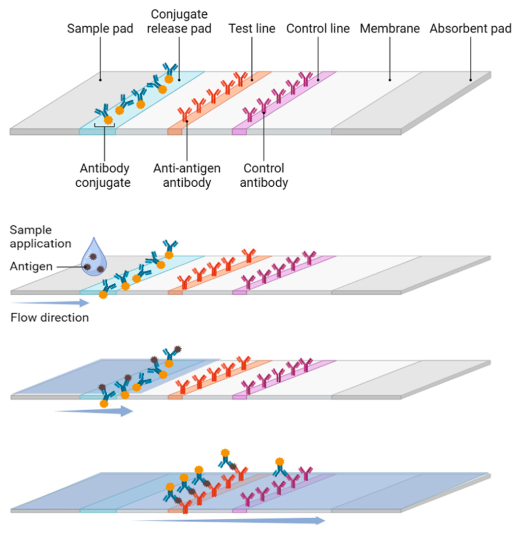
Figure 2.
Schematic representation of diagnostic result for a disease.
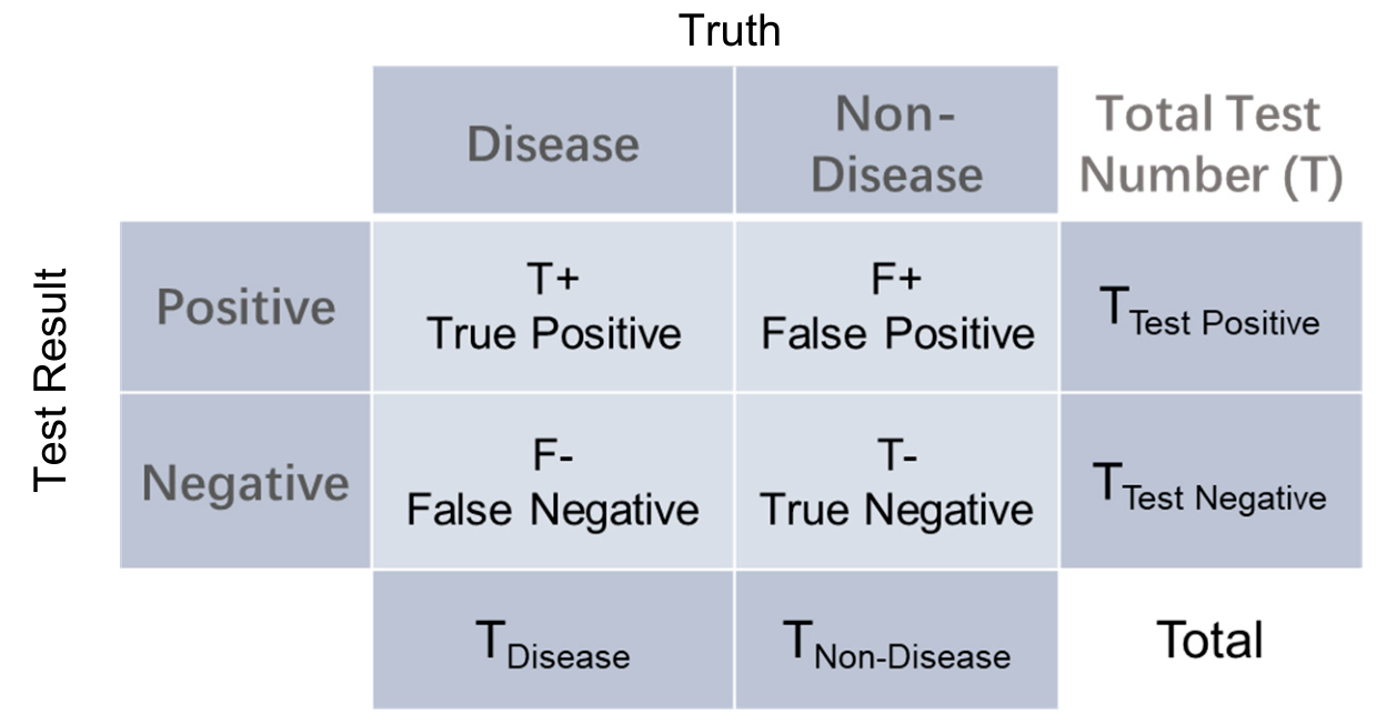
Figure 3.
(a) Multiplex channel layout on Unisart StructSure®. (b) Flow dynamics of Unisart StructSure® membranes with curved narrow channels. (c) Comparison assay with Unisart StructSure® and non-structured membrane, with spot intensities of 0.5, 2, 10 ng/ml Procalcitonin (PCT). Reproduced with permission from ref [39].
Figure 3.
(a) Multiplex channel layout on Unisart StructSure®. (b) Flow dynamics of Unisart StructSure® membranes with curved narrow channels. (c) Comparison assay with Unisart StructSure® and non-structured membrane, with spot intensities of 0.5, 2, 10 ng/ml Procalcitonin (PCT). Reproduced with permission from ref [39].
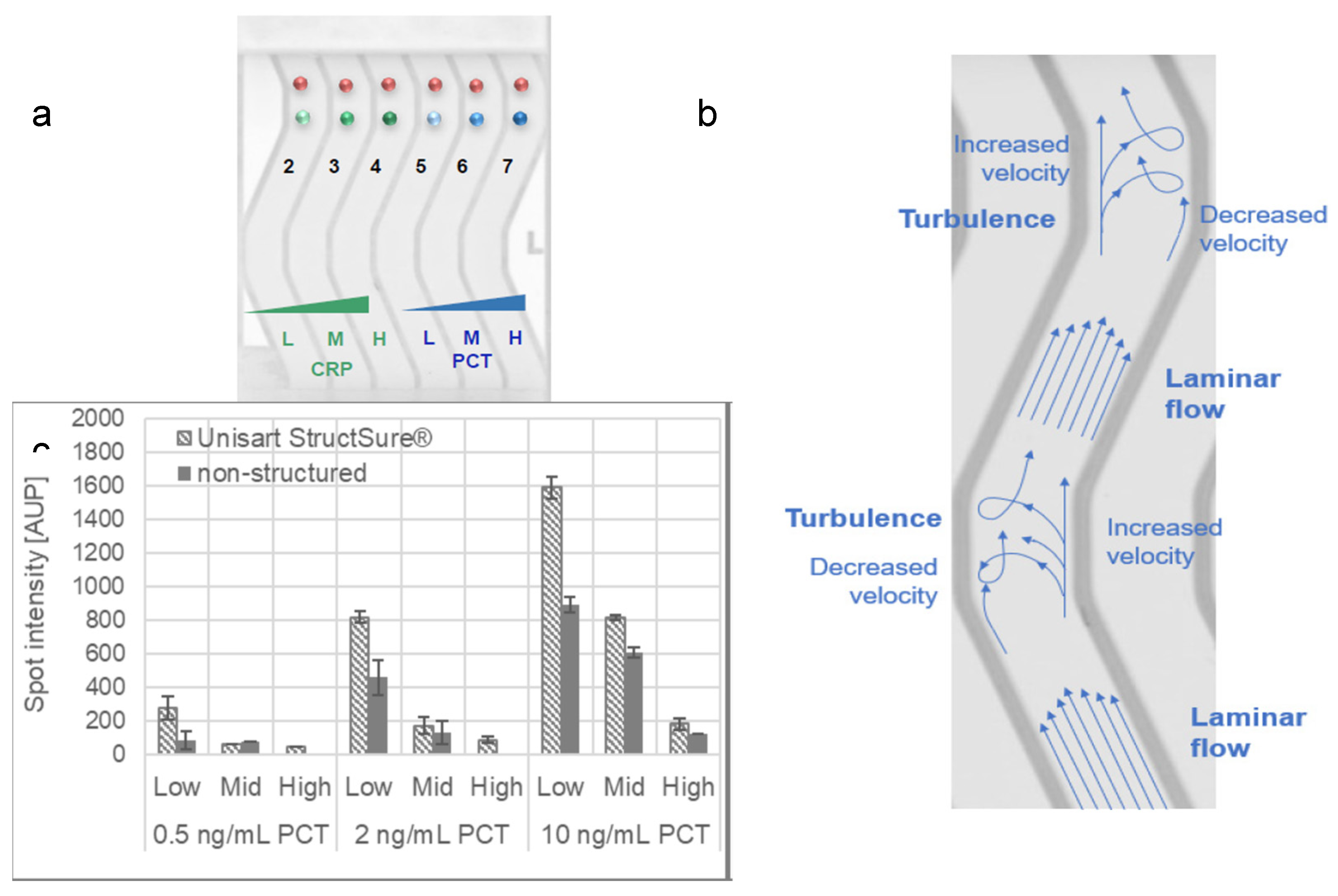
Figure 4.
(A) Layout and design of a rotary lateral flow test device, optimized for the precise delivery of multiple reagents and execution of multistep assays. (B) Elutriation of the top view, highlighting the dynamic transition of the contact zone as the device rotates. (C) Schematics of step-by-step process of strip-based ELISA, encompassing bacteria capture, HRP-IgG labeling, washing, and signal generation steps. Reprint permission from Analytical Chemistry 2016 [40].
Figure 4.
(A) Layout and design of a rotary lateral flow test device, optimized for the precise delivery of multiple reagents and execution of multistep assays. (B) Elutriation of the top view, highlighting the dynamic transition of the contact zone as the device rotates. (C) Schematics of step-by-step process of strip-based ELISA, encompassing bacteria capture, HRP-IgG labeling, washing, and signal generation steps. Reprint permission from Analytical Chemistry 2016 [40].
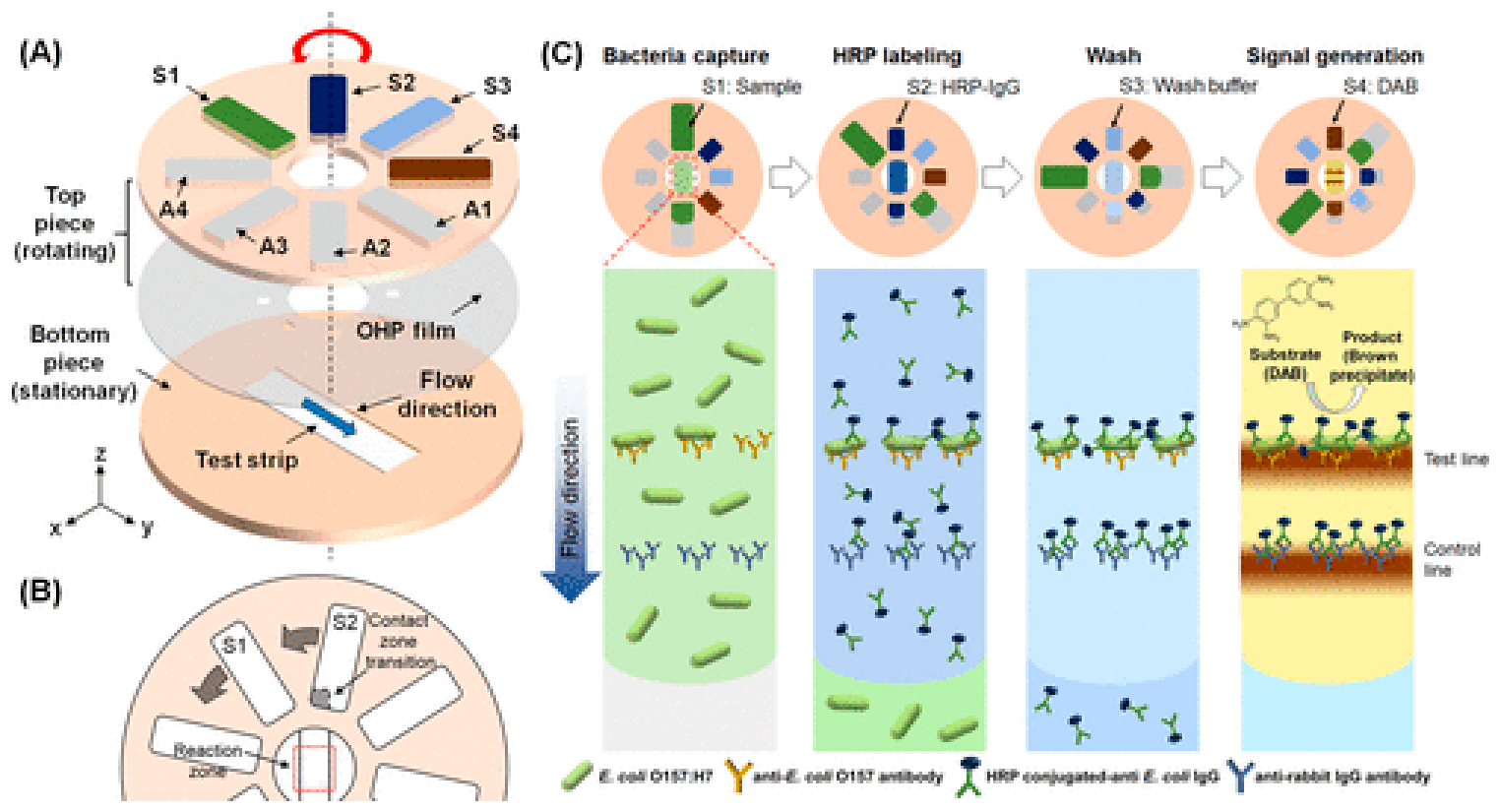
Figure 5.
Principles of preparation and specific identification of analytes for the three kinds of LMs-Abs probes, structure of rainbow latex microspheres lateral flow immunoassay (LMs-LFIA) and detection principles of test strips, and qualitative and quantitative test results. (A) The method to prepare color beads tags; (B) (C) The principle and structure of test strips; (D) The qualitative and quantitative results for the test of analytes. Reprinted (adapted) with permission from ref [70]. Copyright 2022 Analytica Chimica Acta.
Figure 5.
Principles of preparation and specific identification of analytes for the three kinds of LMs-Abs probes, structure of rainbow latex microspheres lateral flow immunoassay (LMs-LFIA) and detection principles of test strips, and qualitative and quantitative test results. (A) The method to prepare color beads tags; (B) (C) The principle and structure of test strips; (D) The qualitative and quantitative results for the test of analytes. Reprinted (adapted) with permission from ref [70]. Copyright 2022 Analytica Chimica Acta.
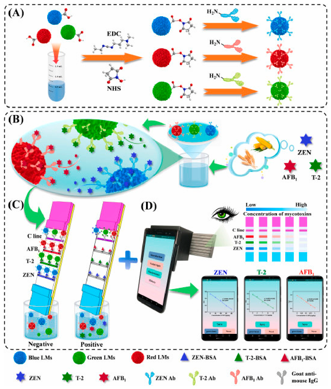
Figure 6.
(a) proposed LFIA biosensor for multiplex and ultrasensitive monitoring of EBOV, MPXV, and SARS-CoV-2 antigens. Reproduced with permission from ref [94]. Copyright 2024 ACS Nano. (b) Schematic Illustration of the Structure and Detection Principle of the Single-Line LFA Strip and Circular Flow Assay Strategy Based on the Single Test-Line Strips for Multiple Detections, inset figure depicts the variations of intensity ratios (IG/IR) between the green emission on an 808 nm excitation and the red emission on a 980 nm excitation on the T line after samples with different amounts of analytes flowing across. Reproduced with permission from ref [96]. Copyright 2021 Analytical Chemistry.
Figure 6.
(a) proposed LFIA biosensor for multiplex and ultrasensitive monitoring of EBOV, MPXV, and SARS-CoV-2 antigens. Reproduced with permission from ref [94]. Copyright 2024 ACS Nano. (b) Schematic Illustration of the Structure and Detection Principle of the Single-Line LFA Strip and Circular Flow Assay Strategy Based on the Single Test-Line Strips for Multiple Detections, inset figure depicts the variations of intensity ratios (IG/IR) between the green emission on an 808 nm excitation and the red emission on a 980 nm excitation on the T line after samples with different amounts of analytes flowing across. Reproduced with permission from ref [96]. Copyright 2021 Analytical Chemistry.
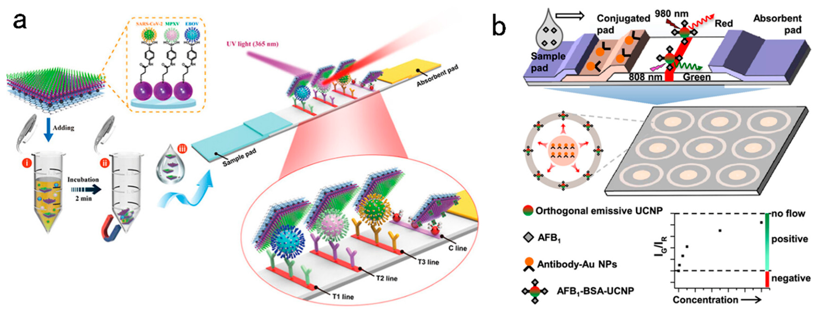
Figure 7.
Comparison of performance for test strip with color beads and fluorescent beads as tracers. (a, b) detection of rotavirus and adenovirus. Reprinted with permission from ref [69]. Copyright 2018 Virology Journal [69,93]. (c, d) detection of influenza. Reproduced with permission from ref [69]. Copyright 2019 Virology Journal.
Figure 7.
Comparison of performance for test strip with color beads and fluorescent beads as tracers. (a, b) detection of rotavirus and adenovirus. Reprinted with permission from ref [69]. Copyright 2018 Virology Journal [69,93]. (c, d) detection of influenza. Reproduced with permission from ref [69]. Copyright 2019 Virology Journal.
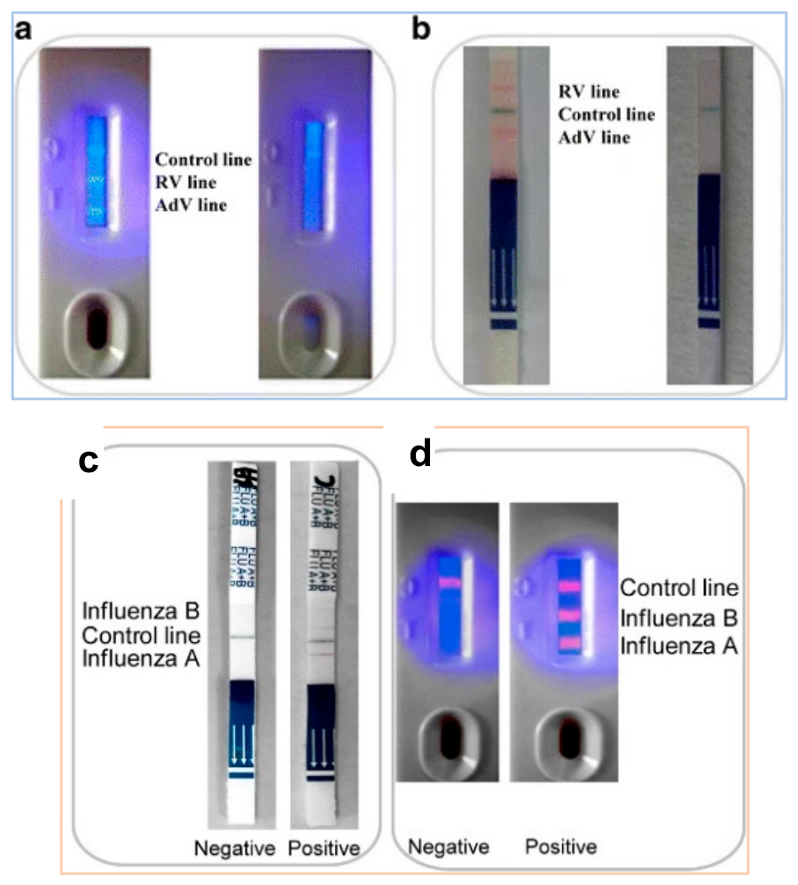
Figure 8.
(a) Schematic diagram of SARS-CoV-2 recognition of ACE2 receptor and schematic diagram of ACE2-based composition and principle, the ACE2 receptor is positioned on the test line composed of a nitrocellulose membrane, while the control line is immobilized with Anti-IgG antibody. Reproduced with permission from ref [110]. Copyright 2021 Biosens Bioelectron. (b) Schematic of the CHA-LFIA method for SARS-CoV-2 viral RNA detection, including the CHA reaction without and with the target RNA and a schematic diagram of the LFIA strip used for detecting digoxigenin-biotin labeled H1-H2 hybrid double stranded complex. Reproduced with permission from ref [112]. Copyright 2021 Actuators B: Chemical.
Figure 8.
(a) Schematic diagram of SARS-CoV-2 recognition of ACE2 receptor and schematic diagram of ACE2-based composition and principle, the ACE2 receptor is positioned on the test line composed of a nitrocellulose membrane, while the control line is immobilized with Anti-IgG antibody. Reproduced with permission from ref [110]. Copyright 2021 Biosens Bioelectron. (b) Schematic of the CHA-LFIA method for SARS-CoV-2 viral RNA detection, including the CHA reaction without and with the target RNA and a schematic diagram of the LFIA strip used for detecting digoxigenin-biotin labeled H1-H2 hybrid double stranded complex. Reproduced with permission from ref [112]. Copyright 2021 Actuators B: Chemical.
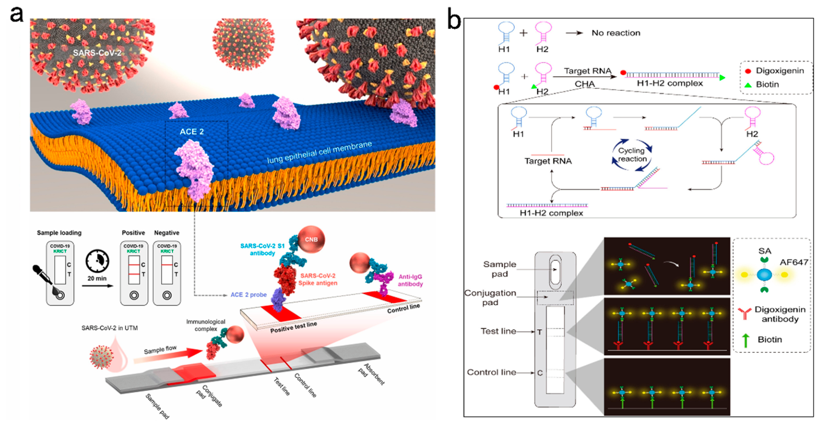
Figure 9.
(a) Schematic of the configuration and the detection mechanism of AIEgen-based test strips. (b) Schematic diagram of an AIEgen-based wearable lateral flow strip for the rapid detection of SARS-CoV-2 RBD protein and N protein. Reproduced with permission from ref [114]. Copyright 2022 Cell Reports Physical Science. (c) Schematic illustration of test strips for the detection of the NIR-emissive AIE nanoparticle-labeled lateral flow immunoassay for detection of IgM and IgG in human serum samples. Reproduced with permission from ref [115]. Copyright 2021 ACS Nano.
Figure 9.
(a) Schematic of the configuration and the detection mechanism of AIEgen-based test strips. (b) Schematic diagram of an AIEgen-based wearable lateral flow strip for the rapid detection of SARS-CoV-2 RBD protein and N protein. Reproduced with permission from ref [114]. Copyright 2022 Cell Reports Physical Science. (c) Schematic illustration of test strips for the detection of the NIR-emissive AIE nanoparticle-labeled lateral flow immunoassay for detection of IgM and IgG in human serum samples. Reproduced with permission from ref [115]. Copyright 2021 ACS Nano.
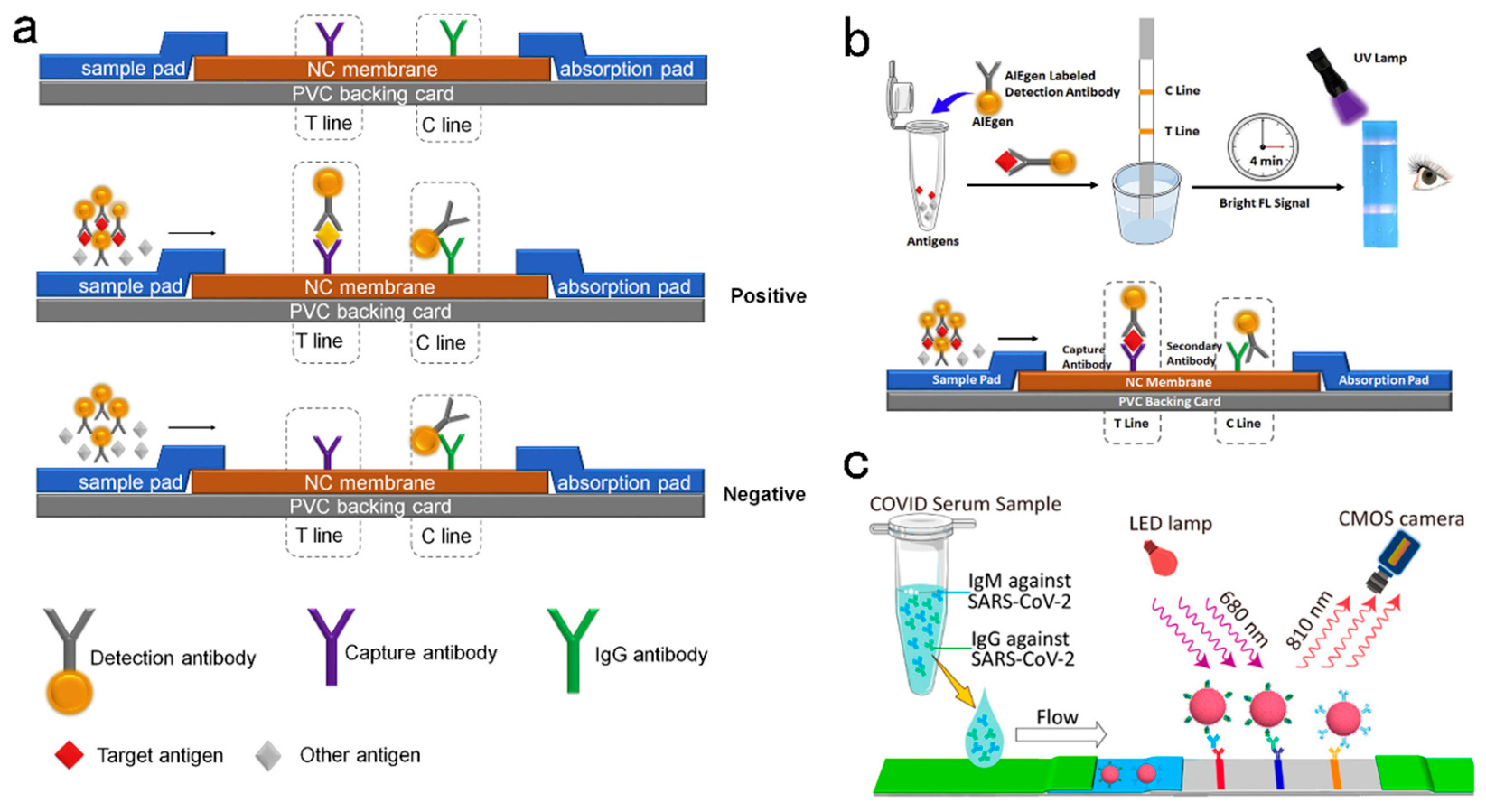
Figure 10.
Schematic illustration of LFA design in the diagnosis of influenza A virus strains H1N1 and H3N2. (a) An overview of the components and (b) procedures for preparation of the carbon nanotag-antibody immunoprobes, and (c) the fabrication of carbon nanotag based lateral flow immunoassay. Reproduced with permission from ref [119]. Copyright 2017 Microchimica Acta.
Figure 10.
Schematic illustration of LFA design in the diagnosis of influenza A virus strains H1N1 and H3N2. (a) An overview of the components and (b) procedures for preparation of the carbon nanotag-antibody immunoprobes, and (c) the fabrication of carbon nanotag based lateral flow immunoassay. Reproduced with permission from ref [119]. Copyright 2017 Microchimica Acta.
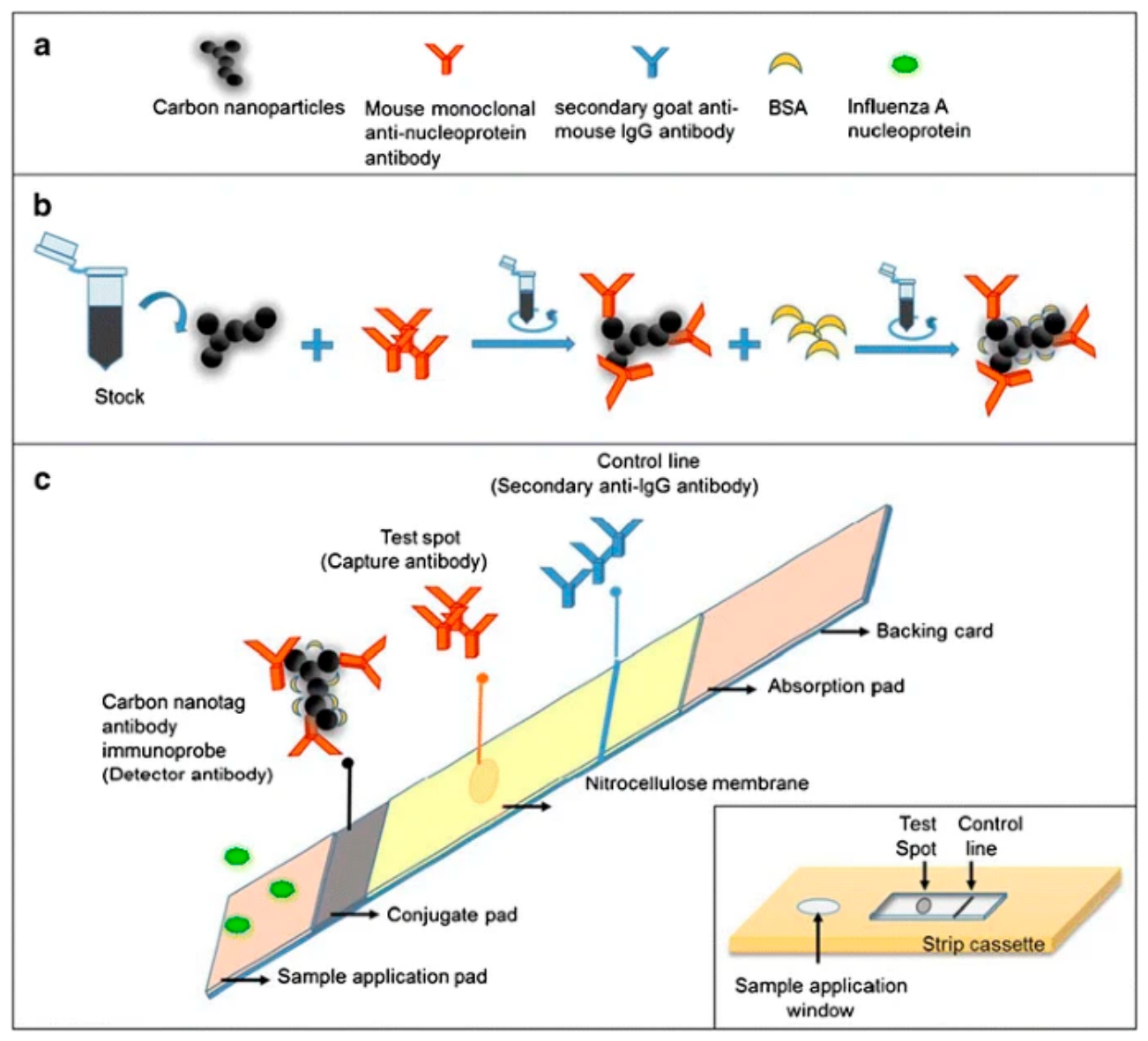
Figure 11.
Schematic depiction of the CM-EUs-based lateral flow assay. The recombinant HBcAg is immobilized on the test line, whereas the anti-RIgG is positioned on the control line. In the assay, the anti-HBcAg monoclonal antibody competes with the anti-HBc present in the sample to bind with the immobilized protein. Quantitative detection is achieved by analyzing the fluorescence signal recorded using a portable TRF strip reader, enabling accurate and rapid results. Reproduced with permission from ref [126].
Figure 11.
Schematic depiction of the CM-EUs-based lateral flow assay. The recombinant HBcAg is immobilized on the test line, whereas the anti-RIgG is positioned on the control line. In the assay, the anti-HBcAg monoclonal antibody competes with the anti-HBc present in the sample to bind with the immobilized protein. Quantitative detection is achieved by analyzing the fluorescence signal recorded using a portable TRF strip reader, enabling accurate and rapid results. Reproduced with permission from ref [126].
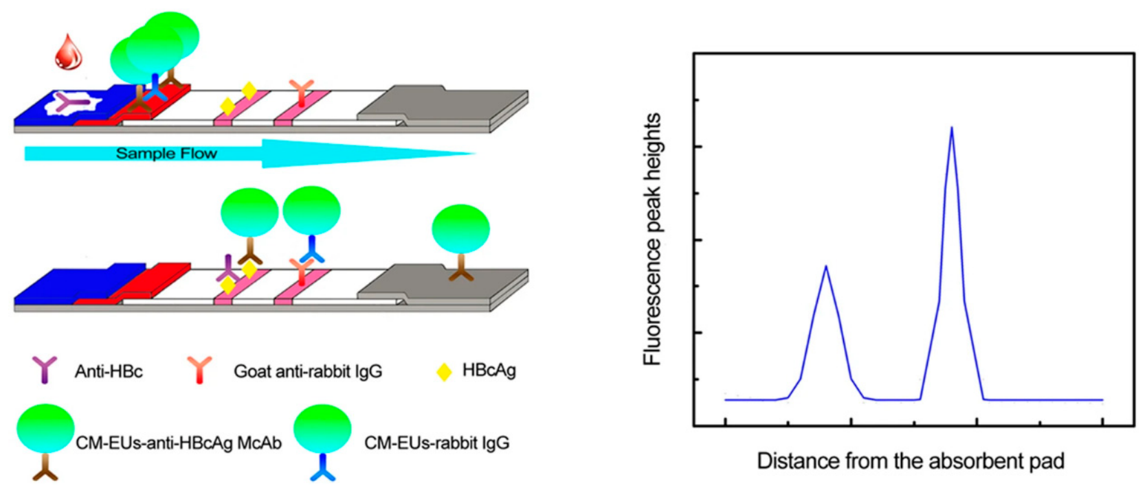
Figure 12.
Schematic showing of HCV Ag-Ab Combo test strip. Reproduced with permission from ref [131].
Figure 12.
Schematic showing of HCV Ag-Ab Combo test strip. Reproduced with permission from ref [131].
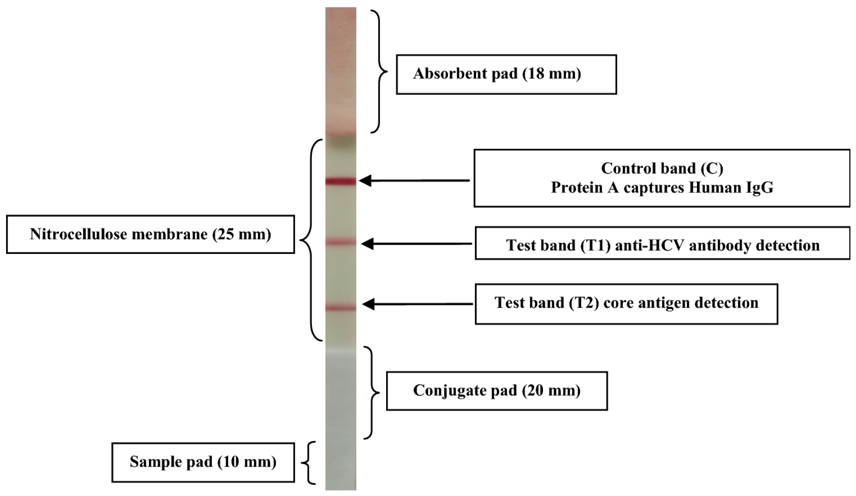
Figure 13.
Schematic illustration of the CuO-based LFT to detect HPV16 DNA. Reproduced with permission from ref [142].
Figure 13.
Schematic illustration of the CuO-based LFT to detect HPV16 DNA. Reproduced with permission from ref [142].
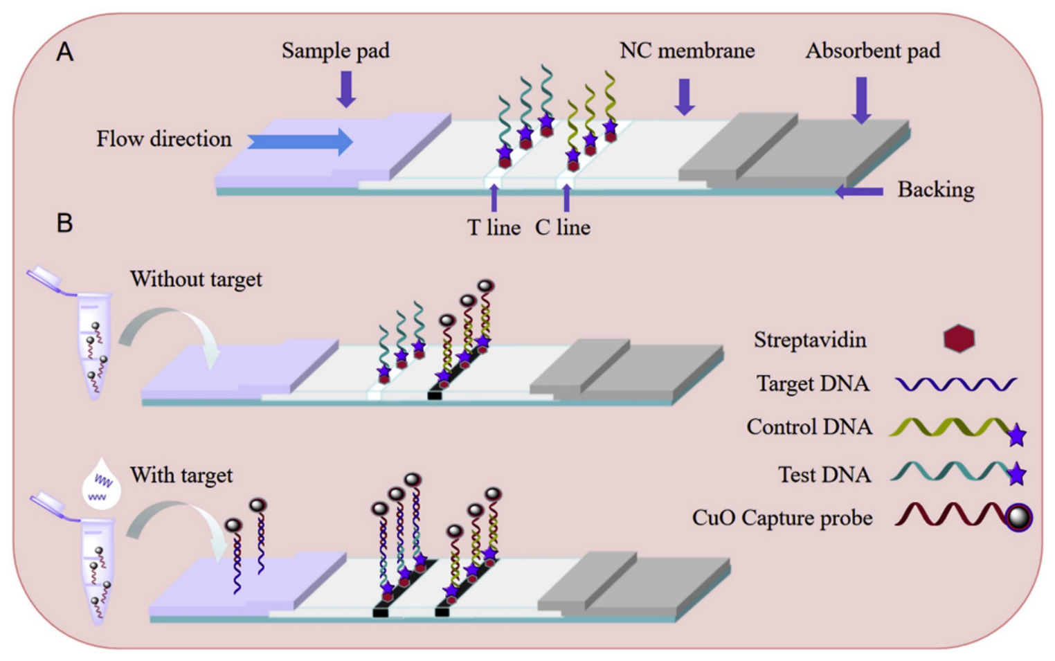
Figure 14.
(A) Standard colloidal gold strip. (B) Nanozyme-strip employing MNPs in place of colloidal gold to form a novel nanozyme probe. The probe with nanozyme activity generates a color reaction with substrates, which significantly enhances the signal so that it can be visualized by the naked-eye. Nanozyme-strip design. Reproduced with permission from [145].
Figure 14.
(A) Standard colloidal gold strip. (B) Nanozyme-strip employing MNPs in place of colloidal gold to form a novel nanozyme probe. The probe with nanozyme activity generates a color reaction with substrates, which significantly enhances the signal so that it can be visualized by the naked-eye. Nanozyme-strip design. Reproduced with permission from [145].
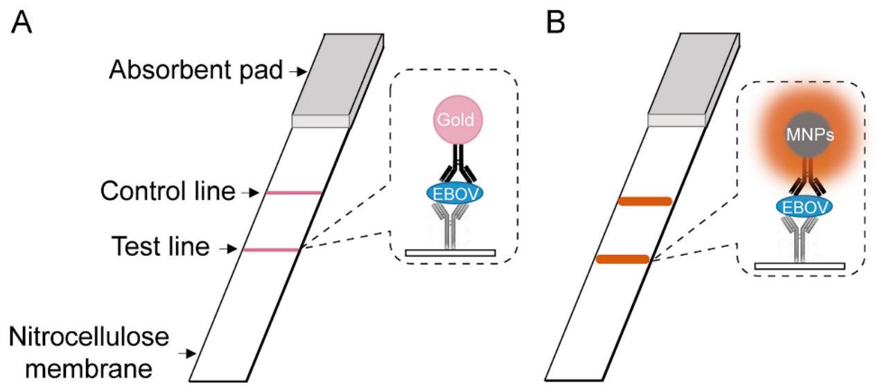
Figure 15.
Design of Lateral Flow Immunoassay for the detection of Ebola with RNs@Au-based nanospheres. Reprint permission obtained from [146].
Figure 15.
Design of Lateral Flow Immunoassay for the detection of Ebola with RNs@Au-based nanospheres. Reprint permission obtained from [146].
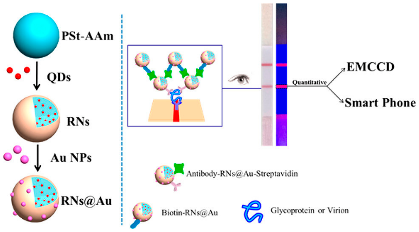
Figure 16.
(a) illustration of Smartphone lateral flow point-of-care test for Ebola virus IgG detection. (b) Lateral flow strip illustration: serum applied onto the sample pad migrates through the analytical area, and subsequently forms complexes between the labeled gold nanoparticles (AuNPs) and the target analytes. Reprint permission obtained from [147].
Figure 16.
(a) illustration of Smartphone lateral flow point-of-care test for Ebola virus IgG detection. (b) Lateral flow strip illustration: serum applied onto the sample pad migrates through the analytical area, and subsequently forms complexes between the labeled gold nanoparticles (AuNPs) and the target analytes. Reprint permission obtained from [147].
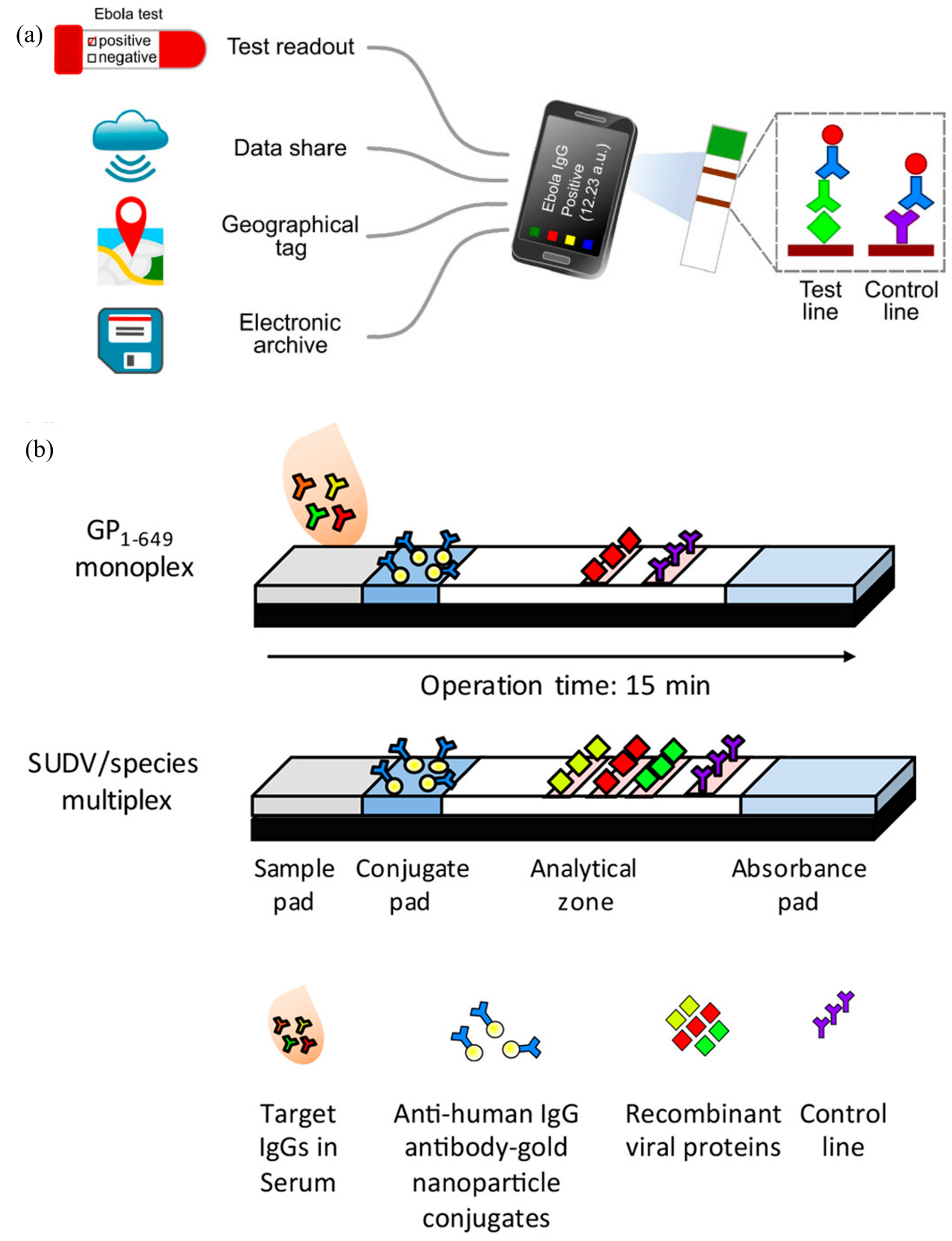
Figure 17.
portable smartphone-based fluorescent LFIA reader for highly sensitive point-of-care detection of ZIKV NS1. Reprint permission from [165].
Figure 17.
portable smartphone-based fluorescent LFIA reader for highly sensitive point-of-care detection of ZIKV NS1. Reprint permission from [165].
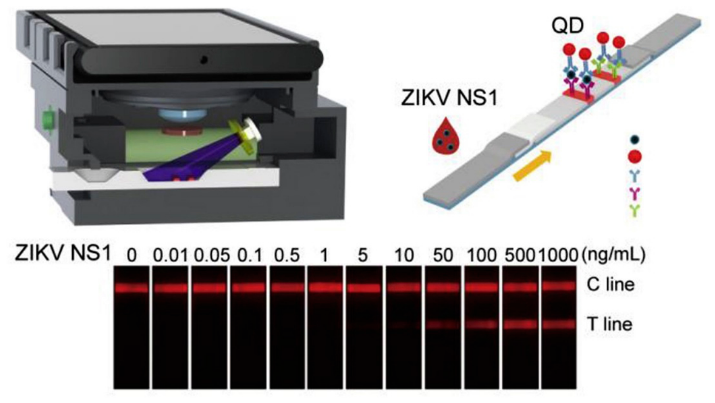
Figure 18.
Schematic illustration of the integrated lateral flow assay platform. (a) Illustration of integrated fluorescent lateral flow assay for multiplex detection of infectious disease (HIV , TP and HCV). (b) Design of disposable strip test cartridge. (c) Image of portable strip reader and test cartridge. Reproduced with permission from ref [173]. Copyright 2021 Sensors and Actuators B: Chemical.
Figure 18.
Schematic illustration of the integrated lateral flow assay platform. (a) Illustration of integrated fluorescent lateral flow assay for multiplex detection of infectious disease (HIV , TP and HCV). (b) Design of disposable strip test cartridge. (c) Image of portable strip reader and test cartridge. Reproduced with permission from ref [173]. Copyright 2021 Sensors and Actuators B: Chemical.
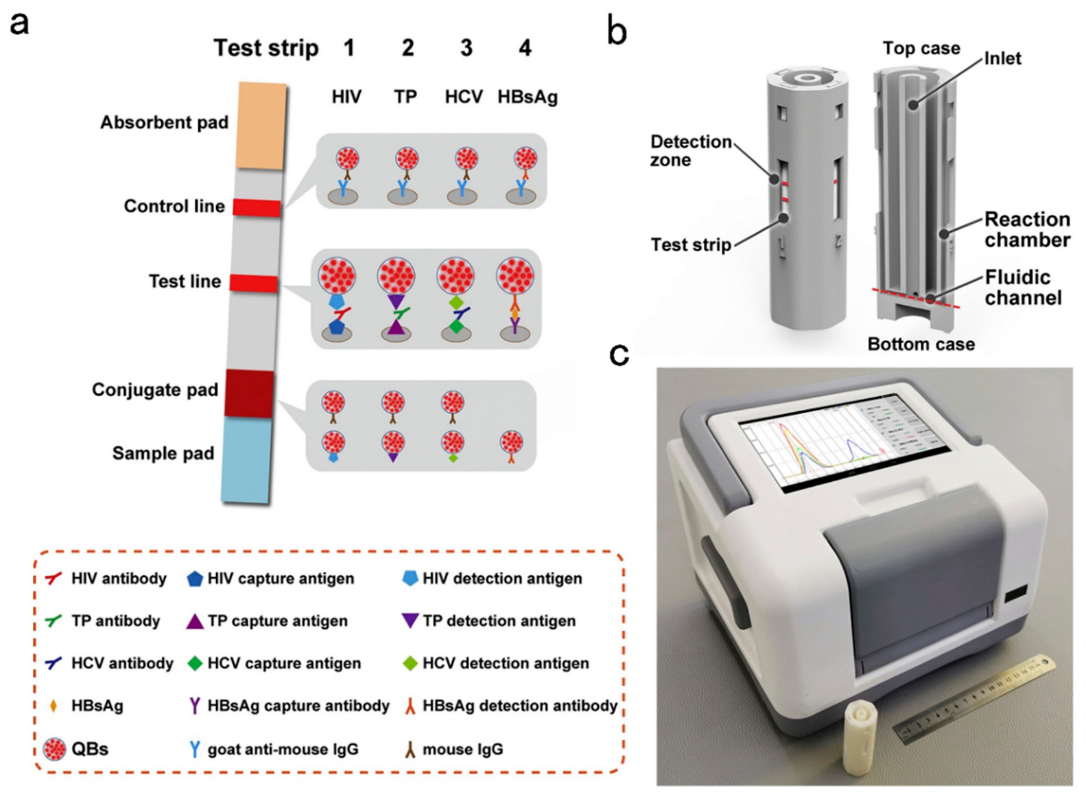
Figure 19.
(a) Schematic of LFIA strip structure and detection principle. Reproduced with permission from ref [183]. Copyright 2020 Journal of Materials Science. (b) Schematic representation of assay principle. L. monocytogenes cells migrate to the conjugation pad region where binding to aptamer gates. (c) schematic representation of the aptamer gated label-free LFA. Reproduced with permission from ref [185]. Copyright 2019 Analytical Biochemistry.
Figure 19.
(a) Schematic of LFIA strip structure and detection principle. Reproduced with permission from ref [183]. Copyright 2020 Journal of Materials Science. (b) Schematic representation of assay principle. L. monocytogenes cells migrate to the conjugation pad region where binding to aptamer gates. (c) schematic representation of the aptamer gated label-free LFA. Reproduced with permission from ref [185]. Copyright 2019 Analytical Biochemistry.
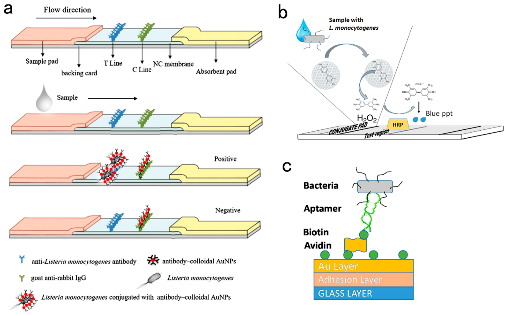
Table 2.
detection targets and labels of LFAs for common pathogenic bacteria.
| Labels or the signal enhancement methods | Recognition element | Read-out way | Reference | |
|---|---|---|---|---|
| Treponema pallidum | Quantum dot bead | TP15 and TP17Antibody | Device | 106 |
| Escherichia coli O157: H7 | Colloidal gold | Antibody | Naked eyes | 108 |
| SERS (gold-shell silica-core (SiO2/Au) nanosphere) | Antibody | Device | 109 | |
| UCNPs (Cucurbit [7] uril-based hydrophilic modified NaYF4:2%Er3+,18%Yb3+@NaYF4) | Antibody | Device | 110 | |
| SERS (DTNB/magnetic Fe3O4/Au-PEI nanoparticles) | Antibody | Device | 111 | |
| Functional nanozyme (mannose modified Prussian blue) | Antibody | Naked eyes/Device | 112 | |
| L. monocytogenes | AEDR-AuNPs | Antibody | Naked eyes | 117 |
| SERS (4-MBA modified AuNPs) | Antibody | Device | 116 | |
| Silica nanoparticles | Aptamer | Naked eyes | 115 |
Disclaimer/Publisher’s Note: The statements, opinions and data contained in all publications are solely those of the individual author(s) and contributor(s) and not of MDPI and/or the editor(s). MDPI and/or the editor(s) disclaim responsibility for any injury to people or property resulting from any ideas, methods, instructions or products referred to in the content. |
© 2026 by the authors. Licensee MDPI, Basel, Switzerland. This article is an open access article distributed under the terms and conditions of the Creative Commons Attribution (CC BY) license (http://creativecommons.org/licenses/by/4.0/).
Copyright: This open access article is published under a Creative Commons CC BY 4.0 license, which permit the free download, distribution, and reuse, provided that the author and preprint are cited in any reuse.