

Submitted:
22 September 2026
Posted:
23 September 2026
You are already at the latest version
Abstract
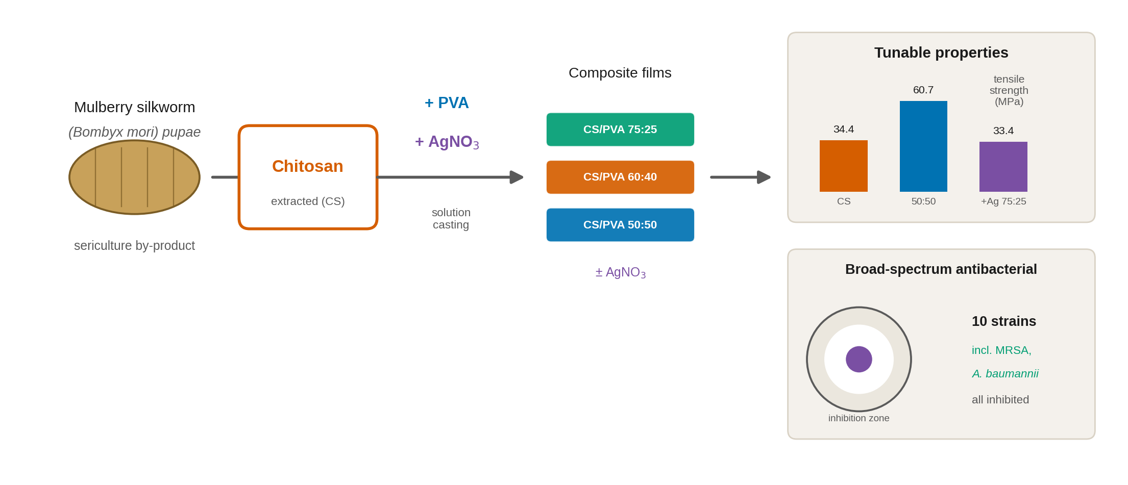
Chitosan-based films are attracting attention as renewable antibacterial materials, but chitosan from crustacean sources remains costly, and insect-derived alternatives are little explored. This study examined how the polymer ratio and silver nitrate (AgNO₃) incorporation govern the properties of films prepared from chitosan (CS) extracted from mulberry silkworm (Bombyx mori) pupae, an abundant by-product of the sericulture industry in Uzbekistan. Films were cast from CS and poly(vinyl alcohol) (PVA) solutions at volume ratios of 75:25, 60:40, and 50:50, with and without AgNO₃, and characterized by infrared spectroscopy, X-ray diffraction, scanning electron microscopy with elemental analysis, and thermal analysis, together with mechanical, swelling, acid-stability, and water vapor transmission measurements. Antibacterial activity was assessed by agar diffusion against ten reference and clinical bacterial strains. The polymer matrix was preserved in all compositions, while the ratio and AgNO₃ modified the intermolecular interactions and structural organization. The CS/PVA 50:50 film reached the highest tensile strength (60.7 ± 7.8 MPa) and the lowest water vapor transmission among the binary films, and AgNO₃ increased the char residue, reduced swelling, and extended the antibacterial spectrum to all ten strains, including methicillin-resistant Staphylococcus aureus (MRSA) and Acinetobacter baumannii. Film properties can therefore be tuned through composition, supporting the development of silkworm-derived chitosan films as antibacterial polymeric materials.
Keywords:
chitosan
; Bombyx mori
; bio-based materials
; poly(vinyl alcohol)
; silver nitrate
; composite films
; structural characterization
; antibacterial activity
; Staphylococcus aureus
; water vapor transmission rate
1. Introduction
The natural polymer chitosan (CS) has attracted considerable interest as a promising material for the development of environmentally friendly and biodegradable polymeric materials due to its biocompatibility, biodegradability, low toxicity, and film-forming ability [1,2,3,4]. Its solubility in dilute aqueous organic acids, particularly acetic acid, facilitates its processing into film materials [2]. The physicochemical, structural, and barrier properties of CS largely depend on its source of origin [5,6,7,8].
Although chitin and CS are renewable biopolymers, their industrial application is limited by the high production cost and the specific extraction procedures required for crustacean-derived materials. Alternative sources, particularly insects, are therefore of increasing interest as environmentally sustainable raw materials for CS production [9]. Mulberry silkworm (Bombyx mori) pupae represent a promising alternative source because they are generated in large quantities as a by-product of the sericulture industry and contain chitinous exoskeletal components. In Uzbekistan, approximately 15,000 tons of mulberry silkworm pupae are generated annually, and the planned increase of cocoon production to approximately 30,000 tons per year further highlights the potential of this secondary biological resource for producing valuable biopolymers and functional materials.
Previous studies have demonstrated the feasibility of obtaining CS from silkworm pupae by chemical extraction, with the resulting materials showing good solubility in dilute acids [10]. Silkworm-pupa-derived CS has also demonstrated high antimicrobial activity and good cytocompatibility, highlighting its potential for food, medical, and biotechnology applications [11,12]. However, CS-based films generally exhibit relatively low water vapor barrier properties [13,14,15,16]. Blending CS with synthetic polymers can improve the mechanical, hydrophilic, and biocompatibility characteristics of the resulting materials compared with the individual components [17].
Polyvinyl alcohol (PVA), owing to its excellent film-forming ability, is a promising component for CS-based composite films. CS/PVA blending enables the regulation of structural, physicomechanical, sorption, and barrier properties through intermolecular interactions and variation of the component ratio. Functionalization with natural extracts, essential oils, and nanomaterials further expands their potential applications, particularly in food packaging and food-quality preservation [18]. Increasing the CS content can reduce crystallinity and elongation while increasing rigidity, fracture resistance, and thermal stability and reducing ultraviolet transmittance [19]. The well-established antibacterial activity of CS further supports its use in functional antimicrobial films [20,21,22,23].
Additional modification of CS/PVA films can provide targeted functional improvements. The incorporation of AgNO₃ is of particular interest for enhancing antimicrobial activity [24], while glycerol plasticization improves flexibility and transparency and enhances water vapor and oxygen barrier properties [25]. Plasticization with a choline chloride/oxalic acid deep eutectic solvent can reduce the environmental impact of coatings and promote agricultural crop growth [26]. Incorporation of chitosan oligosaccharides and gallic acid produces synergistic effects, reducing moisture content, solubility, swelling, and water vapor permeability while improving mechanical, structural, thermal, and UV-barrier properties [27]. Similarly, Pickering emulsions can impart pH-responsive, antibacterial, and sorption properties to CS/PVA films [28], whereas cashew nut extract improves their mechanical, thermal, and antioxidant characteristics and promotes a denser structure [29].
Nanocomposite modifications have also been reported to enhance the functional performance of CS/PVA films. Quaternary ammonium lignin/ZnO nanoparticles increase tensile strength and provide antioxidant and antibacterial activity against Staphylococcus aureus and Escherichia coli [30]. Grape skin anthocyanins combined with selenium nanoparticles provide pronounced antioxidant and antimicrobial properties [31]. Zinc oxide nanoparticles and tannic acid improve mechanical, thermal, barrier, antioxidant, antimicrobial, and UV-blocking properties [32]. In addition, CS/PVA films have demonstrated sorption toward organic dyes, indicating their potential for selective pollutant removal in water treatment and environmental monitoring [33].
CS/PVA films, including CS/PVA membranes containing silver nitrate, have thus already been described [24], and the incorporation of AgNO₃ into a CS/PVA matrix is not in itself new. What remains poorly established is how such systems behave when the chitosan comes from an insect rather than a crustacean source, and how the polymer ratio and the silver component jointly shape the full set of film properties. Previous CS/PVA–silver studies have typically used crustacean-derived chitosan, examined one or two compositions, and characterized a limited subset of properties, most often against a small number of standard bacterial strains. A systematic account that connects composition to structure, thermal and mechanical behavior, water-related properties, and antibacterial performance within a single sample set is still lacking for silkworm-derived chitosan.
The contribution of this study lies in that combination. Films were prepared from chitosan obtained from mulberry silkworm (Bombyx mori) pupae, an underused by-product of the sericulture industry, at three systematically varied CS/PVA solution ratios, with and without AgNO₃. The same sample set was characterized by Fourier transform infrared (FTIR) spectroscopy, X-ray diffraction (XRD), scanning electron microscopy with energy-dispersive X-ray spectroscopy (SEM/EDS), and thermogravimetric and differential thermal analysis (TGA/DTA), and evaluated for mechanical properties, swelling, stability in an acidic medium, and water vapor transmission, and its antibacterial activity was screened against ten reference and clinical strains, including methicillin-resistant Staphylococcus aureus (MRSA) and Acinetobacter baumannii. The specific aim was to determine how the CS/PVA ratio and AgNO₃ incorporation, separately and together, govern the structural organization, physicochemical and mechanical properties, and antibacterial spectrum of films based on silkworm-pupae-derived chitosan.
2. Materials and Methods
2.1. Materials
Chitosan (Mw = 187 kDa; degree of deacetylation, DD = 91%), obtained from chitin isolated from mulberry silkworm (Bombyx mori) pupae in Uzbekistan, was used as the film-forming polymer. Polyvinyl alcohol (PVA), grade V (CAS No. 9002-89-5; average molecular weight ≈ 75 kDa; degree of hydrolysis = 99.27 mol%; viscosity = 30.0 mPa·s), was used as the second polymer. Acetic acid solutions (Beijing Chemical Works, Beijing, China) and distilled water were used to prepare the CS and PVA solutions, respectively. Glycerol (Sigma-Aldrich, G5516; ≥99%) and silver nitrate (AgNO₃) were used as plasticizer and modifying agent, respectively. All reagents were of analytical grade and used without further purification.
2.2. Preparation of CS/PVA Films
CS/PVA composite films were prepared by the solution-casting method according to [34], with minor modifications. Briefly, 1.0 g of chitosan was dissolved in 100 mL of 1% acetic acid under continuous magnetic stirring until homogeneous. PVA (3.0 g) was separately dissolved in 100 mL of distilled water under heating and stirring until complete dissolution.
After cooling to room temperature, the CS and PVA solutions were combined at the predetermined volume ratios to a base volume of 10 mL, and 0.05 mL of glycerol was added to each formulation as a plasticizer. The film compositions are designated throughout this work by the volume ratios of the CS and PVA solutions used for mixing (75:25, 60:40, and 50:50). Because the two solutions differed in polymer concentration (1% and 3% w/v, respectively), these designations refer to the solution volume ratios rather than to the polymer mass ratios in the dried films; the corresponding calculated nominal dry-polymer ratios are given in Table 1. For the silver-containing films, 0.5 mL of 1% (w/v) AgNO₃ solution was added to the mixture, corresponding to approximately 5 mg of AgNO₃ per film. As an example, the CS/PVA/AgNO₃ 75:25 casting mixture comprised 7.5 mL of the CS solution, 2.5 mL of the PVA solution, 0.5 mL of the 1% AgNO₃ solution, and 0.05 mL of glycerol. Each mixture was stirred on a magnetic stirrer at 600 rpm for 30 min, cast into 9 cm diameter Petri dishes (approximately 10 mL per dish), and dried at room temperature for 48 h. Eight formulations were obtained in total: CS, PVA, CS/PVA 75:25, 60:40, and 50:50, and the three corresponding AgNO₃-containing composites (Table 1).
The dried films were immersed in 200 mL of 0.25 M NaOH solution to neutralize residual acetic acid, washed successively with distilled water until neutral pH, and dried at 50 °C.
2.3. Scanning Electron Microscopy and Energy-Dispersive X-Ray Spectroscopy
The surface morphology of the prepared films was analyzed by scanning electron microscopy (SEM) using a JEOL IT210 instrument (JEOL, Tokyo, Japan). Samples were mounted on a metallic holder with conductive carbon tape and examined without additional coating under high-vacuum conditions using a secondary electron detector. SEM images were acquired at an accelerating voltage of 10.0 kV and a working distance of 10.7–11.0 mm at magnifications of ×1000, ×3000, ×6000, and ×10,000. Elemental composition was determined by energy-dispersive X-ray spectroscopy (EDS) integrated into the SEM system, using standardless ZAF quantification.
2.4. Fourier Transform Infrared Spectroscopy
The chemical structure and functional groups of the composite films were analyzed by Fourier transform infrared spectroscopy (FTIR) according to [35], with modifications, using an IRSpirit-T spectrometer (Shimadzu, Kyoto, Japan). Spectra were recorded in transmittance mode over 4000–400 cm⁻¹ at a resolution of 4 cm⁻¹, with five scans per sample. The spectra were used to identify characteristic functional groups and evaluate intermolecular interactions among CS, PVA, and AgNO₃.
2.5. X-Ray Diffraction Analysis
X-ray diffraction (XRD) analysis was performed to evaluate the structural and crystalline properties of the films using a MiniFlex 300/600 diffractometer (Rigaku, Tokyo, Japan) equipped with a D/teX Ultra2 detector. Measurements were conducted in θ/2θ geometry at 40 kV and 15 mA.
Diffraction patterns of CS, PVA, and their composite films were recorded over 2θ = 5–60° with a step size of 0.02° and a scanning rate of 10° min⁻¹. Of the AgNO₃-containing films, the 75:25 composition was selected for XRD analysis because it had the highest chitosan proportion among the composites; comparing it with its silver-free CS/PVA 75:25 counterpart allowed the effect of AgNO₃ on the film structure to be assessed at a fixed polymer ratio. Its pattern was recorded over an extended range of 5–80° with a step size of 0.01° at the same scanning rate. The patterns were processed using the instrument software, including peak identification and fitting with the split pseudo-Voigt function. The analysis was performed according to [36], with modifications to the experimental conditions.
2.6. Thermogravimetric and Differential Thermal Analysis
The thermal behavior of the films was investigated by simultaneous thermogravimetric and differential thermal analysis (TGA/DTA) using a DTG-60 thermal analyzer (Shimadzu, Kyoto, Japan), based on previously reported methods [37,43]. Samples of 1.2–6.8 mg were placed in aluminum pans and heated from 25 to 600 °C at a rate of 11 °C min⁻¹ under an argon atmosphere with a gas flow of 80 mL min⁻¹. The thermograms were used to evaluate thermal stability, the individual decomposition stages, and the associated thermal effects. For comparison among samples of different mass, the DTA signal was normalized to the initial sample mass.
2.7. Determination of the Physicomechanical Properties of Films
The mechanical properties of the prepared films were evaluated by uniaxial tensile testing according to previously reported procedures [38], with modifications, using a Shimadzu AG-Xplus universal testing machine (Shimadzu, Kyoto, Japan) controlled by TRAPEZIUM X software. Film specimens (100 × 10 mm) were tested at a crosshead speed of 10 mm min⁻¹, with four specimens analyzed for each formulation. Tensile strength, Young’s modulus, and elongation at break were determined from the stress–strain curves.
Film thickness was measured at five locations using a digital micrometer (MKTs-25; 0–25 mm range; 0.001 mm resolution), and the mean value was used for mechanical-property calculations [38].
2.8. Determination of the Swelling Degree of Films
The swelling degree (SD) of the films was determined gravimetrically according to previously reported methods [39], with modifications. Film samples (approximately 50 mg) were dried to constant weight, weighed (m₀), and immersed completely in distilled water at 25 °C for 24 h. After immersion, excess surface water was gently removed with filter paper, and the swollen samples were immediately weighed (mₛ).
The swelling degree was calculated using Equation (1):
where mₛ is the mass of the swollen film (g) and m₀ is the initial dry mass (g). Measurements were performed on three independent replicates (n = 3), and the results were expressed as mean ± standard deviation.
SD (%) = [(mₛ − m₀) / m₀] × 100
2.9. Determination of Film Stability in an Acidic Aqueous Medium
The stability of the films in an acidic aqueous medium was evaluated gravimetrically from the mass loss after exposure, according to previously reported methods [39], with modifications. Film samples (approximately 20 mg) were dried at 50 °C to constant weight and weighed (m₀). Each sample was immersed in 2 mL of 0.1 M NaCl solution adjusted to pH 1.6 with HCl and maintained at 25 °C for 24 h. After exposure, the samples were dried at 50 °C for 1 h and reweighed (m₁).
Mass loss (ML, %) was calculated using Equation (2):
where m₀ is the initial dry mass and m₁ is the mass after exposure to the acidic medium (g). Three independent replicates were analyzed (n = 3). Lower mass loss indicates greater film stability under the investigated conditions.
ML (%) = [(m₀ − m₁) / m₀] × 100
2.10. Determination of the Water Vapor Transmission Rate
The water vapor transmission rate (WVTR) was determined by the gravimetric dry-cup method according to ASTM E96, with modifications [40]. Circular film specimens (exposed diameter: 5 cm) were sealed over Petri dishes containing 10 g of anhydrous CaCl₂ and placed in a desiccator at 25 ± 2 °C and 75 ± 5% relative humidity maintained with saturated NaCl solution. After 72 h, the mass increase (Δm = m₁ − m₀) was recorded. The exposed film area was 0.00196 m², and WVTR was calculated using Equation (3):
where Δm is the mass increase (g), A is the exposed film area (m²), and t is the exposure time (h). Measurements were performed on three independent specimens (n = 3), and the results were expressed as mean ± standard deviation.
WVTR = Δm/(A × t)
2.11. Determination of the Antibacterial Activity of Films
The antibacterial activity of CS, PVA, CS/PVA films with different polymer ratios, and AgNO₃-containing composites was evaluated by the agar diffusion method [41,42]. All eight formulations were tested against Acinetobacter baumannii NCTC 13301, Enterobacter cloacae NCTC 13406, Enterococcus faecalis NCTC 13379, Escherichia coli ATCC 25922, Klebsiella oxytoca ATCC 13182, Klebsiella pneumoniae NCTC 13443, Proteus mirabilis ATCC 12453, Salmonella typhimurium NCTC 12023, and isolates of methicillin-resistant Staphylococcus aureus (MRSA) and methicillin-susceptible S. aureus (MSSA). The two S. aureus isolates, together with their methicillin-resistance designations, were supplied by the microbiology laboratory that performed the antibacterial testing. That laboratory has since ceased operation, and its records could not be retrieved; consequently, the strain-collection identifiers of these two isolates and the method used to confirm methicillin resistance are not available, and their MRSA and MSSA designations could not be independently verified in this study.
Fresh 18–24 h cultures were suspended in sterile physiological saline and adjusted to 0.5 McFarland (approximately 1–2 × 10⁸ CFU/mL). The standardized suspensions were uniformly inoculated onto Mueller–Hinton agar. Film samples were cut into 10 mm diameter discs under sterile conditions and placed on the inoculated agar surface with gentle pressure to ensure full contact. Plates were incubated aerobically at 35 ± 2 °C for 18–24 h.
Antibacterial activity was determined by measuring the diameter of the inhibition zone, including the film disc, in two perpendicular directions; the mean value was recorded in millimeters. Absence of a visible inhibition zone was recorded as 0 mm, while growth inhibition directly beneath the film was noted qualitatively when present. The effect of AgNO₃ was evaluated by pairwise comparison of films with identical CS/PVA ratios with and without AgNO₃. CS and PVA films served as controls for the intrinsic antibacterial activity of chitosan and the background effect of the polymer matrix, respectively. A standard antibiotic disc was included as a positive control for each bacterial strain in each experimental series: trimethoprim (5 μg) for A. baumannii; amoxicillin–clavulanic acid (30 μg) for E. cloacae, E. faecalis, E. coli, K. oxytoca, and K. pneumoniae; ampicillin (2 μg) for P. mirabilis and S. typhimurium; and vancomycin (5 μg) for MRSA and MSSA. Because antibiotic discs and polymer-film discs differ in active-agent content, dimensions, and diffusion characteristics, and because no interpretive susceptibility breakpoints were applied, the antibiotic-disc zones served as assay controls rather than as a basis for direct potency comparison with the films.
All experiments were performed in triplicate (n = 3), and results were expressed as mean ± standard deviation. For each strain, differences among the eight formulations were evaluated by one-way analysis of variance (ANOVA), followed by Tukey’s honestly significant difference test for the planned pairwise comparisons between films with and without AgNO₃ at the same polymer ratio. Differences were considered statistically significant at p < 0.05. Statistical analyses were performed with SciPy (version 1.17, Python).
3. Results
3.1. Characterization of the CS/PVA and CS/PVA/AgNO₃ Films
3.1.1. FTIR Analysis
FTIR spectra of CS, PVA, CS/PVA, and AgNO₃-containing CS/PVA films were analyzed to identify characteristic functional groups and assess changes in intermolecular interactions. The main absorption bands are summarized in Table 2, and the spectra are shown in Figure 1.
All investigated films exhibited characteristic absorption bands corresponding to the functional groups of CS and PVA. Their positions varied with the CS/PVA ratio, and the most pronounced changes were observed in the O–H/N–H stretching, amide, and C–O/C–O–C regions. For example, the O–H/N–H band of CS/PVA 50:50 shifted from 3311.23 cm⁻¹ for pure CS to 3273.93 cm⁻¹, indicating changes in the intermolecular environment. The characteristic C–H, amide II, and C–O/C–O–C bands remained detectable in all composite films.
The incorporation of AgNO₃ did not eliminate the characteristic absorption bands of the CS/PVA matrix but changed several band positions. The most noticeable shifts occurred in the O–H/N–H and amide regions, while the principal bands of the polymer matrix remained present in all AgNO₃-containing films. Overall, FTIR analysis confirmed the preservation of the characteristic functional groups of CS and PVA and showed that both the CS/PVA ratio and AgNO₃ incorporation influenced their spectral positions, indicating changes in the intermolecular environment of the composite films.
3.1.2. XRD Analysis
The XRD patterns of pure CS, PVA, CS/PVA films with different ratios, and the CS/PVA/AgNO₃ 75:25 film are presented in Figure 2.
Pure CS exhibited a broad diffraction band at 2θ = 21.3 ± 0.5°, with d = 4.16 ± 0.10 Å and FWHM = 12.4 ± 0.6°, indicating a relatively disordered structure. In contrast, PVA showed a sharper main peak at 2θ = 19.434 ± 0.009° (d = 4.564 ± 0.002 Å; FWHM = 2.42 ± 0.02°), together with weaker reflections at 11.32°, 24.2°, and 40.54°.
The CS/PVA composites retained the main diffraction region at approximately 19–20°, with peak position and width depending on the polymer ratio. The CS/PVA 50:50 film showed a main maximum at 19.450 ± 0.016°, while CS/PVA 60:40 exhibited the narrowest peak (FWHM = 2.3 ± 0.2°). The CS/PVA 75:25 film displayed a less pronounced maximum and a more noticeable amorphous background.
Incorporation of AgNO₃ into the CS/PVA 75:25 film changed the diffraction profile. The main maximum remained at 19.55 ± 0.04°, while an additional broad feature appeared at approximately 23.0 ± 0.3°. No additional narrow and intense reflections were detected over the extended range up to 2θ = 80°; in particular, no reflections attributable to crystalline metallic silver were observed. Overall, the structural characteristics of the films depended on the CS/PVA ratio, the main diffraction region of the polymer matrix was preserved in all compositions, and AgNO₃ incorporation modified the diffraction profile without eliminating the main polymer-matrix peak.
3.1.3. Surface Morphology and Elemental Composition
SEM and EDS were used to investigate the surface morphology and elemental composition of the films (Figure 3). All samples exhibited predominantly dense and continuous surfaces without pronounced macropores or large cracks.
Pure CS and PVA showed relatively homogeneous surfaces, with carbon and oxygen as the predominant elements. The CS/PVA composites remained dense and continuous, although their surface morphology varied with the polymer ratio. The CS/PVA 50:50 film exhibited greater local heterogeneity, whereas the 60:40 and 75:25 films were comparatively more homogeneous.
The incorporation of AgNO₃ preserved the continuous polymer matrix but increased local surface heterogeneity, with dispersed submicron high-contrast features visible at higher magnification. EDS detected C (61.14 ± 1.47 wt%), O (37.88 ± 2.25 wt%), and Ag (0.98 ± 1.47 wt%). The silver signal was identified from the Ag-L emission lines, and elemental mapping showed silver distributed throughout the investigated region with some local variation in intensity (Figure S1). Because the silver content was close to the detection limit of standardless quantification, as reflected in the uncertainty of the measurement, the EDS result is regarded as confirming the presence and distribution of silver rather than providing a precise concentration. Overall, the CS/PVA ratio affected the degree of surface heterogeneity, while AgNO₃ incorporation modified the surface morphology without disrupting the polymer matrix.
3.2. Thermal Behavior of the Films
Figure 4 presents the TGA and DTA curves of pure CS and PVA films, CS/PVA composite films with different ratios, and the corresponding AgNO₃-containing samples. All investigated films exhibited multistage thermal degradation, and the degradation profiles varied with the CS/PVA ratio and AgNO₃ incorporation.
Pure PVA showed a total weight loss of 96.75% and a residual mass of 3.25%, with the main degradation occurring at 212.4–346.2 °C. Pure CS exhibited an initial weight loss of 18.03%, followed by a main degradation stage at 139.8–383.2 °C (73.38% weight loss), leaving 4.36% residual mass.
Blending CS with PVA increased the residual mass to 8.25%, 7.92%, and 10.04% for the 50:50, 60:40, and 75:25 films, respectively. The CS/PVA 75:25 composition thus showed the highest residual mass among the binary films.
AgNO₃ incorporation altered the thermal behavior substantially, reducing the total weight loss to 81.83%, 78.38%, and 75.53% and increasing the residual mass to 18.17%, 21.62%, and 24.47% for the 50:50, 60:40, and 75:25 films, respectively. The CS/PVA/AgNO₃ 75:25 film therefore exhibited the lowest weight loss and the highest residual mass among all investigated samples. Changes in the DTA thermal effects accompanied both the variation of the polymer ratio and the incorporation of AgNO₃. Overall, the thermal behavior depended on both factors, with AgNO₃ producing the most pronounced effect.
3.3. Physical and Mechanical Properties
The tensile strength, Young’s modulus, and elongation at break varied with the CS/PVA ratio and AgNO₃ incorporation (Figure 5).
Pure PVA showed the highest elongation at break (242.47 ± 11.19%), with a tensile strength of 31.91 ± 2.00 MPa and the highest Young’s modulus (77.69 ± 10.69 MPa). CS exhibited slightly higher tensile strength (34.37 ± 3.01 MPa) but much lower elongation (65.16 ± 5.10%) and a Young’s modulus of 46.73 ± 2.53 MPa.
Among the binary composites, CS/PVA 50:50 showed the highest tensile strength (60.71 ± 7.78 MPa), with a Young’s modulus of 35.82 ± 7.02 MPa and elongation of 157.51 ± 11.73%. Increasing the CS solution fraction to 60:40 and 75:25 reduced the tensile strength to 33.92 ± 5.69 and 29.03 ± 2.32 MPa, respectively, while elongation decreased to 99.99 ± 11.67% and 71.62 ± 5.63%.
AgNO₃ incorporation modified the mechanical behavior. The CS/PVA/AgNO₃ 50:50, 60:40, and 75:25 films exhibited tensile strengths of 42.48 ± 2.27, 49.33 ± 2.31, and 33.41 ± 4.21 MPa, respectively. Their Young’s moduli were 32.34 ± 1.31, 36.70 ± 0.91, and 40.96 ± 5.62 MPa, while elongation at break was 136.61 ± 18.08%, 126.00 ± 4.84%, and 59.94 ± 8.08%, respectively. Overall, the mechanical properties were strongly composition dependent. CS/PVA 50:50 provided the highest tensile strength among the binary films, whereas AgNO₃ produced composition-dependent changes in strength, stiffness, and elongation.
3.4. Swelling, Stability in an Acidic Medium, and Water Vapor Transmission
3.4.1. Swelling Degree
The swelling behavior of the films was evaluated after immersion in distilled water for 24 h at 25 °C. The results are presented in Table 3 and Figure 6.
Pure CS and PVA exhibited swelling degrees of 200 ± 10% and 190 ± 2%, respectively. Among the binary composites, CS/PVA 50:50 showed the lowest swelling (180 ± 10%), whereas CS/PVA 60:40 exhibited the highest value (235 ± 10%); increasing the CS solution fraction to 75:25 resulted in a decrease to 220 ± 13%.
AgNO₃ incorporation reduced the swelling of all composite films, to 150 ± 10%, 210 ± 11%, and 207 ± 13% for the 50:50, 60:40, and 75:25 compositions, respectively. Compared with the corresponding AgNO₃-free films, the reductions were 30, 25, and 13 percentage points. The swelling behavior thus showed a nonlinear dependence on the CS/PVA ratio, with the highest value for CS/PVA 60:40 and the lowest for CS/PVA/AgNO₃ 50:50.
3.4.2. Stability in an Acidic Aqueous Medium
The stability of the films in an acidic aqueous medium was evaluated from the mass loss after 24 h of exposure. Pure CS exhibited the highest mass loss among the unmodified films (5%), whereas PVA showed the highest stability (<1%). Incorporation of PVA reduced the mass loss to 2% for CS/PVA 50:50, <2% for 60:40, and <3% for 75:25. AgNO₃ further reduced the mass loss of the 50:50 film to approximately 1%, while the 60:40 and 75:25 AgNO₃-containing films showed mass losses of approximately 2% and 3%, respectively. All CS/PVA composites were therefore more stable than pure CS, with mass losses not exceeding 3% under the investigated conditions.
3.4.3. Water Vapor Transmission Rate
The WVTR was evaluated at 25 ± 2 °C and 75 ± 5% relative humidity over 72 h. The WVTR depended strongly on the CS/PVA ratio and, to a smaller extent, on AgNO₃ incorporation. Pure CS and PVA exhibited WVTR values of 26.88 ± 0.73 and 12.73 ± 0.55 g·m⁻²·h⁻¹, respectively. Among the binary composites, the WVTR increased from 13.16 ± 0.61 g·m⁻²·h⁻¹ for CS/PVA 50:50 to 16.98 ± 0.60 and 22.64 ± 0.85 g·m⁻²·h⁻¹ for the 60:40 and 75:25 films. AgNO₃ incorporation slightly reduced the WVTR, to 11.88 ± 0.40, 16.27 ± 0.56, and 22.49 ± 0.80 g·m⁻²·h⁻¹ for the 50:50, 60:40, and 75:25 compositions, respectively. The lowest WVTR was thus observed for CS/PVA/AgNO₃ 50:50 and the highest for pure CS.
3.5. Antibacterial Activity
The antibacterial activity of the films was evaluated against ten Gram-positive and Gram-negative bacterial strains. Representative plates are shown in Figure 7 and Figure 8, and the inhibition zones are summarized in Table 4.
Pure CS showed antibacterial activity against most tested strains, with inhibition zones of 12.5–18.2 mm, but no measurable activity against E. faecalis or E. coli. PVA exhibited very limited activity, with an inhibition zone of only 8.1 mm against K. oxytoca.
The activity of the AgNO₃-free CS/PVA films depended on the polymer ratio. CS/PVA 50:50 showed limited activity, whereas CS/PVA 60:40 exhibited a broader spectrum, with inhibition zones of up to 16.7 mm against E. cloacae. The CS/PVA 75:25 film was active against only three of the tested strains.
AgNO₃ substantially enhanced the antibacterial activity. One-way ANOVA showed highly significant differences among the formulations for every strain (p < 0.001 in all cases). CS/PVA/AgNO₃ 50:50 and 60:40 exhibited measurable activity against all ten microorganisms, with maximum inhibition zones of 19.6 and 20.4 mm, respectively. The CS/PVA/AgNO₃ 75:25 film was active against nine strains and produced the largest inhibition zone observed in the study (21.1 mm, against E. cloacae). Notably, AgNO₃ conferred activity against E. faecalis and E. coli, for which no inhibition was observed with any AgNO₃-free film. In the pairwise comparisons at equal polymer ratio, the increase associated with AgNO₃ was statistically significant for most film–strain combinations (Table 4); the exceptions were mainly cases in which the AgNO₃-free film was already active at a comparable level or in which both films were inactive.
Under the assay conditions, no inhibition zone was observed around the trimethoprim disc for A. baumannii, around the amoxicillin–clavulanic acid disc for E. cloacae and K. pneumoniae, or around the ampicillin disc for S. typhimurium, whereas the corresponding AgNO₃-containing films produced measurable zones against all of these strains. The vancomycin disc produced zones of 13.4 and 15.0 mm against MRSA in the two experimental series, and on the plate carrying the AgNO₃-containing films those films produced zones of 15.1–20.4 mm. Because the antibiotic discs and the polymer films differ in active-agent concentration, disc dimensions, and diffusion behavior, and because no interpretive susceptibility breakpoints were applied, these observations describe the assay outcome and do not constitute a formal resistance classification or a direct potency comparison.
Overall, CS/PVA/AgNO₃ 50:50 and 60:40 demonstrated the broadest antibacterial spectra, while the most active composition depended on the strain: 60:40 was most active against A. baumannii (20.1 mm) and MRSA (20.4 mm), 75:25 against E. cloacae (21.1 mm), and 50:50 against E. faecalis, E. coli, and MSSA.
4. Discussion
This study examined how the polymer ratio and AgNO₃ incorporation shape the structure and properties of composite films based on chitosan derived from mulberry silkworm (Bombyx mori) pupae and PVA. The discussion below relates the structural observations to the mechanical, water-related, thermal, and antibacterial behavior of the films and compares the findings with previous work.
4.1. Structural Organization and Intermolecular Interactions
The FTIR spectra showed that all CS/PVA compositions retained the characteristic absorption bands of both polymers, indicating preservation of their fundamental chemical structures. Changes in band positions, particularly in the O–H/N–H stretching, amide, and C–O/C–O–C regions, nevertheless indicated modifications of the intermolecular environment after blending. The most notable change was observed for CS/PVA 50:50, where the O–H/N–H band shifted from 3311.23 cm⁻¹ for pure CS to 3273.93 cm⁻¹. This shift may reflect changes in the hydrogen-bonding network between the hydroxyl groups of PVA and the hydroxyl and amino groups of CS, potentially contributing to improved polymer compatibility. The observed spectral regions are consistent with previously reported CS/PVA systems [37,38]. AgNO₃ incorporation caused additional spectral changes without eliminating the principal CS/PVA bands; for example, the O–H/N–H band of CS/PVA/AgNO₃ 50:50 shifted to 3258.15 cm⁻¹, while the bands at 1559.49 and 1065.96 cm⁻¹ were retained. These shifts may indicate changes in hydrogen bonding and the local intermolecular environment. FTIR alone, however, does not provide sufficient evidence to confirm specific coordination bonds between Ag⁺ and CS or PVA, so these changes should be read as possible modifications of intermolecular interactions rather than proof of a particular coordination mechanism.
The XRD results demonstrated clear differences in the structural organization of the pure polymers. Pure CS exhibited a broad diffraction band at 2θ = 21.3 ± 0.5° with an FWHM of 12.4 ± 0.6°, whereas PVA showed a sharper and more intense peak at 2θ = 19.434 ± 0.009° with an FWHM of 2.42 ± 0.02°, indicating a higher degree of structural ordering in PVA. The broad profile of CS suggests a greater contribution of disordered regions. The structural characteristics of CS depend on the origin of the parent chitin, the degree of deacetylation, molecular weight, and processing conditions [44], which may contribute to the observed diffraction profile of the Bombyx mori-derived CS. Blending retained the main diffraction region near 2θ ≈ 19–20°, while peak width and profile varied with the ratio. For CS/PVA 50:50 the main peak broadened to an FWHM of 3.40° compared with 2.42° for pure PVA; similar diffraction features for a PVA/CS 50:50 system were reported by Choo et al. [36]. These changes may be associated with intermolecular interactions and hydrogen bonding between CS and PVA, which affect polymer-chain packing [45], and the FTIR shifts in the O–H/N–H region support such changes. The CS/PVA 60:40 film exhibited the narrowest main peak (FWHM = 2.3 ± 0.2°) and an additional maximum near 21.8°, which may indicate differences in local ordering, although a narrower peak alone does not demonstrate higher overall crystallinity. The film with the highest CS solution fraction (75:25) showed a less pronounced main peak and a more noticeable amorphous background, consistent with a greater contribution of disordered CS regions [36,45].
For the AgNO₃-containing 75:25 film, the main diffraction maximum was retained at approximately 19.55°, an additional broad feature appeared near 23°, and no sharp reflections attributable to crystalline metallic silver were detected over the extended range up to 80°. The formation of a separate crystalline silver phase therefore cannot be confirmed from XRD, and the changes more plausibly reflect a dispersed silver-containing component and modifications of the polymer matrix. Additional analytical methods would be required to establish the chemical state and distribution of silver.
The SEM observations complement this picture. All films exhibited dense, continuous surfaces without pronounced macropores or cracks, and the composites showed composition-dependent differences in local heterogeneity, in line with previous reports on CS/PVA systems [37,46,47]. Interestingly, the CS/PVA 50:50 film had the highest tensile strength despite somewhat greater surface heterogeneity, indicating that mechanical performance is not governed by surface morphology alone but by the combined effects of intermolecular interactions, chain mobility, and internal organization [37,46,47,48]. AgNO₃ incorporation preserved the continuous morphology but increased the number of local high-contrast regions. Similar morphological changes have been reported for silver-containing CS/PVA systems, although the extent of aggregation depends on the type and concentration of the silver component [49,50]. Because the present films were prepared with AgNO₃ rather than pre-synthesized metallic nanoparticles, these regions cannot be identified as silver nanoparticles from SEM images alone. EDS confirmed the presence of silver through the Ag-L lines and elemental mapping, with an indicative content of approximately 1 wt%, but it does not determine the oxidation state or chemical form of the silver.
4.2. Thermal Behavior
The TGA/DTA results showed that thermal degradation depended on the polymer components, their ratio, and AgNO₃ incorporation. Pure PVA degraded mainly at 212.4–346.2 °C, consistent with previously reported behavior in which moisture removal is followed by dehydration, elimination of hydroxyl groups, and backbone degradation [48,51], and its low residual mass (3.25%) indicates extensive decomposition of the organic matrix [48]. Pure CS exhibited an initial weight loss of 18.03%, associated with its hydrophilic structure, followed by a broad degradation region of 139.8–383.2 °C, in agreement with previous descriptions of chitosan degradation [37,48].
Blending increased the residual mass to 8.25–10.04%, consistent with studies showing that CS/PVA composition affects thermal transitions and degradation [37,52]. Hydrogen bonding between the hydroxyl and amino groups of CS and the hydroxyl groups of PVA may influence chain mobility and degradation pathways [52], and the composition-dependent structural organization observed by XRD may also contribute [53], although thermal stability cannot be attributed to any single structural parameter.
The most pronounced effect followed AgNO₃ incorporation, which raised the residual mass to 18.17–24.47%. Increases in residual mass have been reported for silver-containing CS/PVA systems [48], although direct comparison must be made cautiously because that study used Ag/SiO₂ nanoparticles rather than AgNO₃. The increased residue may result from thermal transformation of AgNO₃ into silver-containing inorganic species and from changes in polymer carbonization pathways. TGA/DTA alone cannot determine the chemical form of the silver-containing products, so the increased residue should not be taken as direct evidence of a specific silver phase. The progressive increase in residue with the CS solution fraction, reaching 24.47% for CS/PVA/AgNO₃ 75:25, suggests a combined contribution of the polymer composition and the silver-containing component [37,48,52,53].
4.3. Mechanical Properties
The mechanical behavior was nonlinear in composition, reflecting the combined effects of intermolecular interactions, structural organization, and chain mobility. Pure PVA showed the highest elongation at break (242.47 ± 11.19%) together with the highest Young’s modulus (77.69 ± 10.69 MPa); these observations are not contradictory, because the modulus reflects resistance to initial elastic deformation whereas elongation reflects deformation before fracture. Greater extensibility of PVA relative to CS and CS/PVA films was also reported by Hajji et al. [54], and chitosan incorporation into PVA is known to reduce film extensibility [19]. Pure CS had slightly higher tensile strength than PVA but far lower elongation, likely due to stronger intra- and intermolecular interactions involving its hydroxyl and amino groups, which restrict chain mobility [54].
Among the binary films, CS/PVA 50:50 combined the highest tensile strength (60.71 ± 7.78 MPa) with relatively high elongation (157.51 ± 11.73%), suggesting an effective balance between strength and deformability. Previous studies likewise found that CS/PVA mechanical properties depend strongly on composition and that polymer-chain interactions can improve film performance [43]. The enhanced strength of the 50:50 film may be associated with interactions between PVA hydroxyl groups and the hydroxyl and amino groups of CS [54]; the shift of the O–H/N–H band from 3311.23 to 3273.93 cm⁻¹ points to a modified intermolecular environment and possibly a more interconnected network, although FTIR shifts do not directly prove specific hydrogen bonds. Increasing the CS solution fraction reduced both strength and elongation, giving a nonlinear response with an apparent optimum near 50:50, consistent with earlier CS/PVA studies [43]. Differences from the progressive trends reported by Hajji et al. [54] may arise from differences in CS molecular weight and deacetylation, PVA grade, plasticizer content, and processing conditions. In particular, glycerol was present in all formulations here and may have altered chain mobility, so the mechanical behavior reflects the complete film system rather than the polymer ratio alone.
AgNO₃ produced composition-dependent effects: in the 50:50 film the tensile strength decreased from 60.71 to 42.48 MPa, whereas in the 60:40 film it increased from 33.92 to 49.33 MPa and the elongation rose from 99.99% to 126.00%. Among the silver-containing films, the 60:40 composition offered the most favorable combination of strength and ductility, while 75:25 was the stiffest (40.96 ± 5.62 MPa) and least extensible (59.94 ± 8.08%). These effects should not be attributed solely to Ag⁺ interactions with polymer functional groups, since additive distribution and matrix organization also change. The SEM, EDS, FTIR, and XRD results together indicate that AgNO₃ modified the local structure and intermolecular environment, which is consistent with the observed mechanical changes [19,43,54].
4.4. Swelling, Stability, and Water Vapor Transmission
Although CS and PVA are both hydrophilic, water uptake depended not only on the number of hydrophilic groups but also on their accessibility and the organization of the matrix. The CS/PVA 50:50 film swelled less than either pure polymer, possibly because of a denser network and reduced accessibility of hydrophilic groups. This partially differs from El-Hefian et al., who reported increased swelling after PVA incorporation into CS films [37]; such differences may arise from variations in polymer characteristics, preparation and drying conditions, thickness, and test parameters [37]. The highest swelling of the 60:40 film shows that uptake is not a simple function of composition; a similar composition-dependent effect was reported by Kouchak et al. for CS/PVA wound-dressing films [38]. AgNO₃ reduced swelling in all compositions, most strongly for 50:50 (from 180 ± 10% to 150 ± 10%), which may reflect changes in matrix organization and hydrophilic-group accessibility, although the available data do not establish a specific Ag⁺–polymer coordination mechanism.
In the acidic medium, pure CS lost about 5% of its mass, whereas PVA lost less than 1%, and all composites were more stable than pure CS, consistent with reports that PVA incorporation improves the stability of chitosan-based films [32]. The higher mass loss of CS may be related to protonation of its amino groups, which increases polymer–water interactions and can promote swelling, partial dissolution, or migration of soluble low-molecular-weight fractions, so mass loss should not be equated with complete degradation. The composition dependence of the stability, including the particularly low mass loss of CS/PVA/AgNO₃ 50:50, points to the roles of chain packing, matrix organization, and the accessibility of soluble fractions rather than polymer content alone [19,49,54].
The WVTR was governed primarily by the polymer ratio. Pure CS showed the highest value, in line with its hydrophilic hydroxyl and amino groups, and increasing the CS solution fraction progressively increased the WVTR of the composites, consistent with previous reports [55,56]. The CS/PVA 50:50 film had the lowest WVTR among the binary films, close to pure PVA, possibly because of a relatively dense and well-organized structure in which interactions between the polymers reduce the accessibility of hydrophilic groups [19,54]. AgNO₃ produced only a small additional reduction. Silver-containing components have previously been reported to modify the water transport of CS/PVA films [49], although that work used silver nanoparticles rather than AgNO₃, and the two forms may act through different mechanisms. From a practical perspective, controlled water vapor transmission is relevant to wound-contact applications, where appropriate moisture transport supports moisture management and gas exchange [38,57].
4.5. Antibacterial Activity
The antibacterial performance was shaped by both the polymer ratio and AgNO₃. The intrinsic activity of pure CS against most strains is generally attributed to protonation of its amino groups and electrostatic interaction of the –NH₃⁺ groups with negatively charged bacterial cell surfaces, which perturbs membrane integrity [19,57]; chitosan activity also depends on molecular weight, deacetylation, concentration, and bacterial characteristics [57]. PVA showed almost no activity, consistent with its structural role in the composites. The non-proportional dependence of the activity of the binary films on composition, with the 60:40 film showing the broadest spectrum, indicates that activity depends on matrix organization and functional-group accessibility as well as on CS content [19,54].
AgNO₃ produced the largest improvement. All AgNO₃-containing films inhibited E. faecalis and E. coli, against which no AgNO₃-free film was active, and the 50:50 and 60:40 silver-containing films inhibited all ten microorganisms. The activity remained strain dependent: 60:40 was most active against A. baumannii and MRSA, 75:25 against E. cloacae, and 50:50 against E. faecalis, E. coli, and MSSA, so no single composition maximized activity across the panel. The pronounced activity against A. baumannii is consistent with previous reports on chitosan–silver systems against clinically important pathogens [56]. The strain-to-strain differences may reflect variations in cell-envelope structure, surface charge, and susceptibility to silver-containing species, as well as composition-dependent release and diffusion of active components from the matrix. Agar diffusion results reflect both intrinsic activity and the migration of active components through agar, so zone diameters are influenced by diffusion rate, film thickness, and matrix composition, and a larger zone should not be read as directly proportional to antibacterial potency under all conditions.
The behavior of the antibiotic controls adds a relevant observation. Several strains in the panel produced no inhibition zone around the corresponding antibiotic disc under the assay conditions, yet measurable zones formed around the AgNO₃-containing films on the same plates. Because disc content, dimensions, diffusion characteristics, and interpretive standards differ between antibiotic discs and polymer films, this finding describes the assay outcome and does not amount to a formal resistance determination or a direct potency comparison. The activity against MRSA is nevertheless noteworthy given the clinical importance of methicillin resistance. Agar diffusion alone cannot establish clinical effectiveness or biomedical suitability. A further limitation concerns the two S. aureus isolates: they were used as designated by the supplying laboratory, which has since closed, so their full provenance and the confirmation of methicillin resistance cannot be documented and the MRSA and MSSA results should be read with this caveat. Because Ag⁺ release kinetics, reactive oxygen species formation, and membrane damage were not measured directly, the mechanisms discussed here remain plausible interpretations rather than confirmed pathways. Further studies should include direct-contact assays, Ag⁺ release kinetics, bactericidal activity, cytotoxicity, and biocompatibility to assess the practical biomedical potential of these films.
5. Conclusions
This study set out to determine how the CS/PVA ratio and AgNO₃ incorporation govern the properties of films prepared from silkworm-pupae-derived chitosan and PVA. Both factors markedly influenced the structural, physicochemical, mechanical, barrier, and antibacterial behavior of the films, and their effects were composition dependent rather than additive. FTIR, XRD, and SEM/EDS confirmed the preservation of the polymer matrix and revealed changes in its intermolecular organization following blending and AgNO₃ incorporation, while no crystalline metallic silver phase was detected.
The polymer ratio was the key factor determining the mechanical and water vapor barrier properties. Among the binary composites, CS/PVA 50:50 exhibited the highest tensile strength and the lowest water vapor transmission. AgNO₃ incorporation increased the thermal residue, reduced swelling, and substantially enhanced the antibacterial performance. The CS/PVA/AgNO₃ 50:50 and 60:40 films showed the broadest antibacterial spectra, inhibiting all ten tested microorganisms, including strains for which the antibiotic control discs produced no inhibition zone under the same assay conditions. No single composition maximized the antibacterial activity for every strain.
Overall, the results show that the properties of CS/PVA films can be tailored by adjusting the polymer ratio and incorporating AgNO₃. The developed films show potential as antibacterial polymeric materials, and their further development for wound-contact and related biomedical applications will require assessment of Ag⁺ release, cytotoxicity, and biocompatibility.
Supplementary Materials
The following supporting information can be downloaded at the website of this paper posted on Preprints.org. Figure S1: energy-dispersive X-ray spectroscopy (EDS) spectrum and silver (Ag-L) elemental map of the CS/PVA/AgNO₃ 75:25 film.
Author Contributions
Conceptualization, D.S., N.T. and M.Kod.; methodology, D.S., N.T., M.Kod. and O.E.; validation, N.T., O.E. and M.Kor.; formal analysis, D.S., M.Kod., O.E. and M.Z.; investigation, D.S., N.T., M.Kod., O.E., M.Z., G.I. and R.E.; resources, N.T., M.Kod., O.E. and M.Kor.; data curation, D.S., M.Z., G.I. and R.E.; writing—original draft preparation, D.S., N.T. and M.Kod.; writing—review and editing, D.S., N.T., M.Kod., O.E. and M.Kor.; visualization, D.S., M.Z. and G.I.; supervision, N.T. and M.Kor.; project administration, D.S., N.T. and M.Kod. All authors have read and agreed to the published version of the manuscript.
Funding
This research received no external funding.
Institutional Review Board Statement
Not applicable. The study did not involve human participants or animals; all bacterial strains were used as laboratory cultures supplied by the testing laboratory.
Informed Consent Statement
Not applicable.
Data Availability Statement
The data presented in this study are available from the corresponding author upon reasonable request.
Conflicts of Interest
The authors declare no conflict of interest.
References
- Harugade, A.; Sherje, A.P.; Pethe, A. Chitosan: A review on properties, biological activities and recent progress in biomedical applications. React. Funct. Polym. 2023, 191, 105634. [Google Scholar] [CrossRef]
- Adımcılar, V.; Kalaycıoğlu, Z.; Akın-Evingür, G.; Torlak, E.; Erim, F.B. Comparative physical, antioxidant, and antimicrobial properties of films prepared by dissolving chitosan in bioactive vinegar varieties. Int. J. Biol. Macromol. 2023, 242, 124735. [Google Scholar] [CrossRef] [PubMed]
- Jin, J.; Luo, B.; Xuan, S.; Shen, P.; Jin, P.; Wu, Z.; Zheng, Y. Degradable chitosan-based bioplastic packaging: Design, preparation and applications. Int. J. Biol. Macromol. 2024, 266, 131253. [Google Scholar] [CrossRef] [PubMed]
- Elnaggar, E.M.; Abusaif, M.S.; Abdel-Baky, Y.M.; Ragab, A.; Omer, A.M.; Ibrahim, I.; Ammar, Y.A. Insight into divergent chemical modifications of chitosan biopolymer: Review. Int. J. Biol. Macromol. 2024, 277, 134347. [Google Scholar] [CrossRef] [PubMed]
- Izadi, H.; Asadi, H.; Bemani, M. Chitin: A comparison between its main sources. Front. Mater. 2025, 12, 1537067. [Google Scholar] [CrossRef]
- Joseph, S.M.; Krishnamoorthy, S.; Paranthaman, R.; Moses, J.A.; Anandharamakrishnan, C. A review on source-specific chemistry, functionality, and applications of chitin and chitosan. Carbohydr. Polym. Technol. Appl. 2021, 2, 100036. [Google Scholar] [CrossRef]
- Sáenz-Mendoza, A.I.; Zamudio-Flores, P.B.; García-Anaya, M.C.; Rios Velasco, C.; Acosta-Muñiz, C.H.; Espino-Díaz, M.; Tirado-Gallegos, J.M.; Hernández-González, M.; Vela-Gutiérrez, G.; Salgado-Delgado, R.; Rendón-Villalobos, J.R.; Ortega-Ortega, A. Insects as a potential source of chitin and chitosan: Physicochemical, morphological and structural characterization—A review. Emir. J. Food Agric. 2023, 35, 388–407. [Google Scholar] [CrossRef]
- Machałowski, T.; Amemiya, C.; Jesionowski, T. Chitin of Araneae origin: Structural features and biomimetic applications: A review. Appl. Phys. A 2020, 126, 678. [Google Scholar] [CrossRef]
- Iber, B.T.; Kasan, N.A.; Torsabo, D.; Omuwa, J.W. A review of various sources of chitin and chitosan in nature. J. Renew. Mater. 2022, 10, 1097–1123. [Google Scholar] [CrossRef]
- Thanga Roja, K.; Murugesh, K.A.; Saratha, M. Extraction of chitosan from mulberry silkworm pupae by different methods. Int. J. Chem. Stud. 2024, 12, 137–140. [Google Scholar] [CrossRef]
- Battampara, P.; Sathish, T.N.; Reddy, R.; Guna, V.; Nagananda, G.S.; Reddy, N.; Ramesha, B.S.; Maharaddi, V.H.; Rao, A.P.; Ravikumar, H.N.; Biradar, A.; Radhakrishna, P.G. Properties of chitin and chitosan extracted from silkworm pupae and egg shells. Int. J. Biol. Macromol. 2020, 161, 1296–1304. [Google Scholar] [CrossRef] [PubMed]
- Rabha, M.; Chetia, A.R.; Rahul, K.; Maheswari, M. Extraction and characterization of chitosan from silkworm pupae and egg shells of Bombyx mori L. (Race Nistari). Indian J. Seric. 2024, 58, 28–34. [Google Scholar]
- Shi, B.; Hao, Z.; Du, Y.; Jia, M.; Xie, S. Mechanical and barrier properties of chitosan-based composite film as food packaging: A review. BioResources 2024, 19, 4001–4014. [Google Scholar] [CrossRef]
- Elsabee, M.Z.; Abdou, E.S. Chitosan-based edible films and coatings: A review. Mater. Sci. Eng. C 2013, 33, 1819–1841. [Google Scholar] [CrossRef] [PubMed]
- Cazón, P.; Morales-Sanchez, E.; Velazquez, G.; Vázquez, M. Measurement of the water vapor permeability of chitosan films: A laboratory experiment on food packaging materials. J. Chem. Educ. 2022, 99, 2403–2408. [Google Scholar] [CrossRef]
- Ferreira, A.R.V.; Alves, V.D.; Coelhoso, I.M. Polysaccharide-based membranes in food packaging applications. Membranes 2016, 6, 22. [Google Scholar] [CrossRef] [PubMed]
- Srinivasa, P.C.; Ramesh, M.N.; Kumar, K.R.; Tharanathan, R.N. Properties and sorption studies of chitosan films. Carbohydr. Polym. 2003, 53, 431–438. [Google Scholar] [CrossRef]
- Zhang, W.; Khan, A.; Ezati, P.; Priyadarshi, R.; Alizadeh Sani, M.; Rathod, N.B.; Goksen, G.; Rhim, J.W. Advances in sustainable food packaging applications of chitosan/polyvinyl alcohol blend films. Food Chem. 2024, 443, 138506. [Google Scholar] [CrossRef] [PubMed]
- Bonilla, J.; Fortunati, E.; Vargas, M.; Chiralt, A.; Kenny, J.M. Physical, structural and antimicrobial properties of polyvinyl alcohol–chitosan biodegradable films. Food Hydrocoll. 2014, 35, 463–470. [Google Scholar] [CrossRef]
- Nasaj, M.; Chehelgerdi, M.; Asghari, B.; Ahmadieh-Yazdi, A.; Asgari, M.; Kabiri-Samani, S.; Sharifi, E.; Arabestani, M. Factors influencing the antimicrobial mechanism of chitosan action and its derivatives: A review. Int. J. Biol. Macromol. 2024, 277, 134321. [Google Scholar] [CrossRef] [PubMed]
- Wu, K.; Yan, Z.; Wu, Z.; et al. Recent advances in the preparation, antibacterial mechanisms, and applications of chitosan. J. Funct. Biomater. 2024, 15, 318. [Google Scholar] [CrossRef] [PubMed]
- Liu, M.; Lü, R.; Chen, J.; Jiao, R.; Mi, C.; Li, X.; Ren, D.; Wu, L.; Wang, Q.; Zhou, H. A review of the antibacterial activity of chitosan and its application in antibacterial packaging. Food Sci. 2024, 45, 261–271. [Google Scholar] [CrossRef]
- Dhlamini, N.; et al. Reimagining chitosan-based antimicrobial biomaterials to mitigate antibiotic resistance and alleviate antibiotic overuse: A review. Macromol. Mater. Eng. 2024. [Google Scholar] [CrossRef]
- Koseoglu, T.S.; Coban, E.P.; Kır, E.; Sahin, S.; Biyik, H.H.; Ozmen, I. Preparation and antimicrobial activity of CS/PVA membranes containing silver nitrate. Indian J. Chem. Technol. 2022, 29, 32–39. [Google Scholar] [CrossRef]
- Leceta, I.; Guerrero, P.; de la Caba, K. Functional properties of chitosan-based films. Carbohydr. Polym. 2013, 93, 339–346. [Google Scholar] [CrossRef] [PubMed]
- Yu, J.; Xie, Z.; Han, Y.; et al. Preparation and application of chitosan/PVA films plasticized with deep eutectic solvent. J. Mol. Liq. 2025, 423, 126985. [Google Scholar] [CrossRef]
- Bhowmik, S.; Agyei, D.; Ali, A. Biodegradable chitosan hydrogel film incorporated with polyvinyl alcohol, chitooligosaccharides, and gallic acid for potential application in food packaging. Cellulose 2024, 31, 8087–8103. [Google Scholar] [CrossRef]
- Muhammad, A.H.; et al. Biocompatible Pickering emulsions and films: Unlocking the chitosan-polyvinyl alcohol synergy for multifaceted capabilities. Int. J. Biol. Macromol. 2024, 280, 135790. [Google Scholar] [CrossRef] [PubMed]
- Dang, T.T.; et al. Improving properties of chitosan/polyvinyl alcohol films using cashew nut testa extract: Potential applications in food packaging. R. Soc. Open Sci. 2024, 11, 241236. [Google Scholar] [CrossRef] [PubMed]
- Zhang, X.; Li, G.; Chen, C.; et al. Chitosan/PVA composite film enhanced by ZnO/lignin with high-strength and antibacterial properties for food packaging. Int. J. Biol. Macromol. 2025, 306, 141658. [Google Scholar] [CrossRef] [PubMed]
- Chen, X.; Xiao, N.; Xiang, H.; et al. Fabrication and characterization of double-layer active intelligent film based on chitosan, polyvinyl alcohol, grape skin anthocyanin and selenium nanoparticle. Int. J. Biol. Macromol. 2024, 282, 137211. [Google Scholar] [CrossRef] [PubMed]
- Sultan, M.; Youssef, A.; Baseer, R.A. Fabrication of multifunctional ZnO@tannic acid nanoparticles embedded in chitosan and polyvinyl alcohol blend packaging film. Sci. Rep. 2024, 14, 18533. [Google Scholar] [CrossRef] [PubMed]
- Casey, L.S.; Wilson, L.D. Investigation of chitosan-PVA composite films and their adsorption properties. J. Geosci. Environ. Prot. 2015, 3, 78–84. [Google Scholar] [CrossRef]
- Samadi, A.; Azandeh, S.; Orazizadeh, M.; Bayati, V.; Rafienia, M.; Karami, M.A. Fabrication and characterisation of chitosan/polyvinyl alcohol-based transparent hydrogel films loaded with silver nanoparticles and sildenafil citrate for wound dressing applications. Mater. Technol. 2022, 37, 355–365. [Google Scholar] [CrossRef]
- Jamasri; Yudhanto, F.; Yudha, V.; Syafri, E. Mechanical, physical and thermal characterization of PVA (polyvinyl alcohol)/chitosan bioplastic film. Int. J. Heat Technol. 2023, 41, 687–693. [Google Scholar] [CrossRef]
- Choo, K.; Ching, Y.C.; Chuah, C.H.; Julai, S.; Liou, N.S. Preparation and characterization of polyvinyl alcohol-chitosan composite films reinforced with cellulose nanofiber. Materials 2016, 9, 644. [Google Scholar] [CrossRef] [PubMed]
- El-Hefian, E.A.; Nasef, M.M.; Yahaya, A.H. The preparation and characterization of chitosan/poly(vinyl alcohol) blended films. J. Chem. 2010, 2010, 626235. [Google Scholar] [CrossRef]
- Kouchak, M.; Ameri, A.; Naseri, B.; Kargar Boldaji, S. Chitosan and polyvinyl alcohol composite films containing nitrofurazone: Preparation and evaluation. Iran. J. Basic Med. Sci. 2014, 17, 14–20. [Google Scholar] [PubMed]
- Hajian, M.; Mahmoodi, M.; Imani, R. In vitro assessment of poly(vinyl alcohol) film incorporating Aloe vera for potential application as a wound dressing. J. Macromol. Sci. Part B 2017, 56, 435–450. [Google Scholar] [CrossRef]
- ASTM. Standard Test Methods for Water Vapor Transmission of Materials; ASTM International: West Conshohocken, PA, USA, 1995. [Google Scholar]
- Humphries, R.M.; Ambler, J.; Mitchell, S.L.; et al. CLSI methods development and standardization working group best practices for evaluation of antimicrobial susceptibility tests. J. Clin. Microbiol. 2018, 56, e01934-17. [Google Scholar] [CrossRef] [PubMed]
- Clinical and Laboratory Standards Institute. Performance Standards for Antimicrobial Susceptibility Testing, 21st ed.; Clinical and Laboratory Standards Institute: Wayne, PA, USA, 2011. [Google Scholar]
- El-Hefian, E.A.; Nasef, M.M.; Yahaya, A.H. Preparation and characterization of chitosan/poly(vinyl alcohol) blended films: Mechanical, thermal and surface investigations. J. Chem. 2011, 8, 91–96. [Google Scholar] [CrossRef]
- Kumirska, J.; Czerwicka, M.; Kaczyński, Z.; Bychowska, A.; Brzozowski, K.; Thöming, J.; Stepnowski, P. Application of spectroscopic methods for structural analysis of chitin and chitosan. Mar. Drugs 2010, 8, 1567–1636. [Google Scholar] [CrossRef] [PubMed]
- Nakano, Y.; Bin, Y.; Bando, M.; Nakashima, T.; Okuno, T.; Kurosu, H.; Matsuo, M. Structure and mechanical properties of chitosan/poly(vinyl alcohol) blend films. Macromol. Symp. 2007, 258, 63–81. [Google Scholar] [CrossRef]
- Ke, G.Z.; Zhu, K.D.; Li, Y.F. Structure and properties of chitosan and polyvinyl alcohol blend film. Key Eng. Mater. 2017, 727, 895–899. [Google Scholar] [CrossRef]
- Wardhono, E.Y.; Pinem, M.P.; Susilo, S.; Siom, B.J.; Sudrajad, A.; Pramono, A.; Meliana, Y.; Guénin, E. Modification of physio-mechanical properties of chitosan-based films via physical treatment approach. Polymers 2022, 14, 5216. [Google Scholar] [CrossRef] [PubMed]
- Cai, T.; Ge-Zhang, S.; Zhang, C.; Mu, P.; Cui, J. Excellent antibacterial properties of silver/silica–chitosan/polyvinyl alcohol transparent film. Int. J. Mol. Sci. 2024, 25, 8125. [Google Scholar] [CrossRef] [PubMed]
- Yang, D.; Liu, Q.; Gao, Y.; Wan, S.; Meng, F.; Weng, W.; Zhang, Y. Characterization of silver nanoparticles loaded chitosan/polyvinyl alcohol antibacterial films for food packaging. Food Hydrocoll. 2023, 136, 108305. [Google Scholar] [CrossRef]
- Constantin, M.; Lupei, M.; Bucatariu, S.-M.; Pelin, I.M.; Doroftei, F.; Ichim, D.L.; Daraba, O.M.; Fundueanu, G. PVA/chitosan thin films containing silver nanoparticles and ibuprofen for the treatment of periodontal disease. Polymers 2023, 15, 4. [Google Scholar] [CrossRef] [PubMed]
- Jasrotia, S.; Gupta, S.; Kudipady, M.L.; Puttaiahgowda, Y.M. Development and characterization of chitosan–PVA–tannic acid film for extended shelf life and safety of food products. ACS Omega 2025, 10, 19361–19378. [Google Scholar] [CrossRef] [PubMed]
- Mucha, M.; Pawlak, A. Miscibility and thermal stability of poly(vinyl alcohol)/chitosan mixtures. Thermochim. Acta 2009, 493, 42–48. [Google Scholar] [CrossRef]
- Narasagoudr, S.S.; Shanbhag, Y.; Chougale, R.B.; Baraker, B.M.; Masti, S.P.; Lobo, B. Thermal degradation kinetics of ethyl vanillin crosslinked chitosan/poly(vinyl alcohol) blend films for food packaging applications. Chem. Data Collect. 2021, 34, 100739. [Google Scholar] [CrossRef]
- Hajji, S.; Chaker, A.; Jridi, M.; Maalej, H.; Jellouli, K.; Boufi, S.; Nasri, M. Structural analysis, antioxidant and antibacterial properties of chitosan-poly(vinyl alcohol) biodegradable films. Environ. Sci. Pollut. Res. 2016, 23, 15310–15320. [Google Scholar] [CrossRef] [PubMed]
- Li, L.; Jin, Z.W.; Wang, J.Q. Preparation and characterization of chitosan/poly(vinyl alcohol) composite packaging films. Adv. Mater. Res. 2011, 332–334, 1739–1742. [Google Scholar] [CrossRef]
- Huang, L.; Dai, T.; Xuan, Y.; Tegos, G.P.; Hamblin, M.R. Synergistic combination of chitosan acetate with nanoparticle silver as a topical antimicrobial: Efficacy against bacterial burn infections. Antimicrob. Agents Chemother. 2011, 55, 3432–3438. [Google Scholar] [CrossRef] [PubMed]
- Kong, M.; Chen, X.G.; Xing, K.; Park, H.J. Antimicrobial properties of chitosan and mode of action: A state of the art review. Int. J. Food Microbiol. 2010, 144, 51–63. [Google Scholar] [CrossRef] [PubMed]
Figure 1.
FTIR spectra of the film samples: (a) PVA, CS, and CS/PVA films with ratios of 50:50, 60:40, and 75:25; (b) CS/PVA films with ratios of 50:50, 60:40, and 75:25 and the corresponding AgNO₃-containing films (dashed lines). The spectra are vertically offset for clarity.
Figure 1.
FTIR spectra of the film samples: (a) PVA, CS, and CS/PVA films with ratios of 50:50, 60:40, and 75:25; (b) CS/PVA films with ratios of 50:50, 60:40, and 75:25 and the corresponding AgNO₃-containing films (dashed lines). The spectra are vertically offset for clarity.
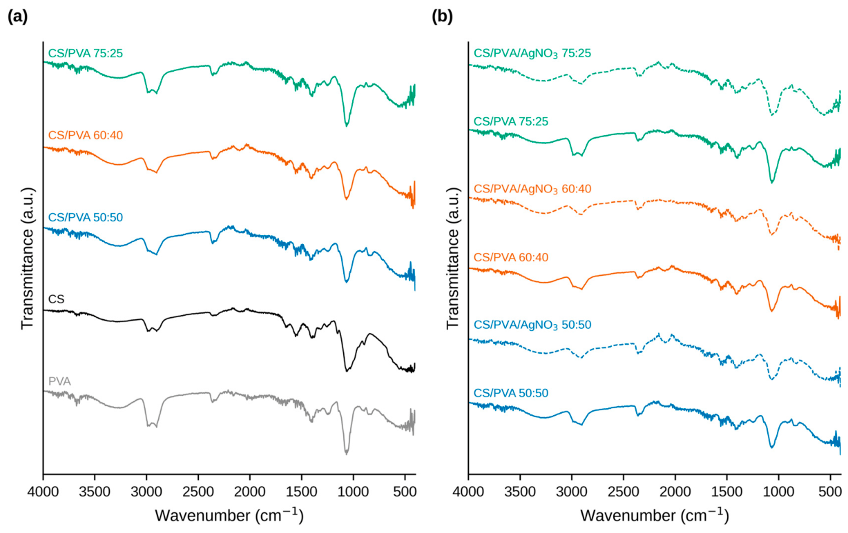
Figure 2.
X-ray diffraction patterns of PVA, CS, CS/PVA 50:50, CS/PVA 60:40, CS/PVA 75:25, and CS/PVA/AgNO₃ 75:25 film samples. The patterns are vertically offset for clarity.
Figure 2.
X-ray diffraction patterns of PVA, CS, CS/PVA 50:50, CS/PVA 60:40, CS/PVA 75:25, and CS/PVA/AgNO₃ 75:25 film samples. The patterns are vertically offset for clarity.
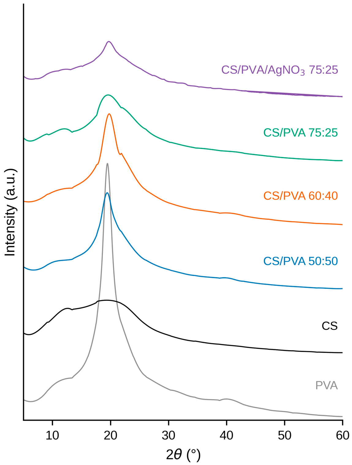
Figure 3.
Surface morphology of films prepared from pure CS (a), pure PVA (b), CS/PVA 50:50 (c), CS/PVA 60:40 (d), CS/PVA 75:25 (e), and CS/PVA/AgNO₃ 75:25 (f). Magnification ×3000; the scale bar in each panel corresponds to 5 μm.
Figure 3.
Surface morphology of films prepared from pure CS (a), pure PVA (b), CS/PVA 50:50 (c), CS/PVA 60:40 (d), CS/PVA 75:25 (e), and CS/PVA/AgNO₃ 75:25 (f). Magnification ×3000; the scale bar in each panel corresponds to 5 μm.
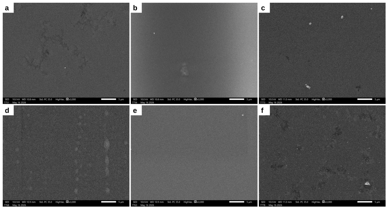
Figure 4.
TGA (a) and DTA (b) curves of the film samples: CS, PVA, CS/PVA 50:50, 60:40, and 75:25, and the corresponding AgNO₃-containing films (dashed lines). The DTA signal is normalized to the initial sample mass.
Figure 4.
TGA (a) and DTA (b) curves of the film samples: CS, PVA, CS/PVA 50:50, 60:40, and 75:25, and the corresponding AgNO₃-containing films (dashed lines). The DTA signal is normalized to the initial sample mass.
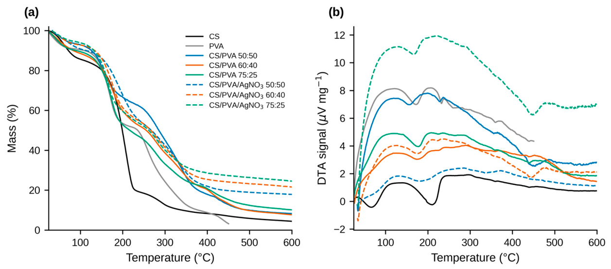
Figure 5.
Tensile strength (a), Young’s modulus (b), and elongation at break (c) of the film samples. Values are mean ± SD (n = 4); hatched bars denote AgNO₃-containing films.
Figure 5.
Tensile strength (a), Young’s modulus (b), and elongation at break (c) of the film samples. Values are mean ± SD (n = 4); hatched bars denote AgNO₃-containing films.
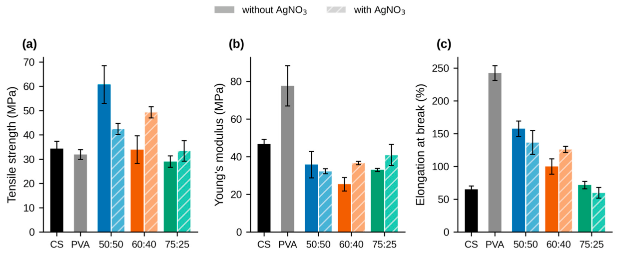
Figure 6.
Swelling degree (a) and water vapor transmission rate (b) of the film samples. Values are mean ± SD; hatched bars denote AgNO₃-containing films.
Figure 6.
Swelling degree (a) and water vapor transmission rate (b) of the film samples. Values are mean ± SD; hatched bars denote AgNO₃-containing films.
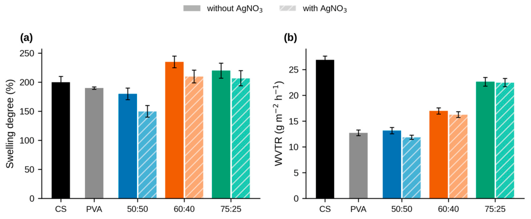
Figure 7.
Antibacterial activity of the CS, PVA, CS/PVA 75:25, and CS/PVA 60:40 films (discs S1–S4) against A. baumannii (a), E. cloacae (b), E. faecalis (c), E. coli (d), K. oxytoca (e), K. pneumoniae (f), P. mirabilis (g), S. typhimurium (h), MRSA (i), and MSSA (j). A standard antibiotic disc served as positive control on each plate. The dish in panel (g) was hand-labelled “E. coli” at the time of photography; its identity as the P. mirabilis plate is established by the ampicillin control disc and the recorded zone data.
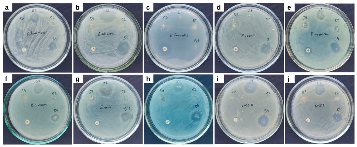
Figure 8.
Antibacterial activity of the CS/PVA 50:50 film and the CS/PVA/AgNO₃ 75:25, 60:40, and 50:50 films (discs S5–S8) against A. baumannii (a), E. cloacae (b), E. faecalis (c), E. coli (d), K. oxytoca (e), K. pneumoniae (f), P. mirabilis (g), S. typhimurium (h), MRSA (i), and MSSA (j). A standard antibiotic disc served as positive control on each plate.
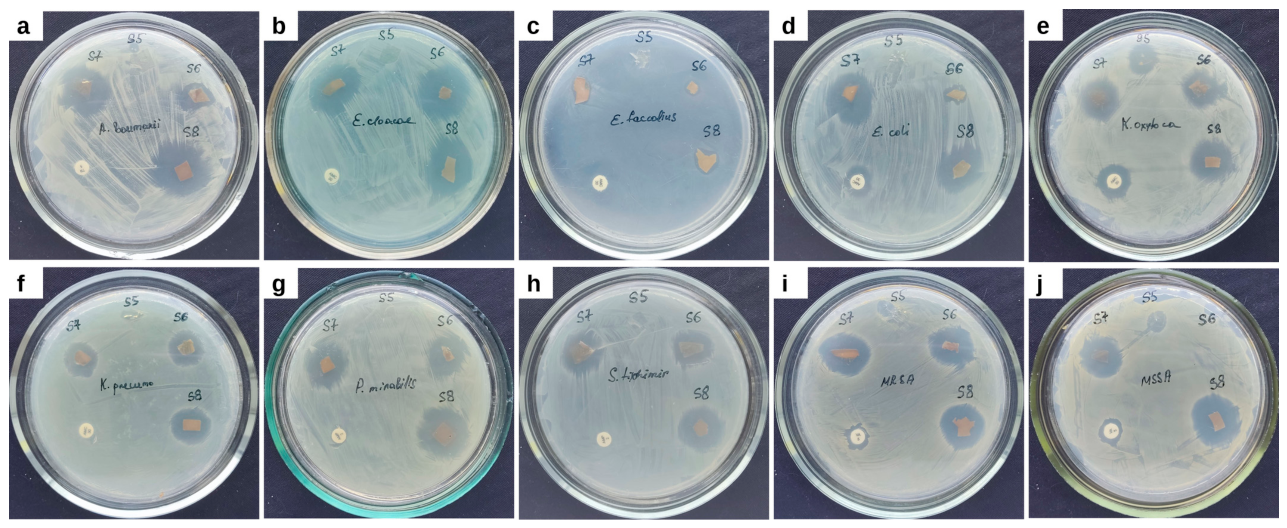

Table 1.
Composition of the investigated film formulations. Sample designations refer to the volume ratios of the CS (1% w/v) and PVA (3% w/v) solutions used for mixing.
Table 1.
Composition of the investigated film formulations. Sample designations refer to the volume ratios of the CS (1% w/v) and PVA (3% w/v) solutions used for mixing.
| Film Designation | CS Solution (vol) | PVA Solution (vol) | Calculated Nominal CS:PVA Dry-Polymer Mass Ratio ¹ | Glycerol Loading | AgNO₃ Loading |
|---|---|---|---|---|---|
| CS | 100 | 0 | 100:0 | 0.05 mL | – |
| PVA | 0 | 100 | 0:100 | 0.05 mL | – |
| CS/PVA 75:25 | 75 | 25 | 50:50 | 0.05 mL | – |
| CS/PVA 60:40 | 60 | 40 | 33:67 | 0.05 mL | – |
| CS/PVA 50:50 | 50 | 50 | 25:75 | 0.05 mL | – |
| CS/PVA/AgNO₃ 75:25 | 75 | 25 | 50:50 | 0.05 mL | 0.5 mL (≈5 mg) |
| CS/PVA/AgNO₃ 60:40 | 60 | 40 | 33:67 | 0.05 mL | 0.5 mL (≈5 mg) |
| CS/PVA/AgNO₃ 50:50 | 50 | 50 | 25:75 | 0.05 mL | 0.5 mL (≈5 mg) |
CS: chitosan; PVA: poly(vinyl alcohol). Glycerol loading is the volume of neat glycerol, and AgNO₃ loading is the volume of 1% (w/v) AgNO₃ solution (≈5 mg AgNO₃) added per film. ¹ Calculated nominal dry-polymer mass ratios derived from the stated solution concentrations (1% w/v CS; 3% w/v PVA) and the confirmed solution volume ratios, excluding glycerol and AgNO₃.
Table 2.
Characteristic FTIR absorption bands (cm⁻¹) of the CS, PVA, and composite films.
| Sample | O–H/N–H | C–H Stretching | C=O/Amide I | N–H/Amide II | C–O/C–O–C | Low-Frequency Bands |
|---|---|---|---|---|---|---|
| CS | 3311.23 | 2988.43; 2886.57 | 1652.75 | 1558.06 | 1150.61; 1057.36 | 892.37 |
| PVA | 3278.23 | 2971.21; 2900.91 | 1652.75 | 1559.49 | 1057.36 | 900.98; 826.37 |
| CS/PVA 50:50 | 3273.93 | 2988.43; 2906.65 | ~1650 ¹ | 1559.49 | 1065.96 | 826.37 |
| CS/PVA 60:40 | 3298.32 | 2933.91 | 1652.75 | 1559.49 | 1065.96 | 921.06; 826.37 |
| CS/PVA 75:25 | 3312.67 | 2988.43; 2910.96 | 1717.31; 1684.31 | 1559.49 | 1065.96 | 892.37; 852.20 |
| CS/PVA/AgNO₃ 50:50 | 3258.15 | 2902.35 | 1700.09 | 1559.49 | 1065.96 | 826.37 |
| CS/PVA/AgNO₃ 60:40 | 3298.32 | 2933.91 | 1652.75 | 1559.49 | 1065.96 | 921.06; 826.37 |
| CS/PVA/AgNO₃ 75:25 | 3357.14 | 2910.96 | 1652.75 | 1550.88 | 1065.96 | 1143.44; 1027.23; 852.20; 826.37 |
¹ The band was weak or overlapped with neighboring vibrational bands and was not separately identified by the automatic peak search.
Table 3.
Thickness, swelling degree, water vapor transmission rate, and mass loss in an acidic aqueous medium of the investigated films. Values are mean ± SD; mass loss after 24 h at pH 1.6.
Table 3.
Thickness, swelling degree, water vapor transmission rate, and mass loss in an acidic aqueous medium of the investigated films. Values are mean ± SD; mass loss after 24 h at pH 1.6.
| Film | Thickness (mm) | Swelling Degree (%) | WVTR (g·m⁻²·h⁻¹) | Mass Loss (%) |
|---|---|---|---|---|
| CS | 0.275 ± 0.004 | 200 ± 10 | 26.88 ± 0.73 | 5 |
| PVA | 0.232 ± 0.031 | 190 ± 2 | 12.73 ± 0.55 | <1 |
| CS/PVA 50:50 | 0.189 ± 0.001 | 180 ± 10 | 13.16 ± 0.61 | 2 |
| CS/PVA 60:40 | 0.161 ± 0.004 | 235 ± 10 | 16.98 ± 0.60 | <2 |
| CS/PVA 75:25 | 0.234 ± 0.002 | 220 ± 13 | 22.64 ± 0.85 | <3 |
| CS/PVA/AgNO₃ 50:50 | 0.209 ± 0.031 | 150 ± 10 | 11.88 ± 0.40 | ~1 |
| CS/PVA/AgNO₃ 60:40 | 0.178 ± 0.001 | 210 ± 11 | 16.27 ± 0.56 | ~2 |
| CS/PVA/AgNO₃ 75:25 | 0.158 ± 0.004 | 207 ± 13 | 22.49 ± 0.80 | ~3 |
Table 4.
Inhibition zone diameters (mm, including the 10 mm film disc) of the films against the tested bacterial strains.
Table 4.
Inhibition zone diameters (mm, including the 10 mm film disc) of the films against the tested bacterial strains.
| Strain | CS | PVA | 50:50 | 50:50 +AgNO₃ | 60:40 | 60:40 +AgNO₃ | 75:25 | 75:25 +AgNO₃ |
|---|---|---|---|---|---|---|---|---|
| A. baumannii | 12.5 ± 2.0 | 0 | 0 | 19.6 ± 0.6 * | 0 | 20.1 ± 2.9 * | 12.3 ± 1.4 | 13.0 ± 1.2 |
| E. cloacae | 16.8 ± 2.3 | 0 | 0 | 19.6 ± 0.8 * | 16.7 ± 2.5 | 18.9 ± 1.8 | 7.6 ± 1.5 | 21.1 ± 1.3 * |
| E. faecalis | 0 | 0 | 0 | 9.3 ± 1.1 * | 0 | 7.9 ± 2.2 * | 0 | 8.7 ± 2.2 * |
| E. coli | 0 | 0 | 0 | 16.6 ± 2.3 * | 0 | 14.6 ± 2.5 * | 0 | 8.2 ± 2.0 * |
| K. oxytoca | 12.8 ± 1.7 | 8.1 ± 1.0 | 13.8 ± 1.2 | 17.4 ± 2.8 | 14.5 ± 1.0 | 16.8 ± 1.9 | 10.2 ± 1.0 | 16.1 ± 1.7 * |
| K. pneumoniae | 14.6 ± 1.7 | 0 | 0 | 15.3 ± 2.0 * | 13.0 ± 2.6 | 15.5 ± 1.5 | 0 | 13.4 ± 1.7 * |
| P. mirabilis | 13.3 ± 2.7 | 0 | 0 | 17.3 ± 0.7 * | 13.9 ± 1.4 | 14.4 ± 1.9 | 0 | 13.7 ± 1.3 * |
| S. typhimurium | 13.1 ± 1.2 | 0 | 0 | 15.4 ± 1.7 * | 16.2 ± 2.0 | 14.4 ± 1.7 | 0 | 14.3 ± 1.7 * |
| MRSA | 18.0 ± 0.5 | 0 | 7.5 ± 1.5 | 15.1 ± 1.6 * | 16.4 ± 1.7 | 20.4 ± 1.9 * | 0 | 18.6 ± 1.5 * |
| MSSA | 18.2 ± 0.9 | 0 | 7.8 ± 1.2 | 18.8 ± 1.7 * | 14.4 ± 1.1 | 15.8 ± 2.5 | 0 | 0 |
Values are mean ± SD (n = 3); 0 indicates no measurable inhibition zone. * p < 0.05 versus the corresponding AgNO₃-free film of the same ratio (Tukey’s HSD following one-way ANOVA within each strain). Positive-control zones: amoxicillin–clavulanic acid 30 μg produced 14.5–17.4 mm zones against E. faecalis, E. coli, and K. oxytoca but no zone against E. cloacae or K. pneumoniae; trimethoprim 5 μg produced no zone against A. baumannii; ampicillin 2 μg produced no zone against S. typhimurium and inhibited P. mirabilis in one of two series (14.3 mm); vancomycin 5 μg produced 13.4–15.0 mm zones against MRSA and 13.5–15.2 mm against MSSA across the two series.
Disclaimer/Publisher’s Note: The statements, opinions and data contained in all publications are solely those of the individual author(s) and contributor(s) and not of MDPI and/or the editor(s). MDPI and/or the editor(s) disclaim responsibility for any injury to people or property resulting from any ideas, methods, instructions or products referred to in the content. |
© 2026 by the authors. Licensee MDPI, Basel, Switzerland. This article is an open access article distributed under the terms and conditions of the Creative Commons Attribution (CC BY) license (http://creativecommons.org/licenses/by/4.0/).
Copyright: This open access article is published under a Creative Commons CC BY 4.0 license, which permit the free download, distribution, and reuse, provided that the author and preprint are cited in any reuse.