

Submitted:
21 September 2026
Posted:
22 September 2026
You are already at the latest version
Abstract
The aim of this paper is to obtain and verify the structures and the biological activity of novel metal complexes derived from 5-carboxy-2-thiouracil. The paper outlines a synthesis method and assesses the antimicrobial activity of newly formed Cu(II), Ni(II) and Co(II) complexes. Several syntheses were carried out with and without NaOH. The structural were verified by melting point analysis, ICP-OES for Cu, Ni and Co, UV-Vis, ATR, solid state and solution 1H NMR, 13C NMR spectroscopy. In addition, the antimi-crobial activity of these complexes and the free ligand are assessed against both Gram-positive and Gram-negative bacteria, as well as yeasts. For the cytotoxic assay we were used the HCT-116 colorectal cell line (ATCC). Only two Ni(II) complexes show reduction in the number of viable tumor cells. All the other compounds did not lead to a significant reduction in the number of viable cells as determined by one-way ANOVA statistical analysis.
Keywords:
5-carboxy-2-thiouracil
; copper
; nickel
; cobalt complexes
; antibacterial activities
; cytotoxic effect
1. Introduction
Ligands containing nitrogen, sulfur, and oxygen donor atoms are widely recognized for their significant contributions to anticancer and antiviral applications. The biological relevance of uracil derivatives [1,2] and their metal complexes has stimulated extensive spectroscopic and biological investigations over recent years [3,4]. Among these compounds, 5-carboxy-2-thiouracil represents a particularly attractive ligand because it offers several potential coordination sites, including the two nitrogen atoms of the pyrimidine ring, the thione (C=S) group at the C2 position, the carbonyl (C=O) group at the C4 position, and the carboxylate (COO⁻) functionality [5,6]. The discovery of new drugs to improve the efficacy and safety of clinically used drugs is the major thrust area in medicinal chemistry. Nitrogen-containing heterocycles are the most researched promising pharmacophores endowed with various pharmacological activities. Figure 1 presents the most popular medical drugs involving uracil derivatives and its metal complexes with biological activities. One such scaffold is uracil and its structural modifications resulted in the development of new derivatives exhibiting anticancer [7,8,9,10], antibacterial [11,12,13], antiviral [14,15], antifungal [16,17], and antidiabetic properties [18,19,20,21,22,23,24]. The anticancer activity of uracils is majorly attributed to the DNA synthesis blockage through inhibition of the enzyme thymidylate synthase. High toxicity, weak tumor affinity, and resistance due to the development of gene mutation in cancer cells are the major concerns.
Thiouracil derivatives have attracted considerable attention as versatile ligands for the synthesis of biologically active transition metal complexes. Recent studies have demonstrated that Cu(II), Pd(II), and Au(III) complexes of 2-thiouracil [25], 6-methyl-2-thiouracil, 6-propyl-2-thiouracil [26,27], and 2,4-dithiouracil [28] can be successfully synthesized and characterized using spectroscopic and physicochemical techniques. Biological evaluation revealed that metal coordination generally enhances the antibacterial activity of the parent ligands, while Au(III) complexes exhibited particularly promising antimicrobial properties [29]. Furthermore, the cytotoxic evaluation of Cu(II), Pd(II), and Au(III) complexes demonstrated significant antiproliferative activity against cancer cell lines, highlighting the potential of thiouracil-based metal complexes as candidates for the development of new antimicrobial [26,27,28] and anticancer agents [29]. Figure 2. presents the possible mechanism of action of metal complexes of pyridine-base compounds.
Copper is an essential trace element for aerobic organisms and plays a key role in various biological processes. At optimal levels, it contributes to plant metabolism as a component of enzymes, structural proteins, and pigments, while excessive concentrations may cause toxic effects [30]. Copper functions as an important cofactor in enzymes such as cytochrome c oxidase, Cu/Zn-superoxide dismutase, lysyl oxidase, tyrosinase, and dopamine β-monooxygenase [31].
Nickel(II) is a biologically relevant metal ion that participates in essential enzymatic processes in microorganisms and plants [32]. It acts as a cofactor for several metalloenzymes, including urease, hydrogenase, and carbon monoxide dehydrogenase, where it contributes to catalytic activity and cellular metabolism [33]. However, excessive Ni(II) accumulation can induce oxidative stress and toxic effects, highlighting the importance of nickel homeostasis in biological systems.
Cobalt(II) is an essential trace metal involved in important biological processes, mainly through its role as the central metal ion in vitamin B₁₂ (cobalamin). It is required for the activity of several cobalt-dependent enzymes involved in DNA synthesis, amino acid metabolism, and cellular energy production. Although Co(II) is biologically important, excessive exposure can promote oxidative stress and cellular toxicity, emphasizing the need for strict cobalt regulation in living organisms [34,35].
To date, a wide range of metal complexes based on uracil and thiouracil derivatives have been synthesized and investigated. Their structural features and compositions have been extensively studied with various metal ions, including copper [6,25,26,36,37,38], silver [39], nickel [6,40,41], cobalt [4,40], zinc [6,40], manganese [6], cadmium [6], gold [25,26,27], vanadium [42,43], as well as palladium [2,25,26,41].
In this study, we report the synthesis and characterization of a Cu(II), Ni(II), Co(II) complexes derived from 5-carboxy-2-thiouracil (see Figure 3).
The coordination compounds were obtained after mixing water solutions of the metal salt and the ligand dissolved in DMSO and water solutions of NaOH, in a metal-to-ligand-to-base ratio of 1:4:2. The ligand and the metal salts were brought into the reaction at room temperature in DMSO and H2O as solvents, respectively. Several syntheses were carried out with NaOH (M:L:OH-=1:4:2) and without (М:L=1:4). The characterization of the metal complexes was conducted through various techniques, including Inductively Coupled Plasma Optical Emission Spectroscopy (ICP-OES) for M and S, attenuated total reflection (ATR), UV-Vis, 1H and 13C NMR spectroscopy. The obtained complexes were investigated of the antimicrobial activity against both Gram-positive, Gram-negative bacteria and yeasts. Тhe cytotoxic assay was tested of the HCT-116 colorectal cell line.
2. Materials and Methods
2.1. Chemicals
All solvents and reagents were purchased from Merck (Merck Bulgaria EAD). Melting points were determined on a Boetius hot stage apparatus and are uncorrected.
Inductively Coupled Plasma Optical Emission Spectroscopy (ICP-OES) Determination of Cu, Co and Ni in the Complexes
A total of 0.0200 g of sample was weighed on an analytical balance and dissolved with 65% nitric acid, p.a. (Chem-Lab NV). Blank solutions were prepared as well. After dilution, the concentration of Co, Cu and Ni was determined via ICP-OES (ICP-OES iCap 6000, Thermo Scientific, Waltham, MA, USA). Calibration standards were prepared from monoelemental standard solutions − 1000 mg L−1 (Merck, Darmstadt, Germany). Conventional ICP-OES operating conditions were used. Analytes were measured on three emission lines for estimation of potential spectral interferences, i.e., 324.754 nm, 327.395 nm, 510.554 nm for Cu, 341.476 nm, 352.454 nm, and 361.939 nm for Ni and 340.512 nm, 345.351 nm, and 350.228 nm for Co. Sulfur was determined by inductively coupled plasma–optical emission spectrometry (ICP-OES iCap 6000, Thermo Scientific,Waltham, MA, USA) on analytical lines 180.731 nm, 182.034 nm, and 182.624 nm in axial mode. Five replicates and 5 s measurements were applied for all lines.
2.2. Synthetic Methods Experimental Protocols
2.2.1. Synthesis of Cu(II), Ni(II) and Co(II) Complexes of 5-Carboxy-2-Thiouracil L, M:L:OH- =1:4:2
The Cu(II), Ni(II) and Co(II) complexes were synthesized by mixing a DMSO solution (10 mL) containing 0.6886 g, (0.004 mol) of the ligand (L) with water solution of the respective metal salts: Cu(CH3COO)2.H2O 0.1996 g, (0.001 mol), Ni(CH3COO)2.4H2O 0.2489 g, (0.001 mol) or Co(CH3COO)2.4H2O 0.2491 g, (0.001 mol). It should be mentioned that the solutions were alkalized with 5 mL 0.08 g, (0.002 mol) of NaOH solution. After stirring with an electromagnetic stirrer for 24 hours, brown (Cu), light green (Ni) and purple (Co) precipitates formed. These were filtered, washed with water, air dried, and further dried over silica gel in a desiccator for two weeks.
2.2.2. Synthesis of Cu(II), Ni(II) and Co(II) Complexes of 5-Carboxy-2-Thiouracil (L), M:L =1:4 Without NaOH
The same procedure for the synthesis of the metal complexes is used, but without the use of sodium hydroxide. After stirring with an electromagnetic stirrer for 24 hours, orange-brown (Cu), light green (Ni) and purple (Co) precipitates formed.
2.3. Spectral Measurements
UV-Vis measurements were done with UV-Vis ONDA apparatus in the range from 1000 nm to 200 nm. The λmax are given below for each compound:
λmax (5-C-2Tu, THF) = 274 nm, 294 nm, 326 nm
λmax (5-C-2Tu+Cu(II) 1:4:2, THF) = 270 nm, 300 nm, 324 nm
λmax (5-C-2Tu, DMSO) = 272 nm, 322 nm
λmax (5-C-2Tu+Cu(II) 1:4:2, DMSO) = 278 nm, 352 nm
λmax (5-C-2Tu, DMF) = 270 nm, 360 nm
λmax (5-C-2Tu+Cu(II) 1:4:2, DMF) = 300 nm, 346 nm
λmax (5-C-2Tu, H2O) = 216 nm, 272 nm, 306 nm
λmax (5-C-2Tu+Cu(II) 1:4:2, H2O) = 214 nm, 272 nm, 306 nm
λmax (5-C-2Tu, CH3OH) = 216 nm, 276 nm, 320 nm
λmax (5-C-2Tu+Cu(II) 1:4:2, CH3OH) = 214 nm, 280 nm, 318 nm
ATR spectra were measured with VERTEX 70 FT-IR instrument (Bruker Optics, Billerica, MA, USA) using MIRacleTM with a one-reflection ZnSe element (PIKE Technology, Madison, WI, USA). ATR data was measured in the range (4000–600) cm−1 with 25 scans and a resolution of 2 cm−1. The ATR-IR data with the assignments are presented below for each compound:
ATR (cm-1) 5-Carboxy-2-Tu: 3254 ν(NH), 3200-2500 ν(OH), 3100 ν(CH), 2968, 1699 ν(C=O), 1675 ν(C=O), 1599, 1530, 1455, 1343, 1327, 1285, 1221, 1177, 1152 ν(C=S), 1068, 980, 936 γ(OH), 806, 768, 719, 654, 611.
ATR (cm-1) 5-Carboxy-2-Tu+Cu(II) (1:4): 3192 ν(NH), 3147 ν(NH), 3032 ν(CH), 1739 ν(C=O), 1638 ν(C=O), 1584, 1530, 1457, 1382, 1356, 1287, 1257, 1172, 1079, 1015, 989, 951, 810, 795, 753, 729, 705, 664, 651, 617.
ATR (cm-1) 5-Carboxy-2-Tu+Cu(II) (1:4:2): 3200-2500 ν(OH), 3140 ν(NH), 3065 ν(CH), 2932, 1704 (C=O), 1635 ν(C=O), 1621, 1579, 1551, 1454, 1391, 1330, 1288, 1270, 1232, 1202, 1167 ν(C=S), 1078, 1008, 988, 973, 949 γ(OH), 887, 835, 814, 806, 770, 706, 652, 610.
ATR (cm-1) 5-Carboxy-2-Tu+Ni(II) (1:4): 3386 ν(OH), 3200-2500 ν(OH), 3187 ν(NH), 3066 ν(CH), 3032 ν(CH), 2783, 1742 ν(C=O), 1668 ν(C=O), 1616, 1576, 1548, 1530, 1462, 1432, 1416, 1381, 1327, 1283, 1230, 1201, 1183, 1171, 1151 ν(C=S), 1087, 1033, 998, 929 γ(OH), 911, 872, 805, 796, 767, 727, 713, 662, 638, 612.
ATR (cm-1) 5-Carboxy-2-Tu+Ni(II) (1:4:2): 3406 ν(OH), 3200-2500 ν(OH), 3144 ν(NH), 3066 ν(CH), 2934, 2831, 1668 ν(C=O), 1620, 1578, 1546, 1530, 1417, 1380, 1328, 1230, 1199, 1170, 1152 ν(C=S), 1085, 1018, 987, 973, 935 γ(OH), 887, 850, 835, 814, 770, 706, 661, 637.
ATR (cm-1) 5-Carboxy-2-Tu+Co(II) (1:4): 3490 ν(OH), 3200-2500 ν(OH), 3192 ν(NH), 3074 ν(CH), 3032 ν (CH), 2945, 2889, 2830, 1675 ν(C=O), 1618, 1582, 1546, 1529, 1460, 1377, 1329, 1230, 1197, 1170, 1152 v(C=S), 1083, 1015, 985, 929 γ(OH), 847, 810, 783, 661, 632.
ATR (cm-1) 5-Carboxy-2-Tu+Co(II) (1:4:2): 3388 ν(OH), 3200-2500 ν(OH), 3150 ν(NH), 3066 ν(CH), 2993, 2911, 1714 ν(C=O), 1662 ν(C=O), 1622, 1580, 1563, 1479, 1456, 1433, 1393, 1357, 1330, 1231, 1204, 1171, 1091, 1078, 1031, 1004, 955 γ(OH), 888, 836, 815, 805, 771, 707, 634, 610.
13C{1H} NMR spectra were recorded on a Bruker Avance III HD 500 spectrometer (Bruker, Billerica, MA, USA) at 500.13 MHz (1H-NMR) and 125.76 MHz (13C-NMR), respectively. Some of the 1H signals for the complexes were not observable in the 1H solution-state NMR spectra due to the low solubility of the compounds in DMSO-d6 used as a solvent. The solid-state NMR spectra were recorded at 298 K. A 2.5 mm Cross-Polarization Magic Angle Spinning (CP MAS) probehead was used for registering the solid-state NMR spectra on a Bruker Avance III HD 500 MHz spectrometer. A MAS speed of 15 kHz as well as α-glycine as external reference (α-glycine carbonyl C—176.03 ppm) were applied for measuring the CP MAS and Cross-Polarization with Polarization Inversion (CPPI) MAS spectra. All NMR experiments were performed at ambient temperature.
2.4. Microbiological Tests
2.4.1. Tested Microorganisms
Antimicrobial Activity Assay
The antimicrobial activity assay was conducted through the agar well diffusion method against the following test microorganisms: Staphylococcus aureus ATCC 25923, Bacillus cereus ATCC 11778, Bacillus subtilis ATCC 6633, Listeria monocytogenes ATCC 8787, Enterococcus faecalis ATCC 19433, Salmonella enterica ssp. enterica ser. Enetritidis ATCC 13076, Pseudomonas aeruginosa ATCC 9027, Proteus vulgaris G, Klebsiella pneumoniae ATCC 13883, Candida albicans ATCC 10231, Saccharomyces cerevisiae. The melted and cooled nutrient medium (Nutrient agar (Merck) for the bacteria and Candida albicans and YPD agar (Merck) for Saccharomyces cerevisiae) was inoculated with a suspension of the test-microorganism and poured into Petri dishes. After the nutrient medium solidified, a hole with a diameter of 6 mm was punched aseptically with a sterile cork borer. A volume of 50 µl of a 1 mg/mL solution of each compound was pipetted into the wells. A control of the solvent (DMSO) was also tested for antimicrobial activity. Then, the agar plates were incubated at 37 °C for 24 hours for the bacteria and Candida albicans and 30 °C for 48 hours for Saccharomyces cerevisiae. After that the diameters of the resulting inhibition zones were measured. Each sample and the control were teste in triplicated and the results are presented as mean values ±SD.
2.4.2. Cytotoxicity Assay
To determine the biological effects of the compounds we performed WST-1 assay (Roche). WST-1 is a tetrazolium salt, which is converted to a dark red formazan product by mitochondrial dehydrogenases in viable cells. Thus, the number of living metabolically active cells is directly proportional to the amount of dye produced. For this assay, we used the HCT-116 colorectall cell line (ATCC). Cells were grown in DMEM/F12 cell culture medium supplemented with 10% serum and 1% penicillin/streptomycin. For the experiments cells were seeded in 96-well plates at a density of 10 000 cells per well and allowed to adhere to the bottom of the wells for 24 hours. Then we treated the cells with several concentrations of the compounds and performed the WST-1 assay 72 hours after treatment. Due to the poor solubility of the compounds, most of them were provided at a concentration of 1 mg/ml in 100% DMSO, which determined the highest possible concentration to be 10 μg/ml (corresponding to a final concentration of 1% DMSO). In a preliminary experiment we tested all 7 compounds in a five-point, twofold serial dilution starting at 10 μg/ml for 48 hours but since most of them did not cause a significant decrease in the number of living cells, we decided to repeat the experiment with only the highest 2 or 3 possible concentrations. For some compounds, whose initial concentration was higher (5 mg/ml in 100% DMSO) – 5-C-2Tu, 5-C-2Tu+Ni(II) 1:4:2, 5-C-2Tu+Ni(II) 1:4, we tested four concentrations in twofold serial dilution, i.e., 50, 25, 12.5 and 6.25 μg/ml. The results of the WST-assay were analyzed in GraphPad using one-way ANOVA followed by Dunnett’s post-hoc test. Graphs were prepared in the GraphPad software.
3. Results and Discussion
The analytical and physical properties of metal(II) complexes with Cu(II), Ni(II) and Co(II) are presented in Table 1.
The data provide preliminary evidence for successful complex synthesis and subsequent coordination with the respective metal ions. The less-than-quantitative yields can be attributed to product loss during purification or side reactions that compete with the desired complex formation. The free ligand exhibited a melting point of 246-247 °C, while the metal complexes displayed higher melting points, all above 325 °C and >350 °C. None of the complexes shared the melting point of the parent ligand, supporting the formation of new coordination compounds. The ligand 5-C-2Tu (C5H4N2O3S, MW 172.16 g/mol) shows as a deep colorless solid with a melting point of 246-247 °C. The complexation with metals gave rise to higher molecular weights and a significant colour change. It was observed that the complexes exhibited higher melting points (325–328 °C for Cu(II) with presents of NaOH in molar ratio 1:4:2 and all rest to > 350 °C). This indicates an increase in thermal stability due to stable chelate rings and stronger metal–ligand interactions, as observed in the Cu(II), Ni(II) and Co(II) complexes (> 350 °C and 325-328 °C, respectively).
Table 2 shows that free ligand displayed solubility in THF, water, DMSO, DMF. CuL (1:4) complex was insoluble in acetone, chloroform, THF, ethanol, water, cyclohexane, methanol and slightly soluble in and dimethyl sulfoxide and DMF. Cobalt, copper, and nickel complexes with NaOH also displayed different solubility behavior in the solvents. The favorable solvent–solute interactions involving the metal center and donor atoms of the solvent molecules resulted in the improved solubility of some complexes in coordinating organic solvents such as DMF and DMSO [44]. The improved solubility in these solvents implies a higher organophilic character post-complexation and indicates successful complex formation. The alteration in polarity and intermolecular interactions accounts for why coordination of ligands with metal ions modifies their solubility. The results in reduced polarity and increased lipophilicity of the complex, leading to lower solubility in water but enhanced solubility in organic solvents like DMF and DMSO [45].
3.1. Electronic Spectra Studies
Table 3 shows the electronic spectra results. The free ligand (5-C-2Tu) shows characteristic π → π* transitions at 274 and 294 nm, and n → π* transitions at 326 nm, indicating aromatic and carboxyl chromophores (this is for THF use as solvent). The free ligand shows characteristic π → π* transitions at 272 nm and n → π* transitions at 322 nm, where the solvent is DMSO. Where the solvent is DMF two maximums were observed at 270 nm and 360 nm, respectively. Where the solvents are water and CH3OH tree maximums are detected at 216 nm, 272 nm, 306 nm and 216 nm, 276 nm, 320 nm, respectively.
Some of the UV-Vis spectra were registered in DMSO. Maxima in the UV-Vis spectra of the free ligand L were observed at λmax= 272, 322 nm. Maxima in the electronic spectra of Cu(II) 1:4:2 complex were observed at 278 nm, 352 nm, respectively. It can be seen that one of the maxima is shifted bathochromically by 30 nm towards longer wavelengths.
3.2. Structure Verification of 5-C-2Tu
The NH protons in 5-carboxy-2-thiouracil are isochronous producing 1H NMR singlet at 13.04 ppm. The OH proton has a broad 1H singlet at 12.70 ppm. Only one HSQC correlation can be found including the chemical shifts of CH carbon (147.81 ppm) and proton (8.02 ppm). There are three strong three-bond HMBC correlations for the CH proton – (8.02 ppm – 176.39 ppm), (8.02 ppm – 160.50 ppm) and (8.02 ppm -163.28 ppm), where the chemicals shift at 176.39 ppm, 160.50 ppm and 163.28 ppm were respectively for the 13C signals of the C=S, C=O and COOH carbons. In addition, there is one weak two-bond HMBC correlation with the signal of C-5 at 106.20 ppm. The NMR assignments for 5-C-2Tu are in good agreement with HOSE predictions by using the NMRShiftDB database (https://nmrshiftdb.nmr.uni-koeln.de/).
Figure 4.
Verified structure of 5-carboxy-2-thiouracil.
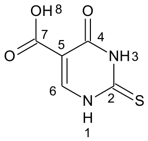

Table 4.
1H and 13C NMR data assignment for 5-carboxy-2-thiouracil [500.13 MHz (1H) and 125.76 MHz (13C)]a.
Table 4.
1H and 13C NMR data assignment for 5-carboxy-2-thiouracil [500.13 MHz (1H) and 125.76 MHz (13C)]a.
| Atom | δ (1H), ppm | Multiplicity (J, Hz) | δ (13C), ppm | DEPT | HMBC |
|---|---|---|---|---|---|
| NH-1 | 13.04 | s | |||
| 2 | 176.39 | C | |||
| NH-3 | 13.04 | s | |||
| 4 | 160.50 | C | |||
| 5 | 106.20 | C | |||
| 6 | 8.02 | s | 147.81 | CH | 2,4,5c,7 |
| 7 | 163.28 | C | |||
| OH-8 | 12.70 | br.s |
aThe solvent used is DMSO-d6 (solvent reference: 1H δref 2.50 ppm, 13C δref 39.51 ppm). 1H-1H COSY showed no observable correlations. b Abbreviations: DEPT, Distortionless Enhancement by Polarization Transfer spectrum; 1H-1H COSY, proton–proton homonuclear correlation spectrum; HSQC, Heteronuclear Single Quantum Correlation experiment; HMBC, Heteronuclear Multiple Bond Correlation experiment. c Weak correlations.
The solid-state 1H NMR spectrum showed three broad peaks at 10.3, 13.0 and 15.0 ppm correspondingly for the CH, NH and OH protons. The 13C-CP-MAS spectrum showed signals at 105.4 ppm, 152.9 ppm, 157.5 ppm, 168.3 ppm and 175.2 ppm respectively for the C-5, CH, C=O, COOH and C=S carbons.
The ATR-IR data for the ligand is in good agreement with some quantum chemical calculations presented previously for the ligand [46]. There is a band at 3254 cm-1 for the NH groups and two C=O bands at 1699 cm-1 (C4=O) and 1675 cm-1 (C7=O). Also, there is a band at 936 cm-1, which is for the out-of-plane bending of the carboxylic OH group. As it was concluded previously in [46], based on the vibrational analysis and the γ(OH) band, it can be stated that 5-C-2-Tu exists as a dimer in solid state through the COOH group. There is also a wide carboxylic ν(OH) band in the range 2500-3200 cm-1. The predictions show a band at 1152 cm-1 for the C=S group which has the same wavenumber as that in the ATR-IR spectrum. Therefore, the observed ATR-IR band at 1152 cm-1 was assigned to ν(C=S).
3.3. Structure Elucidation of Cobalt Complex Without NaOH Addition
The 13C CP-MAS spectrum shows five signals at 182.2, 170.2, 155.5, 144.4 and 108.9 ppm corresponding respectively to the C=S, COOH, C-O, CH and C-5 carbons in the coordinated 5-C-2Tu ligand (Figure 5). There is also one signal at 40.5 ppm indicating the presence of DMSO-h6 in the complex. 13C CPPI-MAS signals that are clearly distinguishable from the noise were observed mainly for the COOH and DMSO-h6 carbons at 170.2 ppm and 40.5 ppm, respectively. As it is expected from 13C CPPI-MAS, the signals for CH carbons were correspondingly suppressed. The solid-state 1H spectrum showed a broad signal at 8.7 ppm that probably includes the signals for the C-H, OH and NH protons in the coordinated ligand. In addition, there is another broad signal in the wing of the latter at 3.3 ppm indicating the possible presence of DMSO and H2O in the complex.
There is an ATR-IR band at 3192 cm-1 for the NH protons in the structure, as well as one OH band at 3490 cm-1. There is only one C=O band in the range (1650 – 1800) cm-1 at 1675 cm-1 assigned for the carboxylic C7=O group which has the same wavenumber as the one for the free ligand. Thus, it was assumed that the ligand was bonded to the metal through a covalent bond between the oxygen of the C-O group and Co(II). There is an ATR band for the out-of-plane vibration of carboxylic OH group at 929 cm-1 in the coordinated ligand. There is also a wide carboxylic ν(OH) band in the range 2500-3200 cm-1. The observed ATR-IR band at 1152 cm-1 was assigned to ν(C=S). For cobalt complex, we assume an octahedral geometry, with the 5-carboxy-2-thiouracil ligand acting in a monodentate manner via O-atom. In the structure of the Co(II) complex there are two molecules of 5-C-2Tu, while the remaining four coordination sites are occupied by water and/or DMSO molecules. This structure is similar to this who presented Singh et al. for Mn(II), Cu(II), Ni(II), Co(II), Zn(II) and Cd(II) complexes [6].
3.4. Structure Elucidation of Nickel Complex Without NaOH Addition
Five signals were observed for the coordinated 5-C-2Tu (Figure 6) in 13C CP-MAS at 180.2 ppm, 168.6 ppm, 154.5 ppm, 135.3 ppm and 107.4 ppm corresponding respectively to C=S, COOH, C=O, C-6 and C-5 carbons. There is a 13C signal at 40.5 ppm indicating the presence of DMSO-h6 in the complex. 13C CPPI-MAS signals that are clearly distinguishable from the noise were observed mainly for the COOH, C-5 and DMSO-h6 carbons, respectively at 168.6 ppm, 107.4 ppm and at 40.5 ppm. 13C CPPI-MAS signals for CH carbons were suppressed. Also, the 1H solid-state spectrum showed a broad peak at 3.2 ppm indicating for the presence of DMSO-h6 and H2O. There are also two broad 1H signals at 7.0 and 12.6 ppm respectively containing the signals for the C-H, O-H and N-H protons in the structure. The solution-state 1H NMR spectrum shows a broad 1H multiplet at 13.02 ppm for the NH and OH protons, as well as one signal at 8.02 ppm for the CH protons. In addition, 1H singlet at 2.54 ppm can be found confirming the presence of DMSO-h6 in the complex.
There is a wide O-H band at 3386 cm-1 in the ATR-IR spectrum. Also, there is a band at 3187 cm-1 assigned for the NH groups. In addition, there are two C=O bands at 1742 and 1668 cm-1, assigned for the C4=O and C7=O groups, respectively. Similarly, to the previous compound, the out-of-plane vibration band of the carboxylic OH group in the coordinated ligand can be found at 929 cm-1. There is also a wide carboxylic ν(OH) band in the range 2500-3200 cm-1. The observed ATR-IR band at 1151 cm-1 was assigned to ν(C=S). For the nickel complex obtained without prior adjustment of the medium’s pH, a dinuclear complex is observed featuring OH-bridging and monodentate coordination via the O-atom of four ligand molecules. It is evident that DMSO and water molecules are also present in the composition of the complex. This is also observed with previously synthesized complexes using similar ligands, namely 6-methyl-2-thiouracil, 6-propyl-2-thiouracil, and 2,4-dithiouracil and their complexes with copper, palladium and gold [26,27,28]. This structure is similar to this who presented Papazoglou et al. for dinuclear Cu(I) complex with monodentate coordination via S-atom [37] with the difference that here coordination took place via the oxygen atom rather than the sulfur atom.
3.5. Structure Elucidation of Copper Complex Without NaOH Addition
There are only four 13C signals for the ligand at 169.3 ppm (C4=O), 161.8 ppm (C2=O), 142.9 ppm (C-6) and 111.9 ppm (C-5) in the 13C CP-MAS spectrum suggesting that 5-C-2Tu undergoes decarboxylation [47,48] followed by subsequent oxidation of the obtained 2-thiouracil that leads to uracil (Figure 7). 13C CPPI MAS spectrum shows the three signals at 169.3 ppm, 161.8 ppm and 111.9 ppm, respectively for C4=O, C2=O and C-5 carbons. The signal at 142.9 ppm has vanished in the 13C CPPI-MAS spectrum because it is for the CH carbon in uracil. There are studies explaining copper-catalyzed oxidative decarboxylation of carboxylic compounds [49,50,51,52,53,54]. In addition, there is a study suggesting the oxidation of a thiouracil derivative to uracil catalyzed by NiO2 [55]. Also, the presence of DMSO-h6 in the complex is confirmed by the solution-state and solid-state 1H spectrum where correspondingly their 1H signals at 2.54 ppm and 2.9 ppm were observed. There are broad 1H solid-state NMR signals at 11.9 ppm and 7.7 ppm that are for NH and CH protons in the coordinated uracil. The solution-state 1H spectrum showed a signal at 12.00 ppm for NH-3 proton and a multiplet at 8.29 ppm where the 1H signals of NH-1 and both CH protons overlap.
The presence of NH groups can be confirmed by the ATR-IR bands at 3147 and 3192 cm-1. There is also an ATR-IR band for C4=O vibration at 1739 cm-1 which is very close as a wavenumber for the one (1742 cm-1) presented in SDBS database for an uncoordinated uracil. The other high-intensity band at 1638 cm-1 was assigned for the coordinated C2=O group in uracil. Consequently, a structure was proposed for complex, where Cu(II) is coordinated to both NH groups and the C2=O group (Figure 7). There is no OH band in the ATR-IR spectrum. In addition, there are no broad ATR-IR bands in the ranges (3200-2500) cm-1 and (950-900) cm-1 for ν(OH) and γ(OH) of carboxylic OH group, respectively. In contrast to the copper complex obtained in an alkaline medium, decarboxylation and desulfurization of the organic ligand are observed in the complex prepared without sodium hydroxide. The solvent DMSO acts as a ligand similar to previously obtained copper and palladium complexes [26]. In contrast, in this copper complex the ligand act as tridentate fashion not bidentate as 2,4-dithiouracil in copper complex [28] or bidentate in palladium complex with 6-propyl-2-thiouracil [26]. This structure is similar to this who presented Masoud et al. for Cu(II) complex with 2-thiouracil and tridentate coordination via O, N, S-atoms [43] with the difference that here coordination took place via the oxygen and two nitrogen atoms rather than the sulfur atom.
3.6. Structure Elucidation of Cobalt Complex with NaOH Addition
Although the signals in the 13C solid state CP-MAS NMR spectrum are accompanied by significant amount of noise, the wide signal around 40 ppm can be attributed to the presence of DMSO-h6 in the complex. There are also some 13C signals that can be distinguished more easily from the noise at 164.5 ppm (COOH carbon), 154.4 ppm (C=O carbon), 135.4 ppm (CH carbon). There is a broad 1H signal at 8.7 ppm in solid-state 1H spectrum for the NH, CH and OH protons, as well as a broad signal in the wing of the latter at 3.2 ppm which can be due to the protons in DMSO-h6 (Figure 8).
The ATR-IR spectrum showed a band at 955 cm-1 which was assigned to the out-of-plane bending of the carboxylic OH group. Also, there is a wide OH band at 3388 cm-1. There is also a wide carboxylic ν(OH) band in the range 2500-3200 cm-1. There are two C=O bands at 1714 cm-1 and 1662 cm-1. For cobalt complex we assume an octahedral geometry, with the free ligand acting in a monodentate fashion via O-atom. In the complex formed in an alkaline medium, there are four ligands and two hydroxyl groups are involved in coordination, the solvent DMSO resides in the outer coordination sphere. The structure is similar to this who presented Singh et al. for Mn(II), Cu(II), Ni(II), Co(II), Zn(II) and Cd(II) complexes with monodentate coordination via oxygen atom [6].
3.7. Structure Elucidation of Copper Complex with NaOH Addition
There are five 13C signals in CP-MAS spectrum at 174.7 ppm, 168.6 ppm, 163.1 ppm, 149.1 ppm and 112.1 ppm, which were respectively for the C=S, COOH, C=O, C-6 and C-5 carbons in the coordinated 5-C-2-Tu ligands. In addition, there are five more low-intensity 13C signals at 171.9 ppm, 170.5 ppm, 165.0 ppm, 144.0 ppm and 108.9 ppm for the both uncoordinated 5-C-2Tu ligands in the outer sphere of the complex (Figure 9). 13C CPPI-MAS spectrum showed each of the signals in the 13C CP-MAS spectrum except the ones at 149.1 ppm and 144.0 ppm which were for the CH carbons, respectively in the coordinated and non-coordinated ligands. There is also a signal at 40.5 ppm that indicates that DMSO-h6 participates in the complex. The 1H solid-state spectrum showed broad signals at 2.9 ppm for DMSO-h6, as well as at 8.1 ppm and 12.6 ppm that include the signals for CH, NH and OH protons in the coordinated ligands. The 1H solution state showed a signal at 2.54 ppm for DMSO-h6 and two singlets at 8.12 ppm and 8.23 ppm that can be assigned for the CH and/or NH protons.
There is ATR-IR bands at 1704 cm-1 and 1635 cm-1 respectively for the C4=O and C7=O groups. There is also a wide carboxylic ν(OH) band in the range 2500-3200 cm-1. In addition, there is a ATR-IR band for an out-of-plane OH bending at 949 cm-1. Also, there is a shoulder ATR-IR band for the NH groups in the compound at 3140 cm-1. There is an ATR-IR band at 1167 cm-1 for the coordinated C=S group in the compound. The copper complex obtained in an alkaline medium is dinuclear, featuring a peroxide bridge between the two metal centers, monodentate coordination via the S-atom of four ligand molecules is observed, along with the participation of the DMSO solvent. This structure is similar to this who presented Papazoglou et al. for dinuclear Cu(I) complex with monodentate coordination via S-atom [37]. In contrast with similar organic ligand as 6-methyl-2-thiouracil and its copper complex where we suggested that in the Cu(II)L1 complex, the ligand could probably coordinate in a monodentate way through S2 and/or O4 atoms [26] in this copper complex the ligand is not desulfurization. The coordination binding site we would suggest for 6-propyl-2-thiouracil in Cu(II)L2 is a monodentate coordination mode binding through S-atom [26] similar to this complex. In all the complexes [26], the solvent DMSO acts as a ligand (one molecule coordinate in Cu(II)L1 and Pd(II)L1 and two molecules in Cu(II)L2 and Pd(II)L2) similar to this complex. In contrast, of the formation of polymeric complexes in a solid state has also been suggested, whereas dissolution in DMSO decreases [26] in this case we don’t obtained polymeric complexes because the solubility is very better of previously synthesis compounds [25,26,27,28].
3.8. Structure Elucidation of Nickel Complex with NaOH Addition
1H solid state spectrum showed three broad signals at 3.3 ppm, 7.1 ppm and 12.1 ppm that include the signals respectively for DMSO-h6, CH, NH and OH protons. There are five signals in 13C CP-MAS spectrum at 173.4 ppm, 167.3 ppm, 161.7 ppm, 147.9 ppm and 110.8 ppm that were correspondingly for the C=S, COOH, C-OH, C-6 and C-5 carbons in the coordinated 5-C-2Tu ligands. The 13C CPPI-MAS spectrum showed the four signals at 173.4 ppm, 167.3 ppm, 161.7 ppm and 110.8 ppm that were for the C=S, COOH, C-OH and C-5 carbons. The 13C signal at 147.9 ppm does not appear in CPPI-MAS spectrum because it is for the CH carbon in the coordinated ligands (Figure 10). The solution-state 1H signal at 2.54 ppm suggests that DMSO-h6 is present in the complex. There is also one singlet at 8.09 ppm for the CH and NH protons in the compound.
There is one ATR-IR band at 1668 cm-1 for C7=O group in the compound. Also, there is a wide OH band at 3406 cm-1, as well as the out-of-plane bending of the carboxylic OH group at 935 cm-1. In addition, there is a NH band at 3144 cm-1, as well as a band at 1152 cm-1 for the C=S group. There is also a wide carboxylic ν(OH) band in the range 2500-3200 cm-1. For the nickel complex in an alkaline medium, we propose the formation of a dinuclear complex featuring a peroxide bridge and monodentate coordination of eight ligand molecules. In this specific case, the ligand has undergone deprotonation and coordinates in a monodentate fashion via the N-3 atom. This structure is similar to this who present Xu et al. [56], Kagilev et al. with dinuclear nickel(II) bridge complexes of 2,2-bibenzimidazole [57], Gao et al. with dinuclear pincer-type Ni(II) complexes bridged by formamidinate/α-carbolinato ligands [58] and Fondo et al. with dinuclear Ni(II) acetylacetonate complexes [59].
3.9. Antimicrobial Activity of the Free Ligand and Its Complexes
The newly synthesized compounds and the ligand were tested for antimicrobial activity against 10 pathogenic bacteria – 5 Gram-positive and 5 Gram-negative, and the yeasts Candida albicans and Saccharomyces cerevisiae. The results are presented in Table 5.
The free ligand exhibited weak activity only against B. subtilis, which was subsequently lost after complexation. The formed complexes with Ni(II) did not show any activity against the used test-microorganisms, as well as the alkalized complex with Co(II) – CoL (1:4:2). The other Co(II) complex was active against S. enterica and P. vulgaris with inhibition zones of 9,67 and 9,33 mm, respectively. Both the largest zones and the largest antimicrobial spectrum were detected for the metal complexes of 5-carboxy-2-thiouracil with copper(II). The alkalized complex was active against all test-microorganisms except B. subtilis, P. vulgaris and K. pneumoniae. The other copper complex inhibited the growth of B. cereus, L. monocytogenes, E. faecalis, S. enterica, P. vulgaris. K. pneumoniae and both yeast strains. This copper complex had stronger antimicrobial effect than the alkalized one against L. monocytogenes, E. faecalis and the yeasts. Unlike CuL (1:4:2), it was also active against P. vulgaris and K. pneumoniae, and lost activity against E. coli and S. aureus. Both metal complexes were most active against L. monocytogenes with inhibition zones of 14,33 and 18,33 mm, respectively.
These results are supported by the work of other authors, investigating the effect of metal complexation on the antimicrobial activity of different ligands. Kamalakannan et al. (2002) reported that metal complexes of 5-morpholinomethyl-2-thiouracil exhibited greater biological activity than the ligand itself in all cases, with cobalt(II) complexes showing the highest antibacterial activity against S. aureus and E. coli [60]. Marinova et al. (2022) found that coordinating 2-thiouracil with copper(II) dramatically increased its antimicrobial effect. The inhibition zone for the ligand against S. aureus was 9 mm, which increased to 29 mm upon complexation with Cu(II) [25]. Research on nano-sized complexes reported that all metal complexes, including those of cobalt(II) and nickel(II), possessed superior antimicrobial activity compared to the free Schiff base ligand [61]. Shobana et al. (2012) noted that mixed ligand complexes of nickel(II) and copper(II) with 5-fluorouracil and amino acids showed more potent antimicrobial activities than the ligands individually [62]. Furthermore, Chandrasekar et al. (2014) evaluated curcumin-based metal complexes and found that Cu(II), Co(II), and Ni(II) chelates all exhibited considerable antimicrobial activity compared to curcumin alone, following the rank order Cu > Co > Ni, which is in line with our results that the strongest antimicrobial effect was exhibited by the copper(II) complexes followed by CoL (1:4) and the rest showed no activity [63].
In view of the activity of the copper(II) complexes against yeasts, further research should be directed at exploration of their antifungal properties, which are very valuable in modern medicine. They show promise in their activity against pathogenic bacteria, which should also be further explored. Investigations into compound toxicity and mechanisms of action would facilitate future pharmacokinetics and structure optimization studies and potentially in vivo efficacy.
3.10. Cytotoxic Effect of the Free Ligand and Its Metal Complexes with Co(II), Cu(II) and Ni(II)
The majority of the hereby described compounds did not show any significant cytotoxic effects on HCT-116 colorectal cells. The ligand 5-carboxy-2-thiouracil and the two nickel complexes, 5-C-2Tu+Ni(II) 1:4:2 and 5-C-2Tu+Ni(II) 1:4, which we tested at higher concentrations exhibited a statistically significant decrease in the number of viable cells after 72 hours of treatment but astonishingly the most significant reduction was measured for the lowest tested concentration. This observation was especially prominent for the 5-C-2Tu+Ni(II) 1:4 complex, whereas for the other two compounds a dosage effect was not detected and cell viability never fell below 50% (Figure 11). All the other compounds did not lead to a significant reduction in the number of viable cells as determined by one-way ANOVA statistical analysis (Figure 12 and Figure 13). This can be due to the fact that the highest possible concentration tested was only 10 μg/ml or due to the nature of the used cell line. We do not exclude that the compounds might show different/stronger cytotoxicity on other types of cells but further experiments are needed to test this hypothesis.
4. Conclusions
The copper complex obtained in an alkaline medium is dinuclear, featuring a peroxide bridge between the two metal centers, monodentate coordination via the S-atom of four ligand molecules is observed, along with the participation of the DMSO solvent. In contrast to the copper complex obtained in an alkaline medium, decarboxylation and desulfonation of the organic ligand are observed in the complex prepared without sodium hydroxide. The solvent DMSO acts as a ligand. For the nickel complex obtained without prior adjustment of the medium’s pH, a dinuclear complex is observed featuring OH-bridging and monodentate coordination via the oxygen atom of four ligand molecules. It is evident that DMSO and water molecules are also present in the composition of the complex. For the nickel complex in an alkaline medium, we propose the formation of a dinuclear complex featuring a peroxide bridge and monodentate coordination of eight ligand molecules. In this specific case, the ligand has undergone deprotonation and coordinates in a monodentate fashion via the N-3 atom. For both cobalt complexes, we assume an octahedral geometry, with the 5-carboxy-2-thiouracil ligand acting in a monodentate manner via O-atom. In the complex formed in an alkaline medium, there are four ligands and two OH- groups are involved in coordination, the solvent resides in the outer coordination sphere. In the other complex, there are two ligand molecules, while the remaining four coordination sites are occupied by water and/or DMSO molecules. Overall the strongest antimicrobial effect was demonstrated by the copper(II) complexes with 5-carboxy-2-thiouracil. The compound was most active against the Gram-positive bacteria Listeria monocytegenes and Enterococcus faecalis and exhibited significant inhibition of the yeasts Candida albicans and Saccharomyces cerevisieae. The free ligand and the two nickel complexes tested at higher concentrations, produced a statistically significant reduction in the number of viable HCT-116 colorectal cells after 72 h of treatment. Interestingly, the greatest decrease in cell viability was observed at the lowest concentration tested.
Supplementary Materials
The following supporting information can be downloaded at the website of this paper posted on Preprints.org, Figure S1: 1H-NMR spectrum of 5-C-2Tu; Figure S2: 13C NMR of 5-C-2Tu; Figure S3: DEPT 135 of 5-C-2Tu; Figure S4: 1H-1H COSY of 5-C-2Tu; Figure S5: HSQC of 5-C-2Tu; Figure S6: HMBC of 5-C-2Tu; Figure S7: 1H NMR of Co(II) complex with NaOH; Figure S8: 1H NMR of Co(II) complex without NaOH; Figure S9: Cu(II) complex with NaOH; Figure S10: Cu(II) complex without NaOH; Figure S11: Ni(II) complex with NaOH; Figure S12: Ni(II) complex without NaOH; Figure S13: Solid-state 1H NMR of 5-C-2Tu; Figure S14: Solid-state 13C NMR spectrum of 5-C-2Tu; Figure S15: Solid-state 1H NMR of Co(II) complex without NaOH; Figure S16: 13C CP-MAS of Co(II) complex without NaOH; Figure S17: 13C CPPI of Co(II) complex without NaOH; Figure S18: Solid-state 1H NMR Ni(II) complex without NaOH; Figure S19: 13C CP-MAS of Ni(II) complex without NaOH; Figure S20: 13C CPPI of Ni(II) complex without NaOH; Figure S21: 1H NMR of Cu(II) complex without NaOH; Figure S22: 13C CP-MAS of Cu(II) complex without NaOH; Figure S23: 13C CPPI of Cu(II) complex without NaOH; Figure S24: 1H NMR Co(II) complex with NaOH; Figure S25: 13C CP-MAS of Co(II) complex with NaOH; Figure S26: 13C CPPI of Co(II) complex with NaOH; Figure S27: 1H NMR of Cu(II) complex with NaOH; Figure S28: 13C CP-MAS of Cu(II) complex with NaOH; Figure S29: 13C CPPI Cu(II) complex with NaOH; Figure S30: 1H NMR of Ni(II) complex with NaOH; Figure S31: 13C CP-MAS of Ni(II) complex with NaOH; Figure S32: 13C CPPI of Ni(II) complex with NaOH; Figure S33: ATR-IR of 5-C-2Tu; Figure S34: ATR-IR of Cu(II) complex without NaOH; Figure S35: ATR-IR of Co(II) complex without NaOH; Figure S36: ATR-IR of Ni(II) complex without NaOH; Figure S37: ATR-IR for Cu(II) complex with NaOH; Figure S38: ATR-IR of Co(II) complex with NaOH; Figure S39: ATR-IR of Ni(II) complex with NaOH; Figure S40: UV-Vis spectra of 5-C-2Tu and Co(II) complexes with NaOH; Figure S41: UV-Vis of 5-C-2Tu and Co(II) complexes without NaOH; Figure S42: UV-Vis spectra of Co(II) complexes without NaOH; Figure S43: UV-Vis of 5-C-2Tu and Co(II) complex without NaOH; Figure S44: UV-Vis spectra of Ni(II) complex without NaOH; Figure S45: UV-Vis spectra of 5-C-2Tu and Ni(II) complexes with NaOH; Figure S46: UV-Vis spectra of 5-C-2Tu and Cu(II) complexes without NaOH; Figure S47: UV-Vis spectra of 5-C-2Tu and Cu(II) complexes with NaOH; Figure S48: UV-Vis spectra of Cu(II) complexes and 5-C-2Tu with NaOH.
Author Contributions
Conceptualization, P.M.; D.S. and M.S.; methodology, P.M.; E.V.; D.S; D.B.; A.S.; I.-I.L. formal analysis, P.M.; E.V.; D.S; D.B.; A.S.; I.-I.L.; investigation, P.M.; E.V.; D.S; D.B.; A.S.; I.-I.L.; M.K.; Y.F.; resources, P.M.; E.V.; D.S; D.B.; A.S.; I.-I.L.; data curation, P.M.; D.S. and M.S.; writing—original draft preparation, P.M.; D.S. and M.S.; Y.F.; M.K.; writing—review and editing, P.M.; D.S. M.K.; Y.F. and D.B.; visualization, D.S.; M.S. supervision, P.M. All authors have read and agreed to the published version of the manuscript.:
References
- Kaur, R.; Roychowdhury, T.; Kinarivala, N.; Kaur, K.; Sanduja, M.; Sharma, S. Recent Expansions in the Potential of Uracil Derivatives as Chemotherapeutic, Antimicrobial, and Antiviral Agents: A Review. Chem. Sel. 2025, 10, e202406021. [Google Scholar] [CrossRef]
- Aly, Ashraf A.; Osman, Esraa M.; Mostafa, Sara M.; Bedair, Tarek M.; Abd-Elmonem, Mohamed; Sadek, Kamal Usef; Mohamed, Asmaa H. 5-Amino- and 6-amino-uracils in the synthesis of various heterocycles as therapeutic agents: a review. RSC Adv. 2025, 15(46), 39235–39287. [Google Scholar] [CrossRef]
- Marinova, P.E.; Tamahkyarova, K.D. Synthesis and Biological Activities of Some Metal Complexes of 2-Thiouracil and Its Derivatives: A Review. Compounds 2024, 4, 186–213. [Google Scholar] [CrossRef]
- Babteen, Nouf A. Advancements in Metal Complexation of Pyridine Derivatives (2022–2024): A Pathway to Enhanced Anticancer Potency. Anti-Cancer Agents Med. Chem. 2025, Volume 25(Issue 20), 1571–1587. [Google Scholar] [CrossRef]
- Rastogi, V.K.; Alcolea Palafox, M.; Singh, C.; Gupta, S.L. Synthesis and characterisation of complex of Cu(II) with 5-carboxy-2-thiouracil. In Spectroscopy of Biological Molecules: New Directions; Greve, J., Puppels, G.J., Otto, C., Eds.; Springer: Dordrecht, 1999. [Google Scholar] [CrossRef]
- Singh, Udai P.; Singh, Sudha; Singh, Sukh Mahendra. Synthesis, Characterization and Antitumour Activity of Metal Complexes of 5-Carboxy-2-Thiouracil. Metal.-Based Drugs 1998, 5(174078), 35–39. [Google Scholar] [CrossRef]
- Agelis, G.; Tzioumaki, N.; Tselios, T.; Botic, T.; Cenci ’ c, A.; Komiotis, D. Synthesis and molecular modelling of unsaturated exomethylene pyranonucleoside analogues with antitumor and antiviral activities. Eur. J. Med. Chem. 2008, 43, 1366–1375. [Google Scholar] [CrossRef]
- González-Díaz, H.; Bonet, I.; Terán, C.; De Clercq, E.; Bello, R.; García, M. M.; Santana, L.; Uriarte, E. ANN-QSAR model for selection of anticancer leads from structurally heterogeneous series of compounds. Eur. J. Med. Chem. 2007, 42, 580–585. [Google Scholar] [CrossRef]
- Mohit Sanduja, J. G.; Rawat, R.; Singh, U.; Verma, S. M. Designing, molecular docking and dynamics simulations studies of 1,2,3-triazole clamped uracil-coumarin hybrids against EGFR tyrosine kinase. J. Appl. Pharm. Sci. 2020, 10, 001–011. [Google Scholar] [CrossRef]
- Nencka, R.; Votruba, I.; Hrebabecký, H.; Tloušt’ová, E.; Horská, K.; Masojídková, M.; Holý, A. Design and synthesis of novel 5,6-disubstituted uracil derivatives as potent inhibitors of thymidine phosphorylase. Bioorg. Med. Chem. Lett. 2006, 16, 1335–1337. [Google Scholar] [CrossRef]
- Zhi, C.; Long, Z.-Y.; Manikowski, A.; Brown, N. C.; Tarantino, P. M.; Holm, K.; Dix, E. J.; Wright, G. E.; Foster, K. A.; Butler, M. M.; Lamarr, W. A.; Skow, D. J.; Motorina, I.; Lamothe, S.; Storer, R. Synthesis and Antibacterial Activity of 3-Substituted-6-(3-ethyl-4-methylanilino)uracils. J. Med. Chem. 2005, 48, 7063–7074. [Google Scholar] [CrossRef]
- Butler, M. M.; Lamarr, W. A.; Foster, K. A.; Barnes, M. H.; Skow, D. J.; Lyden, P. T.; Kustigian, L. M.; Zhi, C.; Brown, N. C.; Wright, G. E.; Bowlin, T. L. Antibacterial activity and mechanism of action of a novel anilinouracil-fluoroquinolone hybrid compound. Antimicrob. Agents Chemother. 2007, 51, 119–127. [Google Scholar] [CrossRef]
- Mahran, M.; Hassan, N. A.; Osman, D. A.; Ragab, S. S.; Hassan, A. A.; Naturforsch, Z. Synthesis and biological evaluation of novel pyrimidines derived from 6-aryl-5-cyano-2-thiouracil. Z. Für Naturforschung C;C. J. Biosci. 2016, vol. 71(no. 5-6 71), 133-140 133–140. [Google Scholar] [CrossRef]
- Asselah, T.; Lada, O.; Moucari, R.; Marcellin, P. Clevudine: a promising therapy for the treatment of chronic hepatitis B. Expert Opin. Investig. Drugs 2008, 17, 1963–1974. [Google Scholar] [CrossRef]
- Kayali, Z.; Schmidt, W. N. Finally sofosbuvir: an oral anti-HCV drug with wide performance capability. Pharmgenomics Pers. Med. 2014, 7, 387–398. [Google Scholar] [CrossRef]
- Mai; Rotili, D.; Massa, S.; Brosch, G.; Simonetti, G.; Passariello, C.; Palamara, A. T. Discovery of uracil-based histone deacetylase inhibitors able to reduce acquired antifungal resistance and trailing growth in Candida albicans. Bioorg. Med. Chem. Lett. 2007, 17, 1221–1225. [Google Scholar] [CrossRef]
- Semenov, V. E.; Voloshina, A. D.; Toroptzova, E. M.; Kulik, N. V.; Zobov, V. V.; Giniyatullin, R. K.; Mikhailov, A. S.; Nikolaev, A. E.; Akamsin, V. D.; Reznik, V. S. Antibacterial and antifungal activity of acyclic and macrocyclic uracil derivatives with quaternized nitrogen atoms in spacers. Eur. J. Med. Chem. 2006, 41, 1093–1101. [Google Scholar] [CrossRef]
- Ahmed, N.M.; Lotfallah, A.H.; Gaballah, M.S.; Awad, S.M.; Soltan, M.K. Novel 2-Thiouracil-5-Sulfonamide Derivatives: Design, Synthesis, Molecular Docking, and Biological Evaluation as Antioxidants with 15-LOX Inhibition. Molecules 2023, 28(4), 1925. [Google Scholar] [CrossRef]
- Mahgoub, S.; Fatahala, S.S.; Sayed, A.I.; Atya, H.B.; El-Shehry, M.F.; Afifi, H.; Awad, S.M.; El-Hameed, R.H.A.; Taha, H. Novel hit of DPP-4Is as promising antihyperglycemic agents with dual antioxidant/anti-inflammatory effects for type 2 diabetes with/without COVID-19. Bioorg Chem. 2022, 128, 106092. [Google Scholar] [CrossRef]
- Sadawarte, G.P.; Rajput, J.D.; Kale, A.D.; Jagrut, V.B. Synthesis and biological evaluation of five-and six-membered heterocycles as an anti-diabetic agent: An overview. J. Chem. Rev. 2024, 6(3), 331–352. [Google Scholar] [CrossRef]
- Alsharif, Abdulaziz; Allahyani, Mamdouh; Aljuaid, Abdulelah; Alsaiari, Ahad A.; Almehmadi, Mazen Mohammed; Asif, Mohammad. Diverse Pharmacological Potential of Various Substituted Pyrimidine Derivatives. Curr. Org. Chem. 2023, 27(20), 1779–1798. [Google Scholar] [CrossRef]
- Rahman, S.S.; Klamrak, A.; Mahat, N.C.; Rahat, R.H.; Nopkuesuk, N.; Kamruzzaman, M.; Janpan, P.; Saengkun, Y.; Nabnueangsap, J.; Soonkum, T.; et al. Thyroid Stimulatory Activity of Houttuynia cordata Thunb. Ethanolic Extract in 6-Propyl-Thiouracil-Induced Hypothyroid and STZ Induced Diabetes Rats: In Vivo and In Silico Studies. Nutrients 2025, 17, 594. [Google Scholar] [CrossRef]
- Spasov, A.A.; Geisman, A.N.; Kosolapov, V.A.; Babkov, D. A.; Rashchenko, A. I.; Babkova, V. A.; Zakhar’yashcheva, O. Yu.; Ozerov, A. A. Synthesis and Antiglycation Activity of Novel S-Carboxyalkyl Derivatives of 2-Thiouracil. Pharm. Chem. J. 2019, 53, 610–615. [Google Scholar] [CrossRef]
- AlRashidi, E.; Ghannay, S.; Albadri, A.; Abid, Majdi; Kadri, Adel. Kaiss Aouadi Design, synthesis, biological evaluation, kinetic studies and molecular modeling of imidazo-isoxazole derivatives targeting both α-amylase and α-glucosidase inhibitors. Heliyon 2024, 10(20), e38376. [Google Scholar] [CrossRef]
- Marinova, Petja; Tsoneva, Slava; Frenkeva, Maria; Blazheva, Denica; Slavchev, Aleksandar; Penchev, Plamen. New Cu(II), Pd(II) and Au(III) complexes with 2-thiouracil: Synthesis, Characteration and Antibacterial Studies. Russ. J. General. Chem. 2022, 92(8), 1578–1584. [Google Scholar] [CrossRef]
- Marinova, Petja; Hristov, Mariyan; Tsoneva, Slava; Burdzhiev, Nikola; Blazheva, Denica; Slavchev, Aleksandar; Varbanova, Evelina; Penchev, Plamen. Synthesis, Characterization and Antibacterial Studies of new Cu(II) and Pd(II) complexes with 6-methyl-2-thiouracil and 6-propyl-2-thiouracil. Appl. Sci. 2023, 13(24), 13150. [Google Scholar] [CrossRef]
- Marinova, P.; Burdzhiev, N.; Blazheva, D.; Slavchev, A. Synthesis and Antibacterial Studies of a New Au(III) Complex with 6-Methyl-2-Thioxo-2,3-Dihydropyrimidin-4(1H)-One. Molbank 2024, 2024, M1827. [Google Scholar] [CrossRef]
- Marinova, P.; Stoitsov, D.; Burdzhiev, N.; Tsoneva, S.; Blazheva, D.; Slavchev, A.; Varbanova, E.; Penchev, P. Investigation of the Complexation Activity of 2,4-Dithiouracil with Au(III) and Cu(II) and Biological Activity of the Newly Formed Complexes. Appl. Sci. 2024, 14, 6601. [Google Scholar] [CrossRef]
- Marinova, P.; Blazheva, D.; Slavchev, A.; Genova-Kalou, P. Investigation of the Cytotoxicity of Cu(II), Au(III), and Pd(II) Complexes with 2,4-Dithiouracil and 6-Propyl-2-thiouracil Derivatives. BioTech 2025, 14(3), 53. [Google Scholar] [CrossRef]
- Han, Jiyeon. Copper trafficking systems in cells: insights into coordination chemistry and toxicity. Dalton Trans. 2023, 52(42), 15277–15296. [Google Scholar] [CrossRef]
- Li, Y.; Liang, J.; Chen, Y.; Wang, Y. The mechanism of copper homeostasis and its role in disease. iLABMED 2023, 1(2), 109–120. [Google Scholar] [CrossRef]
- Huang, X.; Zhang, L.; Li, Y.; et al. Nickel homeostasis and its biological functions: From molecular mechanisms to physiological roles. J. Biol. Inorg. Chem. 2021, 26, 845–860. [Google Scholar] [CrossRef]
- Kaluarachchi, H.; Sutherland, D. E. K.; Young, E. D.; Scott, R. A. Nickel bioinorganic chemistry: Biological roles and mechanisms of nickel utilization. Metallomics 2022, 14, mfac034. [Google Scholar] [CrossRef]
- Vincent, J. B. The bioinorganic chemistry of cobalt. Curr. Opin. Chem. Biol. 2021, 61, 1–8. [Google Scholar] [CrossRef]
- Aaseth, J.; Crisponi, G.; Andersen, O. Chelation therapy in the treatment of metal intoxication: biological aspects of cobalt. J. Trace Elem. Med. Biol. 2022, 70, 126900. [Google Scholar] [CrossRef]
- Palafox, M.A.; Belskaya, N.P.; Hristova-Avakumova, N.G.; Kostova, I.P. Effects of Chlorine and Methyl Substitution on Structure, IR, and Raman Spectra of the Biomolecule 6-Chloro-3-methyluracil in Isolated and Solid States and in Base-Pair Formation. Molecules 2026, 31, 2665. [Google Scholar] [CrossRef]
- Papazoglou; Cox, P.J.; Hatzidimitriou, A.G.; Kokotidou, C.; Choli-Papadopoulou, T.; Aslanidis, P. Copper(I) halide complexes of 5-carbethoxy-2-thiouracil: Synthesis, structure and in vitro cytotoxicity. Eur. J. Med. Chem. 2014, 78, 383–391. [Google Scholar] [CrossRef]
- Kumar, B.; Suman, A. Synthesis, spectroscopic characterization and biological application of copper complex of 5-carbethoxy-2-thiouracil. J. Drug Deliv. Ther. 2020, 10(6), 145–148. [Google Scholar]
- Mastrobuono-Cordeiro, Francisco; Nunes, Julia H. Bormio; Pereira, Gabriele de M.; Nakahata, Douglas H.; Frajácomo, Silmara C.L.; Lustri, Wilton R.; de Carvalho, João Ernesto; Pereira, Douglas H.; Ruiz, Ana Lúcia T.G.; de Paiva, Raphael E.F.; Corbi, Pedro P. Synthesis, structural characterization and biological evaluation of silver(I) complexes with 2-thiouracil and 2,4-dithiouracil. Inorganica Chim. Acta Volume 590(2026), 122971. [CrossRef]
- Al-Shams, Asmaa H. M.; Zearah, Sameerah Ahmed; Al-Riyahee, Ali A. A, Evaluation of the antifungal and anti-hyperthyroidism efficacy of copper(II), nickel(II), cobalt(II) and zinc(II) complexes with thiourea derivatives. J. Kufa Chamical 2022, v. 2(n. 8), 27. Available online: https://openurl.ebsco.com/EPDB%3Agcd%3A6%3A11129814/detailv2?sid=ebsco%3Aplink%3Acrawler-gcd&id=ebsco%3Agcd%3A168770411&crl=c&jrnl=20772351&link_origin=scholar.google.bg.
- Abou- Melha, Khlood Saad. Elaborated studies for the ligitional behavior of thiouracil derivative towards Ni(II), Pd(II), Pt(IV), Cu(II) and UO2+2 ion, M.V. Spectrochim. Acta (A) 2012, vol. 97, 6–16. [Google Scholar] [CrossRef]
- Masoud, M.S.; Ibrahim, A.A.; Khalil, E.A.; El- Marghany, A. Spectral properties of some metal complexes derived from uracil-thiouracil and citrazinic acid compounds. Spectrochim. Acta (A) 2007, vol. 67, 662–668. [Google Scholar] [CrossRef]
- Masoud, M.S.; Amira, M.F.; Ramadan, A.M.; El- Ashry, G.M. Synthesis and characterization of some pyrimidine, purine, amino acid and mixed ligand complexes. Spectrochim. Acta (A) 2008, vol. 69, 230. [Google Scholar] [CrossRef]
- Senac, C.; Desgranges, S.; Contino-Pépin, C.; Urbach, W.; Fuchs, P.F.J.; Taulier, N. Effect of dimethyl sulfoxide on the binding of 1-adamantane carboxylic acid to β- and γ-cyclodextrins 1014–21. ACS Omega 2018, 3(1). [Google Scholar] [CrossRef]
- Antil, N.; Dalal, M.; Kumar, B.; Devi, J.; Bendi, A.; Garg, S. Synthesis, biological evaluation, and computational insights of organyltellurium(IV) complexes with 5-substituted furan ligand. Appl. Organomet. Chem. 2025, 39(4), e70128. [Google Scholar] [CrossRef]
- Palafox, M.; Nagar, K.; Singh, P.; Singh, P.; Mishra, P.; Benial, A.; Rastogi, V. Vibrational analysis, tautomerism and some molecular properties of biomelecule 5-carboxy-2-thiouracil, comparison with uracil and 2-thiouracil: A theoretical study. Asian J. Physiscs 2021, vol.30, 2. [Google Scholar]
- Isono, K.; Suzuki, S.; Tanaka, M.; Nanbata, T.; Shibuya, K. Reaction of 5-Carboxypyrimidine Derivatives with Sodium Bisulfite: A Facile Decarboxylation Method. Agric. Biol. Chem. 1972, 36(9), 1571–1579. [Google Scholar]
- Kreppel, A.; Ochsenfeld, C. The Enzymatic Decarboxylation Mechanism of 5-Carboxy Uracil: A Comprehensive Quantum Chemical Study. J. Chem. Theory Comput. 2021, 17(1), 96–104. [Google Scholar] [CrossRef]
- Serguchev, Y. A.; Beletskaya, I. P. Oxidative decarboxylation of carboxylic acids. Russ. Chem. Rev. 1980, 49(12), 1119–1134. [Google Scholar] [CrossRef]
- Agterberg, F. P. W.; Driessen, W. L.; Reedijk, J.; Oeveringb, H.; Buijs, W. Copper-catalyzed oxidative decarboxylation of aliphatic carboxylic acids. In Studies in Surface Science and Catalysis; Elsevier, 1994; Vol. 82, pp. 639–646. [Google Scholar]
- Darensbourg, D. J.; Holtcamp, M. W.; Longridge, E. M.; Khandelwal, B.; Klausmeyer, K. K.; Reibenspies, J. H. Role of the metal center in the homogeneous catalytic decarboxylation of select carboxylic acids. Copper (I) and zinc (II) derivatives of cyanoacetate. J. Am. Chem. Soc. 1995, 117(1), 318–328. [Google Scholar] [CrossRef]
- Cahiez, G.; Moyeux, A.; Gager, O.; Poizat, M. Copper-Catalyzed Decarboxylation of Aromatic Carboxylic Acids: En Route to Milder Reaction Conditions. Adv. Synth. Catal. 2013, 355(4), 790–796. [Google Scholar] [CrossRef]
- Gong, Y.; Lin, L.; Shi, J.; Liu, S. Oxidative decarboxylation of levulinic acid by cupric oxides. Molecules 2010, 15(11), 7946–7960. [Google Scholar] [CrossRef]
- Liu, R.; Huo, Y.; Liu, Y.; Zhou, Q.; Lv, Y.; Hu, J. Copper-catalyzed decarboxylation of 1, 2, 4-triazine-3, 5 (2H, 4H)-dione with α, α-difluoroarylacetic acid. Tetrahedron 2026, 135275. [Google Scholar]
- Warrener, R. N.; Cain, E. N. Nickel peroxide as a selective oxidant in the pyrimidine series. The synthesis of N1-substituted orotic and 2-thioorotic acids. Aust. J. Chem. 1971, 24(4), 785–807. [Google Scholar] [CrossRef]
- Xu, Wentao; Li, Muzi; Qiao, Liancheng; Xie, Jin. Recent advances of dinuclear nickel- and palladium-complexes in homogeneous catalysis. Chem. Commun. 2020, 56(61), 8524–8536. [Google Scholar] [CrossRef]
- Kagilev, Alexey A.; Gafurov, Zufar N.; Evdokimov, Artem S.; Sakhapov, Il’yas F.; Dobrynin, Alexey B.; Morozov, Vladimir I.; Zaripov, Ruslan B.; Zueva, Ekaterina M.; Bogomyakov, Artem S.; Kantyukov, Artyom O.; Zhukova, Nataliya A.; Sinyashin, Oleg G.; Mamedov, Vakhid A.; Yakhvarov, Dmitry G. Dinuclear Nickel (II) 2,2-Bibenzimidazole Bridged Complexes: Synthesis, Structure, Magnetic, and Electrochemical Properties. Inorganica Chim. Acta 2025, Volume 579, 122555. [Google Scholar] [CrossRef]
- Gao, M.; To, W.-P.; Tong, G. S. M.; Du, L.; Low, K.-H.; Tang, Z.; Lu, W.; Che, C.-M. Dinuclear Cyclometalated Pincer Nickel(II) Complexes with Metal-Metal-to-Ligand Charge Transfer Excited States and Near-Infrared Emission. Angew. Chem. Int. Ed. 2025, 64, e202414411. [Google Scholar] [CrossRef]
- Fondo, Matilde; García-Deibe, Ana M.; Ocampo, Noelia; Sanmartín, Jesús; Bermejo, Manuel R.; Llamas-Saiz, Antonio L. Dinuclear nickel complexes with a Ni2O2 core: a structural and magnetic study. Dalton Trans. 2006, 35(35), 4260–4270. [Google Scholar] [CrossRef]
- Kamalakannan, P.; Venkappayya, D.; Balasubramanian, T. A new antimetabolite, 5-morpholinomethyl-2-thiouracil—Spectral properties, thermal profiles, antibacterial, antifungal and antitumour studies of some of its metal chelates. J. Chem. Soc. Dalton Trans. 2002, 17, 3381–3391. [Google Scholar] [CrossRef]
- Abdel-Rahman, L. H.; Abu-Dief, A. M.; Newair, E. F.; Hamdan, S. K. Some new nano-sized Cr(III), Fe(II), Co(II), and Ni(II) incorporating 2-((E)-(pyridine-2-ylimino)methyl)napthalen-1-ol ligand: Structural characterization, electrochemical, antioxidant, antimicrobial, antiviral assessment and DNA interaction. J. Photochem. Photobiol. B Biol. 2016, 160, 18–31. [Google Scholar] [CrossRef]
- Shobana, S.; Dharmaraja, J.; Kamatchi, P.; Selvaraj, S. Mixed ligand complexes of Cu(II)/Ni(II)/Zn(II) ions with 5-Fluorouracil (5-FU) in the presence of some amino acid moieties: Structural and antimicrobial studies. J. Chem. Pharm. Res. 2012, 4, 4995–5004. [Google Scholar]
- Chandrasekar, T.; Pravin, N.; Raman, N. Biosensitive metal chelates from curcumin analogues: DNA unwinding and anti-microbial evaluation. Inorg. Chem. Commun. 2014, 43, 45–50. [Google Scholar] [CrossRef]
Figure 1.
The most popular medical drugs involving uracil derivatives and its metal complexes with biological activities.
Figure 1.
The most popular medical drugs involving uracil derivatives and its metal complexes with biological activities.
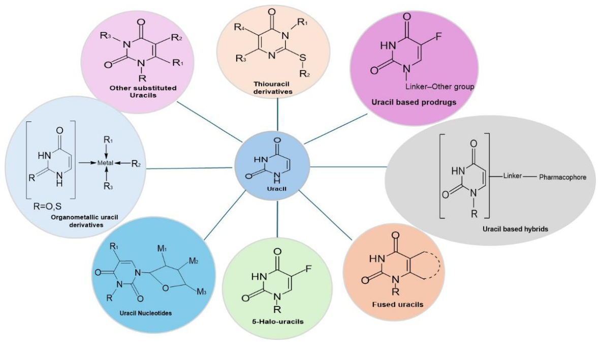
Figure 2.
Progress in metal complexation of pyridine-base compounds a route to improved anticancer effectiveness.
Figure 2.
Progress in metal complexation of pyridine-base compounds a route to improved anticancer effectiveness.
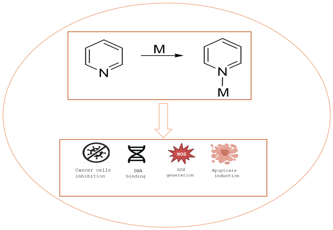
Figure 3.
The structure of free ligand 5-carboxy-2-thiouracil (L).
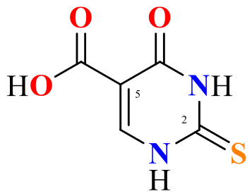
Figure 5.
Possible structure of Co(II) complex without NaOH. X represents one molecule of H2O or DMSO, thus, n can be equal to three or four.
Figure 5.
Possible structure of Co(II) complex without NaOH. X represents one molecule of H2O or DMSO, thus, n can be equal to three or four.
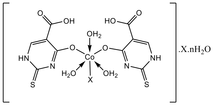
Figure 6.
Possible structure for nickel complex without NaOH.
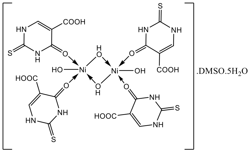
Figure 7.
Possible structure proposed for the Cu(II) complex.
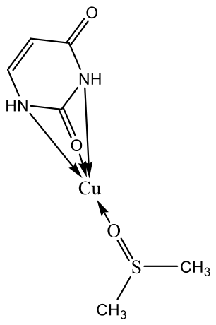
Figure 8.
Possible structure for cobalt (II) complex with NaOH.
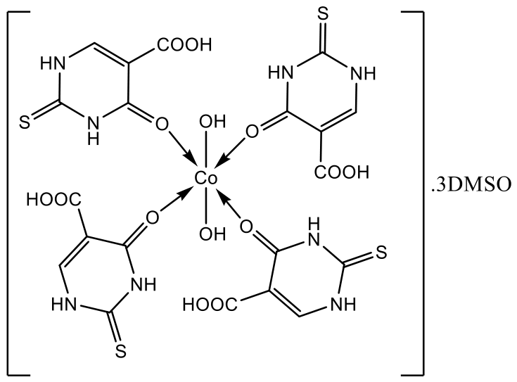
Figure 9.
Possible structure for copper (II) complex with NaOH.
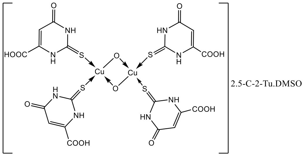
Figure 10.
Possible structure for nickel complex with NaOH.
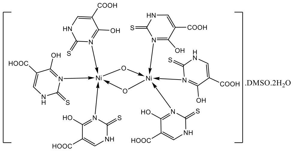
Figure 11.
5-C-2Tu (A), 5-C-2Tu+Ni(II) 1:4:2 (B) and 5-C-2Tu+Ni(II) 1:4 (C) reduce the viability of HCT-116 colorectal cells. The compounds were tested in 4 different concentrations, and their cytotoxic effects were determined with the WST-1 assay after 72 hours of treatment. The asterisks on the graph represent statistically significant differences (* p≤0.05; ** p≤0.01; *** p≤0.001) as determined by one-way ANOVA test. The x axis shows the concentration in μg/ml. The control represents cells treated with cell culture medium containing 1% DMSO.
Figure 11.
5-C-2Tu (A), 5-C-2Tu+Ni(II) 1:4:2 (B) and 5-C-2Tu+Ni(II) 1:4 (C) reduce the viability of HCT-116 colorectal cells. The compounds were tested in 4 different concentrations, and their cytotoxic effects were determined with the WST-1 assay after 72 hours of treatment. The asterisks on the graph represent statistically significant differences (* p≤0.05; ** p≤0.01; *** p≤0.001) as determined by one-way ANOVA test. The x axis shows the concentration in μg/ml. The control represents cells treated with cell culture medium containing 1% DMSO.
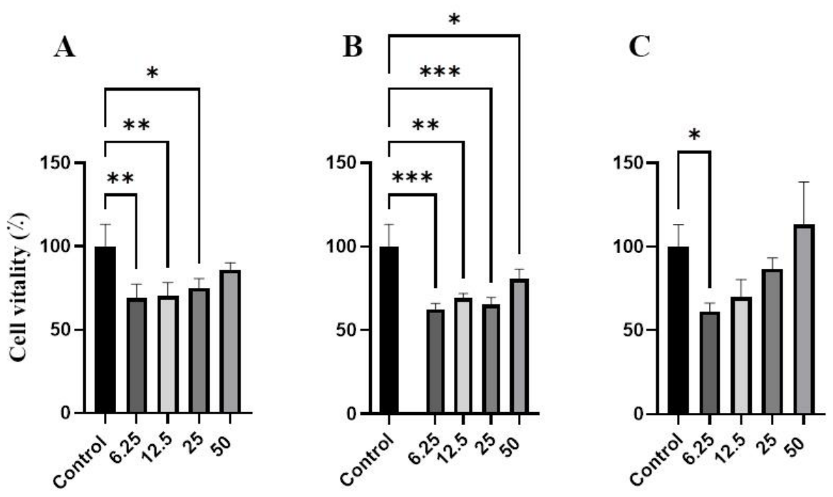
Figure 12.
5-C-Tu+Cu(II) 1:4:2 (A) and 5-C-2Tu+Cu(II) 1:4 (B) do not affect the viability of HCT-116 colorectal cells. The compounds were tested in 2 different concentrations, and their cytotoxic effects were determined with the WST-1 assay after 72 hours of treatment. No significant differences in cell number were observed between the control cells and the treated cells as determined by one-way ANOVA test. The x axis shows the concentration in μg/ml. The control represents cells treated with cell culture medium containing 1% DMSO.
Figure 12.
5-C-Tu+Cu(II) 1:4:2 (A) and 5-C-2Tu+Cu(II) 1:4 (B) do not affect the viability of HCT-116 colorectal cells. The compounds were tested in 2 different concentrations, and their cytotoxic effects were determined with the WST-1 assay after 72 hours of treatment. No significant differences in cell number were observed between the control cells and the treated cells as determined by one-way ANOVA test. The x axis shows the concentration in μg/ml. The control represents cells treated with cell culture medium containing 1% DMSO.
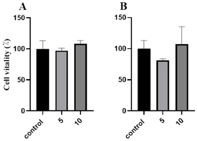
Figure 13.
5-C-Tu+Co(II) 1:4:2 (A) and 5-C-2Tu+Co(II) 1:4 (B) do not affect the viability of HCT-116 colorectal cells. The compounds were tested in 3 different concentrations, and their cytotoxic effects were determined with the WST-1 assay after 72 hours of treatment. No significant differences in cell number were observed between the control cells and the treated cells as determined by one-way ANOVA test. The x axis shows the concentration in μg/ml. The control represents cells treated with cell culture medium containing 1% DMSO.
Figure 13.
5-C-Tu+Co(II) 1:4:2 (A) and 5-C-2Tu+Co(II) 1:4 (B) do not affect the viability of HCT-116 colorectal cells. The compounds were tested in 3 different concentrations, and their cytotoxic effects were determined with the WST-1 assay after 72 hours of treatment. No significant differences in cell number were observed between the control cells and the treated cells as determined by one-way ANOVA test. The x axis shows the concentration in μg/ml. The control represents cells treated with cell culture medium containing 1% DMSO.
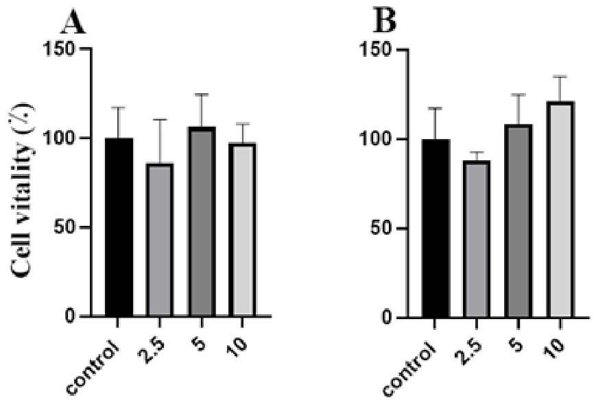
Table 1.
The analytical and physical data for metal complexes.
| Compounds | Molecular formula | Molecular weight, g/mol | colour | Melting point, °C | Experimental metal weight percentage, % | Calculated metal percentage, % | Experimental metal weight percentage, % | Calculated sulfur percentage, % |
|---|---|---|---|---|---|---|---|---|
| CuL (1:4) | C6H10O3N2SCu | 253.77 | brown | >350 | 24.8 ± 2.7 | 25.04 | 13.9 ± 1.4 | 12.64 |
| CuL (1:4:2) | Cu2S7N12C32H30O21 | 1270.19 | brown | 325-328 | 9.7±1.0 | 10.01 | 16.4±1.8 | 17.67 |
| NiL (1:4) | Ni2S5C22H36N8O22 | 1042.27 | light green | >350 | 11.2 ± 1.2 | 11.26 | 15.3 ± 1.6 | 15.38 |
| NiL (1:4:2) | Ni2S9C42H42N16O29 | 1640.84 | light green | >350 | 7.1 ± 0.7 | 7.15 | 17.6 ± 2.0 | 17.59 |
| CoL (1:4) | CoC12H26N4O14S3 | 605.48 | purple | >350 | 9.8 ± 1.2 | 9.73 | 16.0 ± 1.7 | 15.89 |
| CoL (1:4:2) | CoC26H36N8O17S7 | 1015.99 | purple | >350 | 5.8 ± 0.6 | 5.80 | 21.3 ± 2.3 | 22.09 |
Table 2.
Solubility test of the ligand and its metal(II) complexes in selected solvents.
| Compounds | Acetone | Chloroform | THF | Ethanol | Distilled Water | DMSO | DMF | Cyclohexane | Benzene | Methanol |
|---|---|---|---|---|---|---|---|---|---|---|
| C5H4N2O3S | S | S | S | S | S | S | ||||
| CuL (1:4) | IS | IS | IS | IS | IS | SS | SS | IS | IS | IS |
| CuL (1:4:2) | IS | IS | SS | IS | SS | SS | SS | IS | IS | SS |
| NiL (1:4) | IS | IS | IS | SS | IS | SS | IS | IS | IS | IS |
| NiL (1:4:2) | IS | IS | SS | IS | IS | SS | IS | IS | IS | IS |
| CoL (1:4) | SS | IS | IS | IS | SS | SS | IS | IS | IS | IS |
| CoL (1:4:2) | SS | IS | SS | SS | IS | SS | SS | SS | SS | IS |
KEY; L= C5H4N2O3 S, S= Soluble, SS=Slightly Soluble, and IS=Insoluble.
Table 3.
Electronic spectral data for copper complex obtained with sodium hydroxide.
| Compound/solvent | Absorption bands (nm) | Band assignment | Tentative geometry |
|---|---|---|---|
| C5H4N2O3S /THF | 274, 294, 326 | n → π*, π → π* | - |
| CuL (1:4:2) /THF | 270, 300, 324 | n → π*, π → π* | tetrahedral |
| C5H4N2O3S /DMSO | 272, 322 | - | |
| CuL (1:4:2) /DMSO | 278, 352 | n → π*, π → π* | tetrahedral |
| C5H4N2O3S /DMF | 270, 360 | - | |
| CuL (1:4:2)/ DMF | 300, 346 | n → π*, π → π* | tetrahedral |
| C5H4N2O3S /H2O | 216, 272, 306 | - | |
| CuL (1:4:2)/ H2O | 214, 272, 306 | n → π*, π → π* | tetrahedral |
| C5H4N2O3S /CH3OH | 216, 276, 320 | - | |
| CuL (1:4:2)/ CH3OH | 214, 280, 318 | n → π*, π → π* | tetrahedral |
Table 5.
Antimicrobial activity of the free ligand and its complexes with Co(II), Cu(II) and Ni(II).
Table 5.
Antimicrobial activity of the free ligand and its complexes with Co(II), Cu(II) and Ni(II).
| Inhibition zone, mm | |||||||||
|---|---|---|---|---|---|---|---|---|---|
| Compounds | 5-C-2Tu+Co(II) 1:4:2 | 5-C-2Tu+Co(II) 1:4 | 5-C-2Tu+Cu(II) 1:4:2 | 5-C-2Tu+Cu(II) 1:4 | 5-C-2Tu+Ni(II) 1:4:2 | 5-C-2Tu+Ni(II) 1:4 | 5-C-2Tu | DMSO | |
| Test-microorganism | |||||||||
|
Staphylococcus aureus ATCC 25923 |
- | - | 9,33±0,58 | - | - | - | - | - | |
|
Bacillus cereus ATCC 11778 |
- | - | 8,33±0,58 | 7,33±0,58 | - | - | - | - | |
|
Bacillus subtilis ATCC 6633 |
- | - | - | - | - | - | 8,00±0,00 | - | |
|
Listeria monocytogenes ATCC 8787 |
- | - | 14,33±0,58 | 18,33±1,15 | - | - | - | - | |
|
Enterococcus faecalis ATCC 19433 |
- | - | 13,00±1,00 | 16,33±0,58 | - | - | - | - | |
|
Escherichia coli ATCC 8739 |
- | - | 8,33±0,58 | - | - | - | - | - | |
| Salmonella enterica ssp. enterica ser. Enetritidis ATCC 13076 | - | 9,67±0,58 | 13,33±0,58 | 12,33±0,58 | - | - | - | - | |
|
Pseudomonas aeruginosa ATCC 9027 |
- | - | 8,67±0,58 | - | - | - | - | - | |
| Proteus vulgaris G | - | 9,33±0,58 | - | 10,33±0,58 | - | - | - | - | |
| Klebsiella pneumoniae ATCC 13883 | - | - | - | 10,00±0,00 | - | - | - | - | |
|
Candida albicans ATCC 10231 |
- | - | 10,67±0,58 | 14,33±0,58 | - | - | - | - | |
| Saccharomyces cerevisiae | - | - | 10,33±0,58 | 14,00±1,00 | - | - | - | - | |
dwell=6 mm.
Disclaimer/Publisher’s Note: The statements, opinions and data contained in all publications are solely those of the individual author(s) and contributor(s) and not of MDPI and/or the editor(s). MDPI and/or the editor(s) disclaim responsibility for any injury to people or property resulting from any ideas, methods, instructions or products referred to in the content. |
© 2026 by the authors. Licensee MDPI, Basel, Switzerland. This article is an open access article distributed under the terms and conditions of the Creative Commons Attribution (CC BY) license (http://creativecommons.org/licenses/by/4.0/).
Copyright: This open access article is published under a Creative Commons CC BY 4.0 license, which permit the free download, distribution, and reuse, provided that the author and preprint are cited in any reuse.