
Submitted:
20 September 2026
Posted:
21 September 2026
You are already at the latest version
Preprints on COVID-19 and SARS-CoV-2
Abstract
Respiratory viral infections impose a substantial global health burden. Among these, respiratory syncytial virus (RSV) is a leading cause of lower respiratory tract infections in young children, immunocompromised individuals, and older adults. During the COVID-19 pandemic, co-circulation of RSV and COVID-19 led to increasing reports of coinfections, which continue to occur in endemic settings. In this study, we employed a physiologically relevant in vitro model comprising human primary respiratory epithelial cells cultured at an air–liquid interface (ALI), together with the A549-ACE2 cell line model, to investigate the impacts of sequential RSV and SARS-CoV-2 infection. This platform enabled us to examine how host responses primed by an initial RSV infection influence subsequent SARS-CoV-2 infection. Our findings demonstrate that RSV-induced host response significantly restricted SARS-CoV-2 gene expression. Notably, we identified that RSV induced phospholipid scramblase 1 (PLSCR1), an interferon-stimulated gene previously implicated in antiviral defense against SARS-CoV-2, as a novel potential mediator of this effect. Together, these mechanisms are consistent with the phenomenon of viral interference, whereby one respiratory virus transiently reduces susceptibility to another, providing a biologically plausible explanation for cross-protective effects observed in sequential infections.
Keywords:
respiratory syncytial virus
; SARS-CoV-2
; respiratory coinfection
; phospholipid scramblase 1
; innate immunity
1. Introduction
Respiratory infections are a leading cause of mortality, resulting in about 22 percent of deaths per year globally [1]. RSV contributes significantly to this metric [2], causing an estimated 3.6 million hospitalizations per year and 100,000 deaths globally, primarily in young children (children < 5 years) [3,4]. The public health impact of RSV is exacerbated by its frequent role in coinfections with other respiratory viruses, including SARS-CoV-2 [5,6]. Since its emergence, SARS-CoV-2 has led to a total of 7 million deaths to date, and while SARS-CoV-2 is now controlled, the virus still circulates often worldwide, increasing opportunities for co-circulation of both RSV and SARS-CoV-2 [7].
Clinical and epidemiological studies estimate that up to 30% of hospitalized patients with respiratory disease test positive for more than one virus [8]. Respiratory viral coinfections have become increasingly recognized or considered in both pediatric and adult populations; there are many retrospective case reports demonstrating RSV/SARS-CoV-2 coinfections [9]. Previous studies of RSV/SARS-CoV-2 coinfection in vitro and in vivo models have started to study how the viruses influence each other during co-circulation [10,11,12,13]. However, limited studies have been done in human-derived models, especially those physiologically relevant.
Herein, we used human primary respiratory cells cultured in air–liquid interface (ALI) as models to confirm the impact of RSV infection on subsequent SARS-CoV-2 infection from in vivo observation. The importance of this study includes the airway epithelium as a primary target of respiratory viral infection and first-line defender against viral infection, and reports on how the viruses’ interplay within these cells, which may provide critical insights into how we could strategize to control these coinfections. In addition, ALI-cultured primary epithelial cells are increasingly acknowledged to be a more physiologically relevant human in vitro model and are used as a preclinical model for testing therapeutic efficacy against respiratory viral infections [14,15]. We found that RSV infection in ALI-cultured primary airway epithelial cells, derived from both upper and lower respiratory tracts, significantly suppressed SARS-CoV-2 infection. The results were also confirmed in A549 cells overexpressing angiotensin-converting enzyme 2 (hACE2; A549-ACE2). These models allow us to explore and perturb the molecular mechanisms used by RSV to interfere with SARS-CoV-2 infection. We found that innate immunity shaping in RSV-infected epithelial cells likely accounted for the attenuated replication of SARS-CoV-2, improving our understanding of viral-driven regulation of host immunity against viral co-circulation. Our observations are consistent with the phenomenon of viral interference, whereby one respiratory virus transiently reduces susceptibility to another, providing a possible rationale for the previously reported cross-protective effects during viral respiratory coinfections. 2. Materials and Methods
Cell Culture
Human Nasal Epithelial Cells and Cultures
Primary nasal epithelial cells (hNEpCs; PromoCell, Heidelberg, Germany; C-12620) were expanded in Airway Epithelial Basal Medium (PromoCell, C-21260) with SupplementMix (PromoCell, C-39165) and then differentiated in ALI culture using PneumaCult-based media (StemCell Technologies, Vancouver, BC, CA, C-100-1505) following the manufacturer’s instructions. In brief, cells within the passages of 3 and 5, were seeded into Costar® 12-mm Transwell® inserts, 0.4 μm pore polyester membrane (StemCell Technologies, C-38023) at a cell density sufficient to achieve confluence within 3 to 5 days (typical cellular density range was 0.8-1.5x10⁵ cells/insert). The cells were transitioned to ALI culture by withdrawal of apical tissue culture media upon confluence and differentiated until hallmarks of mature airway epithelium were evident (ciliation and mucus production; typically, 21 days total).
Human Small Airway Epithelial Cells and Their Cultures
Primary human small airway epithelial cells (hSAECs; Lonza, Walkersville, Maryland, USACC-2547) were isolated from the distal portion of the lung within the 1-mm bronchiole area and were expanded in flasks using Clonetics® SABM™ Small Airway Cell Basal Medium (Lonza, CC-3119) with the addition of SAGM™ SingleQuots® supplements medium (Lonza, CC-4124) and differentiated in ALI culture using complete PneumaCult-Ex Plus medium (StemCell Technologies, C-05040) and PneumaCult-ALI (StemCell Technologies, C-05001) according to the manufacturer’s instructions. Briefly, the cells at passage 2 (P2) were expanded into T-25 flasks with medium changes every other day. For ALI culture, passage 3 (P3) cells were seeded into Transwell® inserts at 1.1 x 10⁵ cells/insert in 0.5 mL medium per insert, with 1 mL per well medium added to the basal chamber. Cells were grown submerged until 100% confluence was reached, then ALI was initiated by removing apical medium and replacing the basal medium with PneumaCult-ALI. The basal compartment medium was changed every other day, and cultures were differentiated for around 21 days before infection.
Human Primary Cell Ethics Status
Because primary human epithelial cells from multiple donors were used throughout the study, we verified that the commercial suppliers (Lonza and PromoCell) obtained informed consent of the human donors before collection of their tissues, and that the cells were anonymized or de-identifiable.
A549-ACE2 Culture
Human alveolar type II-like epithelial cells expressing human angiotensin-converting enzyme 2 (A549-ACE2) (a gift from Shinji Makino’s lab at the University of Texas Medical Branch [16]) were maintained in high-glucose DMEM (Gibco, Waltham, MA, C-11995065) supplemented with 10% fetal bovine serum (FBS) (Gibco, C-16000044), which was heat inactivated in-house, 10 U/mL penicillin, and 10 μg/mL streptomycin (Gibco, C-15140122) with the addition of Blasticidin S HCl (ThermoFisher Scientific, Waltham, MA, C-A1113903) at a concentration of 10 μg/mL in 37 °C in 5% CO2 [17]. Cells were passaged using standard trypsinization and routinely confirmed to be mycoplasma-free.
Viruses
Respiratory Syncytial Virus (RSV) Preparation
RSV Long strain was propagated in HEp-2 cells and purified by sucrose gradient as previously described [18]. Viral titers were determined by immunostaining in HEp-2 cells using polyclonal biotin-conjugated goat anti-RSV antibody (Ad Direct, Barberton, OH, C-7950-0104) followed by streptavidin peroxidase polymer (Sigma-Aldrich, St. Louis, MO, C-S2438).
SARS-CoV-2 Preparation
SARS-CoV-2 (Omicron BA.1 strain) was obtained from the World Reference Center for Emerging Viruses and Arboviruses (WRCEVA) at the University of Texas Medical Branch (UTMB). Viral stocks were prepared by propagation once in Vero E6 cells (ATCC, Manassas, VA, C-CRL-1586) and maintained in high-glucose DMEM supplemented with 10% FBS, 10 U/mL penicillin, and 10 μg/mL streptomycin. Viral titers were determined by plaque assay as described in [19]. All experiments using live SARS-CoV-2 were performed in a biosafety level 3 (BSL-3) laboratory under the Institutional Biosafety Committee of UTMB (NOU# 2018056 and NOU# 2020043).
Viral Infections
ALI Infections (hNEpCs and hSAECs)
Prior to infection, ALI-cultured cells were apically washed five times with 1X PBS containing Ca2+ and Mg2+ (Corning, Manassas, VA, C-21-030-CV), and the basal surface was washed once with 1X PBS. RSV viruses were diluted to an MOI of 5 in 200 µL MEM (Gibco, C-11095080) and inoculated onto the apical surface. After a 2-hour incubation at 37 °C with 5% CO2, the unbound RSV virus was then removed by washing the apical surface three times with 1X PBS, and ALI conditions were restored. Basal medium was replaced with fresh PneumaCult-ALI, and cells were coinfected with SARS-CoV-2 at approximately 24 hours after RSV infection by apically inoculating with 200 μL of MEM containing SARS-CoV-2, then incubated for 3 hours at 37 °C with 5% CO2 to allow virus attachment. The cells without SARS-CoV-2 infection were also used as controls for mock- or RSV-infected cells. The unbound virus was then removed by washing the apical surface three times with 1X PBS. ALI conditions were restored. Cells, after apical washes and collection, were collected at the indicated time points, such as Day 1 for RSV-only samples, Day 3 for SARS-CoV-2 samples, or Day 4 for coinfection samples (please see the workflow, as illustrated in Figure 1A).
A549-ACE2 Monolayer Infections
A549-ACE2 cells were seeded into 12-well plates 24 hours prior to infection to reach 80–90% confluence the following day. For RSV infection, cells were incubated with the virus in MEM with 2% FBS at an MOI of 1 for 2 hours. Cells were then washed three times with 1X PBS to remove unbound virus and cultured in fresh MEM media containing 2% FBS until harvest or coinfection with SARS-CoV-2 the next day. For SARS-CoV-2 infection, cells were incubated with virus in DMEM containing 10% FBS at an MOI of 0.1 for 1 h. Cells were then washed three times with 1X PBS to remove unbound viruses and cultured in fresh DMEM media containing 10% FBS until harvest. For RSV infection, cells were infected at the indicated MOI under analogous adsorption and wash conditions. For co-infections, cells were exposed to RSV and SARS-CoV-2 sequentially (order and interval specified in Figure 1 schematic), using the same adsorption conditions for each virus. Cell lysates were harvested in TRIzol (Invitrogen, Carlsbad, CA, C-15596026) and/or supernatants were collected at the indicated time-pointsfor downstream analyses.
siRNA Treatment
A549-ACE2 Monolayer Treatments with siRNA
A549-ACE2 cells were seeded into 12-well plates 24 hours before infection, reaching 70–90% confluence the following day. The next morning, upon confirmation of the correct confluency for transfection, siRNAs, target-specific or scrambled, were prepared in Lipofectamine 2000 (Invitrogen, Carlsbad, CA Cat. C-11668027). siRNA targeting PLSCR1 (si-PLSCR1, siRNA ID: 5359) or scrambled si-control (si-CN, C-EHU23051), were ordered from Sigma-Aldrich, St. Louis, MO. Approximately 4 hours before beginning infection with mock or RSV, the siRNAs were applied to monolayer-cultured A549-ACE2.
RNA Isolation
Total cellular RNA was extracted using TRIzol™ Reagent (Invitrogen, C-15596018) according to the manufacturer’s protocol. For ALI cultures, inserts were rinsed with PBS and lysed in TRIzol. For monolayer cultures, cells were lysed directly in TRIzol. RNAs were then extracted by chloroform-based phase separation, isopropanol precipitation, and ethanol washes, then resuspended in nuclease-free water. RNA concentration and quality was assessed by spectrophotometry, and samples were stored at −80 °C until analysis by qRT-PCR.
Quantitative RT-PCR
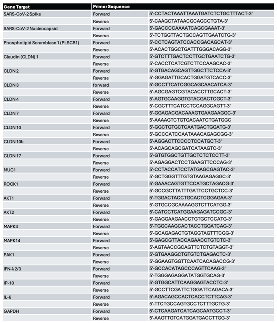
Complementary DNA (cDNA) was synthesized by using iScript Advanced cDNA Synthesis Kit (Bio-Rad, Hercules, CA, C1725037). The sequences of the qPCR primer sets for SARS-CoV-2 S and N gene, human host gene targets, and human cytokine/chemokine genes are listed in Table 1. The PCR assay was performed using the CFX96 real-time PCR system (Bio-Rad). Gene expression was calculated using the formula 2^-[CT(target gene)-CT(GAPDH)] as described before [20].
To quantify RSV N gene expression, cDNA was synthesized with 1 μg of total RNA in a 20-μL reaction mixture using the TaqMan Reverse Transcription Reagents kit from ABI (Applied Biosystems, Foster City, CA, C-N8080234). We used RT primer 5′-CTGCGATGAGTGGCAGGCTTTTTTTTTTTTAACTCAAAGCTC-3′. We incorporated a “tag” (underlined letters) as part of the assay due to self-priming exhibited by viral RNA. The tag sequence was derived from the bacterial chloramphenicol resistance (Cmr) gene. The sequence with bold letters is complementary to the poly(A) tails of the transcribed RSV N gene. The sequence in italics is N gene-specific. The reaction conditions were as follows: 25 °C for 10 min, 48 °C for 30 min, and 95 °C for 5 min. At a 25 °C annealing temperature, the 8 nucleotides (nt) matching N-specific sequences would not be sufficient for stable, efficient priming of cDNA from the antigenome of RSV. On the other hand, 20 nucleotides matching transcribed N (12 T’s and N gene-specific nucleotides) can attain stable annealing to the transcribed N gene. For Quantitative real-time PCR amplification, we used the RSV tag reverse primer CTGCGATGAGTGGCAGGC and the forward primer ACTACAGTGTATTAGACTTRACAGCAGAAG. The PCR was performed with 1 μL of cDNA in a total volume of 25 μL by using iTaq TM Universal SYBR Green Supermix (Bio-Rad, C-1725124). The final primer concentration was 300 nM. GAPDH RNA was used as a housekeeping gene for normalization
Statistical Analysis
Statistical analyses were performed using GraphPad Prism 10. Data are presented as mean ± SEM unless otherwise noted. For comparisons between two groups, paired two-tailed Students T-test were used; for multiple-group comparisons, One-Way or Two-Way ANOVA with appropriate multiple-comparison correction was applied (Tukey’s post-hoc test). A p-value ≤ 0.05 was considered statistically significant.
Generative artificial intelligence (GenAI) has not been used in this paper (e.g., to generate text, data, or graphics, or to assist in study design, data collection, analysis, or interpretation).
3. Results
3.1. RSV Pre-Infection Attenuates SARS-CoV-2 Gene Expression in Cellular Coinfection Models
To model the coinfection of RSV and SARS-CoV-2, we implemented ALI culture for human primary nasal epithelia (hNEpCs) and small airway epithelia (hSAECs) from various donors, similarly as we have previously described [21]. In brief, the well-differentiated cells cultured in ALI were apically infected with RSV at an MOI of 5, as described [22]. Mock infection was used as a control. At Day 1 RSV post-infection (p.i.), the cells were washed three times with PBS from the apical side and followed by the apical infection with or without SARS-CoV-2 (Omicron BA.1) at an MOI of 0.1. After 3 hours, the apical infection was removed, and cells were left in ALI culture. The total cells and the basal compartment were harvested after 3 days post SARS-CoV-2 infection (day 4 of the experiment) for the downstream experimental assays. The experimental timeline and associated conditions are summarized in Figure 1A.
Following the order of RSV infection, then SARS-CoV-2 coinfection, we noticed that prior RSV infection before SARS-CoV-2 coinfection demonstrated a significant reduction in SARS-CoV-2 Spike (S) mRNA expression during coinfection, as shown by qRT-PCR for hNEpCs (Figure 1B).). Coinfected cells were capable to produce compareable IL-6 (Figure 1C) and more IP-10 (Figure 1D) than SARS-CoV-2-infected cells .
Similarly, we discovered that prior RSV infection led to a significant reduction in SARS-CoV-2 Spike (S) mRNA expression (Figure 1E), a comparable IL-6 expression (Figure 1F), and an enhanced IP-10 expression (Figure 1G) in ALI-cultured hSAECs. These data suggest that prior RSV infection primes the host cellular environment for protection against SARS-CoV-2 reaching high S copy number. Additionally, these data recapitulate, on a cellular level, similar observations recently published for ALI-cultured bronchial epithelium cells, specific to this order of coinfection [11], supporting that RSV infection of epithelial cells along the respiratory tract, regardless of the location, generated a protective microenvironment against subsequent SARS-CoV-2 infection.
3.2. RSV-Impacted Antiviral Signaling that May Contribute to SARS-CoV-2 Spike Suppression
To identify host factors that could be influenced by RSV that causes SARS-CoV-2 Spike suppression, we assessed the expression of genes reported to be essential for host antiviral signaling on day 1 post-RSV infection, which is before the SARS-CoV-2 infection. For example, core PI3K-AKT pathway kinases have been reported to be essential for regulating SARS-CoV-2 replication. AKT1/2/3 are downstream effectors of PI3K. The activation of AKTs favors viral SARS-CoV-2 replication [23]. Inflammatory and cell-death-associated kinases such as p38 MAPK (MAPK14) have been reported to promote SARS-CoV-2 replication. Pharmacologic inhibition or genetic knockdown of MAPK14 significantly reduces viral RNA levels and infectious virion production [24,25]. PAKs (p21-activated kinases), especially PAK1, are implicated as pro-viral host kinases in SARS-CoV-2 infection [26,27]. PAK1 inhibition or knockdown reduces viral RNA levels and infectious particle production in vitro [26,28]. ROCK1 (Rho-associated coiled-coil kinase 1) is also a kinase implicated in SARS-CoV-2 infection, mainly as a host regulator of viral entry, cytoskeletal remodeling, and inflammatory pathology [29]. Of the targets considered, AKT1 and MAPK3 had a slight induction of their mRNA expression by RSV in ALI-cultured hNEpCs, while AKT2, PAK1, MAPK14, and ROCK1 demonstrated a non-significant change in mRNA expression (Figure 2).
In ALI-cultured hSAECs, PAK1 was significantly enhanced by RSV at day 1 p.i., while the transcription of AKT1, AKT2, MAPK3, MAPK14, and ROCK1 was not affected by RSV (Figure 3, below).
Phospholipid scramblase 1 (PLSCR1) is a membrane-associated protein that was recently found to be a novel anti-SARS-CoV-2 molecule [30]. RSV could, therefore, induce PLSCR1 to suppress SARS-CoV-2 infection. Generally, PLSCR1 has gained notoriety through its antiviral activities, especially through its categorization as a potent interferon-stimulating gene (ISG). PLSCR1 not only acts in an antiviral manner but also operates as a regulator of endogenous immune components to mediate protection against viruses within both immune and nonimmune cells [31]. To date, there are no reports of RSV inducing PLSCR1 or of how PLSCR1 regulates RSV infection or coinfections involving RSV or SARS-CoV-2. This is why we were so intrigued by this gene target and wanted to explore it further. As shown in Figure 4, RSV significantly increased PLSCR1 expression in both hNEpCs (A) and hSAECs (B) by a relative fold change of 20 and 10, respectively. Taken together with the data shown in Figure 2 and Figure 3, the fold change in PLSCR1 expresion, was more pronounced than that observed for the downstream kinases.
3.3. The Role of PLSCR1 in Viral Infections
To determine the antiviral role that PLSCR1 has during RSV and RSV/SARS-CoV-2 coinfection, we utilized a siRNA method to silence PLSCR1 and to investigate the impact of PLSCR1 on viral gene expression in each infection condition. To do this, we employed a similar model of coinfection with A549-ACE2 cells, which also showed that RSV-infected cells allowed less gene expression of SARS-CoV-2 (Figure 5A). A549 cells are a commonly used model representing lower tract respiratory epithelial cells for respiratory viral infections, including RSV infection [32]. Like ALI-cultured hNEpCs and hSAECs, A549-ACE2 cells had enhanced expression of PLSCR1 by RSV infection (Figure 5B). To investigate the role of RSV-induced PLSCR1 in SARS-CoV-2 infection, we pre-treated A549-ACE2 cells with scrambled si-CN or si-PLSCR1 at a concentration of 100 nM six hours before RSV infection on day 0 of the experimental timeline. RSV infection was done at an MOI of 0.01, followed by SARS-CoV-2 (Omicron BA.1) at an MOI of 0.1. We found that RSV infection with an MOI of 0.01 did not result in significant cell detachment on day 4 p.i. (data not shown). As demonstrated by Figure 5C, efficient knockdown was confirmed by qRT-PCR measurement of PLSCR1 mRNA. As shown in Figure 5D, in the context of SARS-CoV-2 infection only, there was a greater increase in N expression of SARS-CoV-2 by si-PLSCR1 treatment than that in si-PLSCR1-treated cells, compared to si-CN-treated cells. Since SARS-CoV-2 N is widely used as a surrogate marker of viral replication because it is highly expressed during active infection, is essential for viral genome replication and packaging, and accumulates in infected cells in proportion to viral replication, the result implicated the critical anti-SARS-CoV-2 role of PLSCR1, consistent to what is reported for PLSCR1 in SARS-CoV-2 infection [30]. Intriguingly, in response to coinfection (last pair), RSV pre-infection resulted in a 947-fold reduction in SARS-CoV-2 N expression in si-CN-treated cells, while in si-PLSCR1-treated cells, RSV pre-infection led to about a 475-fold reduction in SARS-CoV-2 S expression, suggesting that the impact of RSV-suppressed SARS-CoV-2 gene replication is also PLSCR1-dependent.
3.4. PLSCR1 Is constantly Upregulated in Primary Airway Epithelium During RSV and Coinfection
Now that we had a gene target of interest that could contribute to SARS-CoV-2 restriction during coinfection, which its expression was demonstrated to be influenced by RSV infection, we wanted to validate whether PLSCR1 mRNA expression demonstrated enhancement during not only day 1 RSV-post infection but also at day 4 RSV-post infection to address the question whether increased PLSCR1 mRNA expression was a transient interferon-driven burst or a sustained but regulated antiviral program that is induced upon infection. PLSCR1 mRNA expression remains significantly elevated even at later stages of RSV infection (day 4) within the experimental timeline considered, albeit at reduced levels compared to early timepoints (Figure 6A-C). This is consistent with a sustained rather than a transient early response. Relatedly, mRNA expression of IFNs was considered following the RSV infection, given that PLSCR1 is an ISG. As we know, RSV is widely considered a poor inducer of type I IFN, and two RSV non-structural proteins, NS1 and NS2, as well as the envelope G glycoprotein, are known to suppress type I IFN production [18,33,34]. A robust type I IFN response to RSV does not occur in human infants or neonatal mouse models of RSV infection [35]. We, therefore, measured IFN-lambda (IFN-l), a type-III IFN, which has been shown to directly stimulate the transcription of PLSCR1 in airway epithelial cells [31]. As evidenced further by Figure 6D-G, IFN-l is significantly elevated in both hNEpCs and hSAECs, both at day 1 and day 4 post-RSV infection, respectively. Interestingly, enhanced PLSCR1 did not affect RSV N protein expression, suggesting that RSV evaded the PLSCR1-mediated antiviral effect. Together with results shown in Figure 5 on the suppressive impact of PLSCR1 on SARS-CoV-2 N , these results demonstrated that the impact of PLSCR1 on viruses may be virus-specific (Figure 6H).
4. Discussion
In this study, we identified that upon the development of our physiologically relevant cellular RSV/SARS-CoV-2 coinfection models that recapitulate the nasal and small airway epithelium, SARS-CoV-2 gene expression was restricted by prior RSV, similarly to previous literature in primary human bronchial epithelial cells [11]. Of the targets screened from the literature, PLSCR1 mRNA is rapidly induced by RSV alone by day 1 in hNEpCs and hSAECs. siRNA knockdown of PLSCR1 restored some RSV-suppressed SARS-CoV-2 gene expression during coinfection. Lastly, RSV induced an early sustained innate immune response, possibly through the presence of induced IFN-l. Together, these findings identify RSV-induced PLSCR1 as a host factor that contributes to SARS-CoV-2 restriction during coinfection.
PLSCR1 is an antiviral ISG. Generally, it is induced by all three forms of IFNs [36]. PLSCR1 interferes with viral replication among a variety of viruses, including not only SARS-CoV-2, but also influenza A virus (IAV), Human Immunodeficiency virus (HIV), Epstein-Barr virus (EBV), Human T-lymphotropic virus 1 (HTLV1), Human Cytomegalovirus (HCMV), and Hepatitis B virus (HBV) [30,37,38,39,40,41,42]. However, PLSCR1 also has proviral functions. For example, with Hepatitis C virus (HCV), PLSCR1 directly binds to occludin and HCV proteins E1 and E2, promoting viral attachment [43]. Cheshenko et al. found that the enzymatic activity of PLSCR1 is exploited by Herpes Simplex Virus (HSV)-1 to translocate host protein AKT (and dock for HSV-1) to the outer leaflet of the plasma membrane; thereby promoting HSV-1 viral entry into host cells [44]. Herein, we found that PLSCR1 did not affect RSV N expression while suppressing SARS-CoV-2 N, further confirming the observation that the role of PLSCR1 in viral infections is indeed virus-specific.
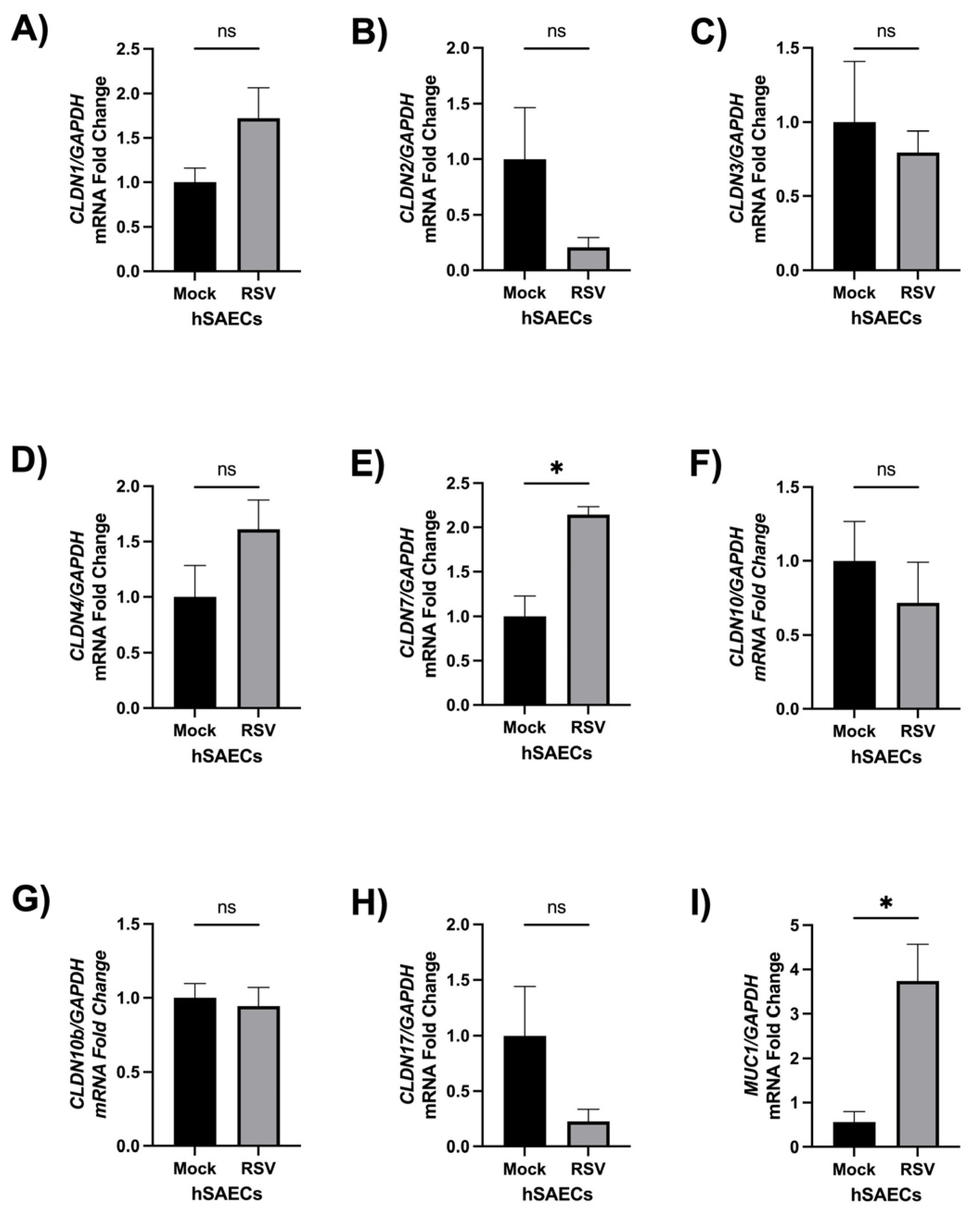
We found that RSV enhanced PLSCR1 expression in ALI-cultured hNEpCs and hSAECs at a fold change of 20 and 10, respectively, at day 1 p.i. We also discovered that PLSCR1 silencing could not fully recover RSV-suppressed SARS-CoV-2 gene expression (Figure 5D). There are several possible reasons. One possibility is that siRNA silencing did not result in complete depletion of PLSCR1. Currently, the lab is developing techniques for gene knockout of PLSCR1 in an ALI-cultured system, and more impact of PLSCR1 on SARS-CoV-2 N expression is expected. The second possibility is that PLSCR1 is, of course, not the sole RSV-induced antiviral molecule. Indeed, Claudin (CLDN) genes were also considered. According to previous knowledge that RSV regulates epithelial barrier integrity across various regions of the respiratory tract during infection [45], it is possible that RSV impacts CLDN expression to regulate SARS-CoV-2 entry [46]. Among the CLDNs considered, CLDN2, CLDN10, CLDN10b and CLDN17 are essential for epithelial cell integration, while CLDN1, CLDN3, CLDN4 and CLDN7 are critical in barrier-forming [45,47,48]. As shown in Supplementary Figure 1 (A-H), in ALI-cultured hSAECs, similar as A549 cells as lower respiratory tract epithelial cells, of the claudins considered and their mRNA expression measured during day 1 post-RSV infection, prior to SARS-CoV-2 challenge, CLDN7 showed a significant increase in expression during day 1 post-RSV infection, while the expression of other tested CLDNs was comparable between mock and RSV-infected cells. However, the fold induction of CLDN7 was much lower than that of PLSCR1. Therefore, here, we primarily focused on PLSCR1, also given its higher induction and being a relatively newer molecule reported to be essential for controlling virus replication. We also examined Mucin-1 (MUC1) and found that it was enhanced by RSV infection (Supplementary Figure 1I). We included MUC1 as it has been reported to physically prevent SARS-CoV-2 from approaching the cell surface and indirectly suppress spike expression [49]. MUC1 mRNA expression was significantly enhanced during day 1 RSV-post infection. However, we did not examine MUC1 further because we considered that, with our ALI-cultured SAECs, when we infected cells with SARS-CoV-2, we fully washed the apical sites with PBS before SARS-CoV-2 infection was applied to eliminate the possible impact of MUC1. In addition, in A549-ACE2 cells cultured in a monolayer, MUC1 secretion was minimal (data not shown). However, in the natural coinfection, it is very feasible that RSV-induced MUC1 could contribute to making respiratory tract cells less susceptible to SARS-CoV-2 infection.
Because of the wide availability of SARS-CoV-2 and RSV vaccines, it may be misunderstood that RSV and SARS-CoV-2 coinfections would likely be ruled out among their similar at-risk populations of these two viruses. But the efficacy of the RSV vaccines can decrease from season one to season two post-vaccination due to the vaccine-induced antibody levels waning over time. Several studies specifically highlighted that the vaccine candidate’s effectiveness can have a dramatic (close to 40%) decrease in immunological benefit to the trial populations they surveyed [52,53,54]. Additionally, a study conducted by The Lancet in mid-2025 demonstrated that even a second subunit vaccine dose administered 1 year after the first dose did not provide additional benefit to the trial population they surveyed [52]. With SARS-CoV-2 vaccinations, generally, similar waning protection over time due to antibody-producing long-lived plasma cells not maturing properly during infection and thereby contributing to immune evasion by new viral variants [55]. Also, respiratory vaccine hesitancy still contributes to reduced rates of protection [56]. Taken together, further understanding of the mechanisms responsible for the discrepancies in phenotypes of disease outcome during RSV and SARS-CoV-2 coinfections remains warranted due to the prospect of the frequency of coinfections increasing because RSV has returned to pre-pandemic seasonality, SARS-CoV-2 is now endemic globally, and the insufficient protection of currently available vaccine options.
Together, the observed reduction in SARS-CoV-2 gene expression following RSV infection suggests that RSV-induced antiviral priming, potentially involving upregulation of PLSCR1, reshapes host permissiveness during coinfection. However, viral interference is highly dependent on infection sequence. Given the well-established IFN antagonism produced by SARS-CoV-2, infection in the reverse order seems to yield distinct outcomes [11] that warrant further investigation, which we plan to investigate further. This question is particularly relevant in the current epidemiological context, characterized by declining SARS-CoV-2 vaccinated populations and restored circulation of RSV to pre-pandemic levels. As vaccines against both viruses are now widely implemented, understanding how prior infection or immune priming alters host determinants such as PLSCR1 could serve as a potential driver hypothesized to have influence over coinfection risk, disease severity, and therapeutic strategies, which warrant further consideration.
Specific Limitations
We wanted to explicitly acknowledge several specific limitations from our study here. This study used physiologically relevant human cell model to validate PLSCR1-mediated RSV-suppressed SARS-CoV-2 gene expression; however, the regulatory function of PLSCR1 needs further characterization within an in vivo animal model. We now have a plausible rationale for viral interference during this viral coinfection; however, we do not know whether SARS-CoV-2 infection will shape the cellular microenvironment to impact RSV infection in our model. In the future, we will validate our findings of SARS-CoV-2 gene expression and associate these observations here with infectivity-based assays (such as focus-forming assay) to determine whether SARS-CoV-2 infectious virions are also affected by RSV. In the future, we ideally want to use iPSCs to develop respiratory epithelia for CRISPR/Cas9 gene editing to generate a complete knockout of PLSCR1 due to the observed incomplete phenotypic recovery after the silence of PLSCR1 in A549-ACE2 cells. Related, this study is a literature reference-based target screen. We anticipate corroborating our findings with PLSCR1 in addition to linking novel genes that are known as host dependency and restriction factors essential for viral replication during coinfection through CRISPRi/CRISPRa screening.
Author Contributions
Manuscript draft: MDRB and WW; Experiment performance and data analysis: MDRB, WW, DD, AA, ESC, EJC, JA, KA, KW, and KK; Sample collection: WW, MDRB, AA, ESC, EJC, HV; Experimental design: WW, TW, and XB; Manuscript finalization: XB.
Funding
This work was supported by grants from the US National Institutes of Health (NIH) R21 AI166543, R61 AG075725, and ERP-1252718 from American Lung Association to XB; R01AI127744, R01 NS125778, and R01 AI176670 to TW; MDRB is supported by Kempner Predoctoral Fellowship; DD was supported by NIAID T35 Infectious Diseases & Inflammatory Disorder Training Program (T35AI0778878, PI: TW).
Data Availability Statement
The numerical data underlying the figures and statistical analysis are available from a corresponding author upon reasonable request. Some are provided in supplementary material. All raw data were obtained with experimentation in the XB laboratory.
Conflicts of Interest
The authors declare no conflicts of interest.
Abbreviations
The following abbreviations are used in this manuscript:
| S | SARS-CoV-2 Spike |
| N | SARS-CoV-2 Nucleocapsid |
| PLSCR1 | Phospholipid scramblase 1 |
| CLDN1 | Claudin1 |
| CLDN2 | Claudin2 |
| CLDN3 | Claudin3 |
| CLDN4 | Claudin4 |
| CLDN7 | Claudin7 |
| CLDN10 | Claudin10 |
| CLDN10b | Claudin10b |
| CLDN17 | Claudin17 |
| MUC1 | Mucin1 |
| AKT1 | AKT serine/threonine kinase 1 |
| AKT2 | AKT serine/threonine kinase 2 |
| MAPK3 | Mitogen-Activated Protein Kinase 3 |
| MAPK14 | Mitogen-Activated Protein Kinase 14 |
| PAK1 | p21-Activated Kinase 1 |
| ROCK1 | Rho-associated, coiled-coil-containing protein kinase 1 |
| IFN-λ | Interferon-lambda |
| IP-10 | Interferon-gamma-inducible protein 10 |
| IL-6 | Interleukin-6 |
| GAPDH | Glyceraldehyde-3-phosphate dehydrogenase |
Appendix A
Appendix A.1. Appendix Figure A1
Figure A1.
Claudin genes were extensively considered in hSAECs during RSV infection but were not always significantly induced. A) CLDN1 mRNA expression 1 day post infection, B) CLDN2 mRNA expression 1 day post infection, C) CLDN3 mRNA expression 1 day post infection, D) CLDN4 mRNA expression 1 day post infection, E) CLDN7 mRNA expression 1 day post infection, F) CLDN10 mRNA expression 1 day post infection, G) CLDN10b mRNA expression 1 day post infection, H) CLDN17 mRNA expression 1 day post infection, and I) MUC1 mRNA day 1 post infection. ). hSAE cells from two donors were used and grown on membrane inserts in 12 well plate with six repeats per group, three independent experiments. *P ≤ 0.05.
Figure A1.
Claudin genes were extensively considered in hSAECs during RSV infection but were not always significantly induced. A) CLDN1 mRNA expression 1 day post infection, B) CLDN2 mRNA expression 1 day post infection, C) CLDN3 mRNA expression 1 day post infection, D) CLDN4 mRNA expression 1 day post infection, E) CLDN7 mRNA expression 1 day post infection, F) CLDN10 mRNA expression 1 day post infection, G) CLDN10b mRNA expression 1 day post infection, H) CLDN17 mRNA expression 1 day post infection, and I) MUC1 mRNA day 1 post infection. ). hSAE cells from two donors were used and grown on membrane inserts in 12 well plate with six repeats per group, three independent experiments. *P ≤ 0.05.
References
- Global age-sex-specific all-cause mortality and life expectancy estimates for 204 countries and territories and 660 subnational locations, 1950-2023: a demographic analysis for the Global Burden of Disease Study 2023. Lancet 2025, 406(10513), 1731–1810. [CrossRef] [PubMed Central]
- Bao, X.; Liu, T.; Shan, Y.; Li, K.; Garofalo, R.P.; Casola, A. Human metapneumovirus glycoprotein G inhibits innate immune responses. PLoS Pathog. 2008, 4(5), e1000077. [Google Scholar] [CrossRef] [PubMed Central]
- Available online: https://www.who.int/news-room/fact-sheets/detail/respiratory-syncytial-virus-(rsv) (accessed on 28 March 2026).
- Halabi, K.C.; Wang, H.; Leber, A.L.; Sánchez, P.J.; Ramilo, O.; Mejias, A. Respiratory syncytial virus and SARS-CoV-2 coinfections in children. Pediatr. Pulmonol. 2022, 57(12), 3158–3160. [Google Scholar] [CrossRef] [PubMed Central]
- Li, Y.; Pillai, P.; Miyake, F.; Nair, H. The role of viral co-infections in the severity of acute respiratory infections among children infected with respiratory syncytial virus (RSV): A systematic review and meta-analysis PMC7295447 reports grants and personal fees from Innovative Medicines Initiative, grants from World Health Organization, personal fees from Bill and Melinda Gates Foundation, grants and personal fees from Sanofi, personal fees from Janssen, personal fees from AbbVie, grants from National Institute of Health Research, outside the submitted work. The authors have completed the ICMJE conflict of interest form (available upon request from the corresponding author), and declare no further conflicts of interest. J. Glob. Health 2020, 10(1), 010426. [Google Scholar]
- Attaianese, F.; Carreras, G.; Trapani, S.; et al. Nirsevimab Immunization to Prevent Pediatric RSV Hospitalizations. JAMA Pediatr. 2026. Angelini Pharma S.P.A, personal fees from Angelini Pharma S.P.A, and grants from Reckitt Benckiser Healthcare SPA outside the submitted work. Dr Azzari reported receiving personal fees from Sanofi, Pfizer, and Moderna and grants from GSK and Merck outside the submitted work. No other disclosures were reported. [Google Scholar] [CrossRef] [PubMed Central]
- Available online: https://data.who.int/dashboards/covid19/cases (accessed on 28 March 2026).
- Georgakopoulou, V.E. Insights from respiratory virus co-infections. World J. Virol. 2024, 13(4), 98600. [Google Scholar] [CrossRef] [PubMed Central]
- Agathis, N.T.; Patel, K.; Milucky, J.; et al. Codetections of Other Respiratory Viruses Among Children Hospitalized With COVID-19. Pediatrics 2023, 151(2). [Google Scholar] [CrossRef]
- Vanetti, C.; Saulle, I.; Artusa, V.; et al. A complex remodeling of cellular homeostasis distinguishes RSV/SARS-CoV-2 co-infected A549-hACE2 expressing cell lines. Microb. Cell 2024, 11, 353–367. [Google Scholar] [CrossRef] [PubMed Central]
- Dee, K.; Schultz, V.; Haney, J.; Bissett, L.A.; Magill, C.; Murcia, P.R. Influenza A and Respiratory Syncytial Virus Trigger a Cellular Response That Blocks Severe Acute Respiratory Syndrome Virus 2 Infection in the Respiratory Tract. J. Infect. Dis. 2023, 227(12), 1396–1406. [Google Scholar] [CrossRef] [PubMed Central]
- Hartwig, S.M.; Odle, A.; Wong, L.Y.; Meyerholz, D.K.; Perlman, S.; Varga, S.M. Respiratory syncytial virus infection provides protection against severe acute respiratory syndrome coronavirus challenge. J. Virol. 2024, 98(9), e0066924. [Google Scholar] [CrossRef] [PubMed Central]
- Adam, A.; Wu, W.; Jones, M.C.; et al. Respiratory syncytial virus infection induces heterologous protection against SARS-CoV-2 through γδ T cell-mediated trained immunity and the activation of SARS-CoV-2-reactive mucosal T cells. J. Virol. 2026, 100(4), e0165825. [Google Scholar] [CrossRef] [PubMed Central]
- Bonneux, B.; Shareef, A.; Tcherniuk, S.; et al. JNJ-7184, a respiratory syncytial virus inhibitor targeting the connector domain of the viral polymerase. Antivir. Res. 2024, 227, 105907. [Google Scholar] [CrossRef]
- Baker, J.; Ombredane, H.; Daly, L.; Knowles, I.; Rapeport, G.; Ito, K. Pan-antiviral effects of a PIKfyve inhibitor on respiratory virus infection in human nasal epithelium and mice. Antimicrob. Agents Chemother. 2024, 68(1), e0105023. [Google Scholar] [CrossRef] [PubMed Central]
- Mossel, E.C.; Huang, C.; Narayanan, K.; Makino, S.; Tesh, R.B.; Peters, C.J. Exogenous ACE2 expression allows refractory cell lines to support severe acute respiratory syndrome coronavirus replication. J. Virol. 2005, 79(6), 3846–3850. [Google Scholar] [CrossRef] [PubMed Central]
- Liu, Y.; Zhang, X.; Liu, J. A live-attenuated SARS-CoV-2 vaccine candidate with accessory protein deletions PMC9326133 from Pfizer, Gilead, Novartis, GSK, Merck, IGM Biosciences, and Atea Pharmaceuticals. P.Y.S. is a member of the Pfizer COVID Antiviral Medical Board, a member of the Scientific Advisory Board of AbImmune, and a founder of FlaviTech. The remaining authors declare no competing interests. Nat. Commun. 2022, 13(1), 4337. [Google Scholar]
- Ren, J.; Liu, T.; Pang, L.; et al. A novel mechanism for the inhibition of interferon regulatory factor-3-dependent gene expression by human respiratory syncytial virus NS1 protein. J. Gen. Virol. 2011, 92 Pt 9, 2153–2159. [Google Scholar] [CrossRef]
- Blanco-Melo, D.; Nilsson-Payant, B.E.; Liu, W.C.; et al. Imbalanced Host Response to SARS-CoV-2 Drives Development of COVID-19. Cell 2020, 181(5), 1036–1045 e1039. [Google Scholar] [CrossRef] [PubMed Central]
- Choi, E.J.; Wu, W.; Cong, X.; et al. Broad Impact of Exchange Protein Directly Activated by cAMP 2 (EPAC2) on Respiratory Viral Infections. Viruses 2021, 13(6), PMC8233786. [Google Scholar] [CrossRef]
- Wu, W.; Choi, E.J.; Wang, B.; et al. Changes of Small Non-coding RNAs by Severe Acute Respiratory Syndrome Coronavirus 2 Infection. Front Mol. Biosci. 2022, 9, 821137. [Google Scholar] [CrossRef] [PubMed Central]
- Kinder, J.T.; Moncman, C.L.; Barrett, C.; Jin, H.; Kallewaard, N.; Dutch, R.E. Respiratory Syncytial Virus and Human Metapneumovirus Infections in Three-Dimensional Human Airway Tissues Expose an Interesting Dichotomy in Viral Replication, Spread, and Inhibition by Neutralizing Antibodies. J. Virol. 2020, 94(20), PMC7527037. [Google Scholar] [CrossRef]
- Fattahi, S.; Khalifehzadeh-Esfahani, Z.; Mohammad-Rezaei, M.; Mafi, S.; Jafarinia, M. PI3K/Akt/mTOR pathway: a potential target for anti-SARS-CoV-2 therapy. Immunol. Res. 2022, 70(3), 269–275. [Google Scholar] [CrossRef] [PubMed Central]
- Mittal, P.; Khandelwal, N.; Chander, Y.; et al. p38-MAPK is prerequisite for the synthesis of SARS-CoV-2 protein. Virusdisease 2024, 35(2), 329–337. [Google Scholar] [CrossRef] [PubMed Central]
- Keeler, S.P.; Wu, K.; Zhang, Y.; et al. A potent MAPK13-14 inhibitor prevents airway inflammation and mucus production. bioRxiv 2023, PMC10246002. [Google Scholar]
- Liu, M.; Lu, B.; Li, Y.; et al. P21-activated kinase 1 (PAK1)-mediated cytoskeleton rearrangement promotes SARS-CoV-2 entry and ACE2 autophagic degradation. Signal Transduct. Target Ther. 2023, 8(1), 385, Astellas Pharma Hong Kong Corporation Limited and was an invited speaker for Gilead Sciences Hong Kong Limited and Luminex Corporation. The other authors declare no competing interests. [Google Scholar] [CrossRef] [PubMed Central]
- Bouhaddou, M.; Memon, D.; Meyer, B.; et al. The Global Phosphorylation Landscape of SARS-CoV-2 Infection. Cell 2020, 182(3), 685–712.e619. [Google Scholar] [CrossRef] [PubMed Central]
- Maruta, H.; He, H. PAK1-blockers: Potential Therapeutics against COVID-19. Med. Drug Discov. 2020, 6, 100039. [Google Scholar] [CrossRef] [PubMed Central]
- Zhang, C.; Li, W.; Lei, X.; et al. Targeting lysophospholipid acid receptor 1 and ROCK kinases promotes antiviral innate immunity. Sci. Adv. 2021, 7(38), eabb5933. [Google Scholar] [CrossRef] [PubMed Central]
- Xu, D.; Jiang, W.; Wu, L.; et al. PLSCR1 is a cell-autonomous defence factor against SARS-CoV-2 infection. Nature 2023, 619(7971), 819–827. [Google Scholar] [CrossRef] [PubMed Central]
- Yang, A.X.; Ramos-Rodriguez, L.; Sorkhdini, P.; et al. Phospholipid scramblase 1 (PLSCR1) regulates interferon-lambda receptor 1 (IFN-λR1) and IFN-λ signaling in influenza A virus (IAV) infection. Elife 2025, 14, PMC12736948. [Google Scholar] [CrossRef]
- Yazici, O.; Vanetti, C.; Clerici, M.; Biasin, M. Experimental Models to Investigate Viral and Cellular Dynamics in Respiratory Viral Co-Infections. Microorganisms 2025, 13(11), PMC12654702. [Google Scholar] [CrossRef]
- Ling, Z.; Tran, K.C.; Teng, M.N. Human respiratory syncytial virus nonstructural protein NS2 antagonizes the activation of beta interferon transcription by interacting with RIG-I. J. Virol. 2009, 83(8), 3734–3742. [Google Scholar] [CrossRef]
- Spann, K.M.; Tran, K.C.; Collins, P.L. Effects of nonstructural proteins NS1 and NS2 of human respiratory syncytial virus on interferon regulatory factor 3, NF-kappaB, and proinflammatory cytokines. J. Virol. 2005, 79(9), 5353–5362. [Google Scholar] [CrossRef]
- Hijano, D.R.; Vu, L.D.; Kauvar, L.M.; Tripp, R.A.; Polack, F.P.; Cormier, S.A. Role of Type I Interferon (IFN) in the Respiratory Syncytial Virus (RSV) Immune Response and Disease Severity. Front Immunol. 2019, 10, 566. [Google Scholar] [CrossRef] [PubMed Central]
- Yang, A.X.; Norbrun, C.; Sorkhdini, P.; Zhou, Y. Phospholipid scramblase 1: a frontline defense against viral infections. Front Cell Infect. Microbiol. 2025, 15, 1573373. [Google Scholar] [CrossRef] [PubMed Central]
- Luo, W.; Zhang, J.; Liang, L.; et al. Phospholipid scramblase 1 interacts with influenza A virus NP, impairing its nuclear import and thereby suppressing virus replication. PLoS Pathog. 2018, 14(1), PMC5792031. [Google Scholar] [CrossRef]
- Kusano, S.; Eizuru, Y. Interaction of the phospholipid scramblase 1 with HIV-1 Tat results in the repression of Tat-dependent transcription. Biochem Biophys. Res. Commun. 2013, 433(4), 438–444. [Google Scholar] [CrossRef]
- Kusano, S.; Ikeda, M. Interaction of phospholipid scramblase 1 with the Epstein-Barr virus protein BZLF1 represses BZLF1-mediated lytic gene transcription. J. Biol. Chem. 2019, 294(41), 15104–15116. [Google Scholar] [CrossRef] [PubMed Central]
- Kusano, S.; Eizuru, Y. Human phospholipid scramblase 1 interacts with and regulates transactivation of HTLV-1 Tax. Virology 2012, 432(2), 343–352. [Google Scholar] [CrossRef]
- Sadanari, H.; Takemoto, M.; Ishida, T.; et al. The Interferon-Inducible Human PLSCR1 Protein Is a Restriction Factor of Human Cytomegalovirus. Microbiol. Spectr. 2022, 10(1), e0134221. [Google Scholar] [CrossRef] [PubMed Central]
- Yuan, Y.; Tian, C.; Gong, Q.; et al. Interactome map reveals phospholipid scramblase 1 as a novel regulator of hepatitis B virus x protein. J. Proteome Res. 2015, 14(1), 154–163. [Google Scholar] [CrossRef]
- Gong, Q.; Cheng, M.; Chen, H.; et al. Phospholipid scramblase 1 mediates hepatitis C virus entry into host cells. FEBS Lett. 2011, 585(17), 2647–2652. [Google Scholar] [CrossRef]
- Cheshenko, N.; Pierce, C.; Herold, B.C. Herpes simplex viruses activate phospholipid scramblase to redistribute phosphatidylserines and Akt to the outer leaflet of the plasma membrane and promote viral entry. PLoS Pathog. 2018, 14(1), PMC5766253. [Google Scholar] [CrossRef]
- Nur Husna, S.M.; Tan, H.T.; Md Shukri, N.; Mohd Ashari, N.S.; Wong, K.K. Nasal Epithelial Barrier Integrity and Tight Junctions Disruption in Allergic Rhinitis: Overview and Pathogenic Insights. Front Immunol. 2021, 12, 663626. [Google Scholar] [CrossRef] [PubMed Central]
- Ashour, L. Roles of the ACE/Ang II/AT1R pathway, cytokine release, and alteration of tight junctions in COVID-19 pathogenesis. Tissue Barriers 2023, 11(2), 2090792. [Google Scholar] [CrossRef] [PubMed Central]
- Li, J.; Zhuo, M.; Pei, L.; Rajagopal, M.; Yu, A.S.L. Comprehensive cysteine-scanning mutagenesis reveals Claudin-2 pore-lining residues with different intrapore locations. J. Biol. Chem. 2014, 289(10), 6475–6484. [Google Scholar] [CrossRef] [PubMed Central]
- Garrido-Urbani, S.; Bradfield, P.F.; Imhof, B.A. Tight junction dynamics: the role of junctional adhesion molecules (JAMs). Cell Tissue Res. 2014, 355(3), 701–715. [Google Scholar] [CrossRef]
- Chatterjee, M.; Huang, L.Z.X.; Mykytyn, A.Z.; et al. Glycosylated extracellular mucin domains protect against SARS-CoV-2 infection at the respiratory surface. PLoS Pathog. 2023, 19(8), PMC10464970. [Google Scholar] [CrossRef]
- Abu-Raya, B.; Viñeta Paramo, M.; Reicherz, F.; Lavoie, P.M. Why has the epidemiology of RSV changed during the COVID-19 pandemic? EClinicalMedicine 2023, 61, 102089. [Google Scholar] [CrossRef] [PubMed Central]
- Trifonov, G.; Büscher, E.; Fistera, D.; et al. Disease Burden of RSV Infection in Adult Patients in Comparison to Influenza Virus Infection. J. Med. Virol. 2025, 97(5), e70373. [Google Scholar] [CrossRef] [PubMed Central]
- Ison, M.G.; Papi, A.; Athan, E.; et al. Efficacy, safety, and immunogenicity of the AS01(E)-adjuvanted respiratory syncytial virus prefusion F protein vaccine (RSVPreF3 OA) in older adults over three respiratory syncytial virus seasons (AReSVi-006): a multicentre, randomised, observer-blinded, placebo-controlled, phase 3 trial. Lancet Respir. Med. 2025, 13(6), 517–529. [Google Scholar] [CrossRef]
- Bajema, K.L.; Bui, D.P.; Yan, L. Durability of Respiratory Syncytial Virus Vaccine Effectiveness Among US Veterans. JAMA Intern Med. 2026, 186(1), 78–88, PMC12645397 of Veterans Affairs and Patient-Centered Outcomes Research Institute as well as being a co-owner of van Breemen and Hynes, LLC outside the submitted work. Dr Ioannou reported grants from US Department of Veterans Affairs during the conduct of the study. No other disclosures were reported. [Google Scholar]
- Fry, S.E.; Terebuh, P.; Kaelber, D.C.; Xu, R.; Davis, P.B. Effectiveness and Safety of Respiratory Syncytial Virus Vaccine for US Adults Aged 60 Years or Older. JAMA Netw. Open 2025, 8(5), e258322, PMC12065041 Instittues of Health (NIH) during the conduct of the study. Dr Kaelber reported grants from the NIH National Center for Advancing Translational Sciences (NCATS)/Clinical and Translational Science (CTSC) during the conduct of the study. No other disclosures were reported. [Google Scholar] [CrossRef]
- Nguyen, D.C.; Hentenaar, I.T.; Morrison-Porter, A.; et al. SARS-CoV-2-specific plasma cells are not durably established in the bone marrow long-lived compartment after mRNA vaccination. Nat. Med. 2025, 31(1), 235–244. [Google Scholar] [CrossRef] [PubMed Central]
- Maggi, S.; Launay, O.; Dawson, R. Respiratory Virus Vaccines: Pathways to Recommendations and Enhanced Coverage for At-Risk Populations. Infect. Dis. Ther. 2025, 14 (Suppl 1), 99–114. [Google Scholar] [CrossRef] [PubMed Central]
Figure 1.
RSV infection primes the host cellular environment to protect against SARS-CoV-2 coinfection. A) Experimental timeline and study conditions (illustration created in BioRender.com, accessed 08/03/2026). B–D) Impact of prior RSV infection on SARS-CoV-2 Spike (S) mRNA expression (B), and viral-induced IL-6 (C) and IP-10 (D) in human primary nasal epithelial cells (hNEpCs) cultured at an air-liquid interface (ALI). E-G) Influence of prior RSV infection on SARS-CoV-2 Spike (S) mRNA expression (E), and viral-induced IL-6 (F) and IP-10 (G) levels in ALI-cultured human primary small airway epithelial cells (hSAECs). For each primary cell type, cells from two donors were used andgrown on membrane inserts in 12 well plate with four to six technical repeats per group, three independent experiments. A One-Way ANOVA was used for statistical analysis. The asterisks *, **, and *** represent the p values ≤ 0.05, 0.01, and 0.001, respectively.
Figure 1.
RSV infection primes the host cellular environment to protect against SARS-CoV-2 coinfection. A) Experimental timeline and study conditions (illustration created in BioRender.com, accessed 08/03/2026). B–D) Impact of prior RSV infection on SARS-CoV-2 Spike (S) mRNA expression (B), and viral-induced IL-6 (C) and IP-10 (D) in human primary nasal epithelial cells (hNEpCs) cultured at an air-liquid interface (ALI). E-G) Influence of prior RSV infection on SARS-CoV-2 Spike (S) mRNA expression (E), and viral-induced IL-6 (F) and IP-10 (G) levels in ALI-cultured human primary small airway epithelial cells (hSAECs). For each primary cell type, cells from two donors were used andgrown on membrane inserts in 12 well plate with four to six technical repeats per group, three independent experiments. A One-Way ANOVA was used for statistical analysis. The asterisks *, **, and *** represent the p values ≤ 0.05, 0.01, and 0.001, respectively.
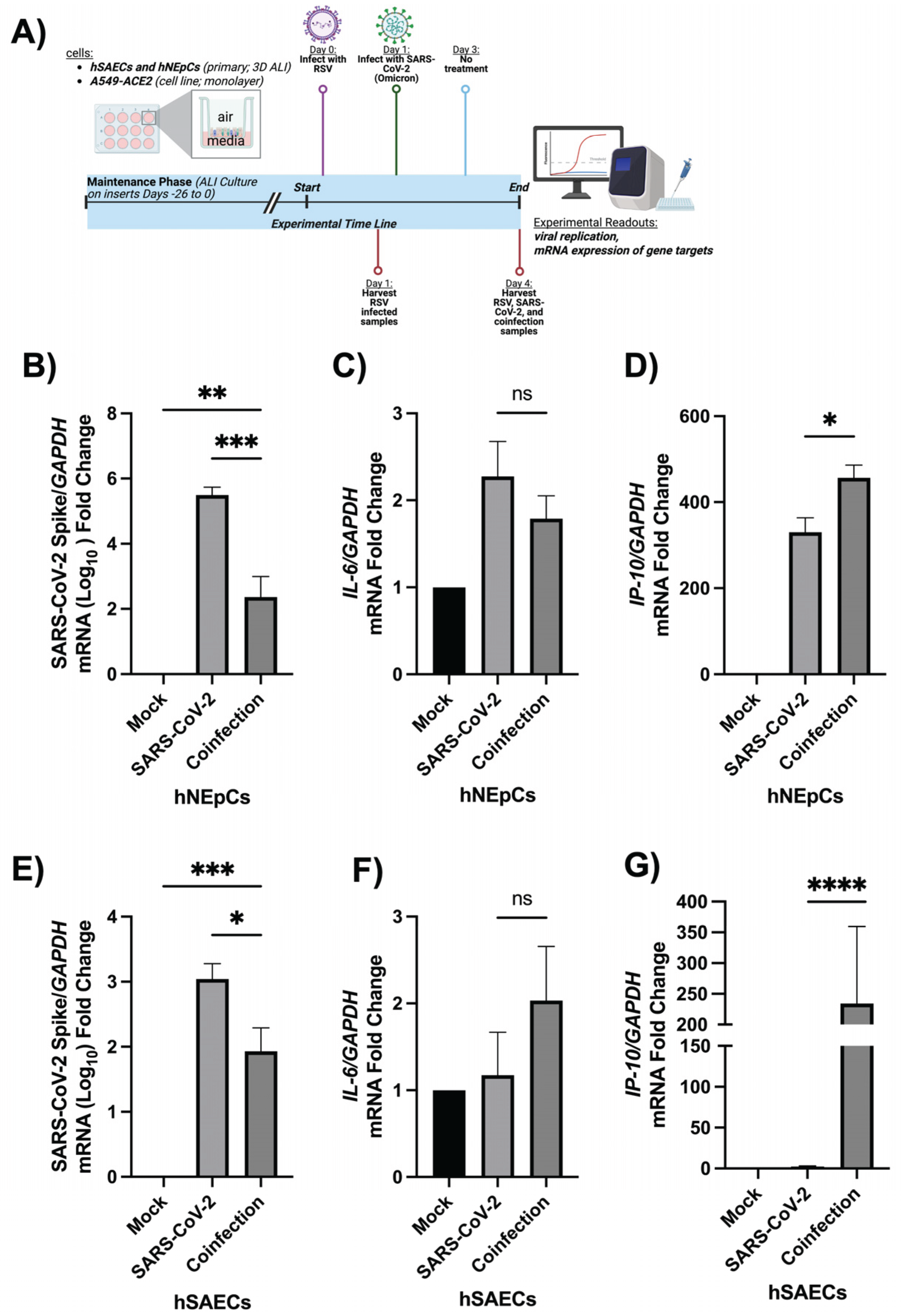
Figure 2.
RSV-induced changes in core kinase expression in hNEpCs. ALI-cultured hNEpCs were infected with RSV for 24 h, with mock infection used as the control. Total cellular RNA was harvested to examine changes in the expression of PI3K-AKT, p38-MAPK, p21-PAKs checkpoints: AKT1 (A), AKT2 (B), MAPK3 (C), MAPK14 (D), PAK1 (E), and ROCK1 (F) by qRT-PCR. Donors and experiments were similar as described above in Figure 1 legend. Cells were cultured in six replicates/group/experiment,and a paired Student’s T--test was used for statistical analysis. *p ≤ 0.05.
Figure 2.
RSV-induced changes in core kinase expression in hNEpCs. ALI-cultured hNEpCs were infected with RSV for 24 h, with mock infection used as the control. Total cellular RNA was harvested to examine changes in the expression of PI3K-AKT, p38-MAPK, p21-PAKs checkpoints: AKT1 (A), AKT2 (B), MAPK3 (C), MAPK14 (D), PAK1 (E), and ROCK1 (F) by qRT-PCR. Donors and experiments were similar as described above in Figure 1 legend. Cells were cultured in six replicates/group/experiment,and a paired Student’s T--test was used for statistical analysis. *p ≤ 0.05.
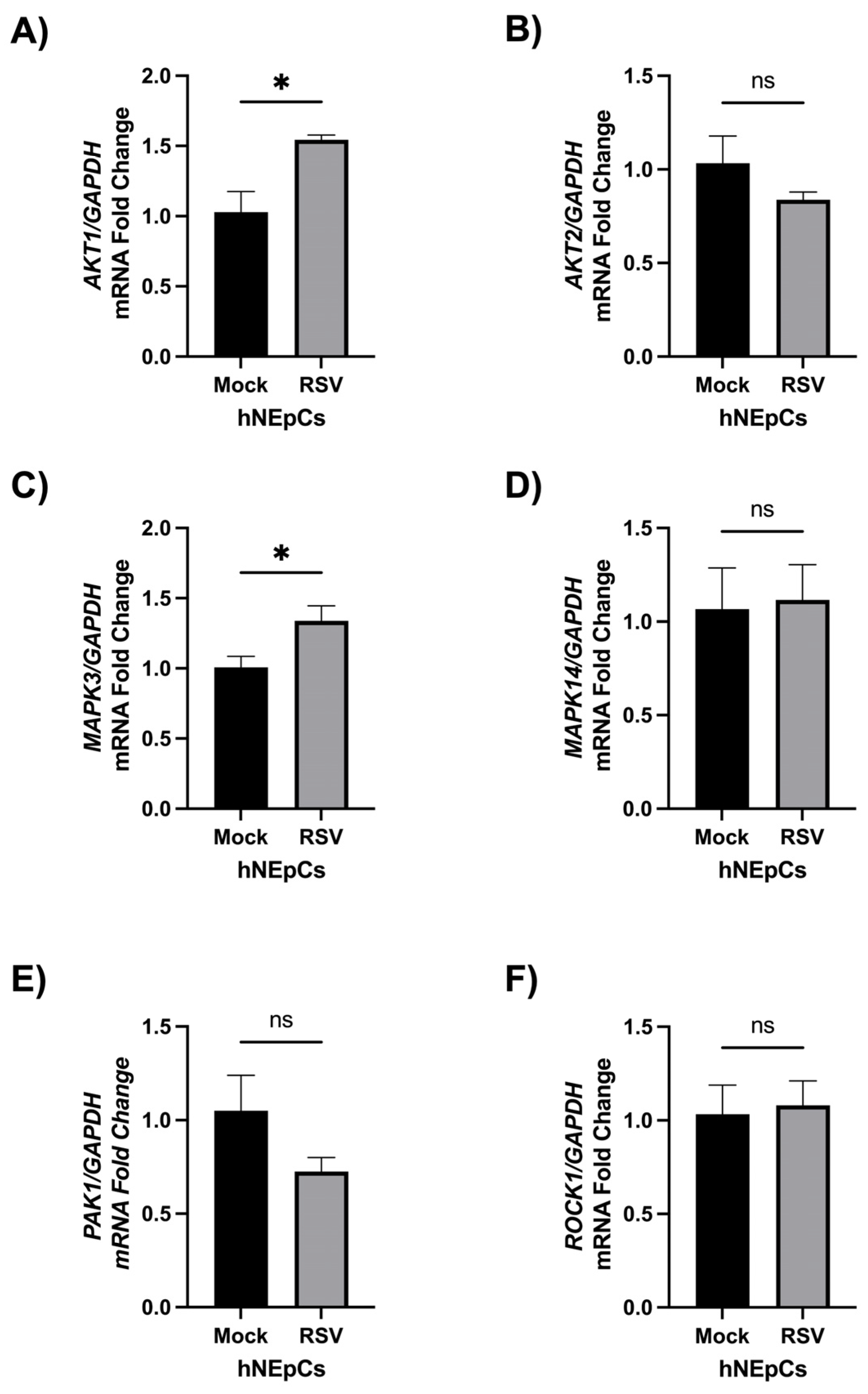
Figure 3.
RSV-induced changes in core kinase expression in hSAECs. ALI-cultured hSAECs were infected with RSV for 24 h, with mock infection used as the control. Total cellular RNA was harvested to examine changes in the expression of PI3K-AKT, p38-MAPK, p21-PAKs checkpoints: AKT1 (A), AKT2 (B), MAPK3 (C), MAPK14 (D), PAK1 (E), and ROCK1 (F) by qRT-PCR. Donors and experiments were similar as described above in Figure 1 legend. Cells were cultured in six replicates/group/experiment,and a paired Student’s T-test was used for statistical analysis. *p ≤ 0.05.
Figure 3.
RSV-induced changes in core kinase expression in hSAECs. ALI-cultured hSAECs were infected with RSV for 24 h, with mock infection used as the control. Total cellular RNA was harvested to examine changes in the expression of PI3K-AKT, p38-MAPK, p21-PAKs checkpoints: AKT1 (A), AKT2 (B), MAPK3 (C), MAPK14 (D), PAK1 (E), and ROCK1 (F) by qRT-PCR. Donors and experiments were similar as described above in Figure 1 legend. Cells were cultured in six replicates/group/experiment,and a paired Student’s T-test was used for statistical analysis. *p ≤ 0.05.
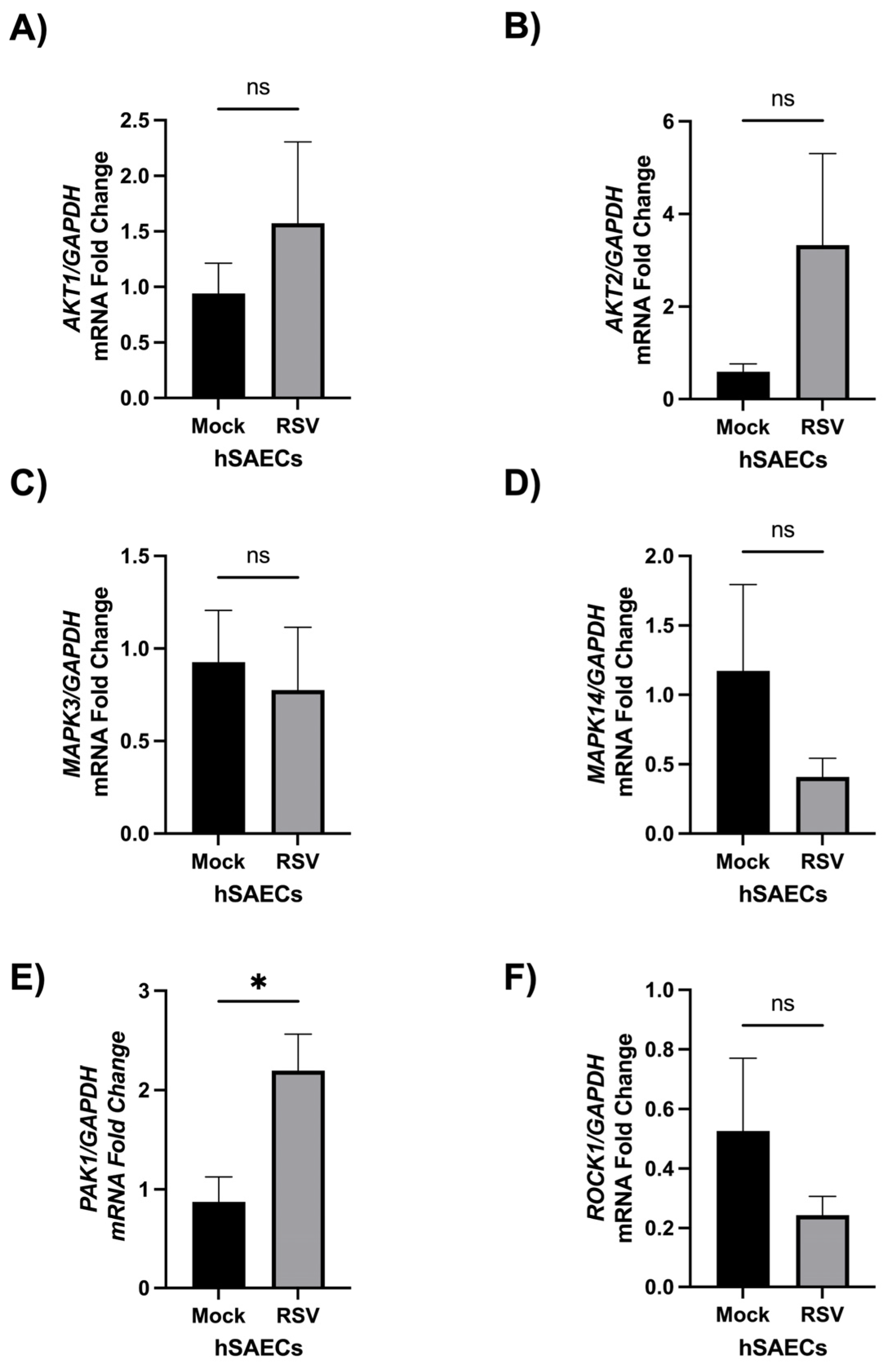
Figure 4.
The impact of RSV infection on PLSCR1 mRNA expression. ALI-cultured hNEpCs (A) or hSAECs (B) grew similarly as described in Figure 1, Figure 2 and Figure 3 and were infected with RSV for 24 h, followed by total cellular RNA harvesting and PLSCR1 quantification by qRT-PCR. A paired t-test was used for statistical analysis. A Student’s T-test was used for analysis. The asterisks ** and *** represent p values ≤ 0.01 and 0.001, respectively.
Figure 4.
The impact of RSV infection on PLSCR1 mRNA expression. ALI-cultured hNEpCs (A) or hSAECs (B) grew similarly as described in Figure 1, Figure 2 and Figure 3 and were infected with RSV for 24 h, followed by total cellular RNA harvesting and PLSCR1 quantification by qRT-PCR. A paired t-test was used for statistical analysis. A Student’s T-test was used for analysis. The asterisks ** and *** represent p values ≤ 0.01 and 0.001, respectively.
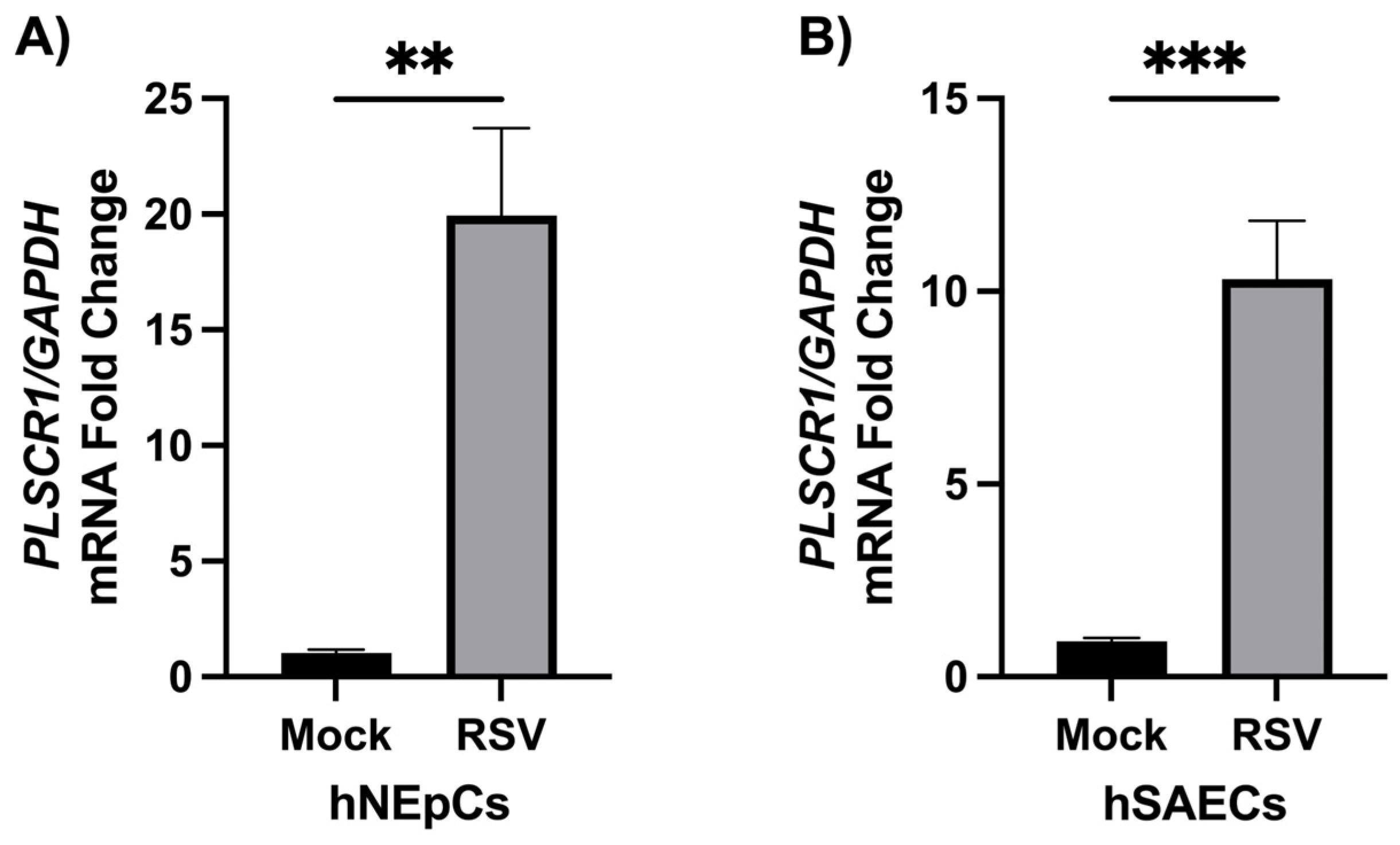
Figure 5.
Determining the antiviral role of RSV-induced PLSCR1 during RSV/SARS-CoV-2 coinfection. (A) A549-ACE2 cells were mock-infected or infected with RSV for 24 h, followed by SARS-CoV-2 infection or mock infection for an additional 3 days as described in the Methods. Total cellular RNA was harvested for detection of SARS-CoV-2 S expression. (B) A549-ACE2 cells were infected with RSV for 24 h, followed by RNA harvesting and PLSCR1 quantification by qRT-PCR. (C-D) A549-ACE2 cells, pretreated with siRNA against PLSCR1 (si-PLSCR1) or scrambled control (si-CN), were mock-infected or infected with RSV for 24 hours, followed by SARS-CoV-2 infection or no additional treatment. At day 3 post SARS-CoV-2 infection, the total cellular RNAs were harvested to investigate the expression of PLSCR1 (C) and SARS-CoV-2 N (D). Technical replicates were four for 2 independent experiments. A One-Way ANOVA was used for statistical analysis for (A-B), and a Two-Way ANOVA, with Tukey’s post-hoc test was used for (D). The asterisks *, **, ***, and **** represent the p values ≤ 0.05, 0.01, 0.001, and 0.0001, respectively.
Figure 5.
Determining the antiviral role of RSV-induced PLSCR1 during RSV/SARS-CoV-2 coinfection. (A) A549-ACE2 cells were mock-infected or infected with RSV for 24 h, followed by SARS-CoV-2 infection or mock infection for an additional 3 days as described in the Methods. Total cellular RNA was harvested for detection of SARS-CoV-2 S expression. (B) A549-ACE2 cells were infected with RSV for 24 h, followed by RNA harvesting and PLSCR1 quantification by qRT-PCR. (C-D) A549-ACE2 cells, pretreated with siRNA against PLSCR1 (si-PLSCR1) or scrambled control (si-CN), were mock-infected or infected with RSV for 24 hours, followed by SARS-CoV-2 infection or no additional treatment. At day 3 post SARS-CoV-2 infection, the total cellular RNAs were harvested to investigate the expression of PLSCR1 (C) and SARS-CoV-2 N (D). Technical replicates were four for 2 independent experiments. A One-Way ANOVA was used for statistical analysis for (A-B), and a Two-Way ANOVA, with Tukey’s post-hoc test was used for (D). The asterisks *, **, ***, and **** represent the p values ≤ 0.05, 0.01, 0.001, and 0.0001, respectively.
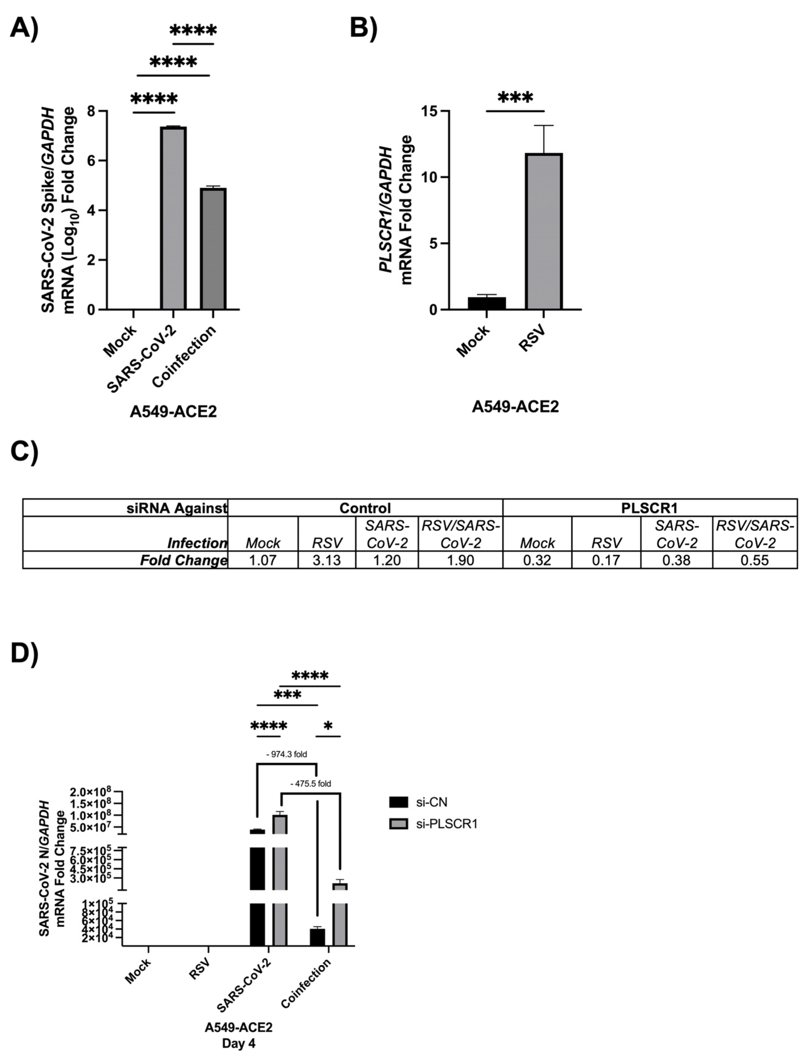
Figure 6.
PLSCR1 is a sustained but regulated antiviral response induced upon both early and later stages of RSV infection. PLSCR1 remained elevated at 4 days post RSV-infection in ALI-cultured hNEpCs (A), hSAECs (B), and A549-ACE2 (C). (D-E) IFN-lambda induction in ALI-cultured hNEpCs at day 1 or day 4 post RSV infection. (F-G). IFN-lambda induction in ALI-cultured hSAECs at day 1 or day 4 post RSV infection. (H) The impact of PLSCR-1 silencing on RSV N protein expression. For the experiments involved with hNEpCs and hSAECs, the repeats and experiments were done as described in Figure 1 legend. For the experiment using the A549-ACE2 cells, the repeats and experiments were done as described in Figure 5 legend. The siRNA treatment was also done similarly as described in Figure 5, followed by RSV infection for 4 days and RSV N gene expression by qRT-PCR. A paired t-test was used for statistical analysis. The asterisks *, **, ***, and **** represent the p values ≤ 0.05, 0.01, 0.001, and 0.0001, respectively.
Figure 6.
PLSCR1 is a sustained but regulated antiviral response induced upon both early and later stages of RSV infection. PLSCR1 remained elevated at 4 days post RSV-infection in ALI-cultured hNEpCs (A), hSAECs (B), and A549-ACE2 (C). (D-E) IFN-lambda induction in ALI-cultured hNEpCs at day 1 or day 4 post RSV infection. (F-G). IFN-lambda induction in ALI-cultured hSAECs at day 1 or day 4 post RSV infection. (H) The impact of PLSCR-1 silencing on RSV N protein expression. For the experiments involved with hNEpCs and hSAECs, the repeats and experiments were done as described in Figure 1 legend. For the experiment using the A549-ACE2 cells, the repeats and experiments were done as described in Figure 5 legend. The siRNA treatment was also done similarly as described in Figure 5, followed by RSV infection for 4 days and RSV N gene expression by qRT-PCR. A paired t-test was used for statistical analysis. The asterisks *, **, ***, and **** represent the p values ≤ 0.05, 0.01, 0.001, and 0.0001, respectively.
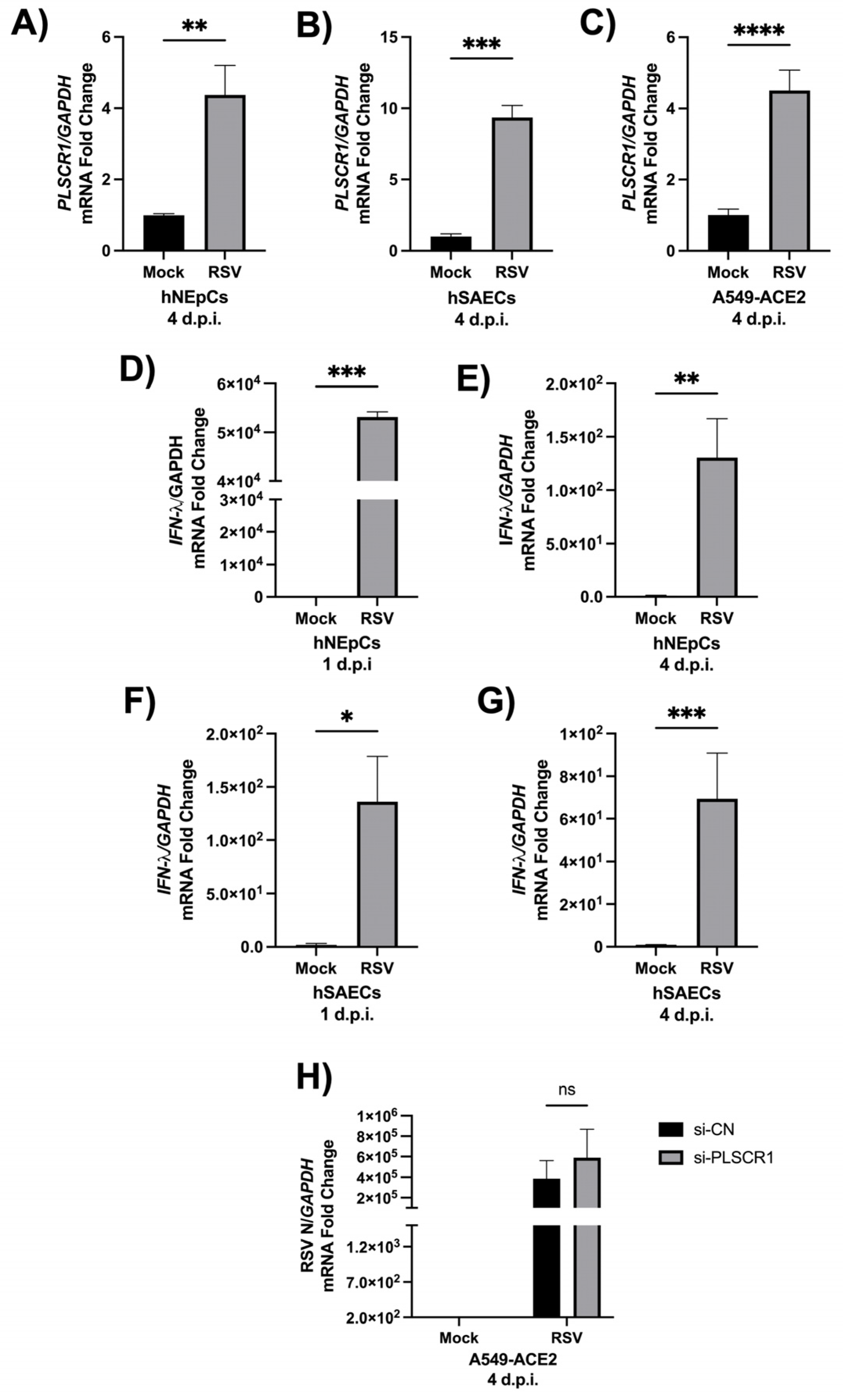

Table 1.
Primers used to quantify viral and host gene expression.
 |
Disclaimer/Publisher’s Note: The statements, opinions and data contained in all publications are solely those of the individual author(s) and contributor(s) and not of MDPI and/or the editor(s). MDPI and/or the editor(s) disclaim responsibility for any injury to people or property resulting from any ideas, methods, instructions or products referred to in the content. |
© 2026 by the authors. Licensee MDPI, Basel, Switzerland. This article is an open access article distributed under the terms and conditions of the Creative Commons Attribution (CC BY) license (http://creativecommons.org/licenses/by/4.0/).
Copyright: This open access article is published under a Creative Commons CC BY 4.0 license, which permit the free download, distribution, and reuse, provided that the author and preprint are cited in any reuse.