

Submitted:
18 September 2026
Posted:
20 September 2026
You are already at the latest version
Abstract
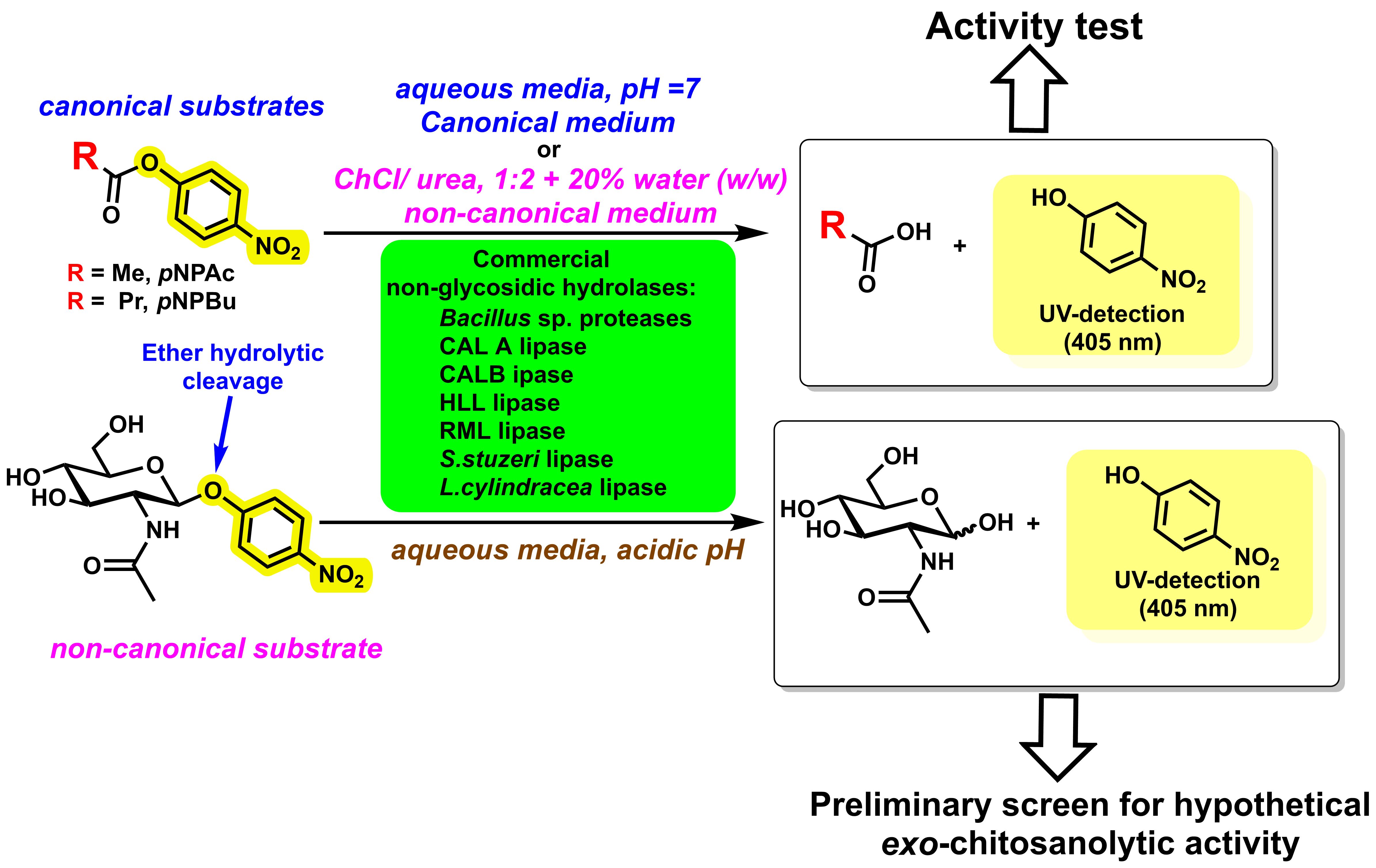
Several commercial hydrolases were screened to assess substrate-dependent activity and the influence of hydrated deep eutectic solvents on canonical and non-canonical hydrolysis. Liquid formulations from Novozymes, comprising proteases from Bacillus species and lipases from Candida antarctica, Rhizomucor miehei, and Thermomyces lanuginosus, and solid lipases from Meito Sangyo derived from Stutzerimonas stutzeri and Limtongozyma cylindracea, were evaluated in aqueous buffer and 20 wt% hydrated reline, composed of choline chloride and urea at a 1:2 molar ratio. Hydrolysis of p-nitrophenyl acetate, p-nitrophenyl butyrate, and p-nitrophenyl N-acetyl-β-D-glucosaminide was monitored spectrophotometrically, and activities were normalized to protein content determined by the bicinchoninic acid assay. SDS-PAGE supported the presence of the principal lipase components in selected liquid formulations. In aqueous buffer, activity profiles depended strongly on substrate structure. Hydrated reline selectively enhanced or suppressed activity, depending on the enzyme–substrate pair, and in some cases abolished measurable activity. Selected liquid lipases and purified CALB hydrolyzed p-nitrophenyl N-acetyl-β-D-glucosaminide under acidic conditions, whereas the solid lipases showed no detectable activity under the tested conditions. These results indicate that this substrate may serve as a preliminary screen for selecting preparations for subsequent chitosan-hydrolysis studies, although its predictive value requires validation by direct analysis of chitosan depolymerization.
Keywords:
enzyme promiscuity
; commercial hydrolases
; lipase activity
; p-Nitrophenyl substrates
; hydrated deep eutectic solvents
; reline
; pNPGlcNAc hydrolysis
1. Introduction
Enzyme promiscuity is the ability of an enzyme to catalyse reactions or act on substrates other than those associated with its evolved, physiological function [1,2,3]. Promiscuous activities are typically much weaker (lower kcat and/or kcat/KM) than the main activity, and they are thought to provide evolutionary starting points for the emergence of new functions [4]. In general, a clear distinction must be made between genuinely promiscuous activities and broad, physiologically intended multispecificity, which was better described as “broad specificity” rather than promiscuity [4,5]. Three archetypal different types of promiscuity are generally accepted, as defined by Hult and Berglund [6] and exemplified in Figure 1.
- Substrate promiscuity (sometimes called substrate ambiguity) is the ability of an enzyme to catalyse the same overall chemical transformation on multiple, structurally distinct substrates using the same active site and mechanism. In this case, the reaction type (bond being made or broken, overall mechanism) is unchanged, but substrate scope is broader than what is required in vivo. The main, “cognate” substrate is usually turned over with higher efficiency than alternative substrates, but noncognate substrates are transformed measurably at the same active site [2,3]. Many hydrolases (e.g. esterases, phosphotriesterases, β-lactamases) show broad substrate ranges and are classic examples of substrate-promiscuous enzymes [7]. From an evolutionary viewpoint, such broad specificity provides raw material for gene duplication and divergence toward new, more specialized functions [8].
- Catalytic promiscuity is the ability of a single active site to catalyse different chemical reactions (different bond-making/breaking patterns or mechanisms) on the same or closely related substrates. Here, the reaction type changes, not just the substrate identity, so that the underlying transition state and overall mechanism are different [3,9]. A classic example is a serine hydrolase (e.g. lipase or esterase) that, in addition to ester hydrolysis, catalyses C–C bond-forming reactions such as aldol, Michael, Mannich or Henry reactions using the same catalytic machinery [10].
- Condition promiscuity (also called environmental promiscuity) refers to the ability of an enzyme to catalyse its native reaction under a wide range of non-physiological conditions (e.g. non-aqueous solvents, extreme pH or temperature) without major changes in mechanism. The reaction and substrates are the same, but the reaction environment deviates strongly from normal cellular conditions. In fact, the use of enzymes in non-conventional media (non-aqueous solutions [11]) boosted from the seminal paper published by Zaks and Klibanov in 1984 [12], where they demonstrated that certain enzymes exhibit remarkable stability and catalytic activity in organic solvents, even at temperatures as high as 100°C. Since then, many lipases, proteases and dehydrogenases have been proven to remain active not only in organic solvents [13], but also in what is called “neoteric solvents”, an expression coined by K.R. Seddon [14,15] in the late 1990s to describe a new class of solvents in which enzymes can work [16], including Room-Temperature Ionic Liquids (RTILs, [17]), Supercritical Fluids (SCFs, [18]), Deep Eutectic Solvents (DESs, [19,20]) or fluorous solvents [21]. Condition and substrate promiscuity often intersect: broad substrate scope may only manifest under certain unconventional media, and non-aqueous conditions can unmask latent activities or alter selectivity [22].
In addition to the three classical promiscuity types, we can also mention another one, product promiscuity, which refers to the capacity of a single enzyme to convert a given substrate into more than one distinct product, typically via competing reaction pathways within the same active site [23]. An archetypal example is the δ-selinene synthase from Abies grandis, a terpene cyclase able to generate 52 different sesquiterpenes from farnesyl diphosphate as the only substrate [24]. This phenomenon is now recognized as a key facet of enzyme promiscuity, alongside substrate and catalytic promiscuity, and plays an important role in shaping the diversity of natural product scaffolds in biosynthetic pathways. In particular, recent work in plant and microbial specialized metabolism has shown how product-promiscuous enzymes can be harnessed or engineered to tune product profiles and expand libraries of bioactive molecules [1,25].
Lipases are particularly relevant in this context because they combine broad substrate tolerance [10,26], operational robustness, and well-documented performance in organic and non-conventional reaction media [27,28,29,30,31,32]. In addition to their native hydrolytic function, lipases are established catalysts for esterification, transesterification, alcoholysis, and a wide range of non-classical transformations [33,34], which has made them emblematic examples of enzymatic promiscuity in biocatalysis. Their activity is often screened using p-nitrophenyl-containing substrates [35], since hydrolysis releases p-nitrophenol (pNP), which can be quantified spectrophotometrically with high sensitivity and converted into rate and specific activity values, as shown in Figure 2.
Among 1. has traditionally been used as a general hydrolase substrate, including esterases [36], lipases [37,38], proteases [39], carbonic anhydrase [40] or acetylcholinesterase [41], whereas , p-nitrophenyl butyrate (pNPBu, R=Pr in Fig.1) is used more commonly in lipase-focused studies because its higher hydrophobicity better reflects interfacial and substrate-recognition features associated with lipase catalysis [42,43].
On the other hand, the enzymatic hydrolysis of p-nitrophenyl N-acetyl-β-D-glucosaminide (pNPGlcNAc), a classic chromogenic substrate for β-N-acetylhexosaminidases [44,45,46,47] and related enzymes such as lysozyme [48], is widely used for activity assays involving glycoside hydrolases (Figure 3).
Additionally, this chromogenic substrate has been also used for determining exo-chitinase activity [49,50,51], as well for activity assays of GH20 enzymes that hydrolyse both chitosan (copolymer formed D-glucosamine units (GlcN, major component) and 2-amino-2-deoxy-D-glucose (N-acetyl-glucosamine, GlcNAc) connected by linear β-(1,4)-links) and chitin (linear polysaccharide composed by a repetition of GlcNAc subunits, also connected by (β1→4) glycosidic bonds) [52,53], as depicted in Figure 4.
In fact, “true chitosanases” are usually assayed with chitosan or defined GlcN/GlcNAc oligomers, whereas pNPGlcNAc-type chromogenic substrates are typically used for β-N-acetylhexosaminidases that may act in chitosan-degrading systems [54].
To the best of our knowledge, there are currently no documented cases of any serin-hydrolases (lipases or proteases) catalyzing the hydrolysis of pNPGlcNAc. Nevertheless, there are examples reporting the hydrolytic activity of these hydrolases on chitosan; thus, the susceptibility of chitosan to serine hydrolases illustrates that its β-1,4-linked GlcN/GlcNAc backbone presents sufficient structural mimicry to canonical ester/amide substrates to support productive binding and glycosidic bond cleavage. Early work by Pantaleone and Yalpani [55,56] showed that food-grade lipase and papain preparations catalyze chitosanolysis, challenging a strict ‘one-enzyme–one-substrate’ view and hinting at shared recognition elements between glycan and peptide backbones. Muzzarelli’s study with a wheat-germ lipase preparation [57] and the later work of Shin et al. with a purified Rhizopus japonicus lipase [58] confirmed that a typical serine-hydrolase catalytic triad can, under mildly acidic conditions, effect non-specific scission of β-1,4 linkages in chitosan, yielding narrowly distributed polymer chains (≈30–50 kDa) and preserving the basic chemical structure of the repeating unit. Finally, a commercial lipase (from Novozymes, non-reporting the source of the microorganism) was proven to be capable of catalysing the hydrolysis of chitosan to glucosamine and chitooligosaccharides (DP 2–6 and higher), with COS yields up to 93.8% after 24 h at 37 °C, acting via combined exo/endo-modes [59].
Likewise, Li et al. [60,61] demonstrated that a neutral protease from Bacillus subtilis follows Michaelis–Menten kinetics on chitosan, with clear dependence on degree of deacetylation, and generates low-molecular-weight chitosan and chitooligosaccharides without detectable alteration of residue-level chemistry, implying that cleavage proceeds via selective attack at glycosidic bonds neighbouring GlcNAc units rather than via side-chain. These observations are consistent with the already commented view of lipases as structurally and mechanistically versatile serine hydrolases capable of exploiting similar oxyanion-stabilizing frameworks in non-classical substrates [28]. Together with pectinase isozymes from Aspergillus niger that hydrolyse both GlcN–GlcN and GlcNAc–GlcN linkages and exhibit distinct KM and Vmax values for chitosan and its derivatives [62] and nonspecific chitosanolysis by pronase [63] and pepsin/papain [64], these systems exemplify biocatalytic promiscuity in which classical serine hydrolase scaffolds exploit overlapping non-covalent recognition motifs to process aminopolysaccharides, even though their natural substrates are structurally unrelated. In any case, the presence of any detectable activity toward pNPGlcNAc by lipase or protease preparations is therefore mechanistically intriguing and potentially informative about hidden promiscuous or contaminating activities in commercial formulations.
The solvent environment is a further determinant of lipase behaviour [27,65]. Conventional aqueous assays provide useful baseline information, but non-conventional media can alter enzyme hydration, substrate partitioning, interfacial activation, and conformational dynamics [30,32,66,67]. Deep eutectic solvents (DESs), and especially hydrated DES systems, have attracted increasing interest because they are easily prepared, tuneable in composition, and capable of modulating enzyme microenvironments while maintaining a practical and potentially greener reaction platform [68,69,70,71]. Reline, the eutectic mixture of choline chloride (ChCl)/urea (1:2 molar ratio) is among the most commonly studied DESs in enzymology [72,73], and modest hydration is often required to obtain manageable viscosity and sustain catalytically relevant water activity [72,74,75].
The present work explores these issues through a comparative screening of commercial lipases and selected proteases in pH 7 aqueous buffer and 20% in weight hydrated reline using pNPAc and pNPBu (canonical ester substrates) as hydrolytic model substrates, complemented by assays with pNPGlcNAc (promiscuous non-canonical probe) under acidic conditions. The principal objective of this study was to evaluate whether hydrolysis of pNPGlcNAc, a small chromogenic substrate containing a GlcNAc-derived glycosidic bond, could serve as a rapid preliminary indicator of the ability of lipase preparations to hydrolyse chitosan through an exo mode; as pNPGlcNAc is structurally much simpler than polymeric chitosan, the assay could be considered a candidate screening tool rather than a direct substitute for an exo- hitosan-degradation assay. In fact, lipase preparations capable of hydrolysing chitosan would be more likely to display any measurable activity toward pNPGlcNAc than preparations lacking chitosanolytic activity.
2. Results
2.1. Protein Content of Commercial Enzyme Formulations
The first requirement for a meaningful comparison among commercial formulations was the estimation of protein, by using the bicinchoninic acid ([2,2'-biquinoline]-4,4'-dicarboxylic acid, BCA) assay (graphically depicted in Figure 5 [76]), which measures the formation of Cu+ from Cu2+ by the Biuret complex in alkaline solutions of protein.
The mechanism of the BCA assay was thought to be similar to the Lowry assay, but it is now known that there are two distinct reactions that take place with copper ions unique to the BCA assay [77]. The first reaction occurs at lower temperatures and is the result of the interaction of copper and BCA with cysteine/cystine, tryptophan, and tyrosine residues in the protein. At elevated temperatures, the peptide bond also is responsible for colour development. Hence, performing the assay at 37°C or 60°C versus room temperature increases the sensitivity and reduces the variation in the response of the assay as a function of protein composition. When possible, the assay should be incubated at 60°C since, after the reaction is complete, the absorbance does not increase appreciably, whereas after cooling samples incubated at 37°C to room temperature, the blank continues to increase in absorbance at ∼2.3% every 10 min [77].
This methodology (Section 4.3) was used to estimate the protein concentration of the different commercial enzymes. Same procedure was repeated for the commercial liquid enzymes; a dilution of these samples was needed to include the absorbance values inside the experimental calibration curve. Results are shown in Table 1.
2.2. Electrophoretic Characterization of the Enzymatic Preparations.
SDS-PAGE analysis was carried out for selected liquid lipases in order to confirm the presence of the dominant catalytic protein in each formulation. The electrophoretic profiles are showed in Figure S1.
Major band for lipases were found at about 45 kDa for Novocor® AD L(assigned to CAL A), a band at about 33 kDa for Lipozyme® CALB L, and bands around 31.6–31.7 kDa for Palatase® 20000L and Lipozyme® TL 100L. These values are consistent with the expected molecular-mass range of the principal lipases and provide qualitative support for the assignment of the formulations used in the hydrolysis experiments.
2.3. Analytical Calibration of p-Nitrophenol Detection
As commented in the Introduction, enzymatic activity in the hydrolysis of pNP-containing substrates was monitored via the release of pNP using UV spectroscopy. In fact, because pNP exists in protonated and deprotonated forms, it is usually monitored at either 405 nm in basic media (due to the formation of its deprotonated form, p-nitrophenolate) or at 348 nm in any other medium (as it is pNP’s isosbestic point [36], see Supplementary materials). Thus, for working at different pH values, calibration curves using different concentrations of pNP both in aqueous media at different pH values and in hydrated reline, both at 348 and 405 nm were obtained (Section 4.5). The values of the extinction coefficients (ε) at different pHs are summarized in Table 2.
2.4. Enzymatic Activity for the Hydrolysis of p-Nitrophenyl Esters
2.4.1. Hydrolysis of pNPAc in Aqueous Medium
Before testing enzyme-catalysed activity, basal hydrolysis assays were performed to determine whether the solvent systems themselves could hydrolyse pNPAc or pNPBu under the assay conditions, as reported in Section 4.7. The raw data for the enzyme assays indeed show blank rates that are non-zero (depicted in red in all the Figures), particularly for pNPAc, and these values were therefore taken into account when interpreting the enzyme-dependent kinetics. The presence of measurable blank hydrolysis further supports the decision to determine initial rates by fitting full time-course data rather than relying on single time-point measurements. The results obtained in the hydrolysis of pNPAc in aqueous buffer using proteases are shown in Supplementary materials (Figure S8 (Alcalase 2.4 L) and Figure S9 (Neutrase 0.8 L)), and summarized in Table 3.
Similar experiments on hydrolysis of pNPAc were conducted using the commercial preparations of lipases, mentioned in the Experimental Section. The results obtained in the hydrolysis of pNPAc in aqueous buffer using these lipases are shown in Supplementary materials (Figures S10 to S16). The specific activities are summarized in Table 4.
2.4.2. Hydrolysis of pNPBu in Aqueous Medium
Another chromogenic substrate tested was pNPBu. For this substrate, none of the proteases tested was capable to catalyse its hydrolysis. In fact, although some proteases can hydrolyse pNPBu (for instance, Sub1, a subtilisin-like serine protease from Pseudomonas sp [78]), this is not a general property of proteases, as this substrate is more suitable for lipases. Results obtained with lipases are shown in Supplementary materials (Figures S17-S23) and summarised in Table 5.
2.4.3. Hydrolysis of pNPAc in Hydrated Reline
The hydrolysis of pNPAc was also tested on hydrated reline (20% water), to check the capability of these commercial preparations to work in a different reaction medium. Results are shown in Supplementary materials (Figures S24 to S30), and summarized in Table 6.
2.4.4. Hydrolysis of pNPBu in Hydrated Reline
In a similar way, hydrolysis of pNPBu was also tested on hydrated reline (20% water), to check the capability of these commercial preparations to work in a different reaction medium. Results are shown in Supplementary materials (Figures S31 to S35), and summarized in Table 7.
2.4.5. Hydrolysis of pNPGlcNAc in Aqueous Medium
To check the capability of commercial enzymes for converting a non canonical ethereal chromogenic substrete, hydrolysis of pNPGlcNAc was tested at 37°C (similar temperature used for pNP esters), but now changing to an acidic buffer (pH =4), as required in the hydrolysis of this substrate by archetypal β-N-acetylglucosaminidases [79,80,81]. Reactions and blank assay (to quantify spontaneous hydrolysis caused by the acidic medium) were analysed at long reaction times to scrutinize any weak promiscuous activity. Palatase ® 20000L was not tested, as its activity against pNP esters was generally lower than that of the other liquid enzymes. Kinetics if the hydrolyses are shown in Supplementary materials (Figures S36 to S38) and the results are shown in Table 8.
Enzymatic hydrolysis of pNPGlcNAc was also evaluated in aqueous media at pH 3, 4, and 5, to assess the effect of pH, but working at a higher temperature (50°C) , in order to increase the activity of this promiscuous reaction. Kinetics are shown in Supplementary materials (Figures S39 to S42, pH =3; Figures S43 to S46, pH = 4; Figures S47 to S50, pH =5) and the results are summarized in Table 9 (pH= 3), Table 10 (pH = 4) and Table 11 (pH = 5).
3. Discussion
3.1. Enzyme Characterization
No electrophoretic characterization of the commercial NovoCor® AD L preparation was found in literature. Dimitrijević and co-workers isolated CALA (in this case, from C. antarctica DSM 70725 obtained from the German Collection of Microorganisms and Cell Cultures (DSMZ)), using hydroxyapatite chromatography, and reporting showed a single protein band at 45 kDa by silver-stained SDS–PAGE showed a single protein band at 45 kDa. Other structural and expression studies describe CALA as a protein of 431 amino acids, with a molecular mass generally reported as 43–45 kDa [82]. Some recombinant preparations display an apparent mass as high as approximately 50–53 kDa, probably reflecting the expression construct, glycosylation or anomalous electrophoretic migration [83].
SDS-PAGE analysis of the commercial Lipozyme CALB L (although in this case Lipozyme® LCN02102, from Novozymes Brazil) reported in the literature by Llerena-Suster et al. [84] showed a most intense band at ~33 kDa corresponding to Candida antarctica lipase B, plus additional bands at approximately 66 and 21 kDa The Bradford assay, using CALB as a standard instead of BSA, quantified the protein content at 13.0 mg/ml after removal of the non-soluble fraction. The crude extract also contains 51.4 mg/ml of non-soluble species and 131.3 mg of nucleic acids plus unidentified substances per milliliter. Size-exclusion chromatography confirmed that the first eluate is protein-rich (1.2 mg/ml) with a 33 kDa band, while the second eluate contains preservatives (benzoate and sorbate) absorbing at 230 and 260 nm. Agarose gel electrophoresis further resolved nucleic acid contaminants present in the crude and filtered extracts.
Electrophoretic analysis of Lipozyme® TL 100L was reported by Duarte et al. [85], is expected to reveal a predominant diffuse protein band at approximately 30–32 kDa, displaying pronounced catalytic activity in transesterification and esterification zymograms, consistent with the calculated molecular mass of Thermomyces lanuginosus lipase (TLL; approximately 31.7 kDa). Several weaker bands also showed activity, indicating that the commercial liquid formulation is not electrophoretically homogeneous and may contain minor active or processed forms of TLL.
Palatase® 20000 L was reported as R. miehei lipase with a measured protein concentration of approximately 2.9–3.7 mg mL⁻¹, although this may vary by batch [86]. Direct electrophoretic analysis of recombinant mature RML has shown a strong band immediately above the 31 kDa marker. Size-exclusion chromatography of the same mature enzyme produced a lower estimate of approximately 27 kDa, illustrating that chromatographic and electrophoretic estimates do not necessarily coincide [87].
Regarding proteases, SDS-PAGE electrophoresis are typically generated by end-users to characterize hydrolysis products or to assess purity, as Novozymes product sheets do not usually include detailed SDS-PAGE profiles for these liquid formulations. Anyhow, not many data are available in literature. For instance, Ye-Won et al. used SDS-PAGE to assess whether Alcalase® 2.4 L remained structurally stable after prolonged treatment at 100 MPa [88]. However, this article did not report numerical molecular weights for these bands, as these authors used relative mobility (Rf) rather than a calibrated molecular-weight axis. Recently, Rodrigues et al. reported the electrophoresis of Alcalase® Pure from Bacillus licheniformis (EC 3.4.21.62), also supplied as a liquid formulation by Novozymes (protein concentration was 17.46 mg mL−1)[89]. These authors reported several low-molecular-weight bands, principally at approximately 13–14, 10 and 8–9 kDa, attributed to extensive autolysis, whereas a distinct band corresponding to intact mature Alcalase at approximately 27–30 kDa was not detected. For Neutrase 0.8 L, Santos-Moriano et al. reported the SDS-PAGE analysis of this preparation [90], revealing a heterogeneous protein profile, with a distinct band at approximately 58 kDa and a prominent broad band at approximately 39 kDa, accompanied by several considerably less intense components in the 20-32 kDa región. The ~58 kDa band is in excellent agreement with the molecular mass of 56.7 kDa reported for the complete neutral-metalloproteinase precursor from Bacillus amyloliquefaciens (UniProt entry Q44677 [91]). Bacillus amyloliquefaciens neutral protease is synthesized with an N-terminal signal peptide and an extensive pro-region that are removed during secretion, yielding a mature enzyme with a predicted molecular mass of approximately 33 kDa. The 37–39 kDa band could therefore represent the processed neutral metalloproteinase (bacillolysin) [92].
The most relevant—and apparently the only detailed—study of the electrophoretic profile of Lipase TL from Meito Sangyo was published by Maraite et al [93], showing a predominant protein band at approximately 30 kDa, assigned to the catalytically active Lipase TL (subsequently designated LipC). The manufacturer reported a molecular mass of approximately 27 kDa, which is compatible with its apparent electrophoretic mobility near 30 kDa. For Lipase OF and Lipase MY-30, both are expected to show a predominant, sometimes diffuse band at approximately 55–62 kDa, corresponding to closely related lipase isoenzymes with calculated molecular masses of about 60 kDa. In fact, Lipase OF reportedly produced one major band near 60–62 kDa, although Isoelectric Focusing (IEF) revealed four or five acidic components clustered around pI 4.0. [94]. Lipase MY-30 similarly gave an unresolved principal band around 55–60 kDa, but IEF demonstrates greater charge heterogeneity, with four major components: one at pI ≈ 5.5, two near pI 4.0 and one near pI 3.5 [95]. As MY-30 is diluted with lactose, equal masses of commercial powder may produce a weaker protein band than Lipase OF.
In all cases, although electrophoresis does not exclude the presence of stabilizers, minor proteins, or traces of side activities in commercial preparations, it supports the view that the observed reactivity mainly derives from the named lipase component.
3.2. Spectrophotometric Analysis
As commented before, it is usual to work at pNP’s isosbestic point, provided that the extinction molar coefficient of both pNP and its corresponding anion is the same, regardless of the pH of the reaction media. Nevertheless, analysing the experimental error of this value (Figure 6), and considering the data showed in Table 2, there is a great variability in the value ε348, so that assuming a universal extinction coefficient at 348 nm across all solvents and pH values would introduce non-negligible error. On the other hand, the value at ε405 is one order of magnitude higher, so that the sensitivity working at that wavelength would be also higher. Therefore, to measure the amount of pNP released from pNPAc or pNPBu, the sample aliquots taken at different reaction times were taken to pH =9 using Tris-HCl 0.05 M pH 9 buffer, so that the absorbance measurements were recorded at 405 nm, using the value of ε405 = 16.05 ± 0.13 mM-1cm-1 as extinction coefficient, with the concomitant increase in the sensibility derived from the higher value of ε. This approach improves sensitivity and reduces ambiguity associated with the acid–base equilibrium of pNP during the assay.
A further reason for basifying the samples and measuring absorbance at 405 nm becomes clear upon examination of the pNPGlcNAc spectra shown in Figure S4. This substrate exhibits appreciable absorbance at 348 nm, which is relatively close to its absorption maximum at 305–310 nm (approximately 300 nm in 0.1 N NaOH, as reported in the literature[96]). Consequently, if substrate hydrolysis is incomplete, the absorbance measured at 348 nm cannot be attributed solely to the released pNP, as residual pNPGlcNAc will also contribute to the signal. This interference would therefore lead to an overestimation of the enzymatic activity.
3.3. pNP Esters Hydrolysis in Aqueous Media
Regarding pNPAc hydrolysis with proteases, as shown in both cases, measurable blank hydrolysis was detected, confirming that pNPAc undergoes slow non-enzymatic hydrolysis even under the assay conditions. Consequently, enzyme activities were calculated from complete kinetic profiles after correcting for this background reaction, an approach that improves the reliability of the initial-rate determination. The experiments shown in Figure 7 clearly demonstrate that Alcalase catalyses the hydrolysis of pNPAc, despite being commercially classified as a protease rather than an esterase. Several observations support that the measured activity is genuinely enzyme-catalysed, as the product formation increases proportionally with enzyme concentration, the initial reaction rate shows an approximately linear dependence on enzyme loading, and the specific activity remains essentially constant over the concentration range studied (≈0.037 U mg⁻¹, Table 3), indicating that the reaction is measured under initial-rate conditions and is not limited by substrate depletion or mass-transfer effects. Although this activity is several orders of magnitude lower than the proteolytic activity normally reported for Alcalase [97,98,99], it is entirely consistent with the known catalytic promiscuity of subtilisin-type serine proteases (main component of Alcalase is subtilisin Carlsberg, a serine protease possessing the classical Asp-His-Ser catalytic triad [100,101]). As far as we know, there are no publications specifically describing the hydrolysis of pNPAc by commercial Alcalase, although it has been reported that subtilisins can hydrolyse various p-nitrophenyl esters through the same acyl-enzyme intermediate responsible for peptide hydrolysis [102]. Therefore, the weak esterase activity observed is mechanistically expected, even though it has rarely been investigated experimentally.
In contrast, as shown in Figure 8, Neutrase exhibits only extremely low activity toward pNPAc, with a specific activity of approximately 1.23 × 10⁻⁴ U mg⁻¹, which is roughly 300-fold lower than Alcalase under identical experimental conditions (Table 3). This pronounced difference is readily explained by the fundamentally different catalytic mechanisms of the two enzymes; unlike Alcalase, Neutrase is a zinc-dependent metalloprotease [90,103,104], obtained from Bacillus amyloliquefaciens (see Table 1) where catalysis proceeds through activation of a water molecule coordinated to Zn²⁺ rather than through formation of a covalent acyl-enzyme intermediate involving an active-site serine residue [105]. Consequently, the catalytic machinery that enables serine hydrolases to hydrolyse both amides and esters is absent. The supporting literature confirms that no published reports describe hydrolysis of pNPAc by Neutrase, and this absence is consistent with current mechanistic understanding. The available literature on Neutrase focuses almost exclusively on protein hydrolysis [99,106,107], production of bioactive peptides [107], and enzyme immobilisation [103], with no recognised esterase activity reported for simple p-nitrophenyl esters. Then, the small increase above the blank observed experimentally may therefore arise from an extremely weak promiscuous catalytic activity, or rather a minor contaminating hydrolases in the commercial preparation, which is feasible considering, Neutrases’ protein profile shown in Section 4.3 [90]. Regardless of its origin, the activity is sufficiently low to confirm that ester hydrolysis is not a significant catalytic function of Neutrase.
The striking difference between the two enzymes illustrates an important aspect of enzyme promiscuity. Although both enzymes are classified as proteases, only the serine protease (Alcalase) possesses a catalytic architecture capable of efficiently stabilising tetrahedral intermediates formed during both amide and ester hydrolysis. The catalytic triad (Ser-His-Asp) and oxyanion hole provide the structural features required for hydrolysis of activated esters such as pNPAc. Conversely, the Zn²⁺-dependent catalytic mechanism of Neutrase is highly specialised for peptide bond cleavage and is poorly suited for hydrolysis of simple activated esters. The approximately 300-fold difference in specific activity observed experimentally therefore reflects intrinsic mechanistic differences rather than merely different substrate affinities. Remarkably, Mostafavi et al [103] have reported that Neutrase immobilized as hybrid nanoflowers [Neutrase-Cu/Ca/Co/Mn)₃(PO₄)₂] exhibits promiscuous catalytic activity for C-C forming reactions such as aldol condensations, Knoevenagel reactions, Hantzsch-type reactions or 4H-pyran synthesis.
The results obtained with the commercial lipase preparations (Table 4) also showed measurable blank hydrolysis, confirming that pNPAc undergoes slow non-enzymatic hydrolysis under the assay conditions: Therefore, once again lipase activities were determined from complete kinetic profiles after accounting for this background reaction to improve the reliability of the initial-rate calculation. Again, the release of pNP increased with enzyme concentration, and the initial reaction rates exhibited an approximately linear dependence on enzyme loading, confirming that the assays were performed under initial-rate conditions and that mass-transfer or substrate-depletion effects were negligible over the concentration range investigated. Furthermore, the specific activities remained essentially constant for each enzyme preparation, indicating that the normalization based on protein content provides a reliable basis for comparing the catalytic efficiencies of the different commercial formulations.
The results summarized in Table 4 clearly demonstrate that several commercial lipases catalyse the hydrolysis of pNPAc in aqueous buffer, although with substantial differences in specific activity. Among the liquid formulations, Lipozyme® CALB L (mainly Candida antarctica lipase B [108,109,110]), exhibited the highest activity (0.065 ± 0.003 U mg⁻¹), followed by NovoCor® AD L (Candida antarctica lipase A [82]) and Palatase® 20000L (Rhizomucor miehei lipase (RML, [111,112,113,114])), which displayed almost identical specific activities (both approximately 0.055 U mg⁻¹), while Lipozyme® TL 100L (non specific lipase from Thermomyces lanuginosus, old name Humicola lanuginosa [115], normally reported as TLL), was less active, with a specific activity of about 9.2 × 10⁻³ U mg⁻¹. TLL is generally recognised as a lipase that exhibits strong interfacial activation and displays its highest catalytic performance towards longer-chain, more hydrophobic esters. Since pNPAc is a highly soluble, short-chain substrate, it may not efficiently promote the conformational opening of the lid domain that characterises the fully active form of TLL, resulting in lower observed activity under homogeneous aqueous conditions [116]. Anyhow, though these four preparations contain lipases belonging to different structural families, they all catalyse the hydrolysis of pNPAc efficiently. Differences in enzymatic activity should be interpreted in light of the strong variability in protein concentration across commercial preparations (Table 1), which makes normalization on a protein basis essential for meaningful comparison. SDS-PAGE analysis of the main liquid lipase formulations (section 2.2) further supports the view that the observed ester hydrolysis mainly derives from the named lipase component, although minor proteins or side activities cannot be excluded completely in commercial products
On the other hand, their similar behaviour is not unexpected because pNPAc is a small, activated ester that can readily access the catalytic serine residue without requiring extensive hydrophobic interactions or interfacial activation. Consequently, the hydrolysis of this substrate mainly reflects the intrinsic catalytic competence of the conserved Ser-His-Asp catalytic triad shared by these enzymes rather than subtle differences in substrate-binding pockets. This observation agrees well with the already mentioned extensive use of pNPAc as a general chromogenic substrate for measuring esterase and lipase activities in the literature.
For Meito solid enzymes, despite its very low protein content in the commercial formulation (see Table 1), the highest specific activity was obtained with Lipase MY30 (containing lipase from Limtongozyma cylindracea [117], historically referred to under the names Candida cylindracea and Candida rugosa [118,119,120]), which reached 0.303 ± 0.016 U mg⁻¹, approximately five times higher than most of the liquid commercial formulations and nearly thirty times higher than the weakest lipase preparation analysed (Lipase TL, lipase from Stutzerimonas stutzeri, basionym Pseudomonas stutzeri [93,121]). Such a remarkably high protein-normalised activity probably reflects the high intrinsic catalytic efficiency of this preparation towards short-chain esters rather than a higher total enzyme loading, since the activities were normalised using the experimentally determined protein concentrations. The excellent performance of Lipase MY30 also illustrates that commercial formulations containing relatively low protein concentrations may nevertheless exhibit outstanding catalytic efficiencies when the active enzyme represents a large fraction of the total protein. An interesting result was obtained for Lipase OF, which displayed a specific activity (0.053 ± 0.004 U mg⁻¹) comparable to those of liquid CALA and RML, despite its considerably lower protein concentration..
Overall, the lipase preparations exhibited specific activities spanning more than one order of magnitude, illustrating the substantial diversity that exists among commercial enzymes even when assayed using the same chromogenic substrate. Nevertheless, all lipases displayed activities significantly higher than those measured for the proteases. These differences are entirely consistent with their catalytic mechanisms. Lipases are true serine hydrolases whose physiological role is the hydrolysis of ester bonds, whereas Alcalase only exhibits weak esterase activity as a consequence of catalytic promiscuity, and Neutrase, a zinc-dependent metalloprotease, lacks the covalent acyl-enzyme intermediate characteristic of serine hydrolases. Consequently, ester hydrolysis constitutes the native reaction for lipases but only a secondary or practically absent activity for the proteases examined. Taken together, these results confirm that pNPAc provides an excellent general substrate for comparing commercial lipase preparations after protein normalisation. The assay successfully discriminates between formulations with markedly different intrinsic catalytic efficiencies while simultaneously highlighting mechanistic relationships among different hydrolase families. Moreover, the substantial differences observed between enzymes derived from distinct microorganisms emphasise that commercial activity cannot be inferred solely from enzyme classification or formulation type, reinforcing the importance of systematic experimental screening when selecting biocatalysts for hydrolytic applications.
When changing from pNPAc to pNPBu, no activity was detected with proteases. For lipases, as shown in Figures 16 to 22 and in Table 5, NovoCor® AD L showed a notably high specific activity of about 4.28 µmol min⁻¹ mg⁻¹, while Lipozyme® CALB L and Palatase® 20000L reached a slightly lower activity values, about 3.0 mol min⁻¹ mg⁻¹. As expected, Lipozyme® TL 100L showed the higher value about 6.85 µmol min⁻¹ mg⁻¹, because of the higher interfacial activation for this more hydrophobic ester [116]. In any case, activity values were much higher than those reported for pNPAc, which indicates that pNPBu is a stronger discriminator of lipase-like binding and interfacial recognition. The Meito lipases were particularly informative in this substrate set because they showed a different ranking and, in some cases, very high activity per unit protein. Lipase TL showed a very high enzymatic activity (4.54 µmol min⁻¹ mg⁻¹, ) clearly comparable to those obtained with preparation form Novozymes. This could be related with the well known affinity of this lipase to covert bulky substrates [93,122]. Conversely, Lipase OF and Lipase MY30 reached lower activity values (an order of magnitude lower). This suggests that low formulation protein content does not imply low catalytic relevance; on the contrary, the active enzyme fraction can be highly efficient when normalized correctly. The pNPBu data therefore reveal a stronger hydrophobicity effect than pNPAc and highlight that substrate promiscuity is not equivalent across all activated esters.
3.4. pNP Esters Hydrolysis in Aqueous Hydrated Reline
When moving from aqueous media to hydrated reline, none of the proteases (Alcalase and Neutrase) tested were capable to hydrolyse pNP-containing substrates. For this last one this behaviour was expected, provided its very low activity in pure aqueous media (Table 3). Regarding alcalase, a recent paper from Álvarez et al [123] reported protease activity and stability in hydrated reline, using an alkaline protease from Bacillus licheniformis. Nevertheless, in this study the activity was tested using a canonical protease substrate (azocasein), so this fact demonstrate the low level of medium promiscuity of proteases.
For lipases, changing the medium from aqueous buffer to 20% hydrated reline produced strongly enzyme-dependent effects during pNPAc hydrolysis. Among the liquid formulations, Lipozyme® TL 100L showed the most pronounced enhancement, with an approximately 317-fold increase, while Palatase® 20000L and Lipozyme® CALB L increased by approximately 2.55-fold and 1.62-fold, respectively. Among the solid preparations, Lipase TL and Lipase OF increased by approximately 34.8-fold and 1.75-fold, respectively. In contrast, NovoCor® AD L activity decreased to approximately 0.35-fold of its aqueous-buffer value, corresponding to a reduction of about 65.5%, whereas Lipase MY30 decreased to approximately 0.016-fold, equivalent to a reduction of about 98.4% or a roughly 64.5-fold loss of activity. These contrasting responses indicate that hydrated reline selectively modulates pNPAc hydrolysis, enhancing some enzyme preparations while substantially inhibiting others, rather than producing a universal stabilizing effect.. The most plausible explanation is that the hydrated DES changes water activity, viscosity, substrate solvation, and protein conformational mobility in a way that benefits some enzymes while impairing others.
The effect of hydrated reline on pNPBu hydrolysis (Table 7) depended strongly on enzyme identity. Neither of the proteases tested was capable of hydrolysing this substrate under the experimental conditions. Among the lipases, Lipozyme® CALB L was the only preparation whose activity increased, reaching 1.28-fold, or approximately 28% above, its aqueous-buffer value. By contrast, NovoCor® AD L, Palatase® 20000L, Lipozyme® TL 100L, and Lipase TL remained active but showed reduced activities in hydrated reline. Their activities decreased to approximately 0.23-, 0.096-, 0.073-, and 0.60-fold of their respective aqueous values, corresponding to reductions of approximately 77%, 90%, 93%, and 40%, respectively. Lipase OF and Lipase MY30, which already showed relatively low activity toward pNPBu in aqueous buffer (Table 5), became completely inactive in hydrated reline. These results indicate that hydrated reline is not a general activator for pNPBu hydrolysis; instead, it strongly modulates activity in an enzyme-specific manner, with CALB displaying the greatest tolerance under the tested conditions.
Thus, switching from buffer to hydrated reline (20% water) significantly modulates the specific activities toward pNPAc and pNPBu. For some biocatalysts (notably CALB and TLL), the activities in hydrated reline remain in the same range or even increase slightly relative to buffer. This behaviour matches previous reports that DESs, and particularly reline [72], can preserve or enhance lipase activity when an appropriate amount of water is present [124,125,126,127], whereas the pure DES can severely rigidify the protein and reduce catalysis. In fact, molecular dynamics and experimental studies carried out by Shehata et al [72] have shown that slightly hydrated reline increases the mobility of key regions such as the lid of thermoalkalophilic lipases, favouring interfacial activation while maintaining overall structural integrity. In other cases, (e.g. RML, some CRL preparations) show a modest loss of activity, as it has been also reported for lipases [128,129]. Our data thus add to the growing evidence that water-in-DES systems can be tuned to balance enzyme stability and flexibility, allowing efficient hydrolysis of hydrophobic pNP-esters in media with reduced conventional solvent content.
3.5. pNPGlcNAc Hydrolysis in Aqueous Buffers
pNPGlcNAc differs fundamentally from pNPAc and pNPBu. It contains a glycosidic bond rather than an ester bond and is classically used as a substrate for beta- -acetylhexosaminidases, exo-chitinases, and related glycoside hydrolases. pNPGlcNAc was selected as a small, chromogenic surrogate substrate to investigate whether a rapid spectrophotometric assay could provide preliminary information about the potential of lipase preparations to hydrolyze chitosan. The rationale is not that pNPGlcNAc reproduces the structure of chitosan, but that both substrates contain GlcNAc-derived glycosidic bonds whose hydrolysis may require recognition features outside the canonical ester-substrate preference of lipases. As previously commented, it requires an acidic pH medium (around pH=4), as previously reported for this substrate the archetypal β-N-acetylglucosaminidases [79,80,81].
Thus, hydrolysis of pNPGlcNAc was tested at this pH and 37°C (similar temperature used for pNP esters), at long reaction times to scrutinize any weak promiscuous activity, and the results are shown in Table 8. Blank assays were also carried out, to quantify spontaneous hydrolysis caused by the acidic medium; oddly, no spontaneous hydrolysis was detected in the first 24 hours, pNPGlcNAc being slowly hydrolysed after that point. Palatase ® 20000L was not tested, as its activity against pNP esters was lower than that of the other liquid enzymes. Interestingly, no reaction was detected for solid lipases (Lipase TL, Lipase OF, or Lipase MY30). Under the tested conditions, Lipozyme® CALB L and NovoCor® AD L exhibited clear pNPGlcNAc hydrolysis, with similar protein-normalized activities of and , respectively, whereas Lipozyme® TL 100L displayed only marginal activity close to the blank level, ,not showing any lag-time. These results demonstrate non-canonical pNPGlcNAc hydrolysis by selected CALA and CALB commercial preparations. To check if this activity could be caused by any formulation component or trace contaminating hydrolases, pure CALB was also tested. Remarkably, this pure enzyme proved to be capable of catalysing the promiscuous hydrolysis of the ethereal bond of pNPGlcNAc at acidic pH, although with a substantially lower activity, . Therefore, the detection of pNPGlcNAc hydrolysis by the purified CALB preparation supports the presence of a non-canonical substrate activity associated with CALB, although it does not by itself establish hydrolysis of polymeric chitosan or exclude the involvement of a minor associated activity.
Comparison of Table 4, Table 5 and Table 8 demonstrates a strongly substrate- and condition-dependent activity profile among the commercial lipases. The preparations that performed best toward pNPAc did not necessarily show the highest activity toward pNPBu or pNPGlcNAc. In particular, the response toward pNPBu was markedly enhanced for several enzymes, especially Lipozyme® TL 100L, indicating a pronounced preference for the more hydrophobic ester substrate. By contrast, pNPGlcNAc hydrolysis was detected only for selected liquid preparations and pure CALB, while no reaction was observed for Lipase TL, Lipase OF, or Lipase MY30 under the tested conditions. NovoCor® AD L and Lipozyme® CALB L showed similar activity toward pNPGlcNAc, whereas pure CALB displayed a weaker response and Lipozyme® TL 100L only marginal activity close to the blank level. Overall, the results indicate that pNPGlcNAc hydrolysis is not a general property of the tested lipases but rather a preparation-dependent, non-canonical activity that may reflect substrate promiscuity and/or contributions from minor components in commercial formulations.
To get more accurate data of the behaviour of liquid lipases (including Palatase® 20000 L) in the hydrolysis of pNPGlcNAc, results were repeated at higher temperature (50 °C) and at different pH values (pH 3, 4, and 5 , Table 9, Table 10 and Table 11). At the same pH 4, increasing the temperature from 37 °C (Table 8) to 50 °C (Table 10) produced markedly different effects among the enzyme preparations. NovoCor® AD L showed an increase of approximately 56% (about 1.56-fold), while Lipozyme® CALB L increased by approximately 506% (about 6.1-fold). Lipozyme® TL 100L increased dramatically by approximately 3,090% (about 31.9-fold). The effect on Palatase® 20000Lcannot be measured, as its activity was not checked at 37°C, although at 50°C its activity is one order of magnitude lower that the other three liquid enzymes, repeating what observed for pNP-containing esters. Thus, the positive effect of temperature for increasing the promiscuous hydrolysis of pNPGlcNAc is very notorious, but the magnitude of the effect was strongly enzyme-dependent. The particularly large increase observed for Lipozyme® TL 100L may be attributed to the thermophilic nature of this lipase [130]. In general, the activity increase with temperature is associated to higher substrate accessibility, molecular mobility, and changes of the productive conformation of the enzyme. Thus, it could be argued that the increase of enzymatic flexibility induced by the higher temperature may increase the promiscuous activity for the hydrolysis of pNPGlcNAc.
Regarding the effect of pH, values in these tables shows that pNPGlcNAc hydrolysis by the tested lipases is strongly dependent on pH. From pH 3 to pH 4, the activity of NovoCor® AD L and Lipozyme® CALB L showed similar increases (18% and 19%). Lipozyme® TL 100L showed an increase of approximately 7%, while Palatase® 20000L also displayed an apparent 3.6% increase. From pH 4 to pH 5, activity for NovoCor® AD L showed an approximately 6% decrease, while this trend was clearer for Lipozyme® CALB L (an approximately 23% decrease). Lipozyme® TL 100L also leaded to an approximately 8% reduction of activity, while Palatase® 20000L presented no changes from the activity displayed at pH 4.
The acidic pH may be relevant for two reasons. First, it may favour the protonation state required for cleavage of the glycosidic bond. Second, it may promote a catalytically productive orientation of the substrate in the active site or in another hydrolase component present in the formulation. Previous observations that serine hydrolases can act on chitosan provide a conceptual basis for considering glycan-related promiscuity, but activity toward polymeric chitosan should not be equated automatically with activity toward the defined chromogenic substrate pNPGlcNAc.
As a conclusion, pNPGlcNAc assay was introduced as a candidate rapid screen for identifying lipase preparations with potential chitosanolytic activity. The rationale was based on the presence of GlcNAc-derived glycosidic bonds in both pNPGlcNAc and chitosan, together with previous reports describing chitosan degradation by some serine-hydrolase preparations. Under the conditions tested, only selected liquid preparations and pure CALB showed detectable pNPGlcNAc hydrolysis, whereas the solid lipase preparations did not. These findings demonstrate preparation-dependent non-canonical hydrolysis of the model substrate and identify formulations suitable for further investigation. However, because pNPGlcNAc is a small, defined substrate whereas chitosan is a heterogeneous polymer, the present results cannot establish that pNPGlcNAc activity predicts chitosan depolymerization; rather, this assay should therefore be regarded as a preliminary selection tool whose predictive value requires validation by direct chitosan-hydrolysis experiments. Additional experiments would also include testing this promiscuous activity in acidic DESs, to check the effect of this reaction media on the enzymatic hydrolysis.
4. Materials and Methods
4.1. Reagents, Enzymes, and Equipment
Unless otherwise stated, all reagents were purchased from Merck Sigma-Aldrich (Madrid, Spain). These included p-nitrophenol (pNP), p-nitrophenyl acetate (pNPAc), p-nitrophenyl butyrate (pNPBu), p-nitrophenyl N-acetyl-β-D-glucosaminide (pNPGlcNAc), choline chloride, urea, and recombinant Lipase B from Candida antarctica expressed in Aspergillus oryzae. Reagents used for the bicinchoninic acid (BCA) assay were supplied as part of the Sigma-Aldrich BCA Protein Assay Kit and included the bicinchoninic acid reagent (Reagent A), a 4% (w/v) copper(II) sulfate pentahydrate solution (Reagent B), and a bovine serum albumin (BSA) protein standard (1 mg/mL)
Liquid commercial proteases (Alcalase® 2.4 L FG and Neutrase 0.8 L) and lipases (NovoCor® AD L, Palatase® 20000, Lipozyme® CALB, and Lipozyme® TL 100 L) were kindly donated by Novozymes (now Novonesis). The solid commercial lipase formulations used in this study (Lipase TL, Lipase OF, Lipase MY-30) were donated by Meito Sangyo. The characterization of these commercial preparations is detailed in Table 12 and Table 13. °
4.2. Electrophoretic Characterization of the Major Commercial Lipases
To confirm the presence of the dominant catalytic protein in each liquid enzyme formulation, sodium dodecyl sulphate–polyacrylamide gel electrophoresis (SDS-PAGE) analysis was carried out. SDS-PAGE analysis was performed using a 12% (w/v) resolving gel and a stacking gel. Details are shown in Supplementary section.
4.3. BCA Analysis of Protein Content
The experimental method was adapted from literature [140]: a stock solution of bovine serum albumin (BSA, 1.0 mg mL-1) in deionized water was used to create samples with different concentrations after dilution. Subsequently, 0.5μL of each diluted sample were added to 1.5 mL microcentrifuge tubes containing 1 mL of the BCA reagent. After that, the samples were incubated for 30 minutes at 37 °C. Subsequently, the absorbance of the samples at a wavelength of 562 nm was measured within the next 10 minutes. This procedure was repeated 3 times to establish a calibration curve. The obtained absorbance values are shown in C (Table S1).
Fitting of the experimental data was done using SIMFIT fitting package (version 7.6, Release 9), a free-of-charge Open Source software for simulation, curve fitting, statistics and plotting [141] (accessible at https://simfit.org.uk/simfit.html). Calibration curve (confidence limits 95%) is shown in Supplementary materials (Figure S2), corresponding to the following equation: Abs562 =(0.90 ± 0.03)·[BSA] + (0.17 ± 0.02), determination coefficient R2 = 0.9857).
For measuring the protein content of liquid formulations, different dilutions from the original commercial preparations were carried out. After that, the same experimental protocol was followed: 0.5μL of the diluted commercial formulations were added to 1mL of the BCA reagent. The resulting samples were incubated for 30 minutes at 37 °C, and the absorbance of the samples was measured at 562 nm within 10 minutes. For the solid preparations, an initial solution containing 1 mg of solid/mL of deionised water was prepared, and the same experimental protocol was used.
4.4. Preparation of Hydrated Reline
Choline chloride-urea DES (also known as ‘reline’ [142]) was synthesized following conventional methodologies [142]. The two components were transferred to a flask following a 1:2 molar ratio (choline chloride: urea) and subjected to heating (70 °C) and mixing (400 rpm) in a magnetic stirrer (P-Selecta, Spain) until a homogeneous, transparent liquid was formed. Subsequently, the flask was retrieved from the magnetic stirrer and, while the DES was cooling off, enough distilled water was added to obtain a 20%-in-weight hydrated reline DES, yielding a final approximate molar ratio of ChCl:urea:water = 1:2:2.88. Finally, the hydrated mixture was thoroughly mixed until a homogeneous, one-phase solvent was obtained.
4.5. Determination of pNP Molar Extinction Coefficients
Standard curves of p-nitrophenol (pNP) were prepared to determine its molar extinction coefficient (ε) in different media. Thus, a series of calibration standard curves of pNP were prepared by diluting a stock solution of 1 mM in acetonitrile to a concentration range of 0 - 0.1 mM in either buffers (pH 3-9) or the 20%-in-weight hydrated reline DES. The dissolved pNP was measured spectrophotometrically at both 348 and 405 nm in a NanoDrop One (Thermo). Calibration curves are shown in Supplementary materials (Figures S3 and S4).
4.6. Basal Hydrolysis Assays
Basal hydrolysis assays were performed to determine whether the solvents used in the hydrolysis experiments (20 wt.% hydrated reline DES or 50 mM Tris-HCl buffer, pH 7.0) promoted the hydrolysis of pNPAc, pNPBu, or pNPGlcNAc. Stock solutions of each substrate (20.5 mM) were prepared in anhydrous acetonitrile. Prior to initiating the reaction, 2 mL of the corresponding solvent were placed in a glass vial and thermally equilibrated at 40 °C under magnetic stirring (400 rpm) using a magnetic stirrer (P-Selecta, Spain). The reaction was initiated by adding 50 µL of the appropriate substrate stock solution, resulting in a final substrate concentration of 0.5 mM in the reaction mix. At predetermined time points over a 2 h reaction period, 50 µL aliquots were withdrawn and immediately transferred to microcentrifuge tubes containing 450 µL of 50 mM Tris-HCl buffer (pH 9.0), yielding a final substrate concentration of 0.05 mM in the sample aliquot. This dilution simultaneously increased the pH to promote the formation of p-nitrophenolate, enabling spectrophotometric measurements at 348 and 405 nm using a NanoDrop One UV–Vis spectrophotometer (Thermo Fisher Scientific, USA). Absorbance values were converted into pNP concentrations using previously determined molar extinction coefficients and the Beer–Lambert law (Section 4.5). The resulting concentration profiles were fitted to an exponential model using module INRATE of the SIMFIT software [141], from which the initial hydrolysis rates were calculated. These assays would potentially show non-zero blank rates that would need to be considered when interpreting the enzyme-dependent kinetics.
4.7. Determination of Enzymatic Activity
Enzymatic hydrolysis assays were performed to evaluate the ability of commercial lipase formulations to hydrolyse the p-nitrophenyl-derived esters pNPAc, pNPBu in either 20 wt.% hydrated reline DES or 50 mM Tris-HCl buffer (pH 7.0). The assays followed the same procedure described for the basal hydrolysis experiments, with the addition of the corresponding enzyme preparation. Thus, commercial enzyme formulations were diluted to the desired concentrations prior to use. Liquid enzyme formulations from Novozymes (now Novonesis) were diluted directly in double-distilled water, whereas solid formulations from Meito Sangyo were first dissolved in double-distilled water and subsequently diluted to the required concentration. All enzyme solutions were kept on ice until use. For each assay, 2 mL of the corresponding solvent were transferred to a glass vial and equilibrated at 40 °C under magnetic stirring (400 rpm) using a magnetic stirrer (P-Selecta, Spain). Subsequently, 100 µL of the enzyme solution were added, followed by 50 µL of the appropriate substrate stock solution (20.5 mM in anhydrous acetonitrile) to initiate the reaction. The final substrate concentration in the reaction mix was 0.5 mM. Aliquots (50 µL) were collected at predetermined time points over a 2 h reaction period and immediately transferred to microcentrifuge tubes containing 450 µL of 50 mM Tris-HCl buffer (pH 9.0), yielding a final substrate concentration of 0.05 mM in the sample aliquots. This dilution simultaneously increased the pH to promote the formation of p-nitrophenolate, enabling spectrophotometric measurements at 348 and 405 nm using a NanoDrop™ OneC spectrophotometer (Thermo Scientific, Madison, WI, USA). The assays were repeated using different enzyme concentrations to evaluate the effect of enzyme loading on the hydrolysis rate. To check the spontaneous hydrolysis of pNPAc and pNPBu under the above-mentioned reaction conditions, blank assays were carried out as reported in Section 4.5. The kinetics (plots of absorbance vs time, converted into product concentration vs time by using ε405) were fitted using the module INRATE of the SIMFIT program [141]; with this approach, curves were fitted to either lineal or exponential mathematical models (the software uses a F-based test to discriminate which model is significative), so that the initial rate of the reactions (expressed as mM·min-1) was calculated. Graphical representation of kinetics are presented in Supplementary materials (Figures S8 to S50).
For the hydrolysis of pNPGlcNAc, assays were initially carried out at acidic pH =4, as required according to the reported protocols for this substrate with β-N-acetylglucosaminidases [79,80,81], and a temperature of 37°C, similar to that used for pNP esters (40°C). A 10 mM pNPGlcNAc stock solution was prepared in pH 4 buffer. Enzyme preparations were diluted to 1 mg/mL in water prior to use. For the CAL A, CAL B, and liquid TL preparations, the corresponding concentrated enzyme solutions were diluted according to their previously determined protein concentrations (53.7, 35.9, and 48.4 mg/mL, respectively). Neutrase, with a reported concentration of 62.1 mg/mL, was diluted by mixing 16 µL of the concentrated preparation with 984 µL of water. For CALB, additional dilutions were prepared from the concentrated enzyme preparation as required. Reactions were initiated by adding 100 µL of the 1 mg/mL enzyme solution to 990 µL of pH 4 buffer, followed by 10 µL of the pNPGlcNAc stock solution, resulting in a total reaction volume of 1.1 mL and a nominal enzyme loading of 100 µg per reaction. Reactions were incubated at 37 °C with shaking at 400 rpm. At predetermined time points, 500 µL aliquots were withdrawn and transferred to tubes containing 400 µL of NaOH solution to basify the samples and promote formation of p-nitrophenolate. The resulting 900 µL samples were subsequently analyzed by UV–Vis spectrophotometry. A basal hydrolysis control without enzyme was also performed under the same reaction conditions to assess substrate hydrolysis in the absence of biocatalyst.
Enzymatic hydrolysis of p-nitrophenyl N-acetyl-β-D-glucosaminide (pNPGlcNAc) was also evaluated in aqueous media at pH 3, 4, and 5 and a higher temperature (50°C) using borate buffer. Reactions were performed in a final volume of 1 mL containing 5 mM pNPGlcNAc and different amounts of enzyme (20, 50, or 100 µg). For the liquid enzyme preparations, 1 mg/mL enzyme stocks were prepared by dilution in water based on previously determined protein concentrations obtained by the bicinchoninic acid (BCA) assay. Specifically, Lypozyme TL (48.4 mg/mL), NovoCor/CALA (53.7 mg/mL), CALB (35.9 mg/mL), and Palatase (5.1 mg/mL) were diluted to 1 mg/mL. Reactions were incubated at 50 °C. At predetermined time points, 5 µL aliquots were withdrawn and diluted with 50 µL borate buffer at pH 9 to promote formation of the p-nitrophenolate species prior to spectrophotometric analysis. Preliminary observations indicated that the tested solid TL preparation, Neutrase, chitosanase, and chitinase did not exhibit detectable activity toward pNPGlcNAc under the conditions evaluated.
In all cases, for the definition of enzymatic activity, an enzyme unit (U) is defined as the amount of enzyme that catalyses the conversion of one micromole (1 µmol) of substrate per minute under defined assay conditions of temperature, pH, and substrate concentration. This definition was originally adopted by the International Union of Biochemistry (IUB) in 1964 [143] and remains widely used in research and industry. However, U was not implemented by the International System of Units, so that the Enzyme Commission proposed in 1978 (and accepted in 1999 [144]) to replace U with katal (kat), which corresponds to one mole of substrate transformed per second. The relationship between the two is 1U=16.67 nkat. Although the katal (mol·s-1) is the official SI standard [145,146], most enzymologists still use the enzyme unit (µmol·min-1) because enzyme assays typically run on the minute timescale. Hence, specific activity should be given in units such as kat·kg-1 or µmol·min-1·mg-1 [145].
Thus, assessing the enzymatic activity for hydrolysis of pNPAc, pNPBu or pNPGlcNAc was carried out by plotting (at a fixed substrate concentration) the initial rates obtained (µmol·mL-1·min-1) vs the enzyme concentration (mg·mL-1). The slope of the line would then correspond to the specific activity expressed as µmol·min-1·mg-1 obtained.
5. Conclusions
This study established a comparative screening strategy for commercial hydrolases using p-nitrophenyl acetate, p-nitrophenyl butyrate, and p-nitrophenyl N-acetyl-β-D-glucosaminide in aqueous buffer and 20% hydrated reline. The results demonstrate that the apparent performance of a commercial enzyme depends strongly on the substrate used, the reaction medium, and the protein content of the formulation. Consequently, enzyme rankings obtained with a single chromogenic substrate should not be generalized to other substrates or applications.
Protein normalization was essential for meaningful comparison because the commercial preparations contained widely different amounts of protein. After normalization, several solid formulations displayed substantial catalytic efficiency despite their low total protein content, with Lipase MY30 showing particularly high activity toward p-nitrophenyl acetate in aqueous buffer. The results also confirm that p-nitrophenyl acetate and p-nitrophenyl butyrate probe different aspects of hydrolase performance: the former provides a useful general esterase assay, whereas the latter better discriminates substrate hydrophobicity and lipase-specific recognition.
Hydrated reline acted as a selective modulator rather than as a generally activating medium. Its effect depended on both the enzyme and the substrate. The medium enhanced the hydrolysis of some ester substrates by selected lipases, while reducing or eliminating the activity of other preparations. These contrasting responses are consistent with changes in water activity, viscosity, substrate solvation, interfacial activation, and enzyme conformational mobility. Thus, the suitability of hydrated reline must be assessed for each enzyme–substrate pair rather than assumed from the performance of one formulation.
The pNPGlcNAc experiments provided evidence for non-canonical hydrolytic activity in selected lipase preparations under acidic conditions. NovoCor® AD L and Lipozyme® CALB L showed measurable activity at 37 °C and pH 4, while purified CALB also hydrolyzed the substrate, supporting the possibility that at least part of the activity is associated with the CALB enzyme itself. At 50 °C, the activity of the liquid formulations increased, although the magnitude of the effect was strongly enzyme-dependent; Lipozyme® TL 100L showed the largest relative increase. The absence of detectable activity in the solid preparations further indicates that pNPGlcNAc hydrolysis is not a general property of all lipases tested.
The pNPGlcNAc assay therefore has potential as a rapid preliminary selection method for identifying lipase preparations worthy of further investigation in chitosan hydrolysis. Nevertheless, the present results do not demonstrate that activity toward pNPGlcNAc predicts chitosan depolymerization. The two substrates differ substantially in size, chemical environment, conformational flexibility, and substrate-recognition requirements, and activity in commercial formulations may also involve minor associated proteins or contaminating hydrolases. The assay should consequently be interpreted as a candidate screening tool, not as a direct substitute for assays using chitosan, chitooligosaccharides, or defined GlcN/GlcNAc oligomers. Further work should correlate pNPGlcNAc activity with direct measurements of chitosan conversion, including changes in molecular weight, formation of glucosamine and chitooligosaccharides, and linkage-specific product profiles. Purified and recombinant enzymes, together with formulation-matched controls and orthogonal analytical methods, will be required to distinguish intrinsic catalytic promiscuity from activity caused by minor formulation components. It will also be important to test the pNPGlcNAc assay in acidic hydrated DES systems and to evaluate the effects of degree of deacetylation, substrate concentration, water content, and temperature on both the model reaction and actual chitosan hydrolysis.
Overall, the study shows that commercial hydrolases are valuable sources of unexpected catalytic activities, but their characterization requires careful control of protein loading, substrate structure, solvent composition, background hydrolysis, and formulation complexity. The combined use of canonical ester probes and the non-canonical pNPGlcNAc probe provides a practical framework for mapping classical and potentially promiscuous activities in hydrolases operating in aqueous and non-conventional media.
Supplementary Materials
The following supporting information can be downloaded at the website of this paper posted on Preprints.org. Figure S1. SDS page of the commercial liquid enzymes from Novozymes; Figure S2, BCA calibration (BSA as standard); Figure S3, UV-visible spectra of pNP at different pHs, [pNP]= 0.1 mM; Figure S4, UV-visible spectra of pNP and pNPGlcNAc at different pHs (3,5, 7 and 9); Figure S5, Calibration curves of pNP at 348 nm in aqueous media (pH from 3 to 9); Figure S6, Calibration curves of pNP at 405 nm in aqueous media (pH from 3 to 9; Figure S7, Calibration curves of pNP at 348 and 405 nm in hydrated reline; Figures S8 to S50, kinetics and graphical representation of the specific activity of the different enzymatic preparations in the hydrolysis of pNPAc in aqueous media (Figures S8 to S16), pNPBu in aqueous media (Figures S17 to S23), pNPAc in hydrated reline (Figures S24 to S30), pNPBu in hydrated reline (Figures S31 to S35), pNPGlcNAc in aqueous buffer pH 4, T = 37°C (Figures S36 to S38), pNPGlcNAc in aqueous buffer pH 3, T = 50°C (Figures S39 to S42), pNPGlcNAc in aqueous buffer pH 4, T = 50°C (Figures S43 to S46) and pNPGlcNAc in aqueous buffer pH 5, T = 37°C (Figures S47 to S50). Table S1, Experimental data of the calibration of BSA concentration using BCA assay.
Author Contributions
Conceptualization and methodology, A.R.A., I.A and N.A.; validation, D.A. D-S. and I. R-V.; formal analysis, A.R:A; investigation, resources and data curation, D.A. D-S. and I. R-V.; writing—original draft preparation, D.A. D-S.; writing—review and editing, visualization, supervision, and project administration, A.R.A., I.A. and N.A.; funding acquisition, A.R.A and I.A. All authors have read and agreed to the published version of the manuscript.
Funding
This project has received funding from the European Union’s HorizonEurope research and innovation programme under the Marie Skłodowska-Curie grant agreement No. 101072731.
Institutional Review Board Statement
Not applicable.
Informed Consent Statement
Not applicable.
Acknowledgments
Authors gratefully acknowledge Novozymes (now Novonesis) and Meito Sangyo Co., Ltd. for providing the enzyme preparations used in this study.
Conflicts of Interest
The authors declare no conflicts of interest.
References
- Hu, F.; Tan, X.; Zuo, S.; Chen, J.; Fan, L.; Lu, Q.; Zhao, Y.; Wang, Y.; Xiao, Z.; Zhang, S.; et al. From Plants to Microbes: Harnessing Enzyme Promiscuity for Natural Product Synthesis. J. Agric. Food Chem. 2025, 73, 28588–28607. [Google Scholar] [CrossRef] [PubMed]
- Malfoy, T.; Alkim, C.; François, J.M. Enzyme promiscuity in the field of synthetic biology applied to white biotechnology: Opportunities and weaknesses. BioDes. Res. 2025, 7. [Google Scholar] [CrossRef] [PubMed]
- Liu, W.L.; Wen, Z.H.; Li, Q.Y.; Liu, H.B.; Li, Q.L.; Deng, S.Z.; Zeng, Z.Y.; Luo, M.C.; Tang, A.X.; Liu, Y.Y. New insights into exploring new functional enzymes through the enzyme promiscuity. Int. J. Biol. Macromol. 2025, 304. [Google Scholar] [CrossRef] [PubMed]
- De Luca, V.; Mandrich, L. Enzyme promiscuous activity: How to define it and its evolutionary aspects. Protein Pept. Lett. 2020, 27, 400–410. [Google Scholar] [CrossRef] [PubMed]
- Khersonsky, O.; Tawfik, D.S. Enzyme Promiscuity: A Mechanistic and Evolutionary Perspective. In Annual Review of Biochemistry; Kornberg, R.D., Raetz, C.R.H., Rothman, J.E., Thorner, J.W., Eds.; 2010; Vol. 79, pp. 471–505. [Google Scholar]
- Hult, K.; Berglund, P. Enzyme promiscuity: mechanism and applications. 2007, 25, 231–238. [Google Scholar] [CrossRef] [PubMed]
- Drexler, L.; F. Fürtges, T.; Rudack, T.; Sterner, R. On the Origin of Substrate Specificity of Enzymes from the Amidohydrolase Superfamily. Angew. Chem. Int. Ed. 2026, 65. [Google Scholar] [CrossRef] [PubMed]
- Thakur, D.; Verma, P.; Pandit, S.B. Deciphering Binding Site Conformational Variability of Substrate Promiscuous and Specialist Enzymes. J. Chem. Inf. Model. 2025, 65, 8740–8753. [Google Scholar] [CrossRef] [PubMed]
- Leveson-Gower, R.B.; Mayer, C.; Roelfes, G. The importance of catalytic promiscuity for enzyme design and evolution. Nat. Rev. Chem. 2019, 3, 687–705. [Google Scholar] [CrossRef]
- Patti, A.; Sanfilippo, C. Stereoselective Promiscuous Reactions Catalyzed by Lipases. 2022, 23, 2675. [Google Scholar] [CrossRef] [PubMed]
- Zhang, N.; Kara, S. Asymmetric biocatalysis in nonconventional media: neat conditions, eutectic solvents, and supercritical conditions. In Biocatalysis in Asymmetric Synthesis: A volume in Foundations and Frontiers in Enzymology; Elsevier, 2024; pp. 323–370. [Google Scholar] [CrossRef]
- Zaks, A.; Klibanov, A.M. Enzymatic catalysis in organic media at 100°C. 1984, 224, 1249–1251. [Google Scholar] [CrossRef] [PubMed]
- Wang, S.; Meng, X.; Zhou, H.; Liu, Y.; Secundo, F.; Liu, Y. Enzyme stability and activity in non-aqueous reaction systems: A mini review. Catalysts 2016, 6. [Google Scholar] [CrossRef]
- Seddon, K.R. Ionic liquids for clean technology. J. Chem. Technol. Biotechnol. 351–356.
- Seddon, K.R. Room-temperature ionic liquids: Neoteric solvents for clean catalysis. Kinet. Catal. 1996, 37, 693–697. [Google Scholar]
- Nieto, S.; Villa, R.; Ruiz, F.J.; Velasco, F.; Lozano, P. Biocatalysis in neoteric ionic systems. 2026, 1–18. [Google Scholar] [CrossRef]
- Dupont, J.; Leal, B.C.; Lozano, P.; Monteiro, A.L.; Migowski, P.; Scholten, J.D. Ionic Liquids in Metal, Photo-, Electro-, and (Bio) Catalysis. 2024. [Google Scholar] [CrossRef] [PubMed]
- Knez, Ž.; Leitgeb, M.; Primožič, M. Biocatalysis in subcritical and supercritical fluids. In Biocatalysis in Green Solvents; Elsevier, 2022; pp. 377–401. [Google Scholar] [CrossRef]
- Domínguez de María, P.; Guajardo, N.; Kara, S. Enzyme Catalysis: In DES, with DES, and in the Presence of DES. In Deep Eutectic Solvents; Ramón, D.J., Guillena, G., Eds.; Wiley-VCH Verlag GmbH & Co. KGaA.: Weinheim, Germany, 2019; pp. 257–271. [Google Scholar] [CrossRef]
- Zhou, D.; Chen, X.; Li, G.; Zhao, M.; Li, D. Effect of deep eutectic solvents on activity, stability, and selectivity of enzymes: Novel insights and further prospects. Int. J. Biol. Macromol. 2025, 284. [Google Scholar] [CrossRef] [PubMed]
- Faisal, M. Green fluorosolvents for biocatalysis. In Green Sustainable Process for Chemical and Environmental Engineering and Science: Green Solvents for Biocatalysis; Elsevier, 2020; pp. 143–162. [Google Scholar] [CrossRef]
- Chen, M.; Jin, T.; Nian, B.; Cheng, W. Solvent Tolerance Improvement of Lipases Enhanced Their Applications: State of the Art. Molecules 2024, 29. [Google Scholar] [CrossRef] [PubMed]
- Atkins, W.M. Mechanisms of promiscuity among drug metabolizing enzymes and drug transporters. FEBS J. 2020, 287, 1306–1322. [Google Scholar] [CrossRef] [PubMed]
- Steele, C.L.; Crock, J.; Bohlmann, J.; Croteau, R. Sesquiterpene synthases from grand fir (Abies grandis): Comparison of constitutive and wound-induced activities, and cDNA isolation, characterization, and bacterial expression of δ-selinene synthase and γ- humulene synthase. J. Biol. Chem. 1998, 273, 2078–2089. [Google Scholar] [PubMed]
- Kreis, W.; Munkert, J. Exploiting enzyme promiscuity to shape plant specialized metabolism. J. Exp. Bot. 2019, 70, 1435–1445. [Google Scholar] [CrossRef] [PubMed]
- Dwivedee, B.P.; Soni, S.; Sharma, M.; Bhaumik, J.; Laha, J.K.; Banerjee, U.C. Promiscuity of lipase-catalyzed reactions for organic synthesis: A recent update. ChemistrySelect 2018, 3, 2441–2466. [Google Scholar] [CrossRef]
- Kamal, Z.; Yedavalli, P.; Deshmukh, M.V.; Rao, N.M. Lipase in aqueous-polar organic solvents: Activity, structure, and stability. Protein Sci. 2013, 22, 904–915. [Google Scholar] [CrossRef] [PubMed]
- Kumar, A.; Dhar, K.; Kanwar, S.S.; Arora, P.K. Lipase catalysis in organic solvents: Advantages and applications. Biol. Proc. Online 2016, 18. [Google Scholar] [CrossRef] [PubMed]
- Vetrano, A.; Daidone, I.; Spreti, N.; Capone, M. A combined experimental and computational approach for the rationalization of the catalytic activity of lipase B from Candida antarctica in water–organic solvent mixtures. J. Chem. Technol. Biotechnol. 2023, 98, 2429–2436. [Google Scholar] [CrossRef]
- Zhang, J.; Chen, J.; Sha, Y.; Deng, J.; Wu, J.; Yang, P.; Zou, F.; Ying, H.; Zhuang, W. Water-mediated active conformational transitions of lipase on organic solvent interfaces. Int. J. Biol. Macromol. 2024, 277. [Google Scholar] [CrossRef] [PubMed]
- Tjørnelund, H.D.; Vind, J.; Brask, J.; Woodley, J.M.; Peters, G.H.J. Candida antarctica lipase B performance in organic solvent at varying water activities studied by molecular dynamics simulations. Comput. Struct. Biotechnol. J. 2023, 21, 5451–5462. [Google Scholar] [CrossRef] [PubMed]
- Tjørnelund, H.D.; Brask, J.; Woodley, J.M.; Peters, G.H.J. Active Site Studies to Explain Kinetics of Lipases in Organic Solvents Using Molecular Dynamics Simulations. J. Phys. Chem. B 2025, 129, 475–486. [Google Scholar] [CrossRef] [PubMed]
- Van Hecke, W.; Martinez-Garcia, M.; Satyawali, Y.; Porto-Carrero, C.; De Wever, H. Unraveling Lipase’s Promiscuous Behavior: Insights into Organic Acid Inhibition during Solventless Ester Production. Org. Process Res. Dev. 2024, 28, 3989–4002. [Google Scholar] [CrossRef]
- Kapoor, M.; Gupta, M.N. Lipase promiscuity and its biochemical applications. 2012, 47. [Google Scholar] [CrossRef]
- Syedd-León, R.; Sandoval-Barrantes, M.; Trimiño-Vásquez, H.; Villegas-Peñaranda, L.R.; Rodríguez-Rodríguez, G. Revisiting the fundamentals of p-nitrophenol analysis for its application in the quantification of lipases activity. A graphical update. Uniciencia 2020, 34, 31–42. [Google Scholar] [CrossRef]
- Peng, Y.; Fu, S.; Liu, H.; Lucia, L. Accurately Determining Esterase Activity via the Isosbestic Point of p-Nitrophenol. 2016, 11. [Google Scholar] [CrossRef]
- Fadiloǧlu, S.; Söylemez, Z. Preliminary Kinetic Studies on the Esteratic and Lipolytic Components of a Commercial Wheat Germ Lipase. J. Agric. Food Chem. 1996, 44, 3015–3017. [Google Scholar] [CrossRef]
- Abuin, E.; Lissi, E.; Biasutti, M.A.; Duarte, R. Kinetics of p-nitrophenyl acetate hydrolysis catalyzed by Mucor javanicus lipase in AOT reverse micellar solutions formulated in different organic solvents. Protein J. 2007, 26, 475–479. [Google Scholar] [CrossRef] [PubMed]
- Kezdy, F.J.; Bender, M.L. Kinetics of alpha-chymotrypsin-catalyzed hydrolysis of p-nitrophenyl acetate. Biochemistry 1962, 1, 1097–&. [Google Scholar] [CrossRef] [PubMed]
- Spyridis, G.T.; Meany, J.E.; Pocker, Y. Carbonic anhydrase catalysis: An experiment on enzyme kinetics. 1985, 62, 1124–1125. [Google Scholar] [CrossRef]
- Forsberg, Å.; Puu, G. Kinetics for the inhibition of acetylcholinesterase from the electric eel by some organophosphates and carbamates. Eur. J. Biochem. 1984, 140, 153–156. [Google Scholar] [CrossRef] [PubMed]
- Verger, R. 'Interfacial activation' of lipases: Facts and artifacts. Trends Biotechnol. 1997, 15, 32–38. [Google Scholar] [CrossRef]
- Reis, P.; Holmberg, K.; Watzke, H.; Leser, M.E.; Miller, R. Lipases at interfaces: A review. Adv. Colloid Interface Sci. 2009, 147-148, 237–250. [Google Scholar] [CrossRef] [PubMed]
- Chen, L.C.; Chung, Y.C.; Chang, Y.M.; Chang, C.T. Characterisation of a β-N-acetylhexosaminidase from a commercial papaya latex preparation. Food Chem. 2011, 124, 1404–1410. [Google Scholar] [CrossRef]
- Li, C.; Jiang, S.; Du, C.; Lu, Z.; He, N.; Zhou, Y.; Jiang S, S.; Zhang G, G. High-Level Extracellular Expression of a New β-N-Acetylglucosaminidase in Escherichia coli for Producing GlcNAc. Front. Microbiol. 2021, 12. [Google Scholar] [CrossRef] [PubMed]
- Jones, C.S.; Kosman, D.J. Purification, properties, kinetics, and mechanism of beta-N-acetylglucosamidase from Aspergillus niger. 1980, 255, 11861–11869. [Google Scholar] [CrossRef]
- Ashida, H.; Yamamoto, K. Enzyme assay for β-N-acetylhexosaminidase. In Glycoscience Protocols (GlycoPODv2); Nishihara, S., Angata, K., Aoki-Kinoshita, K.F., et al., Eds.; Japan Consortium for Glycobiology and Glycotechnology: Saitama (JP), 2021. [Google Scholar]
- Nanjo, F.; Sakai, K.; Usui, T. p-Nitrophenyl penta-N-acetyl-β-chitopentaoside as a novel synthetic substrate for the colorimetric assay of lysozyme. J. Biochem. 1988, 104, 255–258. [Google Scholar] [CrossRef] [PubMed]
- Lobo, M.D.P.; Silva, F.D.A.; Landim, P.G.D.C.; da Cruz, P.R.; de Brito, T.L.; de Medeiros, S.C.; Oliveira, J.T.A.; Vasconcelos, I.M.; Pereira, H.D.; Grangeiro, T.B. Expression and efficient secretion of a functional chitinase from Chromobacterium violaceum in Escherichia coli. BMC Biotechnol. 2013, 13. [Google Scholar] [CrossRef] [PubMed]
- Chernin, L.S.; Winson, M.K.; Thompson, J.M.; Haran, S.; Bycroft, B.W.; Chet, I.; Williams, P.; Stewart, G.S.A.B. Chitinolytic activity in Chromobacterium violaceum: Substrate analysis and regulation by quorum sensing. J. Bacteriol. 1998, 180, 4435–4441. [Google Scholar] [CrossRef] [PubMed]
- Zhao, Q.; Wang, T.; He, X.; Sun, G.; Dong, S.; Wang, C.; Lu, F.; Liu, Y.; Liu, Y. Identification of A Novel Chitinase from Myxococcus landrumensis with Substrate Promiscuity to Produce Various Aminooligosaccharides. J. Agric. Food Chem. 2026, 74, 6398–6408. [Google Scholar] [CrossRef] [PubMed]
- Qu, T.; Zhang, C.; Qin, Z.; Fan, L.; Jiang, L.; Zhao, L. A Novel GH Family 20 β-N-acetylhexosaminidase With Both Chitosanase and Chitinase Activity From Aspergillus oryzae. Front. Mol. Biosci. 2021, 8. [Google Scholar] [CrossRef] [PubMed]
- Nogawa, M.; Takahashi, H.; Kashiwagi, A.; Ohshima, K.; Okada, H.; Morikawa, Y. Purification and characterization of exo-β-D-glucosaminidase from a cellulolytic fungus, Trichoderma reesei PC-3-7. Appl. Environ. Microbiol. 1998, 64, 890–895. [Google Scholar] [CrossRef] [PubMed]
- Weikert, T.; Niehues, A.; Cord-Landwehr, S.; Hellmann, M.J.; Moerschbacher, B.M. Reassessment of chitosanase substrate specificities and classification. Nat. Commun. 2017, 8. [Google Scholar] [CrossRef] [PubMed]
- Pantaleone, D.; Yalpani, M.; Scollar, M. Unusual susceptibility of chitosan to enzymic hydrolysis. Carbohydr. Res. 1992, 237, 325–332. [Google Scholar] [CrossRef]
- Yalpani, M.; Pantaleone, D. An examination of the unusual susceptibilities of aminoglycans to enzymatic hydrolysis. Carbohydr. Res. 1994, 256, 159–175. [Google Scholar] [CrossRef]
- Muzzarelli, R.A.A.; Xia, W.; Tomasetti, M.; Ilari, P. Depolymerization of chitosan and substituted chitosans with the aid of a wheat germ lipase preparation. Enzym. Microb. Technol. 1995, 17, 541–545. [Google Scholar] [CrossRef]
- Shin, S.S.; Lee, Y.C.; Chan, L. The degradation of chitosan with the aid of lipase from Rhizopus japonicus for the production of soluble chitosan. J. Food Biochem. 2001, 25, 307–321. [Google Scholar] [CrossRef]
- Lee, D.X.; Xia, W.S.; Zhang, J.L. Enzymatic preparation of chitooligosaccharides by commercial lipase. Food Chem. 2008, 111, 291–295. [Google Scholar] [CrossRef] [PubMed]
- Li, J.; Du, Y.M.; Liang, H.B.; Yao, P.J.; Wei, Y.A. Effect of immobilized neutral protease on the preparation and physicochemical properties of low molecular weight chitosan and chito-oligomers. J. Appl. Polym. Sci. 2006, 102, 4185–4193. [Google Scholar] [CrossRef]
- Li, J.; Du, Y.; Liang, H. Influence of molecular parameters on the degradation of chitosan by a commercial enzyme. Polym. Degrad. Stab. 2007, 92, 515–524. [Google Scholar] [CrossRef]
- Sashiwa, H.; Fujishima, S.; Yamano, N.; Kawasaki, N.; Nakayama, A.; Muraki, E.; Sukwattanasinitt, M.; Pichyangkura, R.; Aiba, S.I. Enzymatic production of N-acetyl-D-glucosamine from chitin. Degradation study of N-acetylchitooligosaccharide and the effect of mixing of crude enzymes. Carbohydr. Polym. 2003, 51, 391–395. [Google Scholar] [CrossRef]
- Kumar, A.B.V.; Gowda, L.R.; Tharanathan, R.N. Non-specific depolymerization of chitosan by pronase and characterization of the resultant products. Eur. J. Biochem. 2004, 271, 713–723. [Google Scholar] [CrossRef] [PubMed]
- Kumar, A.B.V.; Tharanathan, R.N. A comparative study on depolymerization of chitosan by proteolytic enzymes. Carbohydr. Polym. 2004, 58, 275–283. [Google Scholar] [CrossRef]
- Chai, Y.; Wang, B.; Xu, J.; Zhang, J. Polarity Triggers Interfacial Opening in Lid-Architecture Lipases: A Comparative Study of Pseudomonas cepacia Lipase and Candida antarctica Lipase B. Langmuir 2026, 42, 10623–10632. [Google Scholar] [CrossRef] [PubMed]
- Ingenbosch, K.N.; Vieyto-Nuñez, J.C.; Ruiz-Blanco, Y.B.; Mayer, C.; Hoffmann-Jacobsen, K.; Sanchez-Garcia, E. Effect of Organic Solvents on the Structure and Activity of a Minimal Lipase. J. Org. Chem. 2022, 87, 1669–1678. [Google Scholar] [CrossRef] [PubMed]
- Priyanka, P.; Tan, Y.Q.; Kinsella, G.K.; Henehan, G.T.; Ryan, B.J. Solvent stable microbial lipases: current understanding and biotechnological applications. Biotechnol. Lett. 2019, 41, 203–220. [Google Scholar] [CrossRef] [PubMed]
- Nian, B.B.; Li, X. Can deep eutectic solvents be the best alternatives to ionic liquids and organic solvents: A perspective in enzyme catalytic reactions. Int. J. Biol. Macromol. 2022, 217, 255–269. [Google Scholar] [CrossRef] [PubMed]
- Cao, J.; Wu, R.; Zhu, F.; Dong, Q.; Su, E. Enzymes in nearly anhydrous deep eutectic solvents: Insight into the biocompatibility and thermal stability. Enzym. Microb. Technol. 2022, 157. [Google Scholar] [CrossRef] [PubMed]
- Wang, Y.; Zheng, X.; Li, J.; Chen, Z.; Wang, T.; Miao, W.; Wang, S.; Li, S.; Jiang, T. Green-friendly and biocompatible deep eutectic solvents for pharmaceuticals, biocatalysis, and biosensing applications. Green Chem. 2026, 28, 1316–1345. [Google Scholar] [CrossRef]
- Burke, A.J.; Carreiro, E.P.; Federsel, H.-J. How deep eutectic solvents are currently shaping organocatalytic and enzymatic asymmetric catalysis. 2025. [Google Scholar] [CrossRef]
- Shehata, M.; Unlu, A.; Sezerman, U.; Timucin, E. Lipase and Water in a Deep Eutectic Solvent: Molecular Dynamics and Experimental Studies of the Effects of Water-In-Deep Eutectic Solvents on Lipase Stability. J. Phys. Chem. B 2020, 124, 8801–8810. [Google Scholar] [CrossRef] [PubMed]
- Pätzold, M.; Siebenhaller, S.; Kara, S.; Liese, A.; Syldatk, C.; Holtmann, D. Deep Eutectic Solvents as efficient solvents in biocatalysis. Trends Biotechnol. 2019, 37, 943–959. [Google Scholar] [CrossRef] [PubMed]
- Kumari, P.; Kumari, M.; Kashyap, H.K. How Pure and Hydrated Reline Deep Eutectic Solvents Affect the Conformation and Stability of Lysozyme: Insights from Atomistic Molecular Dynamics Simulations. J. Phys. Chem. B 2020, 124, 11919–11927. [Google Scholar] [CrossRef] [PubMed]
- Rogošić, M.; Cepanec, M.; Damjanović, A.; Logarušić, M.; Jurinjak Tušek, A.; Varenina, I.; Cvjetko Bubalo, M. Understanding Water Activity in Deep Eutectic Solvents: Experimental Screening, Thermodynamic Modeling, and QSAR Predictions. ChemistrySelect 2026, 11, e73572. [Google Scholar] [CrossRef]
- Smith, P.K.; Krohn, R.I.; Hermanson, G.T.; Mallia, A.K.; Gartner, F.H.; Provenzano, M.D.; Fujimoto, E.K.; Goeke, N.M.; Olson, B.J.; Klenk, D.C. Measurement of protein using bicinchoninic acid. Anal. Biochem. 1985, 150, 76–85. [Google Scholar] [CrossRef] [PubMed]
- Wiechelman, K.J.; Braun, R.D.; Fitzpatrick, J.D. Investigation of the bicinchoninic acid protein assay: Identification of the groups responsible for color formation. Anal. Biochem. 1988, 175, 231–237. [Google Scholar] [CrossRef] [PubMed]
- Jabloune, R.; Khalil, M.; Ben Moussa, I.E.; Simao-Beaunoir, A.M.; Lerat, S.; Brzezinski, R.; Beaulieu, C. Enzymatic Degradation of p-Nitrophenyl Esters, Polyethylene Terephthalate, Cutin, and Suberin by Sub1, a Suberinase Encoded by the Plant Pathogen Streptomyces scabies. Microbes Environ. 2020, 35. [Google Scholar] [CrossRef] [PubMed]
- Horak, E.; Hopfer, S.M.; Sunderman, F.W., Jr. Spectrophotometric assay for urinary N-acetyl-β-D-glucosaminidase activity. CLIN. CHEM. 1981, 27, 1180–1185. [Google Scholar] [CrossRef]
- Itoh, T.; Araki, T.; Nishiyama, T.; Hibi, T.; Kimoto, H. Structural and functional characterization of a glycoside hydrolase family 3 β-N-acetylglucosaminidase from Paenibacillus sp. str. FPU-7. J. Biochem. 2019, 166, 503–515. [Google Scholar] [CrossRef] [PubMed]
- Zhang, A.; Mo, X.; Zhou, N.; Wang, Y.; Wei, G.; Chen, J.; Chen, K.; Ouyang, P. A novel bacterial β-N-acetyl glucosaminidase from Chitinolyticbacter meiyuanensis possessing transglycosylation and reverse hydrolysis activities. Biotechnol. Biofuels 2020, 13. [Google Scholar] [CrossRef] [PubMed]
- Monteiro, R.R.C.; Virgen-Ortiz, J.J.; Berenguer-Murcia, A.; da Rocha, T.N.; dos Santos, J.C.S.; Alcantara, A.R.; Fernandez-Lafuente, R. Biotechnological relevance of the lipase A from Candida antarctica. 2021, 362, 141–154. [Google Scholar] [CrossRef]
- Chung, C.F.; Lin, S.C.; Juang, T.Y.; Liu, Y.C. Shaking rate during production affects the activity of escherichia coli surface-displayed candida antarctica lipase a. Catalysts 2020, 10. [Google Scholar] [CrossRef]
- Llerena-Suster, C.R.; Briand, L.E.; Morcelle, S.R. Analytical characterization and purification of a commercial extract of enzymes: A case study. Colloids Surf. B Biointerfaces 2014, 121, 11–20. [Google Scholar] [CrossRef] [PubMed]
- Duarte, J.G.; Leone-Ignacio, K.; Da Silva, J.A.C.; Fernandez-Lafuente, R.; Freire, D.M.G. Rapid determination of the synthetic activity of lipases/esterases via transesterification and esterification zymography. Fuel 2016, 177, 123–129. [Google Scholar] [CrossRef]
- Rios, N.S.; Arana-Peña, S.; Mendez-Sanchez, C.; Ortiz, C.; Gonçalves, L.R.B.; Fernandez-Lafuente, R. Reuse of Lipase from Pseudomonas fluorescens via Its Step-by-Step Coimmobilization on Glyoxyl-Octyl Agarose Beads with Least Stable Lipases. Catalysts 2019, 9. [Google Scholar] [CrossRef]
- Moroz, O.V.; Blagova, E.; Reiser, V.; Saikia, R.; Dalal, S.; Jørgensen, C.I.; Bhatia, V.K.; Baunsgaard, L.; Andersen, B.; Svendsen, A.; et al. Novel Inhibitory Function of the Rhizomucor miehei Lipase Propeptide and Three-Dimensional Structures of Its Complexes with the Enzyme. ACS Omega 2019, 4, 9964–9975. [Google Scholar] [CrossRef] [PubMed]
- Ye-Won, I.; Hyo-Seon, Y.; Eun, B.; Hwa-Jin, L.; Mi-Yeon, L.; Hyung-Yong, C. Selection of Commercial Proteolytic Enzymes for the Preparation of Anchovy Protein Hydrolysates Under Pressurization. 2016, 20, 89–97. [Google Scholar] [CrossRef]
- Rodrigues, T.Q.J.; Zimmermann, V.; Abellanas-Perez, P.; Rodrigues, R.D.C.L.B.; Rodrigues, R.C.; Rocha-Martin, J.; Siar, E.H.; Fernandez-Lafuente, R. Preparation of different immobilized/stabilized biocatalyst of Alcalase by modification with glutaraldehyde. J. Biotechnol. 2026, 419, 45–52. [Google Scholar] [CrossRef] [PubMed]
- Santos-Moriano, P.; Fernandez-Arrojo, L.; Mengibar, M.; Belmonte-Reche, E.; Peñalver, P.; Acosta, F.N.; Ballesteros, A.O.; Morales, J.C.; Kidibule, P.; Fernandez-Lobato, M.; et al. Enzymatic production of fully deacetylated chitooligosaccharides and their neuroprotective and anti-inflammatory properties. Biocatal. Biotransform. 2018, 36, 57–67. [Google Scholar] [CrossRef]
- Consortium, U. UniParc entry UPI00000BB736 (cross-reference Q44677). Available online: https://www.uniprot.org/uniparc/UPI00000BB736/entry/Q44677.
- Brenda. Information on EC 3.4.24.28 - Bacillolysin. 2026. [PubMed]
- Maraite, A.; Hoyos, P.; Carballeira, J.D.; Cabrera, Á.C.; Ansorge-Schumacher, M.B.; Alcántara, A.R. Lipase from Pseudomonas stutzeri: Purification, homology modelling and rational explanation of the substrate binding mode. J. Mol. Catal. B Enzym. 2013, 87, 88–98. [Google Scholar] [CrossRef]
- Liou, Y.C.; Marangoni, A.G.; Yada, R.Y. Aggregation behavior of Candida rugosa lipase. Food Res. Int. 1998, 31, 243–248. [Google Scholar] [CrossRef]
- Food, U.S.; Administration, D. Agency Response Letter: GRAS Notice No. GRN 001161—Triacylglycerol Lipase Enzyme Preparation Produced by Limtongozyma cylindracea; Center for Food Safety and Applied Nutrition, U.S. Food and Drug Administration: College Park, MD, 2024. [Google Scholar]
- Borooah, J.; Leaback, D.H.; Walker, P.G. Studies on glucosaminidase. 2. Substrates for N-acetyl-β-glucosaminidase. 1961, 78, 106–110. [Google Scholar] [CrossRef] [PubMed]
- Di Filippo, G.; Melchior, S.; Plazzotta, S.; Calligaris, S.; Innocente, N. Effect of enzymatic hydrolysis with Alcalase or Protamex on technological and antioxidant properties of whey protein hydrolysates. 2024, 188, 114499. [Google Scholar] [CrossRef] [PubMed]
- Pérez-Flores, J.G.; García-Curiel, L.; Pérez-Escalante, E.; Contreras-López, E.; Rodríguez-Serrano, G.M.; Rivera-Arredondo, M.; Ocampo-Salinas, I.O.; Sánchez-Franco, J.A.; Paz-Samaniego, R.; Guerrero-Solano, J.A. Alcalase for Food-Protein-Derived Bioactive Peptides: Trends, Gaps, and Translational Opportunities. Macromol 2026, 6. [Google Scholar] [CrossRef]
- Zheng, Y.; Yang, F.; Yuan, X.; Ji, Y.; Li, H.; Li, H.; Yu, J.; Zulewska, J. Enzymatic hydrolysis of whey proteins by the combination of Alcalase and Neutrase: Kinetic model and hydrolysis control. Int. Dairy J. 2024, 151. [Google Scholar] [CrossRef]
- Ottesen, M.; Svendsen, I. The Subtilisins. Methods Enzymol. 1970, 19, 199–215. [Google Scholar] [CrossRef]
- Smith, E.L.; DeLange, R.J.; Evans, W.H.; Landon, M.; Markland, F.S. Subtilisin Carlsberg. V. The complete sequence; comparison with subtilisin BPN'; evolutionary relationships. J. Biol. Chem. 1968, 243, 2184–2191. [Google Scholar] [PubMed]
- Philipp, M.; Tsai, I.H.; Bender, M.L. Comparison of the Kinetic Specificity of Subtilisin and Thiolsubtilisin toward n-Alkyl p-Nitrophenyl Esters. Biochemistry 1979, 18, 3769–3773. [Google Scholar] [CrossRef] [PubMed]
- Mostafavi, M.; Mahmoodzadeh, K.; Habibi, Z.; Yousefi, M.; Brask, J.; Mohammadi, M. Immobilization of Bacillus amyloliquefaciens protease “Neutrase” as hybrid enzyme inorganic nanoflower particles: A new biocatalyst for aldol-type and multicomponent reactions. Int. J. Biol. Macromol. 2023, 230. [Google Scholar] [CrossRef] [PubMed]
- Wang, H.; Yang, L.; Ping, Y.; Bai, Y.; Luo, H.; Huang, H.; Yao, B. Engineering of a Bacillus amyloliquefaciens strain with high neutral protease producing capacity and optimization of its fermentation conditions. PLoS ONE 2016, 11. [Google Scholar] [CrossRef] [PubMed]
- Häse, C.C.; Finkelstein, R.A. Bacterial extracellular zinc-containing metalloproteases. 1993, 57, 823–837. [Google Scholar] [CrossRef] [PubMed]
- Ceylan, F.D.; Adrar, N.; Günal-Köroǧlu, D.; Subas, B.G.; Capanoglu, E. Combined Neutrase-Alcalase Protein Hydrolysates from Hazelnut Meal, a Potential Functional Food Ingredient. ACS Omega 2023, 8, 1618–1631. [Google Scholar] [CrossRef] [PubMed]
- Liu, C.; Ma, X.; Che, S.; Wang, C.; Li, B. The Effect of Hydrolysis with Neutrase on Molecular Weight, Functional Properties, and Antioxidant Activities of Alaska Pollock Protein Isolate. J. Ocean Univ. China 2018, 17, 1423–1431. [Google Scholar] [CrossRef]
- Sohail, A.A.; Recacha, R.; Ruddock, L.W. Crystal Structure of Candida antarctica Lipase B with a Putative Pro-Peptide Region. 2025, 15, 927. [Google Scholar]
- Wang, H.h.; Zhang, Q.; Yu, X.; Liang, J.; Zhang, Y.; Jiang, Y.; Su, W. Application of Lipase B from Candida antarctica in the Pharmaceutical Industry. 2023. [Google Scholar] [CrossRef]
- Blaszczyk, J.; Kielbasinski, P. Quarter of a Century after: A Glimpse at the Conformation and Mechanism of Candida antarctica Lipase B. 2020, 10. [Google Scholar] [CrossRef]
- Alcántara, A.R.; De Fuentes, I.E.; Sinisterra, J.V. Rhizomucor miehei lipase as the catalyst in the resolution of chiral compounds: An overview. In Proceedings of Chemistry and Physics of Lipids; pp. 169–184.
- Huang, J.; Zhao, Q.; Chen, L.; Zhang, C.; Bu, W.; Zhang, X.; Zhang, K.; Yang, Z. Improved production of recombinant Rhizomucor miehei lipase by coexpressing protein folding chaperones in Pichia pastoris, which triggered ER stress. Bioengineered 2020, 11, 375–385. [Google Scholar] [CrossRef] [PubMed]
- Luo, W.; Wang, Z.Y.; Miao, C.L.; Lü, P.M.; He, D.; Li, H.W.; Yang, L.M.; Yuan, Z.H. Expression of lipase gene from Rhizomucor miehei in pichia pastoris and properties of lipase. Chem. Ind. For. Prod. 2016, 36, 135–140. [Google Scholar]
- Rodrigues, R.C.; Fernandez-Lafuente, R. Lipase from Rhizomucor miehei as an industrial biocatalyst in chemical process. J. Mol. Catal. B Enzym. 2010, 64, 1–22. [Google Scholar] [CrossRef]
- Berg, O.G.; Cajal, Y.; Butterfoss, G.L.; Grey, R.L.; Alsina, M.A.; Yu, B.Z.; Jain, M.K. Interfacial activation of triglyceride lipase from Thermomyces (Humicola) lanuginosa: Kinetic parameters and a basis for control of the lid. 1998, 37, 6615–6627. [Google Scholar] [CrossRef] [PubMed]
- Skjold-Jørgensen, J.; Vind, J.; Svendsen, A.; Bjerrum, M.J. Altering the activation mechanism in Thermomyces lanuginosus lipase. Biochemistry 2014, 53, 4152–4160. [Google Scholar] [CrossRef] [PubMed]
- Lambré, C.; Barat Baviera, J.M.; Bolognesi, C.; Cocconcelli, P.S.; Crebelli, R.; Gott, D.M.; Grob, K.; Lampi, E.; Mengelers, M.; Mortensen, A.; et al. Safety evaluation of the food enzyme triacylglycerol lipase from non-genetically modified Limtongozyma cylindracea strain MS-5-OF. EFSA J. 2023, 21. [Google Scholar] [CrossRef] [PubMed]
- Rodríguez-Salarichs, J.; García De Lacoba, M.; Prieto, A.; Martínez, M.J.; Barriuso, J. Versatile Lipases from the Candida rugosa-like Family: A Mechanistic Insight Using Computational Approaches. J. Chem. Inf. Model. 2021, 61, 913–920. [Google Scholar] [CrossRef] [PubMed]
- de Maria, P.D.; Alcantara, A.R.; Carballeira, J.D.; de la Casa, R.M.; Garcia-Burgos, C.A.; Hernaiz, M.J.; Sanchez-Montero, J.M.; Sinisterra, J.V. Candida rugosa lipase: A traditional and complex biocatalyst. Curr. Org. Chem. 2006, 10, 1053–1066. [Google Scholar] [CrossRef]
- de Maria, P.D.; Sanchez-Montero, J.M.; Sinisterra, J.V.; Alcantara, A.R. Understanding Candida rugosa lipases: An overview. Biotechnol. Adv. 2006, 24, 180–196. [Google Scholar] [CrossRef] [PubMed]
- Fuentealba, L.; Bobillier, P.; Romero, O.; Urrutia, P.; Wilson, L. Influence of immobilization strategy, temperature and detergent on the reactivation of Pseudomonas stutzeri lipase. Biocatal. Biotransform. 2026. [Google Scholar] [CrossRef]
- van Pelt, S.; Teeuwen, R.L.M.; Janssen, M.H.A.; Sheldon, R.A.; Dunn, P.J.; Howard, R.M.; Kumar, R.; Martínez, I.; Wong, J.W. Pseudomonas stutzeri lipase: A useful biocatalyst for aminolysis reactions. Green Chem. 2011, 13, 1791–1798. [Google Scholar] [CrossRef]
- Álvarez, M.S.; Deive, F.J.; Rodríguez, A.; Longo, M.A. Designing biodegradable aqueous biphasic systems for the selective separation of enzymes. Sep. Purif. Technol. 2025, 353. [Google Scholar] [CrossRef]
- Kim, S.H.; Park, S.; Yu, H.; Kim, J.H.; Kim, H.J.; Yang, Y.H.; Kim, Y.H.; Kim, K.J.; Kan, E.; Lee, S.H. Effect of deep eutectic solvent mixtures on lipase activity and stability. J. Mol. Catal. B-Enzym. 2016, 128, 65–72. [Google Scholar] [CrossRef]
- Juneidi, I.; Hayyan, M.; Hashim, M.A.; Hayyan, A. Pure and aqueous deep eutectic solvents for a lipase-catalysed hydrolysis reaction. Biochem. Eng. J. 2017, 117, 129–138. [Google Scholar] [CrossRef]
- Elgharbawy, A.A.; Hayyan, A.; Hayyan, M.; Rashid, S.N.; Nor, M.R.M.; Zulkifli, M.Y.; Alias, Y.; Mirghani, M.E.S. Shedding light on lipase stability in natural deep eutectic solvents. Chem. Biochem. Eng. Q. 2018, 32, 359–370. [Google Scholar] [CrossRef]
- Oh, Y.; Park, S.; Yoo, E.; Jo, S.; Hong, J.; Kim, H.J.; Kim, K.J.; Oh, K.K.; Lee, S.H. Dihydrogen-bonding deep eutectic solvents as reaction media for lipase-catalyzed transesterification. Biochem. Eng. J. 2019, 142, 34–40. [Google Scholar] [CrossRef]
- Noro, J.; Cabo, J.; Freitas, D.S.; Roque, C.S.; de Castro, M.; Cavaco-Paulo, A.; Silva, C. Deep Eutectic Solvents as Suitable Solvents for Lipase-Catalyzed Transesterification Reactions. ChemSusChem 2023, 16. [Google Scholar] [CrossRef] [PubMed]
- Cao, J.; Wu, R.; Zhu, F.; Dong, Q.; Su, E. How to improve the efficiency of biocatalysis in non-aqueous pure deep eutectic solvents: A case study on the lipase-catalyzed transesterification reaction. Biochem. Eng. J. 2022, 179. [Google Scholar] [CrossRef]
- Liu, W.H.; Beppu, T.; Arima, K. Physical and chemical properties of the lipase of thermophilic fungus humicola lanuginosa S-38. Agric. Biol. Chem. 1973, 37, 2493–2499. [Google Scholar] [CrossRef]
- Novozymes. Biocatalysis brochure: Proteases. Brochure ed.; Novozymes A/S: Bagsvaerd, Denmark, 2016. [Google Scholar]
- Novozymes. Lipases for biocatalysis. Brochure ed.; Novozymes A/S: Bagsvaerd, Denmark, 2016. [Google Scholar]
- Anson, M.L. The estimation of pepsin, trypsin, papain, and cathepsin with hemoglobin. J. Gen. Physiol. 1938, 22, 79–89. [Google Scholar] [CrossRef] [PubMed]
- Cupp-Enyard, C. Sigma's non-specific protease activity assay - Casein as a substrate. J. Vis. Exp. 2008. [Google Scholar] [CrossRef] [PubMed]
- Meito Sangyo Co, L. Enzymes; 2026. [Google Scholar]
- Kokusho, Y.; Machida, H.; Iwasaki, S. Studies on alkaline lipase: Isolation and identification of lipase producing microorganisms. Agric. Biol. Chem. 1982, 46, 1159–1164. [Google Scholar] [CrossRef]
- Naka, Y.; Asada, S. Study on the Assay Method of Lipase with Emulsified Substrate. [CrossRef]
- The effect of Poly (vinyl alcohol) as Emulsifying Agent on the Stability of Emulsified Substrate and Coefficient of Variation (C.V.). 1988, 37, 133–134. [PubMed]
- Fukuda, K.; Kuwahata, O.; Kiyokawa, Y.; Yanagiuchi, T.; Wakai, Y.; Kitamoto, K.; Inoue, Y.; Kimura, A. Molecular cloning and nucleotide sequence of the isoamyl acetate- hydrolyzing esterase gene (EST2) from Saccharomyces cerevisiae. J. Ferment. Bioeng. 1996, 82, 8–15. [Google Scholar] [CrossRef]
- Machida, H. Title not available from the cited reference. 1964, 22, 427–431. [Google Scholar] [PubMed]
- Olson, B.J.; Markwell, J. Assays for determination of protein concentration. Curr. Protoc. Protein Sci. 2007. Chapter 3. [Google Scholar]
- Bardsley, W.G. SIMFIT - A Computer Package for Simulation, Curve Fitting and Statistical Analysis Using Life Science Models. In Modern Trends in Biothermokinetics; Schuster, S., Rigoulet, M., Ouhabi, R., Mazat, J.-P., Eds.; Springer US: Boston, MA, 1993; pp. 455–458. [Google Scholar] [CrossRef]
- Abbott, A.P.; Capper, G.; Davies, D.L.; Rasheed, R.K.; Tambyrajah, V. Novel solvent properties of choline chloride/urea mixtures. Chem. Commun. 2003, 70–71. [Google Scholar] [CrossRef] [PubMed]
- Journals, C.o.E.o.B. Enzyme Nomenclature: Report on the Recommendations (1964) of the International Union of Biochemistry on Nomenclature and Classification of Enzymes. 1965, 150, 719–721. [Google Scholar]
- (NC-IUB), N.C.o.t.I.U.o.B. Units of Enzyme Activity: Recommendations 1978. Eur. J. Biochem. 1979, 97, 319–320. [Google Scholar] [CrossRef]
- Gardossi, L.; Poulsen, P.B.; Ballesteros, A.; Hult, K.; Svedas, V.K.; Vasic-Racki, D.; Carrea, G.; Magnusson, A.; Schmid, A.; Wohlgemuth, R.; et al. Guidelines for reporting of biocatalytic reactions. Trends Biotechnol. 2010, 28, 171–180. [Google Scholar] [CrossRef] [PubMed]
- Malzacher, S.; Meißner, D.; Range, J.; Findrik Blažević, Z.; Rosenthal, K.; Woodley, J.M.; Wohlgemuth, R.; Wied, P.; Nidetzky, B.; Giessmann, R.T.; et al. The STRENDA Biocatalysis Guidelines for cataloguing metadata. 2024, 7, 1245–1249. [Google Scholar] [CrossRef]
Figure 1.
Different types of enzymatic promiscuity.
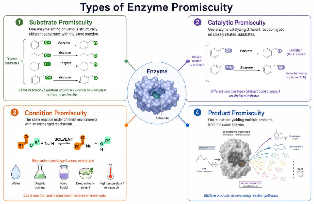
Figure 2.
Test reactions to measure specific activity.
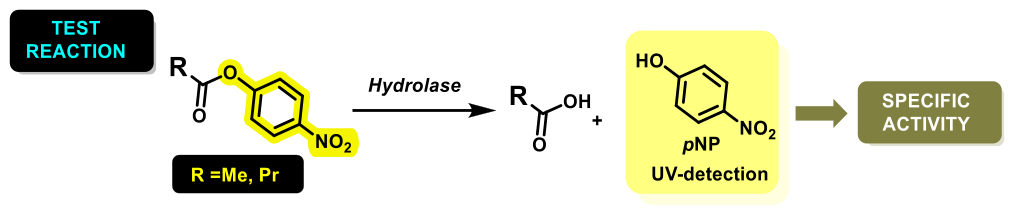
Figure 3.
Enzymatic hydrolysis of p-nitrophenyl N-acetyl-β-D-glucosaminide (pNPGlcNAc).
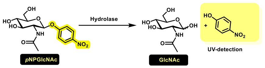
Figure 4.
a) Structure of 2-amino-2-deoxy-D-glucose(GlcNAc) and D-glucosamine (GlcN); b) Structure of natural biopolymers chitin and chitosan.
Figure 4.
a) Structure of 2-amino-2-deoxy-D-glucose(GlcNAc) and D-glucosamine (GlcN); b) Structure of natural biopolymers chitin and chitosan.
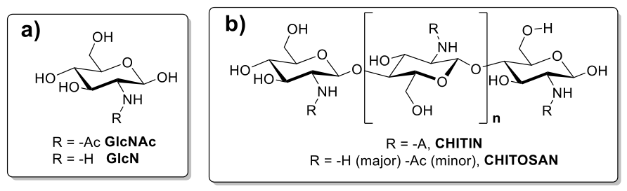
Figure 5.
Bicinchoninic acid (BCA) assay for measuring protein concentration.
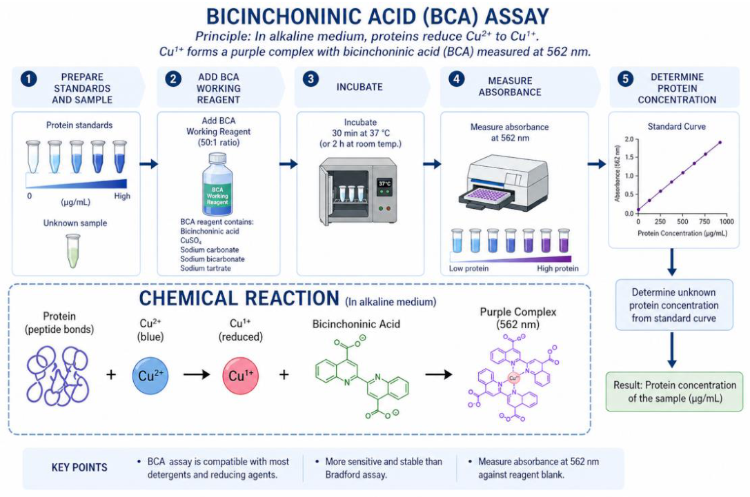

Table 1.
Protein concentration (BCA assay, Section 4.3) of commercial preparations.
Table 1.
Protein concentration (BCA assay, Section 4.3) of commercial preparations.
| Enzyme | [Protein] (mg mL-1) |
[Protein] (mg mL-1) |
|---|---|---|
| Proteases | Alcalase® 2.4 L | 123 ±16 |
| Neutrase® 0.8 L | 62.1 ± 9.7 | |
| Lipases (liquid formulations) | Lipozyme® TL 100L | 48.4 ± 8.5 |
| Lipozyme® CALB L | 35.9 ± 7.2 | |
| Palatase® 20000L | 5.1 ± 1.0 | |
| NovoCor® AD L | 53.7 ± 9.8 | |
| Lipases (solid formulations) | Lipase TL | 0.33 ± 0.02 |
| Lipase OF | 0.22 ± 0.02 | |
| Lipase MY30 | 0.12 ± 0.01 |
Table 2.
Molar extinction coefficients (ε348 and ε405, mM⁻¹ cm⁻¹) of pNP at different pH values (ε348 and ε405).
Table 2.
Molar extinction coefficients (ε348 and ε405, mM⁻¹ cm⁻¹) of pNP at different pH values (ε348 and ε405).
| ε (mM-1 cm-1) | pH 3 | pH 4 | pH 5 | pH 6 | pH7 | pH 8 | pH 9 | Hydrated reline |
|---|---|---|---|---|---|---|---|---|
| λ = 348 nm | 6.44±0.11 | 5.3±0.1 | 5.78±0.05 | 5.30±0.05 | 5.56±0.03 | 5.05±0.04 | 4.99±0.04 | 5.15±0.22 |
| λ = 405 nm | n/d | n/d | 0.22±0.01 | 1.55±0.02 | 8.10±0.03 | 14.5±0.1 | 16.05±0.13 | 11.08±0.47 |
.n/d: no measurable absorbance under the experimental conditions.
Table 3.
Protease activity vs pNPAc in aqueous media.
| ENZYME | Run | E (mg) |
Initial rate (mM·min-1) |
Units (µmol·min-1) |
Specific activity (µmol·min-1·mg-1) |
|---|---|---|---|---|---|
| BLANK (no enzyme) |
#0 | 0 | (6.3 ± 0.1)·10-4 | (1.29±0.45)·10-4 | |
| Alcalase ® 2.4 L FG | #A1 | 0.334 | (6.2 ± 2.7)·10-3 | (1.33±0.58)·10-2 | (3.7±0.6)·10-2 |
| #A2 | 0.835 | (1.45 ± 0.15)·10-2 | (3.13±0.32)·10-2 | ||
| #A3 | 1.67 | (2.8 ± 0.2)·10-2 | (6.06±0.43)·10-2 | ||
| Neutrase® 0.8 L | #N0 | 0 | 1.133·10-4 | (2.4 ± 0.2)·10-4 | (1.23 ± 0.12)·10-4 |
| #N1 | 15 | 1.85·10-4 | (3.9 ± 0.1)·10-4 | ||
| #N2 | 30 | 2.75·10-4 | (5.9 ± 0.2)·10-4 | ||
| #N3 | 60 | 4.54·10-4 | (9.7 ± 0.8)··10-4 |
Table 4.
Lipase activity vs pNPAc in aqueous media.
| ENZYME | Run | E (mg) |
Initial rate (mM·min-1) |
Units (µmol·min-1) |
Specific activity (µmol·min-1·mg-1) |
|---|---|---|---|---|---|
| BLANK (no enzyme) |
#0 | 0 | (1.84 ± 0.17)·10-6 | (2.44 ± 0.22)·10-4 | |
| Novocor® AD L | #CALA1 | 0.25 | (5.39 ± 2.54)·10-3 | (1.08 ± 0.51)·10-2 | (5.5 ± 0.3)·10-2 |
| #CALA2 | 0.5 | (1.71 ± 0.18)·10-2 | (3.23 ± 0.34)·10-2 | ||
| #CALA3 | 1 | (3.54 ± 0.24)·10-2 | (6.48 ± 0.44)·10-2 | ||
| #CALA4 | 2 | (6.86 ± 0.68)·10-2 | (1.08 ± 0.11)·10-1 | ||
| Lipozyme® CALB L | #CALB1 | 0.0625 | (1.97 ± 0.25)·10-2 | (4.24 ± 0.54)·10-2 | (6.5 ± 0.3)·10-2 |
| #CALB2 | 0.125 | (3.78 ± 0.54)·10-2 | (8.14 ± 1.17)·10-2 | ||
| #CALB3 | 0.25 | (7.59 ± 0.62)·10-2 | (1.63 ± 0.13)·10-1 | ||
| Lipozyme® TL 100L | #LTL1 | 0.5 | (9.13 ± 3.50)·10-3 | (1.96 ± 0.75)·10-2 | (1.23 ± 0.12)·10-4 |
| #LTL2 | 1 | (1.41 ± 0.35)·10-2 | (3.02 ± 0.75)·10-2 | ||
| #LTL3 | 2 | (2.74 ± 0.52)·10-2 | (5.89 ± 1.12)·10-2 | ||
| Palatase® 20000L | #PAL1 | 0.65 | (1.68 ± 0.35)·10-2 | (3.62 ± 0.75)·10-2 | (5.5 ± 0.3)·10-2 |
| #PAL2 | 1.3 | (2.84 ± 0.32)·10-2 | (6.11 ± 0.69)·10-2 | ||
| #PAL3 | 2.6 | (6.76 ± 0.77)·10-2 | (1.45 ± 0.17)·10-1 | ||
| Lipase TL | #TL1 | 0.331 | (2.10 ± 0.12) 10-3 | (4.51 ± 0.26)·10-3 | (9.2 ± 1.5)·10-3 |
| #TL2 | 0.662 | (2.93 ± 0.05) 10-3 | (6.29 ± 0.11)·10-3 | ||
| #TL3 | 1.32 | (5.92 ± 2.65) 10-3 | (1.27 ± 0.56)·10-2 | ||
| Lipase OF | #OF1 | 0.27 | (5.76 ± 2.73) 10-3 | (1.23 ± 0.58)·10-2 | (5.3 ± 0.4)·10-2 |
| #OF2 | 0.55 | (1.36 ± 0.42)·10-2 | (2.91 ± 0.90)·10-2 | ||
| #OF3 | 1.09 | (2.64 ± 0.56)10-2 | (5.68 ± 0.60)·10-2 | ||
| Lipase MY30 | #MY1 | 0.015 | (2.02 ± 0.08) 10-3 | (4.85 ± 0.19) 10-3 | (3.03 ± 0.16)·10-1 |
| #MY2 | 0.030 | (3.75 ± 0.23) 10-3 | (8.05 ± 0.49) 10-3 | ||
| #MY3 | 0.060 | (8.62 ± 1.06)·10-3 | (1.85 ± 0.23) 10-2 |
Table 5.
Lipase activity vs pNPBu in aqueous media.
| ENZYME | Run | E mg | Initial rate mM·min-1 |
Units (µmol·min-1) |
Specific activity µmol·min-1·mg-1 |
|---|---|---|---|---|---|
| BLANK (no enzyme) |
#0 | 0 | c | (6.54 ± 0.04)·10-4 | |
| Novocor® AD L | #CALA1 | 0.002 | (3.41 ± 1.94)·10-3 | (7.34 ± 4.17)·10-3 | 4.28 ± 0.73 |
| #CALA2 | 0.004 | (6.17 ± 0.20)·10-3 | (1.41 ± 0.04)·10-2 | ||
| #CALA3 | 0.006 | (1.25 ± 0.52)·10-2 | (2.69 ± 1.12)·10-2 | ||
| Lipozyme® CALB L | #CALB1 | 0.004 | (6.08 ± 1.10)·10-3 | (1.30 ± 0.23)·10-2 | 3.09 ± 0.36 |
| #CALB2 | 0.006 | (9.99 ± 1.11)·10-3 | (2.15 ± 0.24)·10-2 | ||
| #CALB3 | 0.008 | (1.12 ± 0.32)·10-2 | (2.40 ± 0.69)·10-2 | ||
| Lipozyme® TL 100L | #LTL1 | 0.001 | (4.13 ± 0.42)·10-3 | (8.89 ± 0.90)·10-3 | 6.85 ± 0.42 |
| #LTL2 | 0.0015 | (5.68 ± 0.49)·10-3 | (1.22± 0.10)·10-2 | ||
| #LTL3 | 0.002 | (6.50 ± 0.28)·10-3 | (1.40 ± 0.06)·10-2 | ||
| Palatase® 20000L | #PAL1 | 0.002 | (3.50 ± 0.12)·10-3 | (7.50 ± 0.30)·10-3 | 2.92 ± 0.10 |
| #PAL2 | 0.004 | (5.46 ± 0.25)·10-3 | (1.17 ± 0.05)·10-2 | ||
| #PAL3 | 0.006 | (8.72 ± 0.35)·10-3 | (1.87 ± 0.07)·10-2 | ||
| Lipase TL | #TL1 | 0.0025 | (3.21 ± 0.39) 10-3 | (6.89 ± 0.84)·10-3 | 4.54 ± 0.46 |
| #TL2 | 0.0051 | (9.10 ± 4.67) 10-3 | (1.95 ± 1.00) 10-2 | ||
| #TL3 | 0.0102 | (2.13 ± 0.33) 10-2 | (4.57 ± 0.71)·10-2 | ||
| Lipase OF | #OF1 | 0.015 | (3.16 ± 0.06) 10-3 | (6.80 ± 0.14) 10-3 | (4.82 ± 0.15)·10-1 |
| #OF2 | 0.021 | (5.09 ± 0.26)·10-3 | (1.09 ± 0.06)·10-2 | ||
| #OF3 | 0.032 | (7.38 ± 0.13) 10-3 | (1.59 ± 0.03)·10-2 | ||
| Lipase MY30 | #MY1 | 0.004 | (2.17 ± 0.06) 10-3 | (4.66 ± 0.12) 10-3 | (9.92 ± 0.50)·10-1 |
| #MY2 | 0.009 | (4.55 ± 0.13) 10-3 | (9.78 ± 0.28) 10-3 | ||
| #MY3 | 0.018 | (8.59 ± 4.13)·10-3 | (1.85 ± 0.26) 10-2 |
Table 6.
Lipase activity vs pNPAc in hydrated reline.
| ENZYME | Run | E mg | Initial rate mM·min-1 |
Units (µmol·min-1) |
Specific activity µmol·min-1·mg-1 |
|---|---|---|---|---|---|
| BLANK (no enzyme) |
#0 | 0 | (8.13 ± 0.91)·10-4 | (1.74 ± 0.20)·10-3 | |
| Novocor® AD L | #CALA1 | 0.5 | (7.69 ± 1.57) 10-3 | (1.65 ± 0.34)·10-2 | (1.9 ± 0.3)·10-2 |
| #CALA2 | 1 | (1.29 ± 0.40)·10-2 | (2.79 ± 0.85)·10-2 | ||
| #CALA3 | 2 | (1.94 ± 0.60)·10-2 | (4.18 ± 1.29)·10-2 | ||
| Lipozyme® CALB L | #CALB1 | 0.3 | (2.09 ± 0.62)·10-2 | (4.51 ± 1.34)·10-2 | (1.05 ± 0.10)·10-1 |
| #CALB2 | 0.65 | (4.50 ± 0.37)·10-2 | (9.69 ± 0.80)·10-2 | ||
| #CALB3 | 1.3 | (6.49 ± 0.95)·10-2 | (1.39 ± 0.20)·10-1 | ||
| Lipozyme® TL 100L | #LTL1 | 0.25 | (5.26 ± 0.29)·10-3 | (1.13 ± 0.06)·10-2 | (3.9 ± 0.8)·10-2 |
| #LTL2 | 0.5 | (9.21 ± 0.64)·10-2 | (1.98 ± 0.14)·10-2 | ||
| #LTL3 | 1 | (1.89 ± 1.11)·10-2 | (4.07 ± 2.39)·10-2 | ||
| Palatase® 20000L | #PAL1 | 0.030 | (4.47 ± 0.13)·10-3 | (9.60 ± 0.28)·10-3 | (1.4 ± 0.4)·10-1 |
| #PAL2 | 0.065 | (6.07 ± 0.32)·10-3 | (1.30 ± 0.07)·10-2 | ||
| #PAL3 | 0.130 | (7.54 ± 0.53)·10-2 | (1.62 ± 0.11)·10-2 | ||
| Lipase TL | #TL1 | 0.0166 | (2.50 ± 0.16) 10-3 | (5.38 ± 0.34)·10-3 | (3.2 ± 0.4)·10-1 |
| #TL2 | 0.033 | (3.28 ± 0.26) 10-3 | (7.03 ± 0.56)·10-3 | ||
| #TL3 | 0.066 | (1.08 ± 0.29) 10-2 | (2.30 ± 0.62)·10-2 | ||
| Lipase OF | #OF1 | 0.0275 | (1.96 ± 1.00) 10-3 | (4.23 ± 2.16)·10-3 | (9.3 ± 2.1)·10-2 |
| #OF2 | 0.0549 | (3.04 ± 0.80) 10-3 | (6.54 ± 1.72)·10-3 | ||
| #OF3 | 0.0879 | (4.67 ± 2.03) 10-3 | (1.00 ± 0.44)·10-2 | ||
| Lipase MY30 | #MY1 | 0.125 | (1.17 ± 0.10) 10-3 | (2.49 ± 0.21) 10-3 | (4.7 ± 0.6)·10-3 |
| #MY2 | 0.250 | (1.25 ± 0.10) 10-3 | (2.69 ± 0.22) 10-3 | ||
| #MY3 | 0.400 | (1.26 ± 0.13)·10-3 | (3.73 ± 0.39) 10-3 |
Table 7.
Lipase activity vs pNPBu in hydrated reline.
| ENZYME | Run | E mg | Initial rate mM·min-1 |
Units (µmol·min-1) |
Specific activity µmol·min-1·mg-1 |
|---|---|---|---|---|---|
| BLANK (no enzyme) |
#0 | 0 | (5.26 ± 1.05)·10-4 | (1.13 ± 0.23)·10-3 | |
| Novocor® AD L | #CALA1 | 0.005 | (4.91 ± 2.30) 10-3 | (1.06 ± 0.49)·10-2 | (9.9 ± 2.5)·10-1 |
| #CALA2 | 0.01 | (7.19 ± 2.41)·10-3 | (1.54 ± 0.52)·10-2 | ||
| #CALA3 | 0.02 | (1.03 ± 0.56)·10-2 | (2.20 ± 1.12)·10-2 | ||
| Lipozyme® CALB L | #CALB1 | 0.004 | (7.14 ± 2.14)·10-3 | (1.54 ± 0.46)·10-2 | 3.96 ± 0.32 |
| #CALB2 | 0.006 | (1.18 ± 0.22)·10-2 | (2.53 ± 0.47)·10-2 | ||
| #CALB3 | 0.008 | (1.51 ± 0.13)·10-2 | (3.24 ± 0.28)·10-1 | ||
| Lipozyme® TL 100L | #LTL1 | 0.004 | (3.82 ± 0.82)·10-3 | (8.22 ± 1.76)·10-3 | (5.0 ± 1.7)·10-1 |
| #LTL2 | 0.015 | (4.66 ± 1.54)·10-2 | (1.00 ± 0.33)·10-2 | ||
| #LTL3 | |||||
| Palatase® 20000L | #PAL1 | 0.004 | (2.73 ± 0.90)·10-3 | (5.86 ± 0.19)·10-3 | (2.8 ± 0.6)·10-1 |
| #PAL2 | 0.020 | (3.14 ± 0.12)·10-3 | (6.75 ± 0.26)·10-3 | ||
| #PAL3 | 0.040 | (6.72 ± 3.28)·10-3 | (1.44 ± 0.71)·10-2 | ||
| Lipase TL | #TL1 | 0.0025 | (3.41 ± 0.98) 10-3 | (7.34 ± 2.11)·10-3 | 2.71 ± 0.21 |
| #TL2 | 0.0051 | (8.27 ± 1.79) 10-3 | (1.77 ± 0.38)·10-2 | ||
| #TL3 | 0.0103 | (1.33 ± 0.15) 10-2 | (2.86 ± 0.32)·10-2 | ||
| Lipase OF | No reaction | ||||
| Lipase MY30 | No reaction | ||||
Table 8.
Lipase activity vs pNPGlcNAc in aqueous media at pH= 4, T=37°C.
| ENZYME | pH | E mg | Initial rate mM·min-1 |
Units (µmol·min-1) |
Specific activity µmol·min-1·mg-1 |
|
|---|---|---|---|---|---|---|
| BLANK (no enzyme) |
4.0 | 0 | (1.21 ± 0.15)·10-5* | |||
| Novocor® AD L |
4.0 | 0.10 | (2.84 ± 0.20)·10-3 | (5.62 ± 0.39)·10-3 | (5.62 ± 0.39)·10-2 | |
| Lipozyme® CALB L | 4.0 | 0.10 | (2.94 ± 0.31)·10-2 | (5.82 ± 0.61)·10-3 | (5.82 ± 0.39)·10-2 | |
| Lipozyme® TL 100L | 4.0 | 0.10 | (6.26 ± 1.9) 10-6 | (1.24 ± 0.24) 10-5 | (1.24 ± 0.24) 10-4 | |
| Pure CALB |
4.0 | 0.10 | (1.16 ± 0.17) 10-4 | (2.29 ± 0.34) 10-4 | (2.29 ± 0.34)·10-3 | |
| Lipase TL | No reaction | |||||
| Lipase OF | No reaction | |||||
| Lipase MY30 | No reaction | |||||
*Measured after 1440 min.
Table 9.
Lipase activity vs pNPGlcNAc in aqueous media at pH= 3.
| ENZYME | pH | Run | E mg | Initial rate mM·min-1 |
Units (µmol·min-1) |
Specific activity µmol·min-1·mg-1 |
|---|---|---|---|---|---|---|
| BLANK (no enzyme) |
3.0 | #0 | 0 | (1.46 ± 0.25)·10-4 | (1.46 ± 0.05)·10-1 | |
| Novocor® AD L | 3.0 | #CALA1 | 0.02 | (6.39 ± 0.92) 10-3 | (6.39 ± 0.92) 100 | (2.66 ± 0.17)·10-1 |
| #CALA2 | 0.05 | (1.30 ± 0.23)· 10-2 | (1.30 ± 0.23)·101 | |||
| #CALA3 | 0.075 | (2.27 ± 0.29)·10-2 | (2.27 ± 0.29)·101 | |||
| #CALA4 | 0.10 | (2.59 ± 0.30)·10-2 | (2.59 ± 0.30)·101 | |||
| Lipozyme® CALB L | 3.0 | #CALB1 | 0.02 | (1.13 ± 0.04)·10-2 | (1.13 ± 0.04)·101 | (2.97 ± 0.14)·10-1 |
| #CALB2 | 0.05 | (1.80 ± 0.04)· 10-2 | (1.80 ± 0.04)·101 | |||
| #CALB3 | 0.075 | (2.52 ± 0.05)· 10-2 | (2.52 ± 0.05)·101 | |||
| #CALB4 | 0.10 | (3.11 ± 0.03)· 10-2 | (3.11 ± 0.03)·101 | |||
| Lipozyme® TL 100L | 3.0 | #LTL1 | 0.02 | (1.79 ± 0.23) 10-2 | (1.79 ± 0.23) 101 | (3.58 ± 0.34)·10-1 |
| #LTL2 | 0.05 | (1.92 ± 0.24)· 10-2 | (1.92 ± 0.24)·101 | |||
| #LTL3 | 0.075 | (3.24 ± 0.32)·10-2 | (3.24 ± 0.32)·101 | |||
| #LTL4 | 0.10 | (3.90 ± 0.40)·10-2 | (3.90 ± 0.40)·101 | |||
| Palatase® 20000L | 3.0 | #PAL1 | 0.02 | (3.02 ± 0.87) 10-3 | (3.02 ± 0.87) 100 | (6.99 ± 0.63)·10-2 |
| #PAL2 | 0.05 | (4.14 ± 0.65) 10-3 | (4.14 ± 0.65) 100 | |||
| #PAL3 | 0.075 | (5.60 ± 0.92) 10-3 | (5.60 ± 0.92) 100 | |||
| #PAL4 | 0.10 | (7.72 ± 0.89) 10-3 | (7.72 ± 0.89) 100 | |||
| Lipase TL | No reaction | |||||
| Lipase OF | No reaction | |||||
| Lipase MY30 | No reaction | |||||
Table 10.
Lipase activity vs pNPGlcNAc in aqueous media at pH= 4.
| ENZYME | pH | Run | E mg | Initial rate mM·min-1 |
Units (µmol·min-1) |
Specific activity µmol·min-1·mg-1 |
|---|---|---|---|---|---|---|
| BLANK (no enzyme) |
4.0 | #0 | 0 | (1.47 ± 0.06)·10-4 | (1.47 ± 0.06)·10-1 | |
| Novocor® AD L | 4.0 | #CALA1 | 0.02 | (7.93 ± 0.33) 10-3 | (7.93 ± 0.33) 100 | (3.15 ± 0.06)·10-1 |
| #CALA2 | 0.05 | (1.53 ± 0.05)·10-2 | (1.53 ± 0.05)·101 | |||
| #CALA3 | 0.075 | (2.39 ± 0.04)·10-2 | (2.39 ± 0.04)·101 | |||
| #CALA4 | 0.10 | (3.24 ± 0.07)·10-2 | (3.24 ± 0.07)·101 | |||
| Lipozyme® CALB L | 4.0 | #CALB1 | 0.02 | (1.28 ± 0.13)·10-2 | (1.28 ± 0.13)·101 | (3.53 ± 0.16)·10-1 |
| #CALB2 | 0.05 | (2.14 ± 0.04)· 10-2 | (2.14 ± 0.04)·101 | |||
| #CALB3 | 0.075 | (3.01 ± 0.09)· 10-2 | (3.01 ± 0.09)·101 | |||
| #CALB4 | 0.10 | (3.68 ± 0.13)· 10-2 | (3.68 ± 0.13)·101 | |||
| Lipozyme® TL 100L | 4.0 | #LTL1 | 0.02 | (1.04 ± 0.04) 10-2 | (1.04 ± 0.04) 101 | (3.83 ± 0.08)·10-1 |
| #LTL2 | 0.05 | (1.97 ± 0.07)· 10-2 | (1.97 ± 0.07)·101 | |||
| #LTL3 | 0.075 | (3.06 ± 0.05)·10-2 | (3.06 ± 0.05)·101 | |||
| #LTL4 | 0.10 | (3.90 ± 0.08·10-2 | (3.90 ± 0.08)·101 | |||
| Palatase® 20000L | 4.0 | #PAL1 | 0.02 | (2.51 ± 0.12) 10-3 | (2.51 ± 0.12) 100 | (7.24 ± 0.31)·10-2 |
| #PAL2 | 0.05 | (4.25 ± 0.12) 10-3 | (4.25 ± 0.12) 100 | |||
| #PAL3 | 0.075 | (6.45 ± 0.27) 10-3 | (6.45 ± 0.27) 100 | |||
| #PAL4 | 0.10 | (7.46 ± 0.14) 10-3 | (7.46 ± 0.14)·100 | |||
| Lipase TL | No reaction | |||||
| Lipase OF | No reaction | |||||
| Lipase MY30 | No reaction | |||||
Table 11.
Lipase activity vs pNPGlcNAc in aqueous media at pH= 5.
| ENZYME | pH | Run | E mg | Initial rate mM·min-1 |
Units (µmol·min-1) |
Specific activity µmol·min-1·mg-1 |
|---|---|---|---|---|---|---|
| BLANK (no enzyme) |
5.0 | #0 | 0 | (8.77 ± 0.05)·10-5 | (8.77 ± 0.05)·10-2 | |
| Novocor® AD L | 5.0 | #CALA1 | 0.02 | (1.13 ± 0.25) 10-2 | (1.13 ± 0.25) 101 | (2.97 ± 0.36)·10-1 |
| #CALA2 | 0.05 | (2.27 ± 0.45)·10-2 | (2.27 ± 0.45)·101 | |||
| #CALA3 | 0.075 | (2.97 ± 0.45)·10-2 | (2.97 ± 0.45)·101 | |||
| #CALA4 | 0.10 | (3.12 ± 0.96)·10-2 | (3.12 ± 0.96)·101 | |||
| Lipozyme® CALB L | 5.0 | #CALB1 | 0.02 | (6.42 ± 0.30)·10-3 | (6.42 ± 0.30)·100 | (2.71 ± 0.36)·10-1 |
| #CALB2 | 0.05 | (1.64 ± 0.42)·10-2 | (1.64 ± 0.42)·101 | |||
| #CALB3 | 0.075 | (2.00 ± 0.50)·10-2 | (2.00 ± 0.50)·101 | |||
| #CALB4 | 0.10 | (2.77 ± 1.05) 10-2 | (2.77 ± 1.05)·101 | |||
| Lipozyme® TL 100L | 5.0 | #LTL1 | 0.02 | (1.27 ± 0.33) 10-2 | (1.27 ± 0.33) 101 | (3.52 ± 0.50)·10-1 |
| #LTL2 | 0.05 | (2.43 ± 1.11)· 10-2 | (2.43 ± 1.11)·101 | |||
| #LTL3 | 0.075 | (3.15 ± 0.98)·10-2 | (3.15 ± 0.98)·101 | |||
| #LTL4 | 0.10 | (3.57 ± 0.56·10-2 | (3.57 ± 0.56)·101 | |||
| Palatase® 20000L | 5.0 | #PAL1 | 0.02 | (2.32 ± 0.76) 10-3 | (2.32 ± 0.76) 100 | (7.26 ± 0.98)·10-2 |
| #PAL2 | 0.05 | (3.84 ± 1.17) 10-3 | (3.84 ± 1.17) 100 | |||
| #PAL3 | 0.075 | (5.44 ± 2.67) 10-3 | (5.44 ± 2.67) 100 | |||
| #PAL4 | 0.10 | (7.75 ± 1.55) 10-3 | (7.75 ± 1.55)·100 | |||
| Lipase TL | No reaction | |||||
| Lipase OF | No reaction | |||||
| Lipase MY30 | No reaction | |||||
Table 12.
Enzymatic preparations from Novozymes.
| Commercial enzymes from Novozymes | |||||||||
| Proteases [131] | |||||||||
| Strem Catalog Number | Product | EC No | Specificity |
Optimal conditions |
Unit activity* |
Rel. Activity† |
|||
| T (°C) | pH | ||||||||
| 06-3112 | Alcalase® 2.4 L FG | 3.4.21.62 | Serine endo-peptidase from Bacillus licheniformis (mainly subtilisin A) | 30–65 | 7-10 | 2.5 AU/g | 4.4 | ||
| 06-3160 | Neutrase 0.8 L | 3.4.24. | Neutral, zinc metallo endo-protease from Bacillus amyloliquefaciens | 40-50 | 7 | 0.8 AU/g | 1.0 | ||
| Lipases [132] | |||||||||
| Strem Catalog Number | Product | EC No | Specificity |
Optimal conditions |
Unit activity** |
Rel. Activity‡ |
|||
| T (°C) | pH | ||||||||
| 06-3140 | Lipozyme® TL 100L | 3.1.1.3 | Non specific lipase from Thermomyces lanuginosus | 20-50 | 7-10 | 100 KLU/g | 20.0 | ||
| 06-3105 | Lypozyme® CALB L | Non specific lipase from Candida antarctica B | 30-60 | 5-9 | 5 KLU/g | 1.0 | |||
| 06-3118 | Palatase® 20000L | Non specific lipase from Rhizomucor miehie | 30-50 | 7-10 | 20 KLU/g | 4.0 | |||
| 06-3100 | NovoCor® AD L | Non specific lipase from Candida antarctica A | 30-60 | 5-9 | 6 KLU/g | 1.2 | |||
* Proteases activity: K = Kilo, AU = Anson Unit. One AU (AU = Anson unit) is defined as the amount of enzyme that liberates 1.0 μmol (181 μg) of tyrosine from casein per minute at pH 7.5 at 37°C [133,134]. † Reference Standard. The activity is determined relative to a protease A standard. The result is given in the same units as the standard ** K = Kilo, LU = Lipase unit. 1LU is the amount of enzyme activity which liberates 1 μmol of titratable butyric acid from the substrate glycerol tributyrate per minute under defined standard conditions. ‡ Using Lypozyme® CALB L as reference.
Table 13.
Enzymatic preparations from Meito Sangyo [135].
Table 13.
Enzymatic preparations from Meito Sangyo [135].
| Product | Specificity | Molecular weight | Optimal conditions | Activity | ||
|---|---|---|---|---|---|---|
| T (°C) | pH | pH stability | ||||
| Lipase TL | Extracellular enzyme produced by fermentation with Stutzerimonas stutzeri (basionym: Pseudomonas stutzeri ). | 27,000 (SDS-PAGE) | 50 | 7-8 | 6-9 | 50,000U/g powder* |
| 650 U/g powder** | ||||||
| Lipase OF | Extracellular enzyme produced by a strain of Limtongozyma cylindracea (basionym: Candida cylindracea, formerly C. rugosa). | 60,000 (SDS-PAGE) | 40-50 | 6-7 | 3-8 | 360,000U/g powder† |
| Lipase MY30 | 40-50 | 6-7 | 3-8 | 30,000U/g powder (diluted with lactose)† | ||
* Assayed by the method of Kokusho et al. [136] using olive oil emulsified with polyvinylalcohol as a substrate [137]. 1U corresponds to the amount of enzyme which liberates 1µmol fatty acid per min at 37°C. **Assayed by the method of Fukuda et al [138] using p-nitrophenyl acetate as a substrate †Assayed by the method of Machida et al. [139] using olive oil emulsified with polyvinylalcohol as a substrate. 1U corresponds to the amount of enzyme which liberates 1µmol fatty acid per min at 37°C.
Disclaimer/Publisher’s Note: The statements, opinions and data contained in all publications are solely those of the individual author(s) and contributor(s) and not of MDPI and/or the editor(s). MDPI and/or the editor(s) disclaim responsibility for any injury to people or property resulting from any ideas, methods, instructions or products referred to in the content. |
© 2026 by the authors. Licensee MDPI, Basel, Switzerland. This article is an open access article distributed under the terms and conditions of the Creative Commons Attribution (CC BY) license (http://creativecommons.org/licenses/by/4.0/).
Copyright: This open access article is published under a Creative Commons CC BY 4.0 license, which permit the free download, distribution, and reuse, provided that the author and preprint are cited in any reuse.