

Submitted:
17 September 2026
Posted:
18 September 2026
You are already at the latest version
Preprints on COVID-19 and SARS-CoV-2
Abstract
Background/Objectives: Although the COVID-19 pandemic has ended, emerging SARS-CoV-2 variants remain a future threat. This study evaluated the safety and efficacy of an intranasal live attenuated vaccine (LAV) prototype based on a temperature-sensitive (ts) Omicron-like SARS-CoV-2 mutant in a Syrian hamster COVID-19 model. Methods: SARS-CoV-2 Dubrovka (Wuhan-like), Otradnoe (BA.1.1) and FEB2 (BA.5.2) strains and the ts F-F3 mutant derived from FEB2 were characterized by electron microscopy, nanopore sequencing, cytopathic effect titration, and RT-PCR. In vitro studies used Vero CCL-81 cells; in vivo transmissibility and efficacy studies were assessed in female Syrian hamsters. Serum antibody titers were measured by ELISA and neutralization assays. Immunization efficacy was assessed by weight change, viral loads in the lungs, nasal passages, and brain, and histological examination of the lungs at day 4 post-challenge. Safety criteria included evaluation of pathogenicity and transmissibility in hamsters and ts phenotype stability during prolonged in vitro cultivation. Results: The F-F3 strain displayed a stable ts phenotype in vitro, did not infect hamster lungs or brain, and showed no transmission from immunized to naïve co-housed animals. A single intranasal dose induced seroconversion, reduced pneumonia severity by 75% (p < 0.01), protected lungs and nasal passages against infection, and prevented viral transmission after homologous BA.5.2 challenge. Following heterologous Wuhan-like challenge, immunization prevented weight loss and significantly reduced viral loads in the lungs, nasal passages, and brain; however, protection against pneumonia was only modest. Conclusions: Preclinical evaluation of the F-F3-based LAV prototype demonstrated a favorable balance between safety and protective efficacy. These characteristics support its further consideration as a candidate strain for COVID-19 LAV development.
Keywords:
COVID-19 live attenuated vaccine
; temperature-sensitive SARS-CoV-2 mutant
; COVID-19 Syrian hamster model
; immunization efficacy
; transmissibility
; ts phenotype stability
1. Introduction
Members of Betacoronavirus genus within Coronaviridae family play a significant role in human respiratory pathology. Human coronaviruses HCoV-OC43 (Betacoronavirus gravedinis) and HCoV-HKU1 (Betacoronavirus hongkongense) contribute substantially to seasonal acute respiratory diseases morbidity, while zoonotic coronaviruses SARS-CoV-1 (Betacoronavirus pandemicum) and MERS-CoV (Betacoronavirus cameli) have caused outbreaks of severe respiratory diseases with high mortality rates. Between 2020 and 2023, SARS-CoV-2 (Betacoronavirus pandemicum) caused the COVID-19 pandemic, which resulted in at least 7 million deaths worldwide. Even countries with highly developed healthcare systems sustained severe losses during the pandemic. According to WHO estimates, the number of deaths from COVID-19 and its sequelae exceeded 1.2 million in the United States (3,612 deaths per million), 232,000 in the United Kingdom (3,404 deaths per million), and 404,000 in the Russian Federation (2,777 deaths per million) (https://data.who.int/dashboards/covid19/deaths). The true global burden of COVID-19 may be considerably higher than that reflected in publicly accessible data. For instance, due to insufficiently developed epidemiological surveillance systems, accurate assessments of the pandemic’s impact in low-income countries remain unavailable [1]. The severe consequences of the pandemic underscore the inadequate preparedness of national healthcare systems for the emergence of such threats.
To date, as a result of the formation of herd immunity, there is a low incidence of COVID-19, which makes it possible to consider the new coronavirus infection as a common seasonal respiratory infection. At the same time, the emergence risk of new SARS-CoV-2 variants of concern, or the transmission from animals to humans of other SARS-like coronaviruses that escape post-infection and post-vaccination immunity, cannot be excluded [2,3]. In addition, in the post-pandemic period, there is a high proportion of COVID-19 cases with severe course and/or complications in people with concomitant diseases (cerebrovascular and cardiovascular diseases, chronic obstructive pulmonary disease, diabetes) [4]. In this regard, the problem of improving the means of COVID-19 specific prevention remains relevant.
One of the most promising approaches to the specific prophylaxis of respiratory viral infections is the use of intranasally administered mucosal vaccines [5,6]. Mucosal vaccines induce the development of antiviral immunity directly at the respiratory tract mucosa, thereby conferring not only protection against disease but also minimizing the likelihood of viral transmission [5,7,8]. In animal COVID-19 models, intranasally administered mucosal vector vaccines based on replication-incompetent or attenuated human and animal viruses have demonstrated high efficacy [9,10,11,12,13]. Of particular interest are live attenuated vaccines (LAV), which are capable of eliciting robust cellular and humoral immune responses at both the local (mucosal) and systemic levels [14,15,16]. A historical example of the effective use of mucosal LAVs based on ts mutants is provided by live influenza vaccines, which are capable of conferring cross-protection against diverse antigenic variants of influenza viruses [17,18]. A number of preclinical studies have demonstrated the high efficacy of attenuated SARS-CoV-2 mutants in protecting susceptible laboratory animals against homologous challenge with a virulent viral strain, even following a single intranasal administration [19,20,21,22,23,24,25,26,27,28].
Based on the results of preclinical studies, a high efficacy of COVID-19 LAV in humans can be anticipated. This assumption is supported by observations of the herd immunity development, which has been formed primarily by the global spread of SARS-CoV-2 and naturally acquired infection. Evidence for the protective effect of natural infection is provided by Chemaitelly H. et al., who demonstrated that primary SARS-CoV-2 infection conferred 97.3% protection against severe disease or death upon reinfection with an Omicron-like virus, irrespective of the virus variant responsible for the primary infection [29]. Furthermore, in several African countries with low COVID-19 vaccination coverage (such as the Democratic Republic of the Congo, Chad, Burundi, Niger, and Nigeria) [30] population immunity has developed predominantly through natural SARS-CoV-2 infection, which has also contributed to a reduction in morbidity, disease severity, and mortality. The establishment of herd immunity in African countries is corroborated by a decline in the percentage of respiratory samples testing positive for SARS-CoV-2 from over 50% at the peak of morbidity surges in 2021–2022 to below 3% in the first half of 2026, that is comparable to the global average [31]. LAV efficacy is based on the same mechanisms involved in the development of adaptive immunity following natural coronavirus infection [8,32,33], which underscores the substantial potential of LAV in the prevention of diseases caused by SARS-CoV-2-related coronaviruses.
The present study is dedicated to the search for a candidate vaccine strain for COVID-19 LAV development. Previously, we generated SARS-CoV-2 Wuhan-like D-D2 mutant, which was adapted to growth at 23 °C, exhibited sensitivity to 39 °C, and displayed an attenuated (att) phenotype in golden Syrian hamsters [28]. Following a single intranasal immunization, the D-D2 strain reproducibly induced seroconversion in all immunized hamsters and provided highly effective protection against both homologous challenge with the parental Wuhan-like strain and heterologous challenge with Delta- and Omicron-like strains [19,28]. However, since D-D2 temperature-sensitive (ts) mutant, derived from the highly virulent neurotropic Dubrovka strain, retained a limited capacity for replication in hamsters lungs and induced focal interstitial pneumonia [28], it cannot be regarded as entirely safe.
In light of these considerations, for subsequent attenuation efforts, a less virulent Omicron-like strain, FEB2 (BA.5.2), was selected. This strain induced pneumonia in hamsters upon challenge but did not cause weight loss or infect the brain [34]. The choice of the FEB2 strain was further justified by its antigenic relatedness to currently circulating Omicron-like SARS-CoV-2 variants. By passaging the FEB2 strain in Vero cells at reduced temperature, we generated, for the first time, an Omicron-like ts mutant F-F3 strain, which was adapted for growth at 24 °C and exhibited sensitivity to 37 °C and above. F-F3 strain is avirulent for hamsters, having lost the capacity to infect the lungs while retaining the ability to replicate in the nasal passages and maintain immunogenicity [35], thereby positioning it as a candidate strain for LAV development.
In LAVs development researchers may encounter the “hyperattenuation” phenomenon, whereby the mutant virus loses immunogenicity as a result of adaptation to novel conditions, as well as residual virulence or att phenotype instability. Furthermore, a critical limitation to the implementation of any LAV is the potential risk of vaccine strain transmission from immunized individuals to naïve contacts, with subsequent circulation and possible reversion to virulence, as has been documented for the oral poliovirus vaccine [36]. In this regard, the most crucial safety characteristics of a vaccine strain are its non-transmissibility and att phenotype stability.
The aim of the study was to evaluate the safety and efficacy of a live attenuated vaccine prototype based on an Omicron-like SARS-CoV-2 ts mutant in an animal model of coronavirus pneumonia. Syrian hamsters were selected for in vivo studies, since the Syrian hamster COVID-19 model is well characterized and broadly accepted as an animal model of the disease.
2. Materials and Methods
2.1. Viruses
The following SARS-CoV-2 laboratory strains were used in the study: Wuhan-like Dubrovka strain, Omicron-like FEB2 and Otradnoe strains [34], as well as cold-adapted (ca) mutants derived from the FEB2 strain (F-F1, F-F3, F-D3, F-D12, F-C5, and F-C9) which were generated previously in our laboratory [35] (Table 1).
2.2. Animals
Four-week-old female golden Syrian hamsters (Mesocricetus auratus) weighing 40–50 g were obtained from the Nursery for laboratory animals of the Shemyakin and Ovchinnikov Institute of Bioorganic Chemistry of the RAS (Pushchino, Russia). SPF hamsters were screened at the nursery and confirmed free of viral, bacterial, and parasitic infections. Following delivery from the nursery, the animals were acclimatized for 4–7 days prior to the start of the experiments. Hamsters were housed under standard laboratory conditions at a controlled temperature of 20–26 °C and relative humidity of 45–65%, with a 12 h light/12 h dark cycle. Different groups of animals were maintained in individual cages. Standard rodent diet and water were provided ad libitum. Animals were randomly allocated to the experimental groups using a computer-generated random number sequence with Microsoft Excel. Group size was dedicated to allow for an assessment of the significance of between-group differences. No a priori inclusion or exclusion criteria were established for this study. All animals and data points were included in the analysis; no exclusions were applied. Hamsters were inoculated intranasally with SARS-CoV-2 in a volume of 100 μL administered to both nostrils under light ether anesthesia. Upon termination of the experimental procedures, the hamsters were humanely euthanized under chloroform anesthesia. The animals were monitored daily throughout the experiment. No humane endpoints were defined for this study. Hamsters were housed in accordance with the guidelines set forth by the European Convention for the Protection of Vertebrate Animals Used for Experimental and Other Scientific Purposes [37]. All procedures involving animals were approved by the Local Ethics Committee of the I. Mechnikov Research Institute of Vaccines and Sera prior to the commencement of the study (Protocol No. 2 dated 24 May 2021).
2.3. Cell and Virus Cultivation
Vero CCL-81 (American Type Culture Collection, USA) cells were cultured at 37 °C in DMEM medium based on Earle’s buffer (PanEco, Russia) supplemented with 5% fetal bovine serum (FBS) (Gibco), 300 μg/mL L-glutamine (PanEco), and 40 μg/mL gentamicin (PanEco) in a 5% CO₂ atmosphere. SARS-CoV-2 was propagated in Vero cells as previously described [38]. The maintenance medium consisted of DMEM supplemented with 300 μg/mL L-glutamine, 40 μg/mL gentamicin, and 1% FBS. Vero cell monolayers were infected with SARS-CoV-2 and incubated at 37 °C for 3–4 days (strains Dubrovka, Otradnoe, and FEB2, MOI 0.001–0.00001) or at 24 °C for 7–10 days (strain FEB2 ca mutants, MOI 0.01) in a 5% CO₂ atmosphere until pronounced cytopathic effect (CPE) became evident. The virus-containing culture medium was clarified by centrifugation at 5,000 rpm for 5 min, aliquoted, titrated, and stored at −80 °C until use.
2.4. Virus Titration
SARS-CoV-2 titer was determined by TCID₅₀ endpoint dilution assay in Vero CCL-81 cell culture, as previously described [38]. Dubrovka, Otradnoe, and FEB2 strains were titrated at 37 °C, whereas the FEB2 strain ca mutants were titrated at 30 °C. The virus titer was calculated according to Ramakrishnan M.A. et al. [39] and expressed as log TCID₅₀/mL.
2.5. Quantification of SARS-CoV-2 RNA
SARS-CoV-2 RNA was quantified by real-time RT-PCR using a DTprime amplification system (DNA-Technology, Russia), as described previously [19]. Viral RNA was extracted from samples using the MagnoPrime UNI reagent kit (NextBio, Russia). For the detection of viral RNA, primers and a probe targeting SARS-CoV-2 nucleocapsid (N) gene of were employed: CoVN-F (GCGTTCTTCGGAATGTCG), CoVN-R (TTGGATCTTTGTCATCCAATTTG), and CoVN-P (FAM-AACGTGGTTGACCTACACAGGT-BHQ1) [40]. A synthetic oligonucleotide corresponding to the amplified fragment of the SARS-CoV-2 genome was used to construct a standard calibration curve.
2.6. SARS-CoV-2 Genome Sequencing
SARS-CoV-2 genome sequencing was performed on a MinION nanopore sequencer using Flow Cell R9.4 with the MinKNOW software and reagent kits from Oxford Nanopore Technologies (United Kingdom), as described previously [28]. Specific RT-PCR amplification of the SARS-CoV-2 genome was carried out using the NEBNext® ARTIC SARS-CoV-2 Companion Kit (Oxford Nanopore Technologies®). Ligation Sequencing kit and Native Barcoding Kit 1D (Oxford Nanopore Technologies, UK) were used to prepare the resulting pool. Genome assembly was performed in Minimap2 v. 2.24 [41].
2.7. Transmission Electron Microscopy Analysis
The virus-containing cell culture medium was clarified at low centrifugation speed (4000 rpm x 10 min, 4 °C, Eppendorf A-4-44 rotor), and was passed through Amicon Ultra Centrifugal Filters (Millipore) with a cut-off of 100 000 Da (100K) at a low centrifugation speed (4500 rpm x 10, 4 °C; Eppendorf A-4-44 rotor). The concentrated virus particles were re-suspended in a sterile phosphate-buffered saline (PBS, pH 7.2), and aliquots of the virus suspension were frozen (-80 °C) until the usage. To obtain immune complexes, the virus sample was thawed, mixed with rabbit serum against the FEB2 strain, and incubated for 1 h at 37 °C. The virus samples or immune complexes were loaded onto formvar-coated grids (TedPella, США), stained with a 2% water solution of phosphotungstic acid (PTA) (Fluka), pH 7.0, and viewed with a transmission electron microscope JEM-2100 (JEOL, Japan). The research was carried out at the Shared Research Facility “Electron microscopy in life sciences” at Moscow State University (Unique Scientific Equipment “Three-Dimensional Electron Microscopy and Spectroscopy”, ID RFMEFI61919X0014).
2.8. Detection of ts Phenotype in ca SARS-CoV-2 Mutants
Vero cells were infected with parental FEB2 strain and ca mutants at MOI 0.001. Following adsorption for 1 hour, the cells were washed twice with serum-free medium, supplemented with growth medium, and incubated at 37 °C and 39 °C for 4 days in 5% CO₂ atmosphere. From days 1 through 4 post-infection (p.i.), culture supernatant samples were collected daily, and viral titers as well as viral RNA concentrations were determined. A reduction in infectious titer or viral RNA concentration of 4.0 log or more on day 4 p.i. compared to FEB2 strain was considered indicative of a ts phenotype.
2.9. Assessment of F-F3 Strain ts Phenotype Stability
F-F3 strain (propagated at 24 °C) and the parental FEB2 strain (propagated at 37 °C) were serially passaged for 10 passages in Vero cells in 25 cm² culture flasks under two temperature conditions—33 °C and 37 °C. For each subsequent passage, 100 μL of culture supernatant collected on day 4 p.i. was inoculated into flasks containing a 3-day-old cell monolayer and incubated in a 5% CO₂ atmosphere. At each passage level, the presence of CPE was assessed on day 4 p.i. Viral material harvested after the 10th passage was evaluated for infectious titer and viral RNA concentration. Additionally, viral material obtained after the 10th passage was tested for temperature sensitivity at 37 °C and 39 °C based on viral RNA accumulation, as described above.
2.10. Evaluation of Immunization Efficacy
The efficacy of immunization with F-F3 strain was assessed in two independent experiments according to the experimental schedule (Figure 1). A study protocol, including the research question, key design features, and analysis plan, was prepared prior to the study; however, it was not registered.
For each experiment, hamsters were divided into five groups of six animals each (total number of animals, 60). Hamsters in two experimental groups received 100 μL of F-F3 strain at a dose of 5.0 log TCID₅₀ per animal intranasally under light ether anesthesia. Non-immunized hamsters in two control groups received 100 μL of PBS, pH 7.2, intranasally. At 28 p.i., immunized and non-immunized hamsters in the first experiment were challenged intranasally with FEB2 and Otradnoe SARS-CoV-2 strains, while in the second experiment, they were challenged with FEB2 and Dubrovka strains, at a dose of 5.0 log TCID₅₀ per animal. An additional control group consisted of naïve (non-immunized and non-challenged) hamsters. Animals were monitored daily, and body weight was recorded from day 0 to day 4 post-challenge (p.c.). At 4 days p.c., hamsters were humanely euthanized under chloroform anesthesia. The right lung, brain, nasal passages, liver, spleen, heart, and kidneys were excised, rinsed, and homogenized in DMEM supplemented with gentamicin (40 μg/mL) using Bioprep-24R homogenizer (Allsheng, China) (4,000 rpm x 4.5 min, 4 °C). The resulting homogenates were clarified by centrifugation (10,000 rpm x 5 min, 4 °C). Clarified organ homogenates were stored at −80 °C for subsequent virus titration and viral RNA quantification. The left lung was preserved in 10% neutral buffered formalin for histological examination. Immunization efficacy was assessed by comparing weight dynamics, viral load in organs, and histopathological changes in the lungs between immunized and non-immunized hamsters. To minimize the influence of potential confounders, animals in each group were housed in separate cages, and a separate container was used for weighing each group. In addition, cages containing non-immunized animals were isolated from those containing immunized animals prior to challenge to prevent airborne transmission of the virus. The investigators responsible for animal weighing, RT-PCR, virus titration, histological examination, and outcome assessment were blinded to group allocation.
2.11. Evaluation of Protection Against Transmission Upon Challenge of Immunized Hamsters with a Virulent Strain
Two groups of hamsters (n=12 and n=4) were immunized intranasally with F-F3 strain at a dose of 5.0 log TCID₅₀ per animal. At 28 days p.i., one group of hamsters was challenged intranasally with Dubrovka strain, while the second group was challenged with FEB2 strain at a dose of 5.0 log TCID₅₀/mL. On the following day, four naïve hamsters were introduced into each cage containing the challenged animals and were co-housed until the end of the experiment. At day 21 p.c., blood samples were collected from the co-housed hamsters for the detection of IgG antibodies to SARS-CoV-2 by ELISA and neutralization assay. The absence of seroconversion in the co-housed animals was considered the criterion for protection against transmission.
2.12. Assessment of Immunogenicity
F-F3 strain immunogenicity was evaluated in two independent experiments by quantifying total and virus-neutralizing antibodies in hamster sera at 21 days post-immunization (p.i.). Hamsters (n=12) were administered 100 μL of strain F-F3 at a dose of 5.0 log TCID₅₀ per animal intranasally under light ether anesthesia, according to the experimental schedule (Figure 1). In the first experiment, an additional group of hamsters (n=6) was inoculated with the parental FEB2 strain at an equivalent dose. Control group hamsters (n=6) received 100 μL of PBS, pH 7.2, intranasally (K- group).
2.13. Determination of Antibodies to SARS-CoV-2
IgG antibodies to SARS-CoV-2 structural antigens in hamster sera were determined by ELISA, as described previously [42]. For ELISA, 96-well plates were coated with whole-virion preparation of SARS-CoV-2 Dubrovka strain, inactivated by UV irradiation, which was obtained as previously described [43]. Neutralizing antibodies titer was determined in Vero cell culture against the FEB2 and Dubrovka strains, as described earlier [38].
2.14. Histological Examination of the Lungs
The left lungs were fixed in 10% neutral buffered formalin for 24 hours, embedded in histomix, and serial sections of 3–5 μm thickness were prepared. Lung tissue sections were stained with hematoxylin and eosin, subjected to photodocumentation, and the severity of pathomorphological changes was assessed and scored. Each coded histological specimen was assigned a cumulative severity score of the alterative-inflammatory process, calculated as the sum of scores (ranging from 0 to 3) according to the morphological criteria proposed by Gruber A.D. et al. [44]. The morphological scoring criteria included: percentage of affected lung area, distribution pattern of pneumonic foci, intraalveolar neutrophils and macrophages, vasculitis, cellular debris in bronchial and bronchiolar lumina, bronchitis, bronchointerstitial pneumonia, interstitial pneumonia, diffuse alveolar damage (alveolar epithelial necrosis, cellular debris in alveolar spaces, intraalveolar hemorrhage, intraalveolar edema, interstitial edema, inflammatory infiltrate in interalveolar septa), type II pneumocyte hyperplasia, necrosis and desquamation of vascular endothelial cells, damage to the bronchial and bronchiolar epithelial lining, and hyperplasia of bronchial ciliated epithelial cells. The maximum possible total score was 60.
2.15. Assessment F-F3 Strain Transmissibility
In two independent experiments, hamsters (n=12) were immunized intranasally with F-F3 strain at a dose of 5.0 log TCID₅₀ per animal. On the day following immunization, naïve hamsters (n=4) were introduced into the cage and co-housed with the immunized animals throughout the entire experiment. A separate cage housed naïve hamsters serving as the control group (n=4). At 21 days p.i., blood samples were collected from all hamsters and tested for IgG antibodies to SARS-CoV-2 by ELISA and neutralization assay. Seroconversion in the co-housed animals was considered the criterion for transmission.
In an additional experiment, 15 naïve hamsters were divided into 5 equal groups of 3 animals each (groups P0, P1, P2, P3, and P4). P0 group was infected intranasally with F-F3 strain at a dose of 6.0 log TCID₅₀ per animal. At 3 days p.i., nasal washes were collected from the infected animals under light ether anesthesia by flushing the nasal passages with DMEM supplemented with gentamicin (40 μg/mL). The washes were pooled and administered intranasally (50 μL) to P1 group hamsters. This procedure was repeated for a total of 4 sequential passages (groups P1, P2, P3, and P4). All nasal wash samples were analyzed for the presence of viral RNA and infectious virus. At 21 days p.i., blood samples were collected from all hamsters and tested for IgG antibodies to SARS-CoV-2 by ELISA. The criteria for infection of hamsters included the detection of seroconversion, as well as the presence of viral RNA and infectious virus in nasal wash samples.
2.16. Statistical Analysis
Statistical analysis and data visualization was performed using GraphPad Prism software (version 8). Data are presented as mean ± standard error of the mean (SEM). Comparisons between groups were made using the Mann-Whitney U test, appropriate for small sample sizes. Differences were considered statistically significant at p < 0.05.
2.17. Biosafety Requirements
All work with the SARS-CoV-2 virus was carried out under conditions of a Biosafety Level-3 laboratory.
3. Results
3.1. F-F3 Strain Characterization
Previously, through SARS-CoV-2 FEB2 strain cold adaptation, six ca mutant clones capable of replicating in Vero cells at 24 °C were obtained: F-F1, F-F3, F-D3, F-D12, F-C5, and F-C9 (Table 1) [35], and their genomic sequences were determined (GenBank accession numbers PX401966–PX401971). Clones F-C5 and F-C9 exhibited temperature sensitivity at 39 °C, whereas clones F-F1, F-F3, F-D3, and F-D12 were sensitive at 37 °C [35]. Comparative analysis of parental FEB2 strain (GenBank accession number OP920753.1) and its ca mutants genomes revealed genetic changes that occurred as a result of viral adaptation to growth in Vero cells at 24 °C. The genomes of the ca mutants contained 21 to 24 nucleotide substitutions (Supplemental Table S1), predominantly nonsynonymous (Supplemental Table S2). For further investigation, ts strain F-F3 (GenBank accession number PX401966) was selected, which acquired 23 nucleotide substitutions (Supplemental Table S1), 20 of which resulted in amino acid changes in the NSP3, NSP5, NSP6, NSP12, NSP13, NSP14, S, E, M, and N proteins (Table S2). Thus, a set of mutations defining F-F3 strain ca/ts/att phenotype was identified.
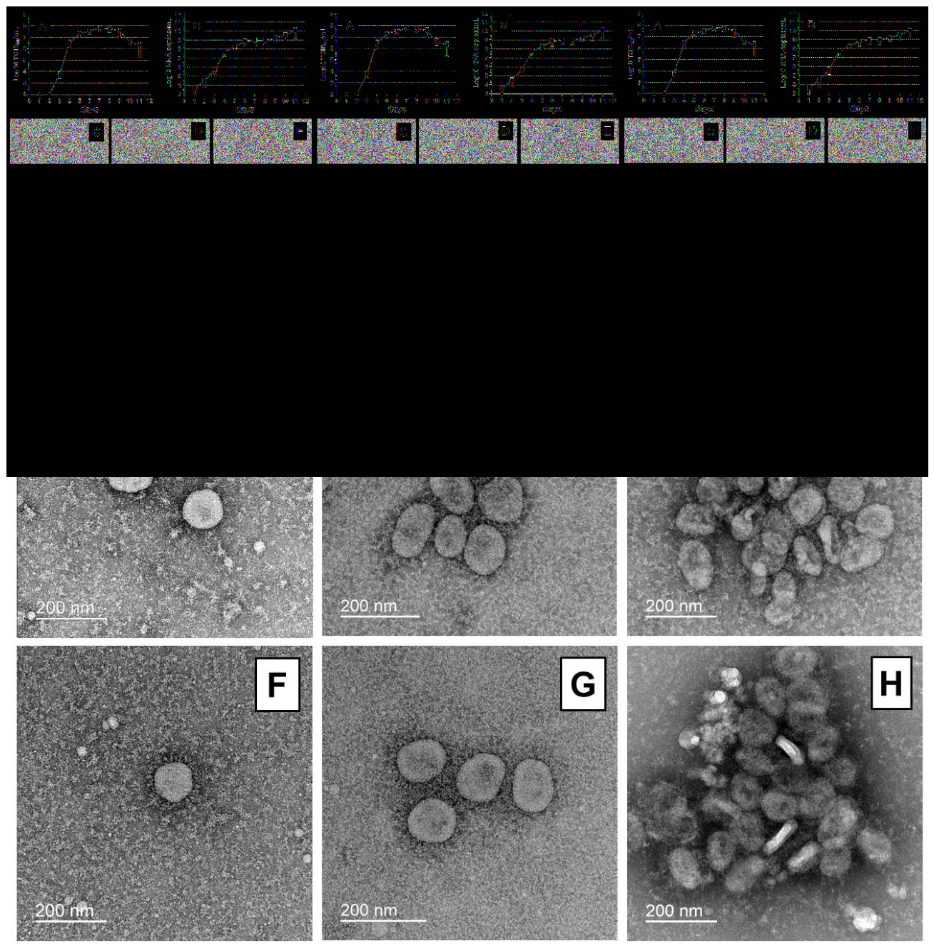
In Vero cells infected with the F-F3 strain (MOI 0.01) at 24 °C, infectious virus accumulated in culture supernatant, reaching peak titers of 7.4–7.7 log TCID₅₀/mL on days 6–9 p.i., after which infectious activity gradually declined by day 11 (A). The accumulation of infectious virus in culture fluid was corroborated by an increase in viral RNA concentration (B). CPE was mild and manifested as alterations in cell morphology. Noticeable detachment of individual virus-infected cells from the flask surface began only after days 6–7 p.i. In preparations of the F-F3 strain propagated at 24 °C, coronavirus virions were observed, which were similar in morphology and size to those of the parental FEB2 strain (cultivated at 37 °C) (C–H). Virions from both virus preparations reacted with hyperimmune rabbit serum against FEB2 strain, forming immune complexes of various sizes, indicating that the surface proteins of F-F3 retained the antigenic properties of the parental strain (D, E, G, H) [45]. Within the immune complexes, a considerable number of elongated structures were observed in both preparations, comparable in length to the diameter of a coronavirus virion. These elongated structures likely represent defective nucleocapsid-deficient (empty) virions, which become flattened upon drying, assume a disc-like shape, and are oriented perpendicular to the grid surface.
Figure 2.
F-F3 strain replication. Accumulation of infectious virus (A) and viral RNA (B) in culture supernatant of Vero cells infected with F-F3 strain (MOI 0.01) at 24 °C. Data from two independent experiments are shown as mean ± SEM. The transmission electron microscopy images of negatively stained virions of FEB2 (C) and F-F3 (F) strains, as well as and immune complexes of FEB2 (D, E) and F-F3 (G, H) strains with immune rabbit serum against FEB2 strain are represented.
Figure 2.
F-F3 strain replication. Accumulation of infectious virus (A) and viral RNA (B) in culture supernatant of Vero cells infected with F-F3 strain (MOI 0.01) at 24 °C. Data from two independent experiments are shown as mean ± SEM. The transmission electron microscopy images of negatively stained virions of FEB2 (C) and F-F3 (F) strains, as well as and immune complexes of FEB2 (D, E) and F-F3 (G, H) strains with immune rabbit serum against FEB2 strain are represented.
3.2. F-F3 Strain ts Phenotype Stability in Vitro
To assess the possibility of ts phenotype reversion (i.e., reversion to the non-ts phenotype characteristic of the parental FEB2 strain), F-F3 strain was serially passaged for 10 passages in Vero cell culture under temperatures 33 °C and 37 °C. At 33 °C, F-F3 strain replicated at all passage levels, inducing pronounced CPE by days 3-4 p.i., with both infectious virus and viral RNA detectable in the culture supernatant. At 37 °C, infection of Vero cells with F-F3 strain was abortive, as evidenced by CPE absence from passages 2 through 10, as well as the lack of infectious virus and viral RNA in the culture fluid (A). FEB2 reference strain replicated efficiently under both temperature conditions.
After 10 passages at 33 °C, F-F3 strain (F-F3_10p) retained its temperature sensitivity at both 37 °C and 39 °C. At 33 °C, F-F3_10p replicated productively, accompanied by pronounced CPE and increases in virus titer and viral RNA concentration in culture supernatant, whereas at 37 °C and 39 °C, no signs of viral replication were detected. FEB2 reference strain replicated efficiently at all three temperatures (33 °C, 37 °C, and 39 °C) (B,C). The obtained results indicate F-F3 strain ts phenotype stability under long-term cultivation in vitro at 33 °C.
3.3. F-F3 Strain Immunogenicity
F-F3 strain immunogenicity was evaluated in two independent experiments according to the scheme (Figure 1). In both experiments, all immunized hamsters developed both IgG antibodies to SARS-CoV-2 antigens and neutralizing antibodies (Nab) in serum at 21 days following a single intranasal immunization. Notably, IgG and Nab titers in hamsters immunized with F-F3 were 3.4-fold (p < 0.001) and 8.2-fold (p < 0.0001) lower, respectively, compared to the group infected with an equivalent dose of the parental FEB2 strain (Figure 4).
3.4. F-F3 Strain Immunization Efficacy
The efficacy of a single intranasal immunization of hamsters with F-F3 strain was assessed in two independent experiments according to the scheme (Figure 1). In the first experiment, immunized hamsters were infected with Omicron-like FEB2 (BA.5.2-like) and Otradnoe (BA.1.1-like) strains, and in the second experiment—with FEB2 (BA.5.2-like) and Dubrovka (Wuhan-like) strains.
3.4.1. Experiment 1
Body weight dynamics of immunized and non-immunized hamsters during the first 4 days post-challenge with the BA.5.2-like and BA.1.1-like strains did not differ significantly, as these strains do not induce substantial weight gain retardation [34] (A). A more informative indicator of immunization efficacy was the viral load in the lungs and nasal passages. On day 4 post-challenge with the homologous BA.5.2-like strain, infectious virus was undetectable in the lungs and nasal passages of immunized hamsters. Following heterologous challenge with the BA.1.1-like strain, infectious virus was also undetectable in the lungs of immunized hamsters, whereas in the nasal passages, the mean virus titer was 4.1 log TCID₅₀/mL, which was 2.2 log lower than that in the non-immunized control group (p < 0.01). In all non-immunized hamsters, infectious virus was detected in the lungs and nasal passages at titers ranging from 5.3 to 6.3 log TCID₅₀/mL. Infectious virus was not detected in the brain of any animal group (C).
Assessment of viral RNA content in the lungs and nasal passages confirmed the protective effect of immunization. Following challenge with the BA.5.2-like strain, viral RNA levels in the lungs and nasal passages of immunized animals were 3.9 log (p < 0.01) and 2.3 log (p < 0.01) lower, respectively, while after challenge with the BA.1.1-like strain, they were 1.9 log (p < 0.01) and 1.9 log (p < 0.01) lower compared to non-immunized animals. In the brain, viral RNA was detected only in individual non-immunized animals at the limit of detection (<3.3 log RNA copies/mL) (D). Determination of viral RNA in other organs and tissues (heart, kidney, liver, spleen, blood) proved to be of limited utility for assessing immunization efficacy (Supplemental Figure S1).
At day 4 post-challenge with the homologous BA.5.2-like and heterologous BA.1.1-like strains, non-immunized hamsters developed bronchointerstitial pneumonia, with the most pronounced morphological manifestations of the inflammatory process observed following BA.5.2-like strain challenge. Lung morphology in hamsters from the intact control group (K−) corresponded to normal. Histological examination of the lungs of immunized hamsters at day 4 post-challenge with both strains revealed a marked reduction in the severity and extent of interstitial pneumonia. In lung sections from hamsters in these groups, small areas of reduced aeration corresponding to interstitial pneumonia foci were observed. Inflammatory changes in bronchi walls, bronchioles, and vessels were absent. Morphological signs of diffuse alveolar damage were not detected (Supplemental Figure S3 and Figure S4). Blinded semi-quantitative scoring demonstrated significantly less severe pathomorphological changes in the lungs of immunized animals compared to non-immunized hamsters (B). Specifically, the cumulative pneumonia severity score in immunized hamsters was reduced by 76.6% (p < 0.01) following homologous challenge and by 45.1% (p < 0.01) following heterologous challenge, relative to non-immunized controls.
3.4.2. Experiment 2
Upon homologous challenge with the BA.5.2-like strain, as in the first experiment, protection against weight loss was not observed, since the challenge strain does not induce substantial weight gain retardation [34]. However, upon heterologous challenge with the highly virulent Wuhan-like strain, significant protection against weight loss (9.2%, p < 0.05) was evident on day 4 p.c. (A).
At day 4 post-challenge with the BA.5.2-like and Wuhan-like strains, infectious virus was undetectable in the lungs of immunized hamsters, whereas in non-immunized animals, the mean virus titers in the lungs were 6.5 and 8.0 log TCID₅₀/mL, respectively. Following homologous challenge, infectious virus was not detected in the nasal passages of the majority of immunized hamsters (virus detected in only one of six animals at a titer of 2.2 log TCID₅₀/mL), while all non-immunized animals had high titers of infectious virus (mean 6.9 log TCID₅₀/mL). Following heterologous challenge, infectious virus was recovered from the nasal passages of all immunized hamsters (mean 5.2 log TCID₅₀/mL), which was 1.8 log lower than in non-immunized animals (p < 0.01). Upon challenge with the Wuhan-like strain, infectious virus was not detected in the brains of immunized hamsters, whereas it was detected in all non-immunized animals at a mean titer of 5.0 log TCID₅₀/mL (C).
The results of viral RNA quantification in the lungs, nasal passages, and brain were well correlated with the data on infectious virus titers. The lungs of immunized hamsters were completely protected, while the nasal passages were partially protected against homologous challenge. Viral RNA was detected in the nasal passages, but its concentration was 2.5 log lower than in non-immunized animals (p < 0.01). Following heterologous challenge, viral RNA levels in the lungs, brain, and nasal passages of immunized hamsters were 3.2 (p < 0.01), 1.7 (p < 0.01), and 1.3 (p < 0.05) log lower, respectively, than in non-immunized animals (D). As in the first experiment, determination of viral RNA in other organs and tissues (heart, kidney, liver, spleen, blood) proved to be of limited utility for assessing immunization efficacy (Supplemental Figure S2).
Histological examination at day 4 post-challenge with the BA.5.2-like and Wuhan-like strains revealed bronchointerstitial pneumonia in the lungs of non-immunized hamsters, with inflammatory lesions involving 70–80% of the histological section area in some lobes. Lung histostructure in non-immunized, non-challenged hamsters (K− group) was normal. In the lungs of immunized hamsters, a marked reduction in the severity and extent of interstitial pneumonia was observed following homologous challenge with the BA.5.2-like strain. Airless areas were absent. In most microscopic fields, inflammatory infiltration of the interalveolar septa was moderate, with no exudate detected in the alveolar lumina. Peribronchiolar and perivascular lymphoid-histiocytic infiltrates were mild. Inflammatory changes in the bronchiolar walls were focal and mild. The epithelial lining of the conducting airways was preserved throughout. Vascular walls showed no pathological alterations. Following heterologous challenge, the severity of inflammatory changes was also reduced, although to a lesser extent. In some lobes, the area of pneumonic involvement reached 40–50%, whereas in other lobes, inflammatory changes were minimal. Pneumonic foci were predominantly localized along the major intrapulmonary airways and were absent in the peripheral regions. Overall, the severity of inflammatory changes in the bronchiolar walls, vessels, and alveoli was lower than in the non-immunized group (Supplemental Figure S5 and Figure S6, S7, S8). Blinded semi-quantitative scoring demonstrated a significant reduction in the cumulative pneumonia severity score in immunized hamsters compared to non-immunized animals following homologous challenge (75.0% reduction, p < 0.01). Following heterologous challenge, only a modest decrease in the cumulative pneumonia severity score was observed in immunized hamsters relative to non-immunized controls (16.0% reduction, p < 0.05) (B).
In addition, protection against transmission of a virulent strain to naïve co-housed hamsters following challenge of immunized animals was evaluated. When immunized hamsters were challenged with the heterologous Wuhan-like strain, co-housed hamsters were found to have total IgG antibodies to SARS-CoV-2 at 21 days p.c., indicating that they had become infected (A). In contrast, when immunized hamsters were challenged with the homologous BA.5.2-like strain, co-housed hamsters were protected from infection, as evidenced by the absence of seroconversion at 21 days p.c. (B). The presence or absence of antibody responses to SARS-CoV-2 was confirmed by virus neutralization assay (A,B).
3.5. F-F3 Strain Transmissibility
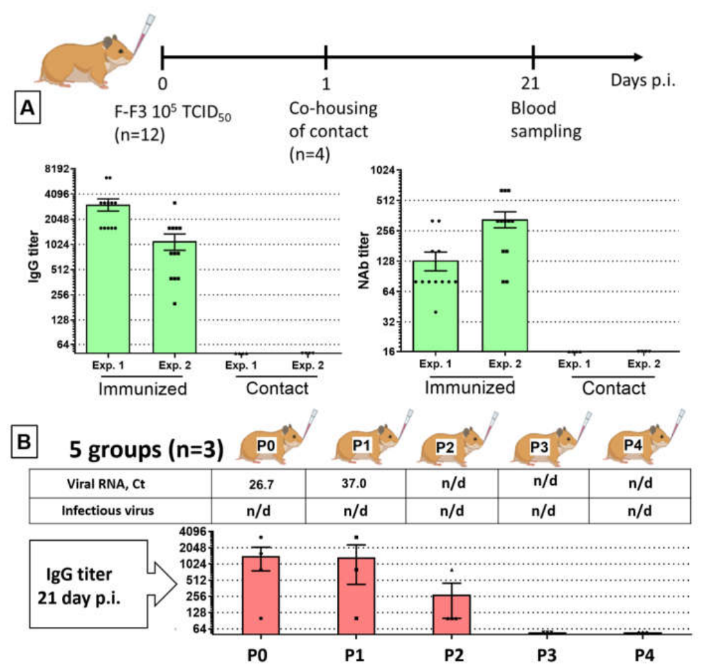
All hamsters immunized with F-F3 strain developed serum IgG antibodies to SARS-CoV-2 structural antigens and neutralizing antibodies by day 21 p.i. In contrast, no seroconversion was detected in the four naïve contact hamsters co-housed with the immunized animals, as assessed by both ELISA and neutralization assay at day 21 p.i. (A). A repeat experiment yielded identical results. Thus, F-F3 strain was reproducibly not transmitted to susceptible contact animals, despite close proximity to immunized hamsters.
Subsequently, an attempt was made to passage F-F3 strain in hamsters through sequential intranasal inoculation with nasal wash supernatants obtained from infected animals. IgG antibodies to SARS-CoV-2 were detected at day 21 p.i. in primarily infected hamsters (group P0) as well as after the first and second passages (groups P1 and P2), although antibody titers tended to decrease with increasing passage number, eventually falling below the detection limit. In P0 and P1 groups, low levels of viral RNA in nasal washes at day 3 p.i. were detected (B), whereas in groups P2–P4, no viral RNA was detectable in the nasal passages. Notably, infectious virus was not detected by culture in nasal wash samples from any of the five groups at day 3 p.i. These findings confirmed that immunized hamsters are non-contagious to naïve susceptible animals.
Figure 8.
Assessment of F-F3 strain transmissibility. (A) Evaluation of contagiousness under co-housing of immunized and naïve hamsters. Titers of total and neutralizing antibodies in sera of immunized (n=12) and co-housed (n=4) hamsters at 21 days p.i. Data from two independent experiments. Exp. 1, Exp. 2—Experiment 1 and Experiment 2. (B) Assessment of transmissibility by serial passaging of F-F3 strain in hamsters. Contact—co-housed naïve hamsters. n/d—not detected. Error bars = SEM.
Figure 8.
Assessment of F-F3 strain transmissibility. (A) Evaluation of contagiousness under co-housing of immunized and naïve hamsters. Titers of total and neutralizing antibodies in sera of immunized (n=12) and co-housed (n=4) hamsters at 21 days p.i. Data from two independent experiments. Exp. 1, Exp. 2—Experiment 1 and Experiment 2. (B) Assessment of transmissibility by serial passaging of F-F3 strain in hamsters. Contact—co-housed naïve hamsters. n/d—not detected. Error bars = SEM.
4. Discussion
Among the approaches employed for SARS-CoV-2 attenuation, the generation of temperature-sensitive (ts) mutants is of particular interest. Sensitivity to 37 °C and above restricts viral replication to peripheral sites of the body, such as the epithelial cells of the upper respiratory tract, while precluding infection of internal organs. Consequently, SARS-CoV-2 ts mutant does not infect the lungs or induce pneumonia, yet retains immunogenicity due to its ability to replicate in the upper respiratory tract. Using this approach, we previously demonstrated that the attenuated Wuhan-like ts D-D2 SARS-CoV-2 strain conferred highly effective protection in hamsters following a single intranasal immunization, not only against homologous but also against heterologous challenge, including Omicron-like strains [19]. In the present study, using the COVID-19 Syrian hamster model, we investigated for the first time the efficacy and safety of a prototype live attenuated vaccine based on a cold-adapted Omicron-like ts mutant (F-F3 strain).
Comparative analysis of all six ca mutant clones genomes derived from the FEB2 strain (including F-F3) does not fully elucidate F-F3 attenuation mechanism, but it does provide insight into its genetic basis. The identified amino acid substitutions can be categorized into those common to all six ca mutants and those specific to individual clones (highlighted in different colors in Supplemental Table S2). Amino acid substitutions shared by all six ca mutants may be responsible for the novel properties acquired by all clones, such as adaptation to a new host (Vero monkey cells), the ca phenotype (ability to replicate at 24 °C), and sensitivity at 39 °C (inability to replicate at 39 °C). Furthermore, the ca mutants can be divided into those sensitive to 37 °C (F-F1, F-F3, F-D3, F-D12) and those insensitive to 37 °C (F-C5 and F-C9). Since temperature sensitivity is a critical attenuation marker in cold-adapted viruses, mutations that distinguish these two groups of clones are of particular interest for understanding the attenuation genetic determinants (underlined in Supplemental Table S2). Such substitutions in 37 °C-sensitive ca mutants were identified in the ORF1ab nsp13 domain (G5892C) in F-F1, F-F3, and F-D3 clones, and, at an adjacent position (G5891D) in F-D12 clone. In addition, F360L substitution in the N protein, present in the 37 °C-insensitive F-C5 and F-C9 clones and absent in all 37 °C-sensitive clones, is noteworthy. Thus, amino acid substitutions at positions 5891–5892 in nsp13 domain of ORF1ab and at position 360 in N protein may play an important role in the F-F3 strain acquiring sensitivity to 37 °C. Furthermore, several other substitutions were identified in some 37 °C-sensitive clones that were not found in F-C5 and F-C9 clones and could theoretically contribute to their ts phenotype, in the nsp2 (V4578L) and nsp10 (K292N and V4578L) domains of ORF1ab, as well as in the structural M (A63T) and N (Q280K) proteins. However, the role of these substitutions in conferring sensitivity at 37 °C appears less likely, as they occur only in individual 37 °C-sensitive clones rather than in all of them.
Phenotypically, F-F3 strain is characterized in vitro by attenuation markers that distinguish it from the parental FEB2 strain, including the ca and ts phenotype. In vivo, F-F3 att phenotype is manifested by reduced immunogenicity (Figure 4) and the inability to infect hamsters lungs [35]. It is important to note that LAV safety is determined not only by the degree of vaccine strain attenuation but also by the att phenotype stability. F-F3 strain sensitivity to 37 °C underlies its inability to infect hamster lungs and induce pneumonia, thus representing a critical marker of viral attenuation. To assess F-F3 strain ts phenotype stability, we passaged it at 33 °C in Vero cells, which models replication of the vaccine strain in the upper respiratory tract of humans. After 10 passages at 33 °C, F-F3 strain retained its sensitivity to 37 °C and above, indicating ts phenotype stability (Figure 3). The unsuccessful attempt to recover infectious virus through serial “blind” passages in cell culture at 37 °C (A) further confirmed F-F3 strain ts phenotype stability.
Following a single intranasal immunization with F-F3 strain, all hamsters developed IgG antibodies to SARS-CoV-2 that exhibited neutralizing activity against the homologous BA.5.2-like strain but did not neutralize the heterologous Wuhan-like strain (Figure 4). The use of both homologous BA.5.2-like and heterologous Wuhan-like strains for challenge of immunized hamsters allowed us to assess F-F3 cross-protective potential. Challenge with the Wuhan-like strain modeled a scenario in which an immunized population is exposed to a novel highly virulent SARS-CoV-2 strain with antigenic properties markedly different from those of the vaccine strain. Following a single intranasal administration, strain F-F3 reproducibly provided protection against bronchointerstitial pneumonia and productive infection in the lungs and nasal passages upon homologous challenge (Figure 5 and Figure 6). Upon heterologous challenge with the Wuhan-like virus, immunization did not prevent pneumonia but did reduce disease severity. So, it protected against weight loss and productive infection in the lungs and brain, as evidenced by significantly lower viral RNA concentrations in the lungs, nasal passages, and brain of immunized animals (Figure 6). Importantly, F-F3 immunization efficacy upon heterologous challenge was observed in the absence of detectable neutralizing activity in the blood against the Wuhan-like virus, suggesting a significant role for cellular immune factors in protection against SARS-CoV-2 infection. Upon homologous challenge, immunization prevented transmission of the challenge virus to naïve contact hamsters co-housed with the experimental animals (Figure 7), in agreement with the findings of Lett M. et al. [20]. The lack of complete protection against heterologous challenge indicates the need for optimization of the immunization regimen. To enhance immunization efficacy, it appears reasonable to consider intranasal administration of a booster dose of a heterologous LAV, or intramuscular immunization with an inactivated COVID-19 vaccine.
It is of interest to compare the efficacy of the Omicron-like F-F3 ts mutant with that of the Wuhan-like D-D2 ts mutant, which we previously characterized [19,28]. Both strains, F-F3 and D-D2, reproducibly protected hamsters from productive infection and pneumonia upon homologous challenge. However, upon heterologous challenge, D-D2 strain protected hamsters from pneumonia [19], whereas F-F3 strain did not. Notably, D-D2 strain exhibits sensitivity to 39 °C and above in cell culture but retains the ability to replicate at 37 °C, which underlies its limited capacity to infect hamster lungs upon immunization [42]. In contrast, F-F3 strain, which is sensitive to 37 °C and above, is unable to infect hamster lungs. These findings suggest that the ability of D-D2 strain to infect the lungs may account for its highly effective protection against pneumonia upon heterologous challenge (Figure 9). This can be explained by the activation of local immune mechanisms not only in nasopharyngeal mucosa (as is characteristic of F-F3 strain) but also in the lower respiratory tract (as is characteristic of D-D2 strain).
Licensed COVID-19 vaccines (including mRNA vaccines, viral vector-based, recombinant protein, and other platforms) are predominantly based on the SARS-CoV-2 S protein as the protective antigen, which exhibits the highest evolutionary variability [46]. The efficacy of such vaccines may decline upon the emergence of novel epidemic SARS-CoV-2 variants, a phenomenon most prominently observed in 2022, when the Omicron VOC achieved global predominance [47]. In 2022, the protection afforded by first-generation COVID-19 mRNA vaccine against infection and symptomatic disease caused by Omicron-like strains was only 40–50% within the first 3 months p.i., followed by a rapid decline to 10–20% [48,49,50,51]. Moreover, Omicron sublineages BF.7, BQ.1, and XBB, which circulated in 2022–2023, were capable of escaping not only vaccine-induced immunity but also post-infection immunity acquired through prior infection with ancestral Omicron sublineages (BA.1, BA.2, etc.) [52]. Further evolution of Omicron descendants throughout 2024–2025 led to the emergence of novel variants, including LP.8.1.1, XEC.25.1, JN.1, NB.1.8.1, and XFG, which were characterized by antigenic changes associated with immune evasion from post-infection immunity [53,54,55]. To address this challenge, various approaches have been proposed, including periodic antigen updating of S-protein-based vaccines [56,57,58,59,60], the development of a universal (pan-SARS-CoV-2) multi-epitope vaccine based on conserved B- and T-cell epitopes of protective viral antigens [61,62], among others. Our studies on prototype LAVs based on cold-adapted Wuhan-like [19] and Omicron-like (the present study) SARS-CoV-2 strains have demonstrated the substantial potential of ts mutants in protecting against new emerging virus variants. Our findings are in good agreement with the data reported by Britzke T. et al. and Schön J. et al., who showed that the attenuated Wuhan-like OTS-228 strain induces sterilizing immunity upon homologous challenge and confers broad protection against Omicron-like genetic variants, including XBB.1.5 [63,64].
An attenuated vaccine strain, by replicating in respiratory epithelial cells, stably express the full repertoire of viral antigens. This enables the host to mount a robust and balanced immune response encompassing both humoral (neutralizing antibodies) and cellular (CD4⁺ and CD8⁺ T-cell activation) components, at both systemic and local (mucosal) levels [8,65,66]. Such a comprehensive response increases the likelihood of recognizing not only the homologous strain but also antigenically related heterologous strains. It is well established that anti-SARS-CoV-2 immunity involves not only the S protein but also other protective antigens carrying conserved T- and B-cell epitopes [67,68,69,70]. Indeed, the immunization efficacy of the recombinant N protein, as well as N protein encoded by mRNA, plasmid, or adenoviral vectors, has been convincingly demonstrated [71,72,73].
In addition to vaccine strain ts phenotype stability, an important safety parameter is its transmissibility. In the context of LAV clinical use, reversion to virulence of the vaccine strain could potentially occur as a result of transmission from immunized individuals to naïve contacts. In two independent experiments, we demonstrated that F-F3 transmission from immunized hamsters to naïve contact hamsters was not possible (A), indicating that F-F3-immunized hamsters are non-contagious and, consequently, that conditions for reversion to a virulent phenotype in vivo are absent. Attempts to propagate F-F3 in hamsters through inoculation with nasal washes were also unsuccessful (B), further confirming the non-contagiousness of immunized hamsters. However, we cannot characterize F-F3 as replication-incompetent strain, since infectious virus from lung homogenates of all immunized animals was isolated in cell culture at 4 days p.i. [35]. A similar inability to passage the attenuated OTS-228 strain in hamsters using nasal washes was described by Britzke T. et al. In the absence of infectious virus in the nasal cavity lumen, the authors performed in vivo passaging of OTS-228 strain using nasal tissues homogenates [63]. An additional important F-F3 strain safety feature is its lack of neurovirulence for hamsters, which is genetically determined by the inability of the parental FEB2 strain to infect the brain [34]. Thus, F-F3 strain possesses a stable ts phenotype and is non-contagious for hamsters, both of which strongly support its safety profile.
A limitation of this study is the difficulty of extrapolating results obtained in Syrian hamsters to humans, since the COVID-19 Syrian hamster model, although recapitulating mild-to-moderate, self-limiting disease, fails to reproduce severe or fatal outcomes, asymptomatic courses, extrapulmonary vascular lesions and thromboses. Nevertheless, the Syrian hamster model has proven highly valuable, as it reproduces key features of human COVID-19, including pathogenesis, viral transmission, and allows to assess the vaccine efficacy.
5. Conclusions
Preclinical studies of the prototype LAV based on the ts Omicron-like F-F3 strain have demonstrated an optimal balance between its safety and efficacy. F-F3 strain does not infect hamster’s lungs and brain, is non-contagious, and possesses a stable ts phenotype. At the same time, F-F3 confers complete protection against homologous challenge and partial protection against heterologous challenge, and it also prevents viral transmission upon homologous challenge. The complex of the F-F3 strain characteristics supports its consideration as a candidate strain for the development of a COVID-19 LAV. Furthermore, the F-F3 strain represents a promising potential “donor of attenuation” for design of LAV, with the possibility of optimizing the antigenic properties of the S protein through genetic engineering, as demonstrated in the study by Kochmann J. et al. [74], as well as by optimizing the immunization regimen.
Supplementary Materials
The following supporting information can be downloaded at the website of this paper posted on Preprints.org. Table S1 and Table S2 show nucleotide and amino acid substitutions acquired by FEB2 strain ca mutants following adaptation to Vero cells. Figure S1-S2 show viral RNA concentration in homogenates of the heart, kidney, liver, spleen, and blood of immunized and non-immunized hamsters obtained in the experiments 1 and 2; Figure S3-S8 show morphology of immunized and non-immunized hamster lungs at day 4 post-challenge with different SARS-CoV-2 strains in the experiments 1 and 2.
Author Contributions
Conceptualization, E.F., O.S. and V.Z.; methodology, E.F., A.G., E.K., G.T. and L.K.; investigation, E.F., A.G., E.K., D.Kh., As.M., D.S., Al.S., D.N., G.T., V.Kh., A.R., L.K., T.T. and An.M.; writing—original draft preparation, E.F., A.G. and E.K.; writing—review and editing, G.T., L.K., O.S. and V.Z.; visualization, E.F., A.G., E.K., G.T., V.Kh., L.K., T.T. and An.M.; supervision, O.S. and V.Z.; funding acquisition, O.S. and V.Z. E.F., A.G. and E.K. contributed equally to this work. All authors have read and agreed to the published version of the manuscript.
Funding
This research was funded by the Ministry of Science and Higher Education of the Russian Federation, research topic under the state assignment FGFS-2025-0001. The electron microscopy experiments were financially supported by the State Assignment of the Lomonosov Moscow State University.
Institutional Review Board Statement
The animal study protocol was approved by the Institutional Ethics Committee of I.I. Mechnikov Research Institute of Vaccines and Sera (Ethics Committee Decision No 2 dated May 24, 2021).
Data Availability Statement
The genomic sequences of FEB2 strain ca mutants (F-F1, F-F3, F-D3, F-D12, F-C5, and F-C9) are available in the GenBank database (accession numbers PX401966–PX401971). The values of the virus titer and the concentration of viral RNA in the organs of individual animals analyzed in this study, as well as antibody titer and other raw data will be made available by the authors on request.
Acknowledgments
The study was performed using virus strains from the Collection of the “I.I.Mechnikov Center for Collective Use”, Moscow, Russia. We express our gratitude to the Nursery for Laboratory Animals of the Shemyakin and Ovchinnikov Institute of Bioorganic Chemistry of the RAS (Pushchino, Russia) for providing Syrian hamsters, on which we have always obtained reproducible results in COVID-19 modeling. We also sincerely acknowledge all of the participants for their invaluable contribution to this study.
Conflicts of Interest
The authors declare no conflicts of interest. The funder had no role in the design of the study; in the collection, analyses, or interpretation of data; in the writing of the manuscript; or in the decision to publish the results.
Abbreviations
The following abbreviations are used in this manuscript:
| att | Attenuated |
| LAV | Live attenuated vaccine |
| MOI | Multiplicity of infection |
| NAb | Neutralizing antibodies |
| p.c. | Post-challenge |
| p.i. | Post-infection or post-immunization (depending on the context) |
| PBS | Phosphate-buffered saline |
| RT-PCR | Reverse transcription polymerase chain reaction |
| SEM | Standard error of the mean |
| TCID₅₀ | 50% tissue culture infectious dose |
| ts | Temperature-sensitive |
References
- Mendoza, C.F.; Spinardi, J.; Chirila, I.; Spencer, C.; Kyaw, M.H.; Yarnoff, B. Estimating the Underreporting of COVID-19 Burden in Selected Countries in Asia, Africa, the Middle East, and Latin America in the Post-Pandemic Era. In Pulm Ther; 2026. [Google Scholar] [CrossRef] [PubMed]
- Rouzine, I.M. Evolutionary Mechanisms of the Emergence of the Variants of Concern of SARS-CoV-2. Viruses 2025, 17, 197. [Google Scholar] [CrossRef] [PubMed]
- Zhao, J.; Kang, M.; Wu, H.; Sun, B.; Baele, G.; He, W.-T.; Lu, M.; Suchard, M.A.; Ji, X.; He, N.; et al. Risk Assessment of SARS-CoV-2 Replicating and Evolving in Animals. Trends Microbiol. 2024, 32, 79–92. [Google Scholar] [CrossRef] [PubMed]
- Chapman, A.; Barouch, D.H.; Lip, G.Y.H.; Pliakas, T.; Polverino, E.; Sourij, H.; Abduljawad, S. Risk of Severe Outcomes from COVID-19 in Comorbid Populations in the Omicron Era: A Systematic Review and Meta-Analysis. Int. J. Infect. Dis. 2025, 158, 107958. [Google Scholar] [CrossRef] [PubMed]
- Tscherne, A.; Sun, W.; Liu, S.T.H.; Krammer, F. Mucosal COVID-19 Vaccines in Clinical Development. Vaccine 2025, 63, 127602. [Google Scholar] [CrossRef] [PubMed]
- Tscherne, A.; Krammer, F. A Review of Currently Licensed Mucosal COVID-19 Vaccines. Vaccine 2025, 61, 127356. [Google Scholar] [CrossRef] [PubMed]
- Yeung, J.; Wang, T.; Shi, P.-Y. Improvement of Mucosal Immunity by a Live-Attenuated SARS-CoV-2 Nasal Vaccine. Curr. Opin. Virol. 2023, 62, 101347. [Google Scholar] [CrossRef] [PubMed]
- Korchevaya, E.R.; Gracheva, A.V.; Dyakov, I.N.; Zverev, З.V.; Faizuloev, E.B. Live Attenuated COVID-19 Vaccines: Approaches to Development and Prospects for Clinical Use. J. Microbiol. Epidemiol. Immunobiol. 2023, 100, 225–236. [Google Scholar] [CrossRef]
- Ponce-de-León, S.; Torres, M.; Soto-Ramírez, L.E.; Calva, J.J.; Santillán-Doherty, P.; Carranza-Salazar, D.E.; Carreño, J.M.; Carranza, C.; Juárez, E.; Carreto-Binaghi, L.E.; et al. Interim Safety and Immunogenicity Results from an NDV-Based COVID-19 Vaccine Phase I Trial in Mexico. npj Vaccines 2023, 8, 67. [Google Scholar] [CrossRef] [PubMed]
- Spearman, P.; Jin, H.; Xiao, P.; Knopp, K.; Radziewicz, H.; Tellier, M.; Larsen, S.E.; Berube, B.J.; Song, X.; Kidd, J.; et al. Safety and Immunogenicity of Intranasal Parainfluenza Virus Type 5 (PIV5)-Vectored COVID-19 Vaccine in Adults and Teens in an Open-Label Phase 1 Trial. Sci. Adv. 2025, 11, eadw0896. [Google Scholar] [CrossRef] [PubMed]
- Stepanova, E.; Isakova-Sivak, I.; Matyushenko, V.; Mezhenskaya, D.; Kudryavtsev, I.; Kostromitina, A.; Chistiakova, A.; Rak, A.; Bazhenova, E.; Prokopenko, P.; et al. Safety and Immunogenicity Study of a Bivalent Vaccine for Combined Prophylaxis of COVID-19 and Influenza in Non-Human Primates. Vaccines 2024, 12, 1099. [Google Scholar] [CrossRef] [PubMed]
- Tioni, M.F.; Jordan, R.; Pena, A.S.; Garg, A.; Wu, D.; Phan, S.I.; Weiss, C.M.; Cheng, X.; Greenhouse, J.; Orekov, T.; et al. Mucosal Administration of a Live Attenuated Recombinant COVID-19 Vaccine Protects Nonhuman Primates from SARS-CoV-2. npj Vaccines 2022, 7, 85. [Google Scholar] [CrossRef] [PubMed]
- Tutykhina, I.L.; Logunov, D.Y.; Shcherbinin, D.N.; Shmarov, M.M.; Tukhvatulin, A.I.; Naroditsky, B.S.; Gintsburg, A.L. Development of Adenoviral Vector-Based Mucosal Vaccine against Influenza. J. Mol. Med. (Berl) 2011, 89, 331–341. [Google Scholar] [CrossRef] [PubMed]
- Minor, P.D. Live Attenuated Vaccines: Historical Successes and Current Challenges. Virology 2015, 479–480, 379–392. [Google Scholar] [CrossRef] [PubMed]
- Pollard, A.J.; Bijker, E.M. A Guide to Vaccinology: From Basic Principles to New Developments. Nat. Rev. Immunol. 2021, 21, 83–100. [Google Scholar] [CrossRef] [PubMed]
- Tang, Y.-D.; Li, Y.; Cai, X.-H.; Yin, X. Viral Live-Attenuated Vaccines (LAVs): Past and Future Directions. Adv. Sci. (Weinh) 2025, 12, e2407241. [Google Scholar] [CrossRef] [PubMed]
- Mohn, K.G.I.; Zhou, F.; Brokstad, K.A.; Sridhar, S.; Cox, R.J. Boosting of Cross-Reactive and Protection-Associated T Cells in Children After Live Attenuated Influenza Vaccination. J. Infect. Dis. 2017, 215, 1527–1535. [Google Scholar] [CrossRef] [PubMed]
- Shinjoh, M.; Furuichi, M.; Sugimoto, R.; Kawakami, C.; Abe, K.; Kusano, C.; Otsubo, G.; Tsumura, Y.; Tezuka, M.; Narabayashi, A.; et al. Effectiveness of Inactivated and Live-Attenuated Influenza Vaccines in Children during the 2025-2026 Influenza Season with Genetically Distinct Influenza A and B Viruses. Vaccine 2026, 88, 128869. [Google Scholar] [CrossRef] [PubMed]
- Faizuloev, E.B.; Gracheva, A.V.; Korchevaya, E.R.; Ammour, Y.I.; Smirnova, D.I.; Khokhlova, D.M.; Drokov, A.O.; Pankratov, A.A.; Trunova, G.V.; Khokhlova, V.A.; et al. Single Intranasal Immunization with Attenuated Wuhan-like SARS-CoV-2 Provides Highly Effective Cross-Protection against Delta and Omicron Variants of Concern. J. Microbiol. Epidemiol. Immunobiol. 2024, 101, 36–51. [Google Scholar] [CrossRef]
- Lett, M.J.; Otte, F.; Hauser, D.; Schön, J.; Kipfer, E.T.; Hoffmann, D.; Halwe, N.J.; Breithaupt, A.; Ulrich, L.; Britzke, T.; et al. High Protection and Transmission-Blocking Immunity Elicited by Single-Cycle SARS-CoV-2 Vaccine in Hamsters. npj Vaccines 2024, 9, 206. [Google Scholar] [CrossRef] [PubMed]
- Liu, S.; Stauft, C.B.; Selvaraj, P.; Chandrasekaran, P.; D’Agnillo, F.; Chou, C.-K.; Wu, W.W.; Lien, C.Z.; Meseda, C.A.; Pedro, C.L.; et al. Intranasal Delivery of a Rationally Attenuated SARS-CoV-2 Is Immunogenic and Protective in Syrian Hamsters. Nat. Commun. 2022, 13, 6792. [Google Scholar] [CrossRef] [PubMed]
- Liu, Y.; Zhang, X.; Liu, J.; Xia, H.; Zou, J.; Muruato, A.E.; Periasamy, S.; Kurhade, C.; Plante, J.A.; Bopp, N.E.; et al. A Live-Attenuated SARS-CoV-2 Vaccine Candidate with Accessory Protein Deletions. Nat. Commun. 2022, 13, 4337. [Google Scholar] [CrossRef] [PubMed]
- Seo, S.H.; Jang, Y. Cold-Adapted Live Attenuated SARS-Cov-2 Vaccine Completely Protects Human ACE2 Transgenic Mice from SARS-Cov-2 Infection. Vaccines 2020, 8, 584. [Google Scholar] [CrossRef] [PubMed]
- Trimpert, J.; Dietert, K.; Firsching, T.C.; Ebert, N.; Thi Nhu Thao, T.; Vladimirova, D.; Kaufer, S.; Labroussaa, F.; Abdelgawad, A.; Conradie, A.; et al. Development of Safe and Highly Protective Live-Attenuated SARS-CoV-2 Vaccine Candidates by Genome Recoding. Cell Rep. 2021, 36, 109493. [Google Scholar] [CrossRef] [PubMed]
- Xu, J.; Liu, M.; Niu, X.; Hanson, J.; Jung, K.; Ru, P.; Tu, H.; Jones, D.M.; Vlasova, A.N.; Saif, L.J.; et al. The Cold-Adapted, Temperature-Sensitive SARS-CoV-2 Strain TS11 Is Attenuated in Syrian Hamsters and a Candidate Attenuated Vaccine. Viruses 2022, 15, 95. [Google Scholar] [CrossRef] [PubMed]
- Ye, Z.-W.; Ong, C.P.; Tang, K.; Fan, Y.; Luo, C.; Zhou, R.; Luo, P.; Cheng, Y.; Gray, V.S.; Wang, P.; et al. Intranasal Administration of a Single Dose of a Candidate Live Attenuated Vaccine Derived from an NSP16-Deficient SARS-CoV-2 Strain Confers Sterilizing Immunity in Animals. Cell Mol. Immunol. 2022, 19, 588–601. [Google Scholar] [CrossRef] [PubMed]
- Yoshida, A.; Okamura, S.; Torii, S.; Komatsu, S.; Miyazato, P.; Sasaki, H.; Ueno, S.; Suzuki, H.; Kamitani, W.; Ono, C.; et al. Versatile Live-Attenuated SARS-CoV-2 Vaccine Platform Applicable to Variants Induces Protective Immunity. iScience 2022, 25, 105412. [Google Scholar] [CrossRef] [PubMed]
- Faizuloev, E.; Gracheva, A.; Korchevaya, E.; Smirnova, D.; Samoilikov, R.; Pankratov, A.; Trunova, G.; Khokhlova, V.; Ammour, Y.; Petrusha, O.; et al. Cold-Adapted SARS-CoV-2 Variants with Different Temperature Sensitivity Exhibit an Attenuated Phenotype and Confer Protective Immunity. Vaccine 2023, 41, 892–902. [Google Scholar] [CrossRef] [PubMed]
- Chemaitelly, H.; Nagelkerke, N.; Ayoub, H.H.; Coyle, P.; Tang, P.; Yassine, H.M.; Al-Khatib, H.A.; Smatti, M.K.; Hasan, M.R.; Al-Kanaani, Z.; et al. Duration of Immune Protection of SARS-CoV-2 Natural Infection against Reinfection. J. Travel Med. 2022, 29, taac109. [Google Scholar] [CrossRef] [PubMed]
- Wariri, O.; Afolabi, M.O.; Mukandavire, C.; Saidu, Y.; Balogun, O.D.; Ndiaye, S.; Okpo, E.A.; Nomhwange, T.; Uthman, O.A.; Kampmann, B. COVID-19 Vaccination Implementation in 52 African Countries: Trajectory and Implications for Future Pandemic Preparedness. BMJ Glob. Health 2023, 8, e013073. [Google Scholar] [CrossRef] [PubMed]
- Summary of Global COVID-19 Circulation through Reported Cases, Percent Positivity, SARS-CoV-2 Variants and National Wastewater System Surveillance. Available online: https://data.who.int/dashboards/covid19/summary (accessed on 24 July 2026).
- Brown, B.; Ojha, V.; Fricke, I.; Al-Sheboul, S.A.; Imarogbe, C.; Gravier, T.; Green, M.; Peterson, L.; Koutsaroff, I.P.; Demir, A.; et al. Innate and Adaptive Immunity during SARS-CoV-2 Infection: Biomolecular Cellular Markers and Mechanisms. Vaccines 2023, 11, 408. [Google Scholar] [CrossRef] [PubMed]
- Shen, J.; Fan, J.; Zhao, Y.; Jiang, D.; Niu, Z.; Zhang, Z.; Cao, G. Innate and Adaptive Immunity to SARS-CoV-2 and Predisposing Factors. Front Immunol. 2023, 14, 1159326. [Google Scholar] [CrossRef] [PubMed]
- Gracheva, A.V.; Drokov, A.O.; Smirnova, D.I.; Khokhlova, D.M.; Korchevaya, E.R.; Pankratov, A.A.; Trunova, G.V.; Khokhlova, V.A.; Vorontsova, M.S.; Leneva, I.A.; et al. Virulence and Tissue Tropism of Different Epidemiologically Significant SARS-CoV-2 Variants for Golden Syrian Hamsters. J. Microbiol. Epidemiol. Immunobiol. 2024, 101, 470–482. [Google Scholar] [CrossRef]
- Faizuloev, E.B.; Gracheva, A.V.; Korchevaya, E.R.; Khokhlova, D.M.; Maslennikova, A.S.; Trifonova, T.S.; Kordyukova, L.V.; Moiseenko, A.V.; Svitich, O.A.; Zverev, V.V. Attenuated Omicron-like SARS-CoV-2 Strain. J. Microbiol. Epidemiol. Immunobiol. 2026, 103, 205–215. [Google Scholar] [CrossRef]
- Quarleri, J. Poliomyelitis Is a Current Challenge: Long-Term Sequelae and Circulating Vaccine-Derived Poliovirus. Geroscience 2023, 45, 707–717. [Google Scholar] [CrossRef] [PubMed]
- National Research Council (US) Guide for the Care and Use of Laboratory Animals, The National Academies Collection: Reports funded by National Institutes of Health, 8th ed.; National Academies Press (US): Washington (DC), 2011; ISBN 978-0-309-15400-0.
- Gracheva, A.V.; Korchevaya, E.R.; Kudryashova, A.M.; Borisova, O.V.; Petrusha, O.A.; Smirnova, D.I.; Chernyshova, I.N.; Svitich, O.A.; Zverev, V.V.; Faizuloev, E.B. Adaptation of the MTT assay for detection of neutralizing antibodies against the SARS-CoV-2 virus. J. Microbiol. Epidemiol. Immunobiol. 2021, 98, 253–265. [Google Scholar] [CrossRef]
- Ramakrishnan, M. Determination of 50% Endpoint Titer Using a Simple Formula. World J. Virol. 2016, 5. [Google Scholar] [CrossRef] [PubMed]
- Chan, J.F.-W.; Yip, C.C.-Y.; To, K.K.-W.; Tang, T.H.-C.; Wong, S.C.-Y.; Leung, K.-H.; Fung, A.Y.-F.; Ng, A.C.-K.; Zou, Z.; Tsoi, H.-W.; et al. Improved Molecular Diagnosis of COVID-19 by the Novel, Highly Sensitive and Specific COVID-19-RdRp/Hel Real-Time Reverse Transcription-PCR Assay Validated In Vitro and with Clinical Specimens. J. Clin. Microbiol. 2020, 58, e00310-20. [Google Scholar] [CrossRef] [PubMed]
- Li, H. Minimap2: Pairwise Alignment for Nucleotide Sequences. Bioinformatics 2018, 34, 3094–3100. [Google Scholar] [CrossRef] [PubMed]
- Faizuloev, E.; Gracheva, A.; Korchevaya, E.; Smirnova, D.; Samoilikov, R.; Pankratov, A.; Trunova, G.; Khokhlova, V.; Ammour, Y.; Petrusha, O.; et al. Cold-Adapted SARS-CoV-2 Variants with Different Temperature Sensitivity Exhibit an Attenuated Phenotype and Confer Protective Immunity. Vaccine 2023, 41, 892–902. [Google Scholar] [CrossRef] [PubMed]
- Gracheva, A.V.; Korchevaya, E.R.; Ammour, Y.I.; Smirnova, D.I.; Sokolova, O.S.; Glukhov, G.S.; Moiseenko, A.V.; Zubarev, I.V.; Samoilikov, R.V.; Leneva, I.A.; et al. Immunogenic Properties of SARS-CoV-2 Inactivated by Ultraviolet Light. Arch. Virol. 2022, 167, 2181–2191. [Google Scholar] [CrossRef] [PubMed]
- Gruber, A.D.; Osterrieder, N.; Bertzbach, L.D.; Vladimirova, D.; Greuel, S.; Ihlow, J.; Horst, D.; Trimpert, J.; Dietert, K. Standardization of Reporting Criteria for Lung Pathology in SARS-CoV-2-Infected Hamsters: What Matters? Am. J. Respir. Cell Mol. Biol. 2020, 63, 856–859. [Google Scholar] [CrossRef] [PubMed]
- Kordyukova, L.V.; Moiseenko, A.V.; Trifonova, T.C.; Akhmetova, A.I.; Gracheva, A.V.; Korchevaya, E.R.; Yaminsky, I.V.; Faizuloev, E.B. Study of a Cold-Adapted Attenuated SARS-CoV-2 Mutant Using Transmission, Cryo-Electron, and Atomic Force Microscopy. Mosc. Univ. Biol.Sci. Bull. 2025, 80, S93–S98. [Google Scholar] [CrossRef]
- Lee, P.; Kim, C.-U.; Seo, S.H.; Kim, D.-J. Current Status of COVID-19 Vaccine Development: Focusing on Antigen Design and Clinical Trials on Later Stages. Immune Netw. 2021, 21, e4. [Google Scholar] [CrossRef] [PubMed]
- Shrestha, L.B.; Foster, C.; Rawlinson, W.; Tedla, N.; Bull, R.A. Evolution of the SARS-CoV-2 Omicron Variants BA.1 to BA.5: Implications for Immune Escape and Transmission. Rev. Med. Virol. 2022, 32, e2381. [Google Scholar] [CrossRef] [PubMed]
- Amir, O.; Goldberg, Y.; Mandel, M.; Bar-On, Y.M.; Bodenheimer, O.; Freedman, L.; Alroy-Preis, S.; Ash, N.; Huppert, A.; Milo, R. Protection against Omicron BA.1/BA.2 Severe Disease 0-7 Months after BNT162b2 Booster. Commun. Biol. 2023, 6, 315. [Google Scholar] [CrossRef] [PubMed]
- Lau, J.J.; Cheng, S.M.S.; Leung, K.; Lee, C.K.; Hachim, A.; Tsang, L.C.H.; Yam, K.W.H.; Chaothai, S.; Kwan, K.K.H.; Chai, Z.Y.H.; et al. Real-World COVID-19 Vaccine Effectiveness against the Omicron BA.2 Variant in a SARS-CoV-2 Infection-Naive Population. Nat. Med. 2023, 29, 348–357. [Google Scholar] [CrossRef] [PubMed]
- Menegale, F.; Manica, M.; Zardini, A.; Guzzetta, G.; Marziano, V.; d’Andrea, V.; Trentini, F.; Ajelli, M.; Poletti, P.; Merler, S. Evaluation of Waning of SARS-CoV-2 Vaccine-Induced Immunity: A Systematic Review and Meta-Analysis. JAMA Netw. Open 2023, 6, e2310650. [Google Scholar] [CrossRef] [PubMed]
- Paul, P.; El-Naas, A.; Hamad, O.; Salameh, M.A.; Mhaimeed, N.; Laswi, I.; Abdelati, A.A.; AlAnni, J.; Khanjar, B.; Al-Ali, D.; et al. Effectiveness of the Pre-Omicron COVID-19 Vaccines against Omicron in Reducing Infection, Hospitalization, Severity, and Mortality Compared to Delta and Other Variants: A Systematic Review. Hum. Vaccin Immunother. 2023, 19, 2167410. [Google Scholar] [CrossRef] [PubMed]
- Guo, L.; Zhang, Q.; Zhong, J.; Chen, L.; Jiang, W.; Huang, T.; Li, Y.; Zhang, Y.; Xu, L.; Wang, X.; et al. Omicron BA.1 Breakthrough Infections in Inactivated COVID-19 Vaccine Recipients Induced Distinct Pattern of Antibody and T Cell Responses to Different Omicron Sublineages. Emerg. Microbes Infect. 2023, 12, 2202263. [Google Scholar] [CrossRef] [PubMed]
- Azhar, L.E.; Samkari, D.A.; Hassan, A.M.; Alsayed, S.M.; Azhar, E.I. The Emergence and Characterization of SARS-CoV-2 Variant XFG (“Stratus”): Comparative Virological, Epidemiological, and Public-Health Perspectives. J. Epidemiol. Glob. Health 2026, 16, 8. [Google Scholar] [CrossRef] [PubMed]
- Jiang, F.; Zheng, M.; Gao, L.; Zhuo, Y.; Liang, X.; Chen, Z.; Huang, X.; Chen, Y.; Zeng, Z.; Xiao, Y.; et al. Immune Evasion, Infectivity, and Membrane Fusion of SARS-CoV-2 Variants LP.8.1.1, XEC.25.1, XFG, and NB.1.8.1. Microbiol. Spectr. 2026, e0382625. [Google Scholar] [CrossRef] [PubMed]
- Tang, H.; Zhuo, Y.; Chen, J.; Zhang, R.; Zheng, M.; Huang, X.; Chen, Y.; Huang, M.; Zeng, Z.; Huang, X.; et al. Immune Evasion, Infectivity, and Membrane Fusion of the SARS-CoV-2 JN.1 Variant. Virol. J. 2025, 22, 162. [Google Scholar] [CrossRef] [PubMed]
- Chen, W.; Tompkins, K.R.; Windsor, I.W.; Martinez, L.T.; Ramos, M.; Li, W.; Shrivastava, S.; Rajput, S.; Chang, J.S.; Sahasrabudhe, P.; et al. Immunologic and Biophysical Features of the BNT162b2 JN.1 and KP.2 Adapted COVID-19 Vaccines. Nat. Commun. 2025, 16, 10914. [Google Scholar] [CrossRef] [PubMed]
- Kurhade, C.; Chen, W.; Li, W.; Tompkins, K.R.; Martinez, L.T.; Rajput, S.; Babiarz, E.; Yam, A.; Lee, S.-A.; Shrivastava, S.; et al. Preclinical Immunogenicity of the LP.8.1-Adapted BNT162b2 COVID-19 Vaccine. npj Vaccines 2026. [Google Scholar] [CrossRef] [PubMed]
- Lee, D.W.; Nasir, A.; Elbashir, S.; Jani, H.; Speidel, T.; Gorrie, A.; Montes Berrueta, D.; Martin, P.; Tan, S.; Hou, Y.J.; et al. mRNA-1273 Vaccines Adapted to JN.1 or KP.2 Elicit Cross-Neutralizing Responses against the JN.1 Sublineages of SARS-CoV-2 in Mice. Vaccine 2025, 54, 126961. [Google Scholar] [CrossRef] [PubMed]
- Patel, N.; Trost, J.F.; Guebre-Xabier, M.; Zhou, H.; Norton, J.; Jiang, D.; Cai, Z.; Zhu, M.; Marchese, A.M.; Greene, A.M.; et al. XBB.1.5 Spike Protein COVID-19 Vaccine Induces Broadly Neutralizing and Cellular Immune Responses against EG.5.1 and Emerging XBB Variants. Sci. Rep. 2023, 13, 19176. [Google Scholar] [CrossRef] [PubMed]
- Shuliakova, I.V.; Grousova, D.M.; Iliukhina, A.A.; Dzharullaeva, A.S.; Lubenets, N.L.; Ordzhonikidze, M.K.; Izhaeva, F.M.; Erokhova, A.S.; Zorkov, I.D.; Azizyan, V.V.; et al. Sputnik V Update: Safety and Neutralizing Antibodies in Healthy Adults and Adolescents. Front Immunol. 17 1736771. [CrossRef] [PubMed]
- Cankat, S.; Demael, M.U.; Swadling, L. In Search of a Pan-Coronavirus Vaccine: Next-Generation Vaccine Design and Immune Mechanisms. Cell Mol. Immunol. 2024, 21, 103–118. [Google Scholar] [CrossRef] [PubMed]
- Wang, C.Y.; Peng, W.-J.; Kuo, B.-S.; Ho, Y.-H.; Wang, M.-S.; Yang, Y.-T.; Chang, P.-Y.; Shen, Y.-H.; Hwang, K.-P. Toward a Pan-SARS-CoV-2 Vaccine Targeting Conserved Epitopes on Spike and Non-Spike Proteins for Potent, Broad and Durable Immune Responses. PLoS Pathog. 2023, 19, e1010870. [Google Scholar] [CrossRef] [PubMed]
- Britzke, T.; Halwe, N.J.; Ulrich, L.; Breithaupt, A.; Barut, G.T.; Wylezich, C.; Ebert, N.; Trüeb, B.S.; Thiel, V.; Hoffmann, D.; et al. Live Attenuated SARS-CoV-2 Vaccine OTS-228 Demonstrates Efficacy, Safety, and Stability in Preclinical Model. npj Vaccines 2025, 10, 104. [Google Scholar] [CrossRef] [PubMed]
- Schön, J.; Barut, G.T.; Trüeb, B.S.; Halwe, N.J.; Berenguer Veiga, I.; Kratzel, A.; Ulrich, L.; Kelly, J.N.; Brügger, M.; Wylezich, C.; et al. A Safe, Effective and Adaptable Live-Attenuated SARS-CoV-2 Vaccine to Reduce Disease and Transmission Using One-to-Stop Genome Modifications. Nat. Microbiol. 2024, 9, 2099–2112. [Google Scholar] [CrossRef] [PubMed]
- Miteva, D.; Peshevska-Sekulovska, M.; Snegarova, V.; Batselova, H.; Alexandrova, R.; Velikova, T. Mucosal COVID-19 Vaccines: Risks, Benefits and Control of the Pandemic. World J. Virol. 2022, 11, 221–236. [Google Scholar] [CrossRef] [PubMed]
- Nouailles, G.; Adler, J.M.; Pennitz, P.; Peidli, S.; Teixeira Alves, L.G.; Baumgardt, M.; Bushe, J.; Voss, A.; Langenhagen, A.; Langner, C.; et al. Live-Attenuated Vaccine sCPD9 Elicits Superior Mucosal and Systemic Immunity to SARS-CoV-2 Variants in Hamsters. Nat. Microbiol. 2023, 8, 860–874. [Google Scholar] [CrossRef] [PubMed]
- Bertoletti, A.; Le Bert, N.; Tan, A.T. SARS-CoV-2-Specific T Cells in the Changing Landscape of the COVID-19 Pandemic. Immunity 2022, 55, 1764–1778. [Google Scholar] [CrossRef] [PubMed]
- Mateus, J.; Grifoni, A.; Tarke, A.; Sidney, J.; Ramirez, S.I.; Dan, J.M.; Burger, Z.C.; Rawlings, S.A.; Smith, D.M.; Phillips, E.; et al. Selective and Cross-Reactive SARS-CoV-2 T Cell Epitopes in Unexposed Humans. Science 2020, 370, 89–94. [Google Scholar] [CrossRef]
- Notarbartolo, S. T-Cell Immune Responses to SARS-CoV-2 Infection and Vaccination. Vaccines 2024, 12, 1126. [Google Scholar] [CrossRef] [PubMed]
- Zhao, J.; Wang, L.; Schank, M.; Dang, X.; Lu, Z.; Cao, D.; Khanal, S.; Nguyen, L.N.; Nguyen, L.N.T.; Zhang, J.; et al. SARS-CoV-2 Specific Memory T Cell Epitopes Identified in COVID-19-Recovered Subjects. Virus Res. 2021, 304, 198508. [Google Scholar] [CrossRef] [PubMed]
- Dolzhikova, I.V.; Grousova, D.M.; Zorkov, I.D.; Ilyukhina, A.A.; Kovyrshina, A.V.; Zubkova, O.V.; Popova, O.D.; Ozharovskaya, T.A.; Zrelkin, D.I.; Savina, D.M.; et al. Protective Potential of Structural Proteins of the SARS-CoV-2 Virus in Protecting against COVID-19. J. Microbiol. Epidemiol. Immunobiol. 2024, 101, 769–778. [Google Scholar] [CrossRef]
- Rabdano, S.O.; Ruzanova, E.A.; Pletyukhina, I.V.; Saveliev, N.S.; Kryshen, K.L.; Katelnikova, A.E.; Beltyukov, P.P.; Fakhretdinova, L.N.; Safi, A.S.; Rudakov, G.O.; et al. Immunogenicity and In Vivo Protective Effects of Recombinant Nucleocapsid-Based SARS-CoV-2 Vaccine Convacell®. Vaccines 2023, 11, 874. [Google Scholar] [CrossRef] [PubMed]
- Rak, A.; Isakova-Sivak, I.; Rudenko, L. Overview of Nucleocapsid-Targeting Vaccines against COVID-19. Vaccines 2023, 11, 1810. [Google Scholar] [CrossRef] [PubMed]
- Kochmann, J.; Britzke, T.; Halwe, N.J.; Ulrich, L.; Breithaupt, A.; Barut, G.T.; Ebert, N.; Trüeb, B.S.; Thiel, V.; Dorhoi, A.; et al. Adapted Live SARS-CoV-2 Vaccine Elicits Rapid Mucosal Immunity, Protects From Disease, and Reduces Shedding of XBB.1.5. Eur. J. Immunol. 2026, 56, e70196. [Google Scholar] [CrossRef] [PubMed]
Figure 1.
Study design for the evaluation of immunogenicity and efficacy immunization with F-F3 strain. The illustration was prepared using BioRender (https://www.biorender.com/).
Figure 1.
Study design for the evaluation of immunogenicity and efficacy immunization with F-F3 strain. The illustration was prepared using BioRender (https://www.biorender.com/).
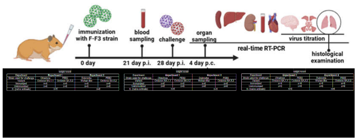
Figure 3.
F-F3 strain ts phenotype stability in vitro. Passaging of F-F3 and FEB2 strains in Vero cells at 33 °C and 37 °C (A). *Analysis of culture supernatant obtained after the 10th passage on day 4 p.i. ** CPE manifestation on day 4 p.i. Evaluation of F-F3 strain temperature sensitivity after 10 passages at 33 °C (F-F3_10p) at 37 °C and 39 °C in Vero cell culture (MOI 0.001) (B, C). FEB2 strain was used as a reference. Virus titer kinetics (B) and viral RNA concentration (C) from days 1 to 4 p.i. Data are from two independent experiments. Error bars = SEM.
Figure 3.
F-F3 strain ts phenotype stability in vitro. Passaging of F-F3 and FEB2 strains in Vero cells at 33 °C and 37 °C (A). *Analysis of culture supernatant obtained after the 10th passage on day 4 p.i. ** CPE manifestation on day 4 p.i. Evaluation of F-F3 strain temperature sensitivity after 10 passages at 33 °C (F-F3_10p) at 37 °C and 39 °C in Vero cell culture (MOI 0.001) (B, C). FEB2 strain was used as a reference. Virus titer kinetics (B) and viral RNA concentration (C) from days 1 to 4 p.i. Data are from two independent experiments. Error bars = SEM.
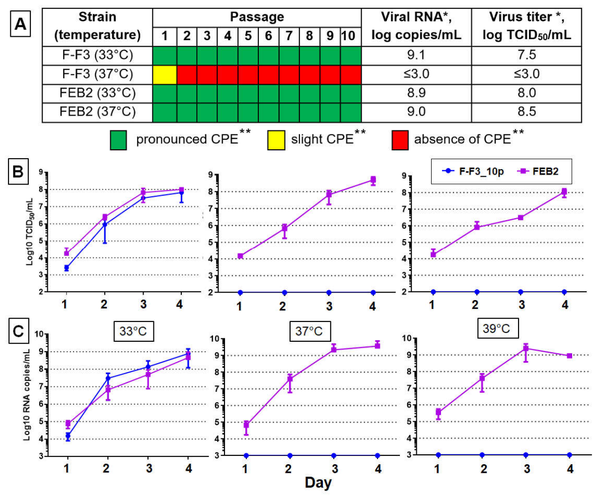
Figure 4.
Antibody titers to SARS-CoV-2 in hamster sera at 21 days after a single intranasal immunization. IgG titers to SARS-CoV-2 antigens measured by ELISA (A). NAb titers against the homologous BA.5.2-like FEB2 strain (B) and the heterologous Wuhan-like Dubrovka strain (C). NAb—neutralizing antibodies. Data from two independent experiments. Exp.1, Exp.2—experiments 1 and 2. ***—p < 0.001; ****—p < 0.0001. Error bars = SEM.
Figure 4.
Antibody titers to SARS-CoV-2 in hamster sera at 21 days after a single intranasal immunization. IgG titers to SARS-CoV-2 antigens measured by ELISA (A). NAb titers against the homologous BA.5.2-like FEB2 strain (B) and the heterologous Wuhan-like Dubrovka strain (C). NAb—neutralizing antibodies. Data from two independent experiments. Exp.1, Exp.2—experiments 1 and 2. ***—p < 0.001; ****—p < 0.0001. Error bars = SEM.
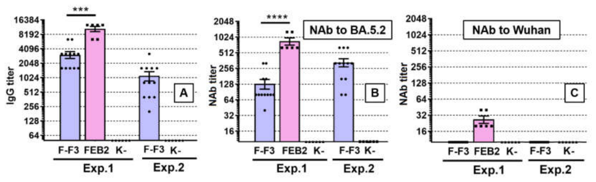
Figure 5.
F-F3 strain efficacy in the COVID-19 Syrian hamster model (Experiment 1). Body weight changes in hamsters on days 1–4 p.c. (A); cumulative pneumonia severity score (B); virus titer (C) and viral RNA concentration (D) in lungs, brain, and nasal passages homogenates. IM—immunized hamsters; unIM—non-immunized hamsters; K−—non-immunized, non-challenged (naïve) hamsters. BA.1.1—challenge with the BA.1.1-like Otradnoe strain; BA.5.2—challenge with the BA.5.2-like FEB2 strain. **—p < 0.01. Error bars = SEM.
Figure 5.
F-F3 strain efficacy in the COVID-19 Syrian hamster model (Experiment 1). Body weight changes in hamsters on days 1–4 p.c. (A); cumulative pneumonia severity score (B); virus titer (C) and viral RNA concentration (D) in lungs, brain, and nasal passages homogenates. IM—immunized hamsters; unIM—non-immunized hamsters; K−—non-immunized, non-challenged (naïve) hamsters. BA.1.1—challenge with the BA.1.1-like Otradnoe strain; BA.5.2—challenge with the BA.5.2-like FEB2 strain. **—p < 0.01. Error bars = SEM.
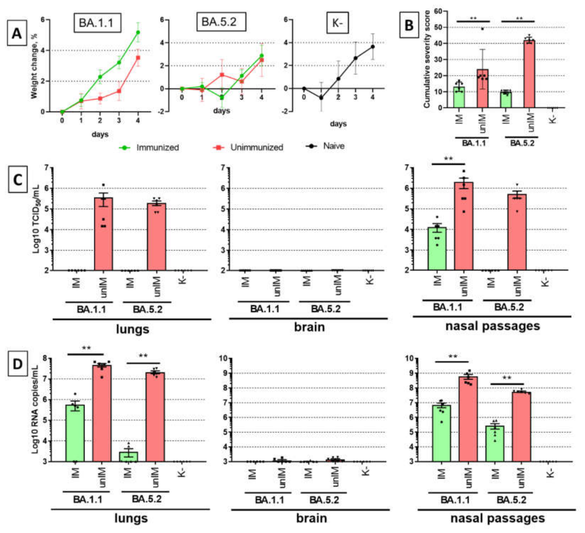
Figure 6.
Efficacy of F-F3 strain in the COVID-19 Syrian hamster model (Experiment 2). Body weight changes in hamsters on days 1–4 p.c. (A); cumulative pneumonia severity score (B); virus titer (C) and viral RNA concentration (D) in lungs, brain, and nasal passages homogenates. IM—immunized hamsters; unIM—non-immunized hamsters; K−—non-immunized, non-challenged (naïve) hamsters. Wuhan—challenge with the Wuhan-like Dubrovka strain; BA.5.2—challenge with the BA.5.2-like FEB2 strain. **—p < 0.01, *—p < 0.05. Error bars = SEM.
Figure 6.
Efficacy of F-F3 strain in the COVID-19 Syrian hamster model (Experiment 2). Body weight changes in hamsters on days 1–4 p.c. (A); cumulative pneumonia severity score (B); virus titer (C) and viral RNA concentration (D) in lungs, brain, and nasal passages homogenates. IM—immunized hamsters; unIM—non-immunized hamsters; K−—non-immunized, non-challenged (naïve) hamsters. Wuhan—challenge with the Wuhan-like Dubrovka strain; BA.5.2—challenge with the BA.5.2-like FEB2 strain. **—p < 0.01, *—p < 0.05. Error bars = SEM.
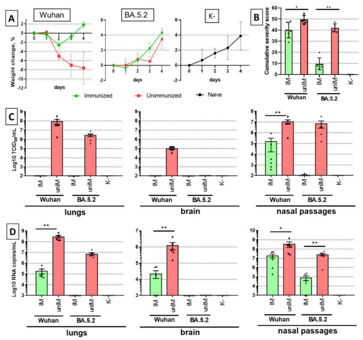
Figure 7.
Protection of naïve contact hamsters from infection upon challenge of immunized animals with a virulent strain. Antibody titers to SARS-CoV-2 following heterologous challenge (21 days p.c.) of immunized hamsters with the Wuhan-like strain (A) and homologous challenge with the BA.5.2-like strain (B). NAb titer was determined against the challenge virus. NAb—neutralizing antibodies, Contact—co-housed naïve hamsters. Error bars = SEM.
Figure 7.
Protection of naïve contact hamsters from infection upon challenge of immunized animals with a virulent strain. Antibody titers to SARS-CoV-2 following heterologous challenge (21 days p.c.) of immunized hamsters with the Wuhan-like strain (A) and homologous challenge with the BA.5.2-like strain (B). NAb titer was determined against the challenge virus. NAb—neutralizing antibodies, Contact—co-housed naïve hamsters. Error bars = SEM.
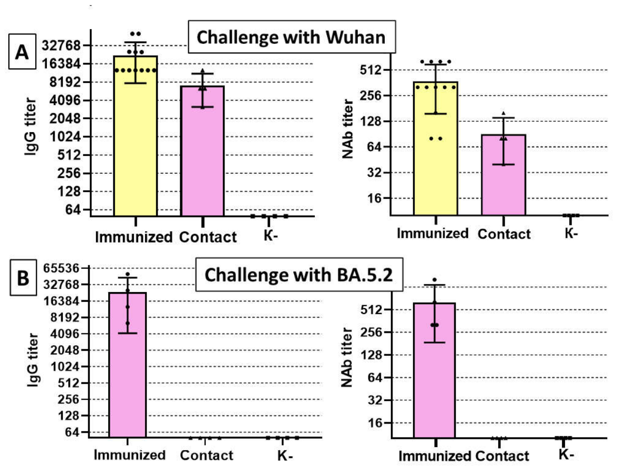
Figure 9.
Protection against pneumonia upon heterologous challenge of hamsters immunized with SARS-CoV-2 ts mutants. The figure schematically illustrates the hypothetical relationship between the ability of a nasal LAV to protect against pneumonia upon heterologous challenge and the capacity of the vaccine strain to infect lungs. F-F3 strain (sensitive to 37 °C and above) does not infect lungs and does not protect against pneumonia, whereas D-D2 strain (sensitive to 39 °C and above) infects lungs and confers protection against pneumonia.
Figure 9.
Protection against pneumonia upon heterologous challenge of hamsters immunized with SARS-CoV-2 ts mutants. The figure schematically illustrates the hypothetical relationship between the ability of a nasal LAV to protect against pneumonia upon heterologous challenge and the capacity of the vaccine strain to infect lungs. F-F3 strain (sensitive to 37 °C and above) does not infect lungs and does not protect against pneumonia, whereas D-D2 strain (sensitive to 39 °C and above) infects lungs and confers protection against pneumonia.
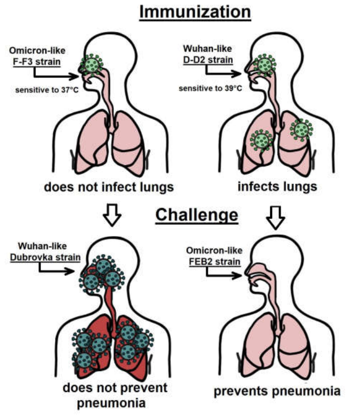

Table 1.
SARS-CoV-2 strains used in the study.
| Strain name | GenBank ID | Pango lineage | Replication capacity at temperature | Ability to infect lungs | Source, date and place of isolation | Passage history in Vero cells | Reference | ||
| 24 °C | 37 °C | 39 °C | |||||||
| Dubrovka | OP920753.1 | B.1.1.317 | no | yes | yes | yes | COVID-19 patient, June 2020, Moscow, Russia | 17 passages at 37 °C | [34] |
| Otradnoe | ON032857.1 | BA.1.1 | no | yes | yes | yes | COVID-19 patient, January 2022, Moscow, Russia | 8 passages at 37 °C | |
| FEB2 | MW514307.1 | BA.5.2 |
no | yes | yes | yes | COVID-19 patient, October 2022, Moscow, Russia | 4 passages at 37 °C | |
| F-F1 | PX401969.1 | BA.5.2 | yes | no | no | n/a | Cold-adapted FEB2 strain mutants | 27 passages at a temperature gradually decreasing from 37 °C to 24 °C | [35] |
| F-F3 | PX401966.1 | BA.5.2 | yes | no | no | no | |||
| F-D3 | PX401970.1 | BA.5.2 | yes | no | no | n/a | |||
| F-D12 | PX401968.1 | BA.5.2 | yes | no | no | n/a | |||
| F-C5 | PX401967.1 | BA.5.2 | yes | yes | no | n/a | |||
| F-C9 | PX401971.1 | BA.5.2 | yes | yes | no | n/a | |||
Abbreviations: n/a—not analized.
Disclaimer/Publisher’s Note: The statements, opinions and data contained in all publications are solely those of the individual author(s) and contributor(s) and not of MDPI and/or the editor(s). MDPI and/or the editor(s) disclaim responsibility for any injury to people or property resulting from any ideas, methods, instructions or products referred to in the content. |
© 2026 by the authors. Licensee MDPI, Basel, Switzerland. This article is an open access article distributed under the terms and conditions of the Creative Commons Attribution (CC BY) license (http://creativecommons.org/licenses/by/4.0/).
Copyright: This open access article is published under a Creative Commons CC BY 4.0 license, which permit the free download, distribution, and reuse, provided that the author and preprint are cited in any reuse.