

Submitted:
17 September 2026
Posted:
18 September 2026
You are already at the latest version
Abstract
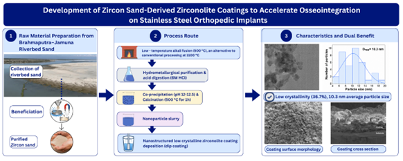
Stainless steel 316L remains a primary material for orthopedic implants, yet its bioinert surface often delays or prevents stable, long-term osseointegration. This study addresses this limitation by developing an energy-efficient method to synthesize nanostructured, low-crystalline zirconolite (CaZrTi2O7) coatings from natural zircon sand. Raw zircon sand from the Brahmaputra-Jamuna riverbed was physically beneficiated and decomposed via low-temperature alkali fusion at 500°C. Following hydrometallurgical purification and acid digestion in 6M HCl, soluble precursors were co-precipitated at pH 12–12.5 and calcined at 500°C to yield single-phase zirconolite nanoparticles. XRD and TEM confirmed the formation of low-crystalline (36.7%) zirconolite nanoparticles with an average size of 10.3 nm and a lattice d-spacing of 0.288 nm, corresponding to the (221) plane. While conventional solid-state synthesis of zirconolite requires temperatures exceeding 1100°C, phase stabilization was achieved at 500°C. Stable, uniform coatings were successfully deposited on SS-316L using a dip-coating slurry. The tailored low crystallinity, nanoscale topography, and trace silica synergy of the zirconolite coatings provide a highly reactive interface that enhances protein adsorption and accelerates bone-like apatite nucleation. This cost-effective synthesis and coating strategy offers a dual benefit: it valorizes local mineral resources and provides a bioactive, structurally durable surface modification to accelerate osseointegration in metallic implants.
Keywords:
zirconolite
; ceramic coatings
; zircon sand
; nanoparticles
1. Introduction
Orthopedic implants are foundational to modern surgical intervention, restoring skeletal function and enabling mobility for patients with fractures or degenerative conditions. However, the long-term clinical success of these devices is fundamentally dictated by the bone-implant interface; failure to establish robust, early-stage biological integration characterized by protein adsorption, osteoblast adhesion, and subsequent mineralization often leads to fibrous encapsulation or aseptic loosening [1]. Stainless steel, particularly the 316L grade, remains a cornerstone of orthopedic manufacturing due to its cost-effectiveness, mechanical reliability, and widespread availability [2]. Despite these advantages, its surface remains inherently bioinert, which can delay osseointegration and elevate the risk of long-term implant failure [2,3]. This limitation necessitates advanced surface modification strategies to convert these bioinert metallic substrates into bio-instructive interfaces.
While traditional coating strategies such as hydroxyapatite, titanium plasma spray, bioactive glass, porous tantalum, and diamond-like carbon have been extensively documented, they are frequently constrained by technical trade-offs, including phase instability, susceptibility to delamination, and complex, high-cost manufacturing requirements [4,5,6,7,8,9] Consequently, there is an urgent research demand for novel, multifunctional bioactive coatings that synthesize structural durability with high biological reactivity. In this context, zirconolite (CaZrTi2O7), a calcium zirconium titanate ceramic, emerges as a highly promising candidate. Its unique monoclinic crystal structure, characterized by alternating titanium-oxygen octahedral layers and calcium/zirconium ion layers, provides exceptional chemical stability in aqueous environments while offering multiple cation sites capable of accommodating atomic substitutions to tailor surface bioactivity [10]. These attributes position zirconolite as an ideal interface for promoting rapid apatite nucleation and sustained protein interaction.
Furthermore, nano-structuring and the reduction of crystallinity have been identified as pivotal strategies for maximizing surface energy and accelerating bio-responsiveness [11]. The synthesis of zirconolite at moderate temperatures using hydrometallurgical and pyrometallurgical techniques, including alkali fusion, leaching, and co-precipitation, allows precise control over crystallinity and particle size [12]. Nanostructured zirconolite coatings, therefore, hold promise for combining mechanical stability with enhanced biological performance, addressing the limitations of highly crystalline or bulk coatings that exhibit reduced surface reactivity [13].
Beyond material innovation, this study addresses a critical gap in sustainable resource management. Bangladesh possesses substantial, underutilized deposits of zircon-bearing sands in coastal regions like Cox’s Bazar and the Brahmaputra-Jamuna riverbeds. Currently, despite this domestic abundance, high-purity zircon precursors remain heavily imported, representing an unexploited economic and strategic opportunity. Existing studies have demonstrated that local zircon can be refined via beneficiation (gravity, magnetic, and electrostatic separation) and chemical extraction to produce high-purity precursors [14,15]. By establishing a cost-effective, domestic pathway to transform local mineral resources into high-value, bioactive ceramic coatings, this study offers a sustainable model for orthopedic implant production.
Collectively, this research introduces a dual-impact strategy: it provides a robust, nanostructured surface modification to enhance the clinical efficacy of 316L stainless steel implants, and it promotes the valorization of local mineral resources. By integrating mineral processing, chemical synthesis, and surface engineering, this work contributes to the advancement of biomaterials science while offering a scalable, sustainable platform for future in vitro and in vivo bioactivity assessments.
2. Experimental Procedure
2.1. Sample Collection and Preparation
The zircon sand used in this study was collected from Everlast Minerals Ltd., which conducts heavy mineral sand mining from the Brahmaputra–Jamuna riverbed located in the Gaibandha district of Bangladesh. The mining and beneficiation processes used a combination of gravity, magnetic, and electrostatic separation to concentrate heavy minerals from river sand. The zircon sand obtained from this beneficiation process was collected as the starting raw material for zirconolite synthesis in this research.
2.2. Characterization of the Raw Sample
Before extraction, the collected sample was characterized to determine its composition and particle-size distribution. XRF was used to determine the chemical composition of the raw zircon sand, specifically to quantify the presence of zirconia (ZrO2), Silica (SiO2), and other associated elements, such as titania (TiO2), calcium oxide (CaO), iron oxide (Fe2O3), and trace elements that may influence the synthesis process. XRD analysis was performed to identify the phases present in the sand. Sieve analysis was conducted to determine the particle size distribution of the zircon sand. This step was crucial for ensuring that the sample was adequately prepared for the synthesis process, as particle size can influence the efficiency of the synthesis method.
2.3. Zirconolite Synthesis from the Collected Sand
The synthesis of zirconolite from the collected zircon sand was carried out through a series of chemical and physical processes. The process involved several stages, including mortaring, alkali fusion, leaching, acid washing, and calcination. The overall experimental procedure for zirconolite synthesis from natural zircon sand is described below:
Initially, 50 g of raw zircon sand was mechanically ground in an agate mortar for 30 min to obtain a fine powder and increase the reactivity of the precursor. Sodium hydroxide pellets were dissolved in a minimal volume of deionized (DI) water to prepare a concentrated alkaline medium. The powdered zircon sand was then gradually added and homogenized under continuous stirring while maintaining a zircon sand-to-NaOH weight ratio of 1:1. The resulting mixture was transferred to a nickel crucible and subjected to alkali fusion at 500 °C for 1 h in a muffle furnace. During this process, the zircon phase decomposed to form sodium zirconate (Na2ZrO3) and sodium silicate (Na2SiO3), while titanium- and hafnium-bearing phases were converted into their corresponding sodium compounds.
After cooling to room temperature, the fused mass was crushed and leached with hot deionized water. This step selectively dissolved the soluble sodium silicate and other alkali-soluble species, while retaining the zirconium-bearing phase in the solid residue. Multiple washing cycles were performed to remove residual soluble impurities and minimize potential nickel contamination originating from the crucible. Simultaneously, sodium titanate and sodium hafnate underwent hydrolysis, yielding insoluble titanium hydroxide and hafnium-containing oxide species.
The washed residue was subsequently digested in 240 mL of 6 M hydrochloric acid at 80–90 °C for approximately 1 h under continuous stirring. During acid treatment, sodium zirconate, titanium hydroxide, and calcium-containing phases dissolved to form zirconyl, titanium, and calcium chloride species in solution, whereas undissolved residues, primarily consisting of refractory hafnium oxide and incompletely reacted zircon particles, remained insoluble. These residues were separated by filtration using Whatman No. 42 filter paper.
To obtain a homogeneous precursor for zirconolite formation, the filtrate was subjected to co-precipitation by the gradual addition of ammonium hydroxide (NH4OH) under continuous stirring until the pH reached 12.0–12.5. Under these alkaline conditions, zirconium, titanium, and calcium species simultaneously precipitated as mixed hydroxides, resulting in the formation of a gel-like precipitate. The precipitate was aged, thoroughly washed with deionized water, and dried overnight at 120 °C to remove residual moisture and physically adsorbed water.
The dried precursor powder was subsequently calcined in air at 500 °C for 1 h. During calcination, the mixed hydroxides underwent dehydration and solid-state transformation, leading to the formation of zirconolite (CaZrTi2O7). The resulting powder was collected and stored for subsequent characterization.
2.4. Zirconolite Coating on Stainless Steel Substrate
Medical-grade stainless steel (SS 316L) substrates were used as the coating substrate. Prior to coating deposition, the substrates were mechanically polished using successive grades of abrasive papers to obtain a smooth surface finish. The polished specimens were rinsed with distilled water to remove residual polishing debris and subsequently ultrasonically cleaned in deionized (DI) water for 10 min. The cleaned substrates were then dried at room temperature before coating deposition.
For slurry preparation, 0.2 g of polyvinyl alcohol (PVA) was dissolved in 10 mL of DI water under continuous magnetic stirring to obtain a homogeneous binder solution. Subsequently, 2 g of synthesized zirconolite powder was dispersed into the solution, followed by the addition of 20 mL of ethanol. The suspension was continuously stirred until a stable and homogeneous coating slurry was obtained.
Zirconolite coatings were deposited onto the SS 316L substrates using a dip-coating process. The cleaned substrates were immersed in the zirconolite slurry and withdrawn at a controlled rate to ensure uniform coating deposition. Following each coating cycle, the coated substrates were dried at 70–80 °C for 10 min to evaporate the solvent and consolidate the deposited layer. The dip-coating and drying sequence was repeated five times to achieve the desired coating thickness and surface coverage.
After the final deposition cycle, the coated specimens were dried at 70–80 °C and stored in a desiccator prior to subsequent characterization.
2.5. Material Characterization and Biological Evaluation
The phase composition and crystallographic characteristics of the synthesized zirconolite powder were investigated using X-ray diffraction (XRD). Diffraction patterns were collected over an appropriate 2θ range using Cu Kα radiation (λ = 1.5406 Å). Phase identification was performed by comparison with standard reference patterns, while the crystallinity of the synthesized zirconolite was evaluated from the diffraction data.
The microstructure and morphology of the synthesized zirconolite nanoparticles were examined using transmission electron microscopy (TEM). High-resolution transmission electron microscopy (HRTEM) was employed to investigate lattice fringes and crystallographic ordering at the nanoscale. Selected area electron diffraction (SAED) analysis was performed to verify the crystalline nature and phase purity of the synthesized particles. The particle size distribution was determined by measuring individual particles from TEM micrographs using ImageJ software. Elemental composition and spatial elemental distribution were further analyzed by energy-dispersive X-ray spectroscopy (EDS) coupled with TEM through elemental mapping.
The cytotoxicity of the synthesized zirconolite nanoparticles was evaluated using the Vero cell line (African green monkey kidney epithelial cells). The cells were cultured in Dulbecco’s Modified Eagle Medium (DMEM) supplemented with 10% fetal bovine serum (FBS), 1% penicillin–streptomycin, and 0.2% gentamycin under standard cell culture conditions (37 °C, 5% CO2, and 95% relative humidity).
For cytotoxicity assessment, cells were seeded into 48-well culture plates at a density of approximately 3.0 × 10⁴ cells per well. Sterilized aqueous suspensions of zirconolite nanoparticles were prepared using deionized water as the dispersion medium at concentrations of 10, 100, and 150 µg mL−1. Following the attachment period, the nanoparticle suspensions were added to the respective wells and incubated with the cells for 48 h under identical culture conditions. Untreated cells maintained under the same conditions served as the control group. After the incubation period, cellular morphology and viability were examined using an inverted optical microscope.
The surface morphology of the zirconolite-coated stainless steel substrates was characterized using scanning electron microscopy (SEM). SEM imaging was employed to assess coating uniformity, surface coverage, and microstructural features. Cross-sectional SEM analysis was additionally performed to evaluate the coating architecture and determine the average coating thickness.
3. Results and Discussion
3.1. Characterization of Raw Zircon Sand
The XRF analysis of the raw zircon sand (Table 1) revealed that, in addition to zircon (ZrSiO4), the precursor contained significant amounts of TiO2 (8.8659 wt%) and CaO (7.6413 wt%).
These oxide constituents are particularly important because both calcium and titanium serve as essential precursor elements for zirconolite formation. The presence of naturally occurring and within the zircon sand significantly reduced the dependency on high-purity external chemical reagents during synthesis, thereby contributing to the cost-effectiveness of the process. Furthermore, the utilization of naturally abundant zircon sand instead of expensive chemically purified precursors substantially lowers the overall production cost, supporting the economic feasibility of large-scale biomedical ceramic synthesis.
The crystalline phases present in the raw zircon sand were further identified by X-ray diffraction (XRD) analysis (Figure 1).
The diffraction peaks corresponded predominantly to crystalline zircon phases, indicating the highly ordered structure of the precursor mineral. In addition to the dominant crystalline zircon phase, minor peaks corresponding to hydroxyapatite were also detected. Furthermore, the strong and sharp diffraction peaks suggested high crystallinity of the raw zircon sand before chemical processing and phase transformation [16].
Particle size distribution analysis of the initial zircon sand (Figure 2) revealed that the majority of the particles were concentrated around approximately 106 µm.
The relatively uniform particle distribution is advantageous for subsequent chemical extraction and synthesis processes because finer and more homogeneous precursor particles promote improved reaction kinetics and enhanced phase transformation during calcination [17]. Furthermore, the reduction of particle size during synthesis facilitates the formation of nanostructured zirconolite with improved bio-functional characteristics [18].
3.2. Phase Formation and Crystallinity of Synthesized Zirconolite
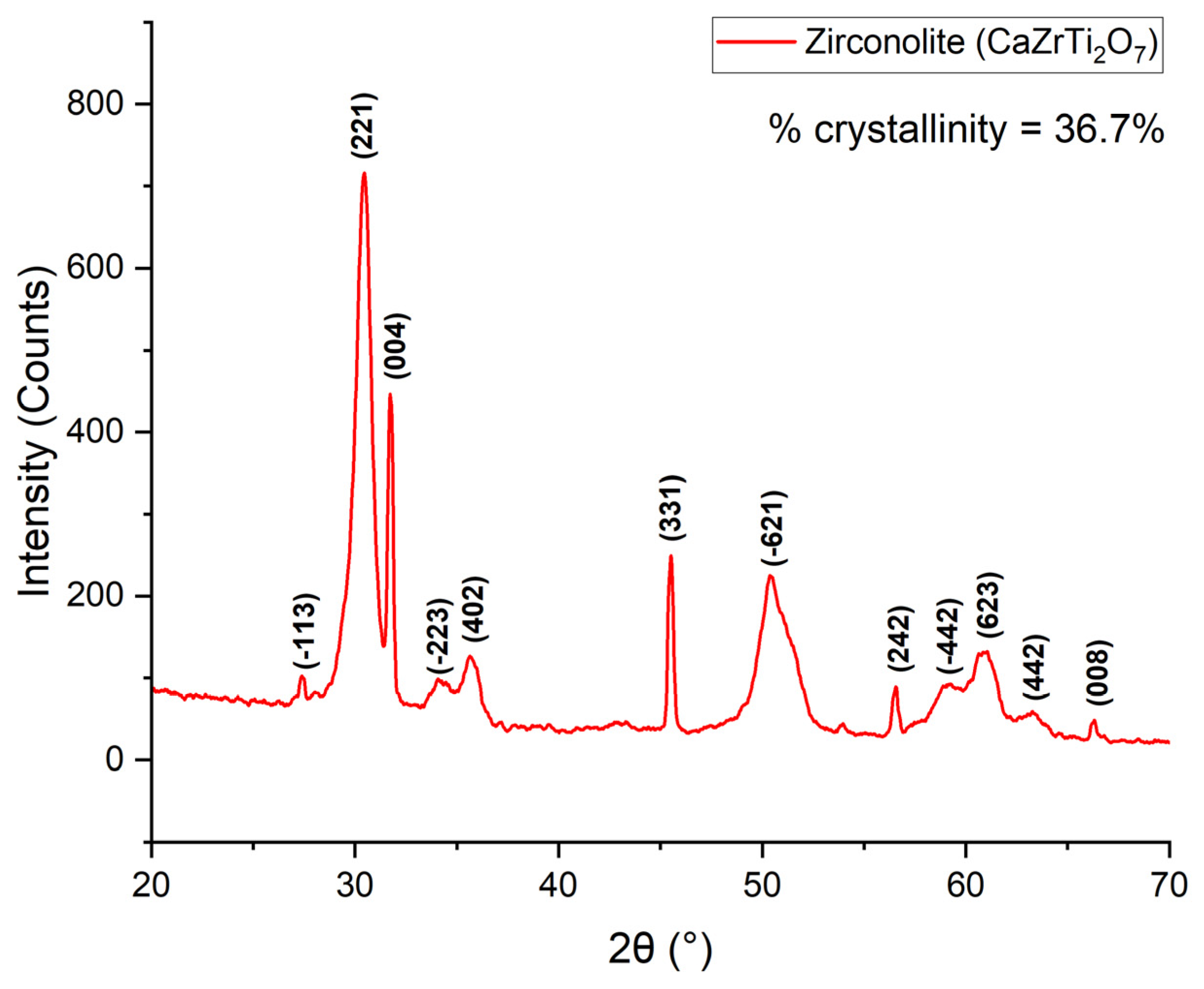
The phase evolution of the synthesized material after calcination at 500 °C for 1 h was investigated using X-ray diffraction (XRD), as shown in Figure 3. The diffraction pattern confirmed the successful formation of single-phase zirconolite. The observed diffraction peaks were indexed to their corresponding crystallographic planes (hkl), which further validated the phase identification and structural consistency of the synthesized material.
The crystallinity of the synthesized zirconolite, calculated from the XRD data, was found to be 36.7%, indicating a relatively low-crystalline structure. The reduced crystallinity is attributed to the low-temperature calcination process, which limits complete atomic ordering within the crystal lattice [19].
Interestingly, while conventional solid-state synthesis of zirconolite typically requires temperatures exceeding 1100 °C to overcome immense kinetic barriers, the single-phase zirconolite in this work was successfully stabilized at a mere 500 °C. This remarkably low-temperature phase formation may be driven by a dual-kinetic mechanism resulting from the co-precipitation processing conditions:
Low Activation Energy via Hydroxide Condensation: During the co-precipitation stage, the constituent metal ions (Ca2+, Zr4+, and Ti4+) are intimately mixed at the molecular level, precluding the formation of isolated elemental oxide grains. Instead, they form a highly cross-linked amorphous oxo-hydrogen polymer network containing -M-O-M- bridges (where M= Ca, Zr, Ti). Upon heating toward 500 °C, neighboring hydroxyl groups (-OH) undergo rapid oxolation reactions, yielding chemical dehydration as follows:
This continuous cross-linking directly constructs the metal-oxygen-metal skeletal framework without relying on long-range thermal bulk diffusion. Consequently, the activation energy required to collapse this highly mixed, amorphous gel precursor into a crystalline lattice is drastically minimized, permitting the onset of zirconolite nucleation at 500 °C [20,21].
-M-OH + OH-M- → -M-O-M- + H2O
Figure 3.
XRD pattern of low-crystalline zirconolite synthesized at 500 °C.
Localized Micro-Flux Effect Facilitated by Trace Silica: This low-temperature atomic rearrangement is further accelerated by a localized fluxing mechanism. Elemental mapping via Energy-Dispersive X-ray Spectroscopy (EDS) revealed the presence of trace silicon distributed within the matrix. Even at trace levels, silica interacts with local calcium species to generate highly reactive, mobile structural configurations. At 500 °C, these transient calcium-silicate-like structural units function as a microscopic “flux,” inducing a localized environment of enhanced atomic mobility. This high-mobility channel dramatically reduces the kinetic barrier for the heavy, highly charged Zr4+ and Ti4+ cations, allowing them to rapidly rearrange into the ordered zirconolite lattice at temperatures hundreds of degrees lower than previously reported in literature [22,23].
Low-crystalline zirconolite coatings can significantly enhance osseointegration by accelerating apatite nucleation and improving biological interactions at the implant–bone interface [24]. The increased structural disorder facilitates ion exchange and promotes rapid formation of biologically active calcium phosphate layers under physiological conditions [25,26].
Furthermore, the successful synthesis of zirconolite at a comparatively low calcination temperature demonstrates an energy-efficient and economically favorable fabrication strategy for bioactive implant coatings.
3.3. HRTEM Analysis of Zirconolite Nanoparticles
High-resolution transmission electron microscopy (HRTEM) images of the synthesized zirconolite nanoparticles are presented in Figure 4. The micrographs revealed nanoscale particles with partially disordered lattice arrangements, further confirming the low-crystalline nature of the synthesized material observed in the XRD analysis.
The measured interplanar spacing obtained from the HRTEM lattice fringes was approximately 0.288 nm. This value closely matches the characteristic d-spacing of the prominent zirconolite (221) plane (~ 0.29 nm), confirming the successful formation of zirconolite nanocrystals [27]. The close agreement between experimental and standard crystallographic values demonstrates the structural reliability of the synthesized phase.
The observed diffuse lattice features indicate the presence of partially ordered nanocrystalline domains rather than highly ordered bulk crystals. Such low-crystalline nanostructures are highly advantageous for orthopedic coating applications because they provide increased surface activity and enhanced biological responsiveness [28].
Nanostructured low-crystalline coatings can improve protein adsorption, osteoblast attachment, and cellular proliferation, thereby facilitating faster osseointegration on metallic implant surfaces [29].
3.4. Selected Area Electron Diffraction (SAED) Analysis
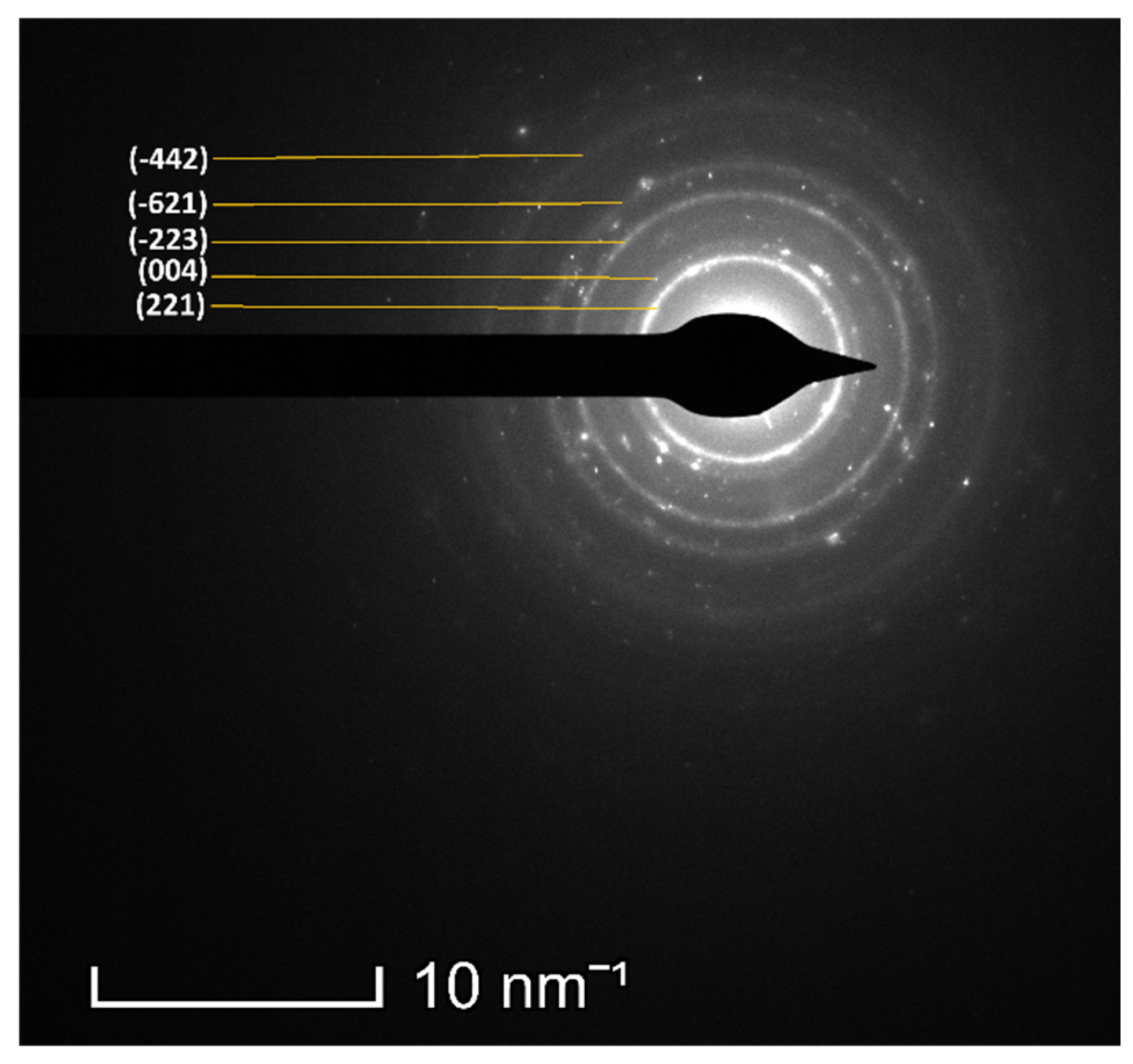
The crystallographic structure of the synthesized zirconolite nanoparticles was further examined using selected area electron diffraction (SAED), as shown in Figure 5. The SAED pattern exhibited diffraction rings corresponding to the crystallographic planes identified in the XRD analysis, confirming the successful formation of zirconolite.
The presence of ring-like diffraction features rather than sharp spot patterns indicates the polycrystalline and nanocrystalline nature of the synthesized particles. The observed diffraction rings also support the low-crystalline structural characteristics identified from XRD and HRTEM analyses.
The consistency between SAED and XRD results confirms the structural homogeneity of the synthesized zirconolite nanoparticles. Such agreement between multiple characterization techniques strengthens the reliability of the synthesized phase identification and validates the effectiveness of the synthesis method.
Figure 5.
Selected Area Electron Diffraction (SAED) pattern of the synthesized zirconolite nanoparticles .
Figure 5.
Selected Area Electron Diffraction (SAED) pattern of the synthesized zirconolite nanoparticles .
3.5. Particle Size Distribution of Zirconolite Nanoparticles
The particle size distribution obtained from TEM analysis is shown in Figure 6. The synthesized zirconolite nanoparticles exhibited an average particle size of approximately 10.3 nm, confirming the successful production of ultrafine nanostructured ceramic particles.
The ultrafine nanoscale dimensions of the synthesized zirconolite are highly advantageous for orthopedic implant coatings because nanoparticles possess significantly higher surface area-to-volume ratios compared to bulk materials. The increased surface area provides a greater number of active sites for protein adsorption and osteoblast attachment, thereby promoting enhanced biological interactions at the implant interface [30]. Additionally, nanoscale bioactive ceramics can accelerate apatite layer formation and improve osteogenic activity, ultimately facilitating rapid bone regeneration and stronger implant fixation [31].
The combination of nanoscale particle size and low crystallinity is expected to synergistically enhance the osseointegration performance of zirconolite-coated stainless-steel implants.
3.6. Elemental Distribution and EDS Mapping Analysis
The elemental composition and spatial distribution of the synthesized zirconolite nanoparticles were analyzed using energy-dispersive spectroscopy (EDS) mapping, as shown in Figure 7. The elemental maps confirmed a uniform distribution of the major constituent elements, namely calcium (Ca), zirconium (Zr), titanium (Ti), and oxygen (O), which collectively confirm the formation of the zirconolite phase.
In addition to the main elements, a weak silicon (Si) signal was detected, attributed to trace silica originating from the natural zircon sand precursor used in the synthesis process. A minor chlorine (Cl) peak was also observed, which may be associated with residual precursor salts or processing-related impurities. Copper (Cu) and carbon (C) signals detected in the EDS spectra originate from the TEM grid and sample holder rather than from the synthesized material itself.
Although present in minor concentration, trace silica may play a highly beneficial role in enhancing the biological performance of the synthesized zirconolite coating. Silicon-containing bioactive materials are widely recognized for their ability to stimulate osteogenic differentiation, enhance extracellular matrix mineralization, and accelerate bone tissue regeneration [32].
In physiological environments, silica-containing surfaces can promote the formation of surface silanol (Si–OH) groups, which serve as favorable nucleation sites for calcium phosphate and hydroxyapatite deposition [33]. The formation of such bioactive apatite layers is critically important for achieving rapid osseointegration between orthopedic implants and surrounding bone tissue.
Furthermore, silicon has been reported to enhance osteoblast proliferation, collagen synthesis, and early-stage bone mineralization processes [34]. Therefore, the presence of trace silica within the zirconolite nanostructure may synergistically improve the bioactivity of the coating by combining the chemical stability of zirconolite with the osteogenic advantages associated with silicon-containing biomaterials.
The homogeneous elemental distribution observed throughout the nanoparticles further confirms the successful synthesis of chemically uniform zirconolite nanostructures. Uniform elemental dispersion is particularly important for biomedical coatings because compositional homogeneity contributes to consistent surface chemistry, stable bioactive behavior, and reliable biological interactions across the implant surface [35].
3.7. Cytotoxicity of Synthesized Zirconolite Nanoparticles
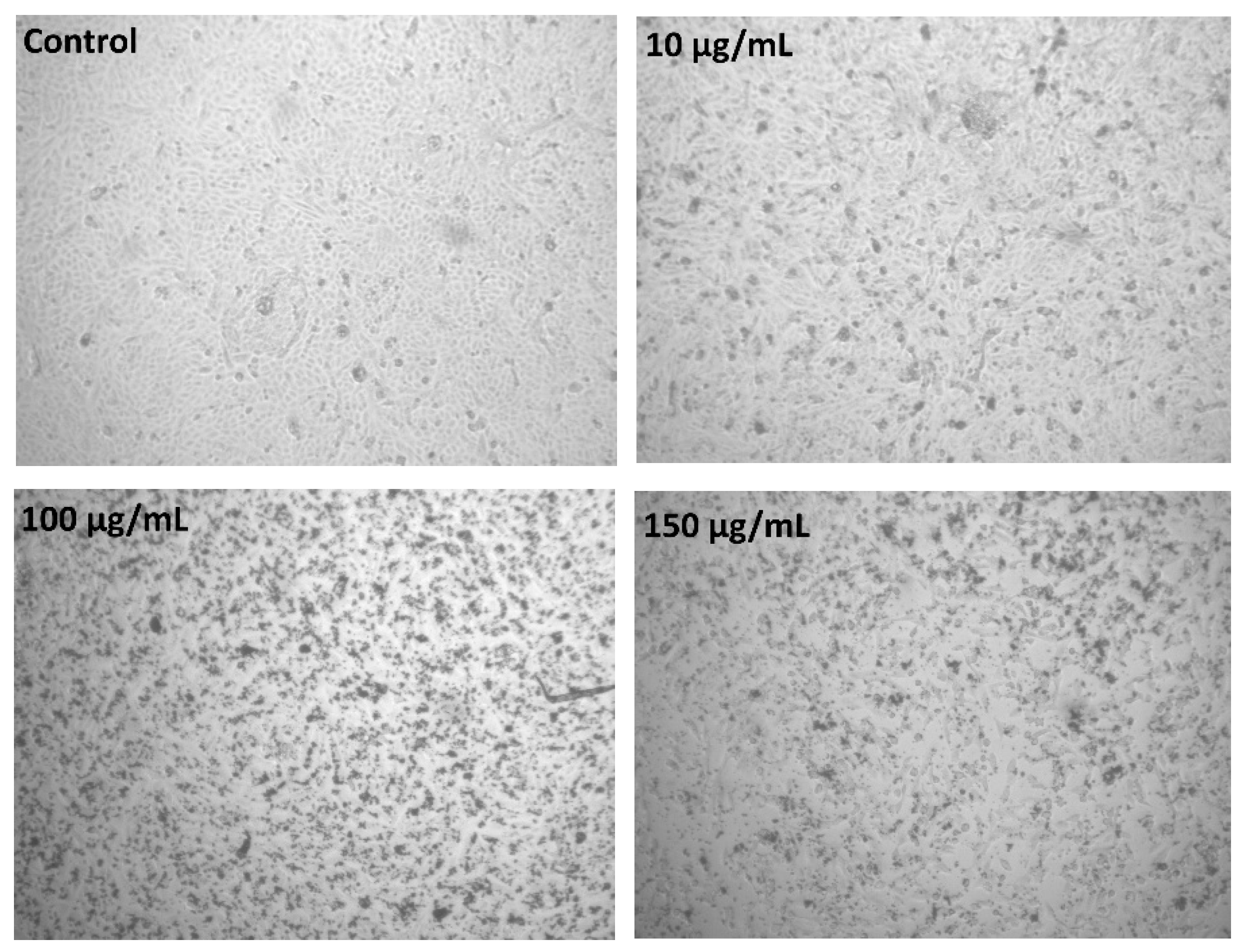
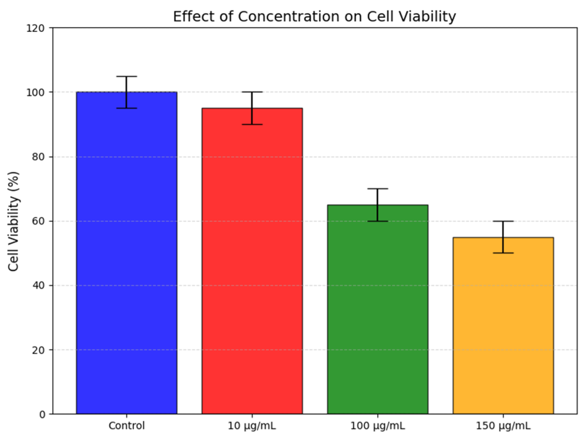
The cytocompatibility of the synthesized zirconolite nanoparticles was evaluated using Vero cells following 48 h of exposure at concentrations of 10, 100, and 150 µg/ml. Representative optical micrographs of the treated and control groups are shown in Figure 8, while the corresponding cell viability results are presented in Figure 9.
As shown in Figure 8, the control cells exhibited normal epithelial morphology with a well-adhered and uniformly distributed cell population. Cells exposed to 10 µg/mL zirconolite nanoparticles maintained morphological characteristics comparable to those of the control group, indicating minimal adverse effects on cellular integrity. No evident signs of cell shrinkage, membrane disruption, or extensive detachment were observed, suggesting excellent cytocompatibility at this concentration.
With increasing nanoparticle concentration, a gradual reduction in cell density and confluency became apparent. Cells treated with 100 and 150 µg/mL zirconolite nanoparticles exhibited a lower population of adherent cells compared with the control and 10 µg/mL groups, indicating a concentration-dependent cellular response. Nevertheless, a substantial number of viable cells remained attached to the culture surface, demonstrating that the nanoparticles did not induce severe cytotoxic damage under the investigated conditions.
The quantitative viability results shown in Figure 9 corroborate the microscopic observations. Cell viability remained high at 10 µg/mL (approximately 95%), indicating negligible cytotoxicity relative to the control. However, viability decreased to approximately 65% and 55% at concentrations of 100 and 150 µg/mL, respectively, demonstrating a dose-dependent reduction in cellular viability. This trend suggests that while low concentrations of zirconolite nanoparticles are well tolerated by Vero cells, higher concentrations may influence cellular metabolic activity and proliferation.
Figure 8.
Representative optical micrographs of Vero cells after 48 h exposure to synthesized zirconolite nanoparticles at different concentrations.
Figure 8.
Representative optical micrographs of Vero cells after 48 h exposure to synthesized zirconolite nanoparticles at different concentrations.
The favorable cellular response observed at low nanoparticle concentrations is consistent with the bioinert nature and chemical stability commonly associated with zirconium-containing ceramic materials [36]. These findings indicate that zirconolite nanoparticles synthesized from natural zircon sand via the hydrometallurgical route possess good cytocompatibility, particularly at lower concentrations relevant to biomedical applications. Collectively, the results highlight the potential of the synthesized zirconolite nanoparticles for use in bone tissue engineering, implant coatings, and other regenerative biomedical applications.
Figure 9.
Effect of zirconolite nanoparticle concentration on Vero cell viability after 48 h of exposure.
Figure 9.
Effect of zirconolite nanoparticle concentration on Vero cell viability after 48 h of exposure.
3.8. Zirconolite Coatings on Stainless Steel Substrates
Visual inspection of the coated specimens (Figure 10) confirms the successful fabrication of a continuous, uniform, and structurally stable light-grey zirconolite layer across the entire active surface of the SS 316L substrate. The coating exhibits complete surface coverage with no macroscopically observable cracks, demonstrating the outstanding coat-ability of the formulated slurry.
The high-magnification surface microstructure of the coated layer was analyzed using field-emission SEM, as shown in Figure 11(a). The surface exhibits a highly textured, rough, and micro-to-nanoporous morphology. This topography is dominated by irregular particulate clusters and localized porous networks formed by the agglomeration of zirconolite nanoparticles within the polymeric PVA matrix. From a biomaterials engineering standpoint, this hierarchical roughness is highly advantageous for orthopedic implants. The complex microtopography mimics the natural porous architecture of trabecular bone, providing an optimal micro-environment that accelerates protein adsorption (such as fibronectin and vitronectin) and supplies structural anchor points to promote the adhesion, spreading, and proliferation of migrating osteoblastic cell lines [37,38].
To evaluate the interfacial integrity and coating thickness, cross-sectional SEM analysis was performed, as shown in Figure 11(b). The cross-sectional micrograph reveals an intimate, continuous, and highly cohesive interface between the ductile metallic SS 316L substrate and the overlying ceramic zirconolite coating. No micro-cracks, interfacial voids, or gaps are observed along the boundary, indicating excellent wet-adhesion and mechanical interlocking between the particulate slurry and the substrate.
The average thickness of the solid zirconolite-based coating was determined to be approximately 104.51 µm across multiple measurements. In clinical applications, a coating thickness of approximately 100 µm represents an optimal threshold. It is sufficiently thick to act as a robust physical barrier that completely isolates the bioinert metal and serves as a long-term bioactive reservoir for biomineralization, while remaining thin enough to suppress thermal-expansion-induced residual stresses that commonly trigger catastrophic delamination and third-body wear debris in thicker ceramic coatings under cyclic physiological loading [39,40].
The elemental composition and spatial homogeneity of the coating surface were characterized using Energy-Dispersive X-ray Spectroscopy (EDS) mapping and quantitative analysis, as illustrated in the spectrum shown in Figure 11(c).
The quantitative elemental composition of the coating surface obtained from the spectrum is summarized in Table 2.
The EDS spectrum confirms the retention of all primary cationic species characteristic of the synthesized zirconolite matrix, specifically zirconium (Zr, 10.78 wt%), calcium (Ca, 3.95 wt%), titanium (Ti, 0.48 wt%), and oxygen (O, 39.83 wt%), along with trace silicon (Si, 0.66 wt%) originating from the natural zircon sand precursor. Crucially, a highly prominent carbon (C) peak is resolved at 0.277 keV, with a quantitative mass concentration of 44.30 wt% (corresponding to 57.38 at%). This significant carbon signal is directly attributed to the incorporation of the polyvinyl alcohol (PVA) binder added during the preparation of the coating slurry.
4. Conclusions
4.1. Findings of This Work
This research successfully developed a novel, energy-efficient synthesis and coating strategy to modify the bioinert surface of grade 316L stainless steel (SS 316L) orthopedic implants. Single-phase, nanostructured zirconolite (CaZrTi2O7) was extracted from natural zircon sand obtained from the Brahmaputra-Jamuna riverbed in Bangladesh, thereby valorizing local mineral resources. Through a hydrometallurgical and low-temperature alkali fusion process, zircon was decomposed and synthesized into zirconolite nanoparticles.
The key crystallographic, microstructural, and biological findings of this research include:
- a)
- Low-Temperature Phase Stabilization: While conventional solid-state reactions require temperatures exceeding 1100 °C, single-phase zirconolite was stabilized at just 500 °C. This low-temperature synthesis was driven by a dual-kinetic mechanism: molecular hydroxide condensation (which bypasses high-temperature bulk diffusion) and a localized micro-flux effect facilitated by trace matrix silica.
- b)
- Tailored Low Crystallinity and Ultrafine Nanostructure: Characterization via XRD, TEM, HRTEM, and SAED confirmed the synthesis of highly homogeneous, ultrafine zirconolite nanoparticles with an average size of 10.3 nm and a tailored low crystallinity of 36.7%.
- c)
- Trace Silica Bio-Functional Synergy: Residual trace silica within the coating matrix acts as a crucial osteogenic catalyst. Upon hydration, it generates active surface silanol (Si-OH) groups, which trigger rapid, biomimetic carbonated hydroxyapatite nucleation, promoting direct chemical bonding between the implant and native bone.
- d)
- Optimized Osteogenic Coating Architecture: A mechanically stable zirconolite coating was successfully engineered on SS 316L substrates, exhibiting a crack-free interface, strong substrate adhesion, and a controlled thickness of approximately 100 µm. The hierarchical micro/nanoporous surface closely resembles the architecture of trabecular bone, creating a bioactive interface that is expected to enhance protein-mediated cell attachment, accelerate osseointegration, and improve the long-term performance of orthopedic implants.
In summary, this research successfully demonstrates a cost-effective, sustainable surface engineering platform that decouples the excellent mechanical durability of SS 316L from its interfacial biological behavior, laying the groundwork for accelerated bone integration.
4.2. Future Perspectives
To build upon the findings of this study and transition this technology from laboratory-scale validation to clinical orthopedic and dental applications, several key research directions are proposed:
- a)
- Quantitative In Vitro Biocompatibility and Osteogenesis: Future work should conduct detailed, quantitative in vitro assays (such as MTT or CCK-8 tests) using human osteoblast-like cell lines (e.g., MG-63 or Saos-2) to assess cell viability, adhesion, and spreading on the zirconolite coatings. Additionally, monitoring osteogenic markers such as alkaline phosphatase (ALP) activity, osteopontin (OPN) expression, and extracellular matrix mineralization will quantitatively evaluate the osteo-inductive potential.
- b)
- Long-Term SBF and Degradation Kinetics: Systematic simulated body fluid (SBF) immersion tests over extended periods (e.g., 28 days or longer) are needed to monitor carbonated hydroxyapatite growth kinetics, structural coating stability, and long-term chemical dissolution or elemental leaching rates under physiological conditions.
- c)
- Tribological and Mechanical Performance: Quantifying the interfacial adhesion strength between the zirconolite coating and the metallic SS 316L substrate is paramount. Standardized micro-scratch tests, nanoindentation, and wear behavior evaluations under cyclic physiological loads will ensure the mechanical stability of the coating and mitigate delamination risks in load-bearing implants.
- d)
- Antibacterial and Infection Prevention Profiling: Periprosthetic joint infection remains a major clinical challenge. Evaluating the antibacterial efficacy of these zirconolite coatings against common clinical pathogens (such as S. aureus and E. coli) and testing their ability to inhibit biofilm formation will be essential to ensure overall implant biosafety.
- e)
- In Vivo Pre-clinical Models: Ultimately, in vivo animal implantation studies must be conducted to evaluate early-stage Bone-Implant Contact (BIC) and quantify secondary mechanical fixation via push-out or pull-out shear tests. These pre-clinical investigations will provide the necessary biological and mechanical validation required for future clinical translation.
Author Contributions
Mehrab Rahman: Conceptualization, Methodology, Investigation, Data curation, Formal analysis, Validation, Visualization, Writing—original draft. Md Shofiqul Islam: Investigation (assistance with X-ray diffraction (XRD) measurements) Fahmida Gulshan: Conceptualization, Supervision, Project administration, Resources, Validation, Writing—review and editing.
Funding
The authors declare that no specific funding was received from any public, commercial, or not-for-profit funding agency for this research. Laboratory facilities were accessed with institutional support.
Data Availability Statement
The data supporting the findings of this study are available upon reasonable request from the corresponding author. The datasets generated and/or analyzed during the current study are not publicly available due to institutional policy, but can be made accessible for academic or research purposes through email or institutional repositories upon request.
Acknowledgments
We would like to express our sincere gratitude to the Bangladesh University of Engineering and Technology (BUET) for providing the essential laboratory facilities throughout this research. We also extend our sincere gratitude to Dr. Sheikh Manjura Hoque, Chief Scientific Officer and Head of the Materials Science Division (MSD) at the Atomic Energy Centre, Bangladesh Atomic Energy Commission, for her valuable assistance with the Transmission Electron Microscopy (TEM) analysis.
Conflicts of Interest
This work was carried out as part of an academic thesis project. The authors declare that there are no known competing financial interests or personal relationships that could have appeared to influence the work reported in this paper.
Declaration: of Generative AI
During the preparation of this manuscript, the authors used OpenAI’s GPT-5.3 solely to refine the language quality, grammar, and clarity of expression. The scientific content, data analysis, interpretation of results, and conclusions were entirely developed, reviewed, and validated by the authors. The authors take full responsibility for the integrity, accuracy, and originality of the content of the published article.
References
- Y. Ahn et al., “Innovations in Implant Osseointegration: Biomaterials, Surface Engineering, and Translational Strategies,” J. Biomed. Mater. Res. A, vol. 114, no. 4, p. e70081, Apr. 2026. [CrossRef]
- Y. K. Erdogan and B. Ercan, “Anodized Nanostructured 316L Stainless Steel Enhances Osteoblast Functions and Exhibits Anti-Fouling Properties,” ACS Biomater. Sci. Eng., vol. 9, no. 2, pp. 693–704, Feb. 2023. [CrossRef]
- S. Ashish Daniel and P. Suya Prem Anand, “A review on the utilization of SS 316L lattice structures for orthopaedics developed by the laser powder bed fusion process,” Proceedings of the Institution of Mechanical Engineers, Part E: Journal of Process Mechanical Engineering, p. 09544089251317120, Feb. 2025. [CrossRef]
- L. Sun, C. C. Berndt, K. A. Gross, and A. Kucuk, “Material fundamentals and clinical performance of plasma-sprayed hydroxyapatite coatings: A review,” J. Biomed. Mater. Res., vol. 58, no. 5, pp. 570–592, Jan. 2001. [CrossRef]
- Wypych, P. Siwak, D. Andrzejewski, and J. Jakubowicz, “Titanium Plasma-Sprayed Coatings on Polymers for Hard Tissue Applications,” Materials, vol. 11, no. 12, 2018. [CrossRef]
- S. Lopez-Esteban, E. Saiz, S. Fujino, T. Oku, K. Suganuma, and A. P. Tomsia, “Bioactive glass coatings for orthopedic metallic implants,” J. Eur. Ceram. Soc., vol. 23, no. 15, pp. 2921–2930, Jan. 2003. [CrossRef]
- S. Bencharit et al., “Development and applications of porous tantalum trabecular metal-enhanced titanium dental implants,” Clin. Implant Dent. Relat. Res., pp. 817–826, 2014. [CrossRef]
- Ul Haq and K. Krukiewicz, “Antimicrobial approaches for medical implants coating to prevent implants associated infections: Insights to develop durable antimicrobial implants,” Applied Surface Science Advances, vol. 18, p. 100532, Dec. 2023. [CrossRef]
- R. Shah et al., “DLC coatings in biomedical applications – Review on current advantages, existing challenges, and future directions,” Surf. Coat. Technol., vol. 487, p. 131006, Jul. 2024. [CrossRef]
- R. Mazinani, S. Hesaraki, N. Nezafati, M. Shahrezaee, and S. Borhan, “In vitro evaluation of porous zirconolite as a novel bioactive and osteoconductive bone substitute,” Ceram. Int., vol. 51, no. 28, pp. 59045–59060, Nov. 2025. [CrossRef]
- R. Garimella and A. E. M. Eltorai, “Nanotechnology in orthopedics,” J. Orthop., vol. 14, no. 1, pp. 30–33, Mar. 2017. [CrossRef]
- L. R. Blackburn et al., “Review of zirconolite crystal chemistry and aqueous durability,” 2021, Taylor and Francis Ltd. [CrossRef]
- V. Pandey and T. Pandey, “Understanding the bio-crystallization: An insight to therapeutic relevance,” Biophys. Chem., vol. 308, p. 107216, May 2024. [CrossRef]
- Y. Dahlan et al., “Characterization and Upgrading of Zircon Sand from Central Kalimantan Placer Gold Mine Tailings for Zirconia Production,” Min. Metall. Explor., vol. 42, no. 2, pp. 1025–1038, 2025. [CrossRef]
- N. S. Nzeh, P. Popoola, D. Okanigbe, S. Adeosun, and A. Adeleke, “Physical beneficiation of heavy minerals – Part 1: A state of the art literature review on gravity concentration techniques,” Heliyon, vol. 9, no. 8, Aug. 2023. [CrossRef]
- M. M. Zaman and S. M. Antao, “Crystal Chemistry and Structural Variations for Zircon Samples from Various Localities,” Minerals, vol. 10, no. 11, 2020. [CrossRef]
- J. R. González-Velasco, R. Ferret, R. López-Fonseca, and M. A. Gutiérrez-Ortiz, “Influence of particle size distribution of precursor oxides on the synthesis of cordierite by solid-state reaction,” Powder Technol., vol. 153, no. 1, pp. 34–42, May 2005. [CrossRef]
- N. S. Alarifi, M. R. Abukhadra, and L. Y. Chang, “A novel bio-nanocatalyst Ni-CS NPs/MWCNT@AlFe2O4 for the click synthesis of 5-phenyl-1H-tetrazole,” J. Mol. Struct., vol. 1339, p. 142353, Sep. 2025. [CrossRef]
- B. M. Patil, G. C. Wadhawa, A. C. Chaskar, and P. S. Walke, “Low-temperature synthesis and thermal expansion of nanocrystalline monoclinic zirconolite ceramics,” J. Therm. Anal. Calorim., vol. 148, no. 15, pp. 7591–7596, 2023. [CrossRef]
- E. R. Vance, C. J. Ball, M. G. Blackford, D. J. Cassidy, and K. L. Smith, “Crystallisation of zirconolite from an alkoxide precursor,” Journal of Nuclear Materials, vol. 175, no. 1–2, pp. 58–66, Dec. 1990. [CrossRef]
- B. M. Patil, G. C. Wadhawa, A. C. Chaskar, and P. S. Walke, “Low-temperature synthesis and thermal expansion of nanocrystalline monoclinic zirconolite ceramics,” J. Therm. Anal. Calorim., vol. 148, no. 15, pp. 7591–7596, 2023. [CrossRef]
- P. Xie, H. Zhu, F. Wang, and Q. Liao, “Crystallization behavior and microstructure characterization of zirconolite glass-ceramic synthesized via controlled cooling,” Ceram. Int., vol. 50, no. 9, pp. 14280–14291, May 2024. [CrossRef]
- R. Camerini, G. Poggi, F. Ridi, and P. Baglioni, “The kinetic of calcium silicate hydrate formation from silica and calcium hydroxide nanoparticles,” J. Colloid Interface Sci., vol. 605, pp. 33–43, Jan. 2022. [CrossRef]
- G. Zhu, G. Wang, and J. J. Li, “Advances in implant surface modifications to improve osseointegration,” Mater. Adv., vol. 2, no. 21, pp. 6901–6927, 2021. [CrossRef]
- L. Degli Esposti and M. Iafisco, “Amorphous calcium phosphate, the lack of order is an abundance of possibilities,” Biomaterials and Biosystems, vol. 5, p. 100037, Mar. 2022. [CrossRef]
- M. M. Babu et al., “Influence of ZrO2 addition on structural and biological activity of phosphate glasses for bone regeneration,” Materials, vol. 13, no. 18, Sep. 2020. [CrossRef]
- Y. Bin Chen et al., “Phase and structural evolution of zirconolite ceramics prepared by solid-state reaction sintering,” Ceram. Int., vol. 49, no. 1, pp. 419–424, Jan. 2023. [CrossRef]
- J. G. Lyons, M. A. Plantz, W. K. Hsu, E. L. Hsu, and S. Minardi, “Nanostructured Biomaterials for Bone Regeneration,” Front. Bioeng. Biotechnol., vol. Volume 8-2020, 2020. [CrossRef]
- K. Lin et al., “Tailoring the Nanostructured Surfaces of Hydroxyapatite Bioceramics to Promote Protein Adsorption, Osteoblast Growth, and Osteogenic Differentiation,” ACS Appl. Mater. Interfaces, vol. 5, no. 16, pp. 8008–8017, Aug. 2013. [CrossRef]
- Y. Yang, R. Cavin, and J. L. Ong, “Protein adsorption on titanium surfaces and their effect on osteoblast attachment,” J. Biomed. Mater. Res. A, vol. 67A, no. 1, pp. 344–349, Oct. 2003. [CrossRef]
- R. Alias et al., “Introduction to the implant nanoscaled advanced materials,” Advanced Nanomaterials in Biomedical Implants: Processing, Structures, Properties and, Applications, pp. 3–28, Jan. 2025. [CrossRef]
- W. Götz, E. Tobiasch, S. Witzleben, and M. Schulze, “Effects of Silicon Compounds on Biomineralization, Osteogenesis, and Hard Tissue Formation,” Pharmaceutics, vol. 11, no. 3, 2019. [CrossRef]
- H. Takadama, H.-M. Kim, T. Kokubo, and T. Nakamura, “Mechanism of Biomineralization of Apatite on a Sodium Silicate Glass: TEM−EDX Study In Vitro,” Chemistry of Materials, vol. 13, no. 3, pp. 1108–1113, Mar. 2001. [CrossRef]
- S. Zou, D. Ireland, R. A. Brooks, N. Rushton, and S. Best, “The effects of silicate ions on human osteoblast adhesion, proliferation, and differentiation,” J. Biomed. Mater. Res. B Appl. Biomater., vol. 90B, no. 1, pp. 123–130, Jul. 2009. [CrossRef]
- E. Avcu, Y. Y. Avcu, B. Yavuzyegit, and M. Guney, “Advances in Surface Engineering and Biocompatible Coatings for Biomedical Applications,” Coatings, vol. 15, no. 12, 2025. [CrossRef]
- B. Huang, M. Chen, J. Wang, and X. Zhang, “Advances in zirconia-based dental materials: Properties, classification, applications, and future prospects,” J. Dent., vol. 147, p. 105111, Aug. 2024. [CrossRef]
- B. D. Boyan, E. M. Lotz, and Z. Schwartz, “Roughness and hydrophilicity as osteogenic biomimetic surface properties,” Tissue Eng. Part A, vol. 23, no. 23–24, pp. 1479–1489, Dec. 2017. [CrossRef]
- Y. Deng et al., “Effect of surface roughness on osteogenesis in vitro and osseointegration in vivo of carbon fiber-reinforced polyetheretherketone– Nanohydroxyapatite composite,” Int. J. Nanomedicine, vol. 10, pp. 1425–1447, Feb. 2015. [CrossRef]
- S. Rahmadani, A. Anawati, M. D. Gumelar, R. Hanafi, and I. N. Jujur, “Optimizing parameter for electrophoretic deposition of hydroxyapatite coating with superior corrosion resistance on pure titanium,” Mater. Res. Express, vol. 9, no. 11, p. 115402, Dec. 2022. [CrossRef]
- R. B. Heimann, “Plasma-Sprayed Hydroxylapatite Coatings as Biocompatible Intermediaries Between Inorganic Implant Surfaces and Living Tissue,” Journal of Thermal Spray Technology, vol. 27, no. 8, pp. 1212–1237, Dec. 2018. [CrossRef]
Figure 1.
XRD analysis of raw zircon sand.
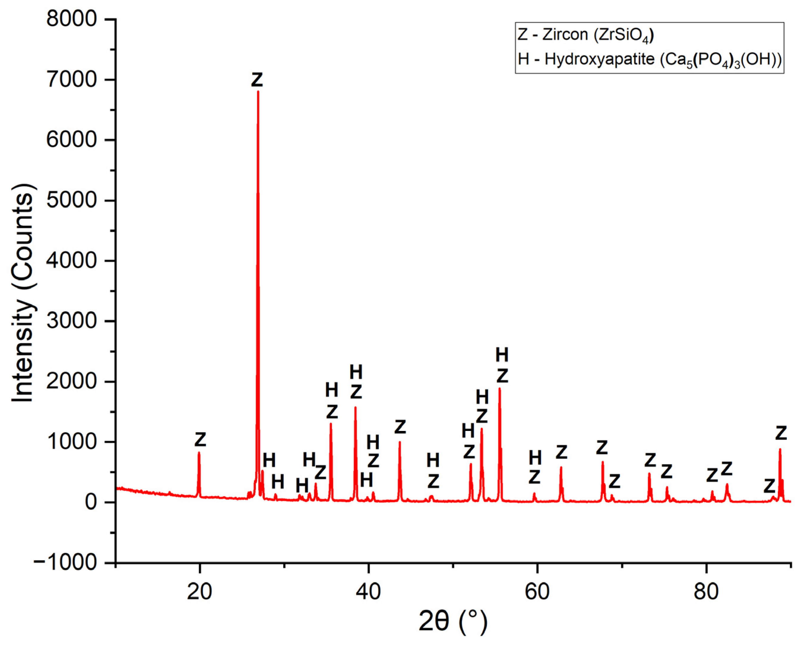
Figure 2.
Particle size distribution of raw zircon sand.
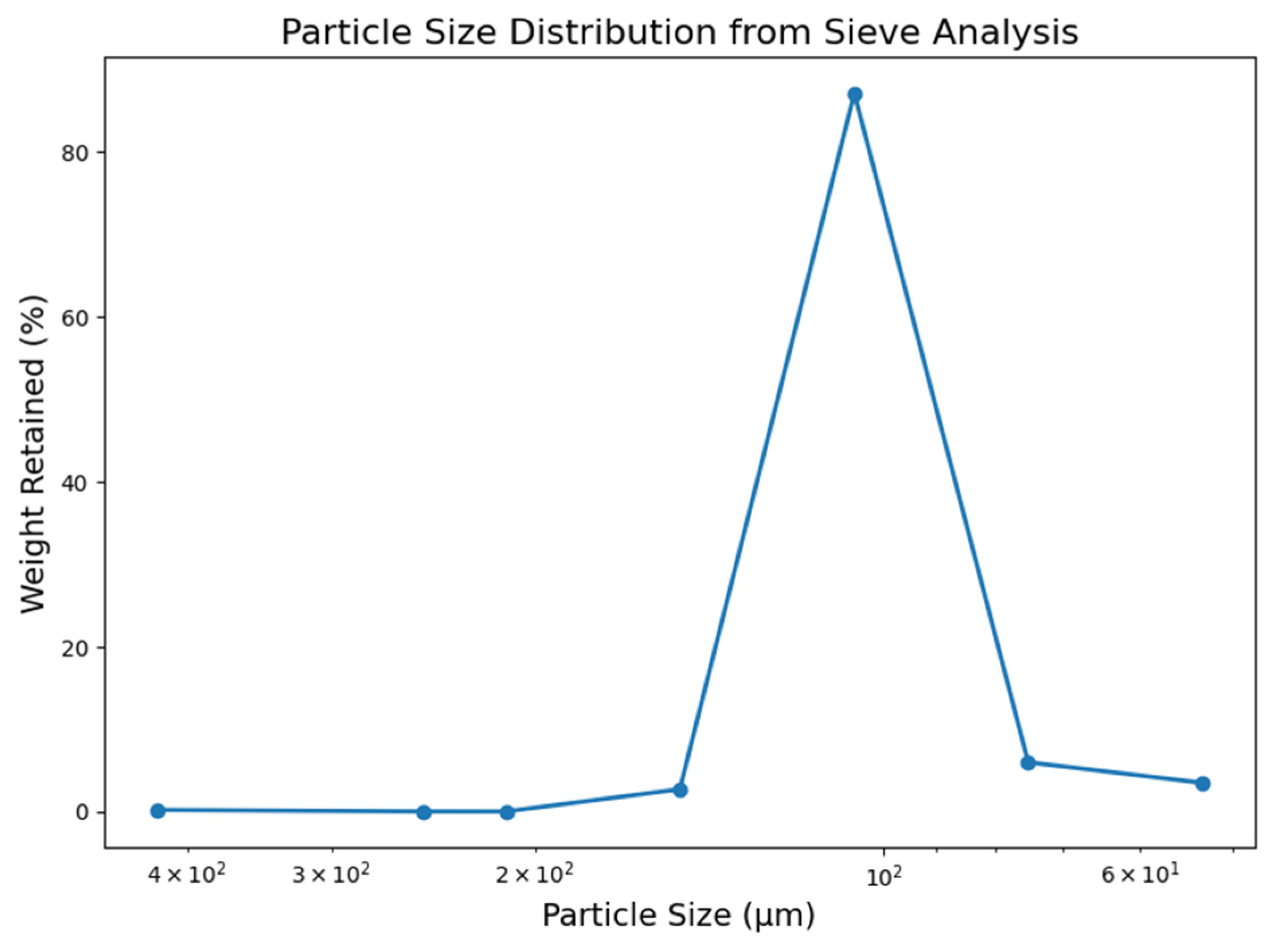
Figure 4.
HRTEM micrograph of synthesized zirconolite nanoparticles exhibiting nanocrystalline lattice structure.
Figure 4.
HRTEM micrograph of synthesized zirconolite nanoparticles exhibiting nanocrystalline lattice structure.
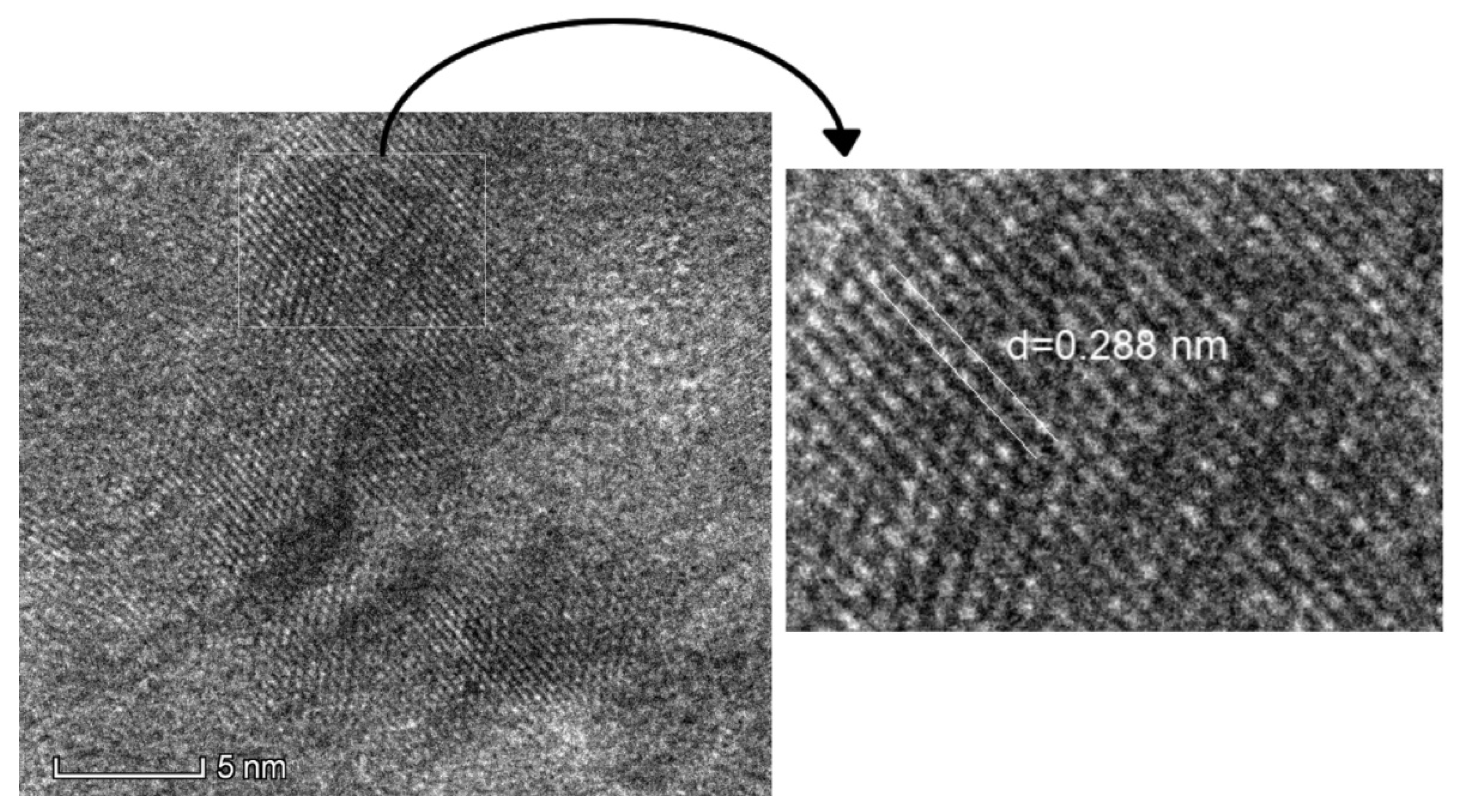
Figure 6.
TEM micrograph and particle size distribution of synthesized zirconolite nanoparticles. .
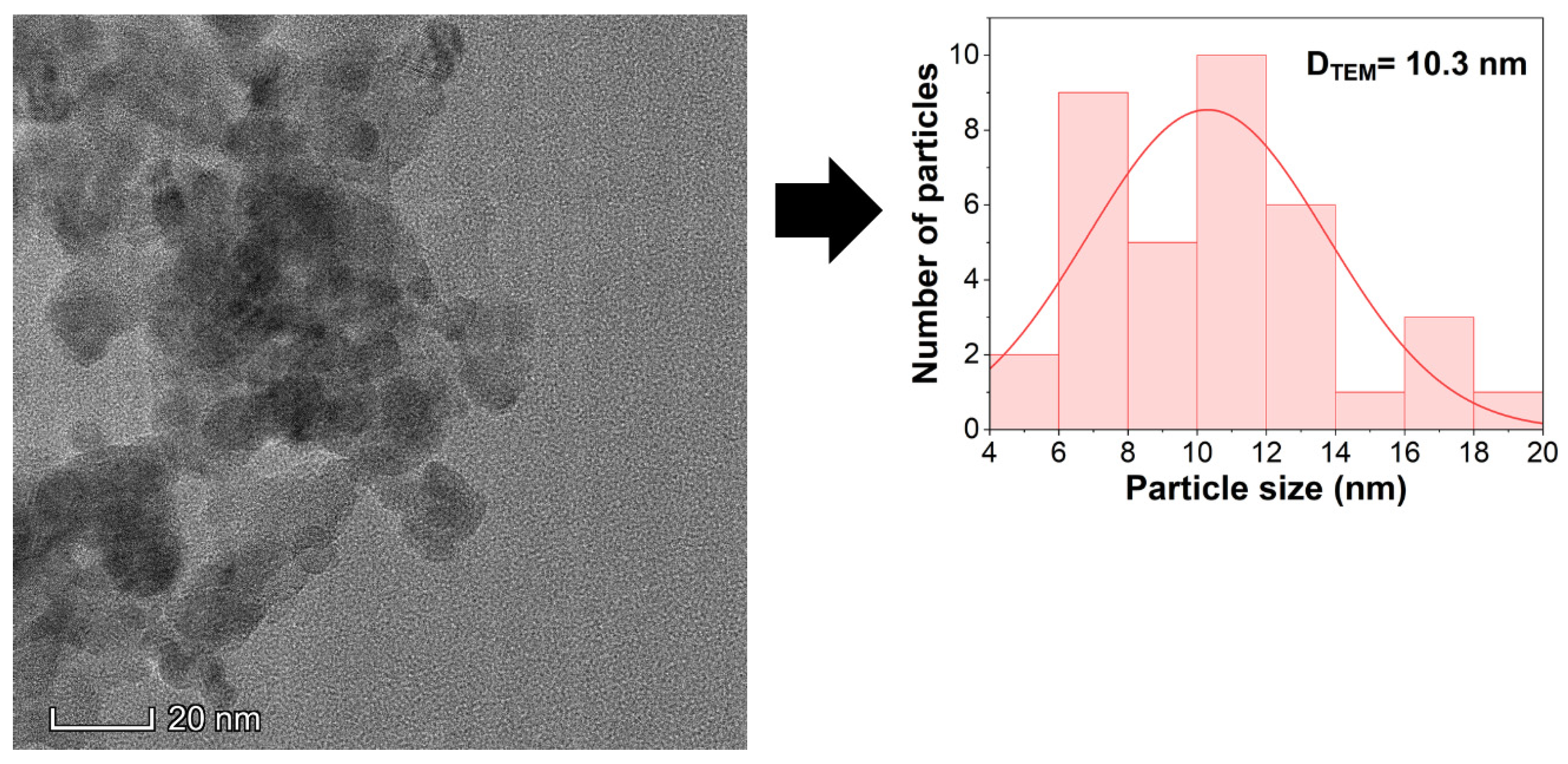
Figure 7.
Energy-dispersive spectroscopy (EDS) elemental mapping of the synthesized zirconolite nanoparticles.
Figure 7.
Energy-dispersive spectroscopy (EDS) elemental mapping of the synthesized zirconolite nanoparticles.
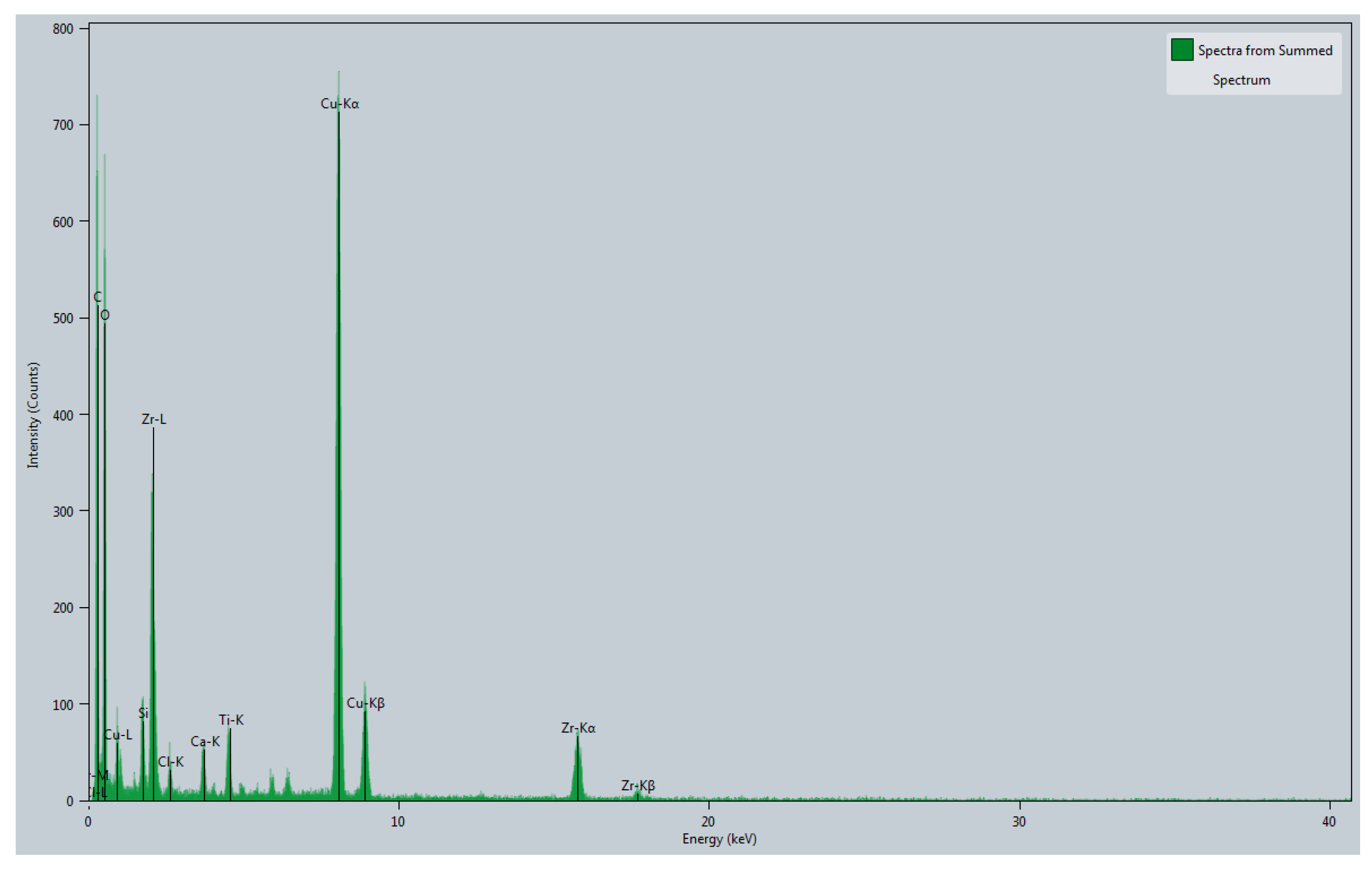
Figure 10.
Macrostructure of Zirconolite coating on SS 316L.
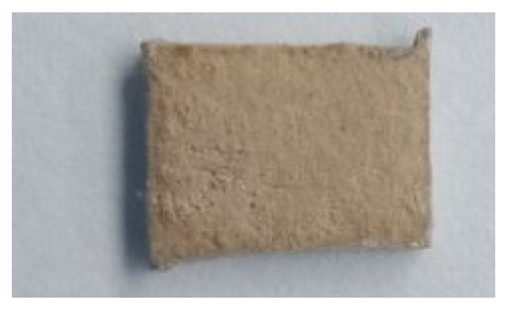
Figure 11.
Scanning electron microscopy (SEM) micrographs and elemental analysis of the zirconolite coating: (a) high-magnification surface morphology showing a rough, micro-to-nanostructured particulate network, (b) cross-sectional SEM image demonstrating a continuous, crack-free interface and coating thickness, and (c) energy-dispersive X-ray spectroscopy (EDS) spectrum of the coating surface.
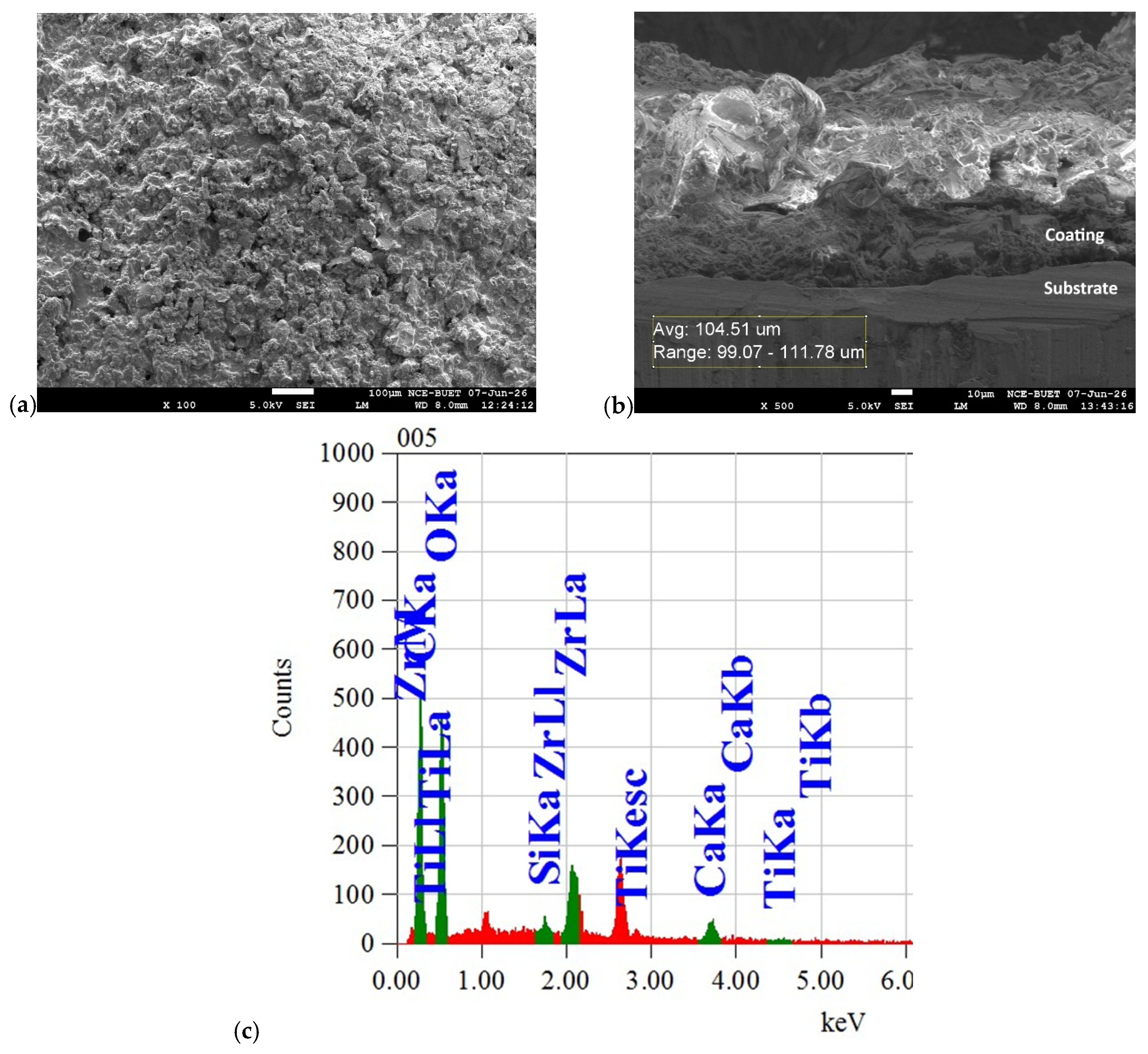

Table 1.
Chemical composition of raw zircon sand determined by XRF analysis.
| Compound | Result (%) | Compound | Result (%) |
|---|---|---|---|
| ZrO2 | 54.1169 | Fe2O3 | 0.3048 |
| SiO2 | 20.9761 | Y2O3 | 0.3021 |
| TiO2 | 8.8659 | MgO | 0.1993 |
| CaO | 7.6413 | K2O | 0.0965 |
| P2O5 | 3.7429 | Cr2O3 | 0.0948 |
| HfO2 | 2.7158 | ThO2 | 0.0872 |
| Al2O3 | 0.8565 |
Table 2.
Quantitative elemental composition of the dip-coated zirconolite surface determined by EDS analysis.
Table 2.
Quantitative elemental composition of the dip-coated zirconolite surface determined by EDS analysis.
| Element | Energy (keV) | Mass (%) |
|---|---|---|
| C | 0.277 | 44.3 |
| O | 0.525 | 39.83 |
| Si | 1.739 | 0.66 |
| Ca | 3.69 | 3.95 |
| Ti | 4.508 | 0.48 |
| Zr | 2.042 | 10.78 |
Disclaimer/Publisher’s Note: The statements, opinions and data contained in all publications are solely those of the individual author(s) and contributor(s) and not of MDPI and/or the editor(s). MDPI and/or the editor(s) disclaim responsibility for any injury to people or property resulting from any ideas, methods, instructions or products referred to in the content. |
© 2026 by the authors. Licensee MDPI, Basel, Switzerland. This article is an open access article distributed under the terms and conditions of the Creative Commons Attribution (CC BY) license (http://creativecommons.org/licenses/by/4.0/).
Copyright: This open access article is published under a Creative Commons CC BY 4.0 license, which permit the free download, distribution, and reuse, provided that the author and preprint are cited in any reuse.