

Submitted:
17 September 2026
Posted:
18 September 2026
You are already at the latest version
Abstract
Ectopic positioning of the gallbladder within the hepatic parenchyma is uncommon and can substantially increase surgical complexity. A 25-year-old woman with gastroschisis, prior abdominal operations, short bowel syndrome, and gastroileostomy bypass presented with recurrent acute cholecystitis despite prior drainage and stenting. Laparoscopy was abandoned due to dense adhesions preventing exposure, prompting open exploration. The gallbladder was discovered buried in the liver and was accessed safely through limited parenchymal transection followed by dome-down dissection. After confirmation of a single outflow tract, the inflamed infundibular-cystic duct region was divided with a linear stapler. Ultimately, subtotal cholecystectomy was avoided due to her altered gastrointestinal anatomy, making postoperative endoscopic management difficult in the event of a leak or retained remnant. Nevertheless, she recovered uneventfully and was discharged on postoperative day 2. This case emphasizes the importance of individualized operative strategy when aberrant anatomy coexists with limited rescue options.
Keywords:
intrahepatic gallbladder
; open cholecystectomy
; partial hepatic resection
An intrahepatic gallbladder is a rare anatomical variant in which the gallbladder is partially or completely embedded within the anteroinferior liver parenchyma, in which its abnormal position can impair emptying, resulting in bile stasis with an increased risk of gallstone formation [1,2]. Resultantly, several of these patients will require surgery, typically more difficult due to the unusual anatomy. However, the anomaly may be difficult to recognize on preoperative imaging and can create substantial difficulties operatively due to the concealment of the gallbladder within the liver and distorted biliary or vascular anatomy [3]. Such challenges are amplified in patients with complex surgical histories, especially in those with surgeries completed for congenital abdominal wall defects and multiple prior laparotomies, in whom adhesions and gastrointestinal reconstruction both limit operative exposure. A 25-year-old woman was transferred to our surgical service from another institution for higher level of care with 3 days of persistent, severe right upper quadrant pain associated with nausea and vomiting. Her past medical and surgical history consists of gastroschisis with silo reduction, recurrent small bowel obstructions with multiple resections totaling approximately 105 cm of small intestine and subsequent short bowel syndrome, and a gastroileostomy bypass. Prior to her admission, she experienced recurrent cholecystitis that was previously managed with a percutaneous cholecystostomy and biliary stent placement.
Her preoperative white blood cell (WBC) count was 12.4 x 10⁹/L and total bilirubin (t. bili) was 1.2 mg/dL. Right upper quadrant ultrasound demonstrated a distended gallbladder with an impacted stone in the neck, wall thickening, pericholecystic fluid, and a positive sonographic Murphy sign. MRCP demonstrated no choledocholithiasis or biliary ductal dilation. Given her recurrent disease despite prior drainage and biliary stenting, definitive cholecystectomy was recommended.
The patient initially underwent a diagnostic laparoscopy, initiated using the left upper quadrant access at Palmer’s point. However, after insufflation and placement of a 5-mm trocar, extensive adhesions prevented safe laparoscopic visualization and dissection. Therefore, the procedure was converted to an exploratory laparotomy in which an incision was made to the patient’s right lower quadrant and then extended towards the midline in anticipation of the gallbladder’s suspected location.
Several hours were required to lyse the adhesions between the abdominal viscera and wall and to obtain access to the liver. After exposure, the gallbladder was noted to be intrahepatic and a partial hepatic resection of the parenchyma provided access to the gallbladder fossa, permitting a dome-down dissection towards a single tubular structure (Figure 1). Needle aspiration confirmed bile within a distended gallbladder, resulting in drainage of hydrops and purulent material. The gallbladder was also perforated, with purulent infection present at the time of surgery, resulting in a class IV dirty/infected operative wound.
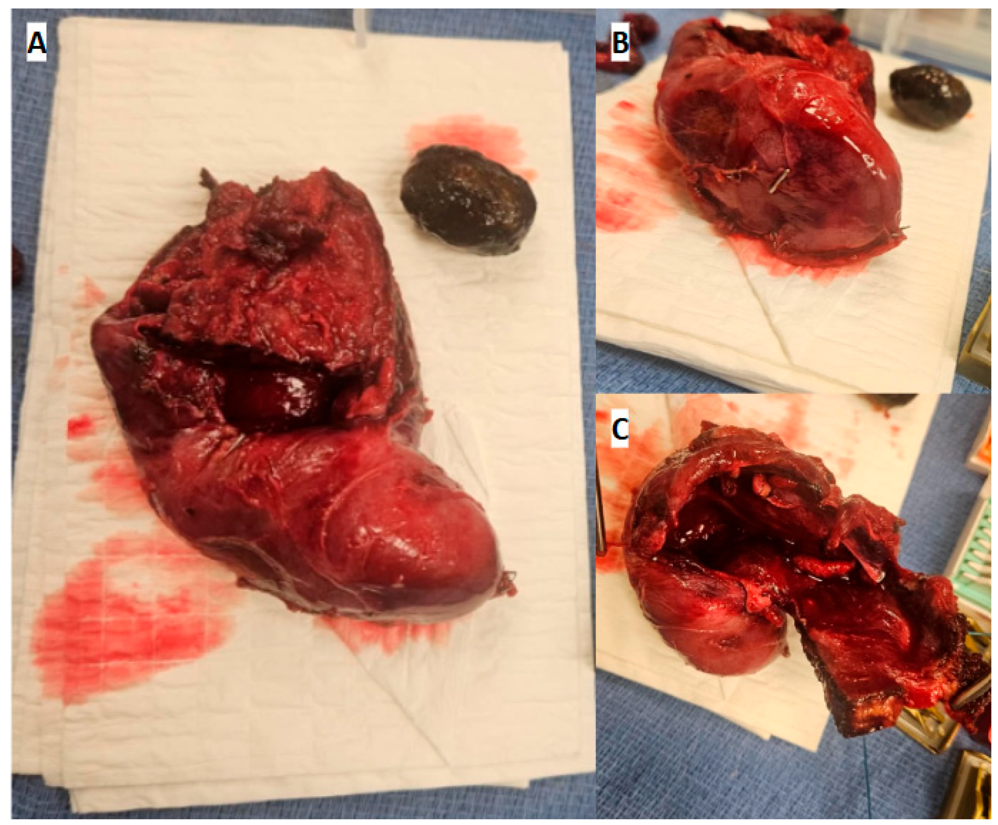
Direct inspection down the gallbladder lumen confirmed only one tubular structure entered the gallbladder. Furthermore, the gallbladder and surrounding inflammatory rind were markedly thickened, preventing secure closure with the largest available hemoclip, necessitating a 60-mm black-load linear stapler to close. The staple site was determined to be the junction between the infundibulum and cystic duct, with the procedure documented as a total cholecystectomy. Gross examination demonstrated the resected gallbladder and extracted gallstone (Figure 2A), the divided cystic duct-infundibular junction (Figure 2B), and the opened gallbladder lumen (Figure 2C). A fenestrating subtotal cholecystectomy was considered intraoperatively but the patient’s altered gastrointestinal anatomy raised concerns postoperative endoscopic retrograde cholangiopancreatography (ERCP) would not be feasible if a bile leak, retained stone, or symptomatic remnant developed. Therefore, a total cholecystectomy was favored after the single outlet was identified.
A 12-french closed-suction drain was placed within the fossa and then fascia and skin were closed in layers. Postoperatively, the patient recovered without major events, and the bile drain was removed on postoperative day (POD) 2 when output had fallen to 25 mL, remaining serosanguineous without bilious output. Her WBC decreased to 5.4 × 10⁹/L, t. bili to 0.7 mg/dL, and ALT to 62 U/L on POD 2. As the patient remained afebrile, hemodynamically stable, without evidence of a bile leak, intra-abdominal abscess, wound infection, or obstruction, the patient was discharged POD 2.
Once recognized, an intrahepatic gallbladder converts a routine cholecystectomy into a hepatobiliary procedure due to the gallbladder wall contiguous with the liver parenchyma. Safe dissection requires partial hepatic resection to mobilize the organ as with our dome-down approach and in the case of Mathis et al. who completed hepatotomy with energy devices for parenchyma transection [4]. Distorted vascular and biliary anatomy within this setting raises the risk of hemorrhage and bile duct injuries with prior recommendations including preoperative MRCP, intraoperative cholangiography, and hepatobiliary surgeon involvement whenever such a variant is suspected [4]. Dense adhesions from prior surgeries compounded the difficulty of our patient’s cholecystectomy and precluded a laparoscopic approach altogether, reinforcing that a preoperative or congenitally abnormal abdomen should raise instead of lower suspicion for anatomical variations.
Furthermore, the choice of total over subtotal cholecystectomy illustrates how a patient’s global anatomy can outweigh local operative findings in surgical decision-making. When critical view of safety is difficult, surgeons require alternative methods to define anatomy, including additional intraoperative biliary imaging, subtotal cholecystectomy, conversion to open, or assistance of a specialist [5]. Subtotal cholecystectomies are an accepted bailout strategy when critical view of safety becomes difficult to obtain, especially in cases of severe inflammation, adhesions, or concern for bile duct or other anatomical injury precluding safe laparoscopic progress [6,7]. However, a subtotal cholecystectomy holds a significantly higher bile leak rate in which a retained stone, leak, or symptomatic remnant have required post-operative ERCP [8]. Given our patient’s gastroileostomy bypass and 105 cm of resected small bowel, it was concluded that any future ERCP for a remnant or leak would have been of significant risk and likely infeasible - our deciding factor favoring definitive total cholecystectomy. Ultimately, this case demonstrates that managing an intrahepatic gallbladder in a reoperative, anatomically complex abdomen requires anticipation of difficult hepatobiliary dissection and tailoring the extent of cholecystectomy to both intraoperative findings as well as the feasibility of postoperative ERCP rescue.
References
- Lee, F.M.; Wong, J.; Haghighi, K.S. Hiding in plain sight: An unusual case of intrahepatic gallbladder. J. Clin. Images Med. Case Rep. 2025, 6, 3410. [Google Scholar] [CrossRef]
- Guiteau, J. J.; Fisher, M.; Cotton, R. T.; Goss, J. A. Intrahepatic gallbladder. J. Am. Coll. ofSurgeons 2009, 209, 672. [Google Scholar] [CrossRef] [PubMed]
- Badrawi, N.; Iqbal, S.S.; Ahmed, A.; Albastaki, U. Intrahepatic gallbladder mimicking a cystic liver lesion: a case report and literature review. Radiol. Case Rep. 2021, 16, 2746–2748. [Google Scholar] [CrossRef] [PubMed]
- Mathis, R.; Stodghill, J.; Shaver, T.; Younan, G. Cholecystectomy of an intrahepatic gallbladder in an ectopic pelvic liver: a case report and review of the literature. Case Rep. Surg. 2017, 2017, 3568768. [Google Scholar] [CrossRef] [PubMed]
- Brunt, L.M.; Deziel, D.J.; Telem, D.A.; et al. Safe cholecystectomy multi-society practice guideline and state-of-the-art consensus conference on prevention of bile duct injury during cholecystectomy. Ann. Surg. 2020, 272, 3–23. [Google Scholar] [CrossRef] [PubMed]
- Strasberg, S.M.; Pucci, M.J.; Brunt, L.M.; Deziel, D.J. Subtotal cholecystectomy—”fenestrating” vs “reconstituting” subtypes and the prevention of bile duct injury: definition of the optimal procedure in difficult operative conditions. J. Am. Coll. Surg. 2016, 222, 89–96. [Google Scholar] [CrossRef] [PubMed]
- Pisano, M.; Allievi, N.; Gurusamy, K.; Borzellino, G.; Cimbanassi, S.; Boerna, D.; Coccolini, F.; Tufo, A.; Di Martino, M.; Leung, J.; Sartelli, M.; Ceresoli, M.; Maier, R.V.; Poiasina, E.; De Angelis, N.; Magnone, S.; Fugazzola, P.; Paolillo, C.; Coimbra, R.; Di Saverio, S.; De Simone, B.; Weber, D.G.; Sakakushev, B.E.; Lucianetti, A.; Kirkpatrick, A.W.; Fraga, G.P.; Wani, I.; Biffl, W.L.; Chiara, O.; Abu-Zidan, F.; Moore, E.E.; Leppäniemi, A.; Kluger, Y.; Catena, F.; Ansaloni, L. 2020 World Society of Emergency Surgery updated guidelines for the diagnosis and treatment of acute calculus cholecystitis. World J. Emerg. Surg. 2020, 15, 61. [Google Scholar] [CrossRef] [PubMed] [PubMed Central]
- Hajibandeh, S.; Hajibandeh, S.; Parente, A.; Laing, R.W.; Bartlett, D.; Athwal, T.S.; Sutcliffe, R.P. Meta-analysis of fenestrating versus reconstituting subtotal cholecystectomy in the management of difficult gallbladder. HPB 2024, 26, 8–20. [Google Scholar] [CrossRef] [PubMed]
Figure 1.
Intraoperative mobilization of the intrahepatic gallbladder. Open operative view demonstrating the gallbladder following partial hepatic parenchymal resection and fundus-first mobilization.
Figure 1.
Intraoperative mobilization of the intrahepatic gallbladder. Open operative view demonstrating the gallbladder following partial hepatic parenchymal resection and fundus-first mobilization.
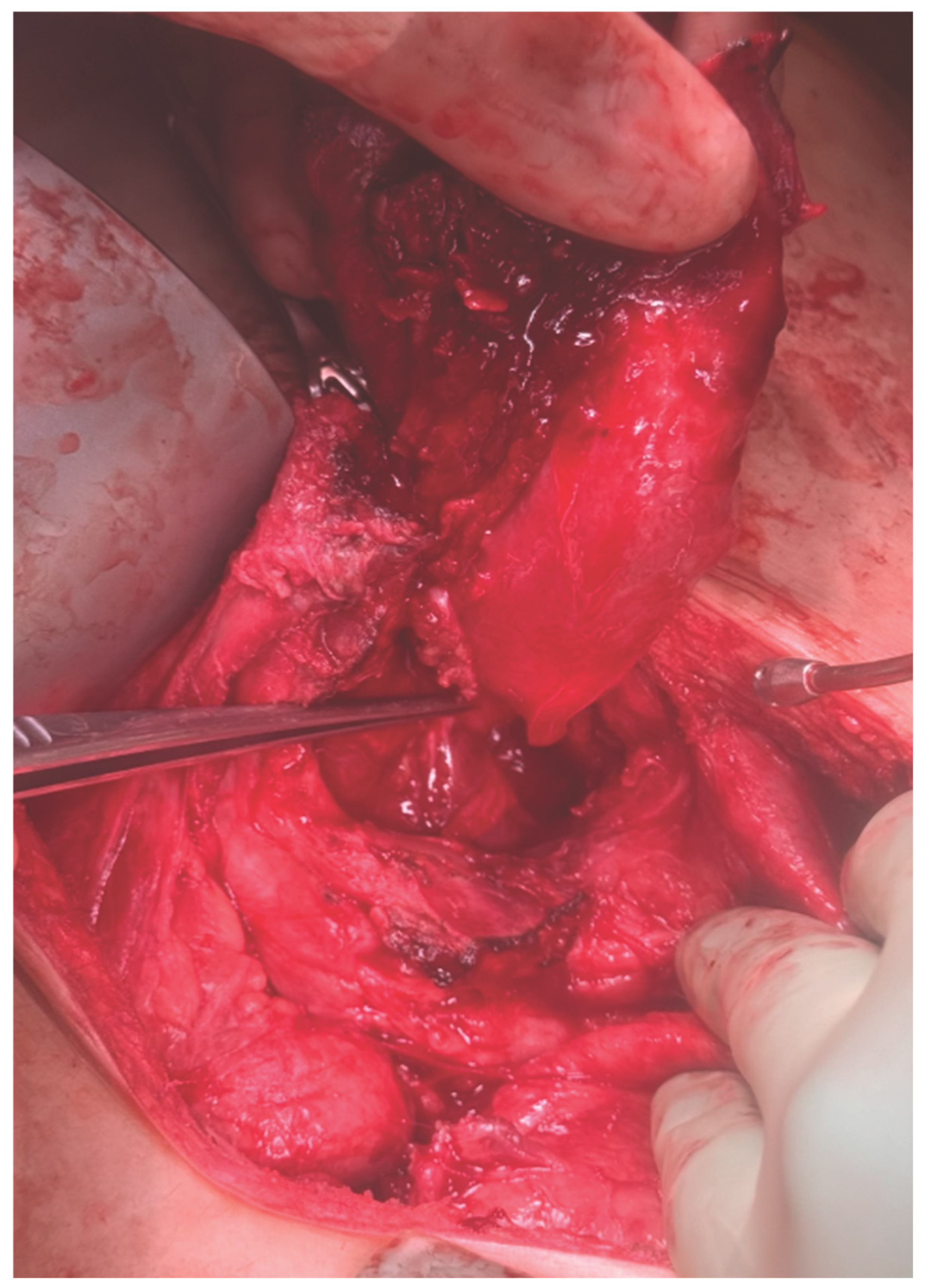
Figure 2.
Gross Examination of resected intrahepatic gallbladder. (A) Resected gallbladder specimen with extracted gallstone. (B) Inferior view demonstrating the divided cystic duct-infundibular junction. (C) Superior view of opened gallbladder demonstrating lumen and thickened wall.
Figure 2.
Gross Examination of resected intrahepatic gallbladder. (A) Resected gallbladder specimen with extracted gallstone. (B) Inferior view demonstrating the divided cystic duct-infundibular junction. (C) Superior view of opened gallbladder demonstrating lumen and thickened wall.
Disclaimer/Publisher’s Note: The statements, opinions and data contained in all publications are solely those of the individual author(s) and contributor(s) and not of MDPI and/or the editor(s). MDPI and/or the editor(s) disclaim responsibility for any injury to people or property resulting from any ideas, methods, instructions or products referred to in the content. |
© 2026 by the authors. Licensee MDPI, Basel, Switzerland. This article is an open access article distributed under the terms and conditions of the Creative Commons Attribution (CC BY) license (http://creativecommons.org/licenses/by/4.0/).
Copyright: This open access article is published under a Creative Commons CC BY 4.0 license, which permit the free download, distribution, and reuse, provided that the author and preprint are cited in any reuse.