

Submitted:
16 September 2026
Posted:
17 September 2026
You are already at the latest version
Abstract
Human papillomavirus (HPV) infection and oral microbiota alterations have been independently associated with oral disease in people living with HIV (PLHIV). However, the relationship between alpha-HPV (α-HPV), beta-HPV (β-HPV), and the oral microbiome remains poorly understood. Aim: This cross-sectional study aimed to describe the association between oral HPV infection and the oral and oropharyngeal microbiome in Mexican PLHIV with sustained virologic control and immune reconstitution. Methods: Oral and oropharyngeal samples were obtained by rinsing and gargling, and participants were classified into HPV-negative, α-HPV, and β-HPV groups. The V3–V4 region of the 16S rRNA gene was sequenced using the Illumina MiSeq platform. A total of 65 participants were included in the final analysis: 19 HPV-negative, 16 -α-HPVpositive, and β-HPV 30 -positive. Results: Alpha diversity was significantly increased in both HPV-positive groups compared with the HPV-negative group, with the highest diversity observed in the α-HPV positive group. The α-HPV group also showed a higher Fusobacteriota/Bacteroidota ratio and enrichment of Fusobacteriota-related taxa. In addition, predicted functional analysis showed enrichment of pathways related to lysine and pyruvate fermentation to butyrate in the α-HPV group. These findings suggest that α- and β-HPV infections may be associated with different oral microbial ecosystems in PLHIV.
Keywords:
HIV
; human papillomavirus
; oral microbiome
; oral dysbiosis
; alpha-papillomavirus
; beta-papillomavirus
1. Introduction
There are several common conditions of the oral cavity in the population living with HIV (PLHIV), such as periodontal disease and dental caries, which involve a local inflammatory process that can lead to an imbalance of the oral microbiota, favoring the establishment of certain microorganisms that allow the development of other conditions such as bacterial translocation and oral cancer [1,2], in addition to HPV infection, tobacco use [3], and other factors such as abundant alcohol consumption or genetic predisposition [4,5].
It has been reported that HIV-positive (HIV+) men who have sex with men (MSM) have a prevalence of alpha-HPV (α-HPV) generating infection of up to 30%. In addition, the predictors or determinants of oral HPV infection in the context of α-HPVs have been well characterized [6]. However, the clinical features and prevalence of beta-HPV (β-HPV) infection have not been well described. β-HPV genera can be found in the oral mucosa and saliva as asymptomatic infections and have been reported with a prevalence from 10 to 50% [7]. They have also been identified in head and neck carcinomas [8].
The oral microbiome is the second most abundant in the body after the intestinal microbiome [9]. It is currently recognized that the oral microbiome interacts with the immune system, playing a role in health and disease at local and systemic levels [10]. Several pathological processes in the oral cavity involve inflammation that can lead to a dysbiotic milieu, promoting the establishment of certain microorganisms that induce local inflammation and bacterial translocation. The microorganisms primarily identified in the oral cavity of PLWHIV are Streptococcus mutans, Streptococcus sobrinus, and Lactobacillus, as well as an increase in Candida species and the main pathogens associated with periodontitis [11](10.1111/odi.12415), such as Porphyromonas gingivalis and Fusobacterium nucleatum. All these factors can interact with potentially malignant lesions in the mouth, contributing to the inflammatory microenvironment and the onset of squamous cell carcinoma (SCC) [12,13].
Infections in the oral mucosa, such as periodontal disease, candidiasis, and the presence of HPV and human herpesvirus, are favored by HIV infection itself, as it decreases the local immune response and dysregulates the microbiota due to chronic inflammation from the presence of chemokines, cytokines, and oral dysbiosis of PLWHIV, with or without treatment [2]. Furthermore, local translocation, products of microbial metabolism, and damage to mucosal surfaces (such as erosions and ulcerations) alter the functioning of the immune response, which is enhanced throughout life [11].
Certain components of the oral microbiota can activate inflammatory pathways, and each of these pathways can modulate the development and behavior of malignant neoplasms at different intensities [12]. It has been shown that the oral microbiota composition influences the balance of cell proliferation and apoptosis, hindering antitumor immunity and affecting the metabolism, like NF-κB, modulating the antitumor immune response and promoting tumor tolerance, inhibition of NK cell and T lymphocyte cytotoxicity, induction of JAK/STAT signaling that promotes tumor survival through gingipains and production of pro-inflammatory cytokines IL-6 and IL-8 produced by the host [14].
In addition to the classic factors associated with the development of oral cancer or dysplasia, such as smoking, HPV infection [2,3,15], alcohol misuse, or genetic predisposition [5]. PLWHIV also has other factors associated with local immunodeficiency. Among these mechanisms, changes in the immunological capacity of the oral cavity stand out because the humoral immune response, composed mainly of secretory immunoglobulin A, and enzymes such as lactoferrin and lysozyme, are diminished in PLHIV, as well as the cellular immune response [2,11,12].
Although the use of antiretroviral therapy (ART) has decreased opportunistic oral infections in PLHIV, the dysbiosis at this anatomical site persists, and the effects of ART have not been fully elucidated [16]. In recent years, the role of oral microbiota in the predisposition and/or clinical outcomes of different viral infections has been increasingly identified. An example of the relationship between viruses and bacteria and oral cancer is found with HPV and SCC, due to HPV proteins such as E6 and E7, as well as with Fusobacterium nucleatum through FAD and FADH2, and with Porphyromonas gingivalis and Treponema denticola through their lipopolysaccharides [13]. In addition, endotoxins and toxic metabolic byproducts of bacteria can damage keratinocyte DNA, inducing mutations in proto-oncogenes and tumor suppressor genes [17].
Despite the growing evidence of high prevalence of alpha and β-HPV infection in PLHIV [18], the interaction between HPV and oral cavity microbiota in PLHIV remains poorly understood, particularly in Mexican populations. Therefore, our study aimed to describe the relationship between HPV infection and the oral cavity microbiota in PLHIV to understand how they jointly influence oral health status. This knowledge may help inform future strategies for the prevention, diagnosis, and treatment of oral diseases.
2. Methods
2.1. Study Design and Participants
We conducted a cross-sectional study from January 2022 to May 2023 at the HIV Unit of Hospital Civil de Guadalajara “Fray Antonio Alcalde,” a reference university hospital in western Mexico. The inclusion criteria were male PLHIV ≥18 years old, treated patients with sustained virologic control (viral load (VL) < 40 copies/mL) for at least 12 months, absolute CD4+ T-cell count ≥350 cells/μL (Considered as immunological responders), and no history of HPV vaccination. The HIV infection stage was assessed and stratified according to the CDC criteria. Subjects under immunosuppressive or immunomodulator treatment, active bleeding in the oral cavity secondary to local or systemic pathology (blood dyscrasia), or subjects with previous treatments for pre-existing oral lesions in the last three months were excluded from the study. In addition, samples that were insufficient, degraded, or contaminated were eliminated. All participants voluntarily signed an informed consent form. Medical and physical examinations were performed, and a questionnaire about the usage of tobacco, alcohol, recreational drugs, exposure to other kinds of drugs, history of sexual activity (beginning of active sexual life, number of partners, and type of sexual relations, whether oral or anal, frequency of condom use in oral sex), and comorbidities was administered. This study was approved by the Ethics Committee of Hospital Civil (156/22).
2.2. Sampling and DNA Extraction
Oral and oropharyngeal samples were obtained via rinses and gargles with Listerine®, according to [19,20], without prior toothbrushing. The participants were requested to gargle for 15 seconds and to rinse for 45 seconds. Five mL of the sample were recollected in a sterile plastic tube and preserved at -80ºC until used for DNA extraction. DNA extraction was performed using the protocol for extracting genomic DNA from oral and oropharyngeal samples with the PureLink Genomic DNA Mini Kit (K182001, Thermo Fisher Scientific, USA). The concentration and purity of the extracted DNA (A260/A280≥1.80) were determined by spectrophotometry using a Thermo Scientific NanoDrop 2000 (Thermo Fisher Scientific, USA).
2.3. HPV Genotyping
To identify the HPV genotypes in each sample, endpoint and nested PCR tests were performed using PGMY and FAP primers, according to the protocol described in [21,22]. Briefly, the primer set PGMY11/09 was used to identify α-HPV, and the degenerate primers FAP59/FAP64 were used to identify β-HPV, according to the PCR reaction conditions described in Li et al. [22]. DNA extracted from the CaSki cell line (containing integrated HPV16 DNA) was used as the HPV-positive control, and FZD2 as the positive human genomic amplification control.
2.4. 16S rRNA Sequencing
The 16S rRNA amplicon sequencing library preparation was performed according to the Illumina MiSeq System’s protocol [23].
The V3 and V4 regions of the 16S rRNA gene were amplified using PCR following the manufacturer’s protocol with Platinum Taq DNA Polymerase High Fidelity (Invitrogen, Waltham, MA, USA) and previously published primers: 341F: 5’TCGTCGGCAGCGTCAGATGTGTATAAGAGACAGCCTACGGGNGGCWGCAG-3’ and 805R: 5’GTCTCGTGGGCTCGGAGATGTGTATAAGAGACAGGACTACHVGGGTATCTAATCC-3’. The obtained amplicons were further purified using AMPure XP (Beckman Coulter, Indianapolis, IN, USA) magnetic beads and quantified using the Qubit 3 dsDNA HS kit (Invitrogen, Waltham, MA, USA) according to the manufacturer’s instructions. Index incorporation was achieved using the Nextera XT Index Kit v2 (Illumina, San Diego, CA, USA). Finally, the indexed amplicons were pooled to equimolar concentrations in a 4 nmol/L solution tube. The resulting library was denatured and further sequenced using the MiSeq Reagent V3 600-cycle (Illumina, San Diego, CA, USA) according to the manufacturer’s protocol.
2.5. Bioinformatic and Statistical Analyses
Analysis of 16S rRNA sequences was performed with QIIME2 version 2024.2 amplicon distribution [24]. Paired-end sequences whose Phred ≥ 30 were accepted and denoised with DADA2 via q2-dada2 [25], at default settings. Taxonomy assignment was performed on obtained amplicon sequence variants (ASVs), using a full-length 16S weighted classifier (via q2-feature-classifier) [26,27], which employs Silva 138.1 as a reference taxonomic database [28]. ASVs identified as mitochondria and chloroplasts were also removed. Then, filtered ASVs were aligned using MAFFT via q2-alignment [29], and phylogeny was built with FastTree2 via q2-phylogeny [30]. Alpha diversity indices [31]; and beta diversity distances (unweighted and weighted Unifrac [32,33], were computed via q2-diversity. PCoA plots were generated to visualize beta diversity distances using Emperor via q2-emperor [34] and further analyzed using PERMANOVA.
Differential abundance analyses were accomplished by means of ANCOM-BC (via q2-composition [35]. Before ANCOM-BC analysis, a frequency filter was applied, in which features that appeared more than 50 times in at least 10% of the samples were retained. A q ≤ 0.05 cut-off was used to assess significance, and a log fold change (LFC) ≥ |1.0| was used to assess the effect size. To evaluate the potential metabolic profile of the oral microbiota, we employed the PICRUSt2 pipeline [36,37,38,39,40], which is coupled with the MetaCyc Database [41]. The resulting pathways were further analyzed using ANCOM-BC, using a frequency filter that retained features that appeared more than 500 times in at least 10% of the samples, with a q ≤ 0.05 and an LFC ≥ |1.0|. Bacillota/Pseudomonadota, Fusobacteriota/Bacillota, and Fusobacteriota/Bacteroidota ratios were calculated similarly to previous publications [42]. Centered log-ratio transformations (clr) were computed using the MicrobiomeAnalyst 2.0 server [43].
Data normality was examined using the D’Agostino-Pearson omnibus test. If significant, a parametric distribution was assumed, and a one-way ANOVA with a Bonferroni post hoc test was performed to assess differences between groups; otherwise, the Kruskal–Wallis test with Dunn’s post hoc test was performed. Beta diversity distances were statistically analyzed using PERMANOVA tests. Both alpha and beta diversity statistical analyses were corrected with Benjamini–Hochberg (BH) multiple testing through the QIIME2 package. Fisher’s exact test was used to evaluate categorical variables. All statistical tests were two-tailed, and a p-value or false discovery rate-adjusted q-value equal to or less than 0.05 were considered statistically significant. Data were analyzed using SPSS version 25.0, unless otherwise specified. Plots were generated using GraphPad Prism version 9.0.0.
3. Results
3.1. Clinical and Demographic Characteristics
A total of 398 participants were evaluated during the study period, of which 99 met the selection criteria and provided oral rinse samples. After DNA extraction, five participants were excluded because their rinse samples were degraded. HPV genotyping was performed on 94 patient samples. For logistical reasons, 73 samples were sequenced. Of these, eight samples were excluded (three because their sequence quality was below Phred >30 and five because they showed co-infection with α- and β-HPV during genotyping due to positivity to the primers PGMY and FAP). Our final study group comprised 65 participants: HPV−, n=19; PGMY+, n=16; and FAP+, n=30 (Table 1).
3.2. Alpha Diversity and Beta Diversity of the Oral and Oropharyngeal Microbiome
To assess alpha diversity (that is, the number of distinguishable nucleotidic bacterial features in each sample), we calculated the following indices: observed features, Chao1, Shannon, and Strong dominance (Figure 1). The observed features index (which accounts for differences among ASVs identified in the bioinformatics analysis) showed a significant increase in alpha diversity in the PGMY+ and FAP+ groups compared to the HPV− group (q<0.05 in both cases). Notably, the PGMY+ group showed greater diversity than the FAP+ group (q<0.05). The same behavior was observed with the Chao1 and Shannon indices, although measurements with the latter index were only trends and did not reach statistical significance. Finally, the strong dominance index, which increases when the microbiota is polarized toward a few bacterial species, showed an upward trend in the HPV− group.
Additionally, no differences were observed in the α diversity index when comparing HPV− with HPV+ individuals, regardless of the HPV genus (Supplemental Figure S1). Notably, we observed that people who reported using condoms during oral sex tended to present diminished lower alpha diversity compared to those who did not use condoms (Supplemental Figure S1). Furthermore, we identified a slight upward trend in bacterial alpha diversity in advanced stages, depending on the clinical stage of HIV infection (clinical category on CDC classification) (Supplemental Figure S1).
Regarding beta diversity distance, the principal coordinates analysis (PCoA, Figure 2) and statistical analysis using PERMANOVA (Table 2) indicated that the oropharyngeal microbiome formed significantly separate clusters comparing PGMY+ (α-papillomaviruses) with HPV− participants, using the unweighted Unifrac metric (q=0.006, pseudo-F=2.056). Conversely, the unweighted Unifrac metric indicated that the distance between the FAP+ (β-papillomaviruses) and PGMY+ groups was greater than that between the FAP+ and HPV− groups. These findings suggest that the oropharyngeal microbiome has a greater phylogenetic similarity between the FAP+ and HPV− groups than between the PGMY+ and HPV− groups. To further explore the distances, we plotted intergroup (PGMY+ vs. PGMY+) and intragroup distances (Figure 3), clearly showing a greater distance between PGMY+ and HPV− than between PGMY+ and FAP+. Notoriously, when the weighted Unifrac metric was calculated, no significant differences were found, pointing towards a qualitative difference rather than a quantitative one, since the unweighted Unifrac metric assesses qualitative phylogenetic aspects of the measured bacterial communities [32].
3.3. Relative Abundances
At the phylum level, Bacillota (previously known as Firmicutes) was the most abundant taxon identified in the three groups, followed by Pseudomonadota (previously known as Proteobacteria) and Bacteroidota. Bacillota showed a greater relative abundance in the HPV− group (48.2%) than in the PMGY+ (46.62%) and FAP+ (41.73%) groups. In contrast, the FAP+ group exhibited a relative abundance of Pseudomonadota at 34.23%, compared to 30.98% in the PGMY+ group and 30.65% in the HPV− group. Interestingly, the PGMY+ group showed a greater relative abundance of Fusobacteriota (3.54%) than the other groups (HPV−: 1.92% and FAP+: 2.3%) (Figure 4A).
At the genus level, a slight expansion of Haemophilus was observed in the FAP+ group (24.45% vs. 22.07% in HPV− and 19.79% in PGMY+), whereas a greater relative abundance of Streptococcus was observed in the PGMY+ group (27.27% vs. 24.49% in HPV− and 19.66% in FAP+). Notably, an expansion of Rothia was observed in both the PGMY+ and FAP+ groups (2.67% and 2.71%, respectively) compared with the HPV− group (1.12%; Figure 4B). Given the relevance of the genera Fusobacterium and Neisseria, median comparisons of clr-transformed counts for these taxa were performed across the three groups (HPV−, PGMY+, and FAP+; Supplementary Figure 3). Although no statistically significant differences were observed in Fusobacterium abundance, a trend toward increased levels was noted in the PGMY+ group (α-HPV), which approached statistical significance compared with the HPV− group (352.0 vs. 203.0, p=0.06). This increasing trend in HPV+ groups was also evident when comparing HPV+ with HPV− individuals (318.0 vs. 203.0; p= 0.07; Supplementary Figures S3A and S3C).
Comparisons of Neisseria did not reveal statistically significant differences; however, modest increases were observed in both the PGMY+ and FAP+ groups compared with healthy controls. This pattern was further supported when grouping patients as HPV− versus HPV+, with higher Neisseria abundance observed in the latter group (Supplementary Figures S3E and S3G).
Finally, some taxa could be resolved at the species level. We observed similar behavior in Haemophilus parainfluenzae, showing a decrease in PGMY+ (15.86% vs. 20.70% in HPV− and 21.81% in FAP+). Conversely, we found that the previously observed expansion of Streptococcus in PMGY+ could be attributed to Streptococcus infantis (22.02%). Notably, a decreased relative abundance of Neisseria perflava and Prevotella melaninogenica was noted in the PGMY+ group compared to the HPV− and FAP+ groups (Figure 4C).
3.4. Oral Microbiome Ratios
We evaluated the Pseudomonadota/Bacillota (formerly known as Proteobacteria/Firmicutes) ratio and proposed the Fusobacteriota/Bacillota and Fusobacteriota/Bacteroidota ratios, which assess increases in Fusobacteriota at the expense of decreases in Bacillota or Bacteroidota (Figure 5). It is noteworthy that the Fusobacteriota/Bacillota ratio showed an increasing trend in the PGMY+ group and even more so in the FAP+ group, compared with the HPV− group, indicating an increase in the Fusobacteriota phylum relative to Bacillota (Firmicutes) species. Additionally, the Fusobacteriota/Bacteroidota ratio was significantly higher in the PMGY+ group than in the HPV− and FAP+ groups (p<0.01 for both comparisons). Finally, the comparison of Fusobacteriota counts across samples showed the same pattern, with a significant increase in the PGMY+ group compared to the HPV− (p<0.01) and FAP+ (p<0.05) groups. This is particularly interesting because it shows a unique dynamic characterized by a generalized increase in Fusobacteriota at the expense of a decrease in Bacteroidota in the oral cavity of PLHIV with alpha papillomavirus.
3.5. Differential Abundance of Bacterial Taxa
To calculate the differential abundance, the ANCOM-BC test (isometric logarithmic relationship) was applied, where we could observe that in the reference group taken (Participants HPV+ with the PGMY primers, mostly of the alpha genus) it is enriched with the Lautropia genera, Filifactor and Johnsonella; On the contrary, this group was observed to be depleted with the Eikenella genus, which indicates that the group of HPV+ patients with FAP primers (generally of the beta genus) is enriched with this same genus) (See Figure 6).
3.6. Functional Pathway Predictions
Metagenome reconstruction and its functional potential were determined using PICRUSt2 in terms of MetaCyc pathways. We found several pathways applying ANCOM-BC to the obtained features in pairing groups; notably, protocatechuate degradation II (PROTOCATECHUATE-ORTHO-CLEAVAGE-PWY), fatty acid salvage (PWY-7094), creatinine degradation (CRNFORCAT-PWY), and methyl ketone biosynthesis (PWY-7007) were statistically enriched in the PGMY+ group compared to the FAP+ group. We identified several pathways associated with the glyoxylate cycle, although none were statistically significant. Notably, no pathways from the FAP+ group that surpassed the LFC threshold were recovered (Figure 7, upper).
Comparison of the PGMY+ group with the HPV− group revealed that some butyrate-related pathways, such as lysine fermentation to acetate and butyrate (P163-PWY), pyruvate fermentation (CENTFERM-PWY), and C. acetobutylicum acidogenic fermentation (PWY-6590), and an enrichment in UDP-2,3-diacetamido-2,3-dideoxy-alpha-D-mannuronate biosynthesis (PWY-7090) were also observed in this group (Figure 7, middle). This pathway is involved in the construction of O-antigens from lipopolysaccharides. Finally, when comparing the FAP+ group with the HPV− group, adenosylcobalamin biosynthesis (PWY-5507) was identified, although it did not reach significance (Figure 7, bottom).
To explore the precise differences among butyrate-producing pathways, such as the fermentation of L-lysine and pyruvate, we compared the associated PICRUSt2 feature counts for L-lysine fermentation (P163-PWY) and pyruvate fermentation to butyrate (CENTFERM-PWY) among the three groups (supplemental Figure S4, A and B). We found significant enrichment in L-lysine fermentation among patients in the PMGY+ group, with a similar trend in pyruvate fermentation. Additionally, we stratified patients according to HIV/AIDS clinical stage, showing a trend suggesting higher pyruvate fermentation to butyrate in AIDS stages (supplemental Figure S4, C).
4. Discussion
In this cross-sectional study, we characterized the oral microbiome of PLHIV according to α- and β-HPV infection status. Our findings indicate that α-HPV infection is associated with qualitative shifts in the oral bacterial community, increased α-diversity, enrichment of Fusobacteriota-related taxa, and a predicted butyrate-producing metabolic profile. In contrast, β-HPV infection showed a microbiome composition more similar to that of HPV− individuals. All these findings suggest that different HPV genera may be associated with distinct oral microbial ecosystems in PLHIV.
Alterations in the oral mucosa and microbiota are well recognized in PLHIV and are largely driven by chronic immune activation, local immunodeficiency, and persistent inflammation [44]. The interaction between oral cavity microbiota, microbial metabolites, and viral infections has gained increasing attention, particularly in immunocompromised populations. Our study provides observational evidence from western Mexico, a region characterized by a predominantly young population of MSM and a high proportion of late HIV diagnoses (around 40%), a factor associated with increased susceptibility to opportunistic infections, including persistent HPV infection [45,46,47]. This regional context reinforces the relevance of our findings and addresses a population that has been underrepresented in oral microbiome–HPV research.
Regarding α-diversity, we observed a generalized increase in bacterial diversity in HPV+ individuals, particularly in the α-HPV group, which includes most oncogenic α-HPV genotypes. This finding contrasts with previous reports in Italian and Puerto Rican cohorts of PLHIV, where no significant differences in α-diversity were detected according to HPV status. However, studies from the same Puerto Rican population have shown that periodontal disease in PLHIV is associated with bacterial richness and α-diversity increased, supporting the notion that higher oral bacterial diversity reflects dysbiosis rather than health [48,49]. Indeed, unlike the intestinal microbiome, increased bacterial diversity in the oral cavity has been consistently associated with periodontal disease, higher plaque indices, and gingival inflammation [50]. Our observation of a trend toward higher α-diversity in HPV+ individuals, even when not statistically significant, aligns with reports from both PLHIV and HIV− populations and supports the concept that HPV infection may coexist with or could contribute to a dysbiotic oral environment [51].
We also observed a tendency toward lower α-diversity in individuals who reported condom use during oral sex. Although these findings should be interpreted cautiously, they are consistent with previous observations suggesting that sexual behaviors influence mucosal microbial communities. Similar associations have been reported in studies of the intestinal microbiota, where protected sexual practices were associated with lower bacterial diversity, potentially reflecting reduced microbial exchange [52].
Beta diversity analyses further highlighted the impact of α-HPV infection on the oral microbiome. Using the unweighted UniFrac metric, which emphasizes qualitative phylogenetic differences, we identified a significant separation between the α-HPV+ and HPV− groups. In contrast, no significant differences were detected using the weighted UniFrac metric, suggesting that α-HPV infection is associated with changes in community composition rather than shifts in dominant taxa abundance. These findings are consistent with previous studies reporting distinct oral microbial profiles associated with oncogenic HPV genotypes and support the hypothesis that HPV-related dysbiosis may involve the acquisition or loss of less abundant but phylogenetically distinct bacterial taxa [48,51].
At the phylum level, changes in relative abundance were modest, consistent with previous studies in PLHIV, with or without high-risk HPV genotypes [48,51]. However, in our study, a slight expansion of Bacillota (formerly Firmicutes) was observed in the α-HPV+ group. To explore this, ratios (such as Bacillota/Pseudomonadota) have served as a semiquantitative tool, showing utility for assessing imbalances in the intestinal microbiota of PLHIV and in other conditions characterized by dysbiosis and immunological alterations [42,53]. In this way, we explored new ratios, such as Fusobacteriota/Bacillota and Fusobacteriota/Bacteroidota, which together show a Fusobacteriota phylo expansion at the expense of a decrease in Bacteroidota in the oral cavity, particularly in the α-HPV+ group.
This dynamic has not been previously described in the context of oral HPV infection. While earlier studies have reported increases in Bacteroidota in HPV-positive individuals [12,51], our data suggest that α-HPV infection may be associated with a distinct microbial signature characterized by expansion of Fusobacteriota. Importantly, although the increase in Fusobacterium abundance did not reach statistical significance, its consistent enrichment in the α-HPV+ group is biologically relevant, given the well-established role of Fusobacterium species, particularly Fusobacterium nucleatum, in oral carcinogenesis and tumor-associated inflammation [54,55,56].
Regarding other taxa, our study identified a greater presence of Neisseria, Porphyromonas, and Prevotella in the HPV+ group (regardless of HPV genus); these taxa are commonly associated with periodontitis, dentoalveolar abscesses, and dysbiosis [57,58]. Furthermore, during oral dysbiosis, pathogenic bacteria become predominant, generating chronic inflammation, disrupting the protective functions of the biofilm, and fueling dysbiosis itself. Consequently, individuals with periodontal disease or other chronic oral infections are at an increased risk of HPV persistence and HPV-related cancers [58,59]. Notably, a positive correlation has been reported between higher HPV16 viral load and higher total bacterial counts, with Prevotella identified as a major trait in these cases [60].
Likewise, Di Spirito and Muthusamy pointed out that Staphylococcus, Streptococcus, Neisseria, Prevotella, Fusobacterium, Porphyromonas, and HPV play an important role in the development and establishment of SCC [61,62], through interference with the elimination of reactive oxygen species generated by human cells, generating a pro-inflammatory microenvironment, favoring the recruitment of tumor cells, and generating type IV collagen dysfunction in the epithelial basement membrane, which can lead to metastasis [63,64].
Differential abundance methods indicated that the α-HPV+ group is enriched in the genera Lautropia, Filifactor, and Johnsonella and depleted in the genus Eikenella. Authors such as Rossman reported enrichment of Lautropia in immunocompromised individuals, including children living with HIV, and although it is not considered a classical pathogen, its presence may reflect immune dysregulation [65]. Jaramillo et al. reported that the genus Eikenella is depleted in patients with destruction of the periodontium [66]. In addition, the enrichment of Filifactor and Johnsonella has been previously associated with colorectal cancer and advanced periodontal disease, suggesting a potential link between α-HPV infection and pro-inflammatory microbial profiles [67]. Previous findings suggest that the presence of HPV, along with other factors such as immunosuppression, smoking, alcohol consumption, and oral inflammatory conditions, may favor the persistence of a microbial profile in the oral cavity, potentially related to SCC development. [68].
Notably, we observed a depletion of several genera with putative probiotic or protective roles, including Lactobacillus, Bifidobacterium, Alloscardovia, and Megasphaera, in HPV+ participants. This pattern parallels observations in intestinal dysbiosis and suggests that HPV infection in PLHIV may be associated with a loss of beneficial microbial functions in the oral cavity [69,70].
It is important to consider that chronic inflammation induced by dysbiosis also modulates the host immune response. Some oral pathogens, such as Porphyromonas gingivalis, Prevotella spp., and Fusobacterium nucleatum, interfere with the functions of macrophages and dendritic cells, hindering viral clearance. This phenomenon favors HPV persistence, replication, and, in certain cases, malignant transformation of infected cells [58,59]. In contrast, lactic acid bacteria, including Lactobacillus, Bifidobacterium, and Streptococcus species, have been widely studied for their protective roles in the oral cavity. These bacteria prevent infections by modulating the immune response, competitively excluding pathogens, producing antimicrobial compounds, and suppressing inflammation. It has been shown that certain Streptococcus species can promote oral health by inhibiting the growth of pathogenic bacteria through metabolites such as bacteriocins and hydrogen peroxide [71,72].
Our study, using PICRUSt2-based functional predictions, revealed enrichment of butyrate-producing pathways in the α-HPV+ group, including lysine and pyruvate fermentation pathways, consistent with other studies in PLHIV and HPV+, which also found enrichment of butyrate and a positive correlation between butyrate and higher bacterial α-diversity [49]. Butyrate is a short-chain fatty acid with complex immunomodulatory effects. While it has been shown to exert anti-inflammatory and pro-apoptotic effects in certain contexts, in others, such as in HPV infection, butyrate exposure has also been shown to promote keratinocyte growth, migration, and immortalization, a phenomenon that could increase the risk of subsequent malignant transformation [73]. Furthermore, its functions as a histone deacetylase inhibitor could induce HIV replication and modulate viral latency; while its accumulation in the oral cavity (often from periodontal pathogens) can be associated with dysbiosis in the tongue and oral microbiome, it has been the subject of intense recent research (Chen et al., 2023; Margolis, 2011).
In particular, increased butyrate levels have been associated with higher abundances of Prevotella histicola, Veillonella atypica, and Streptococcus parasanguinis, as well as depletion of Neisseria subflava. Interestingly, our results showed a slight increase in Neisseria perflava (a variant of N. subflava) in the β-HPV+ group compared with the α-HPV+ group, which may be related to our functional predictions indicating greater butyrate-producing activity in the α-HPV+ group. Additionally, butyrate is produced in large amounts by Fusobacterium nucleatum [74].
The relationship between Fusobacteriota and HPV infection remains complex and incompletely understood (10.3389/fonc.2024.1425545). While in vitro studies support a pro-tumorigenic role for F. nucleatum through activation of pathways such as Wnt/β-catenin via FadA–E-cadherin interactions, clinical studies in oral SCC have yielded conflicting results. Importantly, both α- and β-HPV oncoproteins E6 and E7 can also modulate Wnt/β-catenin signaling, and emerging evidence suggests that these effects may differ by HPV genus [75,76,77]. Our data support the hypothesis that α-HPV infection may synergize with specific microbial profiles to create a permissive microenvironment for viral persistence and epithelial dysregulation.
In the context of HPV infection, the role of butyrate appears to be dual and context-dependent, with evidence supporting both tumor-suppressive and tumor-promoting effects depending on concentration, cellular context, and local microenvironment. Our findings suggest that α-HPV infection in PLHIV may be associated with a butyrogenic oral environment that could favor viral persistence and chronic inflammation, rather than directly driving carcinogenesis [73,74,78,79,80].
From a clinical perspective, our findings highlight the importance of comprehensive oral healthcare in PLHIV, particularly in those with HPV infection. Regular oral screening, management of periodontal disease, and counseling on modifiable risk factors such as smoking, alcohol use, and oral hygiene may help mitigate dysbiosis and reduce the risk of HPV persistence and downstream complications.
This study has several limitations. Its cross-sectional design precludes causal inference and limits assessment of temporal dynamics between HPV infection and microbiome changes. Lifestyle factors such as diet were not systematically assessed, and microbial metabolites or salivary immune components were not measured. Additionally, functional predictions were based on 16S rRNA data rather than shotgun metagenomics. Future longitudinal studies incorporating metabolomics, immune profiling, and higher-resolution sequencing approaches are needed to clarify the mechanistic links between HPV infection, oral dysbiosis, and disease progression in PLHIV.
5. Conclusion
This study demonstrates that oral α-HPV infection in a Mexican cohort of p PLHIV is associated with a distinct oral microbiome profile characterized by increased bacterial α-diversity, qualitative shifts in community composition, enrichment of Fusobacteriota-related taxa, and a predicted enrichment of butyrate-producing metabolic pathways. In contrast, β-HPV infection exhibited a microbial profile more similar to that of HPV-negative individuals, suggesting that different HPV genera may interact differently with the oral microbial ecosystem.
The enrichment of taxa associated with oral dysbiosis and inflammation, together with the predicted increase in butyrate-producing pathways, suggests that α-HPV infection may be associated with a pro-inflammatory oral microenvironment that could favor viral persistence and potentially increase susceptibility to HPV-associated oral disease. Future longitudinal studies integrating direct measurements of local and systemic bacterial metabolites, particularly butyrate, together with functional microbiome analyses, are warranted to clarify the biological impact of HPV infection on the oral microbiome and to determine whether these microbial alterations contribute to the development of SCC.
Supplementary Materials
The following supporting information can be downloaded at the website of this paper posted on Preprints.org, a) Supplementary Figure S1: Oropharyngeal microbiota alpha diversity in analyzed samples stratified according to the positivity for α or β-HPV (PMGY+ and FAP+), or negative results (upper figures), reported condom usage during oral sex (middle figure), and clinical categorization for HIV/AIDS (A, asymptomatic; B, not A or B; C, AIDS indicator condition). Median and interquartile ranges are shown in black lines. ns = not significant. Supplementary Figure S2: Beta diversity PCoA plots for unweighted (left) and weighted Unifrac (right) distances in HPV− (dark blue) and HPV+ (purple) for both alpha and beta papillomaviruses. Supplementary Table S1: PERMANOVA analysis results for unweighted and weighted Unifrac distances between HPV− and HPV+ (both alpha and beta papillomaviruses). Supplementary Figure S3: Fusobacterium and Neisseria counts and log fold changes (lfc) were assessed using ANCOM-BC. (A and E) Centered log ratio transformed counts of Fusobacterium or Neisseria in the three groups. (B and F) Log fold change of Fusobacterium or Neisseria derived from ANCOM-BC analysis. The y-axis shows the logarithmic effect sizes. The paired comparisons are shown below. (C and G) Centered-log ratio transformed counts stratified according to positivity for α or β-HPV (PMGY+ and FAP+) and negative results. (D and H) Log fold change stratified according to alpha or beta HPV positivity and negative results. Dot plots are shown for each individual, and median and interquartile ranges are shown in black lines. p-values are shown above the columns. ns= not significant. Supplementary Figure S4: MetaCyc pathways (according to PICRUSt2 feature counts) related to butyrate biosynthesis. Comparisons were made between the three groups (A and B) and according to the clinical categorization of HIV/AIDS (C). Dot plots are shown for each individual, and the median and interquartile ranges are shown as black lines. b) Informed consent: The form that all participants enrolled in the study reviewed and signed in compliance with the study.
Author Contributions
Conceptualization, Jose Adan Vizcaino Resendiz, Tonatiuh Abimael Baltazar-Díaz,Felipe De Jesús Bustos-Rodríguez, Luis Felipe Jave-Suarez, Miriam Ruth Bueno-Topete, Luz Alicia González-Hernández and Jaime Federico Andrade-Villanueva; Methodology, Jose Adan Vizcaino Resendiz, Tonatiuh Abimael Baltazar-Díaz, Luis Felipe Jave-Suarez, Juan Carlos Vázquez-Limón, Pedro Martínez-Ayala, Luz Alicia González-Hernández and Jaime Federico Andrade-Villanueva; Software, Tonatiuh Abimael Baltazar-Díaz, Pedro Martínez-Ayala, Luz Alicia González-Hernández and Jaime Federico Andrade-Villanueva; Validation, Luis Felipe Jave-Suarez, Miriam Ruth Bueno-Topete, Luz Alicia González-Hernández and Jaime Federico Andrade-Villanueva; Formal analysis, Jose Adan Vizcaino Resendiz, Tonatiuh Abimael Baltazar-Díaz, Felipe De Jesús Bustos-Rodríguez, Miriam Ruth Bueno-Topete, Luz Alicia González-Hernández and Jaime Federico Andrade-Villanueva; Investigation, Jose Adan Vizcaino Resendiz, Tonatiuh Abimael Baltazar-Díaz, Felipe De Jesús Bustos-Rodríguez, Luis Felipe Jave-Suarez, Juan Carlos Vázquez-Limón, Pedro Martínez-Ayala, Dana Alejandra Figueroa-González, Luz Alicia González-Hernández and Jaime Federico Andrade-Villanueva; Resources, Luz Alicia González-Hernández and Jaime Federico Andrade-Villanueva; Data curation, Tonatiuh Abimael Baltazar-Díaz, Luz Alicia González-Hernández and Jaime Federico Andrade-Villanueva; Writing—original draft, Jose Adan Vizcaino Resendiz, Tonatiuh Abimael Baltazar-Díaz, Luz Alicia González-Hernández and Jaime Federico Andrade-Villanueva; Writing—review & editing, Jose Adan Vizcaino Resendiz, Tonatiuh Abimael Baltazar-Díaz, Juan Carlos Vázquez-Limón, Miriam Ruth Bueno-Topete, Luz Alicia González-Hernández and Jaime Federico Andrade-Villanueva; Visualization, Luz Alicia González-Hernández and Jaime Federico Andrade-Villanueva; Supervision, Luz Alicia González-Hernández and Jaime Federico Andrade-Villanueva; Project administration, Luis Felipe Jave-Suarez, Luz Alicia González-Hernández and Jaime Federico Andrade-Villanueva; Funding acquisition, Luz Alicia González-Hernández and Jaime Federico Andrade-Villanueva.
Funding
This research did not receive any external funding.
Institutional Review Board Statement
The study followed the Ethical Principles for Medical Research Involving Human Subjects outlined in the Helsinki Declaration of 1975 (as revised in Brazil 2013) and was approved by the Ethics Committee of Hospital Civil de Guadalajara (Office number HCG/CEI-1200/22) under protocol code 156/22, dated 18/07/2022.
Informed Consent Statement
Written informed consent was obtained from each participant before enrollment.
Data Availability Statement
Available from the first author upon request.
Conflicts of Interest
The authors declare no conflict of interest.
References
- Annavajhala, M.K.; Khan, S.D.; Sullivan, S.B.; Shah, J.; Pass, L.; Kister, K.; et al. Oral and Gut Microbial Diversity and Immune Regulation in Patients with HIV on Antiretroviral Therapy. mSphere 2020, 5, e00798-19. [Google Scholar] [CrossRef] [PubMed]
- Heron, S.E.; Elahi, S. HIV Infection and Compromised Mucosal Immunity: Oral Manifestations and Systemic Inflammation. Front Immunol. 2017, 8. [Google Scholar] [CrossRef] [PubMed]
- Johnson, N.W.; Anaya-Saavedra, G.; Webster-Cyriaque, J. Viruses and oral diseases in HIV-infected individuals on long-term antiretroviral therapy: What are the risks and what are the mechanisms? Oral Dis. 2020, 26, 80–90. [Google Scholar] [CrossRef] [PubMed]
- Mancuso, P.; Djuric, O.; Collini, G.; Serventi, E.; Massari, M.; Zerbini, A.; et al. Risk of cancer in individuals with alcohol and drug use disorders: a registry-based study in Reggio Emilia, Italy. Eur. J. Cancer Prev. 2020, 29, 270–278. [Google Scholar] [CrossRef] [PubMed]
- Riddell, J.; Brouwer, A.F.; Walline, H.M.; Campredon, L.P.; Meza, R.; Eisenberg, M.C.; et al. Oral human papillomavirus prevalence, persistence, and risk-factors in HIV-positive and HIV-negative adults. Tumour Virus Res. 2022, 13, 200237. [Google Scholar] [CrossRef] [PubMed]
- Rajendra Santosh, A.B.; Christian, N.A.; Jones, T.; Thoms-Rodriguez, C.A.; Condappa, A.; Thompson, T.; et al. Molecular epidemiology of human papillomavirus genotypes in oral rinses from HIV-positive and HIV-negative Jamaican patients. J. Investig. Clin. Dent. 2019, 10, e12365. [Google Scholar] [CrossRef] [PubMed]
- Giuliani, M.; Gheit, T.; Rollo, F.; Tommasino, M.; Latini, A.; Benevolo, M.; et al. Predictors of Oral Infection by Mucosal and Cutaneous Human Papillomaviruses in HIV-Infected and Uninfected Men Who Have Sex with Men of the OHMAR Study. J. Clin. Med. 2021, 10, 2804. [Google Scholar] [CrossRef] [PubMed]
- Kerge, S.; Vuorinen, J.; Hurme, S.; Soukka, T.; Gheit, T.; Tommasino, M.; et al. Benign proliferative epithelial lesions of oral mucosa are infrequently associated with α-, β-, or γ human papillomaviruses. Laryngoscope Investig. Otolaryngol. 2019, 4, 43–48. [Google Scholar] [CrossRef] [PubMed]
- Lacunza, E.; Fink, V.; Salas, M.E.; Canzoneri, R.; Naipauer, J.; Williams, S.; et al. Oral and anal microbiome from HIV-exposed individuals: role of host-associated factors in taxa composition and metabolic pathways. npj Biofilms Microbiomes 2023, 9, 48. [Google Scholar] [CrossRef] [PubMed]
- Baker, J.L.; Mark Welch, J.L.; Kauffman, K.M.; McLean, J.S.; He, X. The oral microbiome: diversity, biogeography and human health. Nat. Rev. Microbiol. 2024, 22, 89–104. [Google Scholar] [CrossRef] [PubMed]
- Moyes, D.; Saxena, D.; John, M.; Malamud, D. The gut and oral microbiome in HIV disease: a workshop report. Oral Dis. 2016, 22, 166–170. [Google Scholar] [CrossRef] [PubMed]
- Dahlstrom, K.R.; Sikora, A.G.; Liu, Y.; Chang, C.C.; Wei, P.; Sturgis, E.M.; et al. Characterization of the oral microbiota among middle-aged men with and without human papillomavirus infection. Oral Oncol. 2023, 142, 106401. [Google Scholar] [CrossRef] [PubMed]
- Saikia, P.J.; Pathak, L.; Mitra, S.; Das, B. The emerging role of oral microbiota in oral cancer initiation, progression and stemness. Front Immunol. 2023, 14, 1198269. [Google Scholar] [CrossRef] [PubMed]
- Urzică, R.N.; Crețu, B.; Căruntu, A.; Bucurica, S.; Farcasiu, A.T.; Ciupescu, L.M.; et al. The Molecular Interplay Between Oral Microbiome and Oral Cancer Pathogenesis. Int. J. Mol. Sci. 2025, 26, 10212. [Google Scholar] [CrossRef] [PubMed]
- Wang, C.; Palefsky, J. Human papillomavirus-related oropharyngeal cancer in the HIV -infected population. Oral Dis. 2016, 22, 98–106. [Google Scholar] [CrossRef] [PubMed]
- Alam, S.; Chatterjee, S.; Kang, S.D.; Milici, J.; Biryukov, J.; Chen, H.; et al. Anti-Retroviral Protease Inhibitors Regulate Human Papillomavirus 16 Infection of Primary Oral and Cervical Epithelium. Cancers 2020, 12, 2664. [Google Scholar] [CrossRef] [PubMed]
- Sahan, A.Z.; Hazra, T.K.; Das, S. The Pivotal Role of DNA Repair in Infection Mediated-Inflammation and Cancer. Front Microbiol. 2018, 9, 663. [Google Scholar] [CrossRef] [PubMed]
- Gheit, T.; Rollo, F.; Brancaccio, R.N.; Robitaille, A.; Galati, L.; Giuliani, M.; et al. Oral Infection by Mucosal and Cutaneous Human Papillomaviruses in the Men Who Have Sex with Men from the OHMAR Study. Viruses 2020, 12, 899. [Google Scholar] [CrossRef] [PubMed]
- Heath, E.M.; Morken, N.W.; Campbell, K.A.; Tkach, D.; Boyd, E.A.; Strom, D.A. Use of Buccal Cells Collected in Mouthwash as a Source of DNA for Clinical Testing. Arch. Pathol. Lab Med. 2001, 125, 127–133. [Google Scholar] [CrossRef] [PubMed]
- Mendoza, Á.C.; Volante, B.B.; Hernández, M.E.O.; Mendoza, C.C.C.; Pliego, A.F.; Baptista Gonzalez, H.A.; et al. Design of a protocol for obtaining genomic DNA from saliva using mouthwash: Samples taken from patients with periodontal disease. J. Oral Biol. Craniofacial Res. 2016, 6, 129–134. [Google Scholar] [CrossRef] [PubMed]
- Forslund, O.; Antonsson, A.; Nordin, P.; Stenquist, B.; Göran Hansson, B. A broad range of human papillomavirus types detected with a general PCR method suitable for analysis of cutaneous tumours and normal skin. J. Gen. Virol. 1999, 80, 2437–2443. [Google Scholar] [CrossRef] [PubMed]
- Li, J.; Pan, Y.; Xu, Z.; Wang, Q.; Hang, D.; Shen, N.; et al. Improved detection of human papillomavirus harbored in healthy skin with FAP6085/64 primers. J. Virol. Methods 2013, 193, 633–638. [Google Scholar] [CrossRef] [PubMed]
- Ilumina. 16S Metagenomic Sequencing Librery Preparation [Internet]. San Diego, CA.: Ilumina; 2013. Report 14044223 Rev. B. Available from: Document Number 15044223). Illumina. Available online: https://support.illumina.com/documents/documentation/chemistry_documentation/16s/16s-metagenomic-library-prep-guide-15044223-b.pdf.
- Bolyen, E.; Rideout, J.R.; Dillon, M.R.; Bokulich, N.A.; Abnet, C.C.; Al-Ghalith, G.A.; et al. Reproducible, interactive, scalable and extensible microbiome data science using QIIME 2. Nat. Biotechnol. 2019, 37, 852–857. [Google Scholar] [CrossRef] [PubMed]
- Callahan, B.J.; McMurdie, P.J.; Rosen, M.J.; Han, A.W.; Johnson, A.J.A.; Holmes, S.P. DADA2: High-resolution sample inference from Illumina amplicon data. Nat. Methods 2016, 13, 581–583. [Google Scholar] [CrossRef] [PubMed]
- Bokulich, N.A.; Kaehler, B.D.; Rideout, J.R.; Dillon, M.; Bolyen, E.; Knight, R.; et al. Optimizing taxonomic classification of marker-gene amplicon sequences with QIIME 2’s q2-feature-classifier plugin. Microbiome 2018, 6, 90. [Google Scholar] [CrossRef] [PubMed]
- Kaehler, B.D.; Bokulich, N.A.; McDonald, D.; Knight, R.; Caporaso, J.G.; Huttley, G.A. Species abundance information improves sequence taxonomy classification accuracy. Nat. Commun. 2019, 10, 4643. [Google Scholar] [CrossRef] [PubMed]
- Quast, C.; Pruesse, E.; Yilmaz, P.; Gerken, J.; Schweer, T.; Yarza, P.; et al. The SILVA ribosomal RNA gene database project: improved data processing and web-based tools. Nucleic Acids Res. 2012, 41, D590–D596. [Google Scholar] [CrossRef] [PubMed]
- Katoh, K. MAFFT: a novel method for rapid multiple sequence alignment based on fast Fourier transform. Nucleic Acids Res. 2002, 30, 3059–3066. [Google Scholar] [CrossRef] [PubMed]
- Price, M.N.; Dehal, P.S.; Arkin, A.P. FastTree 2 – Approximately Maximum-Likelihood Trees for Large Alignments. PLoS ONE 2010, 5, e9490. [Google Scholar] [CrossRef] [PubMed]
- Strong, W.L. Assessing species abundance unevenness within and between plant communities. Community Ecol. 2002, 3, 237–246. [Google Scholar] [CrossRef]
- Lozupone, C.A.; Hamady, M.; Kelley, S.T.; Knight, R. Quantitative and Qualitative β Diversity Measures Lead to Different Insights into Factors That Structure Microbial Communities. Appl. Environ. Microbiol. 2007, 73, 1576–1585. [Google Scholar] [CrossRef] [PubMed]
- Lozupone, C.; Knight, R. UniFrac: a New Phylogenetic Method for Comparing Microbial Communities. Appl. Environ. Microbiol. 2005, 71, 8228–8235. [Google Scholar] [CrossRef] [PubMed]
- Vázquez-Baeza, Y.; Pirrung, M.; Gonzalez, A.; Knight, R. EMPeror: a tool for visualizing high-throughput microbial community data. Gigascience 2013, 2, 2047-217X-2–16. [Google Scholar] [CrossRef] [PubMed]
- Lin, H.; Peddada, S.D. Analysis of compositions of microbiomes with bias correction. Nat. Commun. 2020, 11, 3514. [Google Scholar] [CrossRef] [PubMed]
- Barbera, P.; Kozlov, A.M.; Czech, L.; Morel, B.; Darriba, D.; Flouri, T.; et al. EPA-ng: Massively Parallel Evolutionary Placement of Genetic Sequences. Syst Biol. 2019, 68, 365–369. [Google Scholar] [CrossRef] [PubMed]
- Czech, L.; Barbera, P.; Stamatakis, A. Genesis and Gappa: processing, analyzing and visualizing phylogenetic (placement) data. Bioinformatics 2020, 36, 3263–3265. [Google Scholar] [CrossRef] [PubMed]
- Douglas, G.M.; Maffei, V.J.; Zaneveld, J.R.; Yurgel, S.N.; Brown, J.R.; Taylor, C.M.; et al. PICRUSt2 for prediction of metagenome functions. Nat. Biotechnol. 2020, 38, 685–688. [Google Scholar] [CrossRef] [PubMed]
- Louca, S.; Doebeli, M. Efficient comparative phylogenetics on large trees. Bioinformatics 2018, 34, 1053–1055. [Google Scholar] [CrossRef] [PubMed]
- Ye, Y.; Doak, T.G. A Parsimony Approach to Biological Pathway Reconstruction/Inference for Genomes and Metagenomes. PLoS Comput Biol 2009, 5, e1000465. [Google Scholar] [CrossRef] [PubMed]
- Caspi, R.; Altman, T.; Billington, R.; Dreher, K.; Foerster, H.; Fulcher, C.A.; et al. The MetaCyc database of metabolic pathways and enzymes and the BioCyc collection of Pathway/Genome Databases. Nucleic Acids Res. 2014, 42, D459–D471. [Google Scholar] [CrossRef] [PubMed]
- Baltazar-Díaz, T.A.; Andrade-Villanueva, J.F.; Sánchez-Álvarez, P.; Amador-Lara, F.; Holguín-Aguirre, T.; Sánchez-Reyes, K.; et al. A Two-Faced Gut Microbiome: Butyrogenic and Proinflammatory Bacteria Predominate in the Intestinal Milieu of People Living with HIV from Western Mexico. Int. J. Mol. Sci. 2024, 25, 4830. [Google Scholar] [CrossRef] [PubMed]
- Lu, Y.; Zhou, G.; Ewald, J.; Pang, Z.; Shiri, T.; Xia, J. MicrobiomeAnalyst 2.0: comprehensive statistical, functional and integrative analysis of microbiome data. Nucleic Acids Res. 2023, 51, W310–W318. [Google Scholar] [CrossRef] [PubMed]
- Li, S.; Su, B.; He, Q.S.; Wu, H.; Zhang, T. Alterations in the oral microbiome in HIV infection: causes, effects and potential interventions. Chin. Med. J. (Engl) 2021, 134, 2788–2798. [Google Scholar] [CrossRef] [PubMed]
- Ablanedo-Terrazas, Y.; Romero-Mora, K.; Gómez-Palacio, M.; Alvarado-de La Barrera, C.; Ruiz-Cruz, M.; Hernández-Juan, R.; et al. Prevalence and risk factors for oral human papillomavirus infection in Mexican HIV-infected men. Salud Pública México 2018, 60, 653. [Google Scholar] [CrossRef] [PubMed]
- Algarin, A.B.; Lara, M.V.; Chapin-Bardales, J.; Baruch-Dominguez, R.; Sanchez, T.H.; Hernandez-Avila, M.; et al. Examining Geographical Differences in the HIV Care Continuum Among Men Who Have Sex with Men in Mexico. AIDS Behav. 2023, 27, 772–782. [Google Scholar] [CrossRef] [PubMed]
- Azamar-Alonso, A.; Bautista-Arredondo, S.A.; Smaill, F.; Mbuagbaw, L.; Costa, A.P.; Tarride, J.E. Patient characteristics and determinants of CD4 at diagnosis of HIV in Mexico from 2008 to 2017: a 10-year population-based study. AIDS Res. Ther. 2021, 18, 84. [Google Scholar] [CrossRef] [PubMed]
- Escobar Marcillo, D.I.; Privitera, G.F.; Rollo, F.; Latini, A.; Giuliani, E.; Benevolo, M.; et al. Microbiome analysis in individuals with human papillomavirus oral infection. Sci. Rep. 2025, 15, 2953. [Google Scholar] [CrossRef] [PubMed]
- Valerio, Y.A.R.; Delgado, E.S.P.; Sanchez, M.M.; Garcia, R.F.G.; Martinez, A.P.O.; Ferrer, M.M.; et al. Abstract C135: The oral microbiome and short chain fatty acids as contributors to oral HPV disparities in Puerto Rican people with HIV. Cancer Epidemiol. Biomark. Prev. 2023, 32. [Google Scholar] [CrossRef]
- Takeshita, T.; Kageyama, S.; Furuta, M.; Tsuboi, H.; Takeuchi, K.; Shibata, Y.; et al. Bacterial diversity in saliva and oral health-related conditions: the Hisayama Study. Sci. Rep. 2016, 6, 22164. [Google Scholar] [CrossRef] [PubMed]
- Zhang, Y.; D’Souza, G.; Fakhry, C.; Bigelow, E.O.; Usyk, M.; Burk, R.D.; et al. Oral Human Papillomavirus Associated With Differences in Oral Microbiota Beta Diversity and Microbiota Abundance. J. Infect. Dis. 2022, 226, 1098–1108. [Google Scholar] [CrossRef] [PubMed]
- Huang, K.D.; Amend, L.; Gálvez, E.J.C.; Lesker, T.R.; De Oliveira, R.; Bielecka, A.; et al. Establishment of a non-Westernized gut microbiota in men who have sex with men is associated with sexual practices. Cell Rep. Med. 2024, 5, 101426. [Google Scholar] [CrossRef] [PubMed]
- Castaño-Jiménez, P.A.; Baltazar-Díaz, T.A.; González-Hernández, L.A.; García-Salcido, R.; Klimov-Kravtchenko, K.; Andrade-Villanueva, J.F.; et al. Deciphering the Language of Intestinal Microbiota Associated with Sepsis, Organ Failure, and Mortality in Patients with Alcohol-Related Acute-on-Chronic Liver Failure (ACLF): A Pioneer Study in Latin America. Microorganisms 2025, 13, 1138. [Google Scholar] [CrossRef] [PubMed]
- Alon-Maimon, T.; Mandelboim, O.; Bachrach, G. Fusobacterium nucleatum and cancer. Periodontol. 2000 2022, 89, 166–180. [Google Scholar] [CrossRef] [PubMed]
- McIlvanna, E.; Linden, G.J.; Craig, S.G.; Lundy, F.T.; James, J.A. Fusobacterium nucleatum and oral cancer: a critical review. BMC Cancer 2021, 21, 1212. [Google Scholar] [CrossRef] [PubMed]
- Wu, Y.; Ji, G.; Han, D.; Zhang, Y.; Zhu, X.; Li, H.; et al. Fusobacterium lineage profiling facilitates the clarification of the associations between non-nucleatum Fusobacterium and colorectal cancer. npj Biofilms Microbiomes 2025, 11, 197. [Google Scholar] [CrossRef] [PubMed]
- Hajishengallis, G.; Lamont, R.J. Beyond the red complex and into more complexity: the polymicrobial synergy and dysbiosis (PSD) model of periodontal disease etiology. Mol. Oral Microbiol. 2012, 27, 409–419. [Google Scholar] [CrossRef] [PubMed]
- Zhang, Y.; D’Souza, G.; Fakhry, C.; Bigelow, E.O.; Usyk, M.; Burk, R.D.; et al. Oral Human Papillomavirus Associated With Differences in Oral Microbiota Beta Diversity and Microbiota Abundance. J. Infect. Dis. 2022, 226, 1098–1108. [Google Scholar] [CrossRef] [PubMed]
- Yan, X.; Shen, L.; Xiao, Y.; Wang, Q.; Li, F.; Qian, Y. Prevalence, characteristics, and distribution of HPV genotypes in women from Zhejiang Province, 2016–2020. Virol. J. 2021, 18, 208. [Google Scholar] [CrossRef] [PubMed]
- Shigeishi, H.; Sugiyama, M.; Ohta, K.; Yokoyama, S.; Sakuma, M.; Murozumi, H.; et al. High HPV16 E6 viral load in the oral cavity is associated with an increased number of bacteria: A preliminary study. BioMed Rep. 2017. [Google Scholar] [CrossRef] [PubMed]
- Di Spirito, F.; Di Palo, M.P.; Folliero, V.; Cannatà, D.; Franci, G.; Martina, S.; et al. Oral Bacteria, Virus and Fungi in Saliva and Tissue Samples from Adult Subjects with Oral Squamous Cell Carcinoma: An Umbrella Review. Cancers 2023, 15, 5540. [Google Scholar] [CrossRef] [PubMed]
- Muthusamy, M.; Ramani, P.; Poothakulath Krishnan, R.; K, H.; Sukumaran, G.; Ramasubramanian, A. Oral Microflora and Its Potential Carcinogenic Effect on Oral Squamous Cell Carcinoma: A Systematic Review and Meta-Analysis. Cureus 2023. [Google Scholar] [CrossRef] [PubMed]
- Hirota, J.; Yoneda, K.; Osaki, T. Basement membrane type IV collagen in oral squamous cell carcinoma. Head Neck 1990, 12, 400–405. [Google Scholar] [CrossRef] [PubMed]
- Wang, H.; Zhou, H.; Duan, X.; Jotwani, R.; Vuddaraju, H.; Liang, S.; et al. Porphyromonas gingivalis-Induced Reactive Oxygen Species Activate JAK2 and Regulate Production of Inflammatory Cytokines through c-Jun. Infect Immun. 2014, 82, 4118–4126. [Google Scholar] [CrossRef] [PubMed]
- Rossmann, S.N.; Wilson, P.H.; Hicks, J.; Carter, B.; Cron, S.G.; Simon, C.; et al. Isolation of Lautropia mirabilis from Oral Cavities of Human Immunodeficiency Virus-Infected Children. J. Clin. Microbiol. 1998, 36, 1756–1760. [Google Scholar] [CrossRef] [PubMed]
- Jaramillo, R.D.; Suárez, P.; Barraza, B.; Lara, P.; Teherán, L.; Escamilla, J.E. Eikenella corrodens: Pathogenesis and clinic aspects. Colomb. Medica 2006, 37, 228–241. [Google Scholar] [CrossRef]
- Ahn, J.; Sinha, R.; Pei, Z.; Dominianni, C.; Wu, J.; Shi, J.; et al. Human Gut Microbiome and Risk for Colorectal Cancer. JNCI J. Natl. Cancer Inst. 2013, 105, 1907–1911. [Google Scholar] [CrossRef] [PubMed]
- Xia, Q.; Pierson, S. HPV Infection and Oral Microbiota: Interactions and Future Implications. Int. J. Mol. Sci. 2025, 26, 1424. [Google Scholar] [CrossRef] [PubMed]
- Liang, L.; Ma, C.; Li, Y.; Mijiti, Y.; Zhang, L.; Liu, Y. Vaginal microbiota changes of persistent human papillomavirus infection after cervical conization. Front Cell Infect. Microbiol. 2025, 15, 1544794. [Google Scholar] [CrossRef] [PubMed]
- Yan, D.; Liu, Y.; Che, X.; Mi, S.; Jiao, Y.; Guo, L.; et al. Changes in the Microbiome of the Inner Surface of Clear Aligners After Different Usage Periods. Curr. Microbiol. 2021, 78, 566–575. [Google Scholar] [CrossRef] [PubMed]
- Beattie, R.E. Probiotics for oral health: a critical evaluation of bacterial strains. Front Microbiol. 2024, 15, 1430810. [Google Scholar] [CrossRef] [PubMed]
- Zhang, Y.; Ding, Y.; Guo, Q. Probiotic Species in the Management of Periodontal Diseases: An Overview. Front Cell Infect. Microbiol. 2022, 12, 806463. [Google Scholar] [CrossRef] [PubMed]
- Li, M.; McGhee, E.M.; Shinno, L.; Lee, K.; Lin, Y.L. Exposure to Microbial Metabolite Butyrate Prolongs the Survival Time and Changes the Growth Pattern of Human Papillomavirus 16 E6/E7-Immortalized Keratinocytes in Vivo. Am. J. Pathol. 2021, 191, 1822–1836. [Google Scholar] [CrossRef] [PubMed]
- Chen, B.J.; Takeshita, T.; Tajikara, T.; Asakawa, M.; Kageyama, S.; Shibata, Y.; et al. Butyrate as a Potential Driver of a Dysbiotic Shift of the Tongue Microbiota. mSphere 2023, 8, e00490-22. [Google Scholar] [CrossRef] [PubMed]
- Bello, J.; Nieva, L.; Paredes, A.; Gonzalez, A.; Zavaleta, L.; Lizano, M. Regulation of the Wnt/β-Catenin Signaling Pathway by Human Papillomavirus E6 and E7 Oncoproteins. Viruses 2015, 7, 4734–4755. [Google Scholar] [CrossRef] [PubMed]
- Rubinstein, M.R.; Wang, X.; Liu, W.; Hao, Y.; Cai, G.; Han, Y.W. Fusobacterium nucleatum Promotes Colorectal Carcinogenesis by Modulating E-Cadherin/β-Catenin Signaling via its FadA Adhesin. Cell Host Microbe 2013, 14, 195–206. [Google Scholar] [CrossRef] [PubMed]
- Skelin, J.; Tomaić, V. Comparative Analysis of Alpha and Beta HPV E6 Oncoproteins: Insights into Functional Distinctions and Divergent Mechanisms of Pathogenesis. Viruses 2023, 15, 2253. [Google Scholar] [CrossRef] [PubMed]
- Chemudupati, M.; Kenney, A.D.; Smith, A.C.; Fillinger, R.J.; Zhang, L.; Zani, A.; et al. Butyrate Reprograms Expression of Specific Interferon-Stimulated Genes. J Virol. 2020, 94, e00326-20. [Google Scholar] [CrossRef] [PubMed]
- Kantor, B.; Ma, H.; Webster-Cyriaque, J.; Monahan, P.E.; Kafri, T. Epigenetic activation of unintegrated HIV-1 genomes by gut-associated short chain fatty acids and its implications for HIV infection. Proc. Natl. Acad. Sci. 2009, 106, 18786–18791. [Google Scholar] [CrossRef] [PubMed]
- Leonov, G.E.; Varaeva, Y.R.; Livantsova, E.N.; Starodubova, A.V. The Complicated Relationship of Short-Chain Fatty Acids and Oral Microbiome: A Narrative Review. Biomedicines 2023, 11, 2749. [Google Scholar] [CrossRef] [PubMed]
Figure 1.
Oropharyngeal microbiota alpha diversity metrics. These metrics evaluate the richness, underrepresented taxa, diversity, and dominance of the bacterial community in the three study groups. Data were statistically analyzed using the Kruskal-Wallis test with BKY correction for multiple comparisons. Median and interquartile ranges are shown in black lines. ns = not significant, * = q < 0.05.
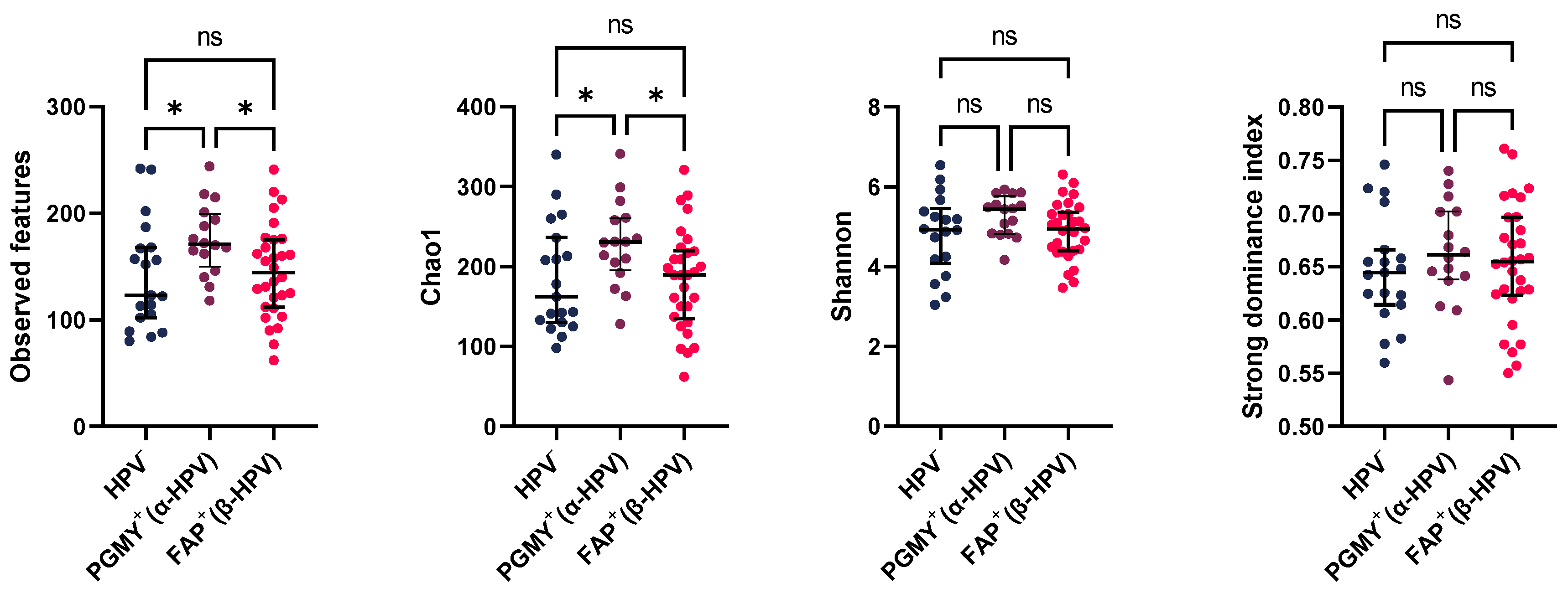
Figure 2.
Beta diversity PCoA plots for unweighted (left) and weighted Unifrac (right) distances in the HPV− (dark blue), FAP+ (purple), and PGMY+ (red) groups.
Figure 2.
Beta diversity PCoA plots for unweighted (left) and weighted Unifrac (right) distances in the HPV− (dark blue), FAP+ (purple), and PGMY+ (red) groups.
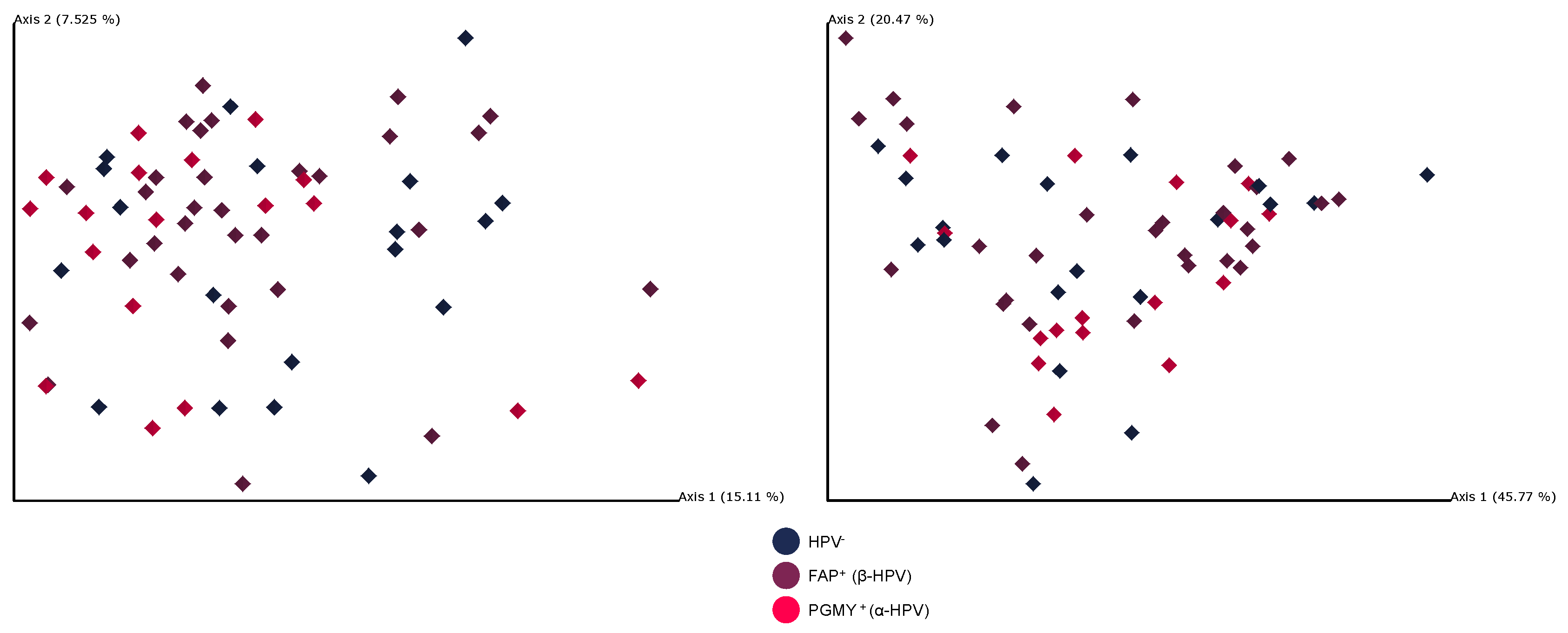
Figure 3.
Unweighted and weighted Unifrac beta diversity distances in comparison to the PGMY+ group. This figure represents the distance given by each evaluated metric. The distances of the PGMY+ group are shown in comparison with itself (PGMY+ vs. PGMY+, in red), against the FAP+ group (purple), and against the HPV− group (blue). A significant distance between the PGMY+ and HPV− groups was observed with the unweighted UniFrac metric, reaching statistical significance. The violin plot shows the median of the distances (thick white dashed line) and interquartile ranges (dotted line). Statistically analyzed with PERMANOVA. ** q = 0.006.
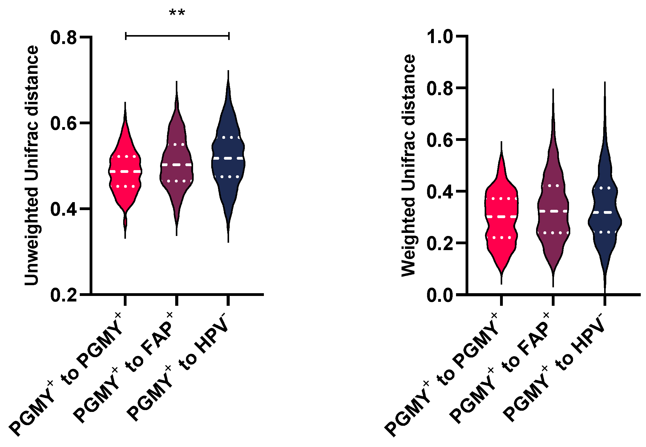
Figure 4.
Relative bacterial abundance across phylum, genus, and species ranges in the three groups. A: Bar chart representing the main phyla in both groups, accompanied by relative abundance percentages. The first five phyla are shown. Minor phyla are summarized in the “other phyla” category. B: Stacked bar plot of bacterial genera in both groups. C: Stacked barplot of the identified bacterial species. The first 25 taxa (ordered by median) are depicted, and key genus and/or species are accompanied by relative abundance percentages. Minor taxa are summarized in the “other taxa” category.
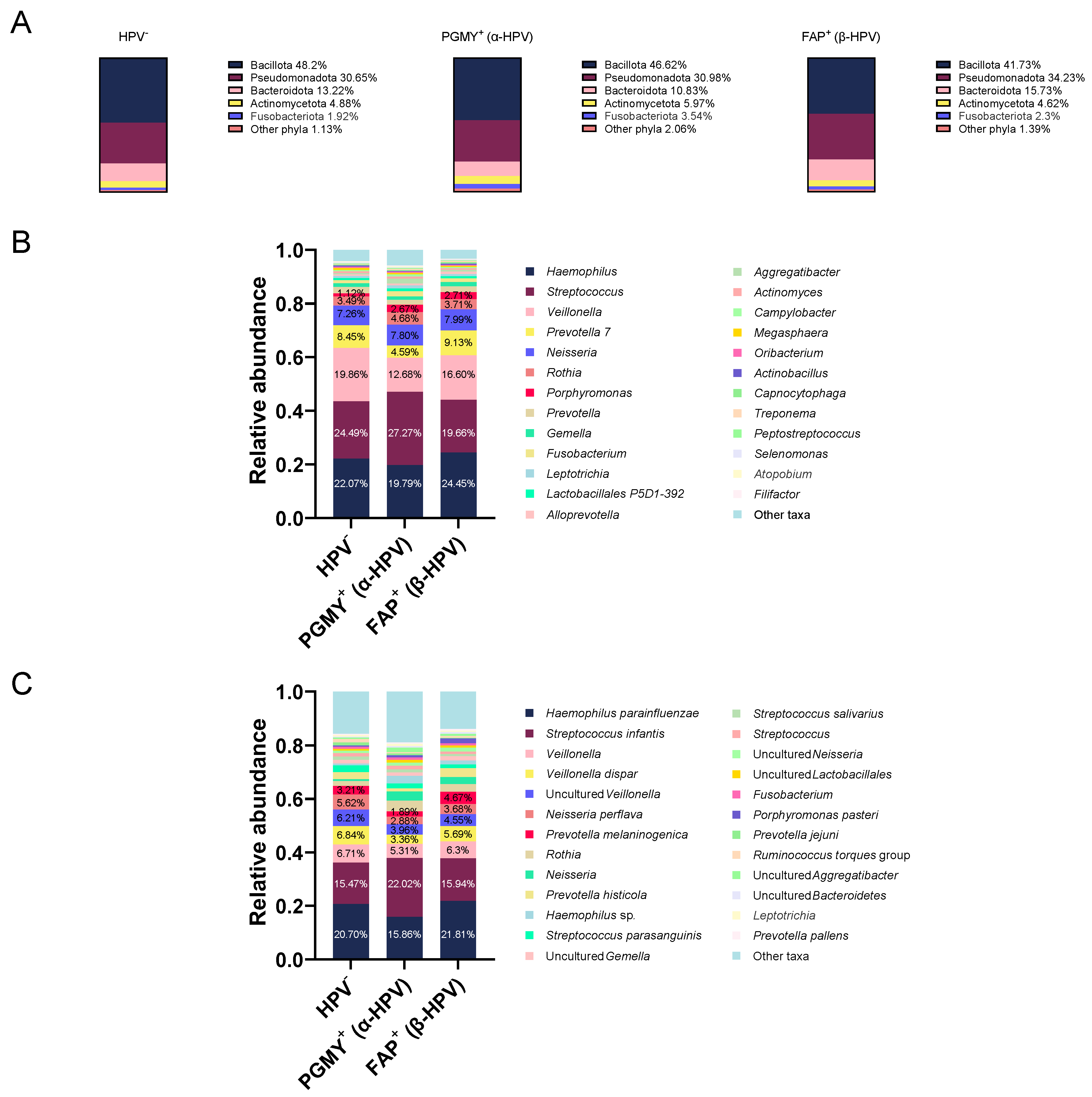
Figure 5.
Dot plots of Pseudomonadota/Bacillota (previously known as Proteobacteria/Firmicutes), Fusobacteriota/Bacillota (Fusobacteria/Firmicutes), Fusobacteriota/Bacteroidota ratios, and Fusobacteria clr-transformed counts in the three groups. The results are presented as median and interquartile range. Analyzed using the Kruskal–Wallis test with Benjamini, Krieger and Yekutieli multiple testing correction (BKY), ns not significant, * q <0.05, ** q < 0.01.
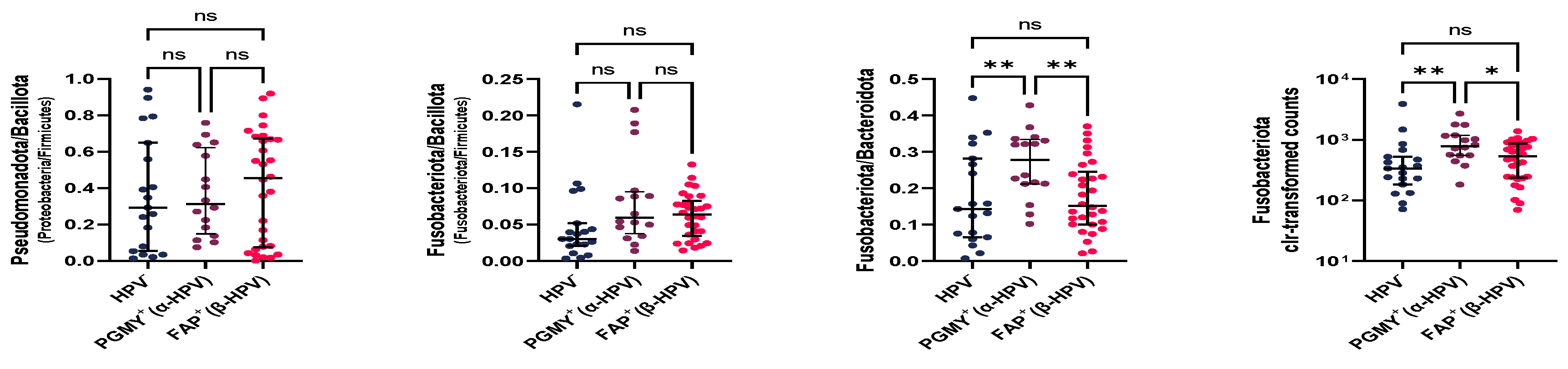
Figure 6.
Differentially abundant taxa in the PGMY+, FAP+, and HPV− groups determined by ANCOM-BC. Bar lengths depict log fold change (LFC) between both groups, and black lines represent standard error. Upper: Comparison between the PGMY+ and FAP+ groups. Red bars indicate taxa enriched in the PGMY+ group, whereas purple bars indicate depleted taxa (Enriched in the FAP+ group). Middle: Comparison between the PGMY+ and HPV- groups. Blue bars indicate taxa enriched in the HPV- group, whereas red bars indicate depleted taxa (Or enriched in the PGMY+ group). Lower: Comparison between the FAP+ and HPV− groups. Blue bars indicate taxa enriched in the HPV− group, whereas purple bars indicate depleted taxa (Or enriched in the FAP+ group). A threshold of LFC ≥ |1.0| was used. * = q < 0.05, ** = q < 0.01, *** = q < 0.001.
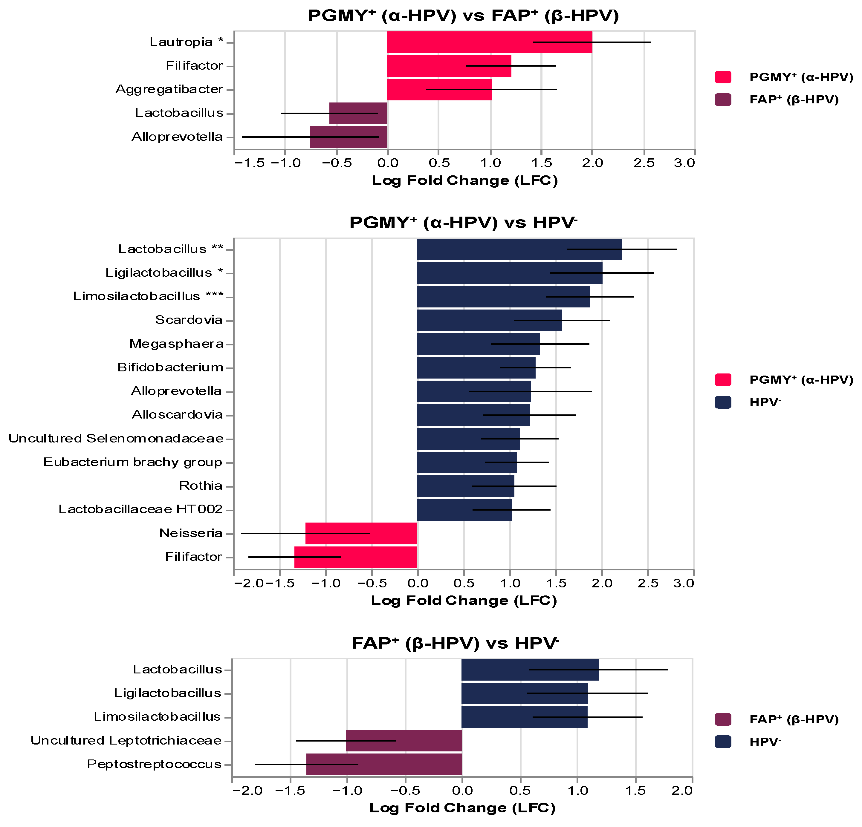
Figure 7.
Differentially enriched metabolic pathways in the oropharyngeal microbiome predicted by PICRUSt2 in the three groups, analyzed using ANCOM-BC. Upper: Comparison between the PGMY+ and FAP+ groups. The red bars indicate the pathways enriched in the PGMY+ group. No significant pathways were identified in the FAP+ group. Middle: Comparison between the PGMY+ and HPV− groups. Blue bars indicate taxa enriched in the HPV− group, whereas red bars indicate depleted taxa (or enriched in the PGMY+ group). Lower: Comparison between the FAP+ and HPV− groups. The purple bar represents the single pathway enriched in the FAP+ group. A threshold of LFC ≥ |1.0| was applied. * = q < 0.05, ** = q < 0.01, **** = q < 0.0001.
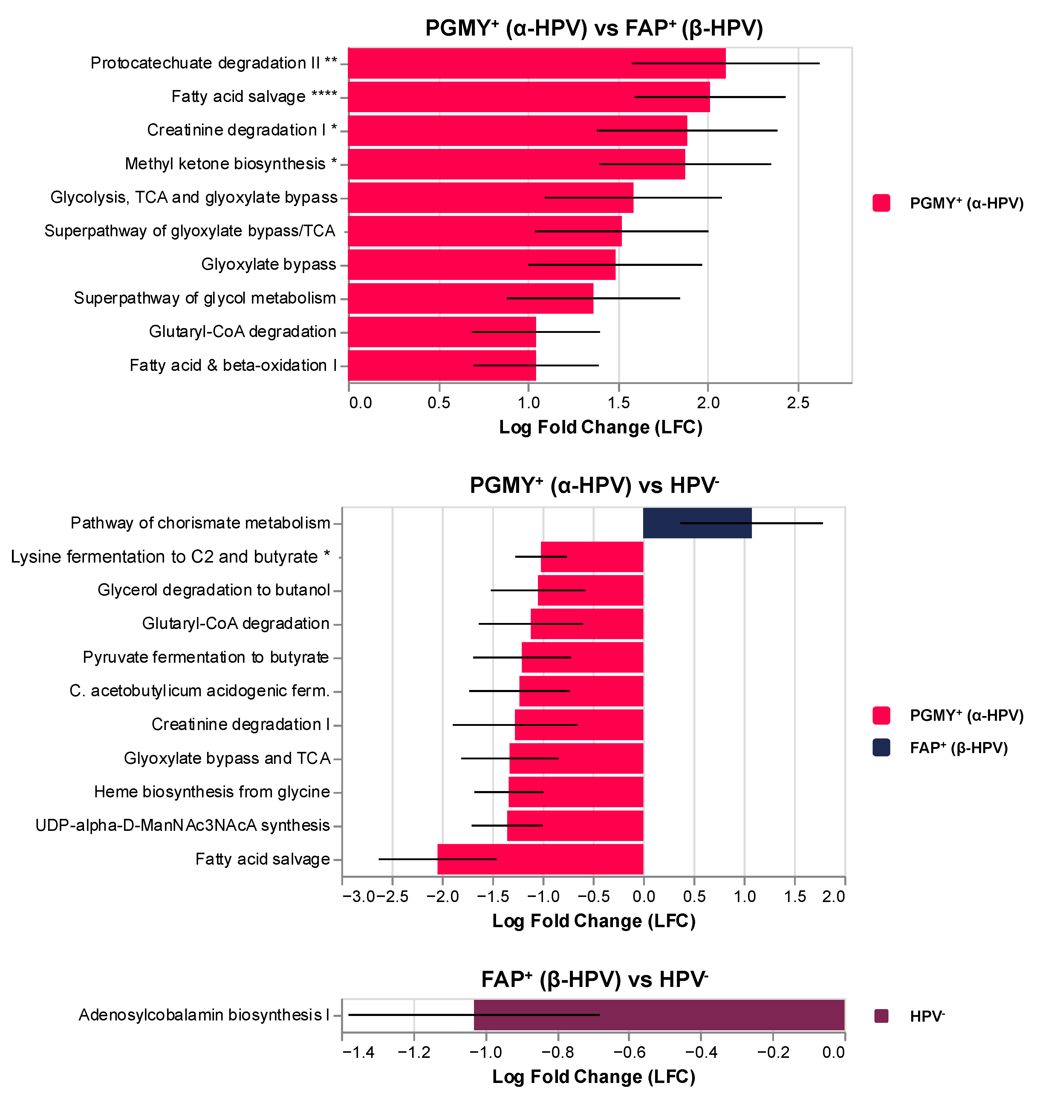

Table 1.
Sociodemographic and clinical characteristics of the recruited participants.
| HPV− n=19 |
PGMY+ (α19. | FAP+ (β-HPV) n=30 |
p | |
|---|---|---|---|---|
| Age (years) | 36.0 (29.0-42.0) | 39.5 (35.5-46.0) | 41.0 (33.5-49.0) | 0.109 b |
| Height (m) | 1.73 ± 0.07 | 1.76 ± 0.05 | 1.72 ± 0.07 | 0.193 a |
| Weight (kg) | 73.26 ± 15.90 | 81.0 ± 9.42 | 77.97 ± 12.14 | 0.234 a |
| BMI (kg/m2) | 23.3 (19.60-26.75) | 24.90 (24.01-27.55) | 26.37 (23.51-29.31) | 0.080 b |
| Periodontal diseases n (%) | 19 (100) | 16 (100) | 30 (100) | 1.000 c |
| Gingivitis n (%) | 17 (89.5) | 16 (100) | 29 (96.7) | 0.443 c |
| Periodontitis n (%) | 2 (10.5) | 0 (0) | 1 (3.3) | |
| Oral lesions‡ n (%) | 1 (5.3) | 3 (18.8) | 3 (10) | 0.458 c |
| Current smoking n (%) | 10 (52.6) | 6 (37.5) | 15 (50) | 0.658 c |
| Current alcohol consumption n (%) | 9 (47.4) | 10 (62.5) | 19 (63.3) | 0.524 c |
| Illicit drug misuse n (%) | 6 (31.6) | 4 (25) | 14 (46.7) | 0.321 c |
| Anal and/or genital HPV lesions n (%) | 9 (47.4) | 3 (18.8) | 11 (36.7) | 0.223 c |
| Absolute T CD4+ (cell count/µL) | 716 (396-863) | 666 (479.5-1009.5) | 618 (443.5-820) | 0.733 b |
| Nadir T CD4+ (cell count/µL) | 337.5 (106.5-427.75) | 223.5 (43.25-418.5) | 135 (40-399.50) | 0.712 b |
| CD4+/CD8+ ratio | 0.37 (0.132-0.965) | 0.11 (0.07-0.35) | 0.14 (0.055-0.7054) | 0.310 b |
| Time since HIV diagnosis (years) | 6 (4-8) | 4.5 (3-8) | 7 (2.5-7) | 0.830 b |
| Duration of ART (years) | 5 (2.5-8) | 6 (4-8.5) | 7 (3-10) | 0.628 b |
| Time of INSTI use (years) | 4.3 (3.4-5.5) | 5.4 (4.325-5.65) | 4.7 (2.8-5.75) | 0.398 b |
Abbreviations: INSTI, integrase strand-transfer inhibitor group; NA, not applicable. ‡ Oral lesions consisted of four benign leukoplakias and three oral squamous papilloma lesions. Data are presented as mean ± standard deviation or median with interquartile range in parentheses. P-values were calculated by (a) One-way ANOVA, (b) Kruskal–Wallis, (c) Fisher’s exact test.
Table 2.
PERMANOVA analysis results for unweighted and weighted Unifrac distances between the three study groups.
Table 2.
PERMANOVA analysis results for unweighted and weighted Unifrac distances between the three study groups.
| Group 1 | Group 2 | Pseudo-F | q-value |
|---|---|---|---|
| PGMY+ (α) | FAP+ (β) | 1.332 | 0.102 |
| HPV− | 2.056 | 0.006 | |
| FAP+ (β) | HPV− | 1.065 | 0.336 |
| PGMY+ (α) | FAP+ (β) | 1.020 | 0.576 |
| HPV− | 1.250 | 0.576 | |
| FAP+ (β) | HPV− | 0.596 | 0.620 |
Disclaimer/Publisher’s Note: The statements, opinions and data contained in all publications are solely those of the individual author(s) and contributor(s) and not of MDPI and/or the editor(s). MDPI and/or the editor(s) disclaim responsibility for any injury to people or property resulting from any ideas, methods, instructions or products referred to in the content. |
© 2026 by the authors. Licensee MDPI, Basel, Switzerland. This article is an open access article distributed under the terms and conditions of the Creative Commons Attribution (CC BY) license (http://creativecommons.org/licenses/by/4.0/).
Copyright: This open access article is published under a Creative Commons CC BY 4.0 license, which permit the free download, distribution, and reuse, provided that the author and preprint are cited in any reuse.