
Submitted:
16 September 2026
Posted:
17 September 2026
You are already at the latest version
Abstract
Spaceflight (FLT) is associated with fluid redistribution, cardiovascular deconditioning, and altered vascular tone. We sought to determine whether FLT induces molecular alterations in human aortic smooth muscle cells (HASMCs) that could provide a mechanistic basis for altered vascular function. We analyzed NASA GeneLab/Open Science Data Repository dataset OSD-635, comprising mRNA sequencing of primary HASMCs cultured for 3 days aboard the International Space Station compared with ground controls. Up- and downregulated differentially expressed genes (DEGs), together with genes ranked by a composite significance-magnitude pi-score, overlapped with GSE225884, a transcriptomic readout of HASMCs exposed to lactacidosis. The gene intersection contained 182 genes shared in the upregulated direction and 382 shared in the downregulated direction for DEG comparisons; corresponding pi-score intersections contained 170 and 180 genes, respectively. Convergent upregulated genes included glucose/glycogen-associated genes, PYGL, PYGM, GCK, ALDOC and PGM2L1, and fatty-acid/mitochondrial metabolism genes ACADS, HADH, IDH2 and LDHD. Multiple genes involved in mitochondrial oxidative phosphorylation and maintenance were downregulated including NDUFAF8, NDUFC2, NDUFS6, COQ2, UQCC4, TIMM23, MFN2 and ACO2. Additional robustness was observed because in the FLT study 98 genes were shared between the DEG-up and pi-score-up intersections and 127 between the DEG-down and pi-score-down intersections. FLT-exposed HASMCs exhibit a lactacidosis-associated transcriptomic remodeling which may contribute to vascular dysfunction.
Keywords:
FLT
; microgravity
; vascular
; dysfunction
; transcriptomics
; energy metabolism
; mitochondria
; lactacidosis
Introduction
Human FLT is entering a historic transition toward extended exploration missions to the Moon and Mars. During these missions, astronauts are exposed to a complex milieu of environmental stressors including microgravity, radiation, altered circadian cycles, and confined ecological life-support systems. Prolonged exposure to microgravity induces profound cephalad fluid shifts, cardiovascular deconditioning, bone loss, and metabolic remodeling. Indeed, FLT induces a cardiovascular deconditioning phenotype that includes fluid redistribution, reduced plasma volume, altered vascular regulation, and impaired orthostatic tolerance after return to Earth [1,2].
Although hemodynamic effects of FLT are now well characterized, the contribution of molecular remodeling within vascular smooth muscle remains incompletely defined. Indeed, vascular smooth muscle cells (VSMCs) are central regulators of arterial tone through coordinated control of the contractile apparatus, intracellular calcium handling, cellular metabolism, extracellular matrix interactions, and responses to vasoactive signaling [3,4]. Changes in VSMC phenotype or energetic state can therefore alter vascular resistance and blood pressure. Recent RNA-seq analysis of human aortic smooth muscle cells (HASMCs) exposed to FLT identified transcriptomic alterations involving contractile and extracellular matrix signaling [2]. These observations provide evidence that vascular smooth muscle responds directly to the space environment rather than being affected solely by systemic hemodynamic changes.
We analyzed FLT-exposed HASMCs, a type of VSMCs, to determine whether transcriptomic remodeling informs, at least, in part, the vascular responses observed in astronauts.
Methods
Transcriptomic Data
Data were obtained from NASA GeneLab/Open Science Data Repository accession OSD-635, comprising bulk RNA sequencing of primary HASMCs (n=3) cultured for 3 days aboard the ISS and corresponding ground controls (n=3) [5]. Up- and downregulated DEGs were identified using adjusted p-value (padj; Benjamini–Hochberg [B-H]) <0.05 [6]. To complement DEG-based thresholding, FLT genes were ranked using the pi-score defined as log2(fold-change) × −log(p-value) [7]. Genes meeting an absolute pi-score ≥4 were included as investigator-defined pi-score up or down genes.
Pathway Activation Signature
Upregulated and downregulated DEGs were submitted to RummaGeo [8] to identify Gene Ontology Biological Process (GOBP) vascular disease-associated transcriptomic signatures based on concordance and gene similarity relative to existing NCBI transcriptomic datasets. Additionally, pi-score-ranked genes were also evaluated for overlap against NCBI transcriptomic using RummaGEO. GSE225884 comprises primary HASMCs exposed to lactacidosis at pH 6.8 for 48 hours compared with pH 7.4 controls [9].
Data Visualization and Analysis
Principal component analysis and heat maps were generated using iDEP [10], while volcano plots and gene-set operations were performed in Microsoft Excel. The analysis used two-tailed Welch’s t-test procedures for group comparisons with B-H correction for DEG identification. A padj <0.05 was deemed statistically significant.
Results
FLT was associated with broad transcriptional changes (Figure 1A and B) with PCA analysis exhibiting separation between FLT and ground control cohorts (Figure 1C).
A total of 1,442 upregulated and 1,961 downregulated DEGs were identified in the supplied analysis (Figure 2A). Submitting the FLT upregulated DEGs to RummaGEO returned a highly significant (padj = 5.28e-74) and concordant 182-gene overlap with GSE225884/lactacidosis (Figure 2B). Common upregulated genes between FLT and lactacidosis included the glycogen/glucose metabolism genes PYGL, PYGM, GCK, ALDOC, and PGM2L1, as well as fatty-acid/mitochondrial oxidation genes HADH, ACADS, and IDH2. The corresponding FLT downregulated analysis returned a highly significant (padj = 6.86e-150) and concordant 382-gene overlap with lactacidosis (Figure 2C). The shared downregulated genes showed a coherent mitochondrial pattern. These included NDUFAF8, NDUFC2, NDUFS6, COQ2, UQCC4, TIMM23, MFN2, ACO2, AIFM1, BOLA3, MRPL54, MRPS7, LARS2, and ATPSCKMT. Collectively, these genes represent electron-transport/oxidative-phosphorylation machinery, coenzyme-Q biology, mitochondrial protein import, mitochondrial dynamics, tricarboxylic-acid-cycle activity, and mitochondrial protein synthesis or maintenance. This broad representation supports a coordinated mitochondrial remodeling signal rather than dependence on a single gene.
The pi-score analysis (Figure 3A) provided an independent ranking strategy that reproduced the direction of the metabolic signal. Of the 170 genes shared between FLT pi-score genes and lactacidosis DEGs in the upregulated direction (Figure 3B), representative genes included ALDOC, GCK, ACADS, IDH2, and LDHD. Of the 180 genes shared in the downregulated direction (Figure 3C), NDUFAF8, NDUFS6, COQ2, and ATPSCKMT were among the recurrent downregulated mitochondrial genes.
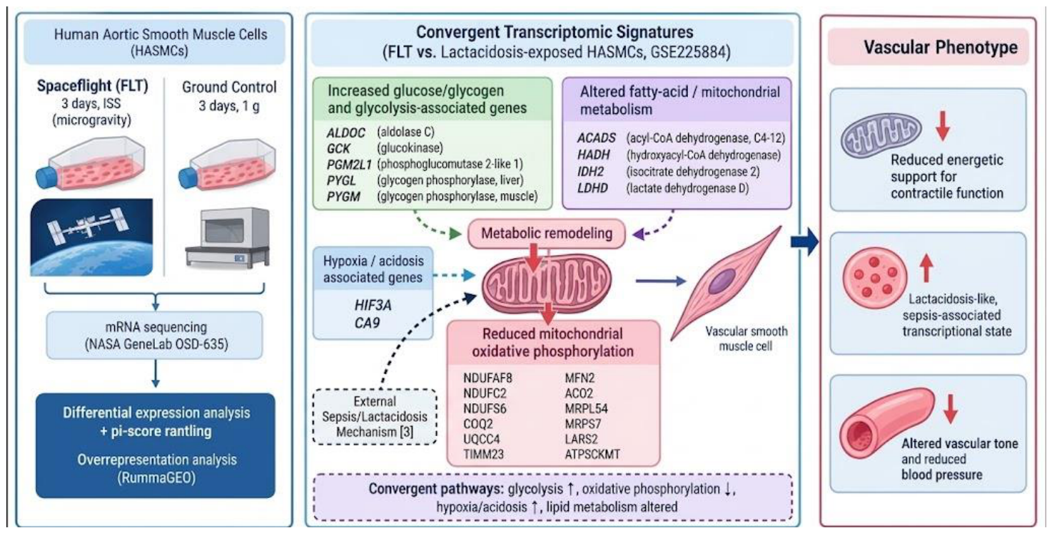
Interestingly, of the 182 FLT DEG-up genes shared with lactacidosis, 98 were also present in the pi-score-up intersection. Among the 382 FLT DEG-down genes shared with lactacidosis, 127 were also present in the pi-score-down intersection. This recurrence supports the persistence of the metabolic signal when gene selection is based on statistical significance alone versus a combined significance-and-effect-size ranking. Taken together, the convergent pattern is characterized by three linked features: remodeling of glucose and glycogen metabolism, altered fatty-acid and mitochondrial carbon metabolism, and reduced expression of multiple genes required for mitochondrial oxidative phosphorylation and maintenance. The combination is consistent with a metabolically stressed or remodeled cellular state. Because the lactacidosis comparator study [9] demonstrated that combined acidosis and lactate can impair glycolysis and glutaminolysis, alter mitochondrial respiration, reduce ATP, and change VSMC phenotype through mechanisms involving intracellular acidification and NAD+/NADH imbalance, the shared transcriptomic architecture provides a plausible molecular bridge between FLT and vascular dysfunction (Figure 4).
Discussion
The principal finding of this study is that HASMCs exposed to FLT exhibit transcriptomic convergence with a lactacidosis-associated state. This convergence was observed in both directions of gene regulation and was reproduced using conventional DEG intersections and pi-score-based ranking. The strongest biological coherence was seen in cellular metabolism. Glucose/glycogen-associated genes were represented among shared upregulated genes, whereas a broad group of mitochondrial oxidative-phosphorylation and mitochondrial-maintenance genes was represented among shared downregulated genes.
FLT-associated vascular deconditioning has traditionally been explained primarily through altered hydrostatic loading, fluid shifts, plasma-volume changes, autonomic adaptation, and vascular remodeling [2,3,11,12,13,14]. This result is biologically relevant because vascular smooth muscle is an energetically active tissue whose ability to maintain vascular tone depends on ATP-dependent contractile processes, calcium handling, and coordinated signaling. The present analysis suggests direct metabolic transcriptomic remodeling within VSMCs may contribute to the cellular phenotype underlying altered vascular tone. The observed mitochondrial signature is particularly notable [15]. NDUFAF8, NDUFC2, and NDUFS6 are associated with respiratory-chain complex I biology; COQ2 and UQCC4 participate in coenzyme-Q/respiratory-chain function; TIMM23 is involved in mitochondrial protein import; MFN2 regulates mitochondrial dynamics; and ACO2 participates in the tricarboxylic acid cycle. The recurrence of these genes across the lactacidosis-associated intersections suggests that the convergence is not driven by a single metabolic gene but rather by multiple components of mitochondrial energy metabolism. This interpretation is consistent with experimental work showing that lactacidosis in HASMCs alters mitochondrial respiration and reduces cellular ATP [9].
The upregulated component of the signature is equally informative. PYGL, PYGM, GCK, ALDOC, and PGM2L1 point to remodeling of glucose and glycogen handling, while ACADS, HADH, IDH2, and LDHD indicate altered mitochondrial and lipid-associated metabolism [9]. These changes should not be interpreted as evidence of increased glycolytic flux. Transcript abundance cannot establish metabolic flux, and the lactacidosis comparator study [9] demonstrated that lactacidosis can inhibit glycolysis and glutaminolysis while perturbing mitochondrial respiration and reducing ATP. Thus, the most defensible interpretation is metabolic reprogramming or remodeling rather than a straightforward increase in glycolysis.
These observations fit with prior experimental work [3] demonstrating that FLT alters HASMC phenotype and signaling. Scotti et al [3] reported broad changes in contractile, synthetic, and osteogenic programs, together with alterations in STAT3, NFκB, PI3K/AKT, HIF1α, and endothelin-associated signaling. The present analysis extends that work by identifying a specific cross-study transcriptomic resemblance to a lactacidosis-associated vascular phenotype. Importantly, Terpe et al [9] provide a mechanistic context for the sepsis connection: in primary human aortic VSMCs, lactacidosis produced greater metabolic and transcriptomic disruption than acidosis or lactate alone, with reduced intracellular NAD+/NADH ratio, impaired glycolysis and glutaminolysis, altered mitochondrial respiration, reduced ATP, and phenotypic changes that may contribute to vasoplegia and hypotension in sepsis. Our findings do not demonstrate that FLT reproduces this pathway in full, but they identify transcriptional features that justify testing whether related metabolic stress mechanisms occur during FLT.
One potential implication is that reduced mitochondrial energetic capacity could diminish the ability of VSMCs to sustain contractile work or respond appropriately to vasoactive signals. This is potentially relevant to both FLT-associated vascular deconditioning and the broader biology of vasoplegia. In sepsis, lactacidosis has been linked experimentally to VSMC energetic and phenotypic impairment, and Terpe et al [9] identified the combined effects of intracellular acidosis and reduced NAD+/NADH ratio as an important determinant of this response. A similar biochemical mechanism cannot be inferred from the present transcriptomic data, but the observed mitochondrial and metabolic convergence provides a testable hypothesis: FLT-associated metabolic remodeling may reduce the energetic reserve of VSMCs and thereby contribute to impaired vascular responsiveness. Because vascular tone is ultimately a functional phenotype, this provides a plausible link between transcriptomic remodeling and altered vascular resistance or blood-pressure regulation documented during and after FLT [2,13,14].
Several limitations should be considered. First, this is a secondary analysis of existing transcriptomic datasets rather than a prospective experiment designed to test lactacidosis as a mediator of FLT effects. Second, the two datasets differ in exposure conditions: OSD-635 represents 3 days of FLT exposure, whereas GSE225884 represents 48 hours of experimentally imposed lactacidosis [9]. Third, FLT includes the broader space environment, and transcriptomic effects cannot be attributed exclusively to microgravity because radiation and other flight-associated factors may contribute [1]. Fourth, the analysis compares gene expression rather than metabolites, protein abundance, enzyme activity, or cellular function. Finally, cross-study gene-set overlap can reflect shared generic stress responses as well as specific pathway convergence. In particular, the present study did not measure lactate concentration, intracellular pH, NAD+/NADH ratio, glycolytic or glutaminolytic flux, ATP, mitochondrial respiration, or vasomotor function; therefore, the sepsis-related mechanism described by Terpe et al [9] should be viewed as biological context and a hypothesis for future testing, not as a demonstrated mechanism of FLT.
These findings stimulate several experimentally testable directions. First, FLT-exposed HASMCs should be evaluated for extracellular and intracellular lactate, intracellular pH, NAD+/NADH ratio, oxygen consumption rate, extracellular acidification rate, ATP, and mitochondrial membrane potential. Second, functional studies should determine whether the transcriptional signature is accompanied by altered calcium handling and contractile responses to vasoconstrictors and vasodilators. Third, targeted perturbation of candidate metabolic nodes, including mitochondrial respiration, glucose/glycogen pathways, TXNIP signaling, and NAD+/NADH metabolism, could test whether the metabolic program is causal for the VSMC phenotype. Fourth, experiments that separately manipulate extracellular pH and lactate would be particularly informative because the sepsis study [9] indicates that their combination can produce stronger effects than either factor alone. Finally, replication in independent flight or simulated-microgravity datasets and integration with proteomics and metabolomics would help distinguish a true lactacidosis-related mechanism from a broader stress-response signature.
Conclusions
HASMCs exposed to FLT exhibit substantial transcriptomic convergence with a lactacidosis-associated HASMC state. The convergence is characterized by remodeling of glucose/glycogen metabolism, altered fatty-acid and mitochondrial metabolism, and reduced expression of multiple mitochondrial oxidative-phosphorylation and maintenance genes. Experimental work in human VSMCs has demonstrated that lactacidosis relevant to sepsis can produce intracellular acidification, a reduced NAD+/NADH ratio, impaired glycolysis and glutaminolysis, mitochondrial dysfunction, ATP depletion, and phenotypic remodeling. These findings therefore support a testable hypothesis that metabolic remodeling of vascular smooth muscle may contribute to FLT-associated vascular dysfunction.
References
- Afshinnekoo E, Scott RT, MacKay MJ, et al. Fundamental biological features of spaceflight: advancing the field to enable deep-space exploration. Cell. 2020;183(5):1162-1184. [CrossRef]
- Baran R, Marchal S, Garcia Campos S, Rehnberg E, Tabury K, Baselet B, Wehland M, Grimm D, Baatout S. The cardiovascular system in space: focus on in vivo and in vitro studies. Biomedicines. 2022;10(1):59. [CrossRef]
- Scotti MM, Wilson BK, Bubenik JL, Yu F, Swanson MS, Allen JB. Spaceflight effects on human vascular smooth muscle cell phenotype and function. npj Microgravity. 2024;10:41. [CrossRef]
- Ludtka C, Allen JB. The effects of simulated and real microgravity on vascular smooth muscle cells. Gravit Space Res. 2024;12(1):46-59. [CrossRef]
- NASA GeneLab/Open Science Data Repository. OSD-635: FLT effects on human vascular smooth muscle cell phenotype and function. NASA OSDR Biological Data and Visualization Portal. Accessed September 11, 2026. [CrossRef]
- Benjamini Y, Hochberg Y. Controlling the false discovery rate: a practical and powerful approach to multiple testing. J R Stat Soc Series B Stat Methodol. 1995;57(1):289-300.
- Xiao Y, Hsiao TH, Suresh U, Chen HI, Wu X, Wolf SE, Chen Y. A novel significance score for gene selection and ranking. Bioinformatics. 2014 Mar 15;30(6):801-7. Epub 2012 Feb 9. PMID: 22321699. [CrossRef]
- Marino GB, Clarke DJB, Lachmann A, Deng EZ, Ma’ayan A. RummaGEO: Automatic mining of human and mouse gene sets from GEO. Patterns (N Y). 2024 Oct 11;5(10):101072. PMID: 39569206. [CrossRef]
- Terpe P, Ruhs S, Dubourg V, Bucher M, Gekle M. The synergism of cytosolic acidosis and reduced NAD+/NADH ratio is responsible for lactic acidosis-induced vascular smooth muscle cell impairment in sepsis. J Biomed Sci. 2024;31:3. [CrossRef]
- Ge SX, Son EW, Yao R. iDEP: an integrated web application for differential expression and pathway analysis of RNA-Seq data. BMC Bioinformatics. 2018;19:534. [CrossRef]
- Scott JM, Stoudemire J, Dolan LB, Downs M. Leveraging spaceflight to advance cardiovascular research on Earth. Circ Res. 2022;130(6):942-957. [CrossRef]
- Gallo C, Ridolfi L, Scarsoglio S. Cardiovascular deconditioning during long-term spaceflight through multiscale modeling. npj Microgravity. 2020;6:27. [CrossRef]
- Hargens AR, Richardson S. Cardiovascular adaptations, fluid shifts, and countermeasures related to space flight. Respir Physiol Neurobiol. 2009;169 Suppl 1:S30-S33. [CrossRef]
- Vernice NA, Meydan C, Afshinnekoo E, et al. Long-term spaceflight and the cardiovascular system. Precis Clin Med. 2020;3(4):284-291. [CrossRef]
- Nguyen HP, Tran PH, Kim KS, Yang SG. The effects of real and simulated microgravity on cellular mitochondrial function. npj Microgravity. 2021;7:44. [CrossRef]
Figure 1.
FLT-associated transcriptomic changes. (A) Volcano plot showing up (red)- and downregulated (blue) genes in FLT vs. a ground control cohort. (B) Heatmap representation of FLT genes. (C) The PCA plot shows separation between the two cohorts.
Figure 1.
FLT-associated transcriptomic changes. (A) Volcano plot showing up (red)- and downregulated (blue) genes in FLT vs. a ground control cohort. (B) Heatmap representation of FLT genes. (C) The PCA plot shows separation between the two cohorts.
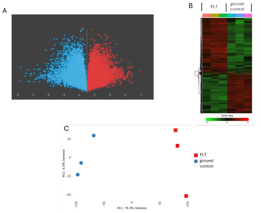
Figure 2.
DEGs associated with FLT. (A) Volcano plot showing up (red)- and downregulated (blue) DEGs in FLT-exposed HASMCs. B) Overlap of FLT-upregulated DEGs with HASMC lactacidosis-associated DEGs. C) Overlap of FLT-downregulated DEGs with HASMC lactacidosis-associated DEGs.
Figure 2.
DEGs associated with FLT. (A) Volcano plot showing up (red)- and downregulated (blue) DEGs in FLT-exposed HASMCs. B) Overlap of FLT-upregulated DEGs with HASMC lactacidosis-associated DEGs. C) Overlap of FLT-downregulated DEGs with HASMC lactacidosis-associated DEGs.
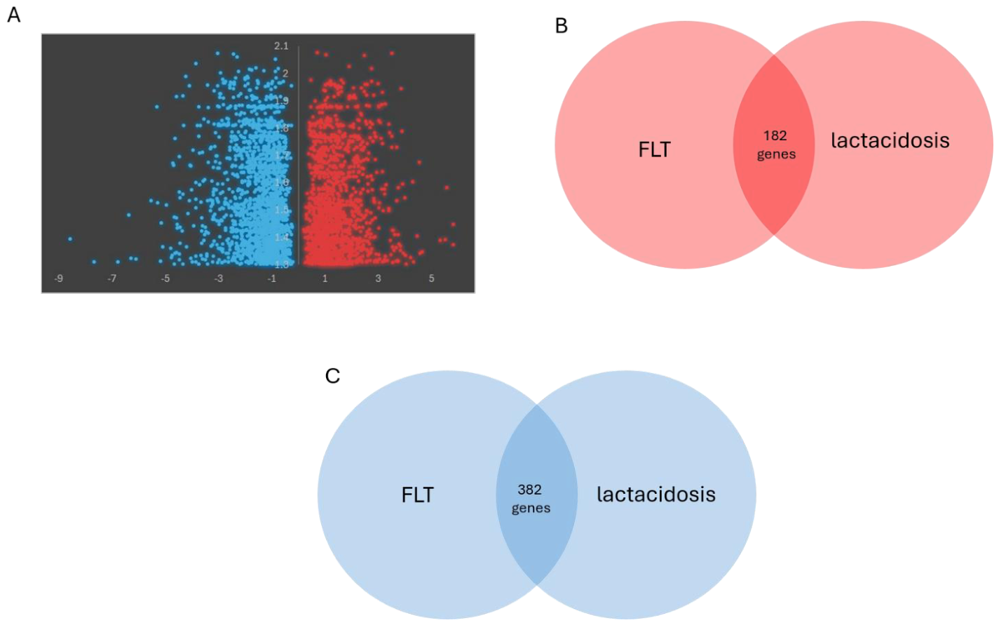
Figure 3.
Pi-score genes associated with FLT. (A) Inverted sigmoid plot of investigator-defined pi-scored genes. B) Overlap of FLT-upregulated pi-score genes with HASMC lactacidosis-associated DEGs. C) Overlap of FLT-downregulated pi score genes with HASMC lactacidosis-associated DEGs.
Figure 3.
Pi-score genes associated with FLT. (A) Inverted sigmoid plot of investigator-defined pi-scored genes. B) Overlap of FLT-upregulated pi-score genes with HASMC lactacidosis-associated DEGs. C) Overlap of FLT-downregulated pi score genes with HASMC lactacidosis-associated DEGs.
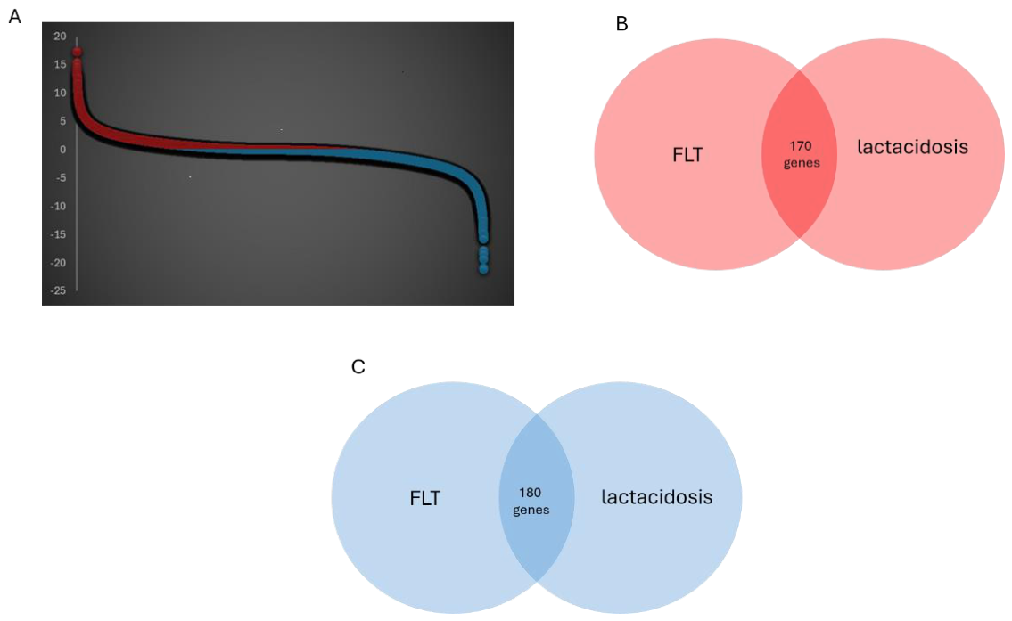
Figure 4.
Proposed metabolic pathway linking FLT-associated transcriptomic remodeling to vascular smooth muscle dysfunction. Solid arrows represent findings or biologically supported relationships derived from the present analysis; the dashed sepsis/lactacidosis branch represents mechanistic precedent from Terpe et al [9]. and a testable hypothesis, not a demonstrated mechanism of FLT.
Figure 4.
Proposed metabolic pathway linking FLT-associated transcriptomic remodeling to vascular smooth muscle dysfunction. Solid arrows represent findings or biologically supported relationships derived from the present analysis; the dashed sepsis/lactacidosis branch represents mechanistic precedent from Terpe et al [9]. and a testable hypothesis, not a demonstrated mechanism of FLT.
Disclaimer/Publisher’s Note: The statements, opinions and data contained in all publications are solely those of the individual author(s) and contributor(s) and not of MDPI and/or the editor(s). MDPI and/or the editor(s) disclaim responsibility for any injury to people or property resulting from any ideas, methods, instructions or products referred to in the content. |
© 2026 by the authors. Licensee MDPI, Basel, Switzerland. This article is an open access article distributed under the terms and conditions of the Creative Commons Attribution (CC BY) license (http://creativecommons.org/licenses/by/4.0/).
Copyright: This open access article is published under a Creative Commons CC BY 4.0 license, which permit the free download, distribution, and reuse, provided that the author and preprint are cited in any reuse.