
Submitted:
09 September 2026
Posted:
17 September 2026
You are already at the latest version
Abstract
Arthrogryposis–renal dysfunction–cholestasis (ARC) syndrome is a rare, usually fatal autosomal recessive multisystem disorder caused by biallelic mutations in VPS33B or VIPAS39, which encode components of a vesicular trafficking complex required for organelle biogenesis. Loss of function causes a platelet α-granule deficiency and a severe bleeding diathesis, and no curative therapy exists. Building on the efficacy of ex vivo lentiviral gene therapy in inherited haematological disorders, we evaluated haematopoietic stem cell (HSC) gene delivery in a tamoxifen-inducible Vps33b knockout mouse model (Vps33bfl/fl-CreERT2). Knockout mice tolerated myeloablative conditioning and supported efficient donor reconstitution after transplantation of healthy lineage-negative cells. We then transduced lineage-negative cells from knockout donors ex vivo with a third-generation lentiviral vector encoding codon-optimised human VPS33B under an elongation factor 1α short (EFS) promoter and transplanted them into conditioned knockout recipients. Transmission electron microscopy showed that platelet α-granule numbers were restored to wild-type levels after both bone marrow transplantation and ex vivo gene therapy. These findings demonstrate the feasibility of haematopoietic VPS33B gene delivery and support further preclinical evaluation of gene therapy for the bleeding phenotype of ARC syndrome.
Keywords:
ARC syndrome
; VPS33B
; platelet α-granules
; haematopoietic stem cell gene therapy
; lentiviral vector
; storage pool disorder
; thrombopoiesis
; rare disease
1. Introduction
Arthrogryposis–renal dysfunction–cholestasis (ARC) syndrome is a severe autosomal recessive multisystem disorder characterised by progressive cholestatic liver disease, renal tubular dysfunction, congenital arthrogryposis, ichthyosis, and a haematological phenotype that combines platelet dysfunction with susceptibility to severe infection [1,2,3]. Most affected children die in the first year of life, commonly from gastrointestinal haemorrhage or infection, and no disease-specific therapy exists [1]. ARC syndrome is caused by biallelic loss-of-function mutations in VPS33B or VIPAS39, encoding VPS33B and VIPAR (also termed VPS16B), which form a cytosolic complex that regulates SNARE-dependent vesicular trafficking required for the biogenesis of specialised organelles in epithelia, megakaryocytes and immune cells [1,2,4,5,6,7,16]. The bleeding tendency reflects a profound platelet storage-pool disorder caused by defective α-granule biogenesis in megakaryocytes, with markedly impaired platelet aggregation [4,9]. Importantly, individuals with hypomorphic VPS33B alleles exhibit attenuated phenotypes [2], indicating that even partial restoration of VPS33B function may be beneficial.
Ex vivo lentiviral transduction of autologous haematopoietic stem and progenitor cells (HSPCs) has produced durable clinical benefit across an expanding range of inherited haematological and immunological disorders [10,11,12,13,14], enabling stable transgene integration without the alloimmune complications of allogeneic transplantation. In parallel work, we have developed liver-directed in vivo lentiviral gene therapy that corrects the hepatic phenotype of ARC syndrome [17], addressing a disease compartment distinct from the haematopoietic defect considered here. In the present study we tested whether ex vivo haematopoietic gene therapy could increase platelet α-granule number in a tamoxifen-inducible Vps33b knockout mouse model (Vps33bfl/fl-CreERT2), evaluating the feasibility of haematopoietic transplantation and ex vivo VPS33B gene delivery.
2. Results
Because haematopoietic stem cell transplantation (HSCT) had not previously been evaluated in the ARC murine model, we first tested whether tamoxifen-induced knockout (KO) mice tolerate myeloablation. Adult KO mice received split-dose total body irradiation (6 + 5 Gy) followed by intravenous transplantation of 0.5×10⁶ lineage-negative (Lin⁻) cells from healthy male donors (Figure 1A). All recipients (n = 3) survived the early post-transplant period and engrafted efficiently, with donor-derived chimerism approaching 100% in peripheral blood (Figure 1B). Spleen weights trended lower after HSCT but remained elevated relative to wild-type (WT), as previously reported [9] (Figure 1C). These data establish the feasibility of haematopoietic transplantation in this model and provided the basis for the gene therapy experiments.
We next transduced Lin⁻ cells from KO donors ex vivo with a third-generation, VSV-G–pseudotyped, self-inactivating lentiviral vector encoding codon-optimised human VPS33B under the EFS promoter (EFS.cohVPS33B), and transplanted them into conditioned KO recipients (Figure 2A,B). At six weeks, gene therapy (LV-GT) recipients showed near-complete donor chimerism in bone marrow (Figure 2C) and stable gene marking (mean vector copy number ≈ 0.4 vg/dg; Figure 2D). Codon-optimised VPS33B transcript was readily detected in bone marrow from LV-GT recipients but not in WT controls, confirming transgene expression (Figure 2E). Spleen weight remained elevated relative to WT (Figure 2F). Serum procalcitonin, markedly raised in untreated KO mice (p = 0.0002 vs WT), was not significantly different from WT after gene therapy (p = 0.10), consistent with partial correction of the systemic inflammatory phenotype (Figure 2G).
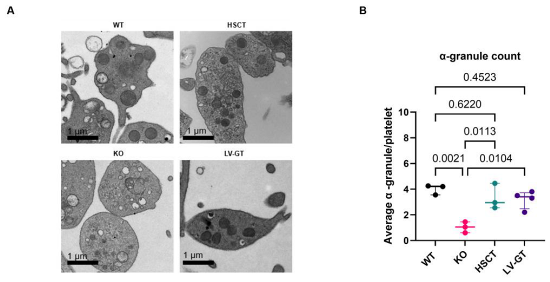
Transmission electron microscopy (TEM) of platelets from untreated ARC mice showed the characteristic ultrastructural defect: markedly reduced α-granule numbers, frequent empty vacuoles and smaller residual α-granule-like structures (Figure 3A). α-Granules were quantified manually in 20 platelets per animal by three investigators blinded to genotype, using established morphological criteria [9]. Both HSCT and ex vivo VPS33B gene therapy significantly increased α-granule counts relative to untreated ARC mice (HSCT vs KO p = 0.0113; LV-GT vs KO p = 0.0104), restoring numbers to levels indistinguishable from WT (HSCT vs WT p = 0.6220; LV-GT vs WT p = 0.4523; Figure 3B). The mean number of α-granules per platelet increased from ≈ 1 in untreated KO mice to 3.0 (HSCT) and 3.5 (LV-GT), comparable to the WT mean of ≈ 4.
3. Discussion
These findings provide proof-of-concept that re-establishing VPS33B expression within the haematopoietic compartment increases platelet α-granule number and ameliorates the systemic proinflammatory phenotype in a mouse model of ARC syndrome. The near-complete restoration of α-granule number was achieved at a modest mean vector copy number (≈ 0.4 copies/cell), suggesting that low transgene dosing is sufficient to influence megakaryocyte organelle number. The EFS promoter, an internal mammalian promoter associated with reduced genotoxicity relative to viral long terminal repeats and used in several ex vivo gene therapy trials [10], supports a translationally favourable profile.
Several considerations temper the interpretation of these results and define the next phase of work. First, our readout is the number of platelet α-granules by TEM; we did not assess α-granule cargo content or platelet function, so we describe restoration of α-granule number rather than full correction of the α-granule deficiency or of haemostasis. Functional confirmation (platelet aggregation, secretion and bleeding time) together with biochemical quantification of α-granule cargo will be required. Of note, only minor changes in platelet aggregation and bleeding time were detectable in this model previously [9], so these murine assays are relatively insensitive here. Second, the study establishes feasibility; formal confirmation of the extent and kinetics of Vps33b ablation in the transplanted donors and recipients, and the use of longer induction intervals as in earlier work [9], would further validate the model. Third, longer follow-up and larger, sex-balanced cohorts will be needed to assess durability and any insertional-mutagenesis risk. Finally, ARC syndrome is a multisystem disorder with hepatic, renal and neurological involvement and severe failure to thrive; haematopoietic gene therapy would address only the platelet/bleeding phenotype, and any clinical application would require careful consideration of patient selection and of the feasibility of stem-cell harvesting and myeloablative conditioning in affected infants. These results should therefore be interpreted as an early feasibility step rather than a complete therapeutic solution.
In principle, the same ex vivo, EFS-driven platform could be explored for other inherited platelet α- or δ-granule disorders, such as VIPAS39/VPS16B-related ARC, Gray platelet syndrome (NBEAL2) and Hermansky–Pudlak syndrome, by substitution of the therapeutic transgene, recognising that disorders with additional non-haematopoietic manifestations would require complementary approaches.
4. Materials and Methods
All animal work was conducted in compliance with UK Home Office regulations under project licence PP9223137. A tamoxifen-inducible conditional Vps33b knockout model (Vps33bfl/fl-CreERT2) was used as previously described [9]. Tamoxifen dosing, conditioning regimens, transplanted cell doses and analysis time points are detailed in the figure legends. Female recipients received lineage-negative bone marrow (Lin⁻) cells from male donors to enable Y-chromosome–based chimerism assessment. Vps33bfl/fl-CreERT2 mice were allocated to WT (no tamoxifen exposure) or tamoxifen-induced knockout (KO) groups; KO mice were further assigned to untreated, HSCT or ex vivo lentiviral gene therapy (LV-GT) arms. Lin⁻ cells were isolated by magnetic lineage depletion (Miltenyi Biotec, Bergisch Gladbach, Germany). For LV-GT, Lin⁻ cells were transduced ex vivo with a third-generation, VSV-G–pseudotyped, self-inactivating lentiviral vector (EFS.cohVPS33B) encoding codon-optimised human VPS33B under the elongation factor 1α short (EFS) promoter at a multiplicity of infection of 2 by spinoculation (600 × g, 60 min). Cells were cultured overnight in StemSpan SFEM with mSCF (100 ng/mL), mFlt3L (100 ng/mL) and hTPO (25 ng/mL) before intravenous transplantation. Donor chimerism and vector copy number (VCN) were quantified by multiplex quantitative PCR (Y-chromosome and proviral targets, respectively). Codon-optimised VPS33B expression was measured by RT-qPCR normalised to β-actin. Blood was collected by cardiac puncture into acid-citrate-dextrose; platelets were processed for transmission electron microscopy and α-granule quantification as described [9], with α-granules counted manually in 20 platelets per animal by three investigators blinded to genotype. Haematopoietic populations were analysed by flow cytometry (BD FACSymphony A5). Statistical comparisons used unpaired Student’s t-test or one-way ANOVA with Tukey post-hoc correction (GraphPad Prism v10); p < 0.05 was considered significant.
Author Contributions
Conceptualization, M.N., C.A.C., M.L., P.G., C.B. and A.J.T.; methodology, M.N., C.A.C., M.L., S.C.-S., A.M., A.E., J.B., D.P. and S.G.; investigation, M.N., C.A.C., D.P., S.C.-S., A.M. and N.V.M.; visualization, M.N., C.A.C., J.J.B. and P.G.; funding acquisition, M.N., P.G., C.A.C., C.B. and A.J.T.; project administration, M.N., C.A.C. and P.G.; supervision, P.G. and C.A.C.; writing—original draft, M.N., P.G. and C.A.C.; writing—review and editing, all authors. All authors have read and agreed to the published version of the manuscript.
Funding
This work was supported by Great Ormond Street Hospital Charity (grant V4420) and LifeArc (grant 586434).
Institutional Review Board Statement
All animal procedures were conducted in compliance with UK Home Office regulations under project licence PP9223137.
Informed Consent Statement
Not applicable.
Data Availability Statement
The data presented in this study are available on reasonable request from the corresponding author. Lentiviral plasmid maps will be deposited in Addgene upon acceptance.
Acknowledgments
The authors acknowledge the University College London LMCB and Biosciences Electron Microscopy Facility (RRID:SCR_027340) for their support. We are grateful to the Kaintz family for their support in memory of Emersynn.
Conflicts of Interest
The authors declare no competing financial interests. P.G. is a Guest Editor of the Special Issue “Individualised Therapies for Rare Disorders” in which this article is submitted; the manuscript was subject to the journal’s standard, independent peer-review process, and P.G. had no role in its editorial handling or in the decision to publish.
References
- Gissen, P.; Johnson, C.A.; Morgan, N.V.; et al. Mutations in VPS33B, encoding a regulator of SNARE-dependent membrane fusion, cause arthrogryposis–renal dysfunction–cholestasis (ARC) syndrome. Nat. Genet. 2004, 36, 400–404. [Google Scholar] [CrossRef] [PubMed]
- Smith, H.; Galmes, R.; Gogolina, E.; et al. Associations among genotype, clinical phenotype, and intracellular localization of trafficking proteins in ARC syndrome. Hum. Mutat. 2012, 33, 1656–1664. [Google Scholar] [CrossRef] [PubMed]
- Akbar, M.A.; Mandraju, R.; Tracy, C.; et al. ARC syndrome-linked Vps33B protein is required for inflammatory endosomal maturation and signal termination. Immunity 2016, 45, 267–279. [Google Scholar] [CrossRef] [PubMed]
- Urban, D.; Li, L.; Christensen, H.; et al. The VPS33B-binding protein VPS16B is required in megakaryocyte and platelet α-granule biogenesis. Blood 2012, 120, 5032–5040. [Google Scholar] [CrossRef] [PubMed]
- Cullinane, A.R.; Straatman-Iwanowska, A.; Zaucker, A.; et al. Mutations in VIPAR cause an arthrogryposis, renal dysfunction and cholestasis syndrome phenotype with defects in epithelial polarization. Nat. Genet. 2010, 42, 303–312. [Google Scholar] [CrossRef] [PubMed]
- Banushi, B.; Forneris, F.; Straatman-Iwanowska, A.; et al. Regulation of post-Golgi LH3 trafficking is essential for collagen homeostasis. Nat. Commun. 2016, 7, 12111. [Google Scholar] [CrossRef] [PubMed]
- Lo, B.; Li, L.; Gissen, P.; et al. Requirement of VPS33B, a member of the Sec1/Munc18 protein family, in megakaryocyte and platelet α-granule biogenesis. Blood 2005, 106, 4159–4166. [Google Scholar] [CrossRef] [PubMed]
- Hanley, J.; Dhar, D.K.; Mazzacuva, F.; et al. Vps33b is crucial for structural and functional hepatocyte polarity. J. Hepatol. 2017, 66, 1001–1011. [Google Scholar] [CrossRef] [PubMed]
- Bem, D.; Smith, H.; Banushi, B.; et al. VPS33B regulates protein sorting into and maturation of α-granule progenitor organelles in mouse megakaryocytes. Blood 2015, 126, 133–143. [Google Scholar] [CrossRef] [PubMed]
- Ferrari, G.; Thrasher, A.J.; Aiuti, A. Gene therapy using haematopoietic stem and progenitor cells. Nat. Rev. Genet. 2021, 22, 216–234. [Google Scholar] [CrossRef] [PubMed]
- Aiuti, A.; Biasco, L.; Scaramuzza, S.; et al. Lentiviral hematopoietic stem cell gene therapy in patients with Wiskott–Aldrich syndrome. Science 2013, 341, 1233151. [Google Scholar] [CrossRef] [PubMed]
- Booth, C.; Masiuk, K.; Vazouras, K.; et al. Long-term safety and efficacy of gene therapy for adenosine deaminase deficiency. N. Engl. J. Med. 2025, 393, 1486–1497. [Google Scholar] [CrossRef] [PubMed]
- Kohn, D.B.; Booth, C.; Kang, E.M.; et al. Lentiviral gene therapy for X-linked chronic granulomatous disease. Nat. Med. 2020, 26, 200–206. [Google Scholar] [CrossRef] [PubMed]
- Magnani, A.; Semeraro, M.; Adam, F.; et al. Long-term safety and efficacy of lentiviral hematopoietic stem/progenitor cell gene therapy for Wiskott–Aldrich syndrome. Nat. Med. 2022, 28, 71–80. [Google Scholar] [CrossRef] [PubMed]
- El-Chemaly, S.; Young, L.R. Hermansky–Pudlak syndrome. Clin. Chest Med. 2016, 37, 505–511. [Google Scholar] [CrossRef] [PubMed]
- Kawarizadeh, K.; Vallez, C.N.; Warrick, K.A.; et al. VPS33B regulates MHC class II antigen presentation in dendritic cells to drive CD4 T cell immunity. iScience 2026, 29, 116052. [Google Scholar] [CrossRef] [PubMed]
- Cozmescu, C.A.; Nazari, M.; Touramanidou, L.; et al. Safety and efficacy analysis of in vivo lentiviral gene therapy in pre-clinical ARC syndrome models. Nat. Commun. 2026, 17, 5074. [Google Scholar] [CrossRef] [PubMed]
Figure 1.
Vps33bfl/fl-CreERT2 mice tolerate myeloablation and support efficient haematopoietic reconstitution. (A) Experimental schematic: Vps33bᶠˡ/ᶠˡ-CreERT2 mice received tamoxifen (d−7, d−5, d−3) to induce Vps33b knockout, split-dose total body irradiation (6 Gy on d0 and 5 Gy on d1) and intravenous transplantation of 0.5×10⁶ Lin⁻ cells from healthy male wild-type donors on d1; recipients were analysed between weeks 5 and 12. (B) Donor-derived chimerism in recipient peripheral blood, quantified by Y-chromosome qPCR. (C) Spleen weight (percentage of body weight) in wild-type (WT), untreated knockout (KO) and haematopoietic stem cell transplantation (HSCT) groups. Graphs show individual values, medians and interquartile ranges (IQRs); n = 3; p-value by one-way ANOVA with Tukey post-hoc correction.
Figure 1.
Vps33bfl/fl-CreERT2 mice tolerate myeloablation and support efficient haematopoietic reconstitution. (A) Experimental schematic: Vps33bᶠˡ/ᶠˡ-CreERT2 mice received tamoxifen (d−7, d−5, d−3) to induce Vps33b knockout, split-dose total body irradiation (6 Gy on d0 and 5 Gy on d1) and intravenous transplantation of 0.5×10⁶ Lin⁻ cells from healthy male wild-type donors on d1; recipients were analysed between weeks 5 and 12. (B) Donor-derived chimerism in recipient peripheral blood, quantified by Y-chromosome qPCR. (C) Spleen weight (percentage of body weight) in wild-type (WT), untreated knockout (KO) and haematopoietic stem cell transplantation (HSCT) groups. Graphs show individual values, medians and interquartile ranges (IQRs); n = 3; p-value by one-way ANOVA with Tukey post-hoc correction.
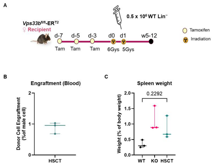
Figure 2.
Ex vivo lentiviral VPS33B gene therapy enables stable engraftment and transgene expression. (A) Schematic of the LV-GT strategy: male donor and female recipient Vps33bfl/fl-CreERT2 mice received tamoxifen (d−14, d−12, d−10); on d0 donor Lin⁻ bone marrow cells were transduced ex vivo with the EFS.cohVPS33B lentiviral vector and 0.5×10⁶ cells were transplanted intravenously on d1 into conditioned recipients (5.5 Gy on d0 and 4 Gy on d1); mice were analysed at 6 weeks. (B) Lentiviral vector schematic (5′LTR, central polypurine tract [cPPT], EFS promoter, codon-optimised human VPS33B [cohVPS33B], 3′LTR and polyA). (C) Donor chimerism in recipient bone marrow by Y-chromosome qPCR. (D) Mean vector copy number per cell. (E) Codon-optimised VPS33B expression in bone marrow by RT-qPCR, normalised to β-actin (2−ΔΔCt). (F) Spleen weight (percentage of body weight) in WT, KO and LV-GT mice. (G) Serum procalcitonin (PCT). Graphs show individual values, medians and IQRs; n = 4 per group; p-values by one-way ANOVA with Tukey post-hoc correction.
Figure 2.
Ex vivo lentiviral VPS33B gene therapy enables stable engraftment and transgene expression. (A) Schematic of the LV-GT strategy: male donor and female recipient Vps33bfl/fl-CreERT2 mice received tamoxifen (d−14, d−12, d−10); on d0 donor Lin⁻ bone marrow cells were transduced ex vivo with the EFS.cohVPS33B lentiviral vector and 0.5×10⁶ cells were transplanted intravenously on d1 into conditioned recipients (5.5 Gy on d0 and 4 Gy on d1); mice were analysed at 6 weeks. (B) Lentiviral vector schematic (5′LTR, central polypurine tract [cPPT], EFS promoter, codon-optimised human VPS33B [cohVPS33B], 3′LTR and polyA). (C) Donor chimerism in recipient bone marrow by Y-chromosome qPCR. (D) Mean vector copy number per cell. (E) Codon-optimised VPS33B expression in bone marrow by RT-qPCR, normalised to β-actin (2−ΔΔCt). (F) Spleen weight (percentage of body weight) in WT, KO and LV-GT mice. (G) Serum procalcitonin (PCT). Graphs show individual values, medians and IQRs; n = 4 per group; p-values by one-way ANOVA with Tukey post-hoc correction.
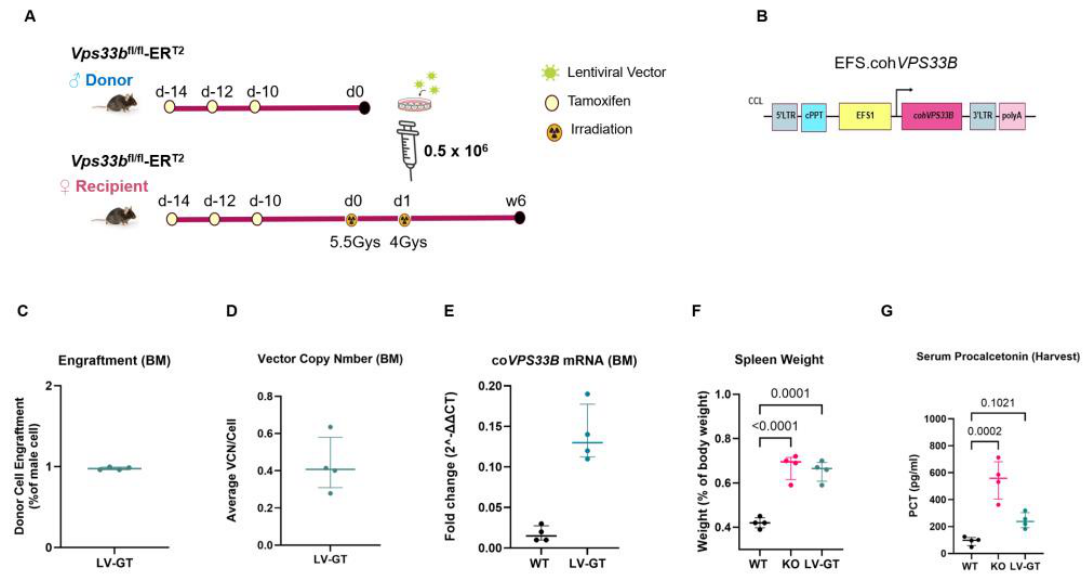
Figure 3.
Restoration of VPS33B expression restores platelet α-granule number. (A) Representative transmission electron micrographs of platelets from wild-type (WT), untreated knockout (KO), haematopoietic stem cell transplantation (HSCT) and gene therapy (LV-GT) mice; platelets from KO mice show markedly reduced α-granule content, with empty vacuoles and smaller α-granule-like structures. Scale bars, 1 µm. (B) Platelet α-granule number per platelet, averaged across 20 platelets per animal by three blinded investigators. Graph shows individual values, medians and IQRs; n = 3 per group (n = 4 for the LV-GT group); p-values by one-way ANOVA with Tukey post-hoc correction.
Figure 3.
Restoration of VPS33B expression restores platelet α-granule number. (A) Representative transmission electron micrographs of platelets from wild-type (WT), untreated knockout (KO), haematopoietic stem cell transplantation (HSCT) and gene therapy (LV-GT) mice; platelets from KO mice show markedly reduced α-granule content, with empty vacuoles and smaller α-granule-like structures. Scale bars, 1 µm. (B) Platelet α-granule number per platelet, averaged across 20 platelets per animal by three blinded investigators. Graph shows individual values, medians and IQRs; n = 3 per group (n = 4 for the LV-GT group); p-values by one-way ANOVA with Tukey post-hoc correction.
Disclaimer/Publisher’s Note: The statements, opinions and data contained in all publications are solely those of the individual author(s) and contributor(s) and not of MDPI and/or the editor(s). MDPI and/or the editor(s) disclaim responsibility for any injury to people or property resulting from any ideas, methods, instructions or products referred to in the content. |
© 2026 by the authors. Licensee MDPI, Basel, Switzerland. This article is an open access article distributed under the terms and conditions of the Creative Commons Attribution (CC BY) license (http://creativecommons.org/licenses/by/4.0/).
Copyright: This open access article is published under a Creative Commons CC BY 4.0 license, which permit the free download, distribution, and reuse, provided that the author and preprint are cited in any reuse.