
Submitted:
15 September 2026
Posted:
17 September 2026
You are already at the latest version
Abstract
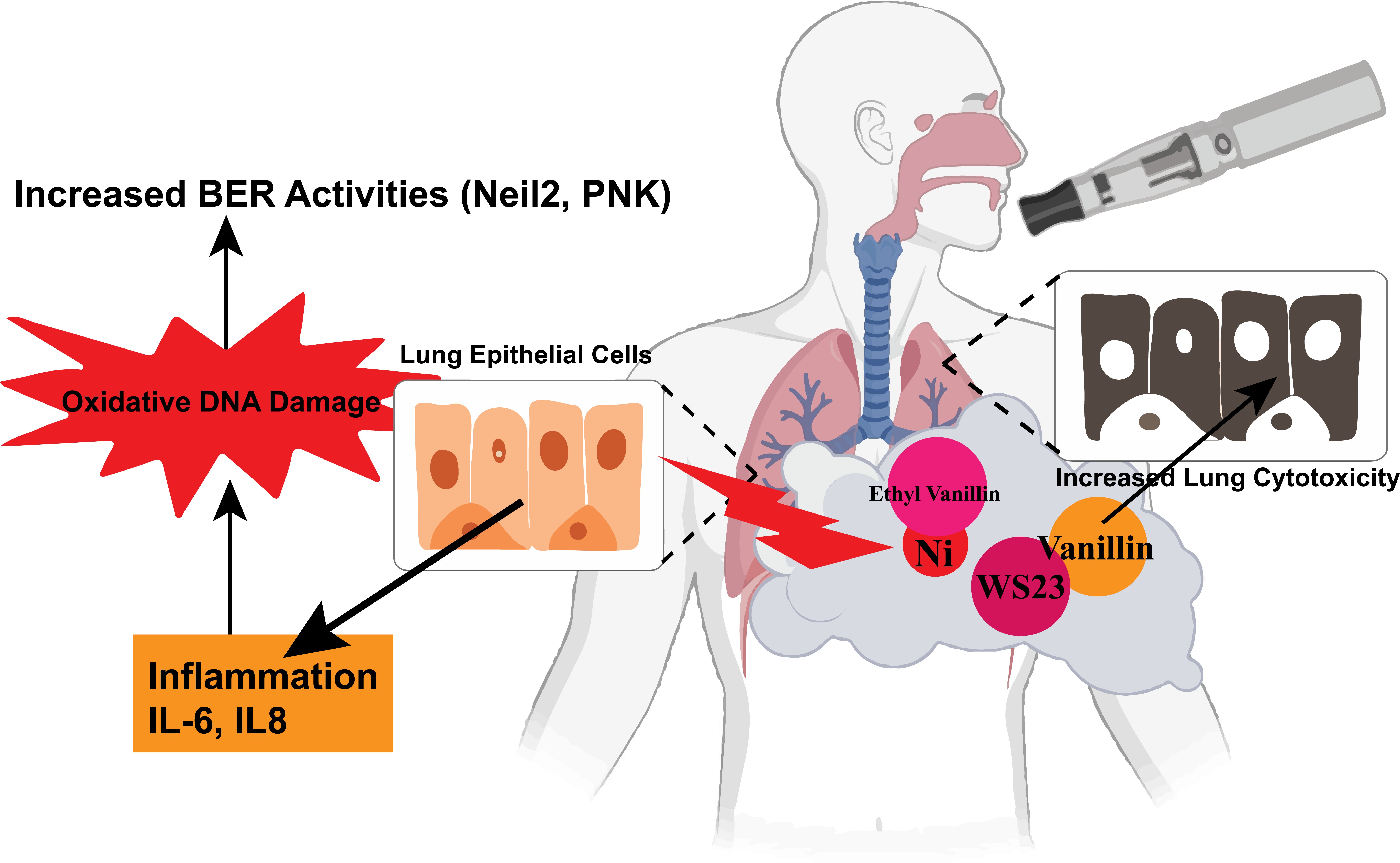
Electronic cigarette aerosols contain complex mixtures of flavoring chemicals and metals released from device components, yet the biological effects of these constituents in combination remain poorly understood. We examined the effects of vanillin, WS23, ethyl vanillin, p-anisaldehyde, and nickel oxide (NiO), alone and in selected mixtures at different ratios, in BEAS-2B human bronchial epithelial cells. Each flavoring chemical reduced metabolic activity and cellular ATP in a concentration-dependent manner. Chou–Talalay analysis showed synergistic interactions between vanillin and WS23 (combination index (CI) < 1) and antagonistic interactions between ethyl vanillin and p-anisaldehyde (CI > 1). Ethyl vanillin (100 µM), NiO (50 µg/cm²), and their combination did not significantly reduce cell viability but were associated with increased interleukin-6 (IL-6) and interleukin-8 (IL-8) in conditioned medium. Total H2AX abundance was unchanged by aldehyde-metal combinations. However, endonuclease VIII-like 2 (NEIL2) showed nonsignificant increases following NiO and combined exposure, whereas polynucleotide kinase 3′-phosphatase (PNKP) increased significantly following ethyl vanillin and combined ethyl vanillin–NiO exposure, suggesting the base-excision repair (BER) pathway mitigating oxidative damage. We show that e-cigarette constituents at relevant concentrations as a mixture may induce oxidative damage but can be repaired by BER. These findings demonstrate that e-cigarette constituents can produce mixture-dependent responses that are not predicted by individual exposures and the inflammatory and DNA-repair-associated responses observed under noncytotoxic conditions warrant further investigation.
Keywords:
ENDS
; electronic cigarette
; flavoring chemicals
; WS23
; nickel oxide
; mixture toxicity
; BEAS-2B
; IL-6
; IL-8
; NEIL2
; PNKP
1. Introduction
Electronic cigarettes (e-cigarettes), also termed electronic nicotine delivery systems (ENDS), generate aerosols from liquids that commonly contain propylene glycol, vegetable glycerin, nicotine, and flavoring chemicals. Aerosol composition reflects device materials, operating conditions, and include carbonyl compounds and metals released from heating components [1] [2] [3] [4]. Because these constituents are inhaled together, evaluation of individual chemicals alone may not predict the biological effects of the resulting mixture.
Many e-cigarette flavoring chemicals are used in foods, but safety for ingestion does not establish safety for inhalation [5] .Flavoring chemicals and flavored e-liquids have produced cytotoxicity, oxidative stress, inflammatory responses, and epithelial dysfunction in experimental models [6] [7] [8] [9]. Flavor aldehydes can also react with propylene glycol and vegetable glycerin to form acetals with properties that differ from those of the parent aldehydes [10].
WS23 (2-isopropyl-N,2,3-trimethylbutyramide) is a synthetic cooling agent detected in e-cigarette products, including disposable products with cooling or menthol-like sensory characteristics [11] [12]. WS23 aerosol exposure can alter airway epithelial responses, including mucin expression and epithelial signaling [13]. The effects of WS23 in mixtures with flavor aldehydes have received less study.
Mixture effects can be additive, synergistic, or antagonistic. Their direction and magnitude may depend on concentration, component ratio, chemical reactivity, cellular uptake, metabolism, and shared biological targets. The Chou-Talalay median-effect method provides a quantitative framework for assessing combined effects, with CI < 1 indicating synergism, CI = 1 additivity, and CI > 1 antagonism [14] [15].
Metals are introduced to ENDS by the electronic/metallic components of the device which adds an additional component of e-cigarette exposure toxicity. Nickel has been measured in e-cigarette liquids and aerosols; and atomizer/coils can contribute nickel to the aerosol [3] [16] [17]. Metal exposure from e-cigarette components has been associated with oxidative, inflammatory, cytotoxic, and genotoxic endpoints [18] [4]. The cellular response to simultaneous exposure to a flavoring chemical and a nickel-containing particle is less well characterized.
Airway epithelial cells contribute to pulmonary innate immune signaling and release cytokines after toxicant exposure [19] [20]. IL-6 and IL-8 are commonly measured indicators of epithelial inflammatory responses [21] [22]. Inflammatory signaling can occur without extensive cell death; therefore, viability/proliferation and cytokine measurements provide complementary information about cellular injury.
Oxidative and inflammatory stress can also affect DNA-damage and DNA-repair pathways. Phospho-histoine-H2AX at Ser 139 is a marker of DNA double-strand-break-associated signaling [23]. Total histone is known to decrease during destabilization of total histones under significant oxidative stress [24]. NEIL2 is a DNA glycosylase/AP lyase involved in repair of oxidized bases, and NEIL2-initiated repair can require polynucleotide kinase 3′-phosphatase (PNKP) to process 3′-phosphate termini [25]. NEIL2 and PNKP also contribute to maintenance of mitochondrial genome integrity [26]. Changes in abundance of these proteins provide insights into DNA lesion burden and repair activity induced by ENDS constituent exposures demonstrated in this study.
This study highlights two complementary challenges in evaluating electronic nicotine delivery systems (ENDS) constituent safety. First, the interactions among flavoring constituents may alter the cytotoxic responses relative to those predicted from individual compounds. Second, co-exposure to chemically distinct constituents such as flavoring chemicals and metals may elicit biological responses that are not apparent from acute cytotoxicity measurements alone. Evaluating both types of mixture effects may therefore provide a more comprehensive assessment of ENDS relevant exposures than single constituents.
The present study was designed to evaluate beyond single-constituent testing by determining whether selected e-cigarette-relevant binary chemical constituents produce distinct non-additive interaction patterns and whether flavoring chemical-metal exposure produces biological responses not recapitulated by acute viability assessment alone. We determined concentration-response relationships for vanillin, WS23, ethyl vanillin, and p-anisaldehyde; evaluated vanillin-WS23 and ethyl vanillin-p-anisaldehyde mixtures using combination-index analysis; and measured cytotoxicty, IL-6, IL-8, H2AX, NEIL2, and PNKP after ethyl vanillin, NiO, or combined exposure. NEIL2/PNKP alterations mitigating oxidative DNA damage and the therapeutic potential of BER pathway targets. This design integrated formal mixture-interaction analysis with inflammatory and DNA-repair-associated protein endpoints in human bronchial epithelial cells. We hypothesized that mixture responses would differ from those predicted from individual constituents. Specifically, we tested whether flavoring-chemical mixtures exhibit distinct non-additive interaction patterns and whether (metallic compounds) ethyl vanillin–NiO co-exposure alters inflammatory and DNA-repair-associated responses at concentrations that produced limited acute cytotoxicity.
2. Materials and Methods
2.1. Cell Culture and Treatment
Human bronchial epithelial BEAS-2B cells were obtained from the American Type Culture Collection (ATCC). Cells were maintained in Gibco Dulbecco’s Modified Eagle Medium/F-12 (DMEM/F-12) containing 5% fetal bovine serum (FBS), 1% glutamine, 1% antibiotic-antimycotic solution, and 15 mM HEPES. Cells were cultured at 37°C in a humidified atmosphere containing 5% CO₂. At approximately 80-85% confluence, cells were serum-deprived for 16-18 h in DMEM/F-12 containing 1% FBS. Treatments were initiated at approximately 90-95% confluence. Conditioned medium and cell pellets were collected 24 h after exposure and stored at -80 °C until analysis.
Cell-line authentication and mycoplasma testing: BEAS-2B cell line was authenticated through short tandem repeat (STR) profiling by ATCC, which confirmed identity and purity. We validated mycoplasma free cultures (InvivoGen, Cat No. rep-mys-50). For the mycoplasma test, supernatant from the BEAS2B T75 culture flasks were combined with reaction mixture and reaction buffer the mixed sample was incubated in 65°C for 40 minutes. Migration buffer was added and the sample was loaded onto the cassette and compared to positive and negative controls provided with the kit.
2.2. Flavoring Chemicals, NiO, and Exposure Conditions
Flavoring-chemical stock solutions were prepared at 100 mM stock concentration in 100% ethanol. The final vehicle concentration in culture medium was ≤1% ethanol and we used vehicle-matched 1% ethanol control for the experiments.
Vanillin (Thermo Scientific, Cat. No. A11169.22), WS23 (TCI, Cat. No. I0729), ethyl vanillin (Sigma-Aldrich, Cat. No. PHR1268), and p-anisaldehyde (TCI, Cat. No. A0480) were evaluated individually and in binary mixtures. Individual concentration-response experiments used relevant concentrations from 10 to 1,000 µM for the MTT assay. CellTiter-Glo experiments used concentrations from 25 to 1,000 µM. Cells were exposed for 24 h.
Vanillin-WS23 and ethyl vanillin-p-anisaldehyde mixtures were prepared at a fixed total concentration of 1,000 µM. Vanillin-WS23 and ethyl vanillin-p-anisaldehyde mixtures were evaluated at multiple component ratios. Individual treatment concentration for the all chemicals was 10, 25, 50, 100, 200, 300, 400, 500, 600, 700, 800, 900, and 1000 µM for MTT assay, and 25, 50, 100, 250, 500, 750, 1000 µM for CellTiter-Glo assay. Ratio series for the binary mixture was 1:1, 1:3, 1:4, 1:9, 1:19, 1:24, 1:49, 3:1, 4:1, 9:1, 19:1, 24:1, and 49:1 for both experiments using their individual doses in MTT assay, and CellTiter-Glo included only 1:1, 1:3, 1:9, 3:1 and 9:1 mixed ratios.
For flavoring chemical-metal experiments, passage-14 BEAS-2B cells were exposed for 24 h to 100 µM ethyl vanillin, 50 µg/cm² NiO, or 100 µM ethyl vanillin plus 50 µg/cm² NiO. Nickel oxide (NiO; Thermo Scientific, Cat# 012359.18, Purity 99%, -325 mesh with particle size ≤44 µm) was prepared at 1 mg/mL using sonication method. We added 0.05% pluronic F-68 for better particle stability into the solution. Then the stock was sonicated for 10 minutes to reduce clumping. Finally, we mixed the sample solution with vortex and treated immediately to ensure uniform delivery.
Treatment preparation for single and binary treatments are listed in supplementary tables 1 and 2. (S and 2S).
2.3. MTT Assay (Cytoxicity/Profileration)
BEAS-2B cells were seeded in 96-well plates at approximately 10,000 cells per well in 200 µL of medium containing 5% FBS. Following serum deprivation in medium containing 1% FBS, cells were exposed to vanillin, WS23, ethyl vanillin, p-anisaldehyde, or the indicated binary mixtures. The total fixed concentration of each binary mixture was 1,000 µM. After 24 h, treatment medium was replaced with fresh medium, 10 µL MTT solution was added to each well, and plates were incubated for 4 h at 37 °C. SDS-HCl solution (100 µL) was then added to each well, followed by a second 4-h incubation at 37 °C. After mixing, absorbance was measured at 570 nm. The MTT response was calculated as (absorbance of treated cells/absorbance of untreated control) × 100. MTT assays were repeated three times, with two technical replicate wells per treatment condition in each repeat. MTT reduction was interpreted as viability-associated metabolic activity rather than as a direct measure of cell number [27].
2.4. CellTiter-Glo ATP Assay
Passage-10 BEAS-2B cells were seeded at 15,000 cells per well in white opaque 96-well plates. At 80-85% confluence, cells were serum-deprived for 16 h in medium containing 1% FBS and then exposed to vanillin, WS23, ethyl vanillin, p-anisaldehyde, or their mixtures. Individual concentrations ranged from 25 to 1,000 µM. Cellular ATP was measured 24 h after treatment using CellTiter-Glo 2.0 (Promega, Cat. No. G9241). ATP standards were 0, 1 nM, 10 nM, 100 nM, 1 µM, 10 µM, and 100 µM. CellTiter-Glo reagent (100 µL) was added to each well, plates were mixed for 2 min and incubated for 10 min to stabilize luminescence, and luminescence was recorded. ATP concentrations were calculated from the standard curve. Luminescence was measured using SpectraMax Id3, Molecular Devices (San Jose, CA, USA).
2.5. Trypan Blue Exclusion Assay (Viability)
At cell collection, 10 µL of cell suspension was mixed with 10 µL trypan blue. Ten microliters of the mixture was loaded into a hemocytometer, and trypan-blue-excluding and trypan-blue-positive cells were counted by microscopy. Percent viability was calculated as the number of trypan-blue-excluding cells divided by the total number of cells counted × 100.
2.6. Combination-Index Analysis (Chemical Interactions)
Chemical interactions were evaluated using the Chou-Talalay median-effect method in CompuSyn (ComboSyn Inc., Paramus, NJ, USA) [14] [15]. MTT-derived dose-effect data for individual chemicals and binary mixtures were entered into CompuSyn to calculate fraction affected (Fa) and CI. CI < 1 indicated synergism, CI = 1 additivity, and CI > 1 antagonism.
CompuSyn version 1.0 was used. All the CI calculations were done based on experiment-level data. We entered all the mixed ratios from MTT assay data as non-constant combo in CompuSyn and used the respective effect (Fa) and CI values to generate Fa-CI curve (S).
2.7. IL-6 and IL-8 ELISA (Inflammatory Mediators)
Conditioned medium was collected 24 h after treatment. Secreted IL-6 and IL-8 were quantified using DuoSet ELISA kits (R&D Systems; IL-6, Cat. No. DY206; IL-8, Cat. No. DY208) according to the manufacturer’s protocol. Cytokine concentrations were reported as pg/mL conditioned medium.
2.8. Immunoblotting (Protein Abundance)
Cells were lysed in RIPA buffer (Thermo Scientific, Cat No. J63306.AP) containing protease inhibitor (Thermo Scientific, Cat No. 87785), and total protein was quantified using a BCA assay kit (Thermo Fisher Scientific, Cat. No. 23225). Twenty micrograms of protein per sample was loaded onto 4-10% polyacrylamide gels (10 wells, 1.5 mm) and separated by electrophoresis at 120 V. Proteins were transferred to polyvinylidene difluoride membranes using a Bio-Rad Trans-Blot Turbo system. Transfer was confirmed by Ponceau S staining (Thermo Fisher Scientific, Cat. No. A40000279).
Membranes were blocked for 1 h at room temperature with gentle shaking in 5% nonfat milk prepared in 1× Tris-buffered saline containing 0.1% Tween-20 (TBST), followed by three 10-min washes in TBST. Membranes were incubated overnight at 4 °C with antibodies against H2AX (R&D Systems, Cat. No. MAB3406), NEIL2 (Novus Biologicals, Cat. No. NBP1-98526), or PNKP (Thermo Fisher Scientific, Cat. No. MA5-55330), each at 1:1,000 in 5% milk/TBST.
After three TBST washes, membranes were incubated with HRP-conjugated goat anti-rabbit IgG (Abcam, Cat. No. AB6721; 1:5,000) for NEIL2 and PNKP or HRP-conjugated goat anti-mouse IgG (Abcam, Cat. No. AB97040; 1:5,000) for H2AX. Membranes were washed three times and developed using SuperSignal West Femto chemiluminescent substrate (Thermo Fisher Scientific, Cat. No. 34094). Images were acquired with a Bio-Rad ChemiDoc XRS+ system.
Membranes were stripped with stripping buffer (Thermo Scientific, Cat No. 46430), blocked again for 1 h in 5% milk/TBST, washed three times, and reprobed overnight with Lamin B1 antibody (Abcam, Cat. No. AB16048-1001). Band density was quantified using Bio-Rad Image Lab software (Version 6.1, Bio-Rad Laboratories). H2AX, NEIL2, and PNKP were normalized to Lamin B1. Approximate molecular weights were H2AX, 25 kDa; NEIL2, 37 kDa; PNKP, 60 kDa; and Lamin B1, 70 kDa. Full-length, uncropped blots are provided in the Supplementary Materials.
2.9. Statistical Analysis
Statistical analyses were performed using GraphPad Prism version 10.5.0. Data are presented as mean ± SEM. Comparisons among treatment groups were performed using one-way analysis of variance followed by Tukey’s multiple-comparisons test. Adjusted P < 0.05 was considered statistically significant. Figure 1 contains n = 2-4 observations depending on the panel, Figure 2 contains n = 3-12, Figure 3 contains n = 3, and immunoblot analyses contain n = 2-3 (Figure 4). In all cases, n number represents independent biological replicates. For figure 1,2, and 3, we used 2 technical replicates too, and immunoblotting used all biological replicates where n number was 2-3 to fit randomly selected samples in 10 well gels.
3. Results
3.1. Vanillin and WS23 Reduce Metabolic Activity and Produce Synergistic Effects in Mixture
Vanillin reduced the MTT endpoint significantly at 200, 400, 600, 700, 800, 900, and 1,000 µM relative to untreated control (Figure 1A). WS23 significantly reduced the MTT endpoint at 50, 200, 300, 400, 500, 600, 700, 800, 900, and 1,000 µM (Figure 1B). Thus, the lowest tested concentrations associated with significant effects were 200 µM for vanillin and 50 µM for WS23.
Vanillin-WS23 mixtures were evaluated at a fixed total concentration of 1,000 µM over multiple component ratios. All tested mixtures significantly reduced the MTT endpoint relative to control, and mixtures containing higher proportions of vanillin produced reductions greater than 50% in MTT-based viability-associated metabolic activity in some conditions (Figure 1C). CI values were <1 for all evaluated mixtures, consistent with synergistic interactions under the experimental conditions (Figure 1D). CellTiter-Glo measurements showed concentration-dependent ATP depletion after individual exposures, and mixtures containing higher proportions of vanillin produced greater ATP depletion. The 1,000 µM vanillin condition and the 9:1 vanillin:WS23 mixture were significantly decreased from control (Figure 1E).
3.2. Ethyl Vanillin and p-Anisaldehyde Produce Antagonistic Effects in Mixture
Passage-20 BEAS-2B cells were exposed for 24 h to ethyl vanillin, p-anisaldehyde, or binary mixtures at a fixed total concentration of 1,000 µM. Ethyl vanillin significantly reduced the MTT endpoint at concentrations from 300 to 1,000 µM (Figure 2A). p-Anisaldehyde significantly reduced the MTT endpoint at concentrations from 200 to 1,000 µM (Figure 2B).
Binary mixtures reduced the MTT endpoint, but mixtures containing higher proportions of p-anisaldehyde were generally not significantly different from control, whereas mixtures containing higher proportions of ethyl vanillin showed significant effects (Figure 2C). CI values were >1 for all evaluated ethyl vanillin-p-anisaldehyde mixtures, consistent with antagonistic interactions under the experimental conditions (Figure 2D). CellTiter-Glo measurements showed concentration-dependent ATP depletion after ethyl vanillin exposure, and mixtures containing higher proportions of ethyl vanillin produced greater ATP depletion (Figure 2E).
We established that flavoring chemical mixtures could produce distinct non additive interaction patterns, we next examined whether co-exposure to chemically dissimilar ENDS constituents could elicit biological responses under conditions without significant acute toxicity. Ethyl vanillin and NiO were therefore evaluated individually and in combination using toxicity and inflammatory end points.
3.3. Ethyl Vanillin and NiO Increase Inflammatory Cytokines Without a Significant Decrease in Viability
Passage-14 BEAS-2B cells were exposed for 24 h to 100 µM ethyl vanillin, 50 µg/cm² NiO, or their combination. Trypan blue exclusion showed no statistically significant difference in percent viability among treatment groups and control (Figure 3A).
IL-6 concentrations were significantly increased after NiO, and combined ethyl vanillin-NiO exposure relative to untreated control (Figure 3B). IL-6 was also significantly higher after combined exposure than after NiO alone. IL-8 concentrations were significantly increased after NiO and combined exposure relative to control and ethyl vanillin alone (Figure 3C). These findings show that inflammatory responses occurred at exposure conditions that did not produce a statistically significant decrease in cell viability.
3.4. Ethyl Vanillin and NiO Exposures are Associated with Differential NEIL2 and PNKP Protein Abundance without Changes in Total H2AX at 24 h
Total H2AX abundance did not differ significantly among control, ethyl vanillin, NiO, and combined ethyl vanillin-NiO groups at 24 h (Figure 4A).
NEIL2 abundance showed a nonsignificant upward trend after NiO alone relative to control (Tukey-adjusted P = 0.0518). For ethyl vanillin + NiO versus control, NEIL2 abundance was increased with a nonsignificant trend (Tukey-adjusted P = 0.0760) (Figure 4B).
PNKP abundance increased significantly after ethyl vanillin (Tukey-adjusted P = 0.0261) and combined ethyl vanillin-NiO exposure (Tukey-adjusted P = 0.0041) relative to control. Combined exposure also produced significantly greater PNKP abundance than NiO alone (Tukey-adjusted P = 0.0174) (Figure 4C).
4. Discussion
This study demonstrated that commonly found selected-e-cigarette-relevant mixtures produced opposite interaction phenotypes in the same bronchial epithelial model: vanillin-WS23 mixtures yielded CI values consistent with synergism, whereas ethyl vanillin-p-anisaldehyde mixtures yielded CI values consistent with antagonism.
A second important finding is that ethyl vanillin-NiO exposure elicited inflammatory cytokine responses under conditions without a statistically significant loss of cell viability, demonstrating a biological response that was not captured by the acute viability/toxicity endpoint. The immunoblot data further identified a DNA-repair-associated response pattern, with an upward NEIL2 pattern in NiO-containing conditions and increased PNKP in ethyl-vanillin-containing conditions while H2AX did not differ significantly among groups at 24 h demonstrating that the induced oxidative stress did not cause histone degradation due to toxicity. These findings suggest that mixture composition influenced NEIL2-associated DNA-repair responses involving proteins that participate in BER mitigating oxidative damage.
WS23 affected the MTT endpoint at a lower concentration than the other flavoring chemicals tested. WS23 is present in marketed e-cigarette products and has been shown to alter airway epithelial responses [11] [12] [13]. Vanillin has also produced cytotoxic effects in bronchial epithelial models [9]. In the present study, CI values <1 across the evaluated vanillin-WS23 mixtures indicate that the combined effects exceeded those predicted from the individual dose-effect relationships. Consistent with our findings, synergistic effects have also been reported for other aldehyde mixtures in BEAS-2B cells [28].
The mechanism underlying vanillin-WS23 synergy was not examined in this study. Potential explanations include changes in cellular uptake, membrane properties, mitochondrial function, or convergence on common stress-response pathways. Flavor aldehydes can react with e-liquid solvents to form acetals [10], but the present study did not evaluate chemical reaction products. The current data therefore establish the interaction phenotype but do not identify its chemical or molecular basis.
Ethyl vanillin-p-anisaldehyde mixtures produced CI values >1, indicating antagonism. In combination-index analysis, antagonism means that the measured combined effect is lower than that predicted from the individual dose-effect relationships; it does not eliminate potential adverse effects [14] [15]. Antagonistic interactions have been found for other vaping ingredients, including limonene and triethyl citrate [29]. Additional dose ranges and mixture ratios would be required to determine whether the interaction observed here is maintained across a broader effect range.
The ethyl vanillin-NiO experiment examined important aldehyde-metal toxicity component of mixture toxicity. Nickel is relevant to e-cigarette exposure because nickel-containing device components can contribute metal to aerosol [3] [16] [17]. A previous study also showed that ethyl maltol enhanced copper-mediated cytotoxicity in lung epithelial cells, demonstrating that flavoring compounds can modify cellular responses to metals [30]. In the present study, 100 µM ethyl vanillin and 50 µg/cm² NiO did not significantly reduce cell viability, but they altered cytokine release.
IL-6 increased after ethyl vanillin, NiO, and combined exposure, and the combination produced higher IL-6 than NiO alone. IL-8 increased after NiO and combined exposure. Airway epithelial cells are active participants in pulmonary inflammatory signaling [19] [20]. The cytokine results therefore indicate a biological response that was not reflected by the cell viability endpoint.
The DNA-repair-associated protein pattern identifies a hypothesis for mechanistic follow-up. NEIL2 abundance showed a nonsignificant upward trend after NiO alone, whereas PNKP increased after ethyl vanillin and combined exposure. NEIL2 is a DNA glycosylase/AP lyase involved in processing selected oxidized bases, and NEIL2-initiated repair of 5-hydroxyuracil has been shown to require polynucleotide kinase activity to generate repair-compatible 3′-OH termini [31] [25]. NEIL2 and PNKP also contribute to mitochondrial genome maintenance [26]. The abundance patterns observed here therefore support further investigation of altered base-damage-response pathways, but protein abundance alone does not establish oxidative DNA lesions, pathway activation, or increased DNA repair activity.
H2AX abundance did not differ significantly among groups at 24 h. The consistency between the upward NEIL2 pattern, increased PNKP abundance, and the absence of a significanH2AX destabilization is biologically important and implicate that these exposures may be associated with altered base-damage-response pathways without producing a prominent persistent H2AX-associated double-strand-break signal at the measured time point. However, this interpretation does not exclude oxidative base lesions, single-strand intermediates, transient DNA damage, or damage repaired before collection [23], and it does not establish activation of base excision repair. Direct measurements of DNA lesions and repair activity are required to test this hypothesis which are included in our ongoing studies of NEIL2-mediated oxidative repair mechanisms in lung injury.
Inflammation provides a plausible context for further mechanistic study because inflammatory processes can generate reactive oxygen and nitrogen species capable of damaging DNA [32]. NEIL2 expression has been altered in models of chemically induced oxidative stress [33], and ATM-dependent stabilization of PNKP has been linked to cellular resistance to oxidative stress [34]. Direct measurement of reactive oxygen species, oxidized DNA bases, and DNA strand-break intermediates would be required to connect the inflammatory response observed here with the changes in NEIL2 and PNKP.
The NEIL2 abundance pattern provides a testable hypothesis for mechanistic follow-up. A time-course experiment could determine whether cytokine induction precedes changes in NEIL2 and PNKP. Oxidative DNA lesions could be measured by 8-oxo-2′-deoxyguanosine analysis or lesion-sensitive comet assays [35] [36]. NEIL2 loss-of-function experiments would determine whether NEIL2 affects lesion burden, inflammatory signaling, or cell survival after NiO and ethyl vanillin-NiO exposure. Such experiments would distinguish an exposure-associated change in NEIL2 abundance from a functional role in the cellular response.
This study has limitations. BEAS-2B cells are an immortalized bronchial epithelial model and do not reproduce the cellular diversity or differentiation state of the human airway. Exposures were performed in submerged culture using concentrations rather than aerosol exposure at an air-liquid interface. The study evaluated a single 24-h exposure period and a limited set of binary mixtures relevant to real-life exposures in ENDS. These limitations restrict mechanistic interpretation and extrapolation to human inhalation exposure.
The results nevertheless establish that the biological effects of the tested mixtures depended on mixture composition and their ratios. Vanillin-WS23 and ethyl vanillin-p-anisaldehyde produced opposite interaction patterns, and ethyl vanillin-NiO exposure produced inflammatory and DNA-repair-associated responses without significant acute loss of viability. Tested constituents, their ratios were caused adverse toxic responses but were repaired by involvement of the BER pathway. These observations support evaluation of e-cigarette constituents as mixtures rather than assuming that combined effects can be predicted from individual-compound toxicity.
5. Conclusions
Vanillin, WS23, ethyl vanillin, and p-anisaldehyde produced concentration-dependent effects on viability-associated metabolic activity and cellular ATP in BEAS-2B cells. Chou-Talalay analysis yielded CI values <1 for the tested vanillin-WS23 mixtures and CI values >1 for the tested ethyl vanillin-p-anisaldehyde mixtures, consistent with synergistic and antagonistic interactions, respectively, under the experimental conditions. Thus, the effects of the tested flavoring chemical mixtures were dependent on mixture composition and were not described by a single assumption of additivity. This study highlights the importance of determining ratio interactions of constituents, organic, inorganic, and metals present in ENDS liquids and emissions.
Author Contributions
Conceptualization, T.M.; Investigation, E.K. and J.F.; Formal Analysis, E.K. and J.F.; Writing—Original Draft Preparation, E.K. and T.M.; Writing—Review and Editing, E.K., J.F. and T.M. All authors have read and agreed to the published version of the manuscript.
Funding
This research was funded by the National Institute of Environmental Health Sciences (NIEHS), National Institutes of Health (NIH), grant number R00ES033835.
Data Availability Statement
The data presented in this study are available from the corresponding author upon reasonable request. Supporting experimental and analytical information is provided in the Supplementary Materials.
Conflicts of Interest
The authors declare no conflict of interest.
References
- Zhu, S.H.; et al. Four hundred and sixty brands of e-cigarettes and counting: implications for product regulation. Tob Control 2014, 23 Suppl 3, iii3–9. [Google Scholar] [CrossRef] [PubMed]
- Cao, D.J.; et al. Review of Health Consequences of Electronic Cigarettes and the Outbreak of Electronic Cigarette, or Vaping, Product Use-Associated Lung Injury. J Med Toxicol 2020, 16(3), 295–310. [Google Scholar] [CrossRef] [PubMed]
- Olmedo, P.; et al. Metal Concentrations in e-Cigarette Liquid and Aerosol Samples: The Contribution of Metallic Coils. Environ Health Perspect 2018, 126(2), 027010. [Google Scholar] [CrossRef] [PubMed]
- Gong, J.Y.; Ghosh, M.; Hoet, P.H. Association between metal exposure from e-cigarette components and toxicity endpoints: A literature review. Regul Toxicol Pharmacol 2023, 144, 105488. [Google Scholar] [CrossRef] [PubMed]
- Barhdadi, S.; et al. Toxicity assessment of flavour chemicals used in e-cigarettes: current state and future challenges. Arch Toxicol 2021, 2879–2881. [Google Scholar] [CrossRef] [PubMed]
- Muthumalage, T.; et al. Inflammatory and Oxidative Responses Induced by Exposure to Commonly Used e-Cigarette Flavoring Chemicals and Flavored e-Liquids without Nicotine. Front Physiol 2017, 8, 1130. [Google Scholar] [CrossRef] [PubMed]
- Gerloff, J. Inflammatory Response and Barrier Dysfunction by Different e-Cigarette Flavoring Chemicals Identified by Gas Chromatography-Mass Spectrometry in e-Liquids and e-Vapors on Human Lung Epithelial Cells and Fibroblasts. Appl In Vitro Toxicol 2017, 3(1), 28–40. [Google Scholar] [CrossRef] [PubMed]
- Lerner, C.A.; et al. Vapors produced by electronic cigarettes and e-juices with flavorings induce toxicity, oxidative stress, and inflammatory response in lung epithelial cells and in mouse lung. PLoS One 2015, 10(2), e0116732. [Google Scholar] [CrossRef] [PubMed]
- Morris, A.M.; et al. Effects of E-Cigarette Flavoring Chemicals on Human Macrophages and Bronchial Epithelial Cells. Int J Environ Res Public Health 2021, 18(21). [Google Scholar] [CrossRef] [PubMed]
- Erythropel, H.C.; et al. Formation of flavorant-propylene Glycol Adducts With Novel Toxicological Properties in Chemically Unstable E-Cigarette Liquids. Nicotine Tob Res 2019, 21(9), 1248–1258. [Google Scholar] [CrossRef] [PubMed]
- Jabba, S.V.; et al. Synthetic Cooling Agents in US-marketed E-cigarette Refill Liquids and Popular Disposable E-cigarettes: Chemical Analysis and Risk Assessment. Nicotine Tob Res 2022, 24(7), 1037–1046. [Google Scholar] [CrossRef] [PubMed]
- Omaiye, E.E.; et al. Disposable Puff Bar Electronic Cigarettes: Chemical Composition and Toxicity of E-liquids and a Synthetic Coolant. Chem Res Toxicol 2022, 35(8), 1344–1358. [Google Scholar] [CrossRef] [PubMed]
- Manevski, M.; et al. E-cigarette synthetic cooling agent WS-23 and nicotine aerosols differentially modulate airway epithelial cell responses. Toxicol Rep 2022, 9, 1823–1830. [Google Scholar] [CrossRef] [PubMed]
- Chou, T.C.; Talalay, P. Quantitative analysis of dose-effect relationships: the combined effects of multiple drugs or enzyme inhibitors. Adv Enzyme Regul 1984, 22, 27–55. [Google Scholar] [CrossRef] [PubMed]
- Chou, T.C. Drug combination studies and their synergy quantification using the Chou-Talalay method. Cancer Res 2010, 70(2), 440–6. [Google Scholar] [CrossRef] [PubMed]
- Palazzolo, D.L.; et al. Trace Met. Deriv. From Electron. Cigar. (ECIG) Generated Aerosol: Potential Problem of ECIG Devices That Contain Nickel. Front Physiol 2016, 7, 663. [Google Scholar] [CrossRef] [PubMed]
- Gray, N.; et al. Toxic Metals in Liquid and Aerosol from Pod-Type Electronic Cigarettes. J Anal Toxicol 2022, 46(1), 69–75. [Google Scholar] [CrossRef] [PubMed]
- Gaur, S.; Agnihotri, R. Health Effects of Trace Metals in Electronic Cigarette Aerosols-a Systematic Review. Biol Trace Elem Res 2019, 188(2), 295–315. [Google Scholar] [CrossRef] [PubMed]
- Hiemstra, P.S.; McCray, P.B., Jr.; Bals, R. The innate immune function of airway epithelial cells in inflammatory lung disease. Eur Respir J 2015, 45(4), 1150–62. [Google Scholar] [CrossRef] [PubMed]
- Wong, J.; Magun, B.E.; Wood, L.J. Lung inflammation caused by inhaled toxicants: a review. Int J Chron Obstruct Pulmon Dis 2016, 11, 1391–401. [Google Scholar] [CrossRef] [PubMed]
- Rincon, M.; Irvin, C.G. Role of IL-6 in asthma and other inflammatory pulmonary diseases. Int J Biol Sci 2012, 8(9), 1281–90. [Google Scholar] [CrossRef] [PubMed]
- Moldoveanu, B.; et al. Inflammatory mechanisms in the lung. J. Inflamm. Res. 2009, 2, 1–11. [Google Scholar] [CrossRef]
- Mah, L.J.; El-Osta, A.; Karagiannis, T.C. gammaH2AX: a sensitive molecular marker of DNA damage and repair. Leukemia 2010, 24(4), 679–86. [Google Scholar] [CrossRef] [PubMed]
- Gruosso, T.; et al. Chronic oxidative stress promotes H2AX protein degradation and enhances chemosensitivity in breast cancer patients. EMBO Mol Med 2016, 8(5), 527–49. [Google Scholar] [CrossRef] [PubMed]
- Das, A.; et al. NEIL2-initiated, APE-independent repair of oxidized bases in DNA: Evidence for a repair complex in human cells. DNA Repair (Amst) 2006, 5(12), 1439–48. [Google Scholar] [CrossRef] [PubMed]
- Mandal, S.M.; et al. Role Hum. DNA Glycosylase Nei-Like 2 (NEIL2) and single strand break repair protein polynucleotide kinase 3'-phosphatase in maintenance of mitochondrial genome. J Biol Chem 2012, 287(4), 2819–29. [Google Scholar] [CrossRef] [PubMed]
- Fotakis, G.; Timbrell, J.A. In vitro cytotoxicity assays: comparison of LDH, neutral red, MTT and protein assay in hepatoma cell lines following exposure to cadmium chloride. Toxicol Lett 2006, 160(2), 171–7. [Google Scholar] [CrossRef] [PubMed]
- Zhang, S.; et al. Combined cytotoxicity of co-exposure to aldehyde mixtures on human bronchial epithelial BEAS-2B cells. Environ Pollut 2019, 250, 650–661. [Google Scholar] [CrossRef] [PubMed]
- Baldovinos, Y. Chemical interactions and cytotoxicity of terpene and diluent vaping ingredients. Chemical Research in Toxicology 2023, 36(4), 589–597. [Google Scholar] [CrossRef] [PubMed]
- Durrani, K.; et al. Ethyl maltol enhances copper mediated cytotoxicity in lung epithelial cells. Toxicol Appl Pharmacol 2021, 410, 115354. [Google Scholar] [CrossRef] [PubMed]
- Myrup Holst, C. Phosphorylation of the Human DNA Glycosylase NEIL2 Is Affected by Oxidative Stress and Modulates Its Activity. Antioxidants 2023, 12(2). [Google Scholar] [CrossRef] [PubMed]
- Kay, J.; et al. Inflammation-induced DNA damage, mutations and cancer. DNA Repair (Amst) 2019, 83, 102673. [Google Scholar] [CrossRef] [PubMed]
- Ma, H.; et al. Induction of NEIL1 and NEIL2 DNA glycosylases in aniline-induced splenic toxicity. Toxicol Appl Pharmacol 2011, 251(1), 1–7. [Google Scholar] [CrossRef] [PubMed]
- Parsons, J.L.; et al. Phosphorylation of PNKP by ATM prevents its proteasomal degradation and enhances resistance to oxidative stress. Nucleic Acids Res 2012, 40(22), 11404–15. [Google Scholar] [CrossRef] [PubMed]
- Rus, L.A. Serum 8-OHdG as a Marker of Oxidative DNA Damage in Acute Exacerbations of COPD: Associations with GOLD Stage and Smoking Status-A Cross Sectional Study. Medicina 2026, 62(7). [Google Scholar] [CrossRef] [PubMed]
- Smith, C.C.; O'Donovan, M.R.; Martin, E.A. hOGG1 recognizes oxidative damage using the comet assay with greater specificity than FPG or ENDOIII. Mutagenesis 2006, 21(3), 185–90. [Google Scholar] [CrossRef] [PubMed]
Disclaimer/Publisher’s Note: The statements, opinions and data contained in all publications are solely those of the individual author(s) and contributor(s) and not of MDPI and/or the editor(s). MDPI and/or the editor(s) disclaim responsibility for any injury to people or property resulting from any ideas, methods, instructions or products referred to in the content. |
© 2026 by the authors. Licensee MDPI, Basel, Switzerland. This article is an open access article distributed under the terms and conditions of the Creative Commons Attribution (CC BY) license (http://creativecommons.org/licenses/by/4.0/).
Copyright: This open access article is published under a Creative Commons CC BY 4.0 license, which permit the free download, distribution, and reuse, provided that the author and preprint are cited in any reuse.