
Submitted:
15 September 2026
Posted:
16 September 2026
You are already at the latest version
Abstract
Skeletal muscle aging arises from interacting defects in proteostasis, mitochondrial quality control, inflammatory signaling, neuromuscular integrity, regenerative capacity, and mechanical-metabolic plasticity, and may culminate in sarcopenia. Circular RNAs (circRNAs) are plausible regulators and biomarker candidates, but most evidence derives from developmental myogenesis, expression profiling, or non-aging atrophy models. This critical review organizes the literature around skeletal muscle aging and applies a four-level framework encompassing discovery, mechanistic support, in vivo causality, and functional intervention. We assess human linkage, species, treatment timing, locus of action, and endogenous or engineered modality separately. circSnd1 and circDdb1 currently provide the strongest combination of functional preclinical evidence and association with aged human skeletal muscle. circFUT10, circBBS9, circNEB, and circGLIS3 define less mature regenerative, discovery, histological, or muscle-quality domains. Non-aging atrophy studies, including circTmeff1 and engineered circmiR-29b, test whether proposed mechanisms remain active under defined catabolic stress. In the targeted literature reviewed here, no reported circulating circRNA has yet met the analytical, biological, and clinical requirements for a useful sarcopenia biomarker. Therapeutic development is constrained by quantitative target credibility, systemic skeletal-muscle exposure, post-onset functional efficacy in aged organisms, safety, reversibility, and independent replication. The field should now prioritize rigorous validation over additional descriptive cataloging.
Keywords:
circular RNA
; skeletal muscle aging
; sarcopenia
; biomarkers
; mechanotransduction
; RNA therapeutics
1. Introduction
Sarcopenia is not synonymous with low muscle mass. Contemporary European, Asian, and global consensus initiatives converge on a clinically important principle. Strength and function are central to the phenotype, whereas muscle quantity and quality provide complementary confirmation and biological context [1,2,3]. A molecular intervention that enlarges fibers without restoring force, power, endurance, or physical performance cannot by itself be assumed to correct sarcopenia. Skeletal muscle aging is better viewed as a progressive loss of adaptive reserve in a tissue that must continuously integrate nutritional, mechanical, neural, metabolic, and inflammatory signals [4,5,6].
This loss of reserve emerges from interacting processes. Aging alters protein turnover and anabolic responsiveness, mitochondrial quantity and quality, redox homeostasis, inflammatory tone, motor-unit connectivity, extracellular-matrix organization, satellite-cell behavior, and responses to mechanical and metabolic stress [4,5,6,7,8]. These domains are interdependent. Denervation modifies fiber composition and mitochondrial demand. Matrix remodeling changes force transmission and mechanosensing. Mitochondrial dysfunction can amplify redox and innate immune signaling, while regenerative impairment alters cellular composition. The network architecture helps explain why changes in a single biochemical readout often fail to predict functional recovery.
Circular RNAs (circRNAs) are attractive within this framework because they are structurally distinct RNA isoforms generated predominantly by back-splicing and can display tissue- and state-selective expression [9,10,11,12,13,14]. Selected circRNAs can interact with microRNAs (miRNAs) or RNA-binding proteins (RBPs), influence host-gene or transcriptional processes, and in some cases support translation [15,16,17]. Engineered circRNAs extend these properties into synthetic decoys and durable protein-expression platforms [18,19,20]. CircRNA abundance can also rise in aging tissues, although accumulation alone does not establish a functional role [21].
Previous reviews have addressed noncoding RNAs in muscle aging, circRNAs in musculoskeletal biology, circRNA-host-gene relationships in myogenesis, and circRNAs in muscle function and disease [22,23,24,25]. A recent review focused on circRNA-RBP mechanisms in exercise-induced skeletal-muscle remodeling and the associated mechanistic controversies [26]. The present review addresses a distinct translational question: how should aging specificity, human linkage, biomarker readiness, and therapeutic maturity be graded? Differential expression, developmental myogenesis, endogenous mechanism, functional rescue, and human association are often discussed together despite their different evidentiary meanings. This review therefore evaluates which circRNA claims meet progressively stricter tests of molecular identity, quantitative mechanism, aging relevance, in vivo function, human linkage, biomarker validation, and therapeutic feasibility.
Three questions organize the review. First, which circRNA mechanisms are supported directly in aging skeletal muscle or in models that meaningfully inform sarcopenia? Second, what evidence would be required before a tissue or circulating circRNA could be considered a biomarker of biological muscle aging instead of a generic disease-associated RNA? Third, which intervention concepts have progressed far enough to justify development in older organisms? Denervation, immobilization, glucocorticoid exposure, inflammation, and cancer cachexia are retained when they test the portability of an aging-relevant mechanism, but they are not treated as interchangeable evidence for sarcopenia.
2. Evidence Review Approach
2.1. Literature Identification, Scope, and Retraction Audit
This article is a structured critical narrative review, not a PRISMA-registered systematic review. The English-language literature search was updated through 12 September 2026. PubMed-indexed primary literature was identified with combinations of 'circular RNA' or 'circRNA' and 'skeletal muscle aging/ageing', 'sarcopenia', 'muscle atrophy', 'denervation', 'disuse', 'immobilization', 'dexamethasone', 'cachexia', 'exercise', 'satellite cell', 'myosteatosis', 'mitochondria', 'inflammation', 'mechanotransduction', 'YAP', 'biomarker', 'delivery', and named circRNAs. Citation-linked searches, publisher records, and full-text verification were used to resolve candidate-specific claims. Candidate claims and evidence levels were evaluated from the primary paper, including figures and supporting information when available, rather than from abstracts or secondary citations. Each candidate was matched to the predefined Level 1–4 criteria, the resulting classifications were checked across studies for internal consistency, and uncertain assignments were resolved by re-examination of the source report. Retraction notices and publisher records were rechecked on 12 September 2026. ClinicalTrials.gov was searched using 'circRNA' or 'circular RNA' with 'sarcopenia', 'skeletal muscle wasting', or 'muscle atrophy'. Human circRNA intervention records were also reviewed by named platform and trial identifier. This critical narrative synthesis does not claim exhaustive coverage. Database non-retrieval alone was not treated as proof of absence, and statements about missing clinical evidence are bounded by the targeted search.
Primary studies were prioritized for mechanistic and readiness judgments. Reviews and consensus documents were used for clinical definitions, field context, analytical standards, and candidate discovery, but not as substitutes for primary evidence. The aging/sarcopenia evidence pool included naturally aged animals, aged human skeletal muscle, aged skeletal-muscle stem cells, aging-associated muscle quality, or exercise modulation of aged muscle. A comparator wasting pool included denervation, unloading or immobilization, glucocorticoid exposure, inflammatory atrophy, and cancer cachexia. Developmental or livestock myogenesis studies were retained only when they established a mechanism with a plausible connection to aging biology, and when their species and developmental context were explicitly stated.
Human linkage was assigned only when a study included directly relevant human skeletal muscle, human myogenic cells, or a clinically relevant human biospecimen. Sequence conservation alone was not counted as human evidence. For human tissue studies, expression association was distinguished from mechanistic or interventional validation.
Editorial status was checked independently of citation frequency. Three reports that could otherwise have entered muscle-circRNA narratives were excluded from the positive synthesis because they had been retracted. These reports concern gastric cancer exosomal ciRS-133, circ-FoxO3 during myoblast differentiation, and the 2018 bovine circFUT10 and miR-133a study [27,28,29]. The last report differs from the 2021 study of circFUT10 in aged skeletal muscle stem cells [30]. Table 1 records how each case was handled.
2.2. Evidence Levels and Independent Modifiers
Four evidence levels are used. Level 1 includes differential expression, profiling, network prediction, exercise responsiveness, or clinical association without candidate-level causal validation. Level 2 requires validated circular identity, candidate perturbation, and a directly tested molecular interaction or pathway, but lacks decisive in vivo evidence in aging or wasting. Level 3 requires circRNA manipulation that changes an aging- or wasting-relevant tissue phenotype in vivo. Level 4 requires an in vivo intervention with a meaningful functional endpoint such as grip, force, contractility, endurance, or power and concordant tissue evidence. Post-onset treatment is recorded separately as an intervention-timing modifier.
The evidence level is separated from five modifiers. These are human linkage, species, intervention timing, anatomical locus of action, and modality. Human linkage is classified as none, human cells, human tissue association, or a clinically phenotyped human cohort. Intervention timing is preventive or post-onset. The locus of action is muscle-intrinsic or systemic and indirect, while the modality is an endogenous target or an engineered circRNA. This separation prevents a small observational human cohort from automatically outranking a rigorous functional animal intervention and prevents non-mammalian evidence from being interpreted as equivalent to human readiness. The evidence levels are summarized in Table 2. These levels are an interpretive framework developed for this review rather than a validated risk-of-bias instrument.
3. Biological Architecture of Skeletal Muscle Aging
3.1. Clinical Phenotype, Proteostasis, and Mitochondrial Function
The European Working Group on Sarcopenia in Older People 2 (EWGSOP2), Asian Working Group for Sarcopenia (AWGS) 2019, and Global Leadership Initiative in Sarcopenia (GLIS) differ in operational details but converge on the concept that sarcopenia is a functional skeletal-muscle disorder rather than a radiologic diagnosis based solely on lean mass [1,2,3]. Molecular studies should connect candidate pathways to strength or performance whenever possible. Muscle quality is equally important. Fibrosis, fat infiltration, denervation, fiber-type remodeling, mitochondrial deficiency, and altered specific force can produce markedly different function at similar muscle quantities. Consensus work on biomarkers of skeletal-muscle aging likewise emphasizes multidimensional assessment and linkage of molecular measures to tissue structure and function [31].
Muscle mass reflects the integrated balance of protein synthesis, degradation, and quality control. The ubiquitin-proteasome and autophagy-lysosome systems are required for normal turnover, but chronic imbalance can lead to the loss of contractile proteins and organelles [4,32]. Aging can also reduce responsiveness to nutritional, insulin, or mechanical stimuli, a concept commonly described as anabolic resistance. Importantly, the human literature is not uniform. Blunted protein-anabolic responses have been demonstrated in older adults under some insulin and exercise conditions [33,34], whereas adequate provision of essential amino acids can elicit preserved responses in healthy older adults [35]. Thus, anabolic resistance is better treated as context- and dose-dependent than as an invariant property of chronological aging.
This nuance matters for circRNA studies. Changes in atrogin-1, MuRF1, phosphorylated mechanistic target of rapamycin (mTOR) pathway proteins, or a single translation marker can support a mechanism but do not demonstrate restoration of protein homeostasis. Stronger studies should connect the molecular pathway to integrated protein turnover, myofibrillar integrity, and force-generating capacity. Human and experimental studies of SESN1 and the HSF1-SIRT3-PGC-1alpha axis illustrate how stress-response pathways can influence proteostasis, mitochondria, and resilience [36,37].
Mitochondrial abnormalities are consistently associated with aging skeletal muscle, including changes in biogenesis, dynamics, mitophagy, respiration, and redox regulation [7,38]. Chronological aging and physical inactivity, however, are difficult to separate. Deeply phenotyped human studies show that exercise status materially modifies mitochondrial capacity and physical function [39], and three-dimensional analyses reveal age-associated remodeling of mitochondrial architecture [40]. CircRNA studies that invoke mitochondrial mechanisms should move beyond adenosine triphosphate (ATP) content or a single respiratory-complex protein and test mitochondrial quality control, respiration, or substrate use together with muscle function.
3.2. Inflammation, Regeneration, and Neuromuscular Remodeling
Chronic low-grade inflammation and oxidative stress can reinforce catabolism, mitochondrial damage, insulin resistance, and regenerative dysfunction [5,8]. Inflammatory markers, however, are not specific to aging muscle. Tumor-derived factors, adipose signals, immune-cell products, vascular changes, and local myofiber signaling can converge on nuclear factor kappa B (NF-kappaB), Janus kinase-signal transducer and activator of transcription (JAK-STAT), cyclic GMP-AMP synthase-stimulator of interferon genes (cGAS-STING), and related stress pathways. For extracellular-vesicle or circulating circRNA studies, source attribution is a central biological requirement.
Aging changes satellite-cell number, quiescence, self-renewal, and regenerative competence, but regenerative failure is not identical to basal sarcopenia. Geriatric muscle stem cells can transition toward senescence-like states, and experimental rejuvenation can improve regenerative function and post-injury strength [41,42]. Conversely, inducible satellite-cell depletion can severely impair regeneration without necessarily accelerating basal sarcopenia in sedentary adult mice [43]. A circRNA that alters myoblast or satellite-cell proliferation should not be labeled a sarcopenia regulator unless the mechanism is connected to maintenance or recovery of aged muscle in vivo.
Neuromuscular aging includes motor-unit loss, repeated denervation-reinnervation, fiber-type grouping, and changes in neuromuscular-junction stability [44,45,46]. Human biopsy studies have identified molecular signatures of denervation in older skeletal muscle [47]. More importantly, recent work across stages of primary human sarcopenia demonstrates progressive neuromuscular impairment using morphological, electrophysiological, and molecular measures [48], while the Study of Muscle, Mobility, and Aging (SOMMA) cohort links higher expression of denervation-responsive genes to lower muscle volume and poorer performance traits [49]. These human data justify retaining denervation models as mechanistic comparators, but an experimentally denervated young muscle remains a stress model and not a surrogate for the full biology of aging.
3.3. Mechanotransduction and Muscle Quality
Aged muscle receives and interprets mechanical signals in a changing physical environment. Physical activity, extracellular matrix composition, cytoskeletal organization, nuclear architecture, and mechanosensitive signaling all change with age. The linker of nucleoskeleton and cytoskeleton (LINC) complex, nuclear lamina, actin cytoskeleton, focal adhesions, and Yes-associated protein/transcriptional coactivator with PDZ-binding motif (YAP/TAZ) provide routes by which force is coupled to transcription, metabolism, and growth [50,51,52,53,54]. In a sarcopenia model, mechanical stimulation has been linked to YAP-mediated transcription of the carnitine transporter OCTN2 [55]. YAP also regulates skeletal-muscle substrate metabolism and can couple altered mechanical state to inflammatory aging pathways in other stromal contexts [56,57]. These observations justify direct tests of exercise-responsive circRNAs in aged muscle, although current evidence remains preliminary.
Myosteatosis provides a complementary muscle-quality axis. Imaging studies in older populations show that muscle density and intramuscular adipose tissue carry information beyond area alone and associate with adverse clinical or functional outcomes [58,59]. CircGLIS3 is relevant to this domain because its manipulation reduced high-fat-diet-associated skeletal-muscle fat infiltration in mice [60]. The model is metabolically relevant but does not represent chronological aging or sarcopenia. CircGLIS3 is classified below as aging-adjacent muscle-quality evidence and is not assigned direct aging linkage. Figure 1 places these candidates within the broader biological architecture of skeletal muscle aging.
4. circRNA Biology Relevant to Aging Muscle
4.1. Circular Identity and Molecular Mechanisms
Back-splicing creates a junction absent from the colinear host transcript. Candidate studies should establish the back-splice junction with orthogonal evidence, typically junction-spanning divergent reverse-transcription polymerase chain reaction (RT-PCR), sequence confirmation, and appropriate assessment of exonuclease resistance or transcript stability. Ribonuclease R resistance is supportive but not absolutely specific [10,11,12,13,14]. Localization should be measured when the proposed mechanism depends on access to nuclear, cytoplasmic, ribosomal, or extracellular compartments.
These experiments prove that a circular species exists, but they do not establish causality. Perturbation must distinguish the circular transcript from the host gene and linear isoforms. Junction-targeting antisense reagents, multiple independent loss-of-function reagents, circularization-deficient controls and rescue with a perturbation-resistant circle provide substantially stronger causal evidence than generic overexpression. Coding claims additionally require direct identification of the back-splice-dependent product and separation of RNA-mediated effects from those of the encoded protein.
The demonstration that selected natural circRNAs can act as potent miRNA sponges established an important mechanism [15], but it also encouraged overgeneralization. Quantitative studies of competing endogenous RNA (ceRNA) interactions show that competition depends on the abundance of the competitor, the miRNA, and the accessible target pool [61]. A predicted site, reciprocal expression, and even a wild-type reporter can establish binding compatibility without demonstrating that endogenous circRNA abundance materially alters miRNA activity.
A convincing circRNA-ceRNA claim should integrate junction-specific perturbation, direct physical interaction, seed-dependent controls, endogenous target responses, and rescue or epistasis. Copy-number estimates, or at minimum abundance measurements relative to the implicated microRNA and target pool, substantially increase credibility. The requirement is especially important in aging muscle, where whole-tissue measurements average across myofibers, satellite cells, fibro-adipogenic progenitors, endothelial cells, immune cells and neural structures.
RNA-binding-protein-centered and translation mechanisms deserve particular attention because several of the strongest muscle-wasting circRNA studies do not rely on classical miRNA sponging. Translation of endogenous circRNAs has been established in myogenic and other systems [16,17], and circRNA production can interact with canonical splicing decisions [14]. Coding claims should demonstrate the unique peptide or protein and its functional dependence on circularization. RBP mechanisms require direct binding and perturbational linkage between the complex and phenotype. These standards also reduce the risk of attributing host-gene effects to a nearby circular isoform.
4.2. Extracellular circRNAs and Biomarker Interpretation
Circular topology can increase RNA stability, but stability is not synonymous with tissue specificity, analytical robustness, or diagnostic value. Hemolysis can distort extracellular RNA measurements, while circRNA studies show marked enrichment in platelets and selective release in platelet-derived extracellular vesicles [62,63,64]. Residual cells and platelets can therefore alter the apparent circulating circRNA profile. These findings do not establish a universal pre-analytical protocol, but they show why hematologic source control and prospective validation of sample handling are essential. A circulating circRNA may originate from hematologic cells, liver, tumor, adipose tissue, or damaged muscle. Source attribution and a prespecified clinical phenotype are required before a blood signal is called a skeletal-muscle-aging biomarker. Figure 2 summarizes the mechanistic routes, evidence levels, and independent study modifiers.
5. circRNA Evidence Relevant to Skeletal Muscle Aging
5.1. Functional Benchmarks with Human Aging Linkage
circSnd1 currently provides one of the most complete experimental programs linking a circRNA to muscle atrophy and aging [65]. The study reported increased circSnd1 in a small human skeletal-muscle cohort of older individuals and examined human myotubes, mice, and C2C12 cells. Eukaryotic translation initiation factor 4A3 (EIF4A3)-associated circSnd1 biogenesis was linked to an interaction between FAT10 (ubiquitin D) and eukaryotic translation elongation factor 1 alpha 1 (EEF1A1). This interaction reduced EEF1A1 ubiquitination and stabilized the protein. The program extended across denervation, immobilization, angiotensin II, dexamethasone, and tumor necrosis factor alpha-associated atrophy settings.
Its strength lies in the combination of structural validation, gain-and-loss experiments, tissue phenotypes, and functional endpoints, including grip and contractile measurements. The reported human comparison included five samples per age group. It was an expression association without clinically adjudicated sarcopenia, longitudinal outcomes, or independent validation. circSnd1 therefore meets Level 4 functional preclinical evidence but not clinical validation. Larger cohorts linked to strength and physical performance are needed.
circDdb1 provides a second high-level example [66]. It is increased in aged human skeletal muscle and multiple experimental atrophy conditions and encodes circDdb1-867aa. The encoded product interacts with eEF2 and increases inhibitory eEF2 Thr56 phosphorylation, directly linking circular RNA expression to reduced translational capacity. In vivo experiments included muscle size, grip strength, exercise capacity, and contractile force.
The mechanism is relevant to aging because it links a circRNA to protein synthesis at the elongation level. The reported human comparison used five young and five aged muscle samples obtained near the knee during arthroscopy. It did not include an accepted sarcopenia phenotype, longitudinal outcomes, or independent validation. circDdb1 meets Level 4 functional preclinical evidence and is associated with aged human skeletal muscle, but it has not been clinically validated.
Age-associated elevation of a circRNA does not by itself establish regulated induction. In post-mitotic muscle, increased RNA stability, reduced turnover, or altered cellular composition may produce apparent accumulation. Distinguishing regulated induction from passive accumulation requires circle-to-linear host-transcript ratios, junction-specific absolute quantification, cell-type resolution, and control for activity or disuse. These caveats particularly limit interpretation of the aged-human-muscle associations for circSnd1 and circDdb1, although they do not negate the functional preclinical findings [10,11,12,13,14,21].
5.2. Regenerative and Exercise-Responsive Candidates
The 2021 circFUT10 study examined aged skeletal muscle stem cells and linked circFUT10 to a miR-365a-3p-HOXA9 mechanism that affects proliferation and differentiation [30]. This provides direct evidence in an aging-related regenerative compartment but does not demonstrate recovery of muscle mass, strength, or performance in an aged organism. The study supports Level 2 mechanistic evidence for aging. Citation identity is important because the earlier 2018 bovine circFUT10-miR-133a myoblast report was retracted in 2026 [29]. That retraction does not apply to the separate 2021 aged-stem-cell study.
Profiling of aging mouse skeletal muscle before and after aerobic exercise identified substantial changes in the circRNA landscape and generated circBBS9-centered network hypotheses [67]. The design is valuable because exercise is a clinically relevant perturbation of aging muscle. Expression responsiveness alone, however, does not establish candidate causality, and predicted ceRNA networks do not prove mechanism. circBBS9 is therefore retained as Level 1 discovery evidence and as a candidate for exercise-response validation.
5.3. Aging Histology and Muscle Quality
circNEB is an instructive protein-coding example with in vivo aging data [68]. Multi-omics analysis identified a circNEB-derived peptide and linked it to SKP1-related control of p27-Kip1 and myogenic proliferation. Four 40-month-old New Zealand white rabbits received paired local plasmid injections in the hindlimbs. Short-term histological measures included muscle-fiber spacing and average fiber area. Grip, force, endurance, and other whole-animal functional outcomes were not reported. The evidence supports Level 3 in vivo causality with aged-organism histology but not a functional sarcopenia intervention.
circGLIS3 inhibits intramuscular adipogenesis and reduces skeletal-muscle fat infiltration in a high-fat-diet mouse model [60]. This provides Level 3 in vivo causal evidence for a muscle-quality phenotype. The biology is relevant because myosteatosis contributes to impaired muscle quality in older adults [58,59]. The study did not examine chronological aging, clinically defined sarcopenia, or functional recovery in old animals. circGLIS3 is therefore classified as aging-adjacent evidence that warrants direct testing in aged muscle. Table 3 compares the candidates with direct aging linkage.
6. Evidence from Non-Aging Atrophy Models
Aging is slow and heterogeneous and is accompanied by changes in activity, innervation, endocrine state, metabolism, and multimorbidity. Acute denervation, immobilization, glucocorticoid exposure, and inflammatory models simplify this complexity but remain experimentally useful. They can show whether a circRNA mechanism remains active under a defined catabolic stress and can support therapeutic timing experiments that are difficult in aging cohorts. Evidence from these models still requires confirmation in naturally old muscle.
6.1. Functional Stress-Test Studies
circTmeff1 is among the strongest comparator atrophy studies [69]. The work combined structural validation, gain- and loss-of-function experiments, multiple wasting conditions, a transactive response DNA-binding protein 43 (TDP-43) interaction mechanism, mitochondrial TDP-43 aggregation, mitochondrial DNA release, cGAS-STING signaling, and an encoded TMEFF1-339aa product. Suppression was tested after atrophy was established, and functional outcomes were measured. These features make circTmeff1 Level 4 comparator evidence and a strong stress test for mitochondrial and innate-immune pathways that are plausible in aging. The study does not establish a direct role in chronological skeletal-muscle aging or human sarcopenia.
Engineered circmiR-29b contains multiple imperfect miR-29b-binding sites and functions as a synthetic circular decoy [70]. It attenuated tissue-level atrophy across several contexts and was tested after immobilization-associated atrophy had developed. No force, grip, endurance, or power endpoint was reported. The study is therefore classified as Level 3 in vivo evidence with a post-onset timing modifier, rather than Level 4. It does not show that an endogenous circRNA naturally controls aging through the same design logic. In vivo expression was vector-mediated, meaning that DNA encoding a circularizing RNA precursor was delivered rather than a purified circRNA molecule. This distinction affects exposure, reversibility, redosing, and safety.
6.2. Systemic, Species-Limited, and Discovery Comparators
circANAPC7 reduces muscle wasting in pancreatic-cancer models through a tumor-centered PH domain leucine-rich repeat protein phosphatase 2 (PHLPP2)-AKT-TGF-beta mechanism [71]. The muscle benefit is biologically meaningful but indirect from the standpoint of skeletal muscle and should not be grouped with muscle-intrinsic aging targets. circPTK2 provides a complementary example of cachexia, centered on adipose lipolysis and adipogenesis [72]. circAGO3 provides in vivo inflammatory-atrophy evidence in chicken through a miR-34b-5p-TRAF3-NF-kappaB axis, whereas circCCDC91 links miR-15-family regulation to IRS1/IGF1-PI3K-AKT signaling predominantly in chicken myogenic systems [73,74]. These studies demonstrate mechanistic possibilities but have important species or anatomical limitations for translation.
Profiling of denervated rat muscle identified hundreds of differentially expressed circRNAs, and circSMOX has been associated with dexamethasone-treated C2C12 cells and amyotrophic lateral sclerosis (ALS)-related muscle contexts [75,76]. These resources are useful for discovery but do not establish causal drivers. circAtxn10 was identified in human skeletal-muscle myogenic cells and was reported to regulate C2C12 differentiation through a proposed miR-143-3p–CHRNA1 axis. The study did not establish quantitative endogenous ceRNA competition or test aging [77]. The TPM1 super-enhancer study linked circTPM1 to myogenic differentiation and regeneration through MYH10 binding and MYL3-dependent actomyosin assembly [78]. It provides Level 2 mechanistic evidence but did not test chronological aging, sarcopenia, or a circRNA-specific in vivo intervention. Table 4 summarizes the comparator and aging-adjacent studies.
7. Readiness of circRNAs as Biomarkers of Skeletal Muscle Aging
7.1. Current Evidence and Source Attribution
The targeted literature and registry search through 12 September 2026 did not identify a circulating circRNA that satisfies the analytical, biological, and clinical requirements for a useful sarcopenia biomarker. This evidence gap does not preclude future utility. It shows that stability and differential expression remain several steps removed from a clinically interpretable measurement. A biomarker of skeletal-muscle aging should add information beyond chronological age and standard phenotyping, and its intended use must be specified before validation begins [31].
The closest human circulating comparator identified was an endurance-exercise study in 11 marathon runners in which plasma circMBOAT2 decreased 24 h after the race [79]. This finding supports exercise responsiveness in plasma, but it does not establish skeletal-muscle source attribution, association with sarcopenia, or clinical validity. Table 5 summarizes the human circulating evidence identified in the targeted search.
Four use cases require separate validation. A tissue marker may reflect biological muscle age or pathway state. A circulating diagnostic marker may associate with clinically defined sarcopenia. A prognostic marker may predict future functional decline, while a pharmacodynamic marker may change with exercise, nutrition, or molecular intervention. Success in one role does not establish performance in another.
Biopsy provides direct source attribution. Age-associated changes in circSnd1 and circDdb1, together with aging/exercise profiling datasets, show that circRNA expression can reflect an aged-muscle state [65,66,67]. Screening utility is nevertheless limited, and biopsy measurements are sensitive to muscle group, fiber composition, physical activity, recent exercise, sex, comorbidity, and cellular admixture. A credible tissue marker requires an independent cohort, a junction-specific assay, and an association with strength, muscle quality, or longitudinal decline, independent of age alone.
Repeated blood sampling makes circulating circRNAs attractive, but the same accessibility creates source ambiguity. Hemolysis can distort extracellular RNA measurements, while the high abundance of circRNAs in platelets and their selective release through platelet-derived extracellular vesicles create a circRNA-specific source problem [62,63,64]. Tube choice, plasma preparation, processing delay, and storage therefore require prospective validation. Exercise, inflammation, renal function, and tissue injury add biological variability. A plasma circRNA can be reproducible yet unrelated to skeletal muscle.
A circulating candidate should be considered muscle-derived only when muscle contribution is supported by tissue enrichment, paired tissue-blood behavior, cell-type localization, release experiments, or another orthogonal strategy. The clinical association should also survive relevant confounders. Evidence from independent human cohorts with prespecified sarcopenia criteria, diagnostic-performance metrics, longitudinal follow-up, and external validation remains sparse. The proposed biomarker-validation pathway is summarized in Table 6.
7.2. Minimum Biomarker Validation Pathway

Table 6.
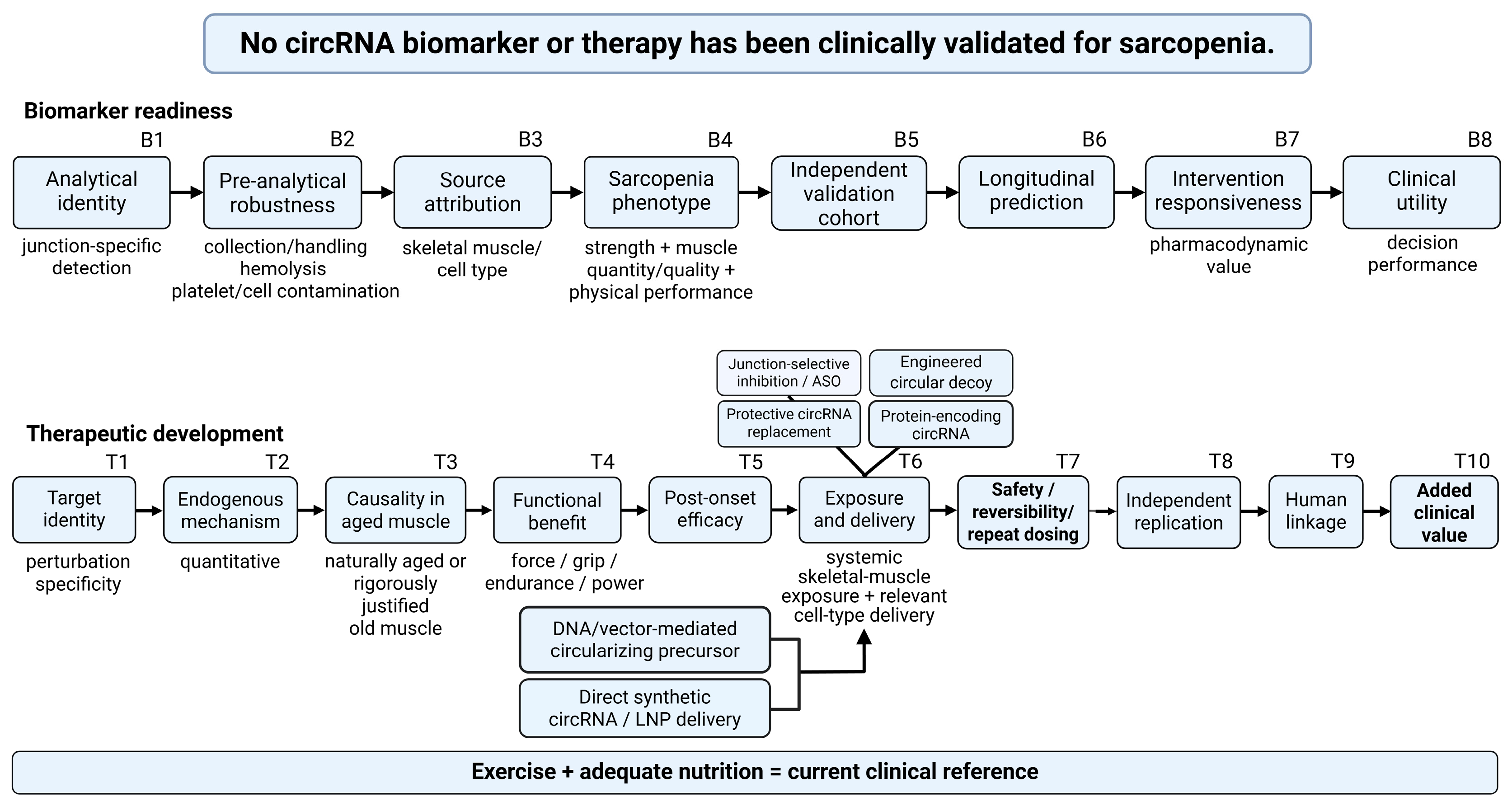
Proposed readiness criteria for circRNA biomarkers of skeletal muscle aging and sarcopenia. B1–B8 correspond to the sequential biomarker checkpoints in Figure 3. General evidence for extracellular RNA and platelet circRNA is summarized in refs. [62,63,64].
| Domain | Minimum requirement | Why it matters |
|---|---|---|
| B1 Analytical identity | Back-splice junction sequence; assay specificity; discrimination from host/linear RNA; reproducibility | Avoids measuring the wrong RNA species |
| B2 Pre-analytical robustness | Tube type; processing delay; freeze-thaw; hemolysis; platelet/cell contamination; storage | Essential for plasma, serum, or extracellular-vesicle studies |
| B3 Source attribution | Muscle enrichment or orthogonal evidence of muscle contribution; cell-type localization where possible | Prevents generic circulating RNA from being mislabeled as muscle-specific |
| B4 Sarcopenia phenotype | Accepted sarcopenia criteria; strength; muscle quantity/quality; physical performance | Links molecular signal to clinical phenotype |
| B5 Independent validation | Separate discovery and validation cohorts; external laboratory where feasible | Limits overfitting and batch-specific findings |
| B6 Longitudinal prediction | Prediction of decline beyond age and standard variables | Demonstrates incremental utility |
| B7 Intervention responsiveness | Prespecified response to exercise/nutrition/therapy with appropriate timing | Supports pharmacodynamic use |
| B8 Clinical utility | Effect size, calibration, discrimination and clinically relevant thresholds for intended use | Separates association from clinically useful classification |
8. Therapeutic Intervention Concepts for Aging Muscle
8.1. Clinical Comparators and Endogenous Targets
Exercise, particularly resistance-oriented training, has the strongest intervention evidence for sarcopenia and improves strength and physical performance across many trials [80]. Nutritional support can be useful, especially when intake is inadequate, but meta-analyses differ on the extent to which protein supplementation enhances resistance training outcomes across populations [81,82]. A circRNA therapy should ultimately be assessed as an adjunct to established care or as an option for people unable to obtain sufficient benefit from exercise and nutrition.
Exercise also provides a mechanistically informative perturbation for RNA biology. In naturally aging rats, distinct exercise regimens alter microRNA profiles together with atrophy-related phenotypes [83], and aging circRNA profiles are exercise-responsive [67]. Human studies should test whether circRNAs track or mediate adaptation to resistance exercise, rather than relying solely on comparisons of old and young tissue at rest.
For pathogenic candidates such as circSnd1 or circDdb1, junction-selective inhibition is the most direct concept. Antisense oligonucleotides (ASOs) or related RNA-targeting strategies could in principle reduce the circle while sparing the host transcript, but selectivity must be demonstrated experimentally. In older adults, repeat dosing, renal and hepatic handling, innate immune activation, off-target hybridization, and distribution across a large muscle mass become central development questions. Dose-exposure-response relationships in relevant muscles are more informative than local proof-of-concept injection alone.
8.2. Protective and Engineered circRNA Modalities
Protective circRNAs could be augmented by direct RNA delivery or by DNA vectors that express a circularizing precursor. These are pharmacologically different modalities. Direct circRNA administration provides a defined RNA dose but faces constraints on uptake, endosomal escape, and biodistribution. Vector-mediated expression may provide longer exposure but is less reversible and introduces vector immunity and redosing issues. These distinctions preclude the blanket statement that an adeno-associated virus (AAV) 'delivers circRNA.'
Engineered circmiR-29b shows that circular topology can serve as a therapeutic platform, not only as an endogenous target [70]. Engineered circular RNAs can also support prolonged protein expression [18,84]. In some settings, a designed molecule may be more tractable than manipulating a low-abundance endogenous circRNA, as sequence, potency, translation, purity, and manufacturing can be deliberately optimized.
Broader circRNA technologies have entered early human testing beyond skeletal muscle. RXRG001 is a lipid-nanoparticle (LNP)-formulated, protein-encoding circRNA registered for radiation-induced xerostomia. RXIM002 is a circRNA-LNP product designed for in vivo CD19-directed chimeric antigen receptor (CAR) T-cell engineering [85,86]. These registrations establish entry into early clinical testing, but they do not establish skeletal-muscle targeting, sarcopenia efficacy, or long-term safety in older adults. A targeted ClinicalTrials.gov search updated on 12 September 2026 did not identify an interventional circRNA trial for sarcopenia or skeletal-muscle wasting. The registered circRNA-encoded peptide dendritic-cell vaccine in breast cancer [87] is an ex vivo cellular vaccine and does not provide evidence for direct systemic circRNA delivery.
8.3. Delivery Constraints for Systemic Muscle Treatment
Skeletal muscle is a large, distributed organ. Effective systemic therapy may require exposure across multiple limb and respiratory muscles while limiting exposure to the liver, spleen, heart, and immune system. LNPs can encapsulate in vitro-transcribed circRNA and provide a clinically familiar manufacturing concept [20,88], but generic LNPs are not inherently muscle-specific. Local intramuscular success should not be interpreted as evidence that a modality can treat generalized sarcopenia.
The required target cell may also differ depending on the mechanism. A myofiber-directed therapy may not correct satellite-cell, fibro-adipogenic progenitor, motor-neuron, vascular, or immune components of aging. The delivery strategy should follow causal cell localization and not precede it.
9. Translational Evaluation Framework
9.1. Criteria for Continued Development
Table 7 and the T1–T10 therapeutic track in Figure 3 define sequential criteria for deciding whether a candidate should advance, be redesigned, or be discontinued. The identifiers indicate checkpoint order rather than current achievement. Failure at an early criterion should not be offset by additional descriptive data.
9.2. Replication and Model Relevance
Several of the strongest muscle-wasting circRNA studies are technically rigorous and provide substantial mechanistic or functional evidence. Multiple advanced candidates were also developed within overlapping investigator networks and related experimental platforms. This concentration does not invalidate the findings, but it limits the current evidence for generalizability. circTmeff1, circDdb1, circSnd1, and engineered circmiR-29b share investigators across studies [65,66,69,70]. Independent reproduction of direction, mechanism, and function should precede expensive delivery, toxicology, or manufacturing programs.
Older organisms differ in immune responsiveness, renal and hepatic handling, tissue perfusion, matrix stiffness, regenerative capacity, neuromuscular reserve, and multimorbidity. A dose that changes a circRNA in young denervated muscle may have different exposure, safety, and efficacy in aged tissue. Translational experiments should include naturally aged animals or a rigorously justified aging model and should report sex, activity, frailty-related phenotype, and baseline function. Across the candidate literature, sex and baseline activity were not reported consistently enough for reliable cross-study stratification; this reporting gap limits generalizability.
Figure 3.
Sequential development checkpoints for circRNA biomarkers and therapeutics in skeletal muscle aging. B1–B8 denote analytical identity, pre-analytical robustness, source attribution, sarcopenia phenotype, independent validation, longitudinal prediction, intervention responsiveness, and clinical utility. T1–T10 denote target identity, endogenous mechanism, causality in aged muscle, functional benefit, post-onset efficacy, exposure and delivery, safety, reversibility and repeat dosing, independent replication, human linkage, and added clinical value. The alphanumeric identifiers mark checkpoint order and are not ratings of current achievement. Arrows show the intended development sequence only; they do not imply biological causality or the likelihood of regulatory approval. The upper statement summarizes the targeted literature reviewed here: no circRNA biomarker or therapy is clinically validated for sarcopenia. Illustrative therapeutic modalities include junction-selective antisense oligonucleotides (ASOs), circular decoys, protective circRNA replacement, protein-encoding circRNAs, vector-mediated expression, and direct synthetic circRNA or lipid nanoparticle (LNP) delivery. Detailed criteria and decision rules are provided in Table 6 and Table 7. Exercise and adequate nutrition provide the current clinical reference against which added therapeutic value should be tested. ASO, antisense oligonucleotide; circRNA, circular RNA; LNP, lipid nanoparticle. Created in BioRender. Lee, W. (2026) https://BioRender.com/f3eln06.
Figure 3.
Sequential development checkpoints for circRNA biomarkers and therapeutics in skeletal muscle aging. B1–B8 denote analytical identity, pre-analytical robustness, source attribution, sarcopenia phenotype, independent validation, longitudinal prediction, intervention responsiveness, and clinical utility. T1–T10 denote target identity, endogenous mechanism, causality in aged muscle, functional benefit, post-onset efficacy, exposure and delivery, safety, reversibility and repeat dosing, independent replication, human linkage, and added clinical value. The alphanumeric identifiers mark checkpoint order and are not ratings of current achievement. Arrows show the intended development sequence only; they do not imply biological causality or the likelihood of regulatory approval. The upper statement summarizes the targeted literature reviewed here: no circRNA biomarker or therapy is clinically validated for sarcopenia. Illustrative therapeutic modalities include junction-selective antisense oligonucleotides (ASOs), circular decoys, protective circRNA replacement, protein-encoding circRNAs, vector-mediated expression, and direct synthetic circRNA or lipid nanoparticle (LNP) delivery. Detailed criteria and decision rules are provided in Table 6 and Table 7. Exercise and adequate nutrition provide the current clinical reference against which added therapeutic value should be tested. ASO, antisense oligonucleotide; circRNA, circular RNA; LNP, lipid nanoparticle. Created in BioRender. Lee, W. (2026) https://BioRender.com/f3eln06.
10. Emerging Directions
10.1. Mechanotransduction and Cell-Resolved Discovery
Mechanotransduction is an attractive but underdeveloped bridge between circRNAs and aging muscle. Nuclear mechanobiology and YAP/TAZ signaling change with aging and unloading [50,51,52,53], while work on the TPM1 super-enhancer and circTPM1 links a skeletal-muscle circRNA program to mechanotransduction and regeneration [78]. The next experiments should identify load-responsive circRNAs in old human muscle, localize them to the relevant cell type, quantify endogenous abundance, and test junction-specific causality under controlled mechanical conditions. They should then determine whether the candidates modify YAP/TAZ, myocardin-related transcription factor/serum response factor (MRTF/SRF), focal-adhesion, ion-channel, or mitochondrial responses. Recent discussion of circRNA-RBP mechanisms in exercise remodeling also supports moving beyond default sponge diagrams [26].
Whole-muscle RNA averages cell populations that change with age. Long-read sequencing can improve isoform definition, while spatial and single-cell-compatible approaches can identify where a circRNA and its molecular partners coexist. These methods are particularly important for low-abundance ceRNA claims and for blood-biomarker source attribution. Standard poly(A)-based single-cell workflows do not uniformly capture circRNAs. The method choice should match the circular species being tested.
10.2. Integrating Biomarkers, Interventions, and Mechanism
A strong translational program should connect biomarker and therapeutic development. A tissue or circulating circRNA that reflects target engagement could support dose selection, stratification, and proof of mechanism. An intervention-responsive circRNA may also be useful as a pharmacodynamic marker even if it is not causal. Separating causal targets from useful markers prevents the assumption that every biomarker should also be a therapeutic target.
Muscle circRNA research should increasingly test RBP interactions, translation, localization-dependent functions, and host-gene relationships instead of defaulting to a circRNA-miRNA-mRNA diagram [11,26]. The strongest current wasting examples already support this shift. circSnd1 acts through protein stability, circDdb1 and circTmeff1 include protein-coding mechanisms, and engineered circles operate as designed therapeutic molecules. Mechanistic diversity is central to identifying biology that can withstand translational scrutiny.
11. Conclusions
CircRNA biology now intersects meaningfully with skeletal-muscle aging, although the maturity of individual claims differs sharply. The most convincing studies combine unambiguous circular identity, specific perturbation, a quantitatively credible mechanism, relevant in vivo causality, and functional endpoints. circSnd1 and circDdb1 currently provide the strongest functional preclinical evidence and are associated with aged human skeletal muscle, but neither has clinical validation. circFUT10, circBBS9, and circNEB address regenerative, discovery, and histological aspects of aging and require deeper functional testing. circGLIS3 is relevant to muscle quality but should be examined directly in aging before it is considered a sarcopenia mechanism.
Progress in biomarker development requires junction-level analytical validation, pre-analytical robustness, source attribution, accepted sarcopenia phenotyping, independent cohorts, and longitudinal or intervention-linked performance. Therapeutic development depends on systemic muscle exposure, post-onset functional efficacy in aged organisms, safety, reversibility, and independent replication. CircRNA platforms have entered human clinical development outside skeletal muscle, but this does not establish skeletal-muscle delivery, efficacy, or long-term safety. Future work should focus on a small number of candidates tested rigorously against these requirements.
Supplementary Materials
The following supporting information can be downloaded at the website of this paper posted on Preprints.org. Table S1, Molecular identity and independent modifiers of the circRNA studies summarized in Table 3 and Table 4.
Author Contributions
Conceptualization, W.L.; investigation, T.H.P.N. and W.L.; writing - original draft preparation, W.L.; writing - review and editing, W.L.; visualization, T.H.P.N. and W.L.; funding acquisition, W.L. The authors have read and agreed to the published version of the manuscript.
Funding
This study was supported by the National Research Foundation of Korea (NRF), which is funded by the Korean government (grant no. RS-2026-25480711).
Institutional Review Board Statement
Not applicable.
Informed Consent Statement
Not applicable.
Data Availability Statement
No new datasets were generated in this study. All information evaluated in this review is available in the cited publications and public clinical trial registries.
Acknowledgments
During the preparation of this manuscript, the authors used OpenAI ChatGPT to assist with preliminary figure schematics and with language and consistency checks. The authors subsequently redrew and edited the figures in BioRender. The authors reviewed and edited all AI-assisted output and take full responsibility for the content of this publication.
Conflicts of Interest
The authors declare no conflict of interest.
References
- Cruz-Jentoft, A.J.; Bahat, G.; Bauer, J.; Boirie, Y.; Bruyère, O.; Cederholm, T.; Cooper, C.; Landi, F.; Rolland, Y.; Sayer, A.A.; et al. Sarcopenia: revised European consensus on definition and diagnosis. Age Ageing 2019, 48, 16–31. [Google Scholar] [CrossRef] [PubMed]
- Chen, L.K.; Woo, J.; Assantachai, P.; Auyeung, T.W.; Chou, M.Y.; Iijima, K.; Jang, H.C.; Kang, L.; Kim, M.; Kim, S.; et al. Asian Working Group for Sarcopenia: 2019 Consensus Update on Sarcopenia Diagnosis and Treatment. J. Am. Med. Dir. Assoc. 2020, 21, 300–307.e2. [Google Scholar] [CrossRef] [PubMed]
- Kirk, B.; Cawthon, P.M.; Arai, H.; Ávila-Funes, J.A.; Barazzoni, R.; Bhasin, S.; Binder, E.F.; Bruyere, O.; Cederholm, T.; Chen, L.K.; et al. The Conceptual Definition of Sarcopenia: Delphi Consensus from the Global Leadership Initiative in Sarcopenia (GLIS). Age Ageing 2024, 53, afae052. [Google Scholar] [CrossRef] [PubMed]
- Larsson, L.; Degens, H.; Li, M.; Salviati, L.; Lee, Y.I.; Thompson, W.; Kirkland, J.L.; Sandri, M. Sarcopenia: Aging-Related Loss of Muscle Mass and Function. Physiol. Rev. 2019, 99, 427–511. [Google Scholar] [CrossRef] [PubMed]
- Grima-Terrén, M.; Campanario, S.; Ramírez-Pardo, I.; Cisneros, A.; Hong, X.; Perdiguero, E.; Serrano, A.L.; Isern, J.; Muñoz-Cánoves, P. Muscle aging and sarcopenia: The pathology, etiology, and most promising therapeutic targets. Mol. Asp. Med. 2024, 100, 101319. [Google Scholar] [CrossRef] [PubMed]
- Lopez-Otin, C.; Blasco, M.A.; Partridge, L.; Serrano, M.; Kroemer, G. Hallmarks of aging: An expanding universe. Cell 2023, 186, 243–278. [Google Scholar] [CrossRef] [PubMed]
- Ferri, E.; Marzetti, E.; Calvani, R.; Picca, A.; Cesari, M.; Arosio, B. Role of Age-Related Mitochondrial Dysfunction in Sarcopenia. Int. J. Mol. Sci. 2020, 21, 5236. [Google Scholar] [CrossRef] [PubMed]
- Chen, M.; Wang, Y.; Deng, S.; Lian, Z.; Yu, K. Skeletal muscle oxidative stress and inflammation in aging: Focus on antioxidant and anti-inflammatory therapy. Front Cell Dev. Biol. 2022, 10, 964130. [Google Scholar] [CrossRef] [PubMed]
- Memczak, S.; Jens, M.; Elefsinioti, A.; Torti, F.; Krueger, J.; Rybak, A.; Maier, L.; Mackowiak, S.D.; Gregersen, L.H.; Munschauer, M.; et al. Circular RNAs are a large class of animal RNAs with regulatory potency. Nature 2013, 495, 333–338. [Google Scholar] [CrossRef] [PubMed]
- Kristensen, L.S.; Andersen, M.S.; Stagsted, L.V.W.; Ebbesen, K.K.E.; Hansen, T.B.; Kjems, J. The biogenesis, biology and characterization of circular RNAs. Nat. Rev. Genet 2019, 20, 675–691. [Google Scholar] [CrossRef] [PubMed]
- Liu, C.X.; Chen, L.L. Circular RNAs: Characterization, cellular roles, and applications. Cell 2022, 185, 2016–2034. [Google Scholar] [CrossRef] [PubMed]
- Jeck, W.R.; Sorrentino, J.A.; Wang, K.; Slevin, M.K.; Burd, C.E.; Liu, J.; Marzluff, W.F.; Sharpless, N.E. Circular RNAs are abundant, conserved, and associated with ALU repeats. RNA 2013, 19, 141–157. [Google Scholar] [CrossRef] [PubMed]
- Salzman, J.; Gawad, C.; Wang, P.L.; Lacayo, N.; Brown, P.O. Circular RNAs are the predominant transcript isoform from hundreds of human genes in diverse cell types. PLoS ONE 2012, 7, e30733. [Google Scholar] [CrossRef] [PubMed]
- Ashwal-Fluss, R.; Meyer, M.; Pamudurti, N.R.; Ivanov, A.; Bartok, O.; Hanan, M.; Evantal, N.; Memczak, S.; Rajewsky, N.; Kadener, S. circRNA biogenesis competes with pre-mRNA splicing. Mol. Cell 2014, 56, 55–66. [Google Scholar] [CrossRef] [PubMed]
- Hansen, T.B.; Jensen, T.I.; Clausen, B.H.; Bramsen, J.B.; Finsen, B.; Damgaard, C.K.; Kjems, J. Natural RNA circles function as efficient microRNA sponges. Nature 2013, 495, 384–388. [Google Scholar] [CrossRef] [PubMed]
- Legnini, I.; Di Timoteo, G.; Rossi, F.; Morlando, M.; Briganti, F.; Sthandier, O.; Fatica, A.; Santini, T.; Andronache, A.; Wade, M.; et al. Circ-ZNF609 Is a Circular RNA that Can Be Translated and Functions in Myogenesis. Mol. Cell 2017, 66, 22–37.e9. [Google Scholar] [CrossRef] [PubMed]
- Yang, Y.; Fan, X.; Mao, M.; Song, X.; Wu, P.; Zhang, Y.; Jin, Y.; Yang, Y.; Chen, L.L.; Wang, Y.; et al. Extensive translation of circular RNAs driven by N6-methyladenosine. Cell Res. 2017, 27, 626–641. [Google Scholar] [CrossRef] [PubMed]
- Wesselhoeft, R.A.; Kowalski, P.S.; Anderson, D.G. Engineering circular RNA for potent and stable translation in eukaryotic cells. Nat. Commun. 2018, 9, 2629. [Google Scholar] [CrossRef] [PubMed]
- O'Leary, E.; Jiang, Y.; Kristensen, L.S.; Hansen, T.B.; Kjems, J. The therapeutic potential of circular RNAs. Nat. Rev. Genet 2025, 26, 230–244. [Google Scholar] [CrossRef] [PubMed]
- Cao, X.; Cai, Z.; Zhang, J.; Zhao, F. Engineering circular RNA medicines. Nat. Rev. Bioeng. 2025, 3, 270–287. [Google Scholar] [CrossRef]
- Knupp, D.; Miura, P. CircRNA accumulation: a new hallmark of aging? Mech. Ageing Dev. 2018, 173, 71–79. [Google Scholar] [CrossRef] [PubMed]
- Shin, Y.J.; Kwon, K.S.; Suh, Y.; Lee, K.P. The role of noncoding RNAs in muscle aging: regulatory mechanisms and therapeutic potential. Front Mol. Biosci. 2024, 10, 1308274. [Google Scholar] [CrossRef] [PubMed]
- Yu, P.; Qi, Y.; Huang, H.; Wang, H.; Xu, J. CircularRNA and Musculoskeletal Diseases. Adv. Exp. Med. Biol. 2025, 1485, 437–448. [Google Scholar] [CrossRef] [PubMed]
- Huang, C.J.; Choo, K.B. Circular RNAs and host genes act synergistically in regulating cellular processes and functions in skeletal myogenesis. Gene 2025, 940, 149189. [Google Scholar] [CrossRef] [PubMed]
- Greco, S.; Cardinali, B.; Falcone, G.; Martelli, F. Circular RNAs in muscle function and disease. Int. J. Mol. Sci. 2018, 19, 3454. [Google Scholar] [CrossRef] [PubMed]
- Liu, J.; Yang, Y.; Chen, H.; Liu, M.; Mao, Z.; Zheng, M. Regulatory roles of the circRNA-RBP axis in exercise-induced skeletal muscle remodeling: mechanistic controversies and translational illusions. Front Genet 2026, 17, 1772541. [Google Scholar] [CrossRef] [PubMed]
- Retraction Exosomal circRNA derived from gastric tumor promotes white adipose browning by targeting the miR-133/PRDM16 pathway. Int. J. Cancer 2023, 152, E7. [CrossRef] [PubMed]
- Cells Editorial Office Retraction: Circular RNA circ-FoxO3 Inhibits Myoblast Cells Differentiation. Cells 2020, 9, 2504. [CrossRef] [PubMed]
- Retraction: CircFUT10 Reduces Proliferation and Facilitates Differentiation of Myoblasts by Sponging miR-133a. J. Cell Physiol. 2026, 241, e70144. [CrossRef] [PubMed]
- Zhu, M.; Lian, C.; Chen, G.; Zou, P.; Qin, B.G. CircRNA FUT10 regulates the regenerative potential of aged skeletal muscle stem cells by targeting HOXA9. Aging 2021, 13, 17428–17441. [Google Scholar] [CrossRef] [PubMed]
- Huang, N.; Ge, M.; Liu, X.; Tian, X.; Yin, P.; Bao, Z.; Cao, F.; Shyh-Chang, N.; Dong, B.; Dai, L.; et al. A framework of biomarkers for skeletal muscle aging: a consensus statement by the Aging Biomarker Consortium. Life Med. 2024, 3, lnaf001. [Google Scholar] [CrossRef] [PubMed]
- Hughes, D.C.; Baehr, L.M.; Waddell, D.S.; Bodine, S.C. Ubiquitin Ligases in Longevity and Aging Skeletal Muscle. Int. J. Mol. Sci. 2022, 23, 7602. [Google Scholar] [CrossRef] [PubMed]
- Fujita, S.; Glynn, E.L.; Timmerman, K.L.; Rasmussen, B.B.; Volpi, E. Supraphysiological hyperinsulinaemia is necessary to stimulate skeletal muscle protein anabolism in older adults: evidence of a true age-related insulin resistance of muscle protein metabolism. Diabetologia 2009, 52, 1889–1898. [Google Scholar] [CrossRef] [PubMed]
- Durham, W.J.; Casperson, S.L.; Dillon, E.L.; Keske, M.A.; Paddon-Jones, D.; Sanford, A.P.; Hickner, R.C.; Grady, J.J.; Sheffield-Moore, M. Age-related anabolic resistance after endurance-type exercise in healthy humans. FASEB J. 2010, 24, 4117–4127. [Google Scholar] [CrossRef] [PubMed]
- Moro, T.; Brightwell, C.R.; Deer, R.R.; Graber, T.G.; Galvan, E.; Fry, C.S.; Volpi, E.; Rasmussen, B.B. Muscle protein anabolic resistance to essential amino acids does not occur in healthy older adults before or after resistance exercise training. J. Nutr. 2018, 148, 900–909. [Google Scholar] [CrossRef] [PubMed]
- Jing, Y.; Zuo, Y.; Sun, L.; Yu, Z.R.; Ma, S.; Hu, H.; Zhao, Q.; Huang, D.; Zhang, W.; Belmonte, J.C.I.; et al. SESN1 is a FOXO3 effector that counteracts human skeletal muscle ageing. Cell Prolif. 2023, 56, e13455. [Google Scholar] [CrossRef] [PubMed]
- Zhang, J.; Hu, M.; Wu, X.; Guo, M.; Ma, Y.; Qiu, J.; Wang, S.; Cao, Y.; Zhong, Y.; Chen, F.; et al. Skeletal Muscle HSF1 Alleviates Age-Associated Sarcopenia and Mitochondrial Function Decline via SIRT3-PGC1alpha Axis. Adv. Sci. (Weinh) 2026, 13, e10368. [Google Scholar] [CrossRef] [PubMed]
- Affourtit, C.; Carre, J.E. Mitochondrial involvement in sarcopenia. Acta Physiol. (Oxf) 2024, 240, e14107. [Google Scholar] [CrossRef] [PubMed]
- Grevendonk, L.; Connell, N.J.; McCrum, C.; Fealy, C.E.; Bilet, L.; Bruls, Y.M.H.; Mevenkamp, J.; Schrauwen-Hinderling, V.B.; Jörgensen, J.A.; Moonen-Kornips, E.; et al. Impact of aging and exercise on skeletal muscle mitochondrial capacity, energy metabolism, and physical function. Nat. Commun. 2021, 12, 4773. [Google Scholar] [CrossRef] [PubMed]
- Scudese, E.; Marshall, A.G.; Vue, Z.; Exil, V.; Rodriguez, B.I.; Demirci, M.; Vang, L.; López, E.G.; Neikirk, K.; Shao, B.; et al. 3D Mitochondrial Structure in Aging Human Skeletal Muscle: Insights Into MFN-2-Mediated Changes. Aging Cell 2025, 24, e70054. [Google Scholar] [CrossRef] [PubMed]
- Sousa-Victor, P.; Gutarra, S.; García-Prat, L.; Rodriguez-Ubreva, J.; Ortet, L.; Ruiz-Bonilla, V.; Jardí, M.; Ballestar, E.; González, S.; Serrano, A.L.; et al. Geriatric muscle stem cells switch reversible quiescence into senescence. Nature 2014, 506, 316–321. [Google Scholar] [CrossRef] [PubMed]
- Cosgrove, B.D.; Gilbert, P.M.; Porpiglia, E.; Mourkioti, F.; Lee, S.P.; Corbel, S.Y.; Llewellyn, M.E.; Delp, S.L.; Blau, H.M. Rejuvenation of the muscle stem cell population restores strength to injured aged muscles. Nat. Med. 2014, 20, 255–264. [Google Scholar] [CrossRef] [PubMed]
- Fry, C.S.; Lee, J.D.; Mula, J.; Kirby, T.J.; Jackson, J.R.; Liu, F.; Yang, L.; Mendias, C.L.; Dupont-Versteegden, E.E.; McCarthy, J.J.; et al. Inducible depletion of satellite cells in adult, sedentary mice impairs muscle regenerative capacity without affecting sarcopenia. Nat. Med. 2015, 21, 76–80. [Google Scholar] [CrossRef] [PubMed]
- Dowling, P.; Gargan, S.; Swandulla, D.; Ohlendieck, K. Fiber-Type Shifting in Sarcopenia of Old Age: Proteomic Profiling of the Contractile Apparatus of Skeletal Muscles. Int. J. Mol. Sci. 2023, 24, 2415. [Google Scholar] [CrossRef] [PubMed]
- Murgia, M.; Toniolo, L.; Nagaraj, N.; Ciciliot, S.; Vindigni, V.; Schiaffino, S.; Reggiani, C.; Mann, M. Single Muscle Fiber Proteomics Reveals Fiber-Type-Specific Features of Human Muscle Aging. Cell Rep. 2017, 19, 2396–2409. [Google Scholar] [CrossRef] [PubMed]
- Hepple, R.T.; Rice, C.L. Innervation and neuromuscular control in ageing skeletal muscle. J. Physiol. 2016, 594, 1965–1978. [Google Scholar] [CrossRef] [PubMed]
- Soendenbroe, C.; Heisterberg, M.F.; Schjerling, P.; Karlsen, A.; Kjaer, M.; Andersen, J.L.; Mackey, A.L. Molecular indicators of denervation in aging human skeletal muscle. Muscle Nerve 2019, 60, 453–463. [Google Scholar] [CrossRef] [PubMed]
- Sarto, F.; Franchi, M.V.; McPhee, J.S.; Stashuk, D.W.; Paganini, M.; Monti, E.; Rossi, M.; Sirago, G.; Zampieri, S.; Motanova, E.S.; et al. Neuromuscular impairment at different stages of human sarcopenia. J. Cachexia Sarcopenia Muscle 2024, 15, 1797–1810. [Google Scholar] [CrossRef] [PubMed]
- Lukasiewicz, C.J.; Tranah, G.J.; Evans, D.S.; Coen, P.M.; Barnes, H.N.; Huo, Z.; Esser, K.A.; Zhang, X.; Wolff, C.; Wu, K.; et al. Higher expression of denervation-responsive genes is negatively associated with muscle volume and performance traits in the Study of Muscle, Mobility, and Aging (SOMMA). Aging Cell 2024, 23, e14115, Correction in: Aging Cell 2024, 23, e14415. https://doi.org/10.1111/acel.14415. [Google Scholar] [CrossRef] [PubMed]
- Iyer, S.R.; Folker, E.S.; Lovering, R.M. The Nucleoskeleton: Crossroad of Mechanotransduction in Skeletal Muscle. Front Physiol. 2021, 12, 724010. [Google Scholar] [CrossRef] [PubMed]
- van Ingen, M.J.A.; Kirby, T.J. LINCing Nuclear Mechanobiology With Skeletal Muscle Mass and Function. Front Cell Dev. Biol. 2021, 9, 690577. [Google Scholar] [CrossRef] [PubMed]
- Iyer, S.R.; Hsia, R.C.; Folker, E.S.; Lovering, R.M. Age-dependent changes in nuclear-cytoplasmic signaling in skeletal muscle. Exp. Gerontol. 2021, 150, 111338. [Google Scholar] [CrossRef] [PubMed]
- Kwon, Y. YAP/TAZ as Molecular Targets in Skeletal Muscle Atrophy and Osteoporosis. Aging Dis. 2025, 16, 299–320. [Google Scholar] [CrossRef] [PubMed]
- Schiaffino, S.; Reggiani, C.; Akimoto, T. Molecular Mechanisms of Skeletal Muscle Hypertrophy. J. Neuromuscul. Dis. 2021, 8, 169–183. [Google Scholar] [CrossRef] [PubMed]
- Lu, Y.; Bai, Y.; Li, W.; Zhou, Z.; Lai, H.; Hu, X.; Yang, T.; Wang, C.; Chen, Y.; Gan, K.; et al. Mechanical Stimulation Induces Yap Mediated OCTN2 Transcription to Enhance Carnitine Metabolism in Sarcopenia. J. Cachexia Sarcopenia Muscle 2025, 16, e70052. [Google Scholar] [CrossRef] [PubMed]
- Watt, K.I.; Henstridge, D.C.; Ziemann, M.; Sim, C.B.; Montgomery, M.K.; Samocha-Bonet, D.; Parker, B.L.; Dodd, G.T.; Bond, S.T.; Salmi, T.M.; et al. Yap regulates skeletal muscle fatty acid oxidation and adiposity in metabolic disease. Nat. Commun. 2021, 12, 2887. [Google Scholar] [CrossRef] [PubMed]
- Sladitschek-Martens, H.L.; Guarnieri, A.; Brumana, G.; Zanconato, F.; Battilana, G.; Xiccato, R.L.; Panciera, T.; Forcato, M.; Bicciato, S.; Guzzardo, V.; et al. YAP/TAZ activity in stromal cells prevents ageing by controlling cGAS-STING. Nature 2022, 607, 790–798. [Google Scholar] [CrossRef] [PubMed]
- Shen, Y.; Luo, L.; Fu, H.; Xie, L.; Zhang, W.; Lu, J.; Yang, M. Chest computed tomography-derived muscle mass and quality indicators, in-hospital outcomes, and costs in older inpatients. J. Cachexia Sarcopenia Muscle 2022, 13, 966–975. [Google Scholar] [CrossRef] [PubMed]
- Hetherington-Rauth, M.; Mansfield, T.A.; Lenchik, L.; Weaver, A.A.; Cawthon, P.M. Associations of CT muscle area and density with functional outcomes and mortality across anatomical regions in older men. J. Am. Geriatr. Soc. 2025, 73, 2717–2726. [Google Scholar] [CrossRef] [PubMed]
- Yu, S.; Wang, J.; Liu, H.; Guo, J.; Kuraz, A.B.; Pan, Y.; Li, A.; Mei, C.; Cheng, G.; Zan, L. CircGLIS3 Inhibits Intramuscular Adipogenesis and Alleviates Skeletal Muscle Fat Infiltration. J. Cachexia Sarcopenia Muscle 2025, 16, e70009. [Google Scholar] [CrossRef] [PubMed]
- Denzler, R.; Agarwal, V.; Stefano, J.; Bartel, D.P.; Stoffel, M. Assessing the ceRNA hypothesis with quantitative measurements of miRNA and target abundance. Mol. Cell 2014, 54, 766–776. [Google Scholar] [CrossRef] [PubMed]
- Kirschner, M.B.; Edelman, J.J.; Kao, S.C.; Vallely, M.P.; van Zandwijk, N.; Reid, G. The impact of hemolysis on cell-free microRNA biomarkers. Front Genet 2013, 4, 94. [Google Scholar] [CrossRef] [PubMed]
- Alhasan, A.A.; Izuogu, O.G.; Al-Balool, H.H.; Steyn, J.S.; Evans, A.; Colzani, M.; Ghevaert, C.; Mountford, J.C.; Marenah, L.; Elliott, D.J.; et al. Circular RNA enrichment in platelets is a signature of transcriptome degradation. Blood 2016, 127, e1–e11. [Google Scholar] [CrossRef] [PubMed]
- Preußer, C.; Hung, L.H.; Schneider, T.; Schreiner, S.; Hardt, M.; Moebus, A.; Santoso, S.; Bindereif, A. Selective release of circRNAs in platelet-derived extracellular vesicles. J. Extracell. Vesicles 2018, 7, 1424473. [Google Scholar] [CrossRef] [PubMed]
- Li, J.; Jin, B.; Yan, Y.; Chen, Y.; Yin, X.; Ren, X.; Li, Q.; Chen, J.; Wang, S.; Yang, T.; et al. EIF4A3-Induced Circular RNA circSnd1 Promotes Muscle Atrophy and Muscle Ageing by Stabilizing EEF1A1. J. Cachexia Sarcopenia Muscle 2026, 17, e70210. [Google Scholar] [CrossRef] [PubMed]
- Zhu, X.; Yang, T.; Zheng, Y.; Nie, Q.; Chen, J.; Li, Q.; Ren, X.; Yin, X.; Wang, S.; Yan, Y.; et al. EIF4A3-Induced Circular RNA CircDdb1 Promotes Muscle Atrophy through Encoding a Novel Protein CircDdb1-867aa. Adv. Sci. (Weinh) 2024, 11, e2406986. [Google Scholar] [CrossRef] [PubMed]
- Guo, M.; Qiu, J.; Shen, F.; Wang, S.; Yu, J.; Zuo, H.; Yao, J.; Xu, S.; Hu, T.; Wang, D.; et al. Comprehensive analysis of circular RNA profiles in skeletal muscles of aging mice and after aerobic exercise intervention. Aging 2020, 12, 5071–5090. [Google Scholar] [CrossRef] [PubMed]
- Huang, K.; Li, Z.; Zhong, D.; Yang, Y.; Yan, X.; Feng, T.; Wang, X.; Zhang, L.; Shen, X.; Chen, M.; et al. A Circular RNA Generated from Nebulin (NEB) Gene Splicing Promotes Skeletal Muscle Myogenesis in Cattle as Detected by a Multi-Omics Approach. Adv. Sci. (Weinh) 2024, 11, e2300702. [Google Scholar] [CrossRef] [PubMed]
- Chen, R.; Yang, T.; Jin, B.; Xu, W.; Yan, Y.; Wood, N.; Lehmann, H.I.; Wang, S.; Zhu, X.; Yuan, W.; et al. CircTmeff1 Promotes Muscle Atrophy by Interacting with TDP-43 and Encoding A Novel TMEFF1-339aa Protein. Adv. Sci. (Weinh) 2023, 10, e2206732. [Google Scholar] [CrossRef] [PubMed]
- Li, J.; Chen, R.; Zheng, Y.; Yuan, W.; Yang, T.; Zhu, X.; Yan, Y.; Jin, B.; Xu, W.; Zhang, Z.; et al. Engineered Circular RNA CircmiR-29b Attenuates Muscle Atrophy by Sponging MiR-29b. Adv. Ther. 2022, 5, 2200029. [Google Scholar] [CrossRef]
- Shi, X.; Yang, J.; Liu, M.; Zhang, Y.; Zhou, Z.; Luo, W.; Fung, K.M.; Xu, C.; Bronze, M.S.; Houchen, C.W.; et al. Circular RNA ANAPC7 Inhibits Tumor Growth and Muscle Wasting via PHLPP2-AKT-TGF-beta Signaling Axis in Pancreatic Cancer. Gastroenterology 2022, 162, 2004–2017.e2. [Google Scholar] [CrossRef] [PubMed]
- Ding, Z.; Sun, D.; Han, J.; Shen, L.; Yang, F.; Sah, S.; Sui, X.; Wu, G. Novel noncoding RNA CircPTK2 regulates lipolysis and adipogenesis in cachexia. Mol. Metab. 2021, 53, 101310. [Google Scholar] [CrossRef] [PubMed]
- Zhao, X.; Tang, S.; Lei, Z.; Shen, X.; Zhang, Y.; Han, S.; Yin, H.; Cui, C. circAGO3 facilitates NF-kappaB pathway-mediated inflammatory atrophy in chicken skeletal muscle via the miR-34b-5p/TRAF3 axis. Int. J. Biol. Macromol. 2024, 283, 137614. [Google Scholar] [CrossRef] [PubMed]
- Zhao, J.; Zhao, X.; Shen, X.; Zhang, Y.; Zhang, Y.; Ye, L.; Li, D.; Zhu, Q.; Yin, H. CircCCDC91 regulates chicken skeletal muscle development by sponging miR-15 family via activating IGF1-PI3K/AKT signaling pathway. Poult. Sci. 2022, 101, 101803. [Google Scholar] [CrossRef] [PubMed]
- Weng, J.; Zhang, P.; Yin, X.; Jiang, B. The Whole Transcriptome Involved in Denervated Muscle Atrophy Following Peripheral Nerve Injury. Front Mol. Neurosci. 2018, 11, 69. [Google Scholar] [CrossRef] [PubMed]
- Reinoso-Sánchez, J.F.; Baroli, G.; Duranti, G.; Scaricamazza, S.; Sabatini, S.; Valle, C.; Morlando, M.; Casero, R.A., Jr.; Bozzoni, I.; Mariottini, P.; et al. Emerging Role for Linear and Circular Spermine Oxidase RNAs in Skeletal Muscle Physiopathology. Int. J. Mol. Sci. 2020, 21, 8227. [Google Scholar] [CrossRef] [PubMed]
- Choe, N.; Jeong, A.; Joung, H.; Jeong, D.; Kim, Y.K.; Kook, H.; Kwon, D.H. Circular RNA circAtxn10 regulates skeletal muscle cell differentiation by targeting miR-143-3p and Chrna1. Korean J. Physiol. Pharmacol. 2025, 29, 637–648. [Google Scholar] [CrossRef] [PubMed]
- Zhang, R.; Feng, W.; Yang, Y.; Pan, Y.; Zou, C.; Wang, L.; Zhang, S.; Zhao, Y.; Wu, Y.; Wang, J.; et al. The Evolutionarily Conserved TPM1 Super-Enhancer Drives Skeletal Muscle Regeneration via Mechanotransduction Signaling. Adv. Sci. (Weinh) 2026, 13, e14271. [Google Scholar] [CrossRef] [PubMed]
- Meinecke, A.; Mitzka, S.; Just, A.; Cushman, S.; Stojanović, S.D.; Xiao, K.; Mooren, F.C.; Fiedler, J.; Thum, T. Cardiac endurance training alters plasma profiles of circular RNA MBOAT2. Am. J. Physiol. Heart Circ. Physiol. 2020, 319, H13–H21. [Google Scholar] [CrossRef] [PubMed]
- Shen, Y.; Shi, Q.; Nong, K.; Li, S.; Yue, J.; Huang, J.; Dong, B.; Beauchamp, M.; Hao, Q. Exercise for sarcopenia in older people: A systematic review and network meta-analysis. J. Cachexia Sarcopenia Muscle 2023, 14, 1199–1211. [Google Scholar] [CrossRef] [PubMed]
- Whaikid, P.; Piaseu, N. The effectiveness of protein supplementation combined with resistance exercise programs among community-dwelling older adults with sarcopenia: a systematic review and meta-analysis. Epidemiol. Health 2024, 46, e2024030. [Google Scholar] [CrossRef] [PubMed]
- Choi, M.; Kim, H.Y.; Bae, J. Does the combination of resistance training and a nutritional intervention have a synergic effect on muscle mass, strength, and physical function in older adults? A systematic review and meta-analysis. BMC Geriatr. 2021, 21, 639. [Google Scholar] [CrossRef] [PubMed]
- Liang, J.; Zhang, H.; Zeng, Z.; Lv, J.; Huang, J.; Wu, X.; Wang, M.; Xu, J.; Fan, J.; Chen, N. MicroRNA profiling of different exercise interventions for alleviating skeletal muscle atrophy in naturally aging rats. J. Cachexia Sarcopenia Muscle 2023, 14, 356–368. [Google Scholar] [CrossRef] [PubMed]
- Unti, M.J.; Jaffrey, S.R. Highly efficient cellular expression of circular mRNA enables prolonged protein expression. Cell Chem. Biol. 2024, 31, 163–176.e5. [Google Scholar] [CrossRef] [PubMed]
- ClinicalTrials.gov. Study of Circular RNA Treatment in Patients with Radiation-Induced Xerostomia-1 (NCT06714253). Available online: https://clinicaltrials.gov/study/NCT06714253 (accessed on 12 September 2026).
- ClinicalTrials.gov. Safety and Efficacy Study of RXIM002 in Severe, Relapsed or Refractory Autoimmune Diseases (NCT07322718). Available online: https://clinicaltrials.gov/study/NCT07322718 (accessed on 12 September 2026).
- ClinicalTrials.gov. A Single Arm Clinical Study of Dendritic Cell Vaccine Loaded with Circular RNA Encoding Cryptic Peptide for Patients with HER2-Negative Advanced Breast Cancer (NCT06530082). Available online: https://clinicaltrials.gov/study/NCT06530082 (accessed on 12 September 2026).
- Juchem, M.; Cushman, S.; Lu, D.; Chatterjee, S.; Bär, C.; Thum, T. Encapsulating In Vitro Transcribed circRNA into Lipid Nanoparticles Via Microfluidic Mixing. Methods Mol. Biol. 2024, 2765, 247–260. [Google Scholar] [CrossRef] [PubMed]
Figure 1.
Biological architecture of skeletal muscle aging and the position of current circRNA evidence. Aging and reduced physical activity converge on interconnected domains, including proteostasis and anabolic responsiveness, mitochondrial quality and redox control, inflammation, regenerative decline, neuromuscular remodeling, mechanotransduction and extracellular matrix change, and myosteatotic remodeling. We separate candidates with direct aging linkage from aging-adjacent evidence and comparator or stress-test evidence. circSnd1 and circDdb1 each combine Level 4 functional preclinical evidence with an aged-human-muscle association; these evidence dimensions remain independent, and the associations do not constitute clinical validation. circFUT10, circBBS9, and circNEB occupy lower preclinical evidence levels. The proteostasis module depicts context-dependent anabolic resistance and dysregulated autophagic flux. circGLIS3 and circTPM1 are classified as aging-adjacent, whereas circTmeff1 and engineered circmiR-29b are comparator or stress-test evidence. Developmental myogenesis and non-aging atrophy are not interchangeable with aging evidence. circRNA, circular RNA; ECM, extracellular matrix; mTOR, mechanistic target of rapamycin; NMJ, neuromuscular junction; ROS, reactive oxygen species; YAP/TAZ, Yes-associated protein and transcriptional coactivator with PDZ-binding motif. Created in BioRender. Lee, W. (2026) https://BioRender.com/osvo17l.
Figure 1.
Biological architecture of skeletal muscle aging and the position of current circRNA evidence. Aging and reduced physical activity converge on interconnected domains, including proteostasis and anabolic responsiveness, mitochondrial quality and redox control, inflammation, regenerative decline, neuromuscular remodeling, mechanotransduction and extracellular matrix change, and myosteatotic remodeling. We separate candidates with direct aging linkage from aging-adjacent evidence and comparator or stress-test evidence. circSnd1 and circDdb1 each combine Level 4 functional preclinical evidence with an aged-human-muscle association; these evidence dimensions remain independent, and the associations do not constitute clinical validation. circFUT10, circBBS9, and circNEB occupy lower preclinical evidence levels. The proteostasis module depicts context-dependent anabolic resistance and dysregulated autophagic flux. circGLIS3 and circTPM1 are classified as aging-adjacent, whereas circTmeff1 and engineered circmiR-29b are comparator or stress-test evidence. Developmental myogenesis and non-aging atrophy are not interchangeable with aging evidence. circRNA, circular RNA; ECM, extracellular matrix; mTOR, mechanistic target of rapamycin; NMJ, neuromuscular junction; ROS, reactive oxygen species; YAP/TAZ, Yes-associated protein and transcriptional coactivator with PDZ-binding motif. Created in BioRender. Lee, W. (2026) https://BioRender.com/osvo17l.
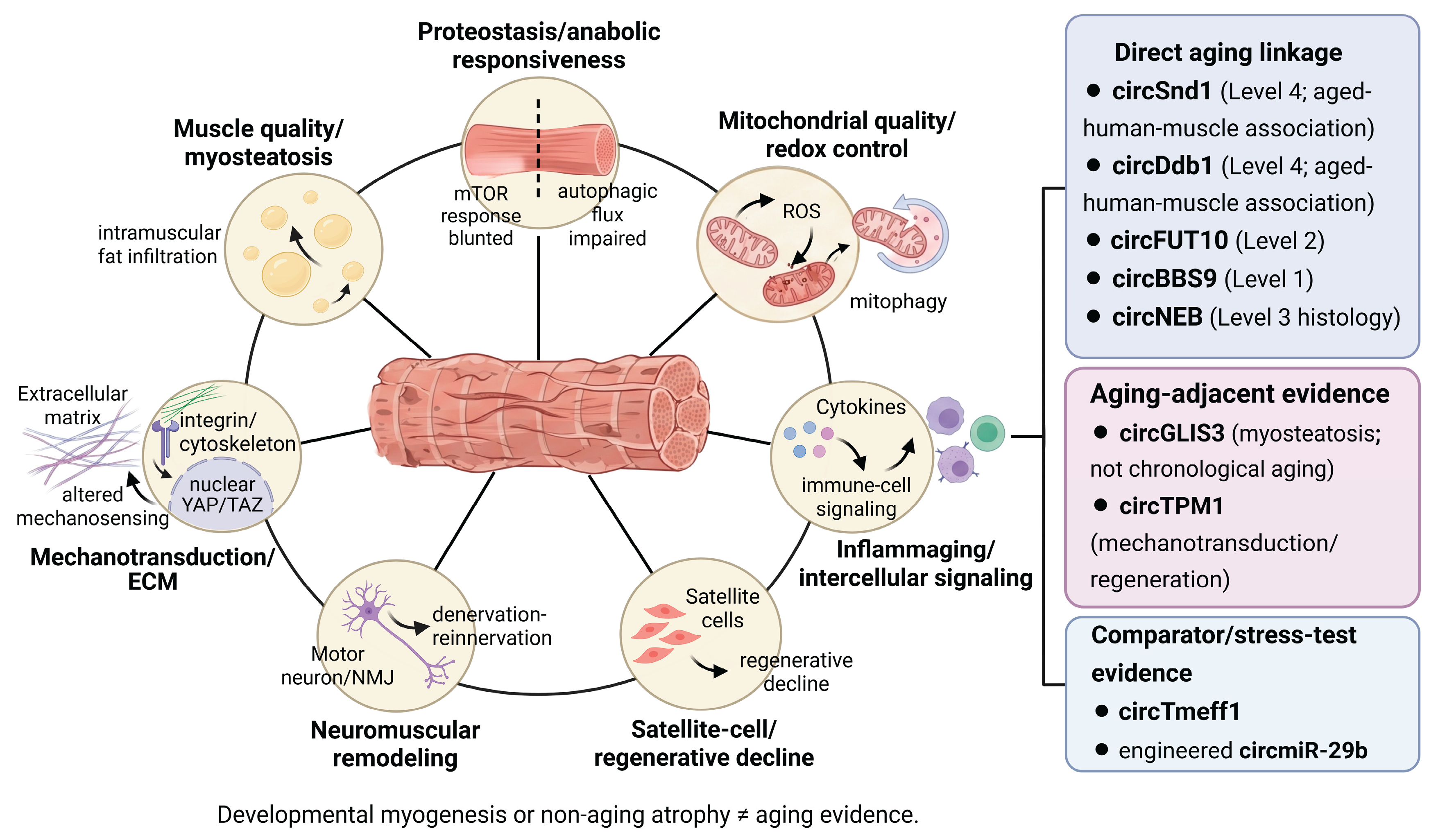
Figure 2.
Mechanisms, preclinical evidence levels, and independent modifiers for circRNA studies in aging muscle. Panel A shows miRNA interaction, RBP interaction, translation, host-gene or transcriptional effects, and extracellular-vesicle signaling. Prediction or binding alone does not establish endogenous ceRNA function; quantitative abundance, stoichiometry, and rescue or epistasis are required. Panel B distinguishes Level 1 discovery or association, Level 2 mechanism, Level 3 in vivo causality, and Level 4 intervention with a meaningful functional endpoint and concordant tissue evidence. The staircase arrows denote increasing evidentiary thresholds and do not indicate biological causality. Panel C records human linkage, species and model, intervention timing, locus of action, and endogenous or engineered modality separately from the preclinical evidence level. Preventive treatment occurs before phenotype onset, whereas post-onset treatment begins after phenotype establishment. Bottom warnings distinguish developmental myogenesis from sarcopenia, mass or CSA from functional recovery, sequence conservation from human validation, and local injection from systemic muscle delivery. ceRNA, competing endogenous RNA; circRNA, circular RNA; CSA, cross-sectional area; miRNA, microRNA; RBP, RNA-binding protein. Created in BioRender. Lee, W. (2026) https://BioRender.com/jb40v4z.
Figure 2.
Mechanisms, preclinical evidence levels, and independent modifiers for circRNA studies in aging muscle. Panel A shows miRNA interaction, RBP interaction, translation, host-gene or transcriptional effects, and extracellular-vesicle signaling. Prediction or binding alone does not establish endogenous ceRNA function; quantitative abundance, stoichiometry, and rescue or epistasis are required. Panel B distinguishes Level 1 discovery or association, Level 2 mechanism, Level 3 in vivo causality, and Level 4 intervention with a meaningful functional endpoint and concordant tissue evidence. The staircase arrows denote increasing evidentiary thresholds and do not indicate biological causality. Panel C records human linkage, species and model, intervention timing, locus of action, and endogenous or engineered modality separately from the preclinical evidence level. Preventive treatment occurs before phenotype onset, whereas post-onset treatment begins after phenotype establishment. Bottom warnings distinguish developmental myogenesis from sarcopenia, mass or CSA from functional recovery, sequence conservation from human validation, and local injection from systemic muscle delivery. ceRNA, competing endogenous RNA; circRNA, circular RNA; CSA, cross-sectional area; miRNA, microRNA; RBP, RNA-binding protein. Created in BioRender. Lee, W. (2026) https://BioRender.com/jb40v4z.
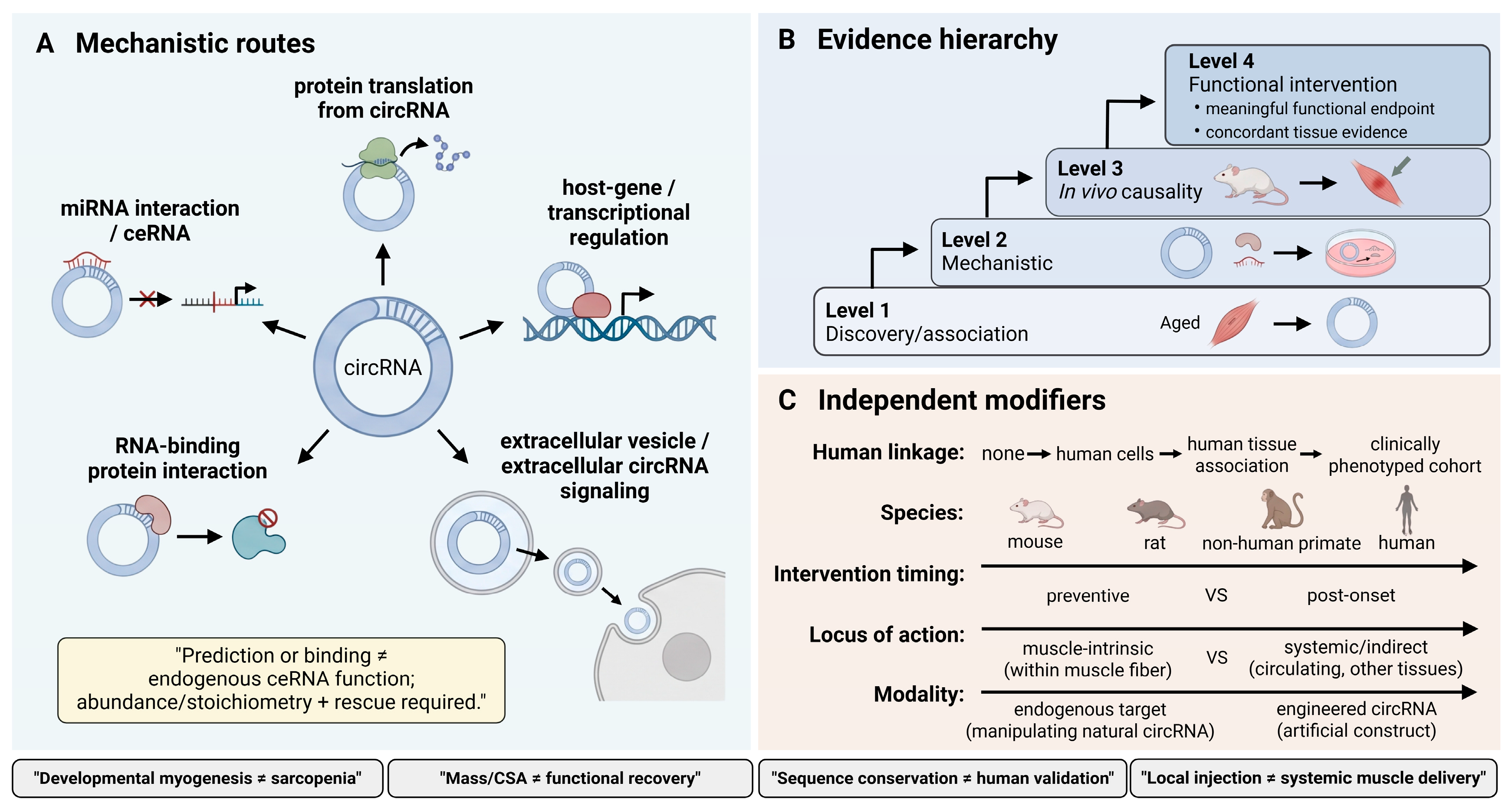
Table 1.
Retraction audit of circRNA reports excluded from the positive evidence synthesis.
| Retracted report | Retraction record | Risk of confusion | Treatment in this review |
|---|---|---|---|
| Exosomal ciRS-133 report in gastric cancer | Retraction published in 2023 [27] | The report linked an extracellular circRNA to systemic adipose browning and could be carried into metabolic or muscle narratives. | Excluded from the positive evidence synthesis. |
| circ-FoxO3 report on myoblast differentiation | Retraction published in 2020 [28] | The original claim concerned a direct myoblast differentiation phenotype. | Excluded from mechanistic and developmental evidence. |
| 2018 bovine circFUT10 and miR-133a report | Retraction published in 2026 [29] | The circRNA name can be confused with the separate 2021 study in aged skeletal-muscle stem cells [30]. | The 2018 report is excluded. The 2021 study is evaluated separately. |
Table 2.
Evidence levels used to classify circRNA studies in skeletal muscle aging and related wasting contexts.
Table 2.
Evidence levels used to classify circRNA studies in skeletal muscle aging and related wasting contexts.
| Evidence level | Minimum evidence | Interpretation |
|---|---|---|
| Level 1 Discovery and association |
Differential expression, profiling, correlation, network prediction or exercise responsiveness | Hypothesis generation; no causal inference |
| Level 2 Mechanistic |
Circular identity + candidate perturbation + directly tested interaction or pathway | Mechanism supported; in vivo aging or wasting relevance incomplete |
| Level 3 In vivo causal |
Manipulation changes an aging- or wasting-relevant tissue phenotype in vivo | Causality supported; therapeutic meaning depends on timing and endpoint |
| Level 4 Functional intervention |
In vivo intervention with a meaningful functional endpoint and concordant tissue evidence | Highest preclinical functional class. Post-onset efficacy is reported separately as a modifier. |
Human linkage, species, treatment timing, locus of action, and engineered or endogenous status are summarized separately in Supplementary Table S1 and do not change the evidence level by themselves.
Table 3.
Selected circRNA studies with direct aging linkage through aging-related models or aged-human-muscle association. Human linkage is assessed separately and does not imply human efficacy.
Table 3.
Selected circRNA studies with direct aging linkage through aging-related models or aged-human-muscle association. Human linkage is assessed separately and does not imply human efficacy.
| Candidate and level | Aging context and models | Mechanistic evidence | Strongest endpoint | Human linkage | Principal limitation |
|---|---|---|---|---|---|
| circSnd1 Level 4 |
Aged human muscle (n = 5 per age group), human myotubes, mice, and C2C12 cells; multiple catabolic stressors | FAT10-EEF1A1 interaction and EEF1A1 stabilization | Muscle mass or CSA, grip, and contractile endpoints | Aged-muscle association and human myotubes | No clinically adjudicated sarcopenia, longitudinal outcome, or independent validation |
| circDdb1 Level 4 |
Aged human muscle (five young and five aged samples), mice, and C2C12 cells; multiple atrophy conditions | Encodes circDdb1-867aa; eEF2 Thr56 phosphorylation and translation control | Muscle mass or CSA, grip, endurance, and contractile endpoints | Aged-muscle association | No accepted sarcopenia phenotype, longitudinal outcome, or independent validation |
| circFUT10 Level 2 |
Aged skeletal-muscle stem cells | miR-365a-3p-HOXA9 axis | Cellular regenerative phenotypes | None reported | No aged-organism mass or functional rescue |
| circBBS9 Level 1 |
Aged mouse skeletal muscle before and after aerobic exercise | Exercise-responsive expression and predicted network | Profiling only | None | Candidate-level causality and biomarker validation are absent |
| circNEB Level 3 |
Four 40-month-old rabbits with paired local hindlimb injections; bovine myoblasts; mouse and tree shrew injury models | Translated peptide; SKP1/p27-Kip1- and TPM1-related mechanisms | Short-term aged-rabbit histology | None | n = 4 aged rabbits; paired-limb local plasmid design; blinding and statistical unit not clearly reported; short-term histology; no functional endpoint |
C2C12, murine myoblast cell line; CSA, cross-sectional area; eEF2, eukaryotic elongation factor 2; EEF1A1, eukaryotic translation elongation factor 1 alpha 1; EIF4A3, eukaryotic translation initiation factor 4A3; FAT10, ubiquitin D; HOXA9, homeobox A9; SKP1, S-phase kinase-associated protein 1; TPM1, tropomyosin 1.
Table 4.
Non-aging atrophy and aging-adjacent studies used to test mechanistic portability without substituting for direct sarcopenia evidence.
Table 4.
Non-aging atrophy and aging-adjacent studies used to test mechanistic portability without substituting for direct sarcopenia evidence.
| Candidate | Comparator context | Mechanistic axis | Strongest endpoint | Level | Use and limitation in an aging review |
|---|---|---|---|---|---|
| circTmeff1 | Denervation; immobilization; inflammatory wasting | TDP-43; mitochondrial stress and cGAS-STING; encoded protein | Functional rescue, including established atrophy | 4 | High mechanistic portability; aging/human sarcopenia not directly tested |
| Engineered circmiR-29b | Immobilization and other atrophy models | Synthetic miR-29b decoy | Post-onset attenuation of established immobilization atrophy without a reported functional endpoint | 3 | Engineered modality proof. No direct aging model or functional endpoint. |
| circANAPC7 | Pancreatic-cancer cachexia | Tumor PHLPP2-AKT-TGF-beta axis | Muscle mass, fiber CSA, and grip strength | 4 | Systemic/tumor-mediated; not muscle-intrinsic aging biology |
| circGLIS3 | HFD-associated muscle fat infiltration | Suppression of intramuscular adipogenesis | Reduced fat infiltration in vivo | 3 | Aging-adjacent muscle-quality evidence; not an aging model |
| circAGO3 | Inflammatory atrophy | miR-34b-5p-TRAF3-NF-kappaB | Causal in vivo atrophy phenotype | 3 | Chicken; species-limited |
| circCCDC91 | Dexamethasone-related atrophy/myogenesis | miR-15 family-IRS1-IGF1-PI3K-AKT | Cell-level rescue | 2 | Primarily in vitro and chicken |
| circSMOX / denervation profiles | Dexamethasone, ALS-related, or denervation-associated expression | Not fully causally resolved | Association / profiling | 1 | Hypothesis pool only |
| circAtxn10 | Human myogenic-cell discovery; C2C12 differentiation | Proposed miR-143-3p–CHRNA1 axis | Myogenic cellular phenotype | 2 | Human-cell discovery with murine mechanistic validation; no direct aging study |
| circTPM1 | Myogenic differentiation and regeneration | MYH10 binding and MYL3-dependent actomyosin assembly downstream of TPM1_SE | Bovine cellular mechanism with cross-species super-enhancer evidence | 2 | Mechanotransduction relevance. No chronological aging, sarcopenia, or circRNA-specific in vivo test. |
AKT, protein kinase B; ALS, amyotrophic lateral sclerosis; cGAS-STING, cyclic GMP-AMP synthase-stimulator of interferon genes; CHRNA1, cholinergic receptor nicotinic alpha 1 subunit; CSA, cross-sectional area; HFD, high-fat diet; IGF1, insulin-like growth factor 1; IRS1, insulin receptor substrate 1; MYH10, myosin heavy chain 10; MYL3, myosin light chain 3; NF-kappaB, nuclear factor kappa B; NMJ, neuromuscular junction; PHLPP2, PH domain leucine-rich repeat protein phosphatase 2; PI3K, phosphoinositide 3-kinase; TDP-43, transactive response DNA-binding protein 43; TGF-beta, transforming growth factor beta; TPM1_SE, TPM1 super-enhancer; TRAF3, TNF receptor-associated factor 3.
Table 5.
Human circulating circRNA evidence identified in the targeted search and its unmet sarcopenia-biomarker requirements.
Table 5.
Human circulating circRNA evidence identified in the targeted search and its unmet sarcopenia-biomarker requirements.
| Candidate | Population and sample | Matrix and timing | Reported finding | Unmet sarcopenia-biomarker domains |
|---|---|---|---|---|
| circMBOAT2 [79] | Marathon runners (n = 11); within-person time course | Plasma; baseline, immediately after the race, and 24 h after the race | Lower abundance at 24 h; correlations with exercise-related measures | No older or clinically defined sarcopenia cohort; no skeletal-muscle source attribution; no independent validation, diagnostic or prognostic performance, or decision utility |
Note: The search was widened from clinically defined sarcopenia to human exercise and aging-adjacent contexts because no circulating circRNA study in a clinically phenotyped sarcopenia cohort was identified. circMBOAT2 is therefore a physiological comparator, not a sarcopenia biomarker. Unmet domains correspond to Table 6.
Table 7.
Criteria for continued development of circRNA therapeutics for skeletal muscle aging. T1–T10 correspond to the sequential therapeutic checkpoints in Figure 3.
Table 7.
Criteria for continued development of circRNA therapeutics for skeletal muscle aging. T1–T10 correspond to the sequential therapeutic checkpoints in Figure 3.
| Criterion | Question | Decision rule |
|---|---|---|
| T1 Target identity | Is the circular species unambiguously distinguished from host transcript and linear isoforms? | Stop if identity or perturbation specificity is unresolved. |
| T2 Endogenous mechanism | Does the proposed interaction operate at endogenous abundance with direct evidence and rescue/epistasis? | Revise if the mechanism depends only on prediction or supraphysiological overexpression. |
| T3 Causality in aged muscle | Does junction-selective manipulation alter a relevant phenotype in naturally aged or rigorously justified old muscle? | Comparator atrophy alone is insufficient. |
| T4 Functional benefit | Does intervention improve force, grip, endurance, power or another meaningful functional endpoint? | Mass or cross-sectional area alone does not meet this criterion. |
| T5 Post-onset efficacy | Can benefit occur after the phenotype is established? | Prioritize post-onset evidence over prophylaxis alone. |
| T6 Exposure and delivery | Can the modality achieve reproducible exposure across relevant muscles and cell types? | Local injection is not systemic delivery. |
| T7 Safety, reversibility, and repeat dosing | Are innate immunity, host-gene effects, off-targets, encoded products, and repeat dosing assessed? | Use aged and multimorbid models when feasible. |
| T8 Independent replication | Is the core result reproduced with independent reagents or cohorts and preferably another laboratory? | Concentration in one investigator network lowers readiness. |
| T9 Human linkage | Is the target associated with human muscle aging, strength or clinically defined sarcopenia? | Sequence conservation alone is insufficient. |
| T10 Added clinical value | Does the strategy add to exercise and nutrition or address patients inadequately served by them? | Defines a credible development path. |
Disclaimer/Publisher’s Note: The statements, opinions and data contained in all publications are solely those of the individual author(s) and contributor(s) and not of MDPI and/or the editor(s). MDPI and/or the editor(s) disclaim responsibility for any injury to people or property resulting from any ideas, methods, instructions or products referred to in the content. |
© 2026 by the authors. Licensee MDPI, Basel, Switzerland. This article is an open access article distributed under the terms and conditions of the Creative Commons Attribution (CC BY) license.
Copyright: This open access article is published under a Creative Commons CC BY 4.0 license, which permit the free download, distribution, and reuse, provided that the author and preprint are cited in any reuse.