

Submitted:
15 September 2026
Posted:
15 September 2026
You are already at the latest version
Abstract
Suspected immune thrombotic thrombocytopenic purpura (iTTP) creates a dual emergency: treatment must begin before ADAMTS13 confirmation, while lethal mimics must be investigated concurrently. A 44-year-old woman presented with fever, severe thrombocytopenia, progressive limb weakness, and multifocal cerebral infarctions. Although the schistocyte proportion was <1% and her PLASMIC score was 5, iTTP could not be safely excluded. ADAMTS13 activity was sampled before treatment, and plasma exchange plus corticosteroids was started. Blood cultures grew methicillin-susceptible Staphylococcus aureus. Despite three negative transthoracic echocardiograms, transesophageal echocardiography demonstrated a 10-mm mitral vegetation, establishing infective endocarditis (IE). Pretreatment ADAMTS13 activity was 35%, allowing TTP-directed therapy to be withdrawn. IE can produce thrombocytopenia, microangiopathic hemolysis, TMA-like presentations, and occasionally marked ADAMTS13 reduction; conversely, schistocytes may be initially inapparent in true iTTP. Thus, neither a low schistocyte count nor an intermediate prediction score should end diagnostic reasoning. When iTTP and IE remain plausible, pretreatment ADAMTS13 sampling, risk-adapted empiric TTP therapy, blood cultures, antimicrobial treatment, and definitive cardiac imaging should proceed in parallel.
Keywords:
ADAMTS13
; diagnostic anchoring
; infective endocarditis
; PLASMIC score
; plasma exchange
; transesophageal echocardiography
; thrombotic microangiopathy
; thrombotic thrombocytopenic purpura
1. Introduction
Immune thrombotic thrombocytopenic purpura (iTTP) is a time-critical thrombotic microangiopathy caused by severe ADAMTS13 deficiency. Because treatment delay may result in preventable death or irreversible organ injury, current diagnostic pathways combine clinical assessment, prediction tools such as the PLASMIC score, pretreatment ADAMTS13 sampling, and empiric plasma exchange before definitive laboratory confirmation [1,2,3,4,5,6]. The PLASMIC score estimates the probability of severe ADAMTS13 deficiency; it is not an etiologic diagnosis and does not exclude competing emergencies [1].
This distinction has become more important as TTP care has accelerated. Systemic infections can produce thrombocytopenia, hemolysis, neurologic abnormalities, and organ dysfunction that resemble TTP [7]. Infective endocarditis (IE) is a consequential mimic because cerebral emboli may resemble TTP-related neurologic injury, and transthoracic echocardiography (TTE) can be negative despite clinically important vegetation [8,9]. Conversely, waiting to complete the evaluation for infection before treating plausible iTTP may expose a patient to avoidable early mortality.
We report methicillin-susceptible Staphylococcus aureus (MSSA) IE that presented with severe thrombocytopenia and progressive neurologic abnormalities, prompting empiric TTP treatment while microbiologic and cardiac investigations continued. We also provide a focused review of IE-associated microangiopathic hemolytic anemia (MAHA), thrombotic microangiopathy (TMA)-like presentations, and ADAMTS13 reduction. The case illustrates a paradox of modern TTP care: algorithms should accelerate life-saving therapy without converting a working diagnosis into diagnostic closure.
2. Case Presentation
A 44-year-old woman with atopic dermatitis developed fever, sore throat, cough, vomiting, and diarrhea. Three days later, progressive weakness of all four limbs caused difficulty walking. She received ceftriaxone at a referring hospital. Diffusion-weighted brain magnetic resonance imaging showed multiple acute infarctions in the right cerebral subcortical region and both cerebellar hemispheres (Figure 1A–C), and she was transferred to our hospital.
On transfer, she was alert. Her temperature was 36.9 °C, pulse 77 beats/min, respiratory rate 18 breaths/min, and blood pressure 97/51 mmHg. Purpuric lesions involved the fingertips and great toes. Manual muscle testing grades (right/left) were 3/3 proximally and 2/2 distally in the upper limbs and 3/1 proximally and 2/2 distally in the lower limbs. Patellar and Achilles tendon reflexes were diminished bilaterally.
Laboratory testing showed a leukocyte count of 6.7 × 10⁹/L, hemoglobin 11.0 g/dL, mean corpuscular volume 82.8 fL, platelet count 37 × 10⁹/L, creatinine 1.0 mg/dL, and C-reactive protein 24 mg/dL. The schistocyte proportion was not increased (<1%). Lactate dehydrogenase was mildly elevated at 347 U/L, whereas total bilirubin was normal at 0.63 mg/dL. Prothrombin time was not prolonged; fibrinogen was 670 mg/dL and fibrin degradation products were 19 μg/mL, arguing against overt disseminated intravascular coagulation (DIC).
Her PLASMIC score was 5: the criteria for no active cancer, no transplantation, mean corpuscular volume <90 fL, international normalized ratio <1.5, and creatinine <2.0 mg/dL were met, whereas the platelet-count and hemolysis criteria were not. Thus, the score indicated an intermediate probability of severe ADAMTS13 deficiency rather than a diagnosis of iTTP [1]. Nevertheless, severe thrombocytopenia and progressive neurologic dysfunction meant that iTTP could not be excluded with acceptable safety. A blood sample for ADAMTS13 testing was obtained before any plasma-containing product or plasma exchange. Daily plasma exchange and methylprednisolone 1000 mg/day were initiated while alternative diagnoses were investigated (Figure 2). Caplacizumab was not administered [2,3].
Because fever, marked inflammation, peripheral purpura, and the embolic distribution of the cerebral lesions suggested infection, blood cultures and empiric meropenem plus vancomycin were obtained and administered in parallel with TTP-directed therapy. Admission cultures grew MSSA in five of six bottles. Antimicrobial therapy was changed to sulbactam/ampicillin 12 g/day. The platelet count began to recover on day 3.
Three TTE examinations, including one at the referring hospital, showed no vegetation. However, MSSA bacteremia and multifocal cerebral infarctions maintained a high clinical probability of IE. Transesophageal echocardiography (TEE) on day 5 demonstrated a mobile 10-mm vegetation on the anterior mitral leaflet with trivial-to-mild mitral regurgitation (Figure 1D,E). The combination of typical blood cultures and echocardiographic evidence established definite IE.
Pretreatment ADAMTS13 activity subsequently returned at 35%, excluding the severe deficiency characteristic of iTTP. Plasma exchange was stopped after eight sessions, and corticosteroids were tapered. No complication attributable to plasma exchange occurred. Blood cultures were negative on day 8, and repeat TEE on day 14 no longer showed the vegetation. Limb strength and tendon reflexes normalized. Antimicrobial therapy was changed to oral cephalexin 2000 mg/day on day 50, and she was discharged on day 53. She later underwent mitral valve repair for severe mitral regurgitation and remained well without recurrent infection or embolism at 20 months of follow-up.
3. Discussion
3.1. A Paradox of Modern TTP Care
This case demonstrates two simultaneous safety obligations. First, plausible iTTP must be treated before a definitive ADAMTS13 result is usually available. Second, empiric treatment must not end the evaluation for other lethal disorders. The PLASMIC score informed but did not determine the treatment decision. A score of 5, absent overt MAHA, and a strong infectious signal reduced the probability of iTTP, but did not reduce the immediate clinical risk to zero. Pretreatment ADAMTS13 sampling created a reliable stopping rule: once activity returned at 35%, TTP-directed therapy could be withdrawn [1,2,3].
Clinical data illustrate the consequences of waiting for diagnostic certainty. In a UK study of 148 first acute TTP episodes, diagnostic uncertainty was the leading reason for treatment delay, and 27.8% of deaths were considered related to delayed treatment initiation [5]. In a French cohort of confirmed iTTP, diagnostic delay beyond 24 h was associated with more neurological events than immediate diagnosis (67% vs. 30%) [4]. These observational findings do not define a risk-free threshold for delay; instead, they support treatment at the stage of clinically meaningful suspicion.
The schistocyte count likewise requires probabilistic interpretation. The International Council for Standardization in Hematology considers >1% schistocytes in adults suspicious for TMA, while explicitly stating that their absence does not exclude TMA a priori [10]. Schistocyte percentage is not an independent component of the PLASMIC score, and manual identification remains dependent on standardized smear preparation, defined morphologic criteria, and observer expertise [1,10]. In the French iTTP cohort, 26% of patients had a schistocyte count below 1% at initial evaluation; diagnostic delay occurred in 71% of patients with counts below 1%, compared with 15% of those with counts of 1% or higher [4]. Thus, the absence of increased schistocytes (<1%) in our patient argued against established MAHA but could not serve as a stand-alone exclusion criterion for iTTP [6,11].
This distinction is particularly relevant in the caplacizumab era. Current guidance supports early caplacizumab, before the ADAMTS13 result is available, when the clinical probability of iTTP is high [2,6]. It does not require identical treatment intensity across all probability strata. In our patient, an intermediate PLASMIC score, absence of overt MAHA, and strong infectious and embolic signals supported immediate plasma exchange and corticosteroids while caplacizumab was withheld pending the pretreatment ADAMTS13 result. This probability-adapted approach provided immediate TTP-directed therapy while avoiding additional anti-von Willebrand factor treatment and its bleeding risk in a patient who was soon shown to have IE; caplacizumab could have been added promptly if severe ADAMTS13 deficiency had been confirmed. At the same time, cultures, antimicrobial therapy, and cardiac imaging proceeded without waiting for the TTP pathway to finish. The central lesson is therefore not that IE should replace suspected iTTP at presentation, but that the two pathways should proceed in parallel until discriminating evidence becomes available.
3.2. Why Infective Endocarditis Can Resemble TTP
IE can reproduce several components of a TTP phenotype through different mechanisms. Thrombocytopenia may reflect inflammation-driven platelet consumption, endothelial injury, coagulation activation, immune-mediated destruction, and recruitment of platelets into vegetation. Cerebral embolization can produce abrupt or fluctuating neurologic deficits. Fragmentation hemolysis has been described in native-valve IE and may result from shear stress across vegetations or abnormal flow, immune erythrocyte injury, and endothelial or coagulation activation [7,12,13,14,15].
Severe infection may also reduce ADAMTS13 activity without establishing immune TTP as the sole process. The degree of reduction, inhibitor status, evidence of DIC, and the overall clinical context must therefore be interpreted together. An activity below 10% strongly supports TTP in an appropriate phenotype, but infection-associated TMA and DIC/TMA overlap remain important competing or coexisting processes. Our patient did not have overt MAHA and had an activity of 35%; accordingly, we do not classify her illness as IE-associated TMA or true iTTP. Instead, the published overlap explains why the initial treatment decision can remain uncertain even when the final diagnosis is IE.
3.3. Focused Literature Review
We conducted a focused PubMed review through 10 September 2026 using combinations of “infective endocarditis”, “thrombotic thrombocytopenic purpura”, “thrombotic microangiopathy”, “microangiopathic hemolytic anemia”, and “ADAMTS13”. We also screened the reference lists of retrieved reports. We included reports describing IE with thrombocytopenia plus MAHA, neurologic abnormalities, a TMA/TTP-like designation, TTP-directed treatment, or reduced ADAMTS13 activity. Because the reports were heterogeneous and most predated standardized ADAMTS13 testing, the review was narrative rather than systematic.
The retrieved reports show a recurring clinical pattern (Table 1) [13,14,15,16,17,18,19]. Most patients had thrombocytopenia with hemolysis, neurologic injury, renal dysfunction, or a combination of these findings. Several received plasma exchange before IE was established. Only the most recent report provided a markedly reduced ADAMTS13 result: activity was 7% with no detectable inhibitor in prosthetic-valve IE complicated by DIC-like coagulopathy [18]. These cases should not be interpreted as proof that IE commonly causes true iTTP. Rather, they demonstrate that infection can enter the same early diagnostic pathway and that neither a TMA phenotype nor reduced ADAMTS13 activity should terminate etiologic evaluation.
3.4. Transesophageal Echocardiography as the Decisive Test
Fever, marked inflammation, MSSA bacteremia, peripheral lesions, and an embolic magnetic resonance imaging pattern increasingly favored IE. TTE is the initial imaging test, but a negative study does not exclude IE when clinical suspicion remains high. Current guidance recommends TEE after negative or inconclusive TTE in this setting [8]. In retrospect, repeating TTE added little after MSSA bacteremia and cerebral emboli were recognized. The actionable lesson is to escalate promptly to TEE rather than allowing repeated negative TTE examinations to provide false reassurance.
3.5. Limitations
This report has limitations. The patient lacked overt MAHA, and the PLASMIC score was developed for patients with suspected thrombotic microangiopathy; therefore, the score should not be interpreted as validating a diagnosis of iTTP in this case. ADAMTS13 turnaround time prolonged empiric therapy, and IE was established before the result became available. Platelet and neurologic improvement occurred while plasma exchange, corticosteroids, and antimicrobial therapy overlapped, so their individual contributions cannot be separated. Finally, the literature consists mainly of heterogeneous case reports that often lacked ADAMTS13 testing, standardized DIC assessment, or complete echocardiographic data.
4. Conclusions
The PLASMIC score and rapid ADAMTS13 testing have improved the recognition of iTTP, but they should be used as components of a dynamic diagnostic process rather than endpoints. When iTTP cannot be safely excluded, obtain a pretreatment ADAMTS13 sample and initiate proportional empiric therapy without delay. Simultaneously, pursue infection with cultures, appropriate antimicrobials, and definitive cardiac imaging. In patients with bacteremia or cerebral emboli, negative TTE should prompt rather than prevent TEE. Treat first, but keep searching.
Author Contributions
Conceptualization, S.S., H.S. and A.T.; investigation, S.S., H.S., K.U. and T.H.; data curation, S.S. and H.S.; writing—original draft preparation, S.S.; writing—review and editing, S.S., H.S., K.U., T.H. and A.T.; visualization, S.S. and H.S.; supervision, A.T. All authors have read and agreed to the published version of the manuscript.
Funding
This research was supported by Grants-in-Aid for Scientific Research (24K11527) and a Health and Labour Sciences Research Grant (23KC2009).
Institutional Review Board Statement
Ethical review and approval were not required for this single-patient case report in accordance with the institutional requirements of Aichi Medical University Hospital.
Informed Consent Statement
Written informed consent was obtained from the patient to publish this paper and the accompanying images.
Data Availability Statement
The data supporting this report are contained within the article. Additional de-identified information may be available from the corresponding author on reasonable request, subject to institutional and patient-privacy requirements.
Acknowledgments
The authors thank the clinical staff involved in the patient’s care.
Conflicts of Interest
A.T. received research funding from AIR WATER; lecture fees from Novartis Pharmaceuticals and argenx Japan; and scholarship donations from Chugai Pharmaceutical and Kyowa Kirin. H.S. received a scholarship grant from the Chugai Foundation for Innovative Drug Discovery Science. The remaining authors declare no conflicts of interest.
Abbreviations
| ADAMTS13 | A disintegrin and metalloproteinase with a thrombospondin type 1 motif, member 13 |
| DIC | Disseminated intravascular coagulation |
| IE | Infective endocarditis |
| iTTP | Immune thrombotic thrombocytopenic purpura |
| MAHA | Microangiopathic hemolytic anemia |
| MSSA | Methicillin-susceptible Staphylococcus aureus |
| TEE | Transesophageal echocardiography |
| TMA | Thrombotic microangiopathy |
| TTE | Transthoracic echocardiography |
References
- Bendapudi, P.K.; Hurwitz, S.; Fry, A.; Marques, M.B.; Waldo, S.W.; Li, A.; Sun, L.; Upadhyay, V.; Hamdan, A.; Brunner, A.M.; et al. Derivation and external validation of the PLASMIC score for rapid assessment of adults with thrombotic microangiopathies: a cohort study. Lancet Haematol. 2017, 4, e157–e164. [Google Scholar] [CrossRef] [PubMed]
- Zheng, X.L.; Al-Housni, Z.; Cataland, S.R.; Coppo, P.; Geldziler, B.; Germini, F.; Iorio, A.; Keepanasseril, A.; Masias, C.; Matsumoto, M.; et al. 2025 focused update of the 2020 ISTH guidelines for management of thrombotic thrombocytopenic purpura. J. Thromb. Haemost. 2025, 23, 3711–3732. [Google Scholar] [CrossRef] [PubMed]
- Zheng, X.L.; Vesely, S.K.; Cataland, S.R.; Coppo, P.; Geldziler, B.; Iorio, A.; Matsumoto, M.; Mustafa, R.A.; Pai, M.; Rock, G.; et al. ISTH guidelines for the diagnosis of thrombotic thrombocytopenic purpura. J. Thromb. Haemost. 2020, 18, 2486–2495. [Google Scholar] [CrossRef] [PubMed]
- Renaud, A.; Caristan, A.; Seguin, A.; Agard, C.; Blonz, G.; Canet, E.; Eveillard, M.; Godmer, P.; Graveleau, J.; Lecouffe-Desprets, M.; et al. Deleterious neurological impact of diagnostic delay in immune-mediated thrombotic thrombocytopenic purpura. PLoS ONE 2021, 16, e0260196. [Google Scholar] [CrossRef] [PubMed]
- HaemStar Collaborators (Ed.) Diagnostic uncertainty presented barriers to the timely management of acute thrombotic thrombocytopenic purpura in the United Kingdom between 2014 and 2019. J Thromb Haemost 2022, 20, 1428–1436. [Google Scholar] [CrossRef] [PubMed]
- Azoulay, E.; Veyradier, A.; Cataland, S.; Joly, B.S.; Knoebl, P.; Zafrani, L.; Wendon, J.; Fakhouri, F.; Lammle, B.; Pavenski, K.; et al. Thrombotic thrombocytopenic purpura: early diagnosis and effective treatment in 2025. Intensive Care Med. 2025, 51, 1225–1239. [Google Scholar] [CrossRef] [PubMed]
- Booth, K.K.; Terrell, D.R.; Vesely, S.K.; George, J.N. Systemic infections mimicking thrombotic thrombocytopenic purpura. Am. J. Hematol. 2011, 86, 743–751. [Google Scholar] [CrossRef] [PubMed]
- Delgado, V.; Ajmone Marsan, N.; de Waha, S.; Bonaros, N.; Brida, M.; Burri, H.; Caselli, S.; Doenst, T.; Ederhy, S.; Erba, P.A.; et al. 2023 ESC Guidelines for the management of endocarditis. Eur. Heart J. 2023, 44, 3948–4042. [Google Scholar] [CrossRef] [PubMed]
- Shively, B.K.; Gurule, F.T.; Roldan, C.A.; Leggett, J.H.; Schiller, N.B. Diagnostic value of transesophageal compared with transthoracic echocardiography in infective endocarditis. J. Am. Coll. Cardiol. 1991, 18, 391–397. [Google Scholar] [CrossRef] [PubMed]
- Zini, G.; d'Onofrio, G.; Erber, W.N.; Lee, S.H.; Nagai, Y.; Basak, G.W.; Lesesve, J.F.; Hematology, I.C.f.S.i. 2021 update of the 2012 ICSH Recommendations for identification, diagnostic value, and quantitation of schistocytes: Impact and revisions. Int. J. Lab Hematol. 2021, 43, 1264–1271. [Google Scholar] [CrossRef] [PubMed]
- de Boisriou, I.; Ellouze, S.; Kassasseya, C.; Feral-Pierssens, A.L.; Gerlier, C.; Chauvin, A.; Beaune, S.; Dubreucq, E.; Pereira, L.; Chocron, R.; et al. Misdiagnosis of thrombotic microangiopathy in the emergency department: a multicenter retrospective study. Intern Emerg. Med. 2024, 19, 115–124. [Google Scholar] [CrossRef] [PubMed]
- Shingu, M.; Ishimaru, N.; Ohnishi, J.; Mizuki, S.; Kanzawa, Y.; Kawano, K.; Nakajima, T.; Sano, N.; Kinami, S. Hemolytic Anemia in a Patient with Subacute Bacterial Endocarditis by Cardiobacterium hominis. Intern Med. 2021, 60, 3489–3495. [Google Scholar] [CrossRef] [PubMed]
- Bayer, A.S.; Theofilopoulos, A.N.; Eisenberg, R.; Friedman, S.G.; Guze, L.B. Thrombotic thrombocytopenic purpura-like syndrome associated with infective endocarditis. A possible immune complex disorder. JAMA 1977, 238, 408–410. [Google Scholar] [CrossRef]
- Roberson, J.C., 2nd; Ludmer, L.M.; Feinman, M.C.; Barth, W.F. Subacute bacterial endocarditis with polyarthritis mimicking thrombotic thrombocytopenic purpura: a case report. J. Clin. Rheumatol. 1995, 1, 118–121. [Google Scholar] [CrossRef] [PubMed]
- Thomas, M.; Heyll, A.; Meckenstock, G.; Vogt, M.; Aul, C. Embolizing aortic valve endocarditis in the differential diagnosis of thrombotic thrombocytopenic purpura. Dtsch. Med. Wochenschr. 1992, 117, 738–742. [Google Scholar] [CrossRef] [PubMed]
- Quinn, K.L.; Osmond, M.; Badiwala, M.; Sermer, M.; Lapinsky, S.E. Severe Staphylococcus aureus Endocarditis Presenting as Peripartum Thrombotic Thrombocytopenic Purpura. J. Obstet. Gynaecol. Can. 2016, 38, 1028–1032. [Google Scholar] [CrossRef] [PubMed]
- Selleng, K.; Warkentin, T.E.; Greinacher, A.; Morris, A.M.; Walker, I.R.; Heggtveit, H.A.; Eichler, P.; Cybulsky, I.J. Very severe thrombocytopenia and fragmentation hemolysis mimicking thrombotic thrombocytopenic purpura associated with a giant intracardiac vegetation infected with Staphylococcus epidermidis: role of monocyte procoagulant activity induced by bacterial supernatant. Am. J. Hematol. 2007, 82, 766–771. [Google Scholar] [CrossRef] [PubMed]
- Guardado, K.E.; Faherty, M.; Lin, J.; Wagner, S. Rothia mucilaginosa Prosthetic Valve Endocarditis Presenting as a Thrombotic Microangiopathy Mimic With Catastrophic Intracranial Hemorrhage. Cureus 2026, 18, e111711. [Google Scholar] [CrossRef] [PubMed]
- Andy Tang, S.O.; Leong, T.S.; Ruixin, T.; Chua, H.H.; Chew, L.P. Thrombotic thrombocytopenic purpura-like syndrome associated with arcanobacterium pyogenes endocarditis in a post-transplant patient: A case report. Med. J. Malays. 2018, 73, 344–346. [Google Scholar]
Figure 1.
Diagnostic imaging linking cerebral embolization to infective endocarditis. (A–C) Diffusion-weighted magnetic resonance images show multiple acute infarctions in the right cerebral subcortical region and both cerebellar hemispheres. (D) Transesophageal echocardiography on day 5 shows a mobile 10-mm vegetation (arrow) on the anterior mitral leaflet. (E) Color Doppler imaging demonstrates mitral regurgitation.
Figure 1.
Diagnostic imaging linking cerebral embolization to infective endocarditis. (A–C) Diffusion-weighted magnetic resonance images show multiple acute infarctions in the right cerebral subcortical region and both cerebellar hemispheres. (D) Transesophageal echocardiography on day 5 shows a mobile 10-mm vegetation (arrow) on the anterior mitral leaflet. (E) Color Doppler imaging demonstrates mitral regurgitation.
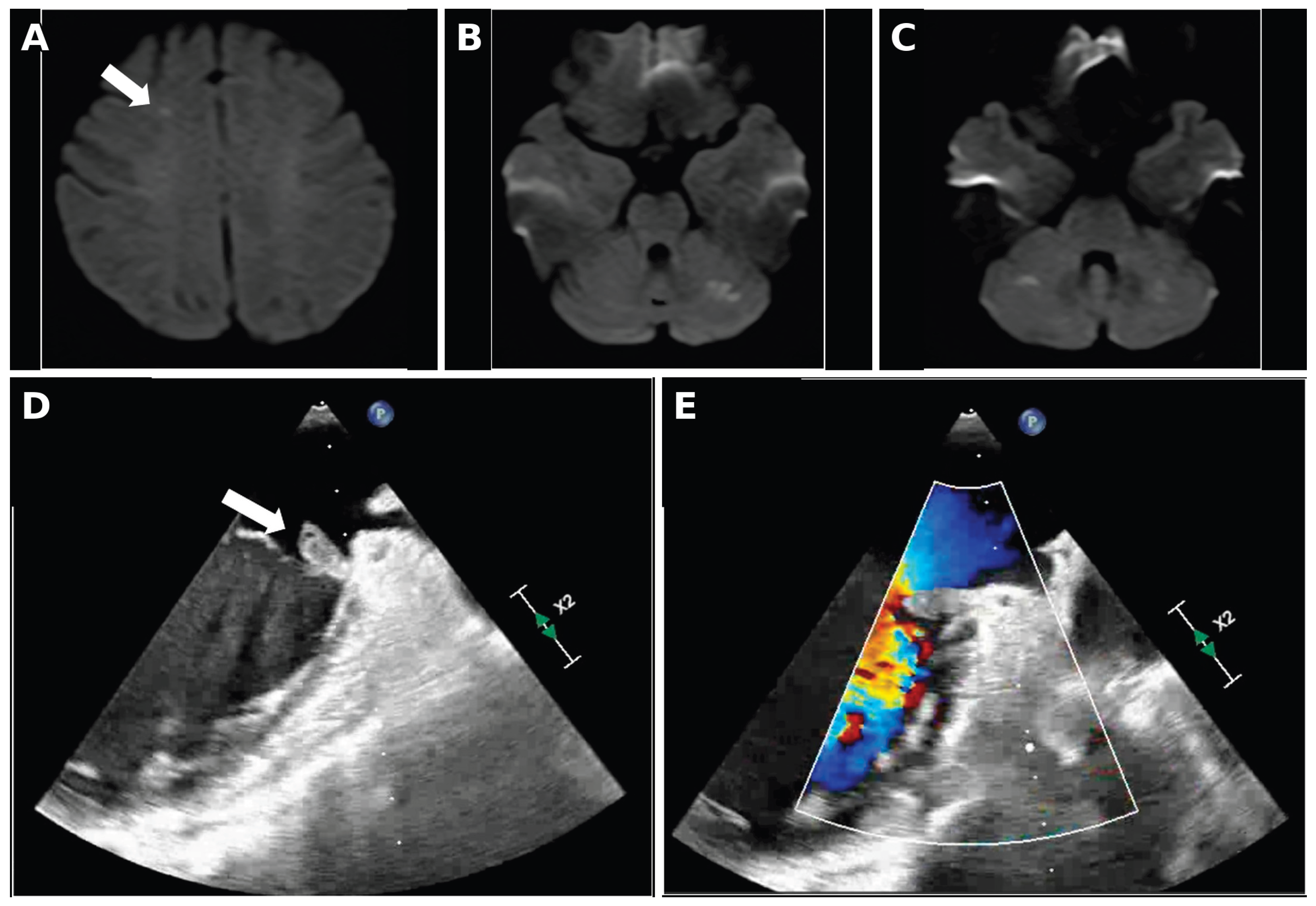
Figure 2.
Clinical course after admission. Eight plasma-exchange sessions were administered while pretreatment ADAMTS13 activity was pending. Platelet recovery and improvement in limb weakness occurred during antimicrobial and TTP-directed treatment. MEPM, meropenem; mPSL, methylprednisolone; PSL, prednisolone; SBT/ABPC, sulbactam/ampicillin; VCM, vancomycin.
Figure 2.
Clinical course after admission. Eight plasma-exchange sessions were administered while pretreatment ADAMTS13 activity was pending. Platelet recovery and improvement in limb weakness occurred during antimicrobial and TTP-directed treatment. MEPM, meropenem; mPSL, methylprednisolone; PSL, prednisolone; SBT/ABPC, sulbactam/ampicillin; VCM, vancomycin.
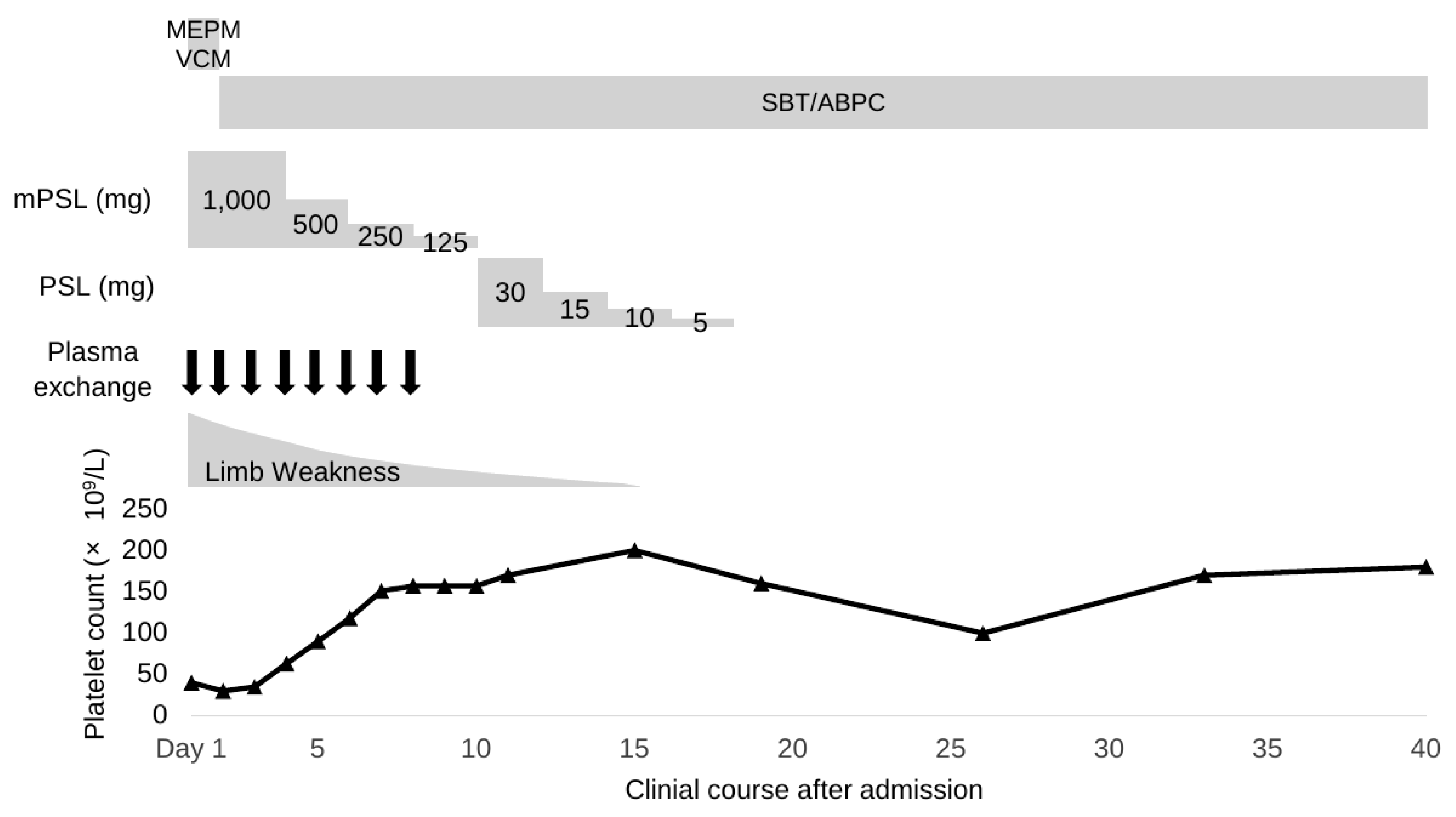

Table 1.
Published reports of infective endocarditis with TTP/TMA-like clinical features.
| Report | Pathogen / Site | Clinical Phenotype | ADAMTS13 / TTP-Directed Therapy | Echocardiography / Outcome |
|---|---|---|---|---|
| Bayer et al. (1977), two cases [13] | Viridans streptococcus / aortic valve; S. aureus / mitral valve | MAHA and thrombocytopenia with renal and/or neurologic abnormalities | NR / corticosteroids in one case | NR / both recovered |
| Thomas et al. (1992) [15] | E. faecalis / aortic valve | Thrombocytopenia and neurologic abnormalities | NR / fresh frozen plasma | NR / recovered |
| Roberson et al. (1995) [14] | NR / mitral and aortic valves | TTP-like syndrome with disorientation | NR / plasma exchange and corticosteroids | TTE NR; TEE positive / recovered |
| Selleng et al. (2007) [17] | S. epidermidis / pacemaker lead and tricuspid valve | Severe thrombocytopenia and fragmentation hemolysis | NR / plasma exchange, corticosteroids, and IVIG | TTE positive; TEE NR / recovered |
| Quinn et al. (2016) [16] | S. aureus / tricuspid valve | Peripartum TTP-like syndrome with altered consciousness | NR / plasma exchange | NR / recovered |
| Tang et al. (2018) [19] | A. pyogenes / mitral valve | MAHA, severe thrombocytopenia, and hemiparesis | NR / none | TTE positive; TEE NR / died |
| Guardado et al. (2026) [18] | R. mucilaginosa / prosthetic aortic valve | TMA mimic with MAHA, DIC-like coagulopathy, and intracranial hemorrhage | 7%, inhibitor negative / emergent plasma exchange | TTE NR; TEE positive / died |
| Present case | MSSA / native mitral valve | Severe thrombocytopenia and multifocal cerebral infarctions; no overt MAHA | 35% / plasma exchange and corticosteroids | TTE negative ×3; TEE positive / recovered |
ADAMTS13, a disintegrin and metalloproteinase with a thrombospondin type 1 motif, member 13; DIC, disseminated intravascular coagulation; IVIG, intravenous immunoglobulin; MAHA, microangiopathic hemolytic anemia; MSSA, methicillin-susceptible Staphylococcus aureus; NR, not reported; TEE, transesophageal echocardiography; TMA, thrombotic microangiopathy; TTE, transthoracic echocardiography; TTP, thrombotic thrombocytopenic purpura.
Disclaimer/Publisher’s Note: The statements, opinions and data contained in all publications are solely those of the individual author(s) and contributor(s) and not of MDPI and/or the editor(s). MDPI and/or the editor(s) disclaim responsibility for any injury to people or property resulting from any ideas, methods, instructions or products referred to in the content. |
© 2026 by the authors. Licensee MDPI, Basel, Switzerland. This article is an open access article distributed under the terms and conditions of the Creative Commons Attribution (CC BY) license (http://creativecommons.org/licenses/by/4.0/).
Copyright: This open access article is published under a Creative Commons CC BY 4.0 license, which permit the free download, distribution, and reuse, provided that the author and preprint are cited in any reuse.