

Submitted:
14 September 2026
Posted:
15 September 2026
You are already at the latest version
Abstract
Wnt signaling plays central roles in embryogenesis/development, skeletal homeostasis, tumour development, and other diseases. The pathway is initiated by Wnt ligands which can be bound and inhibited by sFRP1. Regulation of sFRP1 in lung inflammation needs increased study, and little is known about which cytokines can induce sFRP1, nor the role of gp130 cytokines in its modulation. Here we used AdOSM (Advector expressing mouse Oncostatin M [OSM]) administration to overexpress the gp130 cytokine OSM in mice in vivo, and applied Nanostring, RT-PCR, and immunoblots to measure mRNA and protein levels. OSM induced robust increases in mRNA for sFRP-1 in context of reduced Wnt ligand and receptor mRNA (Wnt10B, Wnt3A, Wnt2, Wnt11, LRP5 and LRP6). Axin-2 mRNA, as a b-catenin target gene, was also suppressed suggesting an inhibition of Wnt signaling pathways. AdOSM could induce sFRP1 in IL-6KO mice, while overexpression of IL-6 (AdIL-6 vector) did not. Immunoblots of total lung extracts showed increases in the 35Kd sFRP1 protein species. In situ hybridization of mouse lung histological sections using specific sFRP1 probes showed staining in the subepithelial layer of the airways, consistent with fibroblast or myofibroblast locations in lung mucosa. In vitro, recombinant OSM stimulated sFRP1 mRNA in cultures of mouse lung fibroblasts and NIH-3T3 cells. The results support the novel observation that OSM can induce sFRP1 in vitro and in vivo, and suggest this axis contributes to the control of Wnt signaling pathways in lung inflammation.
Keywords:
sFRP1
; Wnt signaling
; lung inflammation
; Oncostatin M
1. Introduction
The Wnt pathway has been implicated in multiple processes such as cell proliferation/survival, embryogenesis/lung morphogenesis, liver development, homeostasis of the skeletal system [1,2,3,4], and disease processes such as cancer progression and lung conditions as reviewed by others [5,6,7,8,9]. Wnt ligands bind to cell surface receptors including the seven-pass frizzled (FZ) gene family and the co-receptor LDL receptor-related protein (LRP) family. Signal transduction involves cytoplasmic phosphoprotein disheveled (DSH) which can activate intracellular signaling cascade branches of three major pathways: the (canonical) Wnt-β catenin pathway; the (non-canonical) planar cell polarity (PCP) pathway; and the (non-canonical) Wnt calcium pathway. The best characterized of these is the Wnt β-catenin signaling pathway, where β-catenin protein accumulates in the cytoplasm and translocates into the nucleus where it can regulate the expression of various target genes. In addition to multiple ligands, receptors, signaling systems, and cell type specific responses, Wnt pathways can also be regulated by soluble inhibitors. Soluble Frizzled Related Protein 1 (sFRP1) is a member of the Wnt family and inhibits signaling by binding to Wnt ligands, preventing them from interacting with their receptors and activation of signaling [10,11,12].
As an inhibitor of ligands that stimulate Wnt signaling at the receptor level, sFRP1 function has been studied in various disease pathologies including cancer and other chronic conditions. Abnormal expression or epigenetic alterations in cancer may be involved in dual roles of sFRP1 in certain tumour types [13]. Epigenetic down-regulation of sFRP1 in synovium may lead to self-persistence of joint inflammation in rheumatoid arthritis [14]. Aberrant Wnt signaling has also been observed in kidney fibrogenesis. In loss of function studies, sFRP1 KO mice showed increased kidney fibrosis in the UUO mouse model [15] suggesting a protective effect of sFRP1 [15]. Wnt pathways also have modulatory effects in lung conditions such as fibrosis, Chronic Obstructive Pulmonary Disease (COPD), emphysema, and bronchial asthma [6,16,17]. sFRP1 itself has roles in mouse models of allergic airway disease [18] and has also been implicated in lung fibrosis where sFRP1 derived from fibroblasts appear to inhibit transitional fibroblast invasiveness [19]. However, Burgy et al. have more recently shown that fibroblast-derived extracellular vesicles (ECV) enriched in sFRP1 exacerbated the Bleomycin-induced lung fibrosis model [20]. Thus, the roles of sFRP1 in fibrogenesis appear complex, multifaceted, and require further exploration in animal models as well as clinical studies.
The identification of cytokines and growth factors that regulate sFRP1 will contribute to our understanding the control of Wnt signaling pathways. Cell types that produce sFRP1 include fibroblasts. While factors known to decrease sFRP1 expression in lung fibroblasts in vitro include EGF and TGF [19,20], less is published about cytokines and growth factors that stimulate its levels or function in lung cells. Furthermore, whether cytokines of the IL-6/gp130 family can regulate sFRP1 is not known. Our group has been examining mechanisms of lung inflammation and fibrogenesis in mouse models, and in previous work we have focused on two gp130 members, Interleukin-6 (IL-6) and Oncostatin M (OSM), and their functions in lung inflammation and extracellular matrix (ECM) remodeling. Studies of OSM inflammatory processes in published work by others have suggested its implication in conditions including inflammatory bowel disease (IBD), severe asthma, idiopathic pulmonary fibrosis (IPF), COPD, chronic rhinosinusitis, and scleroderma [22,23,24,25,26,27,28].
OSM functions at receptors present on stromal/structural cells as opposed to hemopoietic cells and we have previously shown in mouse models that OSM can induce genes associated with inflammation and extracellular matrix (ECM) remodeling in lung including Collagen1A1, collagen 3A1, TIMP-1 [28,29,30] as well as Th2 skewed inflammation and chemokines in C57BL/6 mice [31,32,33]. Upon examination of a larger set of genes by Nanostring analysis, we here identify that OSM can regulate genes associated with the Wnt signaling pathway. We explore this observation in more detail examining both mRNA and protein expression of sFRP1, Wnt ligand and receptor mRNAs, sFRP1mRNA localization in lung histological sections, and assessing regulation of sFRP1 by OSM in vitro in mouse fibroblasts. We observe an overall increase in sFRP1 expression, downregulation of several Wnt ligand and receptor mRNAs as well as Axin-2 mRNA suggesting inhibition of Wnt signaling pathways by OSM in mouse lungs.
2. Materials and Methods
2.1. Animals
Female wildtype C57BL/6 or BALB/c mice (6-8 weeks old) and IL-6 deficient mice (IL-6 –/–, C57BL/6 background, 6-8 weeks old) were purchased from The Jackson Laboratory (Bar Harbour, ME, USA) and housed at the McMaster University Central Animal Facility (Hamilton, ON, Canada) under pathogen-free conditions. All experiments were approved by the McMaster University Animal Research Ethics Board AUP# 22-07-24 and its predecessor 18-05-22.
2.2. Adenovirus Administration
Adenoviral vectors AdDl70 (negative control vector), AdOSM, AdIL-6 (as previously described [34]) were thawed on ice and diluted in phosphate buffered saline (PBS) to 5 × 10⁷ PFU dose per mouse. Wildtype C57BL/6 or BALB/c and C57BL/6 background IL-6 –/– mice were endotracheally administered 50µL of virus as previously described [31].
2.3. Lung sample Processing
. At various time points postinfection, mice were humanely euthanized. The left lung was perfused and fixed in formalin for 48 hours, then transferred to 70% EtOH. The right lung was snap frozen in liquid N2 and crushed into a fine powder using a mortar and pestle [34,35]. Two thirds were added to RIPA buffer for protein extraction, and one third to Trizol for RNA extraction. Lung tissue samples were then homogenized using the Brinkmann Homogenizer Polytron PT 3000. RNA was extracted from Trizol following the manufacturers protocol. Homogenized protein was spun down for 1 hour at 13,000 rpm and the supernatant was collected, aliquoted, and stored at -80 °C for Immunoblot analysis.
2.4. Histology and Chromogenic In Situ Hybridization (CISH)
Following formalin fixation, the left lung was cut into 3 sections and embedded in paraffin. 3 µm wide sections were cut and stained with Hematoxylin and Eosin (H&E) to assess histopathology. Slides were scanned at 20x and images were prepared using QuPath as described by Bankhead et al. [36]. Prior to hybridization with target oligo probes, formalin-fixed, paraffin embedded lung tissue was pre-treated with heat and protease. CISH for mouse OSM and SFRP-1 mRNA was performed using the RNAScope 2.5 Duplex Assay Kit (Advanced Cell Diagnostics, Newark, CA, USA) and stained using the BOND 1x staining instrument (Leica) as previously described [37]. Specific RNA staining signals were identified as red (OSM) or green (SFRP-1) dots.
2.5. Immunoblotting
Samples containing 20-40 µg of protein from lung homogenates were prepared by standard methods, loaded onto 12% acrylamide SDS-PAGE, and separated by electrophoresis at 90 V for 90 minutes. Protein was then transferred to nitrocellulose membranes at 400 mA for one hour. Membranes were blocked at room temperature using a 1:1 mixture of TBST (1x tris-buffered saline with 0.15% Tween-20) and TBS Odyssey Blocking Buffer (LIC-927-60001) to prevent non-specific antibody binding, then incubated overnight at 4 °C with primary antibodies for β-Actin C4 (Santa Cruz, Cat# sc-47778), SFRP-1 (Abcam, Cat#ab267466), non-phosphorylated (active) beta-catenin (S33/S37/T41) (Cell Signaling Technology, Cat#8814S), or phosphorylated beta-catenin (Cell Signaling Technology, Cat#9566S). Membranes were then washed for seven minute thrice with TBST and incubated with secondary antibodies IRDye 800CW Donkey anti-Rabbit IgG (LICOR, Cat#LIC-926-32213) or IRDye 800CW Donkey anti-Mouse IgG (LICOR, Cat#LIC-926-32212) for 1 hour at room temperature. After three additional TBST washes, and one 1x tris-buffered saline (TBS) wash, membranes were imaged using an Odyssey LI-COR Imaging System. Band intensities were quantified through densitometric analysis using Image Studio software. When multiple protein targets were probed on the same membrane, the membranes were stripped using a 1:4 mixture of 5x NewBlot Nitro Stripping Buffer (LICOR, Cat#LIC-928-40028) and ddH2O, then re-probed with additional antibodies following the same procedure.
2.6. Cell Culture and Cytokine Stimulations
BALB/c and C57BL/6 mouse lung fibroblasts (MLF) cultures were derived from whole lungs as previously described [38]. Cells were cultured in Minimum Essential Medium Eagle with Earle’s salts and non-essential amino acids (MEM Earles NEAA) medium supplemented with 1% L-glutamine, 1% penicillin-streptomycin antibiotic cocktail, and 10% fetal bovine sera (FBS; Corning). NIH 3T3 cells were purchased from the American Type Culture Collection (ATCC) and maintained in Dulbecco’s Modified Eagle Medium (DMEM) supplemented with 1% L-glutamine, 1% penicillin-streptomycin antibiotic cocktail, and 10% FBS (Gibco). Cells were detached using 0.05% trypsin-EDTA diluted in PBS and plated in 12-well cell culture plates at a density of 1×10⁵ cells/well. Cells were left to adhere overnight under standard cell culture conditions (37 °C and 5% CO2). Media was then aspirated, and cells were washed once with PBS prior to stimulations. Cells were treated with media containing 20ng/mL of mouse recombinant OSM (R&D Systems, Cat #495-MO/CF), 20 ng/mL of mouse recombinant IL-33 (R&D Systems, Cat #3626-ML-010), or both cytokines combined. Cells were subsequently washed twice with PBS, harvested, and immediately processed for RNA extraction.
2.7. RNA Extraction and Reverse Transcription Polymerase Chain Reaction
Total RNA was extracted from cell lysates using the RNeasy Mini RNA Extraction Kit (Qiagen, Cat#74104) according to the manufacturer’s protocol. RNA was quantified using the NanoDrop spectrophotometer. These samples as well as RNA extracted from mouse lung homogenates (described above) were reverse transcribed into cDNA using Super Script IV Reverse Transcriptase (ThermoFisher Scientific, Cat#18090010) and analyzed by Taqman qPCR (Quantstudio3) with pre-determined assay reagents and specific probes for SFRP-1, IL-6, Axin-2, and 18S (Life Technologies, Burlington, ON, Canada). Gene expression of sFRP1, IL-6, and Axin-2 was normalized to housekeeping gene 18S.
2.8. Nanostring Data Analysis
Lung homogenate RNA was probed using nanostring technology and service provided by the McMaster Genomics Facility (https://genomics.healthsci.mcmaster.ca/). Raw data were processed using nSolver software (Nanostring, acquired by Bruker Spatial Biology). Initially, a 2-step normalization procedure was applied using the geometric means of positive controls and the geometric means of housekeeping genes (ACTB, B2M and RPLP2). Subsequently, values have been Log2-transformed and differential analysis was performed using the limma package in R [39] . Genes with an adjusted p-value < 0.05 adjustment for multiple testing [40] were considered to be differentially expressed. All 184 codesets were used for the construction of a Protein-Protein Interaction (PPI) network using ReactomeFI plugin [41] in Cytoscape [42] excluding linker genes (2009 version). The resulting network comprised 149 nodes and was subsequently partitioned into 12 mutually exclusive clusters using spectral partition-based network clustering [43]. Genes found to be differentially expressed (adjusted p-value < 0.05) were highlighted in the network for further examination.
2.9. Statistical Analysis
Statistical analyses for RT-PCR and immunoblot densitometry were carried out using GraphPad Prism 10.0. Students T-test, one or two-way analysis of variance (ANOVA) were used to determine significant differences between groups where *p< 0.05, **p<0.01, ***p<0.001, ****p<0.0001.
3. Results
3.1. AdOSM Regulates mRNA Transcripts Involved in the Wnt Signaling Pathway
We first examined transcriptomics of total RNA from C57BL/6 mouse lungs seven days post treatment with Adenovirus encoding OSM (AdOSM), a timepoint at which we have previously identified prominent inflammatory responses [30,35,44]. Additional analysis here using a 184 Codeset array by Nanostring technology enabled assessment of differentially expressed (DE) genes (AdOSM over control AdDl70) and Protein-Protein Interaction (PPI) Network analysis (Figure 1 and Supp. Table S1). The full list of DE genes that were statistically different among the 184 code-sets used (along with P values and adjusted P values) are shown in Supplementary Table S1. These are arranged in order of fold changes upregulated (left column) and downregulated (right column). As expected, OSM mRNA was induced 15.8-fold by administration of the AdOSM vector (Supp. Table S1). These data were used to construct the PPI Network which comprised 149 nodes and was subsequently partitioned into 10 mutually exclusive clusters (Figure 1).
Three clusters contained genes that we have observed are modulated upon OSM overexpression as previously measured by RT-PCR. Cluster 2 included genes involved in extracellular matrix remodeling (TIMP-1, Col3A1, Col 1A1, fibronectin) consistent with our previous findings and those of others [30,45]. Cluster 0 contained DE genes associated with Th2-like inflammation such as IL-4 and IL-13 as described [31,33]. Cluster 6 included BMP pathway genes such as BMP-2, BMP-4, and Gremlin which have also been described in context of OSM-overexpression [30]. Cluster 4, involving significant modulation of Wnt signaling pathway molecules, was less expected and not analyzed in our prior studies. Of the DE genes in this group, sFRP1 was evident as the most strongly upregulated component (21.4-fold) of this cluster. While ligand Wnt5A was upregulated marginally (1.67-fold), other genes were downregulated including sFRP2 (2.5-fold) as well as Wnt ligands Wnt10B, Wnt3A, Wnt2, and Wnt11. Also significantly downregulated were LRP5 and 6 (co-receptors for Wnt ligand canonical signaling) and Fzd3 (receptor for predominantly non-canonical Wnt signaling [46]).
Absolute counts of mRNAs add to the interpretation of fold changes, and selected targets are shown in Figure 2A. sFRP1 mRNA nanostring counts were the highest (3000 mRNA counts), whereas nanostring counts of Wnt5A were approximately 9-10-fold lower than sFRP1. The Wnt ligands which were downregulated (Wnt 2, Wnt 3A, Wnt 10B, Wnt 11) were 10-fold or lower in nanostring counts than sFRP1 upon AdOSM treatment. This suggested sFRP1 could be an important influence in this system, and we thus went on to investigate in more detail the modulation of sFRP1 at the mRNA and protein level in this model (Figure 2B, 3, 4). In Figure 2B, we analyzed sFRP1 mRNA levels at days 2 and 7 post AdOSM administration in a comparator strain of mice, BALB/c. Similar to results in C57BL/6 mice at day 7, sFRP1 mRNA (Figure 2B left panel) was strongly induced, whereas AdDl70 Control vector or Advector overexpressing IL-6 (IL-6 mRNA shown in Figure 2B right panel) in comparison did not alter sFRP1 mRNA levels.
3.2. AdOSM Induces sFRP1 Protein in Mouse Lungs
To assess whether the mRNA upregulation was associated with an increase in protein expression, we then assayed total lung homogenates by immunoblots as shown in Figure 3. Marked upregulation of bands specific to sFRP1 at approximately 35 kD was evident in lung extracts of both C57BL/6 and BALB/c mice treated with AdOSM (Figure 3B shows densitometry). The immunoblot signals provided confidence that the sFRP1 protein was present as its normally secreted product. AdOSM also induced activated β-catenin and total β-Catenin signals in C57BL/6 mouse total lung homogenates. In contrast, β-catenin signals were not significantly increased in the BALB/c mice treated with AdOSM, as shown by densitometry in Figure 3C,D.
3.3. sFRP1 Expression is Modulated in IL-6KO Mice
Since we have previously shown that IL-6 is elevated due to AdOSM (also shown in Figure 1 and Supp. Table S1) and is required for some of the effects of the AdOSM system and subsequent inflammatory sequalae [31], we assessed sFRP1 expression in IL-6 knockout animals (Figure 4). Figure 4A shows again that AdOSM induced marked sFRP1 protein upregulation in WT mice, and this was reduced at the protein level as shown by immunoblots in IL-6 knockout animals by approximately 50% (densitometry shown in Figure 4B). However, these results suggest that OSM can regulate sFRP1 at least partially through IL-6-independent pathways. The detection of activated β-catenin or total β-catenin was markedly reduced in the IL-6 knockout mice (densitometry shown in Figure 4C,D). Total lung homogenates also include protein species expressed by infiltrating inflammatory cells. The mRNA levels of sFRP1 in WT vs IL-6KO in total lung homogenates in this experiment reflected a similar trend (Figure 4E) to the protein levels detected, showing approximately 50% reduction at the Day 7 time point.
3.4. Geographic Expression of sFRP1 mRNA in Mouse Lung Histology as Detected by CISH
The analysis of total lung RNA is informative in terms of the overall expression induced by OSM in the lung, however, it does not provide information about which cells might be expressing and/or induced in sFRP1 mRNA expression in vivo.
(Of note, we have found that reagents for assessing OSM protein by immunohistochemistry have not been demonstrated to show reliable results in our hands). To answer this question we turned to CISH with specific probes simultaneously assessing sFRP1 and OSM mRNA expression.
Shown in Figure 5, CISH revealed that OSM expressing cells (red probe) upon AdOSM treatment were primarily columnar epithelial cells. This we have reported before [37] and is expected as Advectors infect these cells and drive expression of vector-encoded genes upon administration to mouse lungs. The sFRP1 RNA expression shown in the green probe is clearly differentiated spatially from the epithelial cell population. As shown in Figure 5A–C at 100 or 50 or 20 Micron scaled images, in contrast to the OSM signal, the sFRP1 expression is predominately located to the sub-epithelial cell layer. Control probe for the CISH is shown in Figure 5D confirming the specificity of the CISH probes. Naïve mouse lung sections (Figure 5E) or AdDl70 treated lungs at Day 7 (Figure 5F) showed low/no detectable sFRP1 signals, consistent with the low mRNA and low protein level data in AdDl70 control vector treated whole lungs (Figure 2, Figure 3 and Figure 4). Naïve/AdDl70 sections showed very low amounts of OSM mRNA signal, consistent with our previous studies.
In AdOSM treated lung sections (Day 7) probed by CISH and serial sections stained with alpha smooth muscle actin (ASMA) by Immunohistochemistry (Figure 6), we observed a lack of sFRP1 mRNA correlating to vascular endothelium or subendothelial layer (ASMA+) cells and no co-localization with columnar epithelial cells. sFRP1 signal showed a strong co-localization in submucosal layers underneath airways and closely juxtaposed or overlapping with ASMA+ cells. Shown in (Supplementary Figure S1), Immunohistochemistry for β-catenin showed staining throughout lung sections, and noticeably in greater amounts in cells within the alveolar/parenchymal spaces of AdOSM treated mice (known to have high inflammatory cell infiltrate in C57BL/6 mice). In AdOSM treated mice, β-catenin staining of many epithelial cells appeared sequestered more toward cell membranes away from cell cytoplasm or nuclei (supplemental Figure S1).
3.5. Stimulation of Mouse Lung Fibroblasts In Vitro
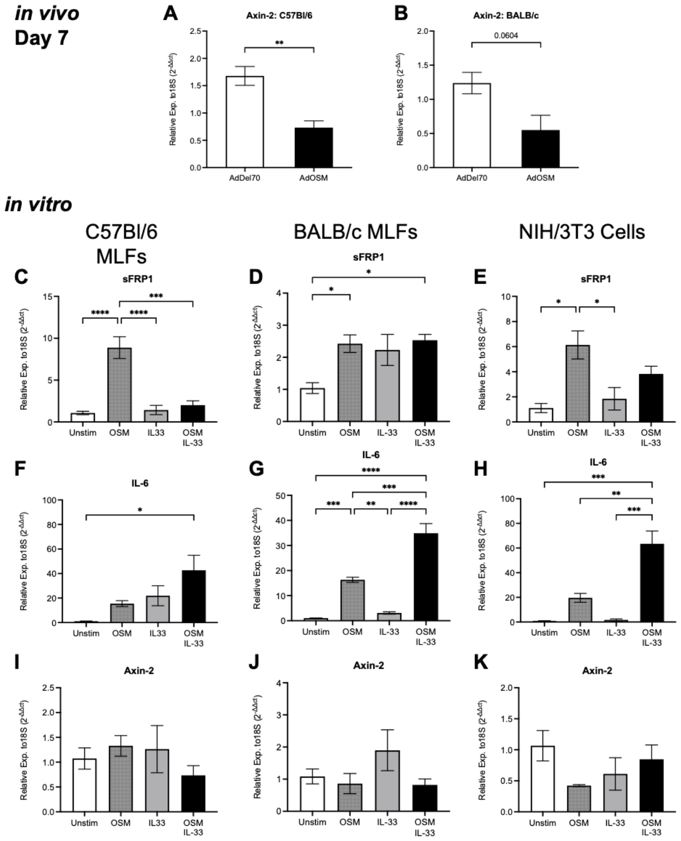
Since the in situ hybridization analysis suggested cell populations consistent with fibroblast and/or smooth muscle actin positive fibroblasts as expressing sFRP1 mRNA, we went on to assess whether OSM could directly regulate the expression of SFRP1 in primary fibroblast cell cultures derived from either C57BL/6 or BALB/c mouse lungs in vitro (Figure 7). As another comparator cell line, we assessed responses of mouse NIH3T3 embryonic fibroblasts. We also assessed the stimulation by IL-33 (known to be induced by AdOSM in vivo [44]) as a comparator stimulus. Since Axin-2 is a recognized as a downstream gene target of β-catenin signaling, we measured Axin-2 in the whole lung homogenates and went on to measure sFRP1, IL-6 and Axin-2 mRNA in the cells cultured in vitro (Figure 7).
In Vivo, Axin-2 mRNA levels in whole lung were decreased at Day 7 of AdOSM treatment (Figure 7A,B) indicating suppression of Wnt/β-catenin signaling to this typical target gene. In vitro, OSM stimulation (24 hours) was able to significantly increase sFRP1 mRNA in primary MLFs derived from either mouse strain and in NIH 3T3 cells (Figure 7C–E). OSM stimulated IL-6 expression, and IL-33/OSM co-stimulation synergized inducing IL-6 mRNA in all 3 fibroblast lines (Figure 7F–H). IL-33/OSM co-stimulation did not upregulate sFRP1 further compared to OSM alone. Neither OSM nor IL-33 showed any consistent or statistically significant direct effect on Axin-2 mRNA levels in these cells in vitro (Figure 7I–K).
Wild-type C57BL/6 and BALB/c mice were administered AdOSM or empty control vector AdDl70 (5 × 10⁷ PFU/mouse) and culled at Day 7. (A, B) Axin-2 expression relative to 18S was measured by RT-qPCR in whole lung homogenates. (C-K) C57BL/6 and BALB/c derived MLFs and NIH 3T3 cells were stimulated with 20 ng/mL of OSM, 20ng/mL IL-33, or 20 ng/mL of both for 24 hours. Relative expression of sFRP1 (C, D, E), IL-6 (F, G, H), and Axin-2 (I, J, K) was quantified by RT-qPCR. Statistical analyses were performed in GraphPad Prism using one-way ANOVA with Tukey’s post-hoc test. Significance was defined where p*<0.05, p<**0.01, p***<0.001, p****<0.0001. Data are presented as mean ± SEM.
4. Discussion
OSM is an IL-6 family member but exhibits distinct biological roles compared to other members of this cytokine group due to differential expression of the receptor chain OSMRβ by different cell types. OSMRβ (along with gp130 receptor chain) is required for OSM activation of cells in the mouse system, and is expressed by lung stromal/structural cells including fibroblasts, epithelial, endothelial, and smooth muscle cells. Here we have shown that in a mouse model, OSM overexpression can regulate the levels of several Wnt pathway ligand and receptor genes in lungs including robust elevation of levels of sFRP1 at the mRNA and protein level, its mRNA level in subepithelial regions of airways in vivo, and that OSM directly stimulates sFRP1 expression in lung fibroblasts in vitro. IL-33 as a comparator cytokine did not induce sFRP1 in vitro. Since IL-6 was regulated in a different fashion in response to OSM and IL-33 in fibroblasts, sFRP1 induction is likely due to different cell regulatory mechanisms than IL-6 induction. OSM strongly activates STAT3 in vitro and in vivo [30] and STAT3 response elements are present in the sFRP1 promoter [47]. Whether STAT3 is essential for OSM regulation of sFRP-1 would need additional deletion studies and/or pharmacological approaches. OSM also induces other cell signaling pathways as reviewed previously by others [22,48] that may also contribute to the regulation of sFRP1 gene expression. Our results suggest that a newly described OSM-sFRP1 pathway exists, which may contribute to some of the unique effects of OSM described previously in lung inflammation.
This study included several Wnt ligands and receptors and although the list was not exhaustive (19 Wnt ligands have been described) those ligands examined here within the nanostring analysis were collectively reduced by the overexpression of OSM (Figure 2, Supp. Table S1) with the exception of Wnt 5A. However, although Wnt5A/B are thought generally to be agonists for the receptors, recent evidence suggests that Wnt 5A and B can inhibit the canonical Wnt signaling pathway in alveolar epithelial cells [49]. Thus, the OSM-induced upregulation of Wnt 5A may also contribute to decreased Wnt signaling in this system. The reduction of mRNA receptor levels for LRP5, LRP6, and FZD3 also suggests a diminished Wnt signaling capability. Collectively, with the observed Axin-2 mRNA in total lung (Figure 7), the results support the concept that canonical β-catenin Wnt signaling is suppressed in this model, at least in certain cell types. The results showing an increase in β-catenin protein overall in total lung homogenates (Figure 3,4) seems at odds with this. However, it is clear that a large number of inflammatory cells infiltrate the AdOSM infected WT C57BL/6 lung, but that this is greatly reduced in IL-6-/- C57BL/6, or in BALB/c mouse lungs (as previously reported [30,31]). These cells stain positive for β-catenin (Supplementary Figure 1) and would contribute to the lung load of total or activated β-catenin as assessed by Immunoblots. Curiously, β-catenin may be present in epithelial cells but not in their nuclei in AdOSM treated lungs, which may affect its function in those cells.
Axin-2 is a well-established downstream target of canonical Wnt/β-catenin signaling and is used widely as a measure of the active status of Wnt pathway in various tissues. β-catenin, once in the nucleus, binds to TCF/LEF transcription factors and the Axin-2 promoter contains multiple TCF/LEF binding sites enabling high responsiveness to β-catenin-induced effects [50,51]. Here we have used Axin-2 mRNA as a surrogate indicator of downstream effects of β-catenin in this system. Figure 7A and B show that OSM overexpression reduces Axin-2 mRNA in total lung homogenates, and this is associated with the induction of sFRP1 and reduction of several Wnt ligands and receptors at the mRNA level as shown in Figure 2, Figure 3 and Figure 4. Which cells of the lung are expressing each one of the Wnt family members analyzed here is not clear, although alveolar epithelial cells and columnar epithelial cells are likely among various lung cell types that can express and/or respond to Wnt ligands. The downregulation of Axin-2 in total lung could also be influenced by multiple cell types. Axin-2 is known to be expressed in type II Alveolar epithelial cells and subsets of lung stromal cells [52], but evaluation of Axin-2 specifically in these cells in this system would need further future study. The assays in MLF cultures in vitro (Figure 7) support the notion that OSM can directly induce sFRP1 but not Axin-2 in fibroblasts. Whether the Axin-2 reduction in vivo is due directly to sFRP1 inhibition of Wnt ligands and/or indirectly through other mechanisms is not discernable in this study.
Wnt pathways are altered in various lung conditions including allergic airway disease and fibrosis [6,16,17]. It has been established that exogenous OSM, either by intranasal administration [24] or by AdOSM [33,34] can induce Th2-skewed inflammation (IL-4, -5, -13) associated with eosinophil, alternatively activated macrophages (AAM) and chemokine accumulation in lungs of C57BL/6 mice. sFRP1 has been implicated in the mouse model of House dust mite (HDM) allergic airway inflammation, where in a HDM-sensitized system, sFRP1-/- mice showed less AAM, IL-5 and eosinophil accumulation than WT. We speculate that OSM-induced effects on Th2-skewed inflammation are due at least in part to its induction of sFRP1, but such cause and effect would have to be demonstrated experimentally. In studies in fibrogenesis, sFRP-1 has been shown to inhibit EMT transition in A549 cells in vitro [53]. Other studies suggested that reduced Sfrp1 levels pose an essential feature in the molecular signature of invading lung fibroblasts, and in vitro studies showed that while sFRP1 inhibited the invasiveness of lung fibroblasts, it was suppressed by TGFβ. This enabled a transition to aggressive invasive fibroblast phenotypes [19] which contributes to TGFβ roles in fibrogenesis. On the other hand, exogenous sFRP1, in the form of sFRP1-enriched extracellular vesicles, exacerbated Bleomycin-induced lung fibrosis [20] suggesting multiple roles of sFRP1 in lung fibrogenesis which may depend on levels and/or timing of expression. OSM can also induce transient lung ECM accumulation [30,33,45]. Interestingly, AdOSM can also exacerbate Bleo-induced lung fibrosis [35] but whether this is mediated through sFRP1 is not known. Although sFRP-1 was elevated in the Bleomycin-induced fibrosis model, the extent of fibrosis observed was no different in sFRP1 KO mice [54]. Thus the role of sFRP1 in mouse models of lung fibrogenesis may be limited to exacerbations engaged by infectious agents or other lung insults. OSM has been shown to play a role Influenza A Virus (IAV) infection models in mice [55] where deletion of OSM in lung macrophages resulted in impaired epithelial barrier repair function. However, the roles of sFRP1 in IAV or other infectious disease models that involve OSM are not known and worth future exploration.
CISH demonstrated the elevation of sFRP1 in subepithelial layers closely aligned to airways (Figure 5 and Figure 6). The results in vitro (Figure 7C–E) show that OSM can directly induce sFRP1 in lung fibroblasts, thus the upregulation of sFRP1 mRNA in subepithelial layers in vivo could be within lung fibroblast or myofibroblast cells in vivo (Figure 5) and is likely due at least in part to direct regulation. Whether mouse airway smooth muscle cells (which can reside in a similar location) respond to OSM with sFRP-1 induction is not known and would require a separate set of studies. It appears that vascular smooth muscle cells do not co-localize sFRP1 with alpha smooth muscle actin (Figure 6) at least at the day seven time point analyzed. There are differences in function (airflow regulation versus blood flow regulation) and phenotypes (receptor expression) between airway smooth muscle cells and lung vascular smooth muscle cells, and we speculate the sFRP-1 expression location (short acting distances) could be affecting airway epithelial cell responses resulting in alterations in β-catenin roles in these cells.
The association of OSM and sFRP-1 activity in other systems may also reflect an OSM-sFRP1 axis. In mouse models, sFRP1-/- mice fed a high fat diet showed alterations in adiposity, glucose metabolism, liver steatosis and inflammatory infiltration, suggesting broad roles in homeostasis of metabolism [56]. Interestingly, OSMRβ-/- mice also show an altered metabolism as they age, including insulin resistance, liver steatosis and obesity [57] indicating an association with levels of OSM pathway activation and metabolic functions in mice. Whether an OSM-sFRP1 axis is involved in these metabolic phenotypes, or other conditions that show elevated levels of OSM, is speculative but would be of interest in future work.
Supplementary Materials
The following supporting information can be downloaded at the website of this paper posted on Preprints.org, Supplementary contains: Table S1: Differentially expressed mRNA genes in lungs of AdOSM treated mice vs control vector at Day 7. Figure S1: Staining for β-catenin in AdOSM-treated lung slices. Series: Full immunoblot images.
Author Contributions
Authors contributed to the manuscript as indicated here “Conceptualization, F.B., CDR, and D.B.; methodology, F.B. and A.D-G., ; validation, A.D-G., M.H, A.C., and L.B. ; formal analysis, F.B., A.D-G., L.S-D., K.Z. .; investigation, FB., A.D-G., L.S-D., K.Z. and L.B. .; resources, D.B. and CDR.; data curation, F.B., L.B.,A.C., A.D-G.; writing—original draft preparation, F.B., M.H and CDR; writing—review and editing L.S-D., A.C., K.Z., D.B., F.B and CDR. ; visualization, F.B., A.D-G., L.B., A.C., K.Z.; supervision, CDR.; project administration, CDR.; funding acquisition, CDR. All authors have read and agreed to the published version of the manuscript.
Funding
This research was funded by the Canadian Institutes of Health Research (CIHR) grant numbers 153084 and 186286. L Somani-Davis was supported by graduate scholarships from the Ontario Graduate Scholarship program and by a Graduate scholarship from the Natural Science and Engineering Research Council (NSERC) of Canada.
Institutional Review Board Statement
The study was conducted in accordance with the Declaration of Helsinki. The animal study protocols were approved by the McMaster University Animal Research Ethics Board AUP# 22-07-24 (approved in 2022), and its predecessor 18-05-22 (approved July 3, 2018).
Data Availability Statement
The datasets generated during the current study supporting the conclusions of the article are available from the corresponding author on reasonable request.
Conflicts of Interest
The authors declare no conflicts of interest. The funders had no role in the design of the study; in the collection, analyses, or interpretation of data; in the writing of the manuscript; or in the decision to publish the results.
References
- Miller, J.R. The Wnts. Genome Biol. 2002, 3. [Google Scholar] [CrossRef] [PubMed]
- Steinhart, Z.; Angers, S. Wnt Signaling in Development and Tissue Homeostasis. Development 2018, 145. [Google Scholar] [PubMed]
- Perugorria, M.J.; Olaizola, P.; Labiano, I.; Esparza-Baquer, A.; Marzioni, M.; Marin, J.J.G.; Bujanda, L.; Banales, J.M. Wnt–β-Catenin Signalling in Liver Development, Health and Disease. Nat. Rev. Gastroenterol. Hepatol. 2019, 16, 121–136. [Google Scholar] [PubMed]
- Baron, R.; Kneissel, M. WNT Signaling in Bone Homeostasis and Disease: From Human Mutations to Treatments. Nat. Med. 2013, 19, 179–192. [Google Scholar] [CrossRef] [PubMed]
- Ng, L.F.; Kaur, P.; Bunnag, N.; Suresh, J.; Sung, I.C.H.; Tan, Q.H.; Gruber, J.; Tolwinski, N.S. WNT Signaling in Disease. Cells 2019, 8. [Google Scholar] [PubMed]
- Reuter, S.; Beckert, H.; Taube, C. Take the Wnt out of the Inflammatory Sails: Modulatory Effects of Wnt in Airway Diseases. Lab. Investig. 2016, 96, 177–185. [Google Scholar] [CrossRef] [PubMed]
- Anastas, J.N.; Moon, R.T. WNT Signalling Pathways as Therapeutic Targets in Cancer. Nat. Rev. Cancer 2013, 13, 11–26. [Google Scholar]
- Xue, C.; Chu, Q.; Shi, Q.; Zeng, Y.; Lu, J.; Li, L. Wnt Signaling Pathways in Biology and Disease: Mechanisms and Therapeutic Advances. Signal Transduct. Target. Ther. 2025, 10. [Google Scholar] [PubMed]
- Qu, J.; Yue, L.; Gao, J.; Yao, H. Perspectives on Wnt Signal Pathway in the Pathogenesis and Therapeutics of Chronic Obstructive Pulmonary Disease. J. Pharmacol. Exp. Ther. 2019, 369, 473–480. [Google Scholar] [PubMed]
- Zhang, W.; Zhang, K.; Ma, Y.; Song, Y.; Qi, T.; Xiong, G.; Zhang, Y.; Kan, C.; Zhang, J.; Han, F.; et al. Secreted Frizzled-Related Proteins: A Promising Therapeutic Target for Cancer Therapy through Wnt Signaling Inhibition. Biomed. Pharmacother. 2023, 166. [Google Scholar] [PubMed]
- Bovolenta, P.; Esteve, P.; Ruiz, J.M.; Cisneros, E.; Lopez-Rios, J. Beyond Wnt Inhibition: New Functions of Secreted Frizzled-Related Proteins in Development and Disease. J. Cell Sci. 2008, 121, 737–746. [Google Scholar] [PubMed]
- Foronjy, R.; Imai, K.; Shiomi, T.; Mercer, B.; Sklepkiewicz, P.; Thankachen, J.; Bodine, P.; D’Armiento, J. The Divergent Roles of Secreted Frizzled Related Protein-1 (SFRP1) in Lung Morphogenesis and Emphysema. Am. J. Pathol. 2010, 177, 598–607. [Google Scholar] [CrossRef] [PubMed]
- Baharudin, R.; Tieng, F.Y.F.; Lee, L.H.; Mutalib, N.S.A. Epigenetics of SFRP1: The Dual Roles in Human Cancers. Cancers 2020, 12. [Google Scholar] [CrossRef] [PubMed]
- Jiang, P.; Wei, K.; Chang, C.; Zhao, J.; Zhang, R.; Xu, L.; Jin, Y.; Xu, L.; Shi, Y.; Guo, S.; et al. SFRP1 Negatively Modulates Pyroptosis of Fibroblast-Like Synoviocytes in Rheumatoid Arthritis: A Review. Front. Immunol. 2022, 13. [Google Scholar] [PubMed]
- Matsuyama, M.; Nomori, A.; Nakakuni, K.; Shimono, A.; Fukushima, M. Secreted Frizzled-Related Protein 1 (Sfrp1) Regulates the Progression of Renal Fibrosis in a Mouse Model of Obstructive Nephropathy. J. Biol. Chem. 2014, 289, 31526–31533. [Google Scholar] [CrossRef] [PubMed]
- Baarsma, H.A.; Königshoff, M. “WNT-Er Is Coming”: WNT Signalling in Chronic Lung Diseases. Thorax 2017, 72, 746–759. [Google Scholar] [PubMed]
- Burgy, O.; Königshoff, M. The WNT Signaling Pathways in Wound Healing and Fibrosis. Matrix Biol. 2018, 68–69, 67–80. [Google Scholar] [CrossRef] [PubMed]
- Rai, N.; Arteaga-Solis, E.; Goldklang, M.; Zelonina, T.; D’Armiento, J. The Role of Secreted Frizzled-Related Protein-1 in Allergic Asthma. Am. J. Respir. Cell Mol. Biol. 2022, 66, 293–301. [Google Scholar] [CrossRef] [PubMed]
- Mayr, C.H.; Sengupta, A.; Asgharpour, S.; Ansari, M.; Pestoni, J.C.; Ogar, P.; Angelidis, I.; Liontos, A.; Rodriguez-Castillo, J.A.; Lang, N.J.; et al. Sfrp1 Inhibits Lung Fibroblast Invasion during Transition to Injury-Induced Myofibroblasts. Eur. Respir. J. 2024, 63. [Google Scholar] [CrossRef] [PubMed]
- Burgy, O.; Mayr, C.H.; Schenesse, D.; Papakonstantinou, E.F.; Ballester, B.; Sengupta, A.; She, Y.; Hu, Q.; Melo-Narvaéz, M.C.; Jain, E.; et al. Fibroblast-Derived Extracellular Vesicles Contain SFRP1 and Mediate Pulmonary Fibrosis. JCI Insight 2024, 9. [Google Scholar] [CrossRef] [PubMed]
- West, N.R.; Hegazy, A.N.; Owens, B.M.J.; Bullers, S.J.; Linggi, B.; Buonocore, S.; Coccia, M.; Görtz, D.; This, S.; Stockenhuber, K.; et al. Oncostatin M Drives Intestinal Inflammation and Predicts Response to Tumor Necrosis Factor–Neutralizing Therapy in Patients with Inflammatory Bowel Disease. Nat. Med. 2017, 23, 579–589. [Google Scholar] [CrossRef] [PubMed]
- West, N.R.; Owens, B.M.J.; Hegazy, A.N. The Oncostatin M-Stromal Cell Axis in Health and Disease. Scand. J. Immunol. 2018, 88, 1–18. [Google Scholar] [CrossRef] [PubMed]
- Headland, S.E.; Dengler, H.S.; Xu, D.; Teng, G.; Everett, C.; Ratsimandresy, R.A.; Yan, D.; Kang, J.; Ganeshan, K.; Nazarova, E. V.; et al. Oncostatin M Expression Induced by Bacterial Triggers Drives Airway Inflammatory and Mucus Secretion in Severe Asthma. Sci. Transl. Med. 2022, 14, 1–16. [Google Scholar] [CrossRef] [PubMed]
- Mozaffarian, A.; Brewer, A.W.; Trueblood, E.S.; Luzina, I.G.; Todd, N.W.; Atamas, S.P.; Arnett, H.A. Mechanisms of Oncostatin M-Induced Pulmonary Inflammation and Fibrosis. J. Immunol. 2008, 181, 7243–7253. [Google Scholar] [CrossRef] [PubMed]
- Pothoven, K.L.; Norton, J.E.; Hulse, K.E.; Suh, L.A.; Carter, R.G.; Rocci, E.; Harris, K.E.; Shintani-Smith, S.; Conley, D.B.; Chandra, R.K.; et al. Oncostatin M Promotes Mucosal Epithelial Barrier Dysfunction, and Its Expression Is Increased in Patients with Eosinophilic Mucosal Disease. J. Allergy Clin. Immunol. 2015, 136, 737–746.e4. [Google Scholar] [CrossRef] [PubMed]
- Pothoven, K.L.; Schleimer, R.P. The Barrier Hypothesis and Oncostatin M: Restoration of Epithelial Barrier Function as a Novel Therapeutic Strategy for the Treatment of Type 2 Inflammatory Disease. Tissue Barriers 2017, 5, 1–18. [Google Scholar] [CrossRef]
- Marden, G.; Wan, Q.; Wilks, J.; Nevin, K.; Feeney, M.; Wisniacki, N.; Trojanowski, M.; Bujor, A.; Stawski, L.; Trojanowska, M. The Role of the Oncostatin M/OSM Receptor β Axis in Activating Dermal Microvascular Endothelial Cells in Systemic Sclerosis. Arthritis Res. Ther. 2020, 22, 1–14. [Google Scholar] [CrossRef] [PubMed]
- Langdon, C.; Kerr, C.; Tong, L.; Richards, C.D. Oncostatin M Regulates Eotaxin Expression in Fibroblasts and Eosinophilic Inflammation in C57BL/6 Mice. J. Immunol. 2003, 170, 548–555. [Google Scholar] [CrossRef] [PubMed]
- Richards, C.D.; Kerr, C.; Tong, L.; Langdon, C. Modulation of Extracellular Matrix Using Adenovirus Vectors. Biochem. Soc. Trans. 2002, 30, 107–111. [Google Scholar] [CrossRef]
- Wong, S.; Botelho, F.M.; Rodrigues, R.M.; Richards, C.D. Oncostatin M Overexpression Induces Matrix Deposition, STAT3 Activation, and SMAD1 Dysregulation in Lungs of Fibrosis-Resistant BALB/c Mice. Lab. Investig. 2014, 94, 1003–1016. [Google Scholar] [CrossRef] [PubMed]
- Botelho, F.M.; Rangel-Moreno, J.; Fritz, D.; Randall, T.D.; Xing, Z.; Richards, C.D. Pulmonary Expression of Oncostatin M (OSM) Promotes Inducible BALT Formation Independently of IL-6, Despite a Role for IL-6 in OSM-Driven Pulmonary Inflammation. J. Immunol. 2013, 191, 1453–1464. [Google Scholar] [CrossRef]
- Botelho, F.; Dubey, A.; Ayaub, E.A.; Park, R.; Yip, A.; Humbles, A.; Kolbeck, R.; Richards, C.D. IL-33 Mediates Lung Inflammation by the IL-6-Type Cytokine Oncostatin M. Mediat. Inflamm. 2020, 2020. [Google Scholar] [CrossRef]
- Fritz, D.K.; Kerr, C.; Fattouh, R.; Llop-Guevara, A.; Khan, W.I.; Jordana, M.; Richards, C.D. A Mouse Model of Airway Disease: Oncostatin M-Induced Pulmonary Eosinophilia, Goblet Cell Hyperplasia, and Airway Hyperresponsiveness Are STAT6 Dependent, and Interstitial Pulmonary Fibrosis Is STAT6 Independent. J. Immunol. 2011, 186, 1107–1118. [Google Scholar] [CrossRef] [PubMed]
- Dubey, A.; Izakelian, L.; Ayaub, E.A.; Ho, L.; Stephenson, K.; Wong, S.; Kwofie, K.; Austin, R.C.; Botelho, F.; Ask, K.; et al. Separate Roles of IL-6 and Oncostatin M in Mouse Macrophage Polarization in Vitro and in Vivo. Immunol. Cell Biol. 2018, 96, 257–272. [Google Scholar] [CrossRef] [PubMed]
- Ayaub, E.A.; Dubey, A.; Imani, J.; Botelho, F.; Kolb, M.R.J.; Richards, C.D.; Ask, K. Overexpression of OSM and IL-6 Impacts the Polarization of pro-Fibrotic Macrophages and the Development of Bleomycin-Induced Lung Fibrosis. Sci. Rep. 2017, 7, 1–16. [Google Scholar] [CrossRef] [PubMed]
- Bankhead, P.; Loughrey, M.B.; Fernández, J.A.; Dombrowski, Y.; McArt, D.G.; Dunne, P.D.; McQuaid, S.; Gray, R.T.; Murray, L.J.; Coleman, H.G.; et al. QuPath: Open Source Software for Digital Pathology Image Analysis. Sci. Rep. 2017, 7. [Google Scholar] [CrossRef] [PubMed]
- Ho, L.; Yip, A.; Lao, F.; Botelho, F.; Richards, C.D. RELMα Is Induced in Airway Epithelial Cells by Oncostatin M Without Requirement of STAT6 or IL-6 in Mouse Lungs In Vivo. Cells 2020, 9. [Google Scholar] [CrossRef] [PubMed]
- Fritz, D.K.; Kerr, C.; Tong, L.; Smyth, D.; Richards, C.D. Eotaxin Expression: Involvement of STAT6 1. 2006. [Google Scholar] [PubMed]
- Ritchie, M.E.; Phipson, B.; Wu, D.; Hu, Y.; Law, C.W.; Shi, W.; Smyth, G.K. Limma Powers Differential Expression Analyses for RNA-Sequencing and Microarray Studies. Nucleic Acids Res. 2015, 43, e47. [Google Scholar] [CrossRef] [PubMed]
- Benjamini, Y.; Hochberg, Y. J. R. Stat. Soc. Ser. B Methological 1995, 57. [CrossRef]
- Wu, G.; Haw, R. Functional Interaction Network Construction and Analysis for Disease Discovery. In Protein Bioinformatics: From Protein Modifications and Networks to Proteomics; Wu, C.H., Arighi, C.N., Ross, K.E., Eds.; Springer New York: New York, NY, 2017; pp. 235–253. ISBN 978-1-4939-6783-4. [Google Scholar]
- Shannon, P.; Markiel, A.; Ozier, O.; Baliga, N.S.; Wang, J.T.; Ramage, D.; Amin, N.; Schwikowski, B.; Ideker, T. Cytoscape: A Software Environment for Integrated Models of Biomolecular Interaction Networks. Genome Res. 2003, 13, 2498–2504. [Google Scholar] [CrossRef] [PubMed]
- Newman, M.E.J. Modularity and Community Structure in Networks. PNAS 2006, 103(23), 8577–82. [Google Scholar] [CrossRef]
- Richards, C.D.; Izakelian, L.; Dubey, A.; Zhang, G.; Wong, S.; Kwofie, K.; Qureshi, A.; Botelho, F. Regulation of IL-33 by Oncostatin M in Mouse Lung Epithelial Cells. Mediat. Inflamm. 2016, 2016. [Google Scholar] [CrossRef] [PubMed]
- Botelho, F.M.; Rodrigues, R.; Guerette, J.; Wong, S.; Fritz, D.K.; Richards, C.D. Extracellular Matrix and Fibrocyte Accumulation in BALB/c Mouse Lung upon Transient Overexpression of Oncostatin M. Cells 2019, 8, 126. [Google Scholar] [CrossRef] [PubMed]
- Li, C.; Nguyen, V.; Clark, K.N.; Zahed, T.; Sharkas, S.; Filipp, F. V.; Boiko, A.D. Down-Regulation of FZD3 Receptor Suppresses Growth and Metastasis of Human Melanoma Independently of Canonical WNT Signaling. Proc. Natl. Acad. Sci. U. S. A. 2019, 116, 4548–4557. [Google Scholar] [CrossRef] [PubMed]
- Marella, S.; Sharma, A.; Ganesan, V.; Ferrer-Torres, D.; Krempski, J.W.; Idelman, G.; Clark, S.; Nasiri, Z.; Vanoni, S.; Zeng, C.; et al. IL-13–Induced STAT3-Dependent Signaling Networks Regulate Esophageal Epithelial Proliferation in Eosinophilic Esophagitis. J. Allergy Clin. Immunol. 2023, 152, 1550–1568. [Google Scholar] [CrossRef] [PubMed]
- Hermanns, H.M. Oncostatin M and Interleukin-31: Cytokines, Receptors, Signal Transduction and Physiology. Cytokine Growth Factor Rev. 2015, 26, 545–558. [Google Scholar] [CrossRef] [PubMed]
- Wu, X.; Van Dijk, E.M.; John-Poul, N.B.; T. Bos, I.S.; Ciminieri, C.; Königshoff, M.; Kistemaker, L.E.M.; Gosens, R. Mesenchymal WNT-5A/5B Signaling Represses Lung Alveolar Epithelial Progenitors. Cells 2019, 8. [Google Scholar] [CrossRef] [PubMed]
- Leung, J.Y.; Kolligs, F.T.; Wu, R.; Zhai, Y.; Kuick, R.; Hanash, S.; Cho, K.R.; Fearon, E.R. Activation of AXIN2 Expression by β-Catenin-T Cell Factor: A Feedback Repressor Pathway Regulating Wnt Signaling. J. Biol. Chem. 2002, 277, 21657–21665. [Google Scholar] [CrossRef] [PubMed]
- Moshkovsky, A.R.; Kirschner, M.W. The Nonredundant Nature of the Axin2 Regulatory Network in the Canonical Wnt Signaling Pathway. Proc. Natl. Acad. Sci. U. S. A. 2022, 119. [Google Scholar] [CrossRef] [PubMed]
- Nabhan, A.N.; Brownfield, D.G.; Harbury, P.B.; Krasnow, M.A.; Desai, T.J. Single-Cell Wnt Signaling Niches Maintain Stemness of Alveolar Type 2 Cells.
- Ren, J.; Wang, R.; Huang, G.; Song, H.; Chen, Y.; Chen, L. SFRP1 Inhibits Epithelial-Mesenchymal Transition in A549 Human Lung Adenocarcinoma Cell Line. Cancer Biother. Radiopharm. 2013, 28, 565–571. [Google Scholar] [CrossRef] [PubMed]
- De Langhe, E.; Aznar-Lopez, C.; De Vooght, V.; Vanoirbeek, J.A.; Luyten, F.P.; Lories, R.J.U. Secreted Frizzled Related Proteins Inhibit Fibrosis in Vitro but Appear Redundant in Vivo. Fibrogenesis Tissue Repair 2014, 7, 1–10. [Google Scholar] [CrossRef] [PubMed]
- Hoagland, D.A.; Rodríguez-Morales, P.; Mann, A.O.; Baez Vazquez, A.Y.; Yu, S.; Lai, A.; Kane, H.; Dang, S.M.; Lin, Y.; Thorens, L.; et al. Macrophage-Derived Oncostatin M Repairs the Lung Epithelial Barrier during Inflammatory Damage 2025.
- Gauger, K.J.; Bassa, L.M.; Henchey, E.M.; Wyman, J.; Bentley, B.; Brown, M.; Shimono, A.; Schneider, S.S. Mice Deficient in Sfrp1 Exhibit Increased Adiposity, Dysregulated Glucose Metabolism, and Enhanced Macrophage Infiltration. PLoS ONE 2013, 8. [Google Scholar] [CrossRef] [PubMed]
- Komori, T.; Tanaka, M.; Senba, E.; Miyajima, A.; Morikawa, Y. Lack of Oncostatin M Receptor β Leads to Adipose Tissue Inflammation and Insulin Resistance by Switching Macrophage Phenotype. J. Biol. Chem. 2013, 288, 21861–21875. [Google Scholar] [CrossRef] [PubMed]
Figure 1.
Protein-Protein Interaction Network of differentially expressed mRNA genes upon overexpression of OSM. C57BL/6 mice were administered 5 × 10⁷ PFU AdOSM or AdDl70 (negative control viral vector) and culled 7 days later (n=3/group). Total lung homogenates were prepared, RNA was purified and analyzed by Nanostring technology, normalized to housekeeping genes (ACTB, B2M and RPLP2) and transformed to differentially expressed genes over AdDl70 controls, as described in Methods. Genes with an adjusted p-value < 0.05 were considered to be differentially expressed as shown in Supplementary Table S1. Fold induction relative to control are shown in red, reduction in green. All 184 codesets were used for the construction of a Protein-Protein Interaction (PPI) network as described in Methods, and subsequently partitioned into 10 mutually exclusive clusters. .
Figure 1.
Protein-Protein Interaction Network of differentially expressed mRNA genes upon overexpression of OSM. C57BL/6 mice were administered 5 × 10⁷ PFU AdOSM or AdDl70 (negative control viral vector) and culled 7 days later (n=3/group). Total lung homogenates were prepared, RNA was purified and analyzed by Nanostring technology, normalized to housekeeping genes (ACTB, B2M and RPLP2) and transformed to differentially expressed genes over AdDl70 controls, as described in Methods. Genes with an adjusted p-value < 0.05 were considered to be differentially expressed as shown in Supplementary Table S1. Fold induction relative to control are shown in red, reduction in green. All 184 codesets were used for the construction of a Protein-Protein Interaction (PPI) network as described in Methods, and subsequently partitioned into 10 mutually exclusive clusters. .
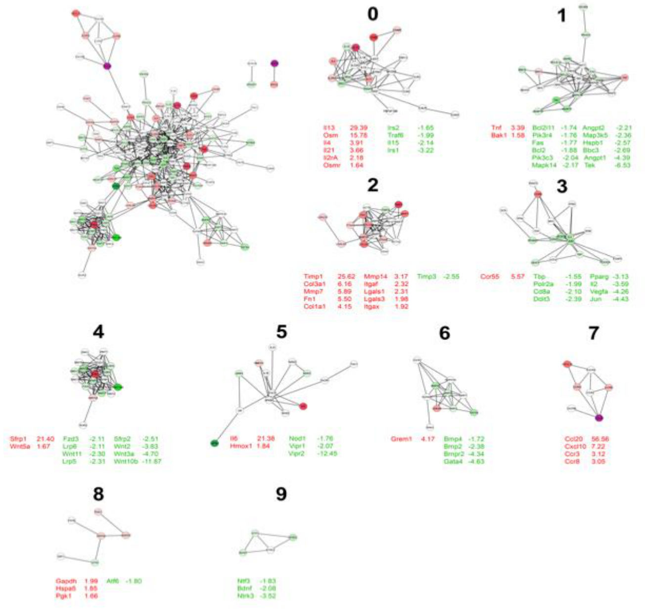
Figure 2.
. Nanostring counts of mRNA induced by overexpression of OSM in Mouse lungs at Day 7: C57BL/6 mice were administered 5x10⁷ PFU AdOSM or AdDl70 (negative control viral vector) and culled after 7 days (n=3/group). Total lung homogenates were prepared, and RNA extracted. Purified RNA was analyzed by Nanostring technology as in Figure 1. (A) Data shows the actual nanostring counts for the most strongly regulated genes in the Wnt pathway module as identified by the probe set. B) BALB/c mice were administered 5x10⁷ PFU AdOSM, 5x10⁷ PFU AdIL-6 or AdDl70 negative control viral vector 2 or 7 days as indicated prior to culling (n=3/group). Whole lung mRNA was extracted and probed by nanostring for sFRP1 and IL-6. Data are presented as mean ± SEM for normalized nanostring counts. Statistical analyses were performed using unpaired t-tests and one way ANOVA, where *p<0.05, **p<0.01. ***p<0.001.
Figure 2.
. Nanostring counts of mRNA induced by overexpression of OSM in Mouse lungs at Day 7: C57BL/6 mice were administered 5x10⁷ PFU AdOSM or AdDl70 (negative control viral vector) and culled after 7 days (n=3/group). Total lung homogenates were prepared, and RNA extracted. Purified RNA was analyzed by Nanostring technology as in Figure 1. (A) Data shows the actual nanostring counts for the most strongly regulated genes in the Wnt pathway module as identified by the probe set. B) BALB/c mice were administered 5x10⁷ PFU AdOSM, 5x10⁷ PFU AdIL-6 or AdDl70 negative control viral vector 2 or 7 days as indicated prior to culling (n=3/group). Whole lung mRNA was extracted and probed by nanostring for sFRP1 and IL-6. Data are presented as mean ± SEM for normalized nanostring counts. Statistical analyses were performed using unpaired t-tests and one way ANOVA, where *p<0.05, **p<0.01. ***p<0.001.
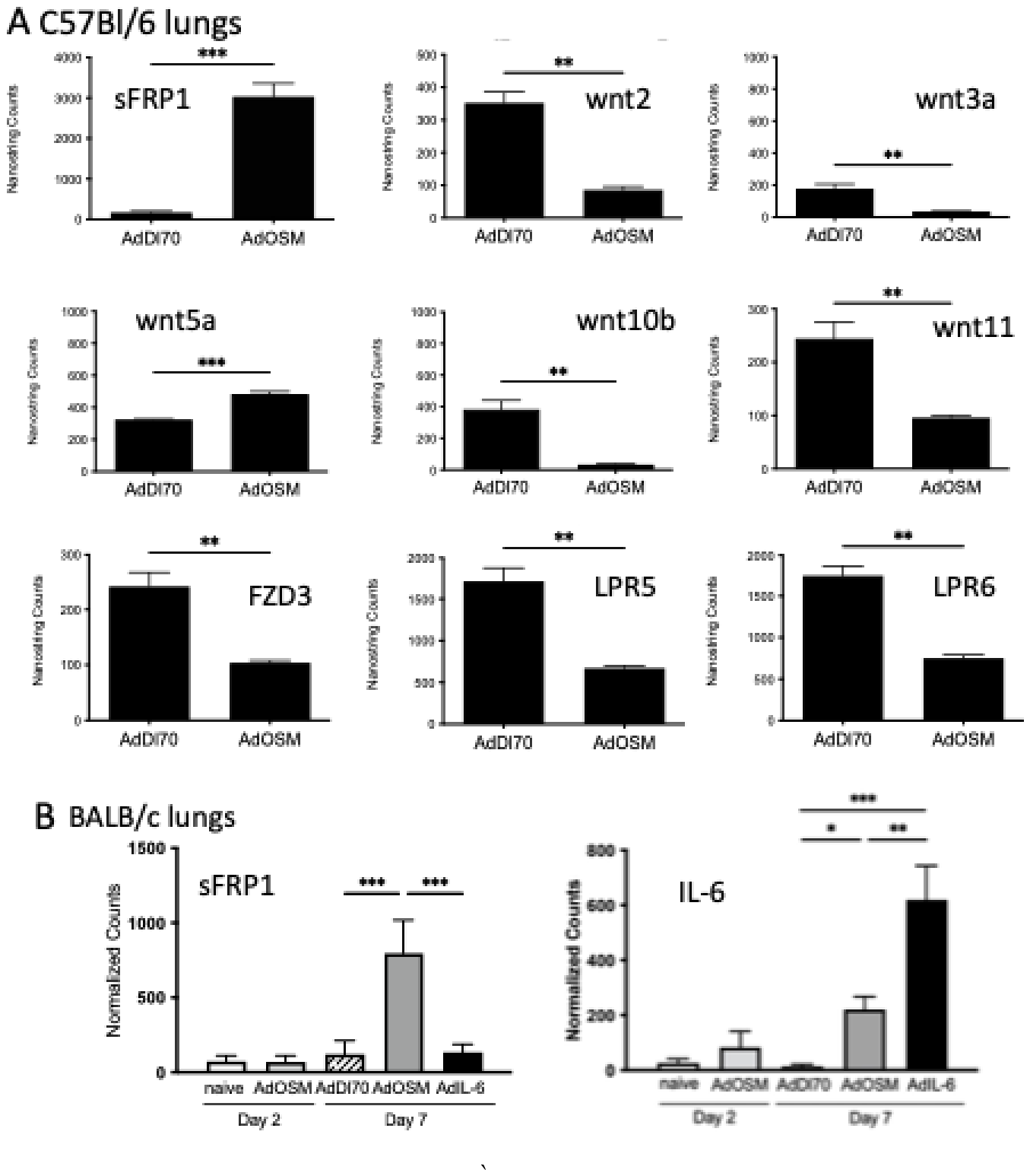
Figure 3.
OSM-induced sFRP1 protein expression in mouse lung. (A) C57BL/6 and BALB/c mice were administered 5 × 10⁷ PFU AdOSM or AdDl70 (negative control viral vector) prior to sacrifice 5 days post-infection. Protein expression of sFRP-1, active-beta-catenin (ABC), or beta-catenin (all forms) was assessed from whole lung extracts by western blotting analysis. β-actin was probed for as a loading control. Each lane represents whole lung extract from a separate mouse (n=3/group). (B-D) Western blot band intensities as assessed by densitometry are shown for (B) sFRP-1, (C) active β-catenin, (D) total β-catenin corrected to β-Actin signals. Data are presented as mean ± SEM. Statistical analyses were performed using unpaired t-tests, where *p< 0.05, **p<0.01, ***p<0.001.
Figure 3.
OSM-induced sFRP1 protein expression in mouse lung. (A) C57BL/6 and BALB/c mice were administered 5 × 10⁷ PFU AdOSM or AdDl70 (negative control viral vector) prior to sacrifice 5 days post-infection. Protein expression of sFRP-1, active-beta-catenin (ABC), or beta-catenin (all forms) was assessed from whole lung extracts by western blotting analysis. β-actin was probed for as a loading control. Each lane represents whole lung extract from a separate mouse (n=3/group). (B-D) Western blot band intensities as assessed by densitometry are shown for (B) sFRP-1, (C) active β-catenin, (D) total β-catenin corrected to β-Actin signals. Data are presented as mean ± SEM. Statistical analyses were performed using unpaired t-tests, where *p< 0.05, **p<0.01, ***p<0.001.
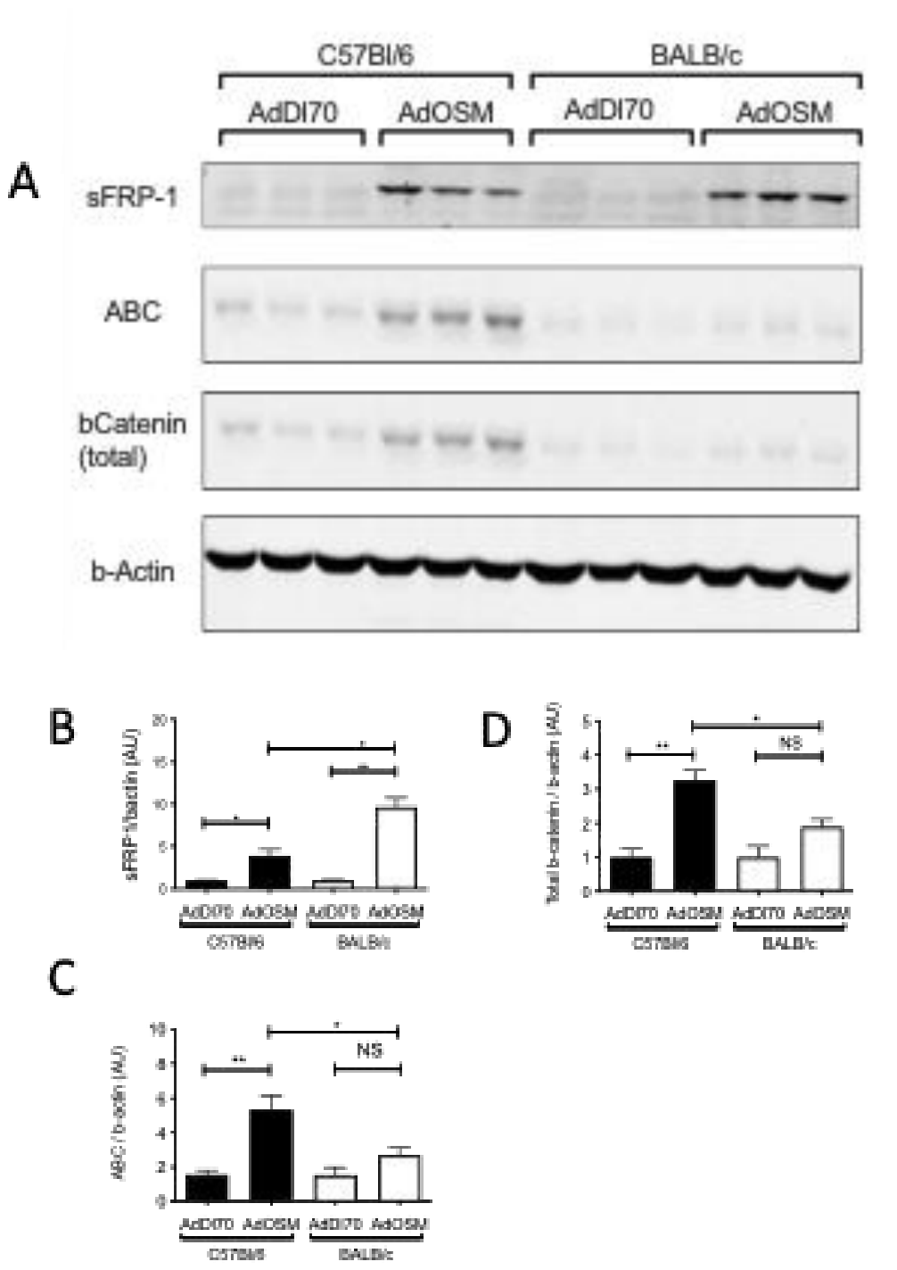
Figure 4.
AdOSM-induced expression of sFRP-1 is partially IL-6-independent while AdOSM-induced levels of β-catenin is IL-6-dependent in the mouse lung. (A) C57BL/6 wildtype or IL-6-knockout (IL-6KO) mice were administered 5 × 10⁷ PFU AdOSM (n=5/group), and naïve mice (n=3) were included as a control. At five days post-infection, mice were culled and expression of sFRP-1, active β-catenin (ABC), or β-catenin (all forms) was assessed from whole lung extracts by western blotting analysis. β-actin was probed for as a loading control. Each lane represents whole lung extract from a separate mouse. Western blot band intensities, in part (A), as assessed by densitometry are shown for (B) sFRP-1, (C) active β-catenin, or (D) total β-catenin, all relative to β-actin. (E) sFRP-1 mRNA levels (nanostring technology) in whole lung of C57BL/6 wildtype (WT) or IL-6 knockout (IL-6KO) mice following treatment with AdOSM. Data are presented as mean ± SEM. Statistical analysis was performed using one-way ANOVA, where *p<0.05, **p<0.01, ***p<0.001.
Figure 4.
AdOSM-induced expression of sFRP-1 is partially IL-6-independent while AdOSM-induced levels of β-catenin is IL-6-dependent in the mouse lung. (A) C57BL/6 wildtype or IL-6-knockout (IL-6KO) mice were administered 5 × 10⁷ PFU AdOSM (n=5/group), and naïve mice (n=3) were included as a control. At five days post-infection, mice were culled and expression of sFRP-1, active β-catenin (ABC), or β-catenin (all forms) was assessed from whole lung extracts by western blotting analysis. β-actin was probed for as a loading control. Each lane represents whole lung extract from a separate mouse. Western blot band intensities, in part (A), as assessed by densitometry are shown for (B) sFRP-1, (C) active β-catenin, or (D) total β-catenin, all relative to β-actin. (E) sFRP-1 mRNA levels (nanostring technology) in whole lung of C57BL/6 wildtype (WT) or IL-6 knockout (IL-6KO) mice following treatment with AdOSM. Data are presented as mean ± SEM. Statistical analysis was performed using one-way ANOVA, where *p<0.05, **p<0.01, ***p<0.001.
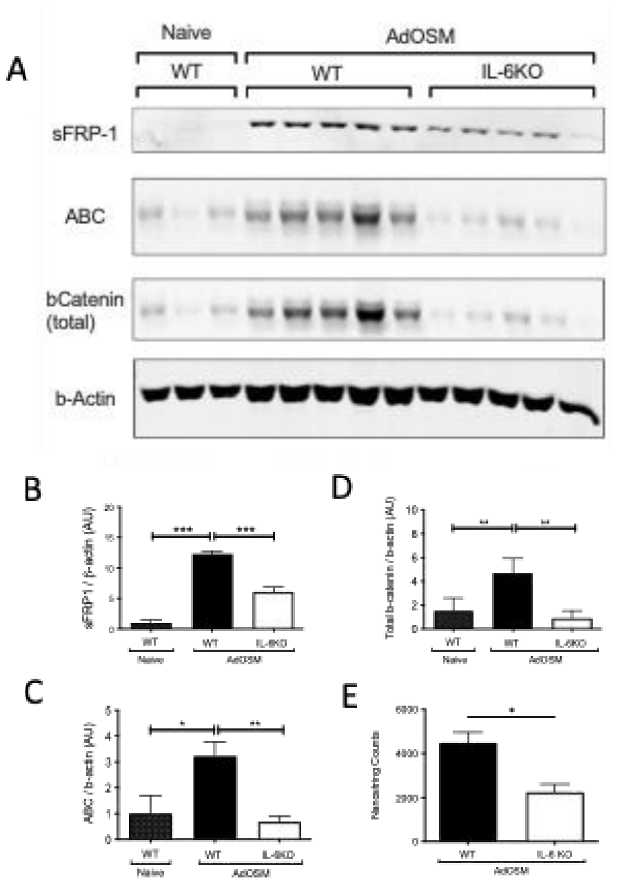
Figure 5.
SFRP1 mRNA detected by chromogenic in situ hybridization (CISH) in mouse lung. AdOSM (5 × 10⁷ PFU) infected C57BL/6 mice were culled at Day 7 and left lung lobes were collected, fixed in formalin, paraffin-embedded, and sections were stained for OSM mRNA (red signal) and SFRP1 mRNA (green signal) simultaneously by CISH. (A-C) Representative scanned images of Day 7 time points at increasing magnifications. (D) is a representative section of AdOSM treated mouse lung probed with control probe (same magnification as (C)). Representative sections stained by CISH for OSM and SFRP1 on lung sections of naïve mice (E) and AdDl70 treated mice (F) are shown.
Figure 5.
SFRP1 mRNA detected by chromogenic in situ hybridization (CISH) in mouse lung. AdOSM (5 × 10⁷ PFU) infected C57BL/6 mice were culled at Day 7 and left lung lobes were collected, fixed in formalin, paraffin-embedded, and sections were stained for OSM mRNA (red signal) and SFRP1 mRNA (green signal) simultaneously by CISH. (A-C) Representative scanned images of Day 7 time points at increasing magnifications. (D) is a representative section of AdOSM treated mouse lung probed with control probe (same magnification as (C)). Representative sections stained by CISH for OSM and SFRP1 on lung sections of naïve mice (E) and AdDl70 treated mice (F) are shown.
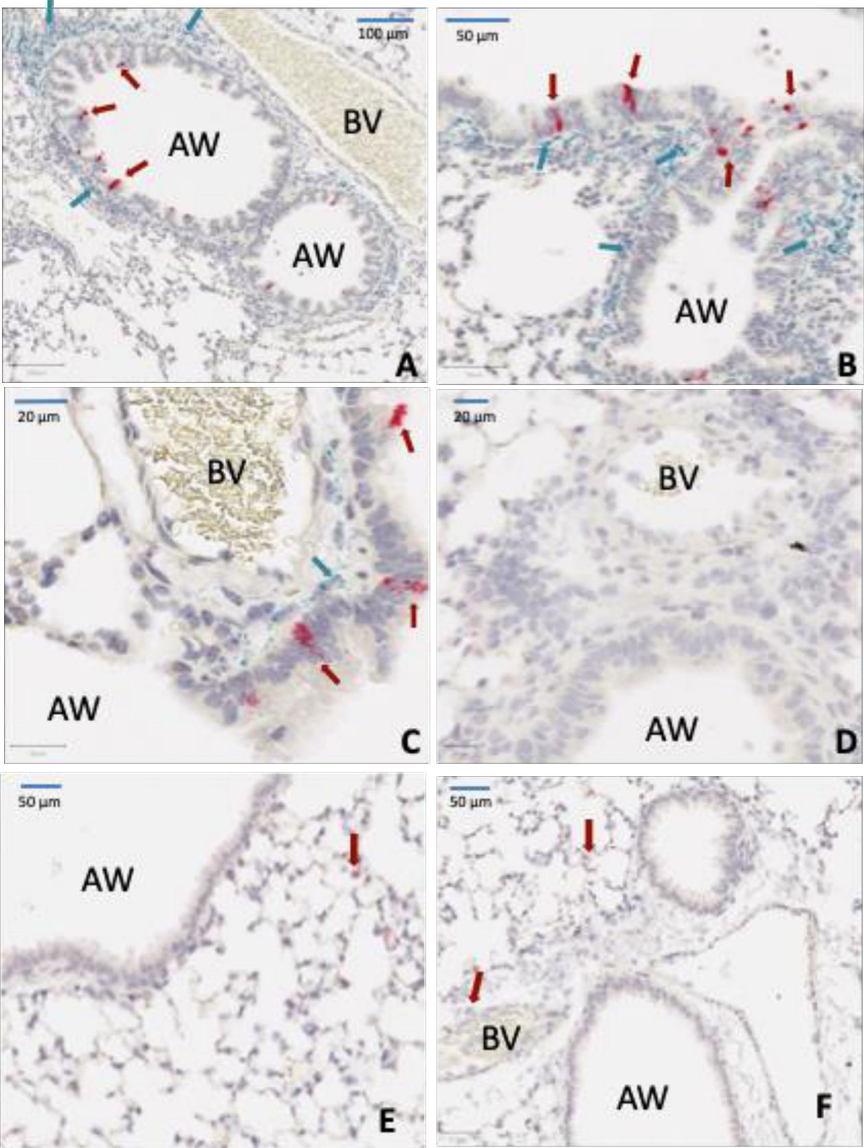
Figure 6.
sFRP1 mRNA detected by CISH and AMSA IHC in serial section of mouse lung. AdOSM (5 × 10⁷ PFU) infected C57BL/6 mice were culled at day 7 and left lung lobes were collected, fixed in formalin, paraffin embedded, and sections were stained (A) for OSM mRNA (red signal) and sFRP1 mRNA (green signal) simultaneously by CISH and (B) for alpha smooth muscle actin by Immunohistochemistry (brown signal) on a serial section. Example locations of airway (AW) and blood vessels (BV) are indicated.
Figure 6.
sFRP1 mRNA detected by CISH and AMSA IHC in serial section of mouse lung. AdOSM (5 × 10⁷ PFU) infected C57BL/6 mice were culled at day 7 and left lung lobes were collected, fixed in formalin, paraffin embedded, and sections were stained (A) for OSM mRNA (red signal) and sFRP1 mRNA (green signal) simultaneously by CISH and (B) for alpha smooth muscle actin by Immunohistochemistry (brown signal) on a serial section. Example locations of airway (AW) and blood vessels (BV) are indicated.
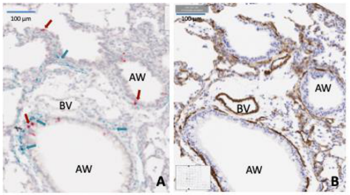
Figure 7.
OSM and IL-33 modulate SFRP1 expression and downstream Wnt signaling pathway.
Disclaimer/Publisher’s Note: The statements, opinions and data contained in all publications are solely those of the individual author(s) and contributor(s) and not of MDPI and/or the editor(s). MDPI and/or the editor(s) disclaim responsibility for any injury to people or property resulting from any ideas, methods, instructions or products referred to in the content. |
© 2026 by the authors. Licensee MDPI, Basel, Switzerland. This article is an open access article distributed under the terms and conditions of the Creative Commons Attribution (CC BY) license.
Copyright: This open access article is published under a Creative Commons CC BY 4.0 license, which permit the free download, distribution, and reuse, provided that the author and preprint are cited in any reuse.