

Submitted:
13 September 2026
Posted:
14 September 2026
You are already at the latest version
Abstract
Launched in 2010 by the National Institutes of Health, the Human Connectome Project (HCP) represents a foundational initiative in systems neuroscience, designed to map the structural and functional architecture of the human brain. Despite its extensive impact, the expanding body of HCP-related research remains methodologically and thematically dispersed. To systematically assess this landscape, we conducted an AI-assisted, intensive review and systematic synthesis of 102 peer-reviewed publications that explicitly referenced the HCP. Using a hybrid computational-human analytic framework integrating natural language processing and manual validation, studies were categorized into six domains: cognition and learning, aging and development, mental health, neurological disorders, movement and motor skills, and behavior and personality. Quantitative and qualitative analyses revealed three dominant trends: (1) inter-individual connectivity patterns exhibit high stability, supporting their utility as neural fingerprints; (2) higher-order association networks undergo postnatal maturation and exhibit age-related decline, confirming lifespan-dependent reorganization; and (3) connectomic measures increasingly function as candidate biomarkers for neuropsychiatric and neurodegenerative disorders, including schizophrenia, Alzheimer's disease, and depression. Critical evaluation identified persistent methodological constraints, including limited spatial resolution in neuroimaging modalities, high attrition and cost in longitudinal paradigms, and data privacy risks associated with large-scale neuroinformatics. This synthesis demonstrates that while the HCP has substantially advanced mechanistic models of human brain organization, future progress will depend on integrating multimodal imaging with computational and AI-driven analytic frameworks. Our review shows that such advancements are essential to overcome current technical limitations and enable precision-level mapping of brain connectivity in health and disease.
Keywords:
neuroscience
; neuroimaging
; human connectome project
; connectomics
; artificial intelligence
1. Introduction
The term “connectome,” coined by Olaf Sporns, Giulio Tononi, and Rolf Kötter in 2005, describes a comprehensive map of the human brain. Despite the impressive advancements in neuroimaging technology at the time, they argued that there was a lack of standardized information on the overall brain’s functionality [1]. Existing models of the human brain were often inaccurate or lacked detail, and most studies of brain function and structure focused on specific parts of the brain with no attention to its overall function. This created countless gaps in knowledge that were made clear in the practice of neuroscience. Thus, in July of 2009, an ambitious project to map one of the most complex structures to exist began. This project, Human Connectome Project (HCP), sought to create a comprehensive description of the functional and structural connectivity of the whole brain [2].
The Human Connectome Project aimed to map and understand the neural pathways in the brain, the specific functions of regions, and how they connect to further understand human cognition and behavior [2]. The HCP uses neuroimaging techniques such as diffusion-weighted magnetic resonance imaging and fMRI to create this map [3]. Through its large sample size, the HCP attempted to discover patterns in connectivity in individuals and as a population, allowing for further research and innovation on the human brain [2].
With over 1,100 participants and easy data sharing through ConnectomeDB, the HCP has enabled a surge of research, totaling more than 27 petabytes of data and over 1,500 papers citing its resources [2]. Beyond the size of the dataset, the project pushed neuroimaging technology forward by incorporating ultra-high-field scanners.
A subset of participants completed resting-state fMRI at 7 T, which later incorporated radiofrequency parallel transmission to improve flip-angle uniformity and boost tSNR and fCNR [4]. These gains helped strengthen functional connectivity estimates in ways that were not possible with standard 3 T protocols. The HCP also developed whole-brain diffusion imaging at 7 T with 1.05 mm resolution, allowing researchers to map white-matter pathways with a level of anatomical detail that had previously only been accessible through ex vivo work. As the group continued to raise spatial resolution, they documented the tradeoffs that come with high-field imaging, including stronger motion sensitivity and challenges in achieving uniform RF performance [5]. Even with those constraints, the combination of advanced hardware, stronger gradients, and new acquisition strategies reshaped what structural and functional imaging could capture inside a living brain [6].
The project was followed up by Lifespan HCP and Aging studies, all of which added to the widely available work now accessible to researchers and the general public [7]. The Human Connectome Project in Aging recruited more than 600 participants for longitudinal assessment to reveal age-related cognitive and physiological changes [8]. This project utilizes MRI data, including task fMRI, structural MRI, resting state fMRI, diffusion MRI, and arterial spin labeling, to observe changes in brain circuits [8]. These imaging techniques allow researchers to assess how the brain functions over time and dynamically.
Machine learning and deep learning techniques have increasingly been applied to HCP data to enhance connectome analysis, including high-accuracy functional connectome fingerprinting for individual identification and cognitive state decoding from task-based fMRI. These conceptual distinctions and applications are summarized in Table 1. For instance, advanced ML classifiers, such as graph convolutional networks and multi-layer perceptrons, achieve robust subject-specific decoding and near state-of-the-art performance on HCP tasks even in dense individual datasets. These computational approaches also support biomarker discovery by modeling brain fluctuation dynamics in resting-state and task conditions, as well as predicting traits like delayed reward discounting from neuroanatomical patterns.
2. Background
2.1. Structural Connectome
The structural connectome refers to the physical wiring of the brain: the white matter pathways that link different regions. These connections form the network through which signals travel, allowing different parts of the brain to communicate [1]. One of the main tools used to study this network is diffusion MRI, which tracks the movement of water molecules in the brain. Since water tends to move along white matter fibers, these scans can be used to estimate the orientation of fiber tracts [9]. Diffusion Tensor Imaging (DTI) was one of the first techniques developed for this purpose. It models diffusion in three dimensions and helps identify major fiber bundles, but it has trouble in areas where fibers cross or bend [10]. High Angular Resolution Diffusion Imaging (HARDI) improves on this by measuring diffusion in more directions, allowing for a more detailed picture in complex regions [3].
Tractography is a method used to reconstruct white matter pathways from diffusion data. It works by following the direction of water diffusion through the brain and creating streamlines that represent possible fiber tracts [11]. These models can be used to study the brain at the level of full networks or to focus on specific pathways. While tractography is a powerful tool, it still lacks sensitivity to noise and can sometimes produce misleading results, especially in the regions of the brain where fibers are densely packed or change direction quickly [12].
To check the accuracy of diffusion-based models, researchers also study the brain using postmortem methods. Tracer studies, which involve injecting dyes or chemicals into brain tissue to track connections, have been widely used in animal studies and sometimes in human tissue. These techniques give high resolution and direct evidence of connection patterns, though they can’t be used on living subjects [13]. A major advancement in postmortem mapping is the BigBrain project [3], which created a high-resolution 3D model of a human brain using thousands of ultra-thin histological slices. This project captures detail at the cellular level, including the structure of individual cortical layers, providing an understanding that imaging alone can’t provide.
Combining diffusion MRI, tractography, and postmortem analysis has given researchers a better understanding of how the brain is structured. Each method brings different strengths, and together they help build a more complete view of how different parts of the brain are structurally connected. This multi-modal approach is important for advancements in the accuracy of results, to validate results, and to overcome the limitations of a single technique.
2.2. Functional Connectome
The functional connectome tracks patterns of simultaneous neural activation across various brain regions. Instead of relying solely on transparent anatomical connections, functional brain connectivity depends upon the temporal correlation between neural signals recorded from distinct areas of the brain. Synchronized activity reveals how the brain forms connections that wire the brain like circuits, supporting various aspects of cognition, physiology, and more [14].
The most common method used to observe functional connectivity is the use of functional magnetic resonance imaging, abbreviated as fMRI. When a neuron cluster becomes inactive, it requires more oxygen to function. As a result, blood vessels dilate to direct more blood to the region, causing alterations to the surrounding tissue [15]. An fMRI tracks brain activity by detecting these shifts in blood oxygenation, called the BOLD (Blood-Oxygen-Level-Dependent) Signal, and the tissue properties through an MRI scanner. An fMRI can therefore be used to accurately map which regions of the brain are activated at any given time. There are two distinct methods of fMRI used to study functional connectivity: resting-state fMRI (rs-fMRI) and task-based fMRI [16].
Resting-state fMRI captures spontaneous brain activity [17] while the subject is lying still, without performing any tasks. This reveals, in passive conditions, networks that are connected functionally, such as the Default Mode Network/DMN (active during internal thought), salience network (active when processing stimuli), and frontoparietal control network (active during cognitive processing). The default mode network, in particular, is one of the first brain networks discovered via resting-state fMRI, and is typically active when a person is not focused on the outside world, such as during daydreaming, self-reflection, or mind wandering [18,19]. These connectivity networks are consistently observed across populations through the Human Connectome Project Data, hence serving as a baseline for understanding the organization of the typical human brain. Especially in clinical environments or for populations such as children or cognitively impaired patients, the resting-state fMRI is a valuable tool. However, due to variability in the mental states of patients and motion, the HCP works to preprocess all imaging to minimize disruptions [16]. Standard preprocessing steps involve physiological noise removal, motion correction, and spatial normalization.
Contrastingly, task-based fMRIs measure the brain activity of a subject when faced with a cognitive, sensory, or motor task. By comparing the BOLD signals during cognitive tasks, researchers can isolate functional networks and decipher which regions are engaged during specific processes such as comprehension, memory, or motor function. These activations are then used to map the task-based functional connectomes of the brain [20].
Through both rs-fMRI and task-based fMRI, the HCP gains a more comprehensive understanding of the functional connectivity of the brain, laying the groundwork for neuroscience and clinical research that could lead to the discovery of the disruptions causing neurodegenerative and neuropsychiatric diseases [21]. Through correlating disruptions in connectivity with behavioral symptoms, functional Connectomic analysis is being increasingly used for investigating schizophrenia, Alzheimer’s disease, and depression.
2.3. Neural Network
The brain is made up of complex systems of neurons that process and share information. These complex systems that work together to form are called brain networks. The main regions of the brain that play a major role are the cerebral cortex, subcortical structures, the cerebellum, and the corpus callosum. The four main structures of the cerebral cortex are the frontal, parietal, temporal, and occipital lobes. These contain the major nodes of brain networks [22]. Each lobe is responsible for a set of tasks. In a study conducted by Olaf Sporns and his team, it was found that the brain areas most involved in the cognitive functions were located in the frontal and parietal lobes. Perception and action limitations are related to frontoparietal brain networks, while working memory capacity limitations are associated with parieto-occipital brain networks [22].
The brain sends fast electrical signals through billions of neurons. Each neuron has branches called dendrites that receive messages, a cell body that processes them, and an axon that sends the signal to the next neuron. When a neuron is activated, it sends an electrical pulse called an action potential down the axon [23]. At the end of the axon, this pulse triggers the release of tiny chemical messengers called neurotransmitters into a gap between neurons called the synapse [24,25]. These neurotransmitters travel across the synapse and bind to receptors on the next neuron, passing along the message [26]. This entire process, called synaptic transmission, allows neurons to communicate rapidly and is the basic building block for all brain activity. Synaptic plasticity, the ability of synapses to strengthen or weaken over time, is critical for learning and memory [26]. These constant signals create patterns of activity that form larger neural networks, which scientists can observe using brain imaging [21]. This synaptic plasticity is needed for memory formation.
For instance, the DMN, a large-scale brain network, includes areas such as the medial prefrontal cortex, posterior cingulate cortex, and hippocampus. It becomes active when we are resting, daydreaming, or thinking about ourselves. Researchers first discovered this in resting-state fMRI studies [18,19]. Later, Greicius et al. (2004) found that the DMN is weaker in people with early Alzheimer’s disease, especially in the hippocampus, which may explain why memory problems occur in these patients [27]. Another key network is the salience network, which helps the brain detect important events. It includes the anterior insula and the dorsal anterior cingulate cortex (dACC). According to Seeley et al. (2007), the salience network helps switch the brain’s focus between internal thoughts like the DMN and external tasks [28]. A study by Ullsperger et al. (2013) found that when people make a mistake, the anterior insula activates and quickly sends a signal to the dACC to help the person adjust [29]. These networks, made of many neurons working together through electrical and chemical signals, help the brain balance rest, attention, memory, and decision-making.
Neuroplasticity allows the brain to form new connections between neurons and reorganize itself based on experiences, learning, or injury. This means the nervous system can reorganize its structure, functions, and connections based on new information or damage [30,31]. Neuroplasticity takes many forms and happens in different contexts, but some important features are common: it depends on experience, is sensitive to timing, and requires motivation and attention to be effective [32,33]. For example, during development, the brain goes through critical periods when it is especially open to change, which helps in learning new skills [34]. Neuroplasticity is also crucial for learning throughout life and for recovering functions after brain injury, as healthy parts of the brain can take over lost functions when motivated training occurs [31,35]. At the cellular level, this process involves changing the strength of synapses, growing new connections, and altering gene activity and protein production to support these changes [36,37]. This process drives dynamic changes in the brain’s structural and functional connectome by continuously remodeling neural connections in response to experience and learning [38].
2.4. Emerging Multimodal Approaches
Connectome research is evolving to integrate diverse data types by using multimodal approaches to build a more comprehensive understanding of the brain’s functions and structure. These multimodal approaches combine functional MRI (fMRI), diffusion MRI (dMRI), electroencephalography (EEG), and magnetoencephalography (MEG), along with transcriptomic and genetic data, going beyond single-source imaging techniques, to fully map the brain’s neural networks with increased accuracy [3,39,40]. Previous methods relied solely on a single data source. However, multimodal approaches combine findings from several modalities, revealing how different aspects of brain connectivity are interrelated by overcoming the limitations of single-method analyses [41,42].
For example, Diffusion Tensor Imaging (DTI) and High Angular Resolution Diffusion Imaging (HARDI) are generally used to map and reconstruct structural connectivity by tracing the diffusion of water molecules along white matter tracts [43]. Diffusion tensor Imaging offers broad structural maps, and on the other hand, High Angular Resolution Diffusion Imaging provides significant angular precision. This precision improves resolution in regions with complex fiber crossings. Resting-state or task-based fMRI is generally used to measure functional connectivity, capturing the correlations in neural activities across different brain regions [44,45].
These methods detect synchronized neural activity, without needing direct anatomical connections, by measuring the Blood Oxygenation Level Dependent (BOLD) signal [44]. This allows researchers the ability to identify networks that are active during sensory processing, internal thought, or cognitive effort. Functional connectivity provides a more dynamic approach to how different brain areas communicate, compared to structural connectivity, as it reflects the brain’s coordination during cognitive tasks [46]. Emerging multimodal approaches combine these modalities with MEG and EEG. In contrast to previous methods, these emerging approaches provide millisecond-level temporal resolution, which MRI cannot offer [47,48]. These electrophysiological tools help examine rapid neural dynamics. Combining the use of MEG or EEG with MRI results in temporal and spatial precision, further enhancing the understanding of how functional and structural dynamics co-exist [49].
Advanced preprocessing tools like FSL (FMRIB Software Library) and Freesurfer allow researchers to combine different types of brain imaging techniques. These tools can prepare structural, functional, and diffusion MRI data for analysis. FSL is widely used in tasks like brain extraction, motion, correction, spatial smoothing, and alignment of brain images to a standardized space. The FEAT pipeline is used for analyzing task-based and resting state fMRI [50], while the diffusion toolkit (FDT) supports tractography, modeling how water molecules move along white matter tracts [51]. Freesurfer is used for structural MRI and can reconstruct cortical surfaces, measure cortical thickness, and segment brain regions based on anatomical landmarks [52]. These reconstructions can map fMRI activity in the brain to study changes on the surface. In large-scale connectome projects like the Human Connectome Project (HCP), both FSL and Freesurfer are used together to ensure consistency across thousands of participants. Their individual strengths help researchers align multimodal data accurately, which is vital when studying how different types of brain connectivity relate to each other [16].
Additionally, recent research studies have started layering and adding transcriptomic data and genetic data into connectomic analyses and multimodal approaches. A landmark study, published in Science, showed that brain regions with synchronous activity were found to share similar gene expression profiles [53]. These findings and correlations suggest that functional connectivity could have connections in molecular architecture, involving a biological dimension to network analysis.
To analyze these complex datasets, researchers have started to integrate tools from graph theory and network science. Albert-László Barabási’s principles of modularity and scale-free networks have been applied to the brain, quantifying the process of how information flows across neural systems [54]. This model offers a quantitative perspective, demonstrating how information flows throughout the connectome.
Multimodal approaches are currently being integrated in large-scale studies. The Human Connectome project integrates dMRI, fMRI, and behavioral data from thousands of participants to generate high-resolution brain maps [41]. The ABCD Study and UK Biobank Imaging Study combine genetic data, lifestyle data, and imaging to investigate the influences of external and internal factors on health and brain development [55,56].
In large-scale initiatives, multimodal approaches play an important role in transforming our ability to map the brain. The major milestones of the Human Connectome Project are summarized in Figure 1. These advancements help expand the understanding of our function and structure while also raising new questions about the biological roots of disease and cognition, by offering improved insight and resolution.
3. Methods
3.1 Research Framework
This literature review synthesized the findings of 102 peer-reviewed papers that discuss the Human Connectome Project (HCP) pipelines, datasets, and methodologies, and allowed for a comprehensive analysis of the advancements in connectomic mapping, multimodal neuroimaging, and neuroinformatics infrastructure. This comprehensive analysis includes diffusion MRI, structural MRI, resting-state and task fMRI, and multimodal extensions [41,57]. We implemented a retrieval-augmented generation (RAG) and human-validation synthesis workflow to ensure reproducibility. This workflow integrated large-language-model outputs, cross-validation, and human validation. Cognition & Learning, Aging & Development, Mental Health, Neurological Disorders, Movement & Motor Skills, and Behavior & Personality.
3.2. Criteria for Papers
In our initial search, we identified 128 papers. However, due to access limitations and other complications, the final sample was reduced to 102 papers. The model’s output was cross-validated by independent systems. Specifically, summaries produced by one system were compared with those generated by the remaining models, and only results confirmed by four models were retained. This approach reduced model-specific bias and misinformation, leading to more reliable categorization and research summaries. We also used a large language model from Meta to conduct a structured meta-analysis and literature review that followed the journal’s standards.
3.3. Categorization
The outputs from the AI ensemble were grouped into six domains, appearing consistently across all models:
- A. Cognition & Learning
- B. Aging & Development
- C. Mental Health
- D. Neurological Disorders
- E. Movement and Motor Skills
- F. Behavior and Personality
Multiple generative AI such as ChatGPT-5.2 and Gemini 2.5 Flash, have been used to refine the summaries for the domains to align with the HCP subprojects and consistency in modality terminology.
3.4. Human Validation and Integration
The human validation assessed methodological rigor using predefined criteria, including pipeline transparency, statistical robustness, and replicable results. The final dataset presents three-tier validation, as seen in Figure 2:
3.4.1. AI Retrieval
The first stage involved AI retrieval using Ollama (Llama 3.1) as a local embedding engine. This system was used to convert the abstracts, methods, and results of the 102 HCP papers into mathematical “vectors.” This allowed the AI to find and pull information based on deep meaning rather than just matching keywords. This process ensured a thorough collection of data while keeping a clear digital trail back to the source papers.
3.4.2. AI Clustering
After gathering the data, AI clustering was used to organize the findings into six main research categories. Using ChatGPT-5.2 and Ollama (Llama 3.1), the system identified shared themes and anatomical terms across the papers. This step automatically sorted a large, unorganized collection of information into a structured format, making it easier to see how different HCP studies and imaging methods relate to each other.
3.4.3. Cross-Validation of Models
To organize the literature efficiently, clustering outputs from multiple AI models (ChatGPT-5.2, Gemini 2.5 Flash, NotebookLM, and Grok 3) were compared against one another to confirm consistent domain assignment across the six categories. A paper’s categorization was retained only when at least four of the five systems agreed. This step reduced individual model bias in sorting the literature but did not extend to writing or interpreting the findings.
3.4.4. Human Validation
The final step was human validation by researchers. The team manually checked the AI’s summaries and categories against the papers to confirm their accuracy. By reviewing the methodology and results for consistency, this manual check ensured that the AI-generated data met high scientific standards and made sense in a real-world medical context.
The literature search, screening, and final data extraction were performed by the authors. Large language models were used for retrieval and clustering of the literature into thematic domains, as described above; all scientific interpretation, writing, and final synthesis were performed manually by the authors.
4. Results
4.1. Cognition and Learning
Research using Human Connectome Project data has provided extensive insights into the neural underpinnings of cognitive performance and learning.
The HCP’s minimal preprocessing pipelines [16,41] ensure standardized alignment of cortical and subcortical regions, facilitating reliable comparisons across participants. Cortical parcellation, particularly through the HCPex multimodal atlas [57,58], has enabled precise mapping of 426 cortical and subcortical regions, including areas involved in memory, attention, and executive control. Diffusion MRI-based tractometry studies [59,60] have linked white matter integrity in tracts such as the superior longitudinal fasciculus and arcuate fasciculus to reasoning ability, reading performance, and general cognitive efficiency. Twin and sibling analyses show strong heritability for cortical thickness, surface area, and global brain volume [61,62], whereas motor and sensory traits exhibit lower genetic influence, suggesting differential contributions of genetics across cognitive domains.
Resting-state fMRI studies [45,63,64] reveal that functional connectivity within frontoparietal, default mode, and salience networks is predictive of cognitive control, working memory, and attention allocation. Task-based fMRI analyses [65,66] demonstrate that multivariate neural activation patterns can classify cognitive states at the individual level, though generalization across participants is limited. Behavioral associations have also been explored: delay discounting, reward sensitivity, and executive function measures correlate with network properties, demonstrating interactions between neural connectivity and decision-making strategies [67,68,69]. Structural-functional integration studies suggest that higher-order association networks are central to adaptive cognitive performance, whereas primary sensorimotor regions remain relatively stable across individuals. These structural-functional integration findings are synthesized in Figure 3.
4.2. Aging and Development
Research using the Human Connectome Project (HCP) and its lifespan extensions has transformed understanding of how the brain evolves from birth through late adulthood. The Developing Human Connectome Project (dHCP) provides unprecedented insight into neonatal neurodevelopment by mapping how cortical folding, myelination, and white matter tracts form and reorganize in the earliest stages of life [70,71,72,73,74]. Motion-tolerant MRI and minimal preprocessing pipelines enable precise reconstruction of cortical surfaces, even in sleeping infants, while deep learning frameworks have dramatically reduced reconstruction times from hours to seconds [70]. Studies have demonstrated that premature birth alters microstructural connectivity in association and limbic regions, potentially predicting later cognitive or emotional difficulties [75]. These advances have positioned the dHCP as a foundation for linking early neural architecture with long-term behavioral outcomes.
During childhood and adolescence, the HCP-Development (HCP-D) and related initiatives such as the BANDA dataset extend this trajectory, examining how hormonal, emotional, and cognitive networks mature from ages five to twenty-one [76,77]. Reward-related and affective circuitry, particularly within the prefrontal cortex and striatum, shows increasing specialization during adolescence, aligning with developmental theories of heightened emotional sensitivity and risk-taking. White matter analyses indicate continued refinement of connectivity strength in frontolimbic and reward networks across adolescence [78].
In parallel, HCP-D analyses suggest that socioeconomic status and cortical myelination are less strongly linked than previously thought, emphasizing the importance of accounting for image quality and motion correction in developmental imaging [79]. Together, these findings portray adolescence as a period of rapid reorganization in both structure and function, where environmental and biological factors jointly shape long-term brain architecture.
In adulthood and aging, the HCP-Aging (HCP-A) project has provided high-resolution insight into how vascular, metabolic, and structural systems co-evolve over time [8,80]. Studies using arterial spin labeling MRI have shown that mean arterial pressure and cerebral perfusion efficiency decline steadily with age, especially in cortical and juxtacortical regions [81,82,83]. These vascular shifts coincide with reductions in gray matter volume, particularly in association cortices, while primary sensorimotor areas remain relatively preserved [84]. Cardiovascular and metabolic health have emerged as key modulators of functional connectivity, linking systemic aging to neural efficiency [85]. Importantly, multimodal integration across HCP-A and dHCP datasets now allows researchers to trace developmental and degenerative trajectories within a unified lifespan framework, bridging early brain formation with late-life decline. An overview of these lifespan trajectories is presented in Figure 4.
4.3. Mental Health
Mental health research leveraging Human Connectome Project (HCP) data has revealed that brain connectivity patterns can help explain variability in psychiatric risk and symptom expression across diagnostic categories. Studies on early psychosis [86] demonstrate that high-resolution structural and functional imaging can detect network abnormalities even before clinical onset, suggesting that connectomic signatures may precede overt illness. The Psychosis HCP [87] has highlighted resting-state functional connectivity as a robust tool for identifying deviations from normative brain communication. Complementary work on visual neurophysiology in psychosis [88] further links disruptions in sensory processing pathways to broader network-level dysfunctions.
Research targeting disordered emotional states [89] adopts dimensional frameworks, aligning with the Research Domain Criteria (RDoC), to connect neural patterns with affective processes such as negative valence and arousal rather than relying solely on categorical diagnoses.
Studies of reward processing and personality [90,91] show that extraversion and internalizing traits correlate with neural activation during reward receipt, suggesting that personality-linked network variations may confer vulnerability to depression and anxiety. Neonatal connectomic studies [53,92] extend this work to early development, revealing that common genetic variants associated with autism spectrum disorder predict cortical and white matter differences at birth, emphasizing that atypical connectome organization may emerge far earlier than behavioral symptoms.
Across these investigations, consistent evidence implicates higher-order association networks, particularly the default mode, salience, and frontoparietal systems, in both psychosis and mood-related traits. Functional connectivity patterns remain stable within individuals but vary meaningfully across populations, supporting their potential as biomarkers for individual differences in personality and symptomatology.
However, studies diverge in how strongly they link these network metrics to clinical outcomes. These psychiatric applications and methodological constraints are summarized in Figure 5. Some report predictive associations between connectivity measures and future symptom trajectories, while others find only modest or inconsistent correlations [87,89].
4.4. Neurological Disorders
Research using Human Connectome Project (HCP) data has advanced understanding of how neurological disorders reflect selective breakdowns in brain network organization rather than uniform degeneration. Seeley et al. (2009) demonstrated that neurodegenerative diseases, including Alzheimer’s disease, preferentially target large-scale human brain networks rather than affecting the brain uniformly.[93] Their work showed that different clinical syndromes correspond to distinct patterns of network degeneration, with the default mode network frequently involved early in Alzheimer’s disease, while other networks (including sensorimotor and attentional systems) can be differentially affected depending on the syndrome. This supports the view that Alzheimer’s disease encompasses multiple network-defined patterns of disruption rather than a single homogeneous process.[93]
Complementary findings from Minami et al. (2020) examined age-related changes in auditory network connectivity using both HCP normative data and tinnitus patient samples [94]. Their study demonstrated that auditory and attention network connectivity declines with aging, but is more pronounced in individuals with tinnitus. This exaggerated reduction in connectivity was interpreted as evidence that maladaptive neural reorganization contributes to persistent auditory perception disturbances. By comparing healthy and clinical populations, Minami et al. (2020) highlighted that age-related alterations in sensory communication are not random but follow identifiable network trajectories [94]. Their use of HCP-derived reference data allowed for standardized comparison, reinforcing the value of large-scale normative datasets in identifying deviations linked to neurological symptoms.
When viewed together, these studies underscore a consistent pattern: functional decline in neurological disorders follows network-specific routes. The findings from Seeley et al. (2009) and Minami et al. (2020) indicate that conditions such as Alzheimer’s disease and tinnitus share a principle of selective network vulnerability [93,94]. The disruption of auditory, default mode, and sensorimotor systems appears central to both sensory dysfunction and cognitive decline, suggesting overlapping mechanisms across conditions. This supports growing evidence within HCP-based research that functional connectivity markers can be used to characterize early stages of neurological impairment and track disease progression.
Despite these advances, both studies demonstrate several collective methodological limitations. The analyses relied primarily on functional MRI, which limits interpretation of the structural or cellular mechanisms underlying connectivity changes. Without integration of diffusion imaging, metabolic data, or electrophysiology, it remains unclear whether observed disruptions represent neuron loss, vascular aging, or adaptive reorganization. Both Seeley et al. (2009) and Minami et al. (2020) used cross-sectional data, which restricts the ability to determine causality or to observe network evolution over time [93,94]. Furthermore, the use of HCP reference data introduces demographic imbalances, since the original dataset consists mostly of younger, healthy adults, creating challenges when comparing to older or clinical samples. The small sample sizes and limited diversity in clinical cohorts further constrain the generalizability and reproducibility of their findings. Together, these limitations reflect ongoing challenges in applying connectome-based methods to neurological disorders, emphasizing the need for longitudinal, multimodal, and demographically representative studies to refine our understanding of disease-related network disintegration. Network-specific degeneration patterns and limitations are illustrated in Figure 6.
4.5. Movement and Motor Skills
Within the Human Connectome Project (HCP), research on movement and motor skills has deepened understanding of how structural and functional networks cooperate to produce coordinated action. Ruck and Schoenemann (2021) investigated handedness measures in the HCP dataset, highlighting that methodological inconsistencies in how laterality is recorded can distort conclusions about motor asymmetry [95]. Their analysis demonstrated that subtle differences in handedness classification, such as whether dominance is treated categorically or along a continuum, can alter connectivity patterns observed in motor cortices. These variations influenced not only measures of cortical thickness but also interhemispheric signal strength, suggesting that apparent hemispheric “dominance” may sometimes reflect measurement artifacts rather than true neural differences. Ruck and Schoenemann (2021) cautioned that inconsistent data coding across studies could lead to contradictory findings regarding how handedness shapes brain organization, underlining the need for standardized laterality metrics within HCP-derived analyses [95].
Complementary structural mapping by Domin and Lotze (2019) used high-resolution HCP imaging to parcellate motor-related regions of the corpus callosum, the primary fiber tract linking the hemispheres [96]. Their results revealed that interhemispheric fibers correspond to distinct motor subregions, including hand, foot, and facial representations, providing a detailed anatomical basis for bilateral coordination. This segmentation demonstrated that callosal communication is organized in a topographically precise manner, supporting efficient integration between homologous cortical regions during movement. Domin and Lotze (2019) emphasized that this structural organization aligns with the HCP’s goal of linking fine-grained white matter pathways to functional performance, illustrating how interhemispheric connectivity supports smooth and adaptive motor control [96].
Viewed together, these studies reinforce a broader shift in HCP motor research, from examining isolated hemispheric activations toward understanding cross-hemispheric integration. Both Ruck and Schoenemann (2021) and Domin and Lotze (2019) contribute evidence that lateralization is not fixed but varies across individuals depending on handedness, skill acquisition, and behavioral experience [95,96]. Rather than treating right- and left-handers as distinct groups, recent findings portray motor networks as flexible systems capable of reorganizing based on use and adaptation. This aligns with emerging HCP-based work suggesting that interhemispheric communication plays a central role in learning, coordination, and the preservation of motor precision across the lifespan. These methodological and anatomical considerations are summarized in Figure 7.
4.6. Behavior and Personality
Research using Human Connectome Project (HCP) data provides critical insight into how brain structure and connectivity relate to behavior and personality. Studies on delay discounting demonstrate that individuals who favor smaller, immediate rewards over larger, delayed ones exhibit distinct patterns of neurocognitive performance [97].
These neural and behavioral associations vary with substance use, indicating that alterations in frontostriatal and limbic circuitry may underlie differences in impulsivity and self-regulation. Social factors further modulate these behaviors: Peeters et al. (2015) found that weaknesses in executive functioning predicted the initiation of adolescent substance use, suggesting that poorer self-regulatory capacity may increase vulnerability to risky decision-making and substance-related behaviors [98].
At the structural level, analyses of cortical morphometry and personality [99,100] have linked the Five-Factor Model traits to measurable variations in cortical thickness, surface area, and gray matter volume. Conscientiousness and openness were associated with expanded cortical regions within higher-order association networks, whereas extraversion showed variability in medial prefrontal and temporal areas implicated in social and emotional processing. Together, these findings support the view that stable individual connectivity profiles are tied to enduring personality and behavioral traits.
Across these studies, several patterns and discrepancies emerge. Most converge on the idea that frontoparietal, limbic, and default-mode networks underlie self-regulation, reward sensitivity, and social cognition. However, inconsistencies remain in the direction and magnitude of reported associations. For example, while Owens et al. (2019, 2021) consistently relate morphometry to personality traits, follow-up analyses using alternative parcellation or preprocessing pipelines often yield attenuated or null effects [99,100]. Similarly, behavioral findings differ in whether neural variation precedes or results from certain behaviors, reflecting the cross-sectional constraints of the HCP dataset [97,98]. Brain-behavior relationships and their limitations are synthesized in Figure 8.
5. Discussion
While the collective findings consistently indicate the importance of stable, higher-order networks for cognition, disagreements exist regarding the magnitude and specificity of these associations. For example, some tractometry studies report strong correlations between white matter integrity and intelligence [59], whereas others find modest or inconsistent effects [101]. Similarly, task-based fMRI decoding is often accurate at the individual level but fails to generalize across broader populations, indicating variability in network-behavior relationships. Limitations across this body of work include heavy reliance on resting-state fMRI, which may not capture task-evoked or dynamic network states, and cross-sectional designs, which preclude causal or developmental inferences [8,16]. The predominance of healthy young adult samples limits generalizability to older adults or clinical populations. Group-level analyses can obscure individual variability, and motion or physiological artifacts remain persistent challenges in fMRI studies [79]. Multimodal integration, combining structural, functional, and genetic data, remains underutilized, and the predictive power of connectivity for individual cognitive outcomes is still debated.
Despite these constraints, HCP studies have provided unprecedented insights into how brain networks support complex cognition, identify individual differences, and inform future directions for personalized neuroscience approaches.
Despite these advances, collective limitations remain consistent across the developmental and aging literature. Most studies rely on cross-sectional designs, making it difficult to distinguish age-related changes from cohort effects [8,84]. Sample representation is another concern, as neonatal and adolescent datasets primarily include Western, high-income populations, while older adults are often underrepresented due to motion and health exclusion criteria [74,80].
Methodologically, resting-state fMRI remains dominant, even though it may not capture the full temporal dynamics of development or the vascular influences of aging [85,102]. Diffusion MRI pipelines, though increasingly sophisticated, still face challenges in resolving crossing fibers in neonates and aging adults with reduced anisotropy [6]. Furthermore, few studies integrate hormonal, vascular, genetic, and behavioral data within the same analytic framework, limiting mechanistic interpretation [75,83]. Together, these constraints underscore the need for longitudinal, multimodal, and demographically diverse studies that can more precisely model how neural systems develop, reorganize, and decline across the human lifespan.
Collectively, these papers share several methodological and conceptual limitations. Most rely heavily on cross-sectional analyses and adult samples, limiting insight into developmental or longitudinal changes. Sample demographics are often skewed toward Western, high-SES populations, restricting generalizability. Resting-state fMRI remains the predominant method, despite its limited ability to capture dynamic or task-evoked connectivity shifts. Multimodal approaches that integrate genetics, behavior, and neurophysiology, though shown to enhance interpretability, are still underrepresented across studies. Small clinical subgroup sizes further constrain statistical power and replication reliability. Finally, most findings are correlational, preventing causal inferences about whether altered connectivity drives symptoms or emerges as a consequence of them. Together, these constraints highlight the need for larger, more diverse, and longitudinally designed connectomic investigations that combine multimodal imaging with genetic and behavioral data to fully capture the neurodevelopmental and mechanistic basis of psychiatric disorders.
However, collectively, these studies share several methodological constraints that limit interpretability. Both Ruck and Schoenemann (2021) and Domin and Lotze (2019) relied primarily on structural and functional MRI data from right-handed adults, restricting generalization to left-handed or ambidextrous populations [95,96]. The exclusion of these groups leaves gaps in understanding how neural asymmetry manifests across the full spectrum of motor behavior. Moreover, fMRI-based motor studies remain constrained by static imaging conditions that cannot capture dynamic movement, forcing participants to remain still during tasks designed to probe action-related processes. This limitation obscures how real-world motor control unfolds through rapid feedback loops between sensory and motor systems. Another shared issue lies in preprocessing dependencies: diffusion and tractography pipelines can overestimate interhemispheric fibers, inflating apparent connectivity strength and complicating cross-study comparisons [9,11]. Neither study integrated vascular or metabolic measures, leaving uncertainty about how blood flow or oxygenation contributes to observed asymmetries. Longitudinal and multimodal approaches also remain rare, meaning the developmental trajectory of hemispheric communication from childhood motor learning to age-related decline has yet to be systematically mapped within HCP frameworks. Collectively, these limitations highlight that while current HCP-based motor studies have refined anatomical understanding of interhemispheric communication, more diverse and temporally sensitive approaches are required to reveal how the motor connectome adapts across experience, development, and aging.
Collectively, these papers share several methodological limitations. First, the heavy reliance on cross-sectional HCP data prevents causal inference and limits understanding of how neural–behavioral relationships evolve across time. Second, the sample primarily includes healthy, young adults with limited demographic diversity, restricting generalizability to broader or clinical populations. Third, many studies depend on self-reported behavioral and personality measures, which introduce subjective bias and measurement error. Fourth, analytic approaches remain largely correlational and univariate, underutilizing the high-dimensional nature of connectomic data. Finally, there is a lack of longitudinal or task-based designs that could clarify whether observed neural correlates reflect stable traits or transient states.
Together, these constraints reveal a need for more diverse sampling, multimodal integration, and longitudinal tracking to strengthen the interpretability and reproducibility of brain–behavior research within the HCP framework.
Machine learning and deep learning techniques address several constraints in HCP research by delivering higher reproducibility in functional and effective connectivity measures, stronger trait prediction from connectomes, and assessments of model vulnerability to data perturbations.
Deep learning pipelines accelerate processing tasks, such as neonatal cortical surface reconstruction in the Developing HCP, while graph-based models reveal structural-functional coupling mechanisms across large cohorts. These methods face persistent challenges in generalizability, robustness to subtle manipulations, and integration with longitudinal or diverse datasets, which demonstrates the value of hybrid AI-human validation in future connectomics efforts.
This computational synthesis portrays that the Human Connectome Project represents more than a neuroimaging dataset. It represents a shift in how human brain organization is studied and interpreted. Across several domains, including cognitive, clinical, developmental, and behavioral domains, human brain function is governed by adaptable and stable networks, instead of isolated regional activations. Through the integration of the findings of 102 peer-reviewed studies, this review shows that association networks serve as organizing hubs, helping build complex behavior, functions, and structures.
These results come from a convergence across multiple domains, providing strong evidence that network-level models would provide a unifying framework for understanding development, disease, and cognition. More importantly, the complexity and scale of HCP-derived data go beyond what traditional reviews can capture. Through the implementation of a hybrid AI and human-validated framework, this review demonstrates how computational synthesis can provide a reproducible and structured interpretation of large scientific research.
By using this approach, bias and fragmentation are reduced, enabling effective and fast identification of cross-domain patterns. As HCP-related studies continue to expand, AI-assisted synthesis will become critical for changing high-quality data into scientific insight.
6. Conclusions
The Human Connectome Project has transformed systems neuroscience from a collection of region-centric studies into a network-based science of human brain organization, providing a rigorously standardized, openly shared reference for structural and functional connectivity in health. Building on ultra-high-quality multimodal imaging, advanced pipelines, and deep behavioral and genetic phenotyping, HCP and its lifespan extensions demonstrate that connectome architecture is both remarkably stable at the individual level and systematically shaped by development, aging, and genetic variation across populations. Across cognition and learning, aging and development, mental health, neurological disorders, movement, and personality, convergent evidence shows that higher-order association networks, particularly frontoparietal, default mode, and salience systems, serve as hubs linking structural integrity, functional dynamics, and complex behavior. Connectomic measures derived from HCP pipelines now function as biomarkers for inter-individual differences and for vulnerability to conditions such as schizophrenia, Alzheimer’s disease, depression, and tinnitus, supporting a shift from symptom-based categories toward circuit-level phenotypes. At the same time, this review demonstrates persistent constraints: heavy reliance on resting-state fMRI and cross-sectional designs, demographic skew toward Western, high-SES cohorts, challenges in resolving complex white-matter architecture, and limited integration of vascular, hormonal, genetic, and environmental data within unified analytic models. These gaps delineate a clear agenda for next-generation connectomics. Longitudinal, demographically diverse cohorts, combined with truly multimodal imaging (fMRI, dMRI, MEG/EEG), molecular and genetic profiling, and AI-driven inference, will be essential for moving from descriptive network maps to mechanistic, predictive models of brain function and dysfunction. Large-scale initiatives that fuse HCP-style pipelines with neuroinformatics, graph theory, and machine learning can enable circuit-level risk stratification, early detection, and individualized treatment planning across psychiatric and neurological conditions. Ultimately, by anchoring brain–behavior relationships in quantitatively defined networks, the HCP provides not only a historical milestone but also a blueprint for precision neuroscience, in which targeted interventions, ranging from behavioral training to neuromodulation, are designed and evaluated at the level of circuits rather than symptoms, bringing the prospect of genuinely personalized brain health within reach.
7. Future Works
Future work should continue to explore the capabilities of 7T MRI, particularly in improving both spatial and temporal resolution for functional and structural imaging. Studies could expand to combine behavioral assessments with MRI, EEG, and fNIRS to capture a more comprehensive picture of brain function. Simultaneous collection of multiple modalities would allow researchers to directly compare neural activity across different measurement techniques. Machine learning and AI could be applied to these datasets to detect subtle patterns that traditional analysis might overlook and to enhance predictive modeling. Incorporating both non-invasive and invasive brain-computer interface approaches may provide new insights into neural encoding and motor control. Advanced techniques for brain decoding could be used to reconstruct neural representations from these diverse datasets. Research could also focus on integrating structural connectivity with functional dynamics to better understand network interactions. Cross-validation across different modalities and populations would strengthen the reliability of these findings. Future studies could explore the application of graph-based models and deep learning to identify novel biomarkers of neurological disorders. Leveraging high-resolution 7T imaging alongside these computational tools could refine the mapping of white matter pathways and cortical microstructure. Ultimately, these approaches could bridge the gap between basic neuroscience and clinical applications, supporting more precise interventions. Expanding the scope of multi-modal and machine learning-driven research will further advance our understanding of human brain function and its variability.
Author Contributions
Conceptualization, S.J., S.A., S.C., J.J., A.K.; methodology, S.A. (lead), S.C., J.J., A.K. (supporting); literature screening and validation, S.A., S.C., J.J., A.K.; writing—original draft, S.A., S.C., J.J., A.K.; writing—review and editing, S.J., S.A., S.C., J.J., A.K.; supervision, S.J. All authors have read and agreed to the published version of the manuscript.
Funding
This research received no external funding.
Institutional Review Board Statement
Not applicable. This review synthesizes previously published, peer-reviewed literature and did not involve the collection of new data from human participants or animals.
Informed Consent Statement
Not applicable. No new human participant data were collected for this review.
Data Availability Statement
The full list of literature reviewed in this synthesis is provided in the References. No new experimental data were generated. Additional materials (e.g., the AI-assisted categorization workflow) are available from the corresponding author upon reasonable request.
Acknowledgments
We thank ASDRP for providing us with the opportunity and resources to conduct scientific research, and researchers who contributed to this project. During the preparation of this manuscript, the authors used ChatGPT-5.2, Gemini 2.5 Flash, NotebookLM, Grok 3, and Ollama (Llama 3.1) for the purposes of literature retrieval and domain clustering — organizing the 102 reviewed papers into the six thematic categories described in Section 3.3 — and used AI-assisted language-editing tools for grammar, coherence, and stylistic flow after manuscript preparation. The authors have reviewed and edited all AI-assisted output and take full responsibility for the content of this publication.
Conflicts of Interest
The authors declare no conflicts of interest.
References
- Sporns, O.; Tononi, G.; Kötter, R. The human connectome: A structural description of the human brain. PLoS Comput. Biol. 2005, 1(4), e42. [Google Scholar] [CrossRef] [PubMed]
- Elam, J. S.; Glasser, M. F.; Harms, M. P.; et al. The Human Connectome Project: A retrospective. NeuroImage 2021, 244, 118543. [Google Scholar] [CrossRef] [PubMed]
- Glasser, M. F.; Smith, S. M.; Marcus, D. S.; et al. The Human Connectome Project’s neuroimaging approach. Nat. Neurosci. 2016, 19(9), 1175–1187. [Google Scholar] [CrossRef] [PubMed]
- Wu, X.; Auerbach, E. J.; Vu, A. T.; et al. Human Connectome Project-style resting-state functional MRI at 7 Tesla using radiofrequency parallel transmission. NeuroImage 2019, 184, 396–408. [Google Scholar] [CrossRef] [PubMed]
- Vu, A. T.; Jamison, K.; Glasser, M. F.; et al. Tradeoffs in pushing the spatial resolution of fMRI for the 7T Human Connectome Project. NeuroImage 2017, 154, 23–32. [Google Scholar] [CrossRef] [PubMed]
- Vu, A. T.; Auerbach, E.; Lenglet, C.; et al. High resolution whole brain diffusion imaging at 7T for the Human Connectome Project. NeuroImage 2015, 122, 318–331. [Google Scholar] [CrossRef] [PubMed]
- Van Essen, D. C.; Smith, S. M.; Barch, D. M.; et al. The WU-Minn Human Connectome Project: An overview. NeuroImage 2013, 80, 62–79. [Google Scholar] [CrossRef] [PubMed]
- Bookheimer, S. Y.; Salat, D. H.; Terpstra, M.; et al. The Lifespan Human Connectome Project in Aging: An overview. NeuroImage 2019, 185, 335–348. [Google Scholar] [CrossRef] [PubMed]
- Jones, D. K.; Knösche, T. R.; Turner, R. White matter integrity, fiber count, and other fallacies: The do’s and don’ts of diffusion MRI. NeuroImage 2013, 73, 239–254. [Google Scholar] [CrossRef] [PubMed]
- Farquharson, S.; Tournier, J. D.; Calamante, F.; et al. White matter fiber tractography: Why we need to move beyond DTI. J. Neurosurg. 2013, 118(6), 1367–1377. [Google Scholar] [CrossRef]
- Maier-Hein, K. H.; Neher, P. F.; Houde, J. C.; et al. The challenge of mapping the human connectome based on diffusion tractography. Nat. Commun. 2017, 8, 1349. [Google Scholar] [CrossRef] [PubMed]
- Schilling, K. G.; Nath, V.; Hansen, C.; et al. Limits to anatomical accuracy of diffusion tractography using modern approaches. NeuroImage 2019, 185, 1–11. [Google Scholar] [CrossRef] [PubMed]
- Thomas, C.; Ye, F. Q.; Irfanoglu, M. O.; et al. Anatomical accuracy of brain connections derived from diffusion MRI tractography is inherently limited. Proc. Natl. Acad. Sci. 2014, 111(46), 16574–16579. [Google Scholar] [CrossRef] [PubMed]
- Bennett, I. J.; Miller, M. W. Effects of white matter integrity on resting-state functional connectivity in aging. Hum. Brain Mapp. 2018, 39(7), 2821–2835. [Google Scholar] [CrossRef] [PubMed]
- Glover, G. H. Overview of functional magnetic resonance imaging. Neurosurg. Clin. N. Am. 2011, 22(2), 133–139. [Google Scholar] [CrossRef] [PubMed]
- Glasser, M. F.; Sotiropoulos, S. N.; Wilson, J. A.; et al. The minimal preprocessing pipelines for the Human Connectome Project. NeuroImage 2013, 80, 105–124. [Google Scholar] [CrossRef] [PubMed]
- Smitha, K. A.; Akhil Raja, K.; Arun, K. M.; et al. Resting state fMRI: A review on methods in resting state connectivity analysis and resting state networks. Neuroradiol. J. 2017, 30(4), 305–317. [Google Scholar] [CrossRef] [PubMed]
- Raichle, M. E.; MacLeod, A. M.; Snyder, A. Z.; et al. A default mode of brain function. Proc. Natl. Acad. Sci. 2001, 98(2), 676–682. [Google Scholar] [CrossRef] [PubMed]
- Greicius, M. D.; Krasnow, B.; Reiss, A. L.; Menon, V. Functional connectivity in the resting brain: A network analysis of the default mode network. Proc. Natl. Acad. Sci. 2003, 100(1), 253–258. [Google Scholar] [CrossRef] [PubMed]
- Barch, D. M.; Burgess, G. C.; Harms, M. P.; et al. Function in the human connectome: Task-fMRI and individual differences in behavior. NeuroImage 2013, 80, 169–189. [Google Scholar] [CrossRef] [PubMed]
- Fornito, A.; Zalesky, A.; Breakspear, M. The connectomics of brain disorders. Nat. Rev. Neurosci. 2015, 16(3), 159–172. [Google Scholar] [CrossRef] [PubMed]
- Sporns, O. Networks of the Brain; MIT Press: Cambridge, MA, 2011. [Google Scholar]
- Hille, B. Ion Channels of Excitable Membranes, 3rd ed.; Sinauer Associates: Sunderland, MA, 2001. [Google Scholar]
- Kandel, E. R.; Schwartz, J. H.; Jessell, T. M.; Siegelbaum, S. A.; Hudspeth, A. J. Principles of Neural Science, 5th ed.; McGraw-Hill Education: New York, NY, 2013. [Google Scholar]
- Purves, D.; Augustine, G. J.; Fitzpatrick, D.; Hall, W. C.; LaMantia, A. S.; Mooney, R. D.; Platt, M. L.; White, L. E. Neuroscience, 6th ed.; Sinauer Associates: New York, NY, 2018. [Google Scholar]
- Bear, M. F.; Connors, B. W.; Paradiso, M. A. Neuroscience: Exploring the Brain, 4th ed.; Wolters Kluwer: Philadelphia, PA, 2016. [Google Scholar]
- Greicius, M. D.; Srivastava, G.; Reiss, A. L.; Menon, V. Default-mode network activity distinguishes Alzheimer’s disease from healthy aging: Evidence from functional MRI. Proc. Natl. Acad. Sci. 2004, 101(13), 4637–4642. [Google Scholar] [CrossRef] [PubMed]
- Seeley, W. W.; Menon, V.; Schatzberg, A. F.; Keller, J.; Glover, G. H.; Kenna, H.; Reiss, A. L.; Greicius, M. D. Dissociable intrinsic connectivity networks for salience processing and executive control. J. Neurosci. 2007, 27(9), 2349–2356. [Google Scholar] [CrossRef] [PubMed]
- Ullsperger, M.; Danielmeier, C.; Jocham, G. Neurophysiology of performance monitoring and adaptive behavior. Physiol. Rev. 2014, 94(1), 35–79. [Google Scholar] [CrossRef] [PubMed]
- Kleim, J. A.; Jones, T. A. Principles of experience-dependent neural plasticity: Implications for rehabilitation after brain damage. J. Speech Lang. Hear. Res. 2008, 51(1), S225–S239. [Google Scholar] [CrossRef] [PubMed]
- Zeiler, S. R.; Krakauer, J. W. The interaction between training and plasticity in the poststroke brain. Curr. Opin. Neurol. 2013, 26(6), 609–616. [Google Scholar] [CrossRef] [PubMed]
- Sale, A.; Berardi, N.; Maffei, L. Environment and brain plasticity: Towards an endogenous pharmacotherapy. Physiol. Rev. 2014, 94(1), 189–234. [Google Scholar] [CrossRef] [PubMed]
- Dinse, H. R.; Ragert, P.; Pleger, B.; Schwenkreis, P.; Tegenthoff, M. Pharmacological modulation of perceptual learning and associated cortical reorganization. Science 2003, 301(5630), 91–94. [Google Scholar] [CrossRef]
- Holtmaat, A.; Svoboda, K. Experience-dependent structural synaptic plasticity in the mammalian brain. Nat. Rev. Neurosci. 2009, 10(9), 647–658. [Google Scholar] [CrossRef] [PubMed]
- Nudo, R. J.; Wise, B. M.; SiFuentes, F.; Milliken, G. W. Neural substrates for the effects of rehabilitative training on motor recovery after ischemic infarct. Science 1996, 272(5269), 1791–1794. [Google Scholar] [CrossRef] [PubMed]
- Bramham, C. R.; Wells, D. G. Dendritic mRNA: Transport, translation and function. Nat. Rev. Neurosci. 2007, 8(10), 776–789. [Google Scholar] [CrossRef] [PubMed]
- Sjöström, P. J.; Nelson, S. B. Spike timing, calcium signals and synaptic plasticity. Curr. Opin. Neurobiol. 2002, 12(3), 305–314. [Google Scholar] [CrossRef] [PubMed]
- Park, H. J.; Friston, K. Structural and functional brain networks: From connections to cognition. Science 2013, 342(6158), 1238411. [Google Scholar] [CrossRef] [PubMed]
- Calhoun, V. D.; Sui, J. Multimodal fusion of brain imaging data: A key to finding the missing link(s) in complex mental illness. Biol. Psychiatry Cogn. Neurosci. Neuroimaging 2016, 1(3), 230–244. [Google Scholar] [CrossRef] [PubMed]
- Sotiropoulos, S. N.; Zalesky, A. Building connectomes using diffusion MRI: Why, how and but. NeuroImage 2019, 197, 655–671. [Google Scholar] [CrossRef] [PubMed]
- Van Essen, D. C.; Smith, S. M.; Barch, D. M.; et al. The WU-Minn Human Connectome Project: An overview. NeuroImage 2013, 80, 62–79. [Google Scholar] [CrossRef] [PubMed]
- Jeurissen, B.; Leemans, A.; Jones, D. K.; Tournier, J. D.; Sijbers, J. Probabilistic fiber tracking using the residual bootstrap with constrained spherical deconvolution. Hum. Brain Mapp. 2011, 32(3), 461–479. [Google Scholar] [CrossRef] [PubMed]
- Tournier, J. D.; Calamante, F.; Connelly, A. MRtrix: Diffusion tractography in crossing fiber regions. Int. J. Imaging Syst. Technol. 2012, 22(1), 53–66. [Google Scholar] [CrossRef]
- Bullmore, E.; Sporns, O. Complex brain networks: Graph theoretical analysis of structural and functional systems. Nat. Rev. Neurosci. 2009, 10(3), 186–198. [Google Scholar] [CrossRef] [PubMed]
- Smith, S. M.; Fox, P. T.; Miller, K. L.; et al. Correspondence of the brain’s functional architecture during activation and rest. Proc. Natl. Acad. Sci. 2009, 106(31), 13040–13045. [Google Scholar] [CrossRef] [PubMed]
- Fox, M. D.; Raichle, M. E. Spontaneous fluctuations in brain activity observed with functional magnetic resonance imaging. Nat. Rev. Neurosci. 2007, 8(9), 700–711. [Google Scholar] [CrossRef] [PubMed]
- Bastos, A. M.; Schoffelen, J. M. A tutorial review of functional connectivity analysis methods and their interpretational pitfalls. Front. Syst. Neurosci. 2016, 9, 175. [Google Scholar] [CrossRef] [PubMed]
- Baillet, S. Magnetoencephalography for brain electrophysiology and imaging. Nat. Neurosci. 2017, 20(3), 327–339. [Google Scholar] [CrossRef] [PubMed]
- Hari, R.; Salmelin, R. Magnetoencephalography: From SQUIDs to neuroscience. NeuroImage 2012, 61(2), 386–396. [Google Scholar] [CrossRef] [PubMed]
- Smith, S. M.; Jenkinson, M.; Woolrich, M. W.; et al. Advances in functional and structural MR image analysis and implementation as FSL 2004, 23 (Suppl 1), S208–S219. [CrossRef] [PubMed]
- Behrens, T. E. J.; Berg, H. J.; Jbabdi, S.; Rushworth, M. F. S.; Woolrich, M. W. Probabilistic diffusion tractography with multiple fibre orientations: What can we gain? NeuroImage 2007, 34(1), 144–155. [Google Scholar] [CrossRef] [PubMed]
- Fischl, B. FreeSurfer. NeuroImage 2012, 62(2), 774–781. [Google Scholar] [CrossRef] [PubMed]
- Richiardi, J.; Altmann, A.; Milazzo, A. C.; et al. Correlated gene expression supports synchronous activity in brain networks. Science 2015, 348(6240), 1241–1244. [Google Scholar] [CrossRef] [PubMed]
- Barabási, A. L. Network Science; Cambridge University Press: Cambridge, UK, 2016. [Google Scholar]
- Casey, B. J.; Cannonier, T.; Conley, M. I.; et al. The Adolescent Brain Cognitive Development (ABCD) study: Imaging acquisition across 21 sites. Dev. Cogn. Neurosci. 2018, 32, 43–54. [Google Scholar] [CrossRef] [PubMed]
- Miller, K. L.; Alfaro-Almagro, F.; Bangerter, N. K.; et al. Multimodal population brain imaging in the UK Biobank prospective epidemiological study. Nat. Neurosci. 2016, 19(11), 1523–1536. [Google Scholar] [CrossRef] [PubMed]
- Huang, C. C.; Rolls, E. T.; Feng, J.; Lin, C. P. An extended Human Connectome Project multimodal parcellation atlas of the human cortex and subcortical areas. Brain Struct. Funct. 2022, 227(3), 763–778. [Google Scholar] [CrossRef] [PubMed]
- Glasser, M. F.; Coalson, T. S.; Robinson, E. C.; et al. A multi-modal parcellation of human cerebral cortex. Nature 2016, 536(7615), 171–178. [Google Scholar] [CrossRef] [PubMed]
- Kruper, J.; Hagen, M. P.; Rheault, F.; et al. Tractometry of the Human Connectome Project: Resources and insights. Front. Neurosci. 2024, 18, 1389680. [Google Scholar] [CrossRef] [PubMed]
- Sotiropoulos, S. N.; Jbabdi, S.; Xu, J.; et al. Advances in diffusion MRI acquisition and processing in the Human Connectome Project. NeuroImage 2013, 80, 125–143. [Google Scholar] [CrossRef] [PubMed]
- Panizzon, M. S.; Fennema-Notestine, C.; Eyler, L. T.; et al. Distinct genetic influences on cortical surface area and cortical thickness. Cereb. Cortex 2009, 19(11), 2728–2735. [Google Scholar] [CrossRef] [PubMed]
- Schmitt, J. E.; Neale, M. C.; Fassassi, B.; et al. The dynamic role of genetics on cortical patterning during childhood and adolescence. Proc. Natl. Acad. Sci. 2021, 118(20), e2024743118. [Google Scholar]
- Cole, M. W.; Ito, T.; Bassett, D. S.; Schultz, D. H. Activity flow over resting-state networks shapes cognitive task activations. Nat. Neurosci. 2016, 19(12), 1718–1726. [Google Scholar] [CrossRef] [PubMed]
- Finn, E. S.; Shen, X.; Scheinost, D.; et al. Functional connectome fingerprinting: Identifying individuals using patterns of brain connectivity. Nat. Neurosci. 2015, 18(11), 1664–1671. [Google Scholar] [CrossRef] [PubMed]
- Rosenberg, M. D.; Finn, E. S.; Scheinost, D.; et al. A neuromarker of sustained attention from whole-brain functional connectivity. Nat. Neurosci. 2016, 19(1), 165–171. [Google Scholar] [CrossRef] [PubMed]
- Lerman-Sinkoff, D. B.; Sui, J.; Rachakonda, S.; et al. Multimodal neural correlates of cognitive control in the Human Connectome Project. NeuroImage 2017, 163, 293–303. [Google Scholar] [CrossRef] [PubMed]
- Liégeois, R.; Li, J.; Kong, R.; et al. Resting brain dynamics at different timescales capture distinct aspects of human behavior. Nat. Commun. 2019, 10, 2317. [Google Scholar] [CrossRef] [PubMed]
- Owens, M. M.; et al. Cortical morphometry and the Five-Factor Model of personality: Findings from the Human Connectome Project. Personal. Neurosci. 2019, 2, e5. [Google Scholar] [CrossRef] [PubMed]
- Li, J.; Kong, R.; Liégeois, R.; et al. Global signal regression strengthens association between resting-state functional connectivity and behavior. NeuroImage 2019, 196, 126–141. [Google Scholar] [CrossRef] [PubMed]
- Makropoulos, A.; Robinson, E. C.; Schuh, A.; et al. The developing human connectome project: A minimal processing pipeline for neonatal cortical surface reconstruction. NeuroImage 2018, 173, 88–112. [Google Scholar] [CrossRef] [PubMed]
- Bastiani, M.; Andersson, J. L. R.; Cordero-Grande, L.; et al. Automated processing pipeline for neonatal diffusion MRI in the developing Human Connectome Project. NeuroImage 2019, 185, 750–763. [Google Scholar] [CrossRef] [PubMed]
- Fitzgibbon, S. P.; Harrison, S. J.; Jenkinson, M.; et al. The developing Human Connectome Project (dHCP) automated resting-state functional processing pipeline for newborns. NeuroImage 2020, 223, 117303. [Google Scholar] [CrossRef] [PubMed]
- Bozek, J.; Makropoulos, A.; Schuh, A.; et al. Construction of a neonatal cortical surface atlas using Multimodal Surface Matching in the Developing Human Connectome Project. NeuroImage 2018, 179, 11–29. [Google Scholar] [CrossRef] [PubMed]
- Edwards, A. D.; Rueckert, D.; Smith, S. M.; et al. The Developing Human Connectome Project neonatal data release. Front. Neurosci. 2022, 16, 886772. [Google Scholar] [CrossRef] [PubMed]
- Ball, G.; Aljabar, P.; Zebari, S.; et al. Rich-club organization of the newborn human brain. Proc. Natl. Acad. Sci. 2014, 111(20), 7456–7461. [Google Scholar] [CrossRef] [PubMed]
- Somerville, L. H.; Bookheimer, S. Y.; Buckner, R. L.; et al. The Lifespan Human Connectome Project in Development: A large-scale study of brain connectivity development in 5–21 year olds. NeuroImage 2018, 183, 456–468. [Google Scholar] [CrossRef] [PubMed]
- Casey, B. J.; Cannonier, T.; Conley, M. I.; et al. The Adolescent Brain Cognitive Development (ABCD) study: Imaging acquisition across 21 sites. Dev. Cogn. Neurosci. 2018, 32, 43–54. [Google Scholar] [CrossRef] [PubMed]
- Simmonds, D. J.; Hallquist, M. N.; Asato, M.; Luna, B. Developmental stages and sex differences of white matter and behavioral development through adolescence: A longitudinal diffusion tensor imaging (DTI) study. NeuroImage 2014, 92, 356–368. [Google Scholar] [CrossRef] [PubMed]
- Weissman, D. G.; Hatoum, A. S.; Sheridan, M. A.; et al. Family income is not significantly associated with T1w/T2w ratio in the Human Connectome Project in Development. Imaging Neurosci. 2023, 1, imag-1-00021. [Google Scholar] [CrossRef] [PubMed]
- Harms, M. P.; Somerville, L. H.; Ances, B. M.; et al. Extending the Human Connectome Project across the lifespan: Imaging and behavioral phenotypes of the HCP-Development and HCP-Aging projects. NeuroImage 2018, 183, 972–984. [Google Scholar] [CrossRef] [PubMed]
- Juttukonda, M. R.; Li, B.; Almaktoum, R.; et al. Characterizing cerebral hemodynamics across the adult lifespan with arterial spin labeling MRI. NeuroImage 2021, 240, 118356. [Google Scholar]
- Lu, H.; Xu, F.; Rodrigue, K. M.; et al. Alterations in cerebral metabolic rate and blood supply across the adult lifespan. Cereb. Cortex 2011, 21(6), 1426–1434. [Google Scholar] [CrossRef] [PubMed]
- Juttukonda, M. R.; Donahue, M. J. Neuroimaging of vascular contributions to cognitive impairment and dementia. NeuroImage 2021, 240, 118356. [Google Scholar]
- Fjell, A. M.; Walhovd, K. B. Structural brain changes in aging: Courses, causes and cognitive consequences. Rev. Neurosci. 2010, 21(3), 187–221. [Google Scholar] [CrossRef] [PubMed]
- Tsvetanov, K. A.; Henson, R. N. A.; Tyler, L. K.; et al. The effect of ageing on fMRI: Correction for the confounding effects of vascular reactivity evaluated by joint fMRI and MEG analysis. Hum. Brain Mapp. 2015, 36(6), 2248–2269. [Google Scholar] [CrossRef] [PubMed]
- Cannon, T. D.; Chung, Y.; He, G.; et al. Progressive reduction in cortical thickness as psychosis develops: A multisite longitudinal neuroimaging study of youth at elevated clinical risk. Biol. Psychiatry 2015, 77(2), 147–157. [Google Scholar] [CrossRef] [PubMed]
- Demro, C.; Mueller, B. A.; Kent, J. S.; et al. The Psychosis Human Connectome Project: An overview. NeuroImage 2021, 236, 118063. [Google Scholar]
- Schallmo, M. P.; Weldon, K. B.; Burton, P. C.; et al. Assessing the relationship between contrast sensitivity and functional connectivity in the visual system of people with psychosis. J. Neurosci. 2020, 40(30), 5811–5823. [Google Scholar]
- Tozzi, L.; Staveland, B.; Holt-Gosselin, B.; et al. The human connectome project for disordered emotional states: Protocol and rationale for a research domain criteria study of brain connectivity in young adults. NeuroImage 2020, 214, 116734. [Google Scholar] [CrossRef] [PubMed]
- Barch, D. M.; Pagliaccio, D.; Luking, K. Mechanisms underlying motivational deficits in psychopathology: Similarities and differences in depression and schizophrenia. Curr. Top. Behav. Neurosci. 2016, 27, 411–449. [Google Scholar] [CrossRef] [PubMed]
- Van Essen, D. C.; Ugurbil, K.; Auerbach, E.; et al. The Human Connectome Project: A data acquisition perspective. NeuroImage 2012, 62(4), 2222–2231. [Google Scholar] [CrossRef] [PubMed]
- Hazlett, H. C.; Gu, H.; Munsell, B. C.; et al. Early brain development in infants at high risk for autism spectrum disorder. Nature 2017, 542(7641), 348–351. [Google Scholar] [CrossRef] [PubMed]
- Seeley, W. W.; Crawford, R. K.; Zhou, J.; Miller, B. L.; Greicius, M. D. Neurodegenerative diseases target large-scale human brain networks. Neuron 2009, 62(1), 42–52. [Google Scholar] [CrossRef] [PubMed]
- Minami, S. B.; Oishi, N.; Watabe, T.; et al. Age-related change of auditory functional connectivity in Human Connectome Project data and tinnitus patients. Laryngoscope Investig. Otolaryngol. 2020, 5(1), 132–139. [Google Scholar] [CrossRef] [PubMed]
- Ruck, L.; Schoenemann, P. T. Handedness measures for the Human Connectome Project: Implications for data analysis. Laterality 2021, 26(5), 584–606. [Google Scholar] [CrossRef] [PubMed]
- Domin, M.; Lotze, M. Parcellation of the human corpus callosum based on diffusion tensor imaging and connectivity analysis. Hum. Brain Mapp. 2019, 40(12), 3589–3601. [Google Scholar] [CrossRef] [PubMed]
- Owens, M. M.; Gray, J. C.; Amlung, M. T.; et al. Neuroanatomical foundations of delayed reward discounting decision making. NeuroImage 2017, 161, 261–270. [Google Scholar] [CrossRef] [PubMed]
- Peeters, M.; Janssen, T.; Monshouwer, K.; et al. Weaknesses in executive functioning predict the initiation of adolescent substance use. Dev. Cogn. Neurosci. 2015, 16, 139–146. [Google Scholar] [CrossRef] [PubMed]
- Owens, M. M.; Hyatt, C. S.; Gray, J. C.; et al. Cortical morphometry of the five-factor model of personality: Findings from the Human Connectome Project full sample. Soc. Cogn. Affect. Neurosci. 2019, 14(4), 381–395. [Google Scholar] [CrossRef] [PubMed]
- Owens, M. M.; Hyatt, C. S.; Gray, J. C.; et al. Cortical thickness of the five-factor model of personality: Findings from the Human Connectome Project full sample. Personal. Neurosci. 2020, 3, e8. [Google Scholar] [CrossRef] [PubMed]
- Kievit, R. A.; Davis, S. W.; Mitchell, D. J.; et al. Distinct aspects of frontal lobe structure mediate age-related differences in fluid intelligence and multitasking. Nat. Commun. 2014, 5, 5658. [Google Scholar] [CrossRef] [PubMed]
- Burgess, G. C.; Kandala, S.; Nolan, D.; et al. Evaluation of denoising strategies to address motion-correlated artifacts in resting-state functional magnetic resonance imaging data from the Human Connectome Project. Brain Connect. 2016, 6(9), 669–680. [Google Scholar] [CrossRef] [PubMed]
Figure 1.
This figure summarizes the major milestones of the Human Connectome Project, including protocol development, public data releases, and key methodological advances that supported large-scale mapping of human brain connectivity. The study primarily used 3T MRI scanners, with 7T MRI applied to a subset of participants to obtain ultra-high-resolution data.
Figure 1.
This figure summarizes the major milestones of the Human Connectome Project, including protocol development, public data releases, and key methodological advances that supported large-scale mapping of human brain connectivity. The study primarily used 3T MRI scanners, with 7T MRI applied to a subset of participants to obtain ultra-high-resolution data.
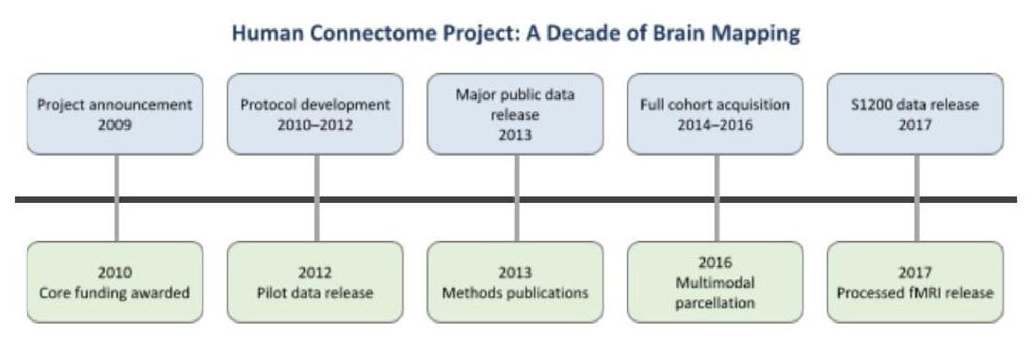
Figure 2.
By using this hybrid approach, the findings synthesized in this comprehensive literature review, in the Results section, are verified computationally and by expert analysis.
Figure 2.
By using this hybrid approach, the findings synthesized in this comprehensive literature review, in the Results section, are verified computationally and by expert analysis.
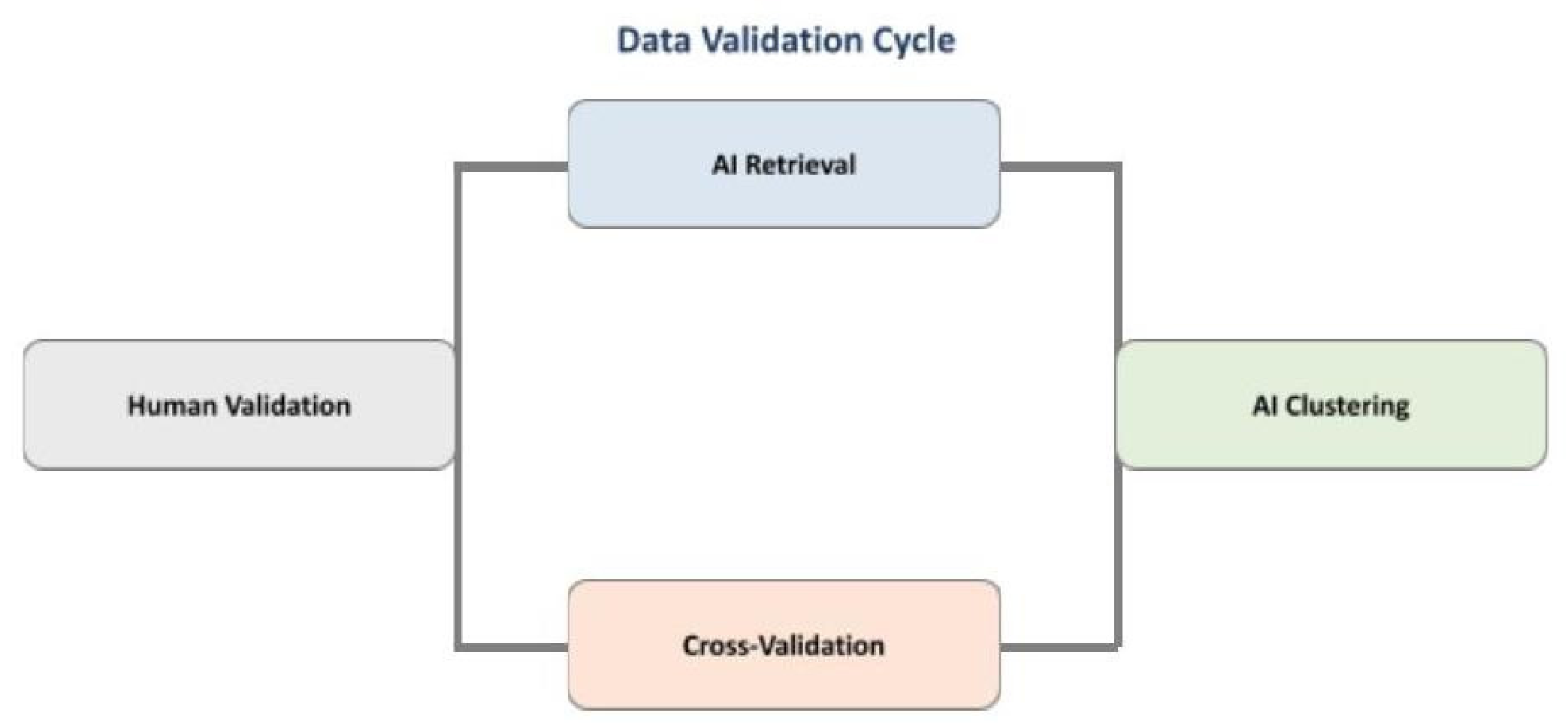
Figure 3.
Summary of the Human Connectome Project’s contributions to cognition and learning research, highlighting advances in preprocessing, cortical parcellation, large-scale network mapping, and genetic influences on brain structure, as well as key methodological challenges.
Figure 3.
Summary of the Human Connectome Project’s contributions to cognition and learning research, highlighting advances in preprocessing, cortical parcellation, large-scale network mapping, and genetic influences on brain structure, as well as key methodological challenges.
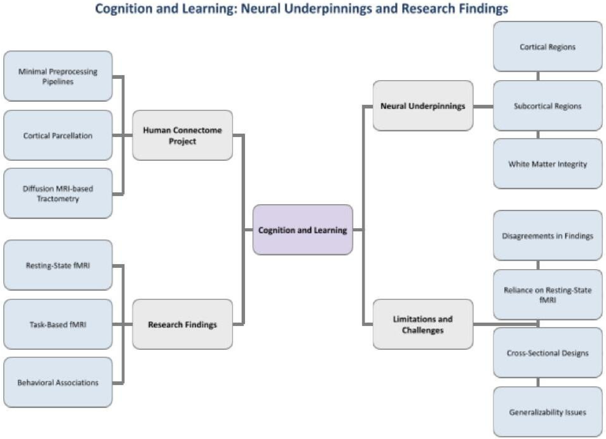
Figure 4.
This figure shows an overview of brain development and aging, illustrating neonatal neurodevelopment, changes in adulthood and aging, cognitive-emotional-hormonal networks in childhood and adolescence, and key limitations of current research.
Figure 4.
This figure shows an overview of brain development and aging, illustrating neonatal neurodevelopment, changes in adulthood and aging, cognitive-emotional-hormonal networks in childhood and adolescence, and key limitations of current research.
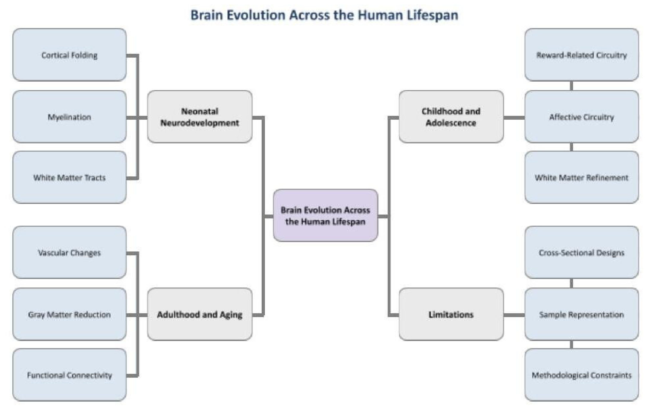
Figure 5.
This figure illustrates mental health research applications of Human Connectome Project data, including early psychosis, personality traits, higher-order networks, emotional states, neonatal connectomics, and key methodological limitations.
Figure 5.
This figure illustrates mental health research applications of Human Connectome Project data, including early psychosis, personality traits, higher-order networks, emotional states, neonatal connectomics, and key methodological limitations.
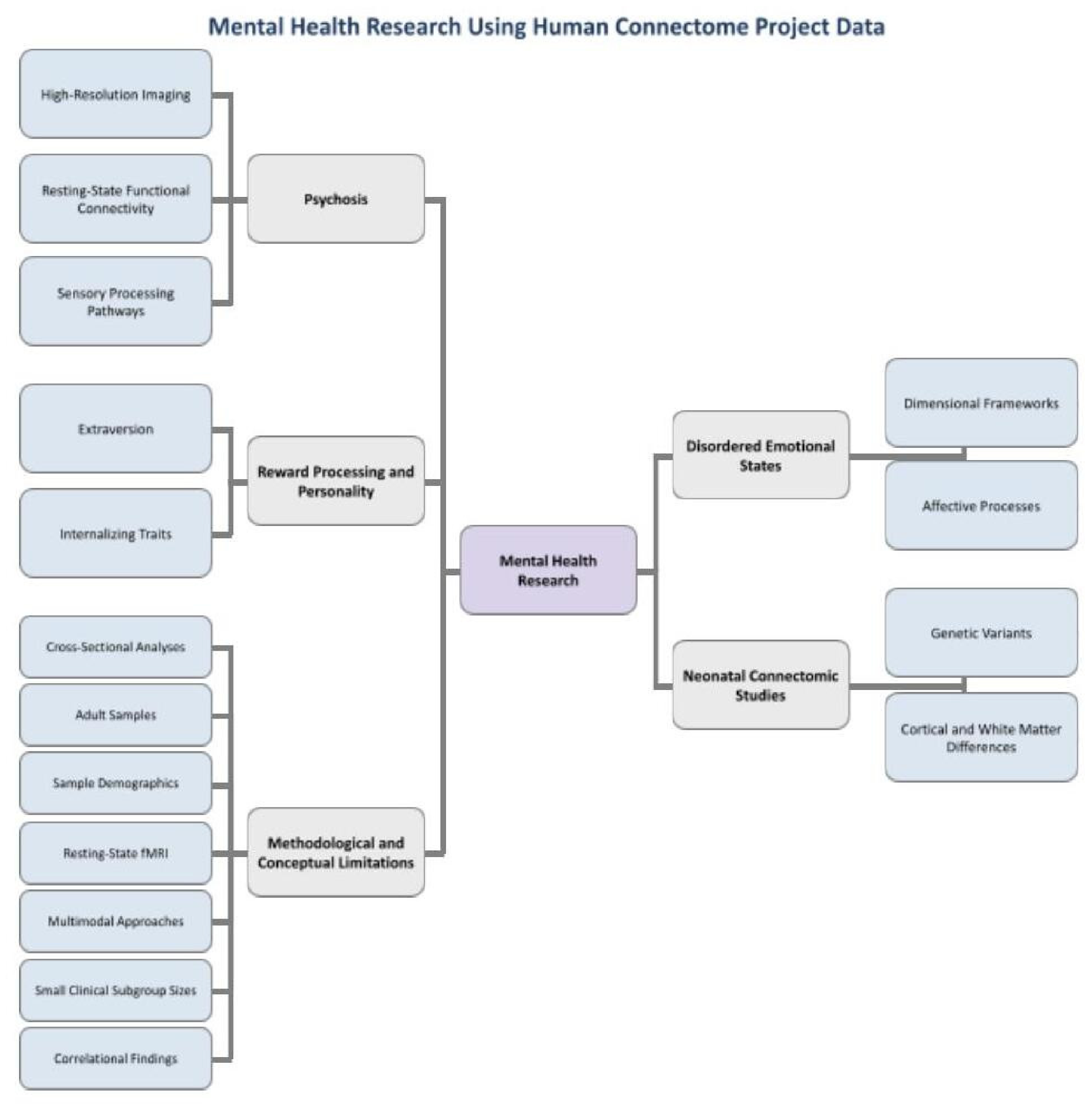
Figure 6.
This figure illustrates how neurological disorders, including Alzheimer’s disease and tinnitus, are linked to disruptions in large-scale brain networks and highlights key methodological limitations in current neuroimaging research.
Figure 6.
This figure illustrates how neurological disorders, including Alzheimer’s disease and tinnitus, are linked to disruptions in large-scale brain networks and highlights key methodological limitations in current neuroimaging research.
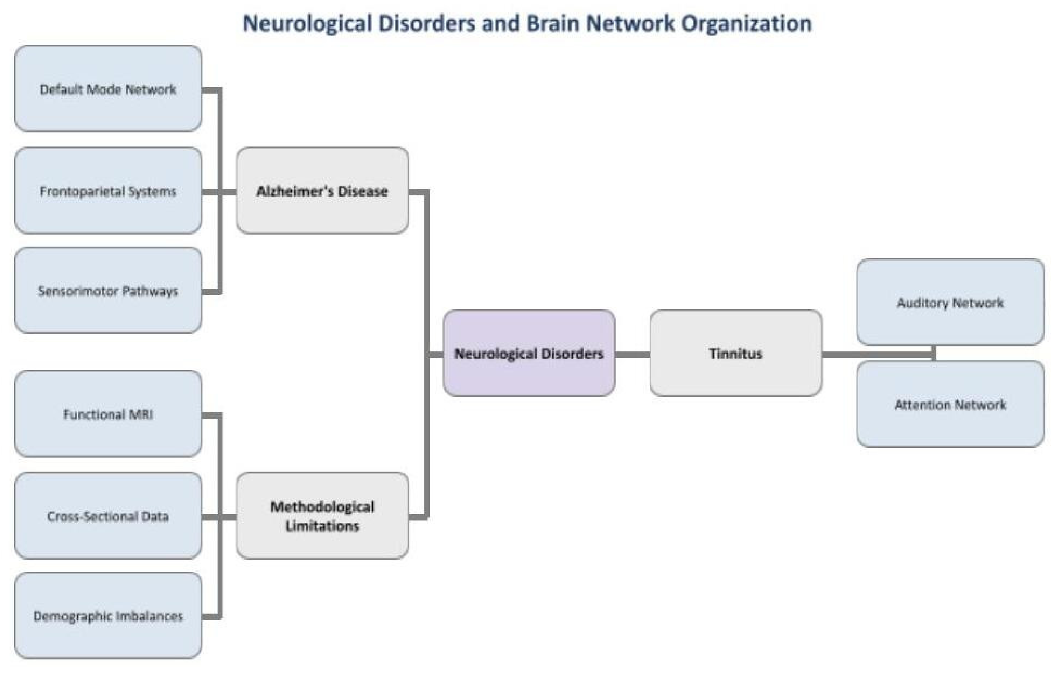
Figure 7.
This figure illustrates the methodological challenges and anatomical considerations in studying interhemispheric communication in motor control, highlighting how handedness asymmetries, imaging constraints, and corpus callosum segmentation precision affect our understanding of bilateral motor coordination.
Figure 7.
This figure illustrates the methodological challenges and anatomical considerations in studying interhemispheric communication in motor control, highlighting how handedness asymmetries, imaging constraints, and corpus callosum segmentation precision affect our understanding of bilateral motor coordination.
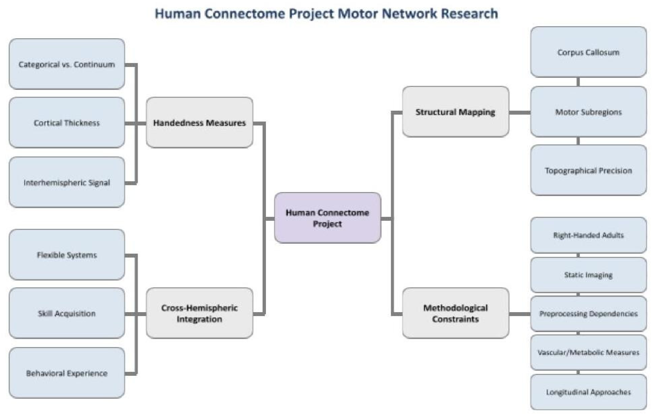
Figure 8.
This figure illustrates the relationship between brain structure and connectivity from Human Connectome Project (HCP) data and behavioral and personality traits, while highlighting key methodological limitations, including cross-sectional design, limited diversity, self-reported measures, and correlational analysis constraints.
Figure 8.
This figure illustrates the relationship between brain structure and connectivity from Human Connectome Project (HCP) data and behavioral and personality traits, while highlighting key methodological limitations, including cross-sectional design, limited diversity, self-reported measures, and correlational analysis constraints.
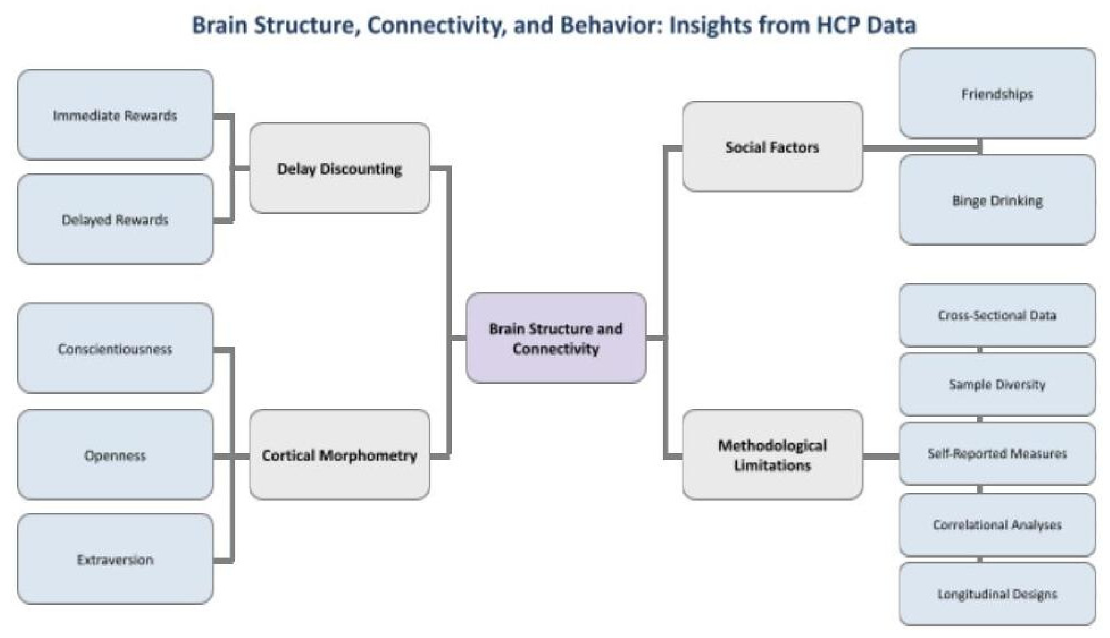

Table 1.
This table describes neuroplasticity as the brain’s ability to adapt for clinical rehabilitation, while brain decoding focuses on reconstructing complex information like speech from neural signals. The Human Connectome Project maps comprehensive brain connectivity to identify disease biomarkers, and Brain Mapping identifies specific therapeutic targets to enhance surgical precision and minimize tissue loss.
Table 1.
This table describes neuroplasticity as the brain’s ability to adapt for clinical rehabilitation, while brain decoding focuses on reconstructing complex information like speech from neural signals. The Human Connectome Project maps comprehensive brain connectivity to identify disease biomarkers, and Brain Mapping identifies specific therapeutic targets to enhance surgical precision and minimize tissue loss.
| Characteristics | Neuroplasticity | Brain Decoding | HCP | Brain Mapping |
|---|---|---|---|---|
| Definition | Ability to adapt and change | Reconstructing from neural activity | Mapping brain connections | Identifying therapeutic targets |
| Mechanisms | Synaptic plasticity, pathway reorganization | Cross-domain reconstruction, speech decoding | Large-scale mapping, biomarker identification | Precision neuromodulation improved surgery |
| Clinical Applications | Depression treatment, neurological rehabilitation | Decoding words, predicting autism | Classification markers for brain disorders | Reducing abnormalities, minimizing resection |
Short Biography of Authors
Sadhana Arivoli is a senior at Heritage High School and a researcher in the Aspiring Scholars Directed Research
Program. Her interests are in neuroscience, psychology, and medicine. She plans to pursue a career in medicine
while continuing neurological research.
Jiya Jagani is a freshman at UCLA studying psychobiology on a pre-medicine track. She aims to dedicate her
undergraduate career to neurological research and preparing for a future career in medicine.
Sindhu Chavali is a senior at Mission San Jose High School and will graduate in 2027.
Aarushi Kulshrestha is a freshman at UC San Diego.
Disclaimer/Publisher’s Note: The statements, opinions and data contained in all publications are solely those of the individual author(s) and contributor(s) and not of MDPI and/or the editor(s). MDPI and/or the editor(s) disclaim responsibility for any injury to people or property resulting from any ideas, methods, instructions or products referred to in the content. |
© 2026 by the authors. Licensee MDPI, Basel, Switzerland. This article is an open access article distributed under the terms and conditions of the Creative Commons Attribution (CC BY) license (http://creativecommons.org/licenses/by/4.0/).
Copyright: This open access article is published under a Creative Commons CC BY 4.0 license, which permit the free download, distribution, and reuse, provided that the author and preprint are cited in any reuse.