

Submitted:
11 September 2026
Posted:
14 September 2026
You are already at the latest version
Abstract
Autonomic innervation governs cardiac development, functional maturation, and electrophysiological integration, yet in vitro models rarely reproduce the spatial organization through which sympathetic neurons reach the heart. We first established planar neuro-cardiac cocultures of intact neonatal rat superior cervical ganglia (SCG) and embryonic rat cardiac aggregates. Sympathetic axons were tyrosine hydroxylase (TH) positive, penetrated the cardiac aggregates, and increased the spontaneous contraction rate. RT-qPCR on these planar cocultures showed that innervated cardiac tissue upregulated contractile, gap-junction, and adrenergic transcripts (Actc1, Actn2, Myh6, Adrb1, Gja5/Cx40, Cx43, Cx45) relative to both freshly isolated tissue and cardiac-only cultures, without re-expression of the fetal isoform Myh7. We then developed 3D Tissue Engineered Innervated Cardiac Units (TE-ICUs), in which SCG and cardiac aggregates occupy opposite ends of a 3–10 mm methacrylated hyaluronic acid microcolumn. Sympathetic axons in isolation projected up to ~6 mm and released ~170 nM norepinephrine upon electrical stimulation. Within TE-ICUs, fasciculated TH+ tracts bridged the microcolumn and penetrated the cardiac unit, while cardiac aggregates self-organized into chamber-like compartments; the contraction rate was significantly higher than that of cardiac-only controls at 8 days in vitro. TE-ICUs provide an anatomically inspired platform for studying neuro-cardiac development and engineering innervated cardiac tissue.
Keywords:
cardiac innervation
; sympathetic neurons
; superior cervical ganglion
; neuro-cardiac coculture
; tissue engineering
; hyaluronic acid hydrogel
; cardiac maturation
; norepinephrine
; microphysiological system
1. Introduction
The rhythm and growth of the heart are profoundly influenced by the autonomic nervous system. Sympathetic and parasympathetic neurons both innervate developing cardiac tissue and orchestrate its maturation and physiology in complementary ways [1,2,3]. Sympathetic innervation promotes cardiomyocyte proliferation, electrical conduction maturity, and increases in heart rate and contractile force during both development and adulthood [1]. Parasympathetic innervation exerts an opposing but equally crucial influence by modulating heart rate and coordinating atrioventricular conduction, thereby refining the developing heart’s rhythm [4]. Cardiac neurons and cardiomyocytes undergo co-maturation: signals from nascent cardiomyocytes regulate the growth and neurotransmitter properties of innervating neurons, while neuronal inputs accelerate cardiomyocyte electrophysiological and metabolic maturation [4]. Disruption of this interplay is associated with abnormal cardiac development and heightened disease susceptibility; genetic ablation of sympathetic guidance cues produces hypo- or hyper-innervation together with arrhythmia and impaired cardiac performance [5,6], underscoring the need to model autonomic influences on the heart in vitro.
Cardiac innervation is highly organized, with sympathetic and parasympathetic neurons occupying distinct anatomical niches [7]. Parasympathetic preganglionic fibers from the vagus nerves project to the heart and synapse onto small intrinsic cardiac ganglia embedded within the surface plexuses [8]. In contrast, sympathetic innervation derives predominantly from postganglionic neurons in the cervical and upper thoracic sympathetic chain—notably the bilateral stellate ganglia—which send long axons to innervate the cardiac chambers [9]. This produces a topographic pattern in which vagal inputs are relatively localized to the pacemaking and atrial regions, whereas sympathetic fibers broadly innervate both atria and ventricles.
This spatial segregation is critical for coordinated autonomic regulation. Because sympathetic somata reside in extracardiac ganglia, a single ganglion distributes a common signal across a wide myocardial territory through bundled long-range axons, allowing a graded, regionally biased chronotropic and inotropic response rather than uniform stimulation [7,9]. Conversely, the intracardiac location of parasympathetic ganglia places cholinergic output immediately adjacent to nodal tissue, supporting rapid, spatially restricted rate control [8]. Segregation also separates the site of neuromodulator synthesis from the site of release, so that transmitter delivery is determined by axon terminal distribution rather than by somatic position, and it preserves the polarity that allows retrograde target-derived trophic signals to reach distant neuronal cell bodies [5,10]. An in vitro model aiming to recapitulate cardiac neurobiology must therefore preserve this spatial pattern—discrete ganglionic sources and long-range projections—to authentically mimic cardiac innervation.
Despite the biological importance of cardiac innervation, most existing in vitro platforms for studying neuro-cardiac interactions are highly simplified and planar. These cocultures lack the structured architecture of the in vivo heart and provide little control over neuronal subtype or spatial arrangement. In many cases, investigators have resorted to heterogeneous or surrogate cell sources, establishing rodent–human hybrid systems that raise concerns due to species differences in maturation and signaling [11,12,13]. Moreover, without guided patterning, the neurons in these setups may not faithfully represent a purely sympathetic or parasympathetic phenotype, as neural cell lines (e.g., SH-SY5Y neuroblastoma cells) or generic induced neurons are often used without robust differentiation into a cardiac-autonomic subtype. This lack of control over cellular phenotype and organization can lead to inconsistent neuromodulatory effects.
Microfluidic systems have significantly improved spatial segregation in neuro-cardiac cocultures by compartmentalizing neuronal and cardiac cells into distinct chambers connected via microchannels that permit directed axonal growth. Human-induced pluripotent stem cell (hiPSC)-derived neurons and cardiomyocytes have been cultured in such devices, most notably in a cardiac innervation chip, in which compartmentalized microfluidics enabled human-on-human neuro-cardiac innervation and functional coupling in an electrophysiologically active system [14]. Early prototypes using polydimethylsiloxane (PDMS) microchips incorporated rat sympathetic neurons and cardiomyocytes separated by microconduits, demonstrating that electrical stimulation of the neuronal compartment could modulate cardiomyocyte beating rates in a frequency-dependent manner [12,15]. Additional platforms integrating hiPSC-derived autonomic neurons with ventricular-like hiPSC cardiomyocytes have used microfluidic designs to dissect neuronal influences on contractility and calcium handling [14,16]. However, these systems largely remain planar or quasi-three-dimensional, confining cellular organization to two dimensions within thin channels and consequently failing to reproduce the volumetric tissue architecture, axon fasciculation, or three-dimensional (3D) polarity observed in vivo.
Recognizing the limitations of two-dimensional (2D) culture, efforts have shifted toward 3D-engineered tissues and organoids that include both neurons and cardiomyocytes [17,18]. While 3D culture offers a more biomimetic microenvironment, current neuro-cardiac 3D models still fall short of recapitulating native heart innervation. A common drawback is the absence of spatial segregation between neuronal and cardiac compartments. In many cardiac organoids or engineered heart tissues, neurons—when present at all—are randomly distributed or intermingled with cardiomyocytes rather than residing in distinct ganglion-like clusters. This blurs the polarity of neuronal projections: instead of extending long axons to distant targets, neurons in unguided 3D cocultures form only short local neurites, so long-range axonal tracts and the clear delineation of axon terminals from cell bodies are rarely observed. Compartmentalized devices address the geometry but not the outcome; even when neurons and cardiomyocytes are physically separated and must connect purely by axon outgrowth, the axons that cross often remain sparse and fail to establish neuromodulation strong enough to alter cardiomyocyte activity, a shortfall that has been particularly persistent in fully human systems where neuronal maturation is slow [19]. A second limitation of current cardiac organoids is incomplete architectural development. Despite some self-organization into chamber-like structures, proper cardiac septation or multi-chambered anatomy is rarely achieved in vitro [20], and human embryonic stem cell-derived heart organoids typically form a single ventricular cavity or primitive heart tube with minimal septation [21,22]. Together, these gaps create a need for a platform that introduces true spatial organization of neurons relative to cardiac tissue, including long-distance aligned axon projections, while supporting the maturation of a more anatomically representative cardiac tissue.
To address these limitations, we developed Tissue Engineered Innervated Cardiac Units (TE-ICUs), which combine tissue engineering and developmental cues to recreate the anatomical and functional coupling of cardiac muscle and nerves. TE-ICUs are fabricated in a modular fashion (Figure 1) and comprise rodent-derived sympathetic ganglia (Figure 1C) seeded at one end of a methacrylated hyaluronic acid (MeHA) hydrogel-based hollow microcolumn (Figure 1A). These ganglia extend long, aligned axonal tracts that innervate rodent embryo-derived cardiac tissue aggregates, termed cardiac units (Figure 1B), located at the opposite end. This configuration establishes neuron-to-cardiac spatial segregation. The sympathetic neuron cell bodies remain clustered within the ganglion source, while their axons are guided to grow longitudinally through the hydrogel column toward the distant cardiac tissue. The resulting architecture mirrors the parallel, bundled sympathetic nerve pathways that innervate the heart in vivo. The TE-ICU design builds on our group’s prior development of micro-tissue engineered neural networks (µTENNs), 3D neural microtissues encased in hydrogel-based hollow microcolumns in which long axonal tracts span the full length of the column, recreating the native architecture of long-range projection pathways such as corticospinal [23], nigrostriatal [24], or peripheral sensorimotor circuits [25]. Here we describe the fabrication of TE-ICUs and evaluate their capacity to recapitulate cardiac autonomic anatomy and physiology. We first characterize cellular morphology, phenotype, contractile behavior and transcriptional responses in planar neuro-cardiac cocultures, in which all gene expression analyses in this study were performed, and then characterize the structure and function of the 3D TE-ICU platform.
2. Results
2.1. SCG-Derived Neurons Exhibit a Robust Sympathetic Phenotype and Innervate Cardiac Aggregates in Planar Coculture
Pieces of SCG isolated from postnatal rats (Figure 1C) were cultured as aggregates on laminin-coated planar surfaces for 10 days in vitro (DIV) (Figure 2A). Immunofluorescence imaging of SCG-only cultures revealed extensive neurite outgrowth from the ganglia. These neurites stained positively for tyrosine hydroxylase (TH) and β-tubulin III, indicating a robust sympathetic neuronal phenotype (Figure 2B). Subsequent coculture of SCG with cardiac tissue aggregates revealed neuronal processes projecting toward and intermingling with cardiac aggregates stained for cardiac troponin T (cTnT), confirming structural contact between sympathetic axons and cardiomyocytes (Figure 2C–F). High-magnification images showed TH-positive fibers penetrating the cardiac tissue aggregates. Beating frequency was assessed by video microscopy at 3 and 5 days after plating of the cardiac aggregates. Cocultured cardiac tissue exhibited a trend toward an increased rate of contraction by 5 DIV, although this difference did not reach statistical significance (Figure 2G).
2.2. Sympathetic Neurons Upregulate Contractile, Conduction and Adrenergic Genes in Planar Neuro-Cardiac Coculture
All transcriptional analyses in this study were performed on planar neuro-cardiac cocultures. RT-qPCR was performed on total RNA isolated from planar cardiac-only cultures and planar neuro-cardiac cocultures to assess transcripts involved in contractility, conduction, adrenergic signaling, cardiac identity, and synaptic machinery (Figure 3). Freshly isolated cardiac tissue from E16 rat embryos was analyzed as the DIV 0 reference sample, and expression was normalized to the housekeeping gene Gapdh.
After 7 days of planar culture (DIV 7), innervated cocultures showed significantly higher transcript levels than the freshly isolated DIV 0 cardiac tissue for the actin-related genes Actc1 and Actn2, the adult myosin isoform Myh6, the β1-adrenergic receptor Adrb1, and the gap junction genes Gja5 (Cx40), Cx43 and Cx45 (Figure 3). The same genes were also significantly elevated in DIV 7 cocultures relative to DIV 7 cardiac-only cultures (Gja5, p < 0.0001; Actc1, Actn2, Adrb1, Cx43 and Cx45, p < 0.01; Myh6, p < 0.05); by contrast, DIV 7 cardiac-only cultures did not differ significantly from DIV 0 tissue for any transcript examined. The presence of sympathetic neurons was therefore associated with a coordinated upregulation of contractile, conduction and adrenergic transcripts over the culture period, whereas cardiac tissue cultured alone remained transcriptionally indistinguishable from freshly isolated tissue.
The cardiac transcription factor Nkx2-5 and cardiac troponin T (Tnnt2) followed the same directional pattern; Nkx2-5 was significantly higher in cocultures than in cardiac-only cultures at DIV 7 (p < 0.05), although neither transcript differed significantly from DIV 0 tissue. No significant differences were detected for the fetal myosin isoform Myh7, the presynaptic marker Snap25, or for TH relative to DIV 0 tissue. Notably, TH remained low in both DIV 0 tissue and DIV 7 cardiac-only cultures, whereas TH levels in the neuro-cardiac cocultures at DIV 7 significantly exceeded those of the cardiac-only group (p < 0.05), reflecting the contribution of SCG-derived sympathetic neurons to the total RNA pool (Figure 3).
2.3. Sympathetic Ganglia Project Functional Noradrenergic Axons in 3D Hydrogel Microcolumns
SCG explants were embedded at one end of MeHA hydrogel-based hollow microcolumns and maintained in culture for at least 30 days. Axons extended longitudinally along the inner walls of the microcolumn in a highly directed manner, with a mean neurite length of ~2 mm by 4 DIV increasing to ~6.0 mm by 17 DIV (Figure 4A–C). Repeated-measures ANOVA yielded a significant effect of time on both neurite length (p < 0.0001) and growth rate (p = 0.0381), with a growth rate of ~0.3–0.55 mm/day sustained throughout the culture period (Figure 4C,D).
Fast-scan cyclic voltammetry (FSCV) was performed at 30 DIV to assess the functional competence of these 3D sympathetic cultures (Figure 4E–G), confirming that the microtissues remain metabolically active well beyond the period over which axon growth was quantified. The ganglion region was electrically stimulated, and the resulting catecholamine release was recorded with a carbon fiber electrode. Peak release reached ~170 nM (Figure 4F). The corresponding cyclic voltammogram displayed an oxidation peak at ~0.67 V and a reduction trough near −0.3 V, consistent with the electrochemical signature of catecholamines such as norepinephrine (Figure 4G). These data establish, as a proof of concept, that 3D sympathetic microtissues respond to stimulation by releasing evoked neurotransmitters.
2.4. TE-ICUs Recapitulate Three-Dimensional Neuro-Cardiac Anatomy
TE-ICUs were fabricated by seeding cardiac aggregates at one end of a MeHA hydrogel microcolumn and SCG explants at the opposite end, and the microtissues were visualized periodically by phase microscopy. Axons were observed projecting from the SCG by 10 DIV; these subsequently bundled into fascicles and reached the cardiac unit (Figure 5). In the ~5 mm construct shown in Figure 5, a physical connection between the two aggregates was established by 14 DIV. Because axon extension proceeds at a relatively constant rate (Section 2.3), the time required to bridge the column scales with its length: shorter columns were connected within several days, whereas the longest constructs (up to 10 mm) required correspondingly more time. This tunability is a feature of the microcolumn format, allowing innervation distance to be set independently of cell source.
Cardiac units within the microcolumns underwent progressive self-organization. Chamber-like cavities emerged around 10 DIV and further resolved into distinct compartments by 14 DIV, reminiscent of rudimentary myocardial partitioning (Figure 5). Axonal bundles remained attached to the cardiac units and maintained structural integrity even while the cardiac units contracted spontaneously within the microcolumn (Video S1).
2.5. Sympathetic Innervation Accelerates Cardiac Beating in TE-ICUs
Confocal microscopy of innervated TE-ICUs revealed TH-positive axons projecting linearly along the column and penetrating the cardiac aggregates, which stained positively for cTnT (Figure 6A,B). Serial z-stacks were merged to visualize the spatial segregation between neuronal and cardiac domains and the established polarity of axonal projection within the column. Control microcolumns containing a cardiac aggregate alone were confirmed by phase microscopy and cTnT immunolabeling (Figure 6C,D); no intracardiac TH signal was detected in these non-innervated constructs.
Contraction rate was quantified as beats per minute at 5 and 8 DIV in microcolumns containing both sympathetic and cardiac aggregates, as well as in cardiac-only microcolumns. Two-way ANOVA revealed significant effects of both culture type (p = 0.0034) and time (p = 0.0089) on contraction rate. Sidak’s multiple comparisons test identified a significant difference between innervated and cardiac-only constructs at 8 DIV (p < 0.01), at which point innervated cardiac units beat at ~24 beats/min compared with ~13 beats/min for cardiac-only controls. At 5 DIV the innervated group showed a higher mean contraction rate (~14 versus ~9 beats/min) that did not reach significance after correction for multiple comparisons (Figure 6E).
2.6. Cardiac Aggregates Do Not Influence Sympathetic Neurite Outgrowth, and Innervated Constructs Show a Trend Toward β-Adrenergic Dependence
To test whether the presence of a cardiac target influences sympathetic axon growth, microcolumns were prepared with SCG explants alone (SCG-only) or with both SCG and cardiac aggregates (SCG-Cardiac). Neurite length and growth rate were quantified 4 and 7 days after addition of the SCG to the microcolumn. Neurite length increased from ~2.0–2.4 mm at 4 DIV to ~3.8–4.1 mm at 7 DIV in both groups, and neither length nor growth rate differed significantly between SCG-only and SCG-Cardiac constructs at either time point (Figure 7A,B).
To probe whether the accelerated beating of innervated constructs depends on β-adrenergic signaling, contraction rate was measured before and after acute application of the β-adrenergic antagonist propranolol (1 µM, 5 min; Figure 7C). Cardiac-only constructs were unaffected by propranolol, whereas innervated SCG-Cardiac constructs showed a reduction in mean contraction rate. Expressed as the ratio of beats after to beats before treatment, cardiac-only constructs gave a ratio of ~1.13 ± 0.28 while SCG-Cardiac constructs gave ~0.80 ± 0.19 (mean ± SEM; Figure 7D). This difference was not statistically significant given the variability across constructs, but the direction of the effect is consistent with a component of the innervation-associated increase in beating rate being mediated by tonic β-adrenergic drive from the sympathetic neurons.
3. Discussion
The TE-ICU described here is a 3D neuro-cardiac platform that reproduces the structural organization of cardiac sympathetic innervation and supports functional coupling between the two tissue compartments. Coculture of cardiac units and sympathetic ganglia within MeHA hydrogel microcolumns produced noradrenergic axonal bundles that spanned the length of the column to innervate the target cardiac unit, chamber-like partitioning of the cardiac unit, and a significantly increased spontaneous beating rate. In parallel planar cocultures, sympathetic innervation was associated with sustained expression of genes encoding contractile and gap-junction proteins.
3.1. Cell Sourcing for Neuro-Cardiac Models
Multiple studies have described neuro-cardiac coculture across planar and 3D platforms. Obtaining appropriate cellular biomass, particularly of autonomic neurons, has been a major limiting factor and a source of variability, with studies relying on neuronal cell lines such as PC12 [12,26,27] or SH-SY5Y [11], or on hybrid cocultures combining neuronal and cardiac cells from different species. Human iPSC lines have emerged as a reliable source of sympathetic and parasympathetic neurons that exhibit structural interactions with and modulation of cardiac activity [16,28,29,30]. However, deriving autonomic neurons from hiPSC lines is non-trivial, time-consuming and expensive, and such protocols often yield low numbers of pure neurons with limited functional maturation.
We therefore used primary sympathetic neurons isolated from the SCG of postnatal rat pups (Figure 1C). Plated on planar surfaces, these explants exhibited robust axonal outgrowth and prominent TH expression, indicating good cellular health and a sympathetic phenotype (Figure 2). Separately, we generated cardiac units from whole heart tissue of E16 rat embryos using the forced aggregation method previously reported by our group [31]. This method produces aggregates structurally similar to spheroids or organoids while affording control over cell number, cell type, and aggregate size. We have previously reported that force-aggregated cardiac tissue exhibits coordinated en masse contraction and stronger local field potentials than dissociated cultures, indicating enhanced functional maturation [31].
3.2. Temporal Matching of Cardiac and Sympathetic Populations
Beyond cell sourcing, the timing of cell seeding matters because innervation occupies a defined window during cardiogenesis. The developmental stages of the heart are highly conserved across species, and functional sympathetic innervation of the rat heart does not occur until E16–E17 [32]; in mice, the first signs of cardiac autonomic innervation appear around E15.5 [33]. We therefore plated cardiac units derived from E16 rat embryos 1 day before introducing SCG-derived sympathetic neurons, so that the cardiac tissue would be present before neuronal arrival, approximating the sequence of innervation in vivo. We emphasize that this is an approximation of developmental sequence rather than a reproduction of it: the interval between cardiac seeding and axonal arrival at the target is set by the geometry of the culture rather than by developmental signaling, and we did not characterize the maturation state of the cardiac unit at the moment sympathetic axons reached it. Cocultures were maintained in nerve growth factor (NGF)-rich medium, consistent with reports of NGF’s requirement in sympathetic cultures. Planar neuro-cardiac cocultures demonstrated innervation of the cardiac unit by sympathetic axonal projections from the SCG, accompanied by a trend toward increased cardiac beat rate (Figure 2).
3.3. Transcriptional Consequences of Sympathetic Coculture in Planar Cultures
Understanding the transcriptional consequences of neuro-cardiac interaction is essential for uncovering how autonomic innervation shapes cardiomyocyte development, electrophysiological competence, and long-term functional integration. Previous studies examining the genetic impact of neuro-cardiac coculture have been limited in scope, often focusing on a narrow set of structural or electrophysiological genes and using rodent–human mixed systems [13]. Our study expands this space by profiling a broader panel of contractile, conduction, and adrenergic transcripts in rodent cardiac tissue cocultured with rodent sympathetic neurons.
In planar coculture, innervated cardiac tissue at DIV 7 showed significantly higher levels of the contractile protein genes Actc1 and Actn2 than both freshly isolated E16 tissue and cardiac-only cultures, while the structural transcript Tnnt2 trended upward without reaching significance, a profile compatible with the enhanced contractility observed in these cocultures (Figure 3). We also assessed Myh6 and Myh7, which encode the α- and β-myosin heavy chain isoforms, respectively [34]. In rodents, Myh7 is highly expressed in fetal and neonatal ventricles and is gradually downregulated postnatally, whereas Myh6 becomes the predominant adult isoform, associated with faster ATPase activity and increased contractile performance [2,34,35,36]. This isoform switch reflects the transition from an energy-efficient fetal phenotype to a more mature, force-generating myocardial state, and re-expression of Myh7 is a documented marker of pathological hypertrophy and normal aging in rodents [37]. In our planar cocultures, Myh6 was elevated relative to both DIV 0 tissue and DIV 7 cardiac-only cultures without a corresponding rise in Myh7, suggesting that the coculture environment supports ventricular-like transcriptional maturation without inducing a stress-associated reversion to the fetal gene program (Figure 3).
The developing heart possesses intrinsic adrenergic activity before sympathetic innervation [38,39], mediated by region-specific and transient expression of enzymes including TH. Intrinsic cardiac TH expression is scattered from ~E11.5, increases markedly in the interventricular septum around E16.5, and declines by E19.5 as functional sympathetic innervation is established [38]. In our hands, TH transcript in E16 cardiac tissue was low at DIV 0 and remained low over 7 days of cardiac-only culture, consistent with the intrinsic adrenergic program already being in decline by this stage (Figure 3). Planar neuro-cardiac cocultures at DIV 7 instead showed significantly higher TH than cardiac-only cultures, which we attribute to TH transcript contributed by the SCG neurons present in the same RNA pool rather than to maintenance of the intrinsic cardiac adrenergic program.
The gap junction genes Gja5 (Cx40), Cx43 and Cx45, which encode proteins central to electrical coupling, were also elevated in innervated planar cocultures relative to both DIV 0 tissue and DIV 7 cardiac-only cultures, suggesting improved intercellular connectivity and conduction potential. These observations are broadly consistent with Kowalski et al. [13], who reported sympathetic neuron-mediated upregulation of sarcomeric and ion channel genes in hiPSC-derived cardiomyocytes. That study observed no change in Cx43, whereas Cx43 was among the transcripts elevated in our planar cocultures. The comparison should be drawn cautiously, as the two systems differ in species, cell source, and neuronal preparation. One possible explanation is that intact ganglia provide a denser and more sustained noradrenergic input than dissociated neurons do, but our data do not test this directly. Adrb1 was significantly elevated in innervated cocultures relative to both DIV 0 tissue and DIV 7 cardiac-only cultures, indicating that sustained sympathetic contact upregulates the receptor transcript itself and not only downstream β-adrenergic responsiveness, in contrast to prior in vitro work in which functional responsiveness was detectable without a corresponding transcriptional change [40,41]. Our propranolol experiments (Figure 7C,D) speak to this point: acute β-blockade reduced the beating rate of innervated constructs by ~20% on average while leaving cardiac-only constructs unaffected, a trend consistent with tonic β-adrenergic drive from the sympathetic neurons, although the effect did not reach significance in our sample. The cardiac transcription factor Nkx2-5 was likewise higher in cocultures than in cardiac-only cultures, consistent with preservation of cardiac identity under innervation, whereas the presynaptic marker Snap25 did not differ across groups, indicating that the transcriptional shift is not simply a function of added neuronal RNA. Overall, these data indicate that structured sympathetic coculture supports transcriptional maturation of cardiac tissue and enhances molecular features underlying excitation–contraction coupling.
3.4. Engineering 3D Sympathetic Networks with Long-Range Noradrenergic Projections
A principal aim of this study was to engineer 3D neuronal microtissue in which a discrete sympathetic population sends long, aligned, noradrenergic axonal projections to innervate cardiac tissue. Our group has developed µTENNs to restore or replace damaged neural circuits. These constructs comprise hydrogel-based hollow microcolumns with phenotypically specific neuronal populations segregated at opposite ends and connected by long axonal tracts that span the microcolumn. We have fabricated and characterized such microtissues using cortical [23,42,43,44], dopaminergic [24,45], and motor and sensory neurons [25], and applied them to the reconstruction of brain pathways and spinal tracts, to neuromodulation as living electrodes, and as peripheral neuromuscular interfaces [23,24,25,46]. Here, we extended this approach to a 3D sympathetic network by placing SCG from postnatal rat pups inside a MeHA microcolumn (Figure 1). SCG within the 3D hydrogel produced rapid, highly directed axonal projections that spanned the microcolumn by 17 DIV (Figure 4). This directed outgrowth contrasts with the radial pattern of planar SCG cultures (Figure 2) and resembles in vivo postganglionic projections toward target organs. These 3D sympathetic networks robustly expressed TH and released transient peaks of norepinephrine (~170 nM) in response to electrical stimulation during FSCV (Figure 4). This concentration is within the range of electrically evoked catecholamine transients measured by FSCV in rodent brain tissue, though the two preparations differ substantially in geometry and electrode–source distance and the comparison is therefore indicative only. Notably, most hiPSC-derived sympathetic cultures either do not exhibit detectable norepinephrine secretion or produce very small amounts, indicating limited functional maturation [40,41].
3.5. Spatial Organization and Cardiac Self-Organization in TE-ICUs
The heart receives sympathetic innervation from axonal projections emanating from the stellate ganglia, a fusion of thoracic and cervical ganglia. During development, these axons grow along the vasculature, passing through the subepicardial layers toward intracardiac targets, including the sinoatrial node, atrioventricular node, and ventricular myocardium. In contrast, parasympathetic input arises from localized intracardiac ganglia on the cardiac wall. Reflecting this spatial relationship, we constructed TE-ICUs by placing cardiac units and SCG-derived sympathetic aggregates at opposite ends of MeHA hydrogel microcolumns 3–10 mm in length. Cardiac aggregates within the microcolumn self-organized into chamber-like cavities reminiscent of the partitioning reported in some human pluripotent stem cell-derived cardiac organoids (Figure 5); formation of internal cavities is considered a key indicator of cardiogenic modeling [21,22]. We have previously observed that dissociated cardiac cultures do not exhibit such chamber-like structures [31]. Within TE-ICUs, sympathetic aggregates extended long aligned projections that fasciculated into thick axonal bundles reaching the cardiac unit (Figure 5). These projections did not break as the cardiac units contracted, indicating durable structural neuro-cardiac coupling (Figure 5, Video S1). At terminal time points, TH+ sympathetic terminals were detected penetrating the cardiac units (Figure 6A,B), whereas no intracardiac TH signal was observed in non-innervated cardiac microtissue (Figure 6C,D), in line with the low intrinsic cardiac TH expression measured in non-innervated planar cultures (Figure 3). TE-ICUs also showed a significant increase in cardiac beating rate at 8 DIV, whereas the corresponding increase in planar coculture remained a non-significant trend (Figure 6E, Figure 2G).
3.6. Cardiac Target Influence on Axon Growth
Rather than being a passive recipient of innervation, the developing heart actively induces and patterns sympathetic growth through vascular guidance structures, bioelectrical feedback and expression of axon guidance molecules [5,6]. Among these, NGF, produced by cardiac myocytes and vascular cells, is the most critical molecule that directs sympathetic innervation [10]. We anticipated that the presence of a beating cardiac unit in TE-ICUs would accelerate axonal outgrowth from the SCG. Instead, cardiac units had no measurable effect on the rate of sympathetic outgrowth along the microcolumn (Figure 7A,B). We attribute this to the high NGF concentration in the medium used for both cardiac-only and neuro-cardiac groups, which may have saturated the trophic requirement and masked any cardiac-derived contribution.
3.7. Limitations
To our knowledge, this is the first report of an in vitro system that models cardiac autonomic anatomy and recapitulates the spatial pattern of cardiac sympathetic innervation, though several limitations should be noted. First, the model lacks parasympathetic input. Although parasympathetic neurons have been derived from human iPSC lines, their isolation from rodent primary tissue is not well established, so we restricted this study to sympathetic effects. Second, we combined E16 embryonic cardiac tissue with postnatal sympathetic neurons, and we acknowledge that this mismatch in maturation state is a limitation. It reflects a practical constraint: postnatal cardiac myocytes are difficult to isolate, require harsher tissue digestion, and survive poorly beyond 2–3 days in culture, while isolating SCG from E16 rat embryos is extremely challenging given their small size and limited anatomical access. Third, TE-ICUs could not be built with fully consistent architecture across constructs. Sympathetic axons occasionally projected along unintended trajectories, and aggregates sometimes shifted position along the column or migrated out of it, contributing to variability in the structural and functional readouts. Fourth, the transcriptional analyses were performed on total RNA from planar cocultures, which limits our ability to resolve cell-specific contributions; the elevated TH signal in cocultures is the clearest instance of this ambiguity. Finally, gene expression was not assessed in the 3D constructs. Future work should compare the transcriptional state of cardiac aggregates in 3D microcolumn culture with planar culture, and of innervated versus non-innervated 3D constructs, to determine how much of the transcriptional response is attributable to dimensionality versus innervation itself. Cell-type-resolved approaches, such as single-nucleus RNA sequencing, would address both the attribution and the cell-specificity limitations.
4. Conclusions
We describe the fabrication and characterization of TE-ICUs as a biofidelic model for studying neuro-cardiac interactions. In planar coculture, SCG-derived neuronal aggregates exhibited a robust sympathetic phenotype, established structural coupling with cardiac units, and were associated with sustained expression of genes encoding cardiac cytoskeletal components, gap junction proteins, and the adult myosin isoform. In 3D microcolumns, sympathetic aggregates produced fasciculated, aligned axonal tracts with demonstrable noradrenergic function. TE-ICUs combined these elements, demonstrating structural and functional neuro-cardiac coupling via intracardiac penetration of sympathetic projections and a significantly accelerated cardiac rate. The platform is modular and can be adapted to human-scale dimensions using hiPSC-derived cells, and extended to incorporate dual autonomic input to more fully recapitulate cardiac autonomic biology.
5. Materials and Methods
5.1. Primary Isolation and Culture of Rat Cardiac Myocytes and Sympathetic Neurons
All procedures were approved by the Institutional Animal Care and Use Committees at the University of Pennsylvania and the Corporal Michael Crescenz Veterans Affairs Medical Center and adhered to the NIH Public Health Service Policy on Humane Care and Use of Laboratory Animals. Primary cardiac myocytes were obtained from E16 Sprague-Dawley rat embryos following previously established protocols (Figure 1) [31]. Hearts were dissected and dissociated using 0.05% trypsin-EDTA for 10–15 min at 37 °C with manual trituration. After centrifugation, dissociated cells were resuspended in Cardiac Medium comprising 78% high-glucose DMEM, 17% Medium-199, 4% horse serum, and 1% penicillin/streptomycin. To form aggregates, 14 µL of a 7 × 10⁶ cells/mL suspension was transferred to pyramidal microwells within a PDMS mold, centrifuged at 1500 rpm for 5 min, supplemented with medium, and incubated overnight prior to seeding [47].
Sympathetic neuron aggregates were isolated from the SCG of P0–P1 Sprague-Dawley rat pups as previously described (Figure 1) [48]. Pups were euthanized by hypothermia and decapitation. The SCG were located at the bifurcation of the carotid arteries lateral to the trachea, extracted, and cleaned of pre- and postganglionic nerves and debris. Pieces of SCG were cultured as sympathetic neuron aggregates without further dissociation. SCG-only cultures were maintained at 37 °C and 5% CO2 in medium comprising RPMI 1640 with 0.4% penicillin/streptomycin, 1% heat-inactivated horse serum, 10 µM uridine/5-fluorodeoxyuridine and 100 ng/mL NGF. For coculture with cardiac aggregates, the medium consisted of Cardiac Medium supplemented with 100 ng/mL NGF, given the requirement for this growth factor in sympathetic neuron survival and growth. For planar culture, surfaces were coated with 20 µg/mL poly-L-lysine, followed by 20 µg/mL laminin, before seeding cardiac and/or sympathetic aggregates.
5.2. Fabrication and Seeding of MeHA Hydrogel Microcolumns
Cardiac and sympathetic aggregates were cultured under 3D conditions using MeHA hydrogel microcolumns of 3–10 mm in length with outer and inner diameters of 701 and 300 µm, respectively (Figure 1A). MeHA was synthesized by esterification of hyaluronic acid with methacrylic anhydride for ~3.5 h at pH 8.5, purified by dialysis for 5–7 days and recovered by lyophilization [49]. The degree of functionalization of the hyaluronic acid disaccharides was ~44% as determined by 1H NMR. A 3% w/v MeHA solution in Dulbecco’s phosphate-buffered saline (DPBS) with 0.05% Irgacure 2959 was drawn by capillary action into glass capillary tubes (inner diameter 701 µm) containing an inserted acupuncture needle (outer diameter 300 µm). The solution was photo-crosslinked under 10 mW/cm2 ultraviolet light for 5 min. The needle was then withdrawn and the gelled microcolumns were removed into DPBS, sterilized for 30 min under ultraviolet light, and rinsed with fresh DPBS to remove residual free radicals. Columns were cut to the desired length, and an extracellular matrix solution of 1 mg/mL rat tail collagen type I plus 1 mg/mL mouse laminin in Neurobasal (pH 7.2–7.5) was added to the lumen and polymerized for 15 min at 37 °C. Dishes containing microcolumns were then flooded with culture medium and incubated at 37 °C and 5% CO2 until seeding.
For seeding, cardiac aggregates and SCG were cut with fine forceps under a dissection scope into pieces that fit within the ends of the columns. For cardiac-only cultures, one piece of a cardiac aggregate was placed at one end of the microcolumn. Unidirectional sympathetic microcolumns were fabricated with a sympathetic aggregate at only one end. For TE-ICUs, the cardiac aggregate was seeded first, and a sympathetic aggregate was introduced at the opposite end one day later. Microcolumns were incubated at 37 °C and 5% CO2 to allow cell attachment to the extracellular matrix and/or MeHA shell and subsequent sympathetic neurite growth.
5.3. RNA Isolation and RT-qPCR Gene Expression Analysis
Gene expression analyses were performed exclusively on planar neuro-cardiac cocultures and planar cardiac-only cultures; no RT-qPCR was performed on 3D microcolumn constructs. Total RNA was isolated after 7 days of culture using TRIzol (Invitrogen, Carlsbad, CA, USA) according to the manufacturer’s instructions. Freshly isolated E16 cardiac tissue served as the DIV 0 reference sample. Complementary DNA was synthesized from 2 µg of RNA using the High-Capacity cDNA Reverse Transcription Kit (Applied Biosystems, Waltham, MA, USA). Real-time PCR reactions of 10 µL were assembled with PowerUp SYBR Green Master Mix (Applied Biosystems), 10 ng cDNA and 300 nM gene-specific primers; primer sequences are listed in Table 1. Reactions were run on a QuantStudio 6 Pro Real-Time PCR System (Applied Biosystems) with one cycle each at 50 °C and 95 °C for 2 min, followed by 40 cycles of 95 °C for 3 s and 60 °C for 30 s, followed by melt curve analysis. Gapdh served as the endogenous reference gene. Relative expression was calculated for each sample as 2^(−ΔCt), where ΔCt = Ct(target) − Ct(Gapdh); values are therefore normalized to the reference gene within each sample and are presented without further normalization to a calibrator group.
5.4. Neurite Growth Characterization
Planar and 3D cultures were imaged in phase-contrast on a Nikon Eclipse Ti-S microscope with a QiClick camera and Nikon Elements software (Nikon, Tokyo, Japan). Imaging was used to quantify the length and growth rate of neurites projected from the sympathetic aggregate as a function of time. Length was measured as the distance between the tip of the longest observed neurite and the edge of the sympathetic aggregate; growth rate was estimated using the backward difference method.
5.5. Immunocytochemistry
At terminal time points, planar and 3D cultures were fixed in 4% paraformaldehyde for 35 min and rinsed in 1× phosphate-buffered saline (PBS). Cultures were permeabilized with 0.3% Triton X-100 in 4% horse serum for 60 min and incubated overnight at 4 °C with primary antibodies in 4% horse serum. Primary antibodies were directed against β-tubulin III (1:500, Sigma-Aldrich, St. Louis, MO, USA, T8578), a neuron-specific microtubule protein; tyrosine hydroxylase (1:500, Abcam, Cambridge, UK, ab113), the rate-limiting enzyme in norepinephrine biosynthesis; and cardiac troponin I (1:250, Abcam, ab47003), a cardiac-specific regulator of actin–myosin interaction. Cultures were then exposed to secondary antibodies (Alexa-488, Alexa-568, Alexa-647; all 1:500) for 2 h at 18–24 °C, followed by 10 min in 1:10,000 Hoechst in PBS. Phalloidin staining was used to visualize actin filaments. Stained cultures were imaged on a Nikon A1RSI laser scanning confocal microscope, with z-stacks presented as maximum intensity projections.
5.6. Fast-Scan Cyclic Voltammetry for Catecholamine Release
To assess evoked catecholamine release from 3D sympathetic aggregates and axon tracts, microtissues were incubated for 2 DIV in medium containing Neurobasal, 2% B-27, 1% fetal bovine serum, 2.0 mM L-glutamine, 100 µM ascorbic acid, 4 ng/mL basic fibroblast growth factor, 100 ng/mL NGF and 0.3% penicillin/streptomycin. Cultures were then incubated in the same medium containing 100 µM L-3,4-dihydroxyphenylalanine (L-DOPA) for at least 30 min. A single microtissue was transferred to a recording chamber and perfused at 37 °C with Neurobasal, 2.0 mM L-glutamine, 100 µM ascorbic acid and 100 µM L-DOPA bubbled with 95% O2/5% CO2. A bipolar stimulating electrode was placed to span the sympathetic aggregate region and a carbon fiber electrode (outer fiber length ~300 µm) was positioned in the same area. The carbon fiber potential was scanned linearly from −0.4 V to 1.2 V to −0.4 V versus Ag/AgCl at 400 V/s using a voltammeter/amperometer. After 8 s of recording, a monophasic stimulation train of 20 Hz, 10 pulses of 5 ms width and 8 V amplitude was applied. Cyclic voltammograms were recorded and analyzed with Demon Voltammetry and Analysis Software [50]. The reported current/concentration trace is the average of six recording sessions 10–12 min apart. Current at the peak oxidation potential of catecholamines in consecutive voltammograms was converted to concentration using the slope of the linear regression of current elicited by injection of 3, 6 and 12 µM norepinephrine hydrochloride in 126 mM NaCl, 2.5 mM KCl, 1.2 mM NaH2PO4·H2O, 2.4 mM CaCl2·2H2O, 1.2 mM MgCl2·6H2O, 25 mM NaHCO3 and 0.4 mM L-ascorbic acid.
5.7. Analysis of Spontaneous Cardiac Contraction
The effect of sympathetic aggregates on cardiac beating rate was analyzed using video recordings of 1–1.5 min acquired with a Nikon Eclipse Ti-S microscope. Planar cultures were recorded at 3 and 5 DIV, and 3D microcolumn cultures at 5 and 8 DIV, with DIV counted from plating of the cardiac aggregate. Beats per minute were quantified manually in Fiji. Sample sizes were as follows: planar cardiac-SCG coculture and planar cardiac-only cultures, and 3D cardiac-SCG and cardiac-only microcolumns, with n = 8 and n = 7 constructs per group, respectively.
5.8. β-Adrenergic Blockade
To assess the contribution of β-adrenergic signaling to spontaneous contraction, beating rate was recorded in cardiac-only and SCG-cardiac microcolumns immediately before and after acute application of the non-selective β-adrenergic antagonist propranolol. Constructs were incubated with 1 µM propranolol for 5 min before the post-treatment recording. Recordings before and after treatment were acquired from the same construct, and the response was expressed as the ratio of beats per minute after treatment to beats per minute before treatment.
5.9. Statistical Analysis
Statistical analyses were performed in Prism 8.1.1 (GraphPad, San Diego, CA, USA). For neurite growth over the full culture period in 3D microcolumns, repeated-measures one-way ANOVA with Tukey’s multiple comparisons test was used to evaluate the effect of time. For the comparison of SCG-only and SCG-Cardiac microcolumns at 4 and 7 DIV, the effects of group and time were analyzed by two-way ANOVA with Sidak’s multiple comparisons test. For contraction rate, normality was confirmed with the Kolmogorov-Smirnov test, and the effects of DIV and culture type were analyzed by two-way ANOVA, with between-group differences at each time point assessed using Sidak’s multiple comparisons test. Gene expression data were compared across DIV 0 cardiac tissue, DIV 7 cardiac-only cultures, and DIV 7 neuro-cardiac cocultures by one-way ANOVA with Tukey’s multiple comparisons test. Gene expression data are presented as mean ± SD; neurite growth and contraction rate data are presented as mean ± SEM, as indicated in the individual figure captions. Significance thresholds are indicated as * p < 0.05, ** p < 0.01, *** p < 0.001 and **** p < 0.0001; ns denotes not significant.
Supplementary Materials
The following supporting information can be downloaded at the website of this paper posted on Preprints.org. Video S1: Beating cardiac unit and long axonal projections within a 3D hydrogel microcolumn.
Author Contributions
Conceptualization, S.D. and D.K.C.; methodology, S.D., W.G.V., E.M. and J.D.; formal analysis, S.D., W.G.V., E.M., J.D., and A.D.; investigation, W.G.V., E.M., M.T., M.H., J.D., and A.D.; writing—original draft preparation, S.D., W.G.V., and D.K.C.; writing—review and editing, E.M. and J.D.; visualization, S.D. and W.G.V.; supervision - D.K.C.; funding acquisition, D.K.C. All authors have read and agreed to the published version of the manuscript.
Funding
This research was funded by the Department of Health of the Commonwealth of Pennsylvania (Health Research Formula Fund Grant 585499, Cullen); the US Department of Defense through the Medical Research and Materiel Command (W81XWH-19-1-0867, Cullen); the National Institutes of Health (R01-NS117757, R01-NS127895 and R01-AR083489, Cullen); the Department of Veterans Affairs (Merit Review I01-RX005045 and Merit Review I01-BX003748, Cullen); and the National Science Foundation (Graduate Research Fellowship DGE-1845298, Gordián-Vélez). Any opinions, findings, conclusions or recommendations expressed in this material are those of the authors and do not necessarily reflect the views of the Commonwealth of Pennsylvania, the Department of Defense, the National Institutes of Health, the Department of Veterans Affairs or the National Science Foundation.
Institutional Review Board Statement
All animal procedures were approved by the Institutional Animal Care and Use Committees of the University of Pennsylvania and the Corporal Michael Crescenz Veterans Affairs Medical Center, and were conducted in accordance with the NIH Public Health Service Policy on Humane Care and Use of Laboratory Animals.
Informed Consent Statement
Not applicable.
Data Availability Statement
The data presented in this study are available on request from the corresponding authors.
Conflicts of Interest
The authors declare no conflicts of interest.
Abbreviations
The following abbreviations are used in this manuscript: 2D, two-dimensional; 3D, three-dimensional; cTnT, cardiac troponin T; DIV, days in vitro; DPBS, Dulbecco’s phosphate-buffered saline; E16, embryonic day 16; FSCV, fast-scan cyclic voltammetry; HA, hyaluronic acid; hiPSC, human induced pluripotent stem cell; L-DOPA, L-3,4-dihydroxyphenylalanine; MeHA, methacrylated hyaluronic acid; NGF, nerve growth factor; PBS, phosphate-buffered saline; PDMS, polydimethylsiloxane; SCG, superior cervical ganglion; TE-ICU, tissue engineered innervated cardiac unit; TH, tyrosine hydroxylase; µTENN, micro-tissue engineered neural network.
References
- Hasan, W. Autonomic cardiac innervation. Organogenesis 2013, 9, 176–193. [Google Scholar] [CrossRef]
- Franco, D.; Lamers, W.H.; Moorman, A.F.M. Patterns of expression in the developing myocardium: towards a morphologically integrated transcriptional model. Cardiovasc. Res. 1998, 38, 25–53. [Google Scholar] [CrossRef]
- Végh, A.; Duim, S.; Smits, A.; Poelmann, R.; ten Harkel, A.; DeRuiter, M.; Goumans, M.; Jongbloed, M. Part and Parcel of the Cardiac Autonomic Nerve System: Unravelling Its Cellular Building Blocks during Development. J. Cardiovasc. Dev. Dis. 2016, 3, 28. [Google Scholar] [CrossRef]
- Habecker, B.A.; Anderson, M.E.; Birren, S.J.; Fukuda, K.; Herring, N.; Hoover, D.B.; Kanazawa, H.; Paterson, D.J.; Ripplinger, C.M. Molecular and cellular neurocardiology: development, and cellular and molecular adaptations to heart disease. J. Physiol. 2016, 594, 3853–3875. [Google Scholar] [CrossRef]
- Manousiouthakis, E.; Mendez, M.; Garner, M.C.; Exertier, P.; Makita, T. Venous endothelin guides sympathetic innervation of the developing mouse heart. Nat. Commun. 2014, 5, 3918. [Google Scholar] [CrossRef]
- Ieda, M.; Kanazawa, H.; Kimura, K.; Hattori, F.; Ieda, Y.; Taniguchi, M.; Lee, J.-K.; Matsumura, K.; Tomita, Y.; Miyoshi, S.; Shimoda, K.; Makino, S.; Sano, M.; Kodama, I.; Ogawa, S.; Fukuda, K. Sema3a maintains normal heart rhythm through sympathetic innervation patterning. Nat. Med. 2007, 13, 604–612. [Google Scholar] [CrossRef]
- Zandstra, T.E.; Notenboom, R.G.E.; Wink, J.; Kiès, P.; Vliegen, H.W.; Egorova, A.D.; Schalij, M.J.; Ruiter, M.C.D.; Jongbloed, M.R.M. Asymmetry and Heterogeneity: Part and Parcel in Cardiac Autonomic Innervation and Function. Front. Physiol. 2021, 12, 665298. [Google Scholar] [CrossRef]
- Campos, I.D.; Pinto, V.; Sousa, N.; Pereira, V.H. A brain within the heart: A review on the intracardiac nervous system. J. Mol. Cell. Cardiol. 2018, 119, 1–9. [Google Scholar] [CrossRef]
- Pardini, B.J.; Lund, D.D.; Schmid, P.G. Organization of the sympathetic postganglionic innervation of the rat heart. J. Auton. Nerv. Syst. 1989, 28, 193–201. [Google Scholar] [CrossRef]
- Nam, J.; Onitsuka, I.; Hatch, J.; Uchida, Y.; Ray, S.; Huang, S.; Li, W.; Zang, H.; Ruiz-Lozano, P.; Mukouyama, Y. Coronary veins determine the pattern of sympathetic innervation in the developing heart. Development 2013, 140, 1475–1485. [Google Scholar] [CrossRef]
- Hernandez, I.; Ramirez, S.P.; Salazar, W.V.; Mendivil, S.; Guevara, A.; Patel, A.; Loyola, C.D.; Dorado, Z.N.; Joddar, B. A Semi-Three-Dimensional Bioprinted Neurocardiac System for Tissue Engineering of a Cardiac Autonomic Nervous System Model. Bioengineering 2023, 10, 834. [Google Scholar] [CrossRef]
- Bernardin, A.A.; Colombani, S.; Rousselot, A.; Andry, V.; Goumon, Y.; Delanoë-Ayari, H.; Pasqualin, C.; Brugg, B.; Jacotot, E.D.; Pasquié, J.-L.; Lacampagne, A.; Meli, A.C. Impact of Neurons on Patient-Derived Cardiomyocytes Using Organ-On-A-Chip and iPSC Biotechnologies. Cells 2022, 11, 3764. [Google Scholar] [CrossRef]
- Kowalski, W.J.; Garcia-Pak, I.H.; Li, W.; Uosaki, H.; Tampakakis, E.; Zou, J.; Lin, Y.; Patterson, K.; Kwon, C.; Mukouyama, Y.-S. Sympathetic Neurons Regulate Cardiomyocyte Maturation in Culture. Front. Cell Dev. Biol. 2022, 10, 850645. [Google Scholar] [CrossRef]
- Häkli, M.; Jäntti, S.; Joki, T.; Sukki, L.; Tornberg, K.; Aalto-Setälä, K.; Kallio, P.; Pekkanen-Mattila, M.; Narkilahti, S. Human Neurons Form Axon-Mediated Functional Connections with Human Cardiomyocytes in Compartmentalized Microfluidic Chip. Int. J. Mol. Sci. 2022, 23, 3148. [Google Scholar] [CrossRef]
- Takeuchi, A.; Nakafutami, S.; Tani, H.; Mori, M.; Takayama, Y.; Moriguchi, H.; Kotani, K.; Miwa, K.; Lee, J.; Noshiro, M.; Jimbo, Y. Device for co-culture of sympathetic neurons and cardiomyocytes using microfabrication. Lab Chip 2011, 11, 2268–2275. [Google Scholar] [CrossRef]
- Wu, H.; Hamilton, C.; Porritt, H.; Winbo, A.; Zeltner, N. Modelling neurocardiac physiology and diseases using human pluripotent stem cells: current progress and future prospects. J. Physiol. 2024. [Google Scholar] [CrossRef]
- Zeltner, N.; Wu, H.-F.; Saito-Diaz, K.; Sun, X.; Song, M.; Saini, T.; Grant, C.; James, C.; Thomas, K.; Abate, Y.; Howerth, E.; Kner, P.; Xu, B. A modular platform to generate functional sympathetic neuron-innervated heart assembloids. Res. Sq. 2024, rs.3.rs–3894397. [Google Scholar]
- Branco, M.A.; Marzec, J.; Nunes, M.M.; Bica, M.; Rayagra, A.L.; Tenreiro, M.F.; Cabral, J.M.S.; Diogo, M.M. Modeling embryonic heart vascular plexus development and sympathetic innervation on a human heart organoid. iScience 2026, 29, 115071. [Google Scholar] [CrossRef]
- Takayama, Y.; Kida, Y.S. In Vitro Reconstruction of Neuronal Networks Derived from Human iPS Cells Using Microfabricated Devices. PLoS ONE 2016, 11, e0148559. [Google Scholar] [CrossRef]
- Thomas, D.; de Jesus Perez, V.A.; Sayed, N. An evidence appraisal of heart organoids in a dish and commensurability to human heart development in vivo. BMC Cardiovasc. Disord. 2022, 22, 122. [Google Scholar] [CrossRef]
- Hofbauer, P.; Jahnel, S.M.; Papai, N.; Giesshammer, M.; Deyett, A.; Schmidt, C.; Penc, M.; Tavernini, K.; Grdseloff, N.; Meledeth, C.; Ginistrelli, L.C.; Ctortecka, C.; Šalic, Š.; Novatchkova, M.; Mendjan, S. Cardioids reveal self-organizing principles of human cardiogenesis. Cell 2021, 184, 3299–3317.e22. [Google Scholar] [CrossRef]
- Lewis-Israeli, Y.R.; Wasserman, A.H.; Gabalski, M.A.; Volmert, B.D.; Ming, Y.; Ball, K.A.; Yang, W.; Zou, J.; Ni, G.; Pajares, N.; Chatzistavrou, X.; Li, W.; Zhou, C.; Aguirre, A. Self-assembling human heart organoids for the modeling of cardiac development and congenital heart disease. Nat. Commun. 2021, 12, 5142. [Google Scholar] [CrossRef]
- Adewole, D.O.; Struzyna, L.A.; Burrell, J.C.; Harris, J.P.; Nemes, A.D.; Petrov, D.; Kraft, R.H.; Chen, H.I.; Serruya, M.D.; Wolf, J.A.; Cullen, D.K. Development of optically controlled “living electrodes” with long-projecting axon tracts for a synaptic brain-machine interface. Sci. Adv. 2021, 7. [Google Scholar] [CrossRef]
- Gordián-Vélez, W.J.; Browne, K.D.; Galarraga, J.H.; Chouhan, D.; Duda, J.E.; España, R.A.; Chen, H.I.; Burdick, J.A.; Cullen, D.K. Dopaminergic Axon Tracts Within a Hyaluronic Acid Hydrogel Encasement to Restore the Nigrostriatal Pathway. Adv. Healthc. Mater. 2025, 14, e2402997. [Google Scholar] [CrossRef]
- Burrell, J.C.; Das, S.; Laimo, F.A.; Katiyar, K.S.; Browne, K.D.; Shultz, R.B.; Tien, V.J.; Vu, P.T.; Petrov, D.; Ali, Z.S.; Rosen, J.M.; Cullen, D.K. Engineered neuronal microtissue provides exogenous axons for delayed nerve fusion and rapid neuromuscular recovery in rats. Bioact. Mater. 2022, 18, 339–353. [Google Scholar] [CrossRef]
- Bernardin, A.; Colombani, S.; Brugg, B.; Jacotot, E.; Meli, A.; Lacampagne, A. Neuro-cardiac organ-a-chip using patient specific induced pluripotent stem cells and microfluidic devices. Arch. Cardiovasc. Dis. Suppl. 2022, 14, 209. [Google Scholar] [CrossRef]
- Greene, L.A.; Tischler, A.S. Establishment of a noradrenergic clonal line of rat adrenal pheochromocytoma cells which respond to nerve growth factor. Proc. Natl. Acad. Sci. USA 1976, 73, 2424–2428. [Google Scholar] [CrossRef]
- Wu, H.-F.; Huang, C.-W.; Daga, K.R.; Marklein, R.A.; Ivanova, N.; Zeltner, N. Human pluripotent stem cell-derived functional sympathetic neurons express ACE2 and RAAS components: a framework for studying the effect of COVID-19 on sympathetic responsiveness. Clin. Auton. Res. 2022, 32, 59–63. [Google Scholar] [CrossRef]
- Wu, H.-F.; Saito-Diaz, K.; Huang, C.-W.; McAlpine, J.L.; Seo, D.E.; Magruder, D.S.; Ishan, M.; Bergeron, H.C.; Delaney, W.H.; Santori, F.R.; Krishnaswamy, S.; Hart, G.W.; Chen, Y.-W.; Hogan, R.J.; Liu, H.-X.; Ivanova, N.B.; Zeltner, N. Parasympathetic neurons derived from human pluripotent stem cells model human diseases and development. Cell Stem Cell 2024, 31, 734–753.e8. [Google Scholar] [CrossRef]
- Wu, H.-F.; Art, J.; Saini, T.; Zeltner, N. Protocol for generating postganglionic sympathetic neurons using human pluripotent stem cells for electrophysiological and functional assessments. STAR Protoc. 2024, 5, 102970. [Google Scholar] [CrossRef]
- Serruya, M.D.; Das, S.; Katiyar, K.S.; Struzyna, L.A.; Burrell, J.C.; Cullen, D.K. Cardiac myocyte microtissue aggregates broadcast local field potentials. bioRxiv 2018, 376418. [Google Scholar] [CrossRef]
- Shigenobu, K.; Tanaka, H.; Kasuya, Y. Changes in Sensitivity of Rat Heart to Norepinephrine and Isoproterenol during Pre- and Postnatal Development and Its Relation to Sympathetic Innervation. Dev. Pharmacol. Ther. 1988, 11, 226–236. [Google Scholar] [CrossRef]
- Salamon, R.J.; Halbe, P.; Kasberg, W.; Bae, J.; Audhya, A.; Mahmoud, A.I. Parasympathetic and sympathetic axons are bundled in the cardiac ventricles and undergo physiological reinnervation during heart regeneration. iScience 2023, 26, 107709. [Google Scholar] [CrossRef]
- Lu, P.; Wu, B.; Feng, X.; Cheng, W.; Kitsis, R.N.; Zhou, B. Cardiac Myosin Heavy Chain Reporter Mice to Study Heart Development and Disease. Circ. Res. 2022, 131, 364–366. [Google Scholar] [CrossRef]
- England, J.; Loughna, S. Heavy and light roles: myosin in the morphogenesis of the heart. Cell. Mol. Life Sci. 2013, 70, 1221–1239. [Google Scholar] [CrossRef]
- Lyons, G.E.; Schiaffino, S.; Sassoon, D.; Barton, P.; Buckingham, M. Developmental regulation of myosin gene expression in mouse cardiac muscle. J. Cell Biol. 1990, 111, 2427–2436. [Google Scholar] [CrossRef]
- Pandya, K.; Kim, H.-S.; Smithies, O. Fibrosis, not cell size, delineates β-myosin heavy chain reexpression during cardiac hypertrophy and normal aging in vivo. Proc. Natl. Acad. Sci. USA 2006, 103, 16864–16869. [Google Scholar] [CrossRef]
- Ebert, S.N.; Thompson, R.P. Embryonic Epinephrine Synthesis in the Rat Heart Before Innervation. Circ. Res. 2001, 88, 117–124. [Google Scholar] [CrossRef]
- Huang, M.H.; Friend, D.S.; Sunday, M.E.; Singh, K.; Haley, K.; Austen, K.F.; Kelly, R.A.; Smith, T.W. An intrinsic adrenergic system in mammalian heart. J. Clin. Investig. 1996, 98, 1298–1303. [Google Scholar] [CrossRef]
- Takayama, Y.; Kushige, H.; Akagi, Y.; Suzuki, Y.; Kumagai, Y.; Kida, Y.S. Selective Induction of Human Autonomic Neurons Enables Precise Control of Cardiomyocyte Beating. Sci. Rep. 2020, 10, 9464. [Google Scholar] [CrossRef]
- Winbo, A.; Ramanan, S.; Eugster, E.; Jovinge, S.; Skinner, J.R.; Montgomery, J.M. Functional coculture of sympathetic neurons and cardiomyocytes derived from human-induced pluripotent stem cells. Am. J. Physiol. Heart Circ. Physiol. 2020, 319, H927–H937. [Google Scholar] [CrossRef]
- Struzyna, L.A.; Browne, K.D.; Burrell, J.C.; Gordián-Vélez, W.J.; Wofford, K.L.; Kaplan, H.M.; Murthy, N.S.; Chen, H.I.; Duda, J.E.; España, R.A.; Cullen, D.K. Axonal Tract Reconstruction Using a Tissue-Engineered Nigrostriatal Pathway in a Rat Model of Parkinson’s Disease. Int. J. Mol. Sci. 2022, 23, 13985. [Google Scholar] [CrossRef]
- Cullen, D.K.; Gordián-Vélez, W.J.; Struzyna, L.A.; Jgamadze, D.; Lim, J.; Wofford, K.L.; Browne, K.D.; Chen, H.I. Bundled Three-Dimensional Human Axon Tracts Derived from Brain Organoids. iScience 2019, 21, 57–67. [Google Scholar] [CrossRef]
- Struzyna, L.A.; Wolf, J.A.; Mietus, C.J.; Adewole, D.O.; Chen, H.I.; Smith, D.H.; Cullen, D.K. Rebuilding Brain Circuitry with Living Micro-Tissue Engineered Neural Networks. Tissue Eng. Part A 2015, 21, 2744–2756. [Google Scholar] [CrossRef]
- Struzyna, L.A.; Browne, K.D.; Brodnik, Z.D.; Burrell, J.C.; Harris, J.P.; Chen, H.I.; Wolf, J.A.; Panzer, K.V.; Lim, J.; Duda, J.E.; España, R.A.; Cullen, D.K. Tissue engineered nigrostriatal pathway for treatment of Parkinson’s disease. J. Tissue Eng. Regen. Med. 2018, 12, 1702–1716. [Google Scholar] [CrossRef]
- Struzyna, L.A.; Adewole, D.O.; Gordián-Vélez, W.J.; Grovola, M.R.; Burrell, J.C.; Katiyar, K.S.; Petrov, D.; Harris, J.P.; Cullen, D.K. Anatomically Inspired Three-dimensional Micro-tissue Engineered Neural Networks for Nervous System Reconstruction, Modulation, and Modeling. J. Vis. Exp. 2017. [Google Scholar] [CrossRef]
- Ungrin, M.D.; Joshi, C.; Nica, A.; Bauwens, C.; Zandstra, P.W. Reproducible, Ultra High-Throughput Formation of Multicellular Organization from Single Cell Suspension-Derived Human Embryonic Stem Cell Aggregates. PLoS ONE 2008, 3, e1565. [Google Scholar] [CrossRef]
- Zareen, N.; Greene, L.A. Protocol for Culturing Sympathetic Neurons from Rat Superior Cervical Ganglia (SCG). J. Vis. Exp. 2009. [Google Scholar] [CrossRef]
- Rodell, C.B.; Dusaj, N.N.; Highley, C.B.; Burdick, J.A. Injectable and Cytocompatible Tough Double-Network Hydrogels through Tandem Supramolecular and Covalent Crosslinking. Adv. Mater. 2016, 28, 8419–8424. [Google Scholar] [CrossRef]
- Yorgason, J.T.; España, R.A.; Jones, S.R. Demon Voltammetry and Analysis software: Analysis of cocaine-induced alterations in dopamine signaling using multiple kinetic measures. J. Neurosci. Methods 2011, 202, 158–164. [Google Scholar] [CrossRef]
Figure 1.
Modular fabrication strategy for Tissue Engineered Innervated Cardiac Units (TE-ICUs). (A) Schematic workflow for generating hollow hyaluronic acid (HA)-based hydrogel microcolumns with an extracellular matrix lumen. Methacrylated HA (MeHA) containing 0.05% Irgacure 2959 is injected into a capillary mold surrounding an acupuncture needle (inner diameter 180 or 300 µm; outer diameter 300 or 710 µm), polymerized under ultraviolet light (10 mW/cm2 for 5 min), after which the needle is removed; the resulting lumen is coated with collagen and laminin matrix at 37 °C for 15 min to establish a cardiomyocyte-supportive core. Adapted from Gordián-Vélez et al. [24]. (B) Generation of cardiac aggregates from embryonic day 16 (E16) rat cardiac tissue: dissociated cardiomyocytes (~7 × 10⁶ cells/mL) are seeded into microwell molds, centrifuged to form spheroidal aggregates, and collected for downstream use. (C) Isolation of intact superior cervical ganglia (SCG) from postnatal day P0–P1 rat pups: ganglia are dissected from the carotid bifurcation adjacent to the trachea, cleaned of pre- and postganglionic nerves and connective tissue, and prepared for incorporation at one end of the hydrogel microcolumn.
Figure 1.
Modular fabrication strategy for Tissue Engineered Innervated Cardiac Units (TE-ICUs). (A) Schematic workflow for generating hollow hyaluronic acid (HA)-based hydrogel microcolumns with an extracellular matrix lumen. Methacrylated HA (MeHA) containing 0.05% Irgacure 2959 is injected into a capillary mold surrounding an acupuncture needle (inner diameter 180 or 300 µm; outer diameter 300 or 710 µm), polymerized under ultraviolet light (10 mW/cm2 for 5 min), after which the needle is removed; the resulting lumen is coated with collagen and laminin matrix at 37 °C for 15 min to establish a cardiomyocyte-supportive core. Adapted from Gordián-Vélez et al. [24]. (B) Generation of cardiac aggregates from embryonic day 16 (E16) rat cardiac tissue: dissociated cardiomyocytes (~7 × 10⁶ cells/mL) are seeded into microwell molds, centrifuged to form spheroidal aggregates, and collected for downstream use. (C) Isolation of intact superior cervical ganglia (SCG) from postnatal day P0–P1 rat pups: ganglia are dissected from the carotid bifurcation adjacent to the trachea, cleaned of pre- and postganglionic nerves and connective tissue, and prepared for incorporation at one end of the hydrogel microcolumn.
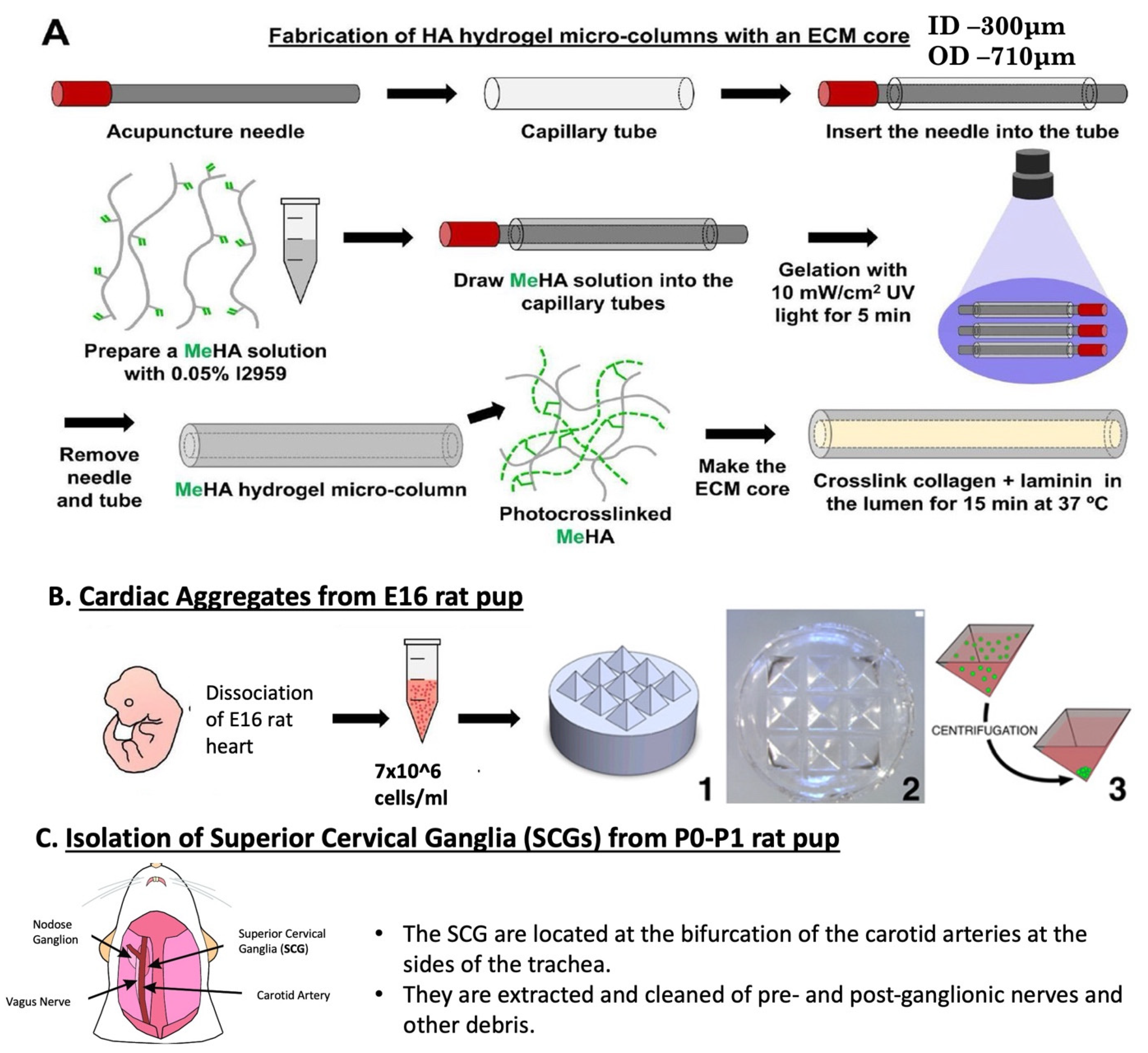
Figure 2.
Characterization of planar cocultures of cardiac aggregates and SCG-derived sympathetic aggregates. (A,B) Pieces of SCG isolated from postnatal rats were cultured as aggregates on laminin-coated planar surfaces for 10 DIV and stained for neurons/axons (Tuj1/β-tubulin III, green), noradrenergic neurons (TH, far red) and nuclei (Hoechst, blue), confirming the TH+ phenotype expected of sympathetic neurons. (C–E) Phase contrast images of SCG cocultured with aggregated cardiac microtissue on laminin-coated planar surfaces. (D,E) Sympathetic axons project into the cardiac aggregates and interact with myocytes (white arrowheads). (F) Planar coculture of cardiac aggregate and SCG showing expression of cardiac troponin T (cTnT, green), tyrosine hydroxylase (TH, red) and the pan-axonal marker β-tubulin III (Tuj1, purple). The inset shows a higher-magnification view near the SCG–cardiac interface, revealing sympathetic axons projecting into the cardiac aggregate. (G) Cardiac beats per minute quantified in the presence and absence of SCG at 3 and 5 DIV; data are mean ± SEM with individual values shown. Scale bars: (A) 250 µm, (B,C) 500 µm, (D,E) 100 µm, (F) 250 µm.
Figure 2.
Characterization of planar cocultures of cardiac aggregates and SCG-derived sympathetic aggregates. (A,B) Pieces of SCG isolated from postnatal rats were cultured as aggregates on laminin-coated planar surfaces for 10 DIV and stained for neurons/axons (Tuj1/β-tubulin III, green), noradrenergic neurons (TH, far red) and nuclei (Hoechst, blue), confirming the TH+ phenotype expected of sympathetic neurons. (C–E) Phase contrast images of SCG cocultured with aggregated cardiac microtissue on laminin-coated planar surfaces. (D,E) Sympathetic axons project into the cardiac aggregates and interact with myocytes (white arrowheads). (F) Planar coculture of cardiac aggregate and SCG showing expression of cardiac troponin T (cTnT, green), tyrosine hydroxylase (TH, red) and the pan-axonal marker β-tubulin III (Tuj1, purple). The inset shows a higher-magnification view near the SCG–cardiac interface, revealing sympathetic axons projecting into the cardiac aggregate. (G) Cardiac beats per minute quantified in the presence and absence of SCG at 3 and 5 DIV; data are mean ± SEM with individual values shown. Scale bars: (A) 250 µm, (B,C) 500 µm, (D,E) 100 µm, (F) 250 µm.
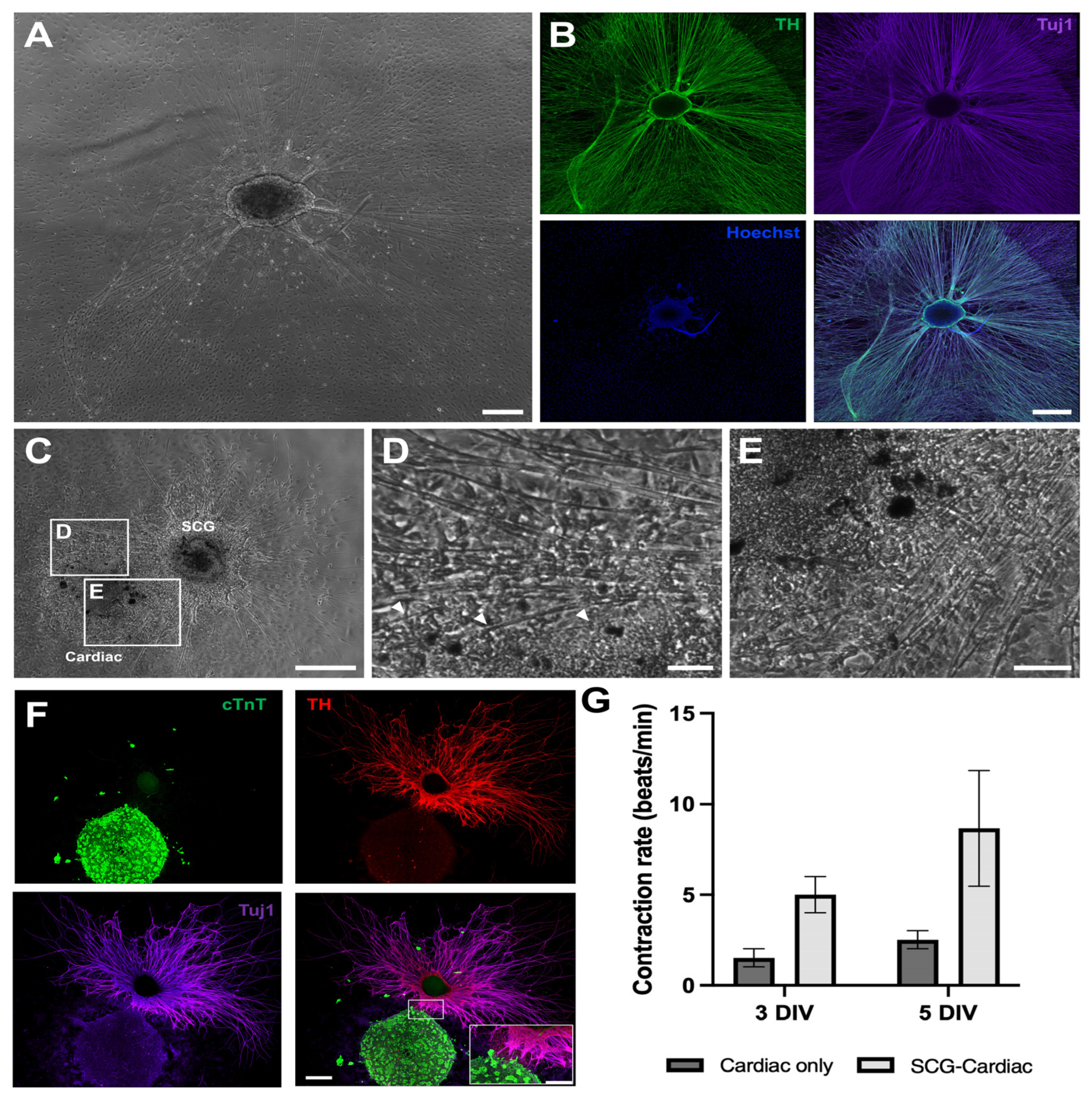
Figure 3.
Gene expression in planar neuro-cardiac cocultures. RT-qPCR analysis of contractile, conduction-, adrenergic-, cardiac transcription factor- and synaptic-associated genes in planar cultures: freshly isolated E16 cardiac tissue (DIV 0 CM), cardiac-only cultures at DIV 7 (DIV 7 CM), and neuro-cardiac cocultures at DIV 7 (DIV 7 CM+SCG). All samples derive from planar culture. Relative expression was calculated as 2^(−ΔCt) using Gapdh as the endogenous reference gene. Actc1, Actn2, Adrb1, Myh6, Gja5 (Cx40), Cx43 and Cx45 were significantly elevated in DIV 7 CM+SCG cocultures relative to both DIV 0 CM and DIV 7 CM (* p < 0.05, ** p < 0.01, **** p < 0.0001), and Nkx2-5 and Th were significantly elevated in DIV 7 CM+SCG relative to DIV 7 CM (* p < 0.05); no comparison reached significance for Tnnt2, Myh7 or Snap25, and DIV 7 CM did not differ from DIV 0 CM for any gene (ns). Data are mean ± SD with individual values shown; n ≥ 3 biological replicates per group.
Figure 3.
Gene expression in planar neuro-cardiac cocultures. RT-qPCR analysis of contractile, conduction-, adrenergic-, cardiac transcription factor- and synaptic-associated genes in planar cultures: freshly isolated E16 cardiac tissue (DIV 0 CM), cardiac-only cultures at DIV 7 (DIV 7 CM), and neuro-cardiac cocultures at DIV 7 (DIV 7 CM+SCG). All samples derive from planar culture. Relative expression was calculated as 2^(−ΔCt) using Gapdh as the endogenous reference gene. Actc1, Actn2, Adrb1, Myh6, Gja5 (Cx40), Cx43 and Cx45 were significantly elevated in DIV 7 CM+SCG cocultures relative to both DIV 0 CM and DIV 7 CM (* p < 0.05, ** p < 0.01, **** p < 0.0001), and Nkx2-5 and Th were significantly elevated in DIV 7 CM+SCG relative to DIV 7 CM (* p < 0.05); no comparison reached significance for Tnnt2, Myh7 or Snap25, and DIV 7 CM did not differ from DIV 0 CM for any gene (ns). Data are mean ± SD with individual values shown; n ≥ 3 biological replicates per group.
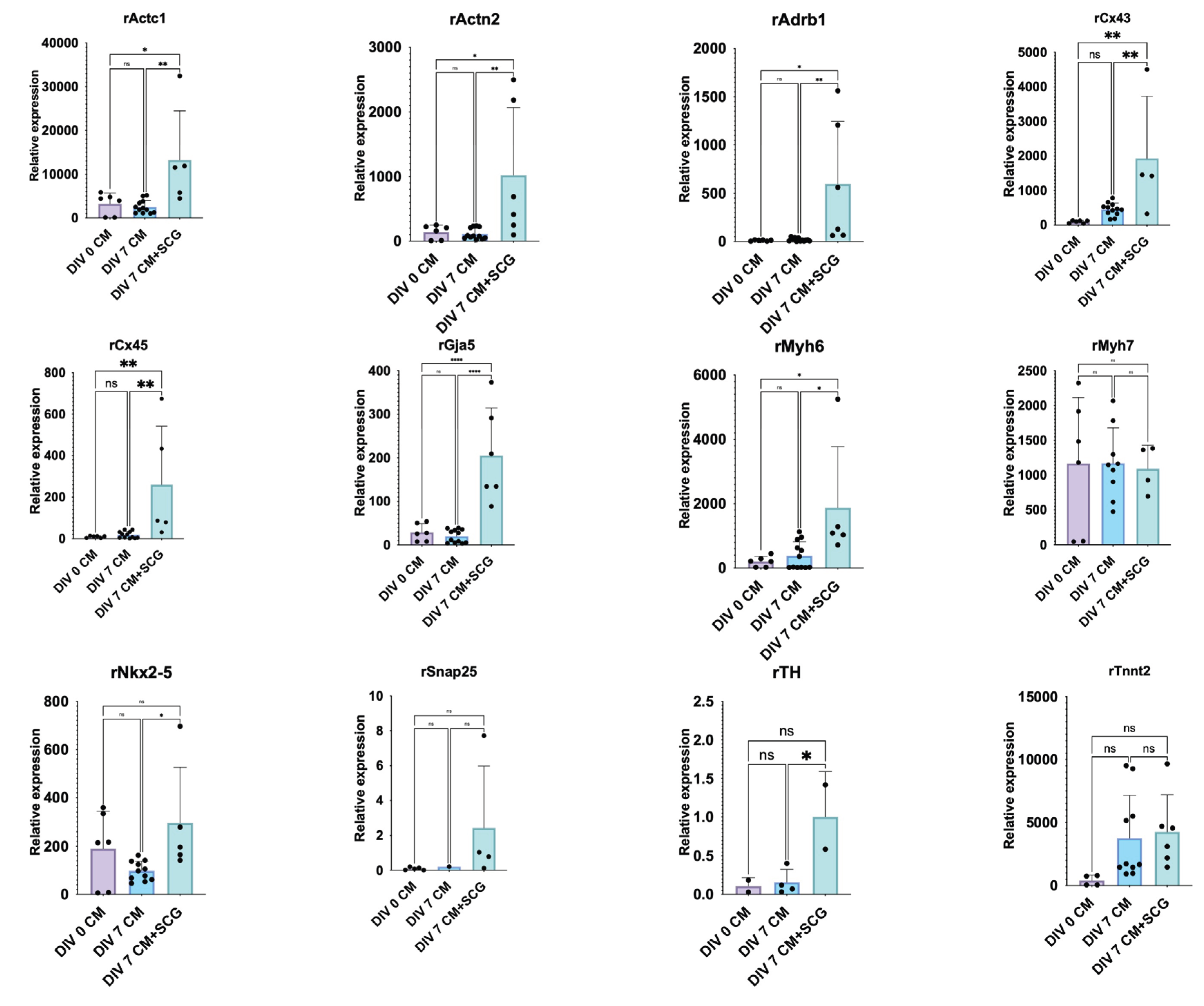
Figure 4.
Phenotype, growth and function of 3D sympathetic aggregates in hydrogel microcolumns. (A) SCG-derived sympathetic aggregate within a hydrogel microcolumn expressing axonal (Tuj1, green) and noradrenergic (TH, red) markers, with Hoechst-stained nuclei clustered within the aggregate. (B) Phase contrast images of a representative 3D MeHA hydrogel column containing a sympathetic aggregate, showing the progression of neurite growth from 1 to 17 DIV. (C,D) Neurite growth length (C) and growth rate (D) quantified as a function of time. Repeated measures ANOVA yielded significant effects of time on neurite length (p < 0.0001) and growth rate (p = 0.0381). Data are mean ± SEM with individual values shown (* p < 0.05, ** p < 0.01, *** p < 0.001, **** p < 0.0001). (E) Functional assessment by electrically evoked catecholamine release using fast-scan cyclic voltammetry at 30 DIV; a stimulating electrode was placed to span the aggregate while a carbon fiber electrode recorded in the adjacent region. (F) Concentration trace recorded in the aggregate region; the black bar indicates the period of electrical stimulation. (G) Cyclic voltammogram at peak release, confirming the catecholaminergic nature of the released transmitter. Scale bars: (A,B,E) 500 µm.
Figure 4.
Phenotype, growth and function of 3D sympathetic aggregates in hydrogel microcolumns. (A) SCG-derived sympathetic aggregate within a hydrogel microcolumn expressing axonal (Tuj1, green) and noradrenergic (TH, red) markers, with Hoechst-stained nuclei clustered within the aggregate. (B) Phase contrast images of a representative 3D MeHA hydrogel column containing a sympathetic aggregate, showing the progression of neurite growth from 1 to 17 DIV. (C,D) Neurite growth length (C) and growth rate (D) quantified as a function of time. Repeated measures ANOVA yielded significant effects of time on neurite length (p < 0.0001) and growth rate (p = 0.0381). Data are mean ± SEM with individual values shown (* p < 0.05, ** p < 0.01, *** p < 0.001, **** p < 0.0001). (E) Functional assessment by electrically evoked catecholamine release using fast-scan cyclic voltammetry at 30 DIV; a stimulating electrode was placed to span the aggregate while a carbon fiber electrode recorded in the adjacent region. (F) Concentration trace recorded in the aggregate region; the black bar indicates the period of electrical stimulation. (G) Cyclic voltammogram at peak release, confirming the catecholaminergic nature of the released transmitter. Scale bars: (A,B,E) 500 µm.
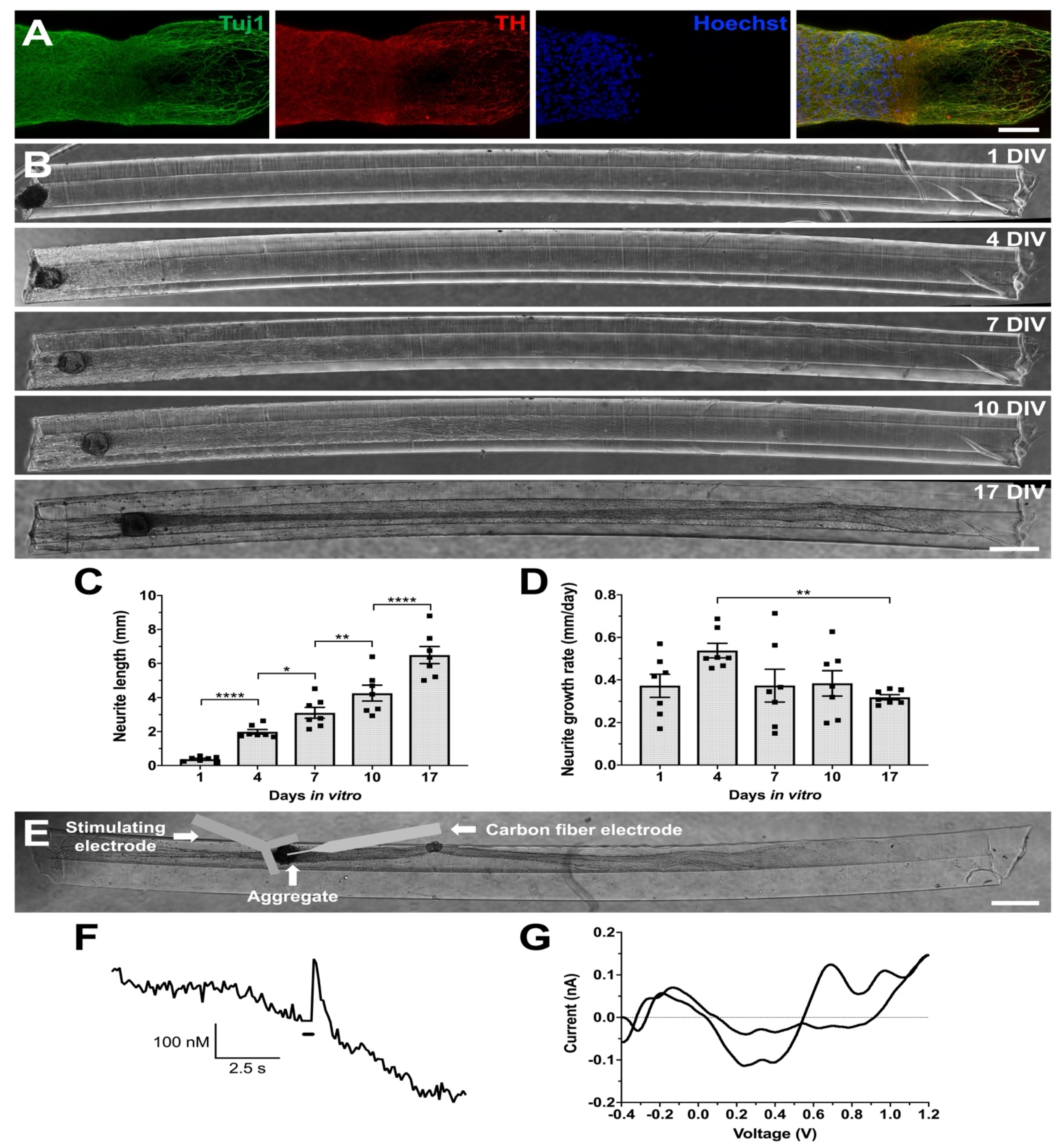
Figure 5.
Cellular architecture within a TE-ICU. Phase contrast images of a 3D cardiac–sympathetic ganglion coculture in a ~5 mm microcolumn at 10 DIV (top) and 14 DIV (middle and bottom). Sympathetic axons projecting from the SCG fasciculate along the length of the microcolumn and attach to the cardiac aggregate at the opposite end. The cardiac aggregate self-organizes into a multi-chambered structure reminiscent of myocardial partitioning during development. Scale bars: 300 µm.
Figure 5.
Cellular architecture within a TE-ICU. Phase contrast images of a 3D cardiac–sympathetic ganglion coculture in a ~5 mm microcolumn at 10 DIV (top) and 14 DIV (middle and bottom). Sympathetic axons projecting from the SCG fasciculate along the length of the microcolumn and attach to the cardiac aggregate at the opposite end. The cardiac aggregate self-organizes into a multi-chambered structure reminiscent of myocardial partitioning during development. Scale bars: 300 µm.
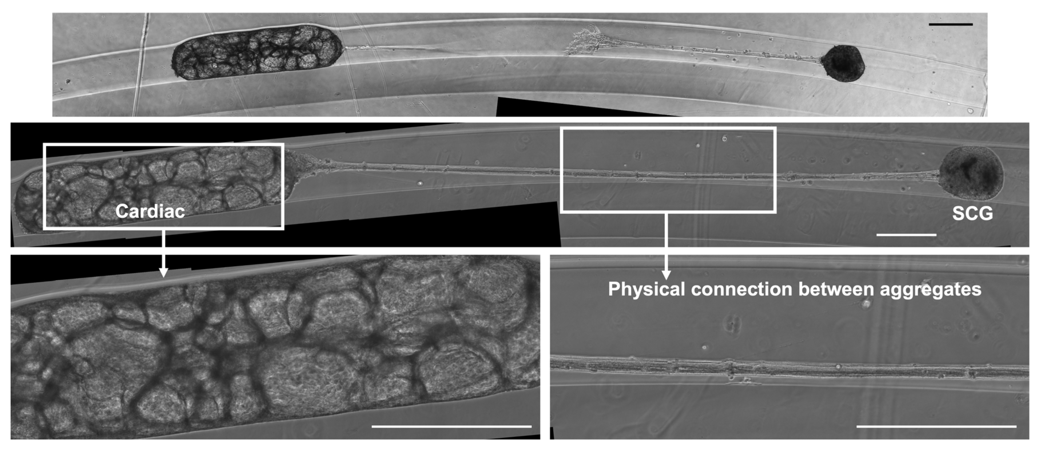
Figure 6.
Structural and functional characterization of TE-ICUs. (A) Phase contrast image of a ~3 mm MeHA hydrogel microcolumn containing sympathetic and cardiac aggregates at the left and right ends, respectively. (B) Confocal reconstruction of the cardiac aggregate region of the microtissue in (A), stained for cardiac myocytes (troponin, green), neurons/axons (Tuj1/β-tubulin III, far red), noradrenergic neurons (TH, red) and nuclei (Hoechst, blue). (C) Phase contrast image of a control microcolumn fabricated with a cardiac myocyte population only, at 5 DIV. (D) The cardiac-only microtissue stained for the same markers as (B), showing the cytoarchitecture of the cardiac aggregate in the absence of innervation. (E) Contraction rate, quantified as beats per minute at 5 and 8 DIV, in microcolumns containing both sympathetic and cardiac aggregates (SCG-Cardiac) and in cardiac-only microcolumns. Two-way ANOVA yielded significant effects of culture type (p = 0.0034) and time (p = 0.0089); Sidak’s multiple comparisons test identified a significant difference between groups at 8 DIV (** p < 0.01) but not at 5 DIV. Data are mean ± SEM. Scale bars: (A,C) 400 µm, (B) 100 µm, (D) 500 µm.
Figure 6.
Structural and functional characterization of TE-ICUs. (A) Phase contrast image of a ~3 mm MeHA hydrogel microcolumn containing sympathetic and cardiac aggregates at the left and right ends, respectively. (B) Confocal reconstruction of the cardiac aggregate region of the microtissue in (A), stained for cardiac myocytes (troponin, green), neurons/axons (Tuj1/β-tubulin III, far red), noradrenergic neurons (TH, red) and nuclei (Hoechst, blue). (C) Phase contrast image of a control microcolumn fabricated with a cardiac myocyte population only, at 5 DIV. (D) The cardiac-only microtissue stained for the same markers as (B), showing the cytoarchitecture of the cardiac aggregate in the absence of innervation. (E) Contraction rate, quantified as beats per minute at 5 and 8 DIV, in microcolumns containing both sympathetic and cardiac aggregates (SCG-Cardiac) and in cardiac-only microcolumns. Two-way ANOVA yielded significant effects of culture type (p = 0.0034) and time (p = 0.0089); Sidak’s multiple comparisons test identified a significant difference between groups at 8 DIV (** p < 0.01) but not at 5 DIV. Data are mean ± SEM. Scale bars: (A,C) 400 µm, (B) 100 µm, (D) 500 µm.
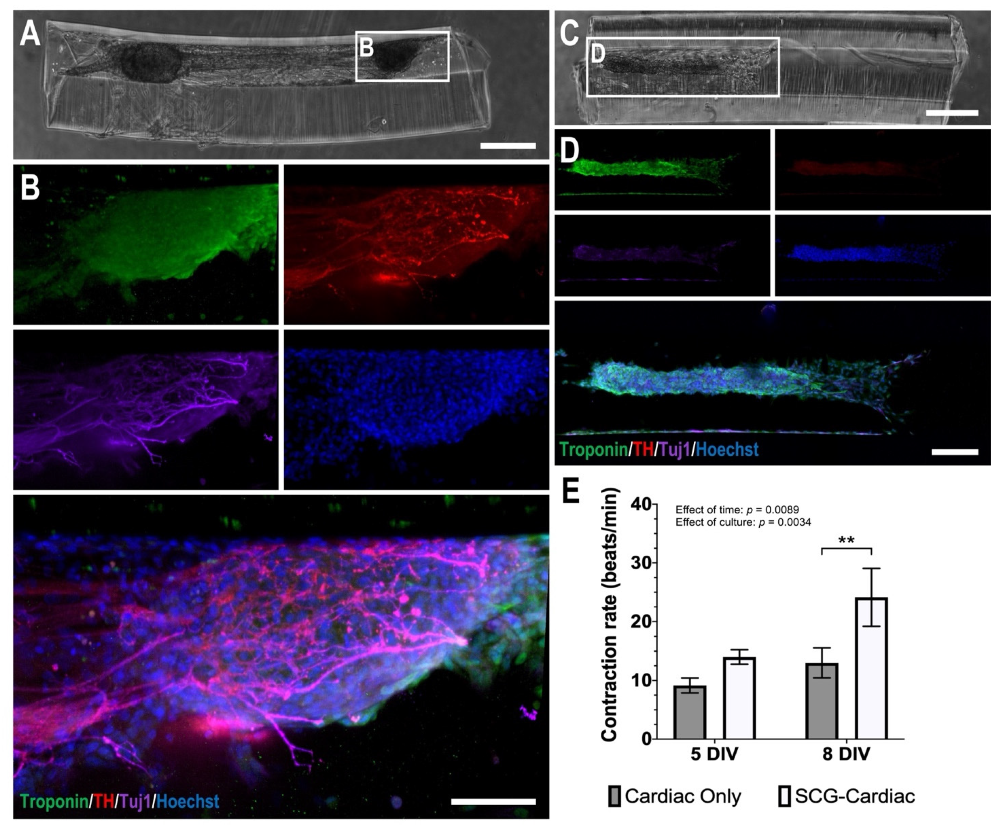
Figure 7.
Effect of the cardiac target on sympathetic outgrowth and β-adrenergic dependence of contraction in TE-ICUs. (A,B) Neurite growth length (A) and growth rate (B) were quantified 4 and 7 days after the addition of SCG to microcolumns containing a sympathetic aggregate alone (SCG Only) or both sympathetic and cardiac aggregates (SCG-Cardiac). The presence of a cardiac target did not alter either measure. (C) Contraction rate before and after acute application of the β-adrenergic antagonist propranolol (1 µM, 5 min) in cardiac-only and SCG-Cardiac microcolumns. (D) Response expressed as the ratio of beats per minute after treatment to beats per minute before treatment; cardiac-only constructs were unaffected, while SCG-Cardiac constructs showed a reduction in beating rate that did not reach statistical significance. Data are mean ± SEM with n = 8 per group in (A,B).
Figure 7.
Effect of the cardiac target on sympathetic outgrowth and β-adrenergic dependence of contraction in TE-ICUs. (A,B) Neurite growth length (A) and growth rate (B) were quantified 4 and 7 days after the addition of SCG to microcolumns containing a sympathetic aggregate alone (SCG Only) or both sympathetic and cardiac aggregates (SCG-Cardiac). The presence of a cardiac target did not alter either measure. (C) Contraction rate before and after acute application of the β-adrenergic antagonist propranolol (1 µM, 5 min) in cardiac-only and SCG-Cardiac microcolumns. (D) Response expressed as the ratio of beats per minute after treatment to beats per minute before treatment; cardiac-only constructs were unaffected, while SCG-Cardiac constructs showed a reduction in beating rate that did not reach statistical significance. Data are mean ± SEM with n = 8 per group in (A,B).
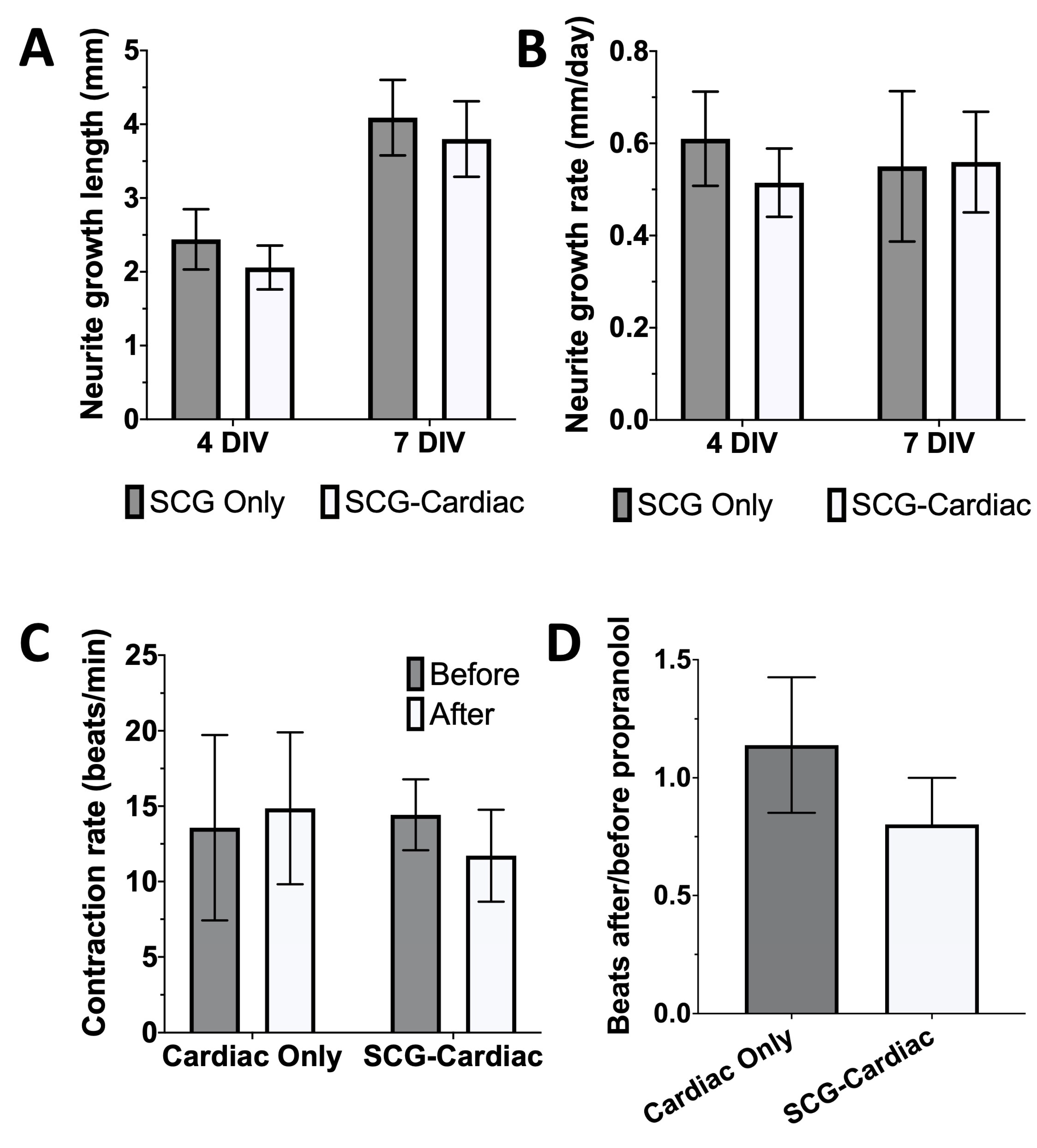

Table 1.
Forward and reverse primer sequences used for RT-qPCR. Gapdh served as the endogenous reference gene.
Table 1.
Forward and reverse primer sequences used for RT-qPCR. Gapdh served as the endogenous reference gene.
| Gene | Forward Primer | Reverse primer |
| Actc1 | AAAGCACGCCTACAGATCCC | CCCACGATGGATGGGAAGAC |
| Actn2 | GTCCCTGACGGAAGTTCGAG | CCTGAGCAATGGCTGCAATC |
| Adrb1 | GCTCTGGACTTCGGTAGACG | CAGGCTCTGGTAGCGAAAGG |
| Cx43 | AGCCTCCAAGGAGTTCCACC | GGAGTAGGCTTGGACCTTGT |
| Cx45 | TCAGCCTCCAAGGAGTTCCA | AACGCGTTTACAAAGCACAGT |
| Gja5 (Cx40) | CTGAAGAAGCCAACTCCAGGG | AGACCTTGCCGATGACCGTA |
| Myh6 | ACCGGAGTTTAAGAGTGACAGG | TCTGTGCGGATGTCAAAGGG |
| Myh7 | CCAAGGGCCTGAATGAAGAGT | TGTGTTTCTGCCTAAGGTGCT |
| TH | CTCCTTGTCTCGGGCTGTAA | AAATACTCCAGGTGGGGGCT |
| Tnnt2 | GAACAGCAGCGTATTCGCAA | CTTCCGGGCCTCATCTTCAG |
| Nkx2-5 | CAACTTCGTGAACTTCGGCG | TTTCCCTACCAGGCTCGGAT |
| Snap25 | ATGTTGGATGAGCAAGGCGA | AGGCCACAGCATTTGCCTAA |
| Gapdh | GCGAGATCCCGCTAACATCA | GGCGGAGATGATGACCCTTT |
Disclaimer/Publisher’s Note: The statements, opinions and data contained in all publications are solely those of the individual author(s) and contributor(s) and not of MDPI and/or the editor(s). MDPI and/or the editor(s) disclaim responsibility for any injury to people or property resulting from any ideas, methods, instructions or products referred to in the content. |
© 2026 by the authors. Licensee MDPI, Basel, Switzerland. This article is an open access article distributed under the terms and conditions of the Creative Commons Attribution (CC BY) license (http://creativecommons.org/licenses/by/4.0/).
Copyright: This open access article is published under a Creative Commons CC BY 4.0 license, which permit the free download, distribution, and reuse, provided that the author and preprint are cited in any reuse.