

Submitted:
10 September 2026
Posted:
11 September 2026
You are already at the latest version
Abstract
Background/Objectives: Escherichia coli is one of the most problematic bacterial species in hospital and community settings and a leading cause of resistant infections worldwide. This reinforces the need of new antibacterial tools like phage therapy. This study aimed to isolate and characterise lytic bacteriophages (phages) targeting clinical E. coli strains with different resistance profiles and to assess their potential for future therapeutic development. Methods: Environmental and clinical samples were used to isolate lytic phages active against clinical E. coli strains, and to evaluate their potential for future therapeutic applications. A phage collection was tested against E. coli strains from human bloodstream infections to determine their bacteriolytic activity, host range and specificity. Two phages were selected for further phenotypic and genomic characterisation. Their infectivity and antibacterial activity were evaluated under growth medium- and filtered human urine-related conditions and across different temperature and pH ranges. Results: Both phages demonstrated consistent antimicrobial activity against susceptible clinical strains, exhibiting a narrow host range and specificity. They retained infectivity under growth medium- and filtered human urine-related conditions, significantly reducing bacterial populations, and maintaining lytic activity across a range of temperatures and pH values. Genomic characterisation confirmed their lytic nature and supported their safety. Taxonomic analysis revealed that one phage was classified as a new podovirus-morphotype species within the genus Xuquatrovirus. The other displayed characteristics consistent with a novel genus, showing myovirus morphology. Conclusions: Environmental reservoirs represent a source of lytic phages active against clinically relevant E. coli strains. The new coliphages provide a basis for future development of phage-based strategies against multidrug-resistant E. coli strains.
Keywords:
coliphages
; phage therapy
; bacteraemia
; antimicrobial resistance
; One Health
; SDGs
1. Introduction
The therapeutic value of conventional antibiotics continues to be threatened by antimicrobial resistance (AMR), with Escherichia coli remaining one of the most problematic bacterial species in both hospital and community settings. This organism is highly relevant due to its ease of recovery and propagation, genetic diversity, frequent acquisition of mobile resistance determinants and broad ecological distribution in humans, animals, wastewater and other environmental reservoirs. Recent estimates of mortality associated with AMR place E. coli among the leading contributors to resistant infection worldwide [1], which reinforces the need to identify new antibacterial tools that can be tested under clinically relevant conditions [2,3].
E. coli is best known for being a dominant facultative anaerobe in the intestinal microbiota of humans and other animals. However, its biology extends far beyond commensalism. The species comprises strains pathogenic to humans and can cause both extraintestinal and intestinal infections. In fact, E. coli is one of the leading causes of nosocomial infections, notably bloodstream infections (BSIs) and urinary tract infections [4,5,6]. Although to a lesser extent, it can also be associated with intestinal diseases (diarrhoea or colitis) and even extraintestinal conditions such as nosocomial pneumonia and ventilator-associated pneumonia [7]. The most common primary source of E. coli BSI is urinary tract infection, accounting for approximately 50–60% of cases [8]. Furthermore, the incidence of E. coli BSI caused by multidrug-resistant (MDR) strains, including those that produce extended-spectrum beta-lactamases (ESBL), is increasing, with E. coli BSI-associated mortality being one of the leading causes of death [5].
In recent years, there has been a significant increase in interest regarding the use of bacteriophages to treat bacterial infections. Within the One Health framework and in line with the Sustainable Development Goals (SDGs), phages have re-emerged as promising antibacterial agents because of their ability to infect and lyse specific bacterial hosts through mechanisms that differ fundamentally from those of antibiotics [9,10]. For therapeutic purposes, strictly virulent or lytic phages are preferred, since they replicate within the target bacterium and promote bacterial killing without establishing lysogeny. Their high specificity, self-amplifying capacity at the site of infection and potential synergy with antibiotics have renewed interest in phage therapy against MDR bacterial infections [2]. However, therapeutic translation requires careful selection of candidates. Phages intended for clinical use must demonstrate consistent lytic activity, an appropriate host range against clinically relevant strains and high specificity to avoid damaging the natural microbiota. They must also be stable under physiological and storage conditions and lack undesirable genomic features, such as integrases, virulence genes, and antimicrobial resistance determinants [2,9].
Environmental sources are particularly valuable for the isolation of E. coli phages. Wastewater, surface waters and other faecally contaminated environments contain high densities of E. coli and therefore provide ecological niches in which diverse phages naturally co-evolve with their bacterial hosts [11,12]. This makes environmental sampling an effective method of obtaining lytic coliphages with potential relevance for clinical development. Recent studies have revealed that phages isolated from sewage or similar environments can exhibit activity against MDR or beta-lactamase-producing E. coli, suggesting their potential as biocontrol or therapeutic agents [11,12]. Nevertheless, environmental origin alone is not sufficient to support biomedical application; each isolate must be subjected to phenotypic and genomic characterization before being considered for further development. Candidate phages must remain viable in human matrices, such as urine, be active at body temperature, and be able to tolerate formulation and storage conditions that are compatible with clinical use. Further, the high diversity of phages and their bacterial hosts together with the narrow host range of many coliphages highlights the need to isolate and study well-characterized phages adapted to local bacterial populations. Genomic analysis is essential to confirm their strictly lytic nature, assign taxonomy, detect novelty and exclude genes that could compromise safety.
The aim of this study was to isolate and characterise bacteriophages active against a panel of local clinical E. coli strains recovered from bloodstream infections, most of which displayed a MDR phenotype, and to assess their potential as candidates for future therapeutic development. Following the initial screening of a collection of lytic coliphages isolated from environmental water samples, two were selected for a comprehensive biological, morphological, genomic and taxonomic characterisation to assess their bacteriolytic activity in growth medium and a biological matrix, as well as their stability, genomic safety and taxonomic classification.
2. Material and Methods
2.1. Bacterial Strains, Culture, Antimicrobial Susceptibility and Preservation Conditions
The E. coli CECT 101, from the Spanish Collection of Type Cultures (CECT), was used as reference strain for this species, which is a standard reference strain for lytic phage propagation [13]. This strain was routinely used in bacterial and phage assays, unless otherwise indicated. The clinical E. coli strains used in this study (8410, 8415, 8436, 8445, 8446, 8466, 8481, 8493, 8530, 8542, 8560, 8577, 8594, 8613, 8615) were obtained from the Microbiology Service at the University and Polytechnic Hospital La Fe (HUyPLaFe) in Valencia. Clinical E. coli strains were obtained from bloodstream infections. They were anonymized to reduce the risk of indirect identification. Individual patient data, exact isolation dates, specific clinical units, and any clinical information not required for microbiological interpretation were not included.
Antimicrobial susceptibility testing was performed by Microscan NMDRM2 panel as a reference method for clinically suspected cases of extended resistance (Beckman Coulter, L'Hospitalet de Llobregat, Spain) and disk diffusion on Müller-Hinton according to European Committee on Antimicrobial Susceptibility Testing (EUCAST) criteria, but with the possible drawback of incubating both in a 5% CO2 atmosphere, according to Sahuquillo-Arce et al. [14]. Results were reported as susceptible (S), intermediate (I) or resistant (R) according to the EUCAST interpretative criteria in use at the time of the study. As described [15], E. coli was classified as MDR if it was resistant to three or more antimicrobial classes.
All strains of E. coli studied, together with strains of other bacterial species (Alcaligenes faecalis CECT 928, Bacillus cereus CECT 495, Enterococcus faecalis CECT 481, Klebsiella pneumoniae CECT 143, Pseudomonas fluorescens CECT 378, Proteus hauseri CECT 484, Salmonella enterica CECT 443, Serratia marcescens CECT 159 and Staphylococcus aureus CECT 4013), were used to test the host range and specificity of the isolated phages. Strains were usually grown in a general liquid LB medium (tryptone 10 g/L, yeast extract 5 g/L, NaCl 10 g/L) [16] alone or with agar (agar 15 g/L) added (LBA) at temperatures of 28 °C and/or 37 °C for 24 h. The strains were cultured in LB with shaking at 150 rpm until an optical density at 600 nm (OD₆₀₀) of 0.1 ± 0.01 (approximately 10⁸ colony-forming units (CFU)/mL) was reached.
Initially, clinical E. coli strains were grown on McConkey’s selective and differential agar plates for enterobacteria [17] at 37°C for up to 48 h. This medium contains crystal violet and bile salts to select Gram-negative enteric bacteria, and lactose and the pH indicator phenol red to differentiate those able of producing lactic acid from this sugar. MacConkey agar was also used to try to isolate novel hospital-related E. coli strains from 24 hospital samples (washbasin and medicine trap wastes, human urine and skin samples from patients and clinical staff, and computer keyboards) taken using sterile swabs from three boxes in the Intensive Care Unit (ICU) at HUyPLaFe. The identity of the clinical E. coli strains was confirmed by conventional PCR using oligonucleotides targeting the 16S ribosomal gene, followed by sequencing; and a specific multiplex PCR for E. coli, as described below. Presumptive hospital-associated E. coli isolates from MacConkey plates, after their purification on LBA. Were tested by the specific PCR for E. coli.
All bacterial strains and isolates were handled in accordance with their biological risk level under biosafety levels 1 and 2 conditions. The strains and isolates were cryopreserved in LB medium supplemented with either 20% or 25% (v/v) glycerol, and stored at either −20 °C or −80 °C.
2.2. Identity Confirmation of Clinical E. coli Strains
To confirm the identity of the clinical E. coli strains studied, the 16S ribosomal gene was amplified by PCR and subsequently sequenced. Partial amplification of the 16S rRNA gene from the bacterial strains was performed by PCR using primers 616V (5′-AGAGTTTGATYMTGGCTCAG-3′) and 699R (5′-GGGTYKCGCTCGTTR-3′), according to Arahal et al. [18]. The resulting amplicons were purified and sequenced by the Sanger methodology at the Sequencing Service of the Central Experimental Research Service (SCSIE) at the University of Valencia (Spain). The partial 16S rRNA gene sequences obtained were analysed using BLASTn [19] against the NCBI database to identify the taxonomic classification at the genus level. This was based on the highest sequence similarity and query coverage. Moreover, the species-level identity of the clinical strains was confirmed using the multiplex PCR protocol described by Zimoń et al. [20], targeting three E. coli-specific genes: cydA (F: 5′- CGTATGGAGATGGTGAG-3′; R: 5′- GTAGAACCAGAACGCAGT-3′), lacY (F: 5′- TTCCCACCGATGCGATT-3′; R: 5′- GTCACTGTATGTTATTGGCG-3′), and ydiV (F: 5′- CCATTTCTCCAGTGAAGAT-3′; R: 5′- CCTAACACAAGGGGATAC-3′). E. coli CECT 101 and Pseudomonas fluorescens CECT 378 were included as positive and negative controls, respectively. Subsequently, the presumptive hospital-associated E. coli strains, once purified, were analysed using the same specific PCR test for this species.
2.3. Isolation, Purification, Amplification and Preservation of Phages
Phages were isolated from 31 environmental water samples of various origins, 28 taken from irrigation channels and 3 from wastewater, in the surrounding areas of Valencia, Easter Spain, using the E. coli reference strain CECT 101. In addition, the 24 above-mentioned clinical samples from the ICU at HUyPLaFe were processed for phage isolation using the reference strain CECT 101 and one MDR clinical strain 8436. Sterile bottles were used to collect the environmental water samples, while sterile swabs in SM buffer (50 mM Tris-HCl, pH 7.5, with 100 mM NaCl, 10 mM MgSO₄ and 0.01% gelatin) [21], and sterile tubes for liquids were used for hospital samples. The samples were stored at 4°C until use. Phage isolation was carried out similarly to Biosca et al. [22,23] by enriching the filtered samples (using sterile filters with a pore diameter of 0.22 μm) with 2X LB medium in equal proportions. Hereinafter, references to filtered samples will refer to 0.22 μm filters. The mixture was inoculated with the bacterial host strains separately, adjusted to an OD600nm of 0.1 ± 0.01, and incubated at 37°C with agitation. After incubation, the positive cultures (in which lysis was observed) were centrifuged at 10,000 rpm for 10 min and the supernatants filtered to separate the cellular debris from the viral fraction. LB medium inoculated with the host bacterium without the addition of the sample was used as a negative control for lysis, and uninoculated LB medium as a sterility control.
To purify the phages obtained, serial decimal dilutions of single plaques were prepared in SM buffer and inoculated using the double-layer agar plating method to obtain isolated plaque-forming units (PFU) similar to Biosca et al. [22,23]. For this purpose, molten and tempered top agar was used, which was inoculated with bacterial culture (OD600nm 0.1 ± 0.01) and 0.1 mL of the phage dilution. The mixture was poured onto LBA plates, which were incubated at 37 °C until the first plaques appeared. Subsequently, plaques of varying sizes and morphologies were then selected and purified by repeating the described method until uniform plaques were observed. Once the desired phages were purified, they were amplified overnight in LB broth with a culture of the host bacterium in the exponential phase. The resulting samples were then processed and preserved at -20°C and -80°C in 25% and 30% glycerol, respectively.
2.4. Host Range and Specificity of E. coli Phages
The host range assay for the selected phages was carried out using the spot test method [22,23]. To do this, molten top agar was mixed with 0.2 mL of bacterial inoculum (DO600nm 0.1 ± 0.01) and poured onto LBA plates. Next, 10 μL of the viral suspensions (at a concentration of approximately 10⁸ PFU/mL) were placed on the solidified top agar. Once the droplets had been absorbed, the plates were incubated at 37 °C. Clinical and reference strains of E. coli, as well as strains of other bacterial species from various sources (A. faecalis CECT 928, B. cereus CECT 495, E. faecalis CECT 481, K. pneumoniae CECT 143, P. fluorescens CECT 378, P. hauseri CECT 484, S. enterica CECT 443, S. marcescens CECT 159 and S. aureus CECT 4013) were used. This assay was performed in duplicate at 37 ºC and repeated at 28 °C in LB broth and LBA. Positive results were defined as the appearance of lysis zones on the bacterial lawn on LBA plates and a reduction in turbidity in the bacterial cultures in the LB tubes following incubation. LB and LBA were included as negative controls, and E. coli strains grown without phages in these media were included as bacterial growth controls.
2.5. In Vitro Phage-E. coli Dynamic Interactions
The growth dynamics of E. coli strains in the presence of selected phages were monitored using a Tecan Infinite M Nano spectrophotometer (Männedorf, Switzerland). For potential clinical use phages were incubated with shaking for either 8 or 16 h at 37 °C with selected clinical strains and OD600nm readings taken at regular 60-min intervals. The 96-well microplates were inoculated with separate co-cultures of the bacterial strains at concentrations of 10⁷, 10⁶ and 10⁵ CFU/mL with single phages at a concentration of 10⁸ PFU/mL, in either LB broth or filtered human urine samples as a model of human biological matrix. LB broth and filtered urine were included as negative controls and E. coli strains in the absence of phages were included as positive controls. At least three replicates were performed per experimental condition.
The growth of E. coli strains in the presence or absence of phages was assessed during the incubation period by calculating the area under the curve (AUC). After normalising the data using the Shapiro–Wilk test (α = 0.05), we compared the AUCs of the different experimental groups using Brown–Forsythe ANOVA tests (α = 0.05) and Dunnett's T3 post hoc analysis (α = 0.05) [22,23].
2.6. Phage Stability Under Physicochemical Relevant Conditions
The stability of E. coli selected phages was assessed to determine whether they remained infective under the relevant physicochemical conditions tested. These conditions were chosen to reflect potential storage, laboratory handling and potential application rather than to define physicochemical limits. Selected phages were suspended in SM buffer and incubated for four weeks at 4, 28 and 37 °C and at pH 4.5, 7.2 and 8.0 at 28 °C. Thus, aliquots were prepared from viral suspensions of each selected phage in LB broth at an approximate concentration of 10⁹ PFU/ml and ten-fold diluted at 108 PFU/ml in SM buffer at pH 7.2 for incubation at three temperatures: 4, 28 and 37 °C for one month. Similarly, to study the effect of pH on the stability of each selected phage, aliquots were also prepared in SM buffer at three different pH values (4.5, 7.2 and 8) and incubated at 28 °C for four weeks. Two independent experiments were carried out, each with triplicate counts for the conditions tested. An initial titration was performed at t=0 to establish a baseline. The phage titer was then monitored weekly under each of the studied conditions for four weeks. Statistical differences among conditions at the final time point were assessed using non-parametric Kruskal–Wallis test, followed by Dunn’s post hoc test with Holm correction for multiple comparisons.
2.7. E. coli Phages Transmission Electron Microscopy
Samples for transmission electron microscopy (TEM) were prepared using filtered, purified phage suspensions with high titres (approximately 10¹⁰ PFU/mL). Prior to depositing the samples onto carbon-coated copper grids, the grids were subjected to a glow discharge (30 seconds at 7.2 V using a Bal-Tec MED 020 coating system). The samples were then placed on the grids immediately afterwards and left for 10 minutes. After two brief washes with distilled water, the samples were stained with either 1% uranyl acetate or 1% phosphotungstic acid, for either five or one minute, respectively. Any excess liquid was then removed, and the grids were left to air-dry. Visualization of the virions was carried out using the FEI Tecnai G2 Spirit TEM (Thermo Fisher Scientific, Waltham, USA) in the Electron Microscopy Service at the Príncipe Felipe Research Centre (CIPF, Valencia) or the TEM Hitachi HT7800 at the Central Service for Experimental Research (SCSIE) facility (Universitat de València, Burjassot, Spain). Finally, when appropriate, the dimensions of the capsids and tails were estimated from the resulting micrographs using ImageJ software (version 1.54p) [24], with measurements taken from at least 10 viral particles from each phage to support the preliminary morphotype assignment.
2.8. Genomic and Phylogenetic Analysis of E. coli Phages
Whole-genome sequencing was performed on two coliphages (5 and 12), which were selected from different environmental waters. The bacterial nucleic acids in the filtered phage lysates were removed using a combination of DNase and RNase for 1 h at 37 °C. The enzymes were then inactivated using EDTA for 10 minutes at the same temperature. CTAB protocol [25] was used to extract the phage DNA. The concentration, purity and integrity of the DNA were subsequently evaluated using a NanoDrop spectrophotometer. It was also verified using 1% agarose gel electrophoresis.
The sequencing of phage 5 was performed using the MinION Mk1B (Oxford Nanopore Technologies, Oxford, UK), the Rapid Barcoding Kit 24 and an R10.4.1 flow cell. Base-calling was performed using Dorado v7.4.14 (Super-Accurate Base-Calling v4.3.0, 400 bp) (https://github.com/nanoporetech/dorado). For the phage 12 genome, sequencing was conducted using a TruSeq Nano DNA Library preparation protocol (META). This was followed by high-throughput sequencing on a NovaSeq X platform, generating 150 base pair paired-end reads (150 × 2 bp). The raw reads were analysed using the nf-core/bacass v2.6.0 pipeline [26], which involved trimming the reads, assessing their quality, screening for contamination and conducting de novo assembly using Unicycler [27]. After assembly, genome completeness was assessed using CheckV v1.0.1 [28], and structural and functional annotation was performed with Pharokka v1.9.1 using the v1.8.0 database [29]. The identification of virulence factor genes and antimicrobial resistance genes was carried out using Pharokka v1.9.1 [29] and PhageScope [30]. Lifestyle predictions were performed using PhageScope [30]. Depolymerase predictions were made using DePolymerase Predictor v1.0.0 [31] and PhageDPO v0.1.0 [32] on a Galaxy server [33]. Thereafter, the complete set of assembled genomes was submitted to the NCBI GenBank database.
For the comparative genomic analysis, phages with both nucleotide identity of at least 75% and query coverage of at least 25% at the BLAST core nucleotide database [34] were selected as related phages. Genome-wide similarities between the isolated phages and related phages were calculated using VIRIDIC [35]. The resulting similarity matrix was then visualized as a heat map. Species- and genus-level relationships were defined using VIRIDIC’s default thresholds of 95% and 70% nucleotide similarity, respectively. Viral genomes were further analysed with ViPTree [36] to generate a proteomic tree based on overall genomic similarity, thereby supporting comparative taxonomic classification. For each phage, 5 and 12, an individual analysis was performed together with its closest BLAST hits from the ViPTree prokaryotic dsDNA viruses database, and the phage of interest was displayed alongside its nearest related phages and the closest reference clade. Comparative visualizations of phages 5 and 12, and their closest phages, were generated using LoVis4u v0.2.0 [37].
3. Results and Discussion
3.1. E. coli Clinical Strain Identity Confirmation and Antibiotic Susceptibility
To confirm the identity of the E. coli clinical strains under study, molecular identification was performed after inoculation on McConkey agar plates, a selective and differential medium for Enterobacteriaceae. On this medium, characteristic E. coli colonies are deep pink and surrounded by a halo of precipitated bile salts (Figure 1A), whereas in the general LB medium, the colonies are beige (Figure 1B). Bile salts and crystal violet dye confer selectivity by inhibiting the growth of non-enteric bacteria, while lactose and the pH indicator phenol red allow differentiation of lactose-fermenting strains that acidify the medium. This causes the E. coli colonies to turn pink and precipitate the surrounding bile salts. Initially, the strains produced dark pink colonies, except for the clinical strains 8415, 8446, 8481 and 8530. Figure 1C shows the growth of some E. coli strains on MacConkey plates after 24 h at 37 °C. However, after 48 h, all strains turned pink. Thus, some clinical E. coli strains were delayed-lactose fermenters, producing colourless colonies on MacConkey agar plates after 24 h.
Hospital-related presumptive E. coli strains, selected based on their colonies appearance when grown on McConkey agar plates, were obtained only in eight of the 24 samples analysed from ICU boxes. These eight samples were obtained from the washbasin and medication siphons of sampled boxes. No growth was observed on the MacConkey plates in the remaining samples. Following the random selection and purification of E. coli-like colonies on LBA, molecular identification was performed.
The identity of all E. coli clinical strains from BSI was confirmed by partial amplification and Sanger sequencing of the 16S rRNA [18] and by multiplex-specific PCR for this species [20]. Although several isolates recovered from the hospital environment were initially considered as presumptive E. coli based on their colony morphology on MacConkey agar, molecular confirmation did not support their assignment to this species. Thus, these isolates were excluded from the study. This result is consistent with the absence of confirmed E. coli strains in the three boxes sampled of the ICU environment at the time of sampling. This observation may be compatible with adequate environmental cleaning and disinfection practices, which are considered essential measures to reduce the risk of healthcare-associated infections, particularly in patient-care areas [38,39].
Antimicrobial susceptibility testing of the 15 clinical E. coli strains revealed a highly resistant bacterial collection. Thirteen strains (86.7%) were resistant to at least 10 of the 21 antibiotics tested, including ampicillin, aztreonam, cefepime, cefixime, cefotaxime, ceftazidime, cefuroxime, ticarcillin and tobramycin, while 12 strains (80.0%) exhibited resistance to additional antimicrobial agents (Figure 2). Phenotypic testing classified 13 of the 15 strains (86.7%) as ESBL producers, whereas only two clinical strains were ESBL-negative (Figure 2).
The broad-spectrum resistance profile involving beta-lactams and other antimicrobial classes is consistent with a multidrug-resistant phenotype. Resistance to penicillins, extended-spectrum cephalosporins, and aztreonam is characteristic of ESBL-mediated resistance. In contrast, concomitant resistance to tobramycin and other agents may reflect the co-localisation of additional resistance genes on plasmids and other mobile genetic elements [40]. The high prevalence of ESBL production and multidrug resistance considerably restricts conventional therapeutic options, supporting the investigation of alternative antimicrobial approaches.
3.2. E. coli Phages Were Isolated Only from Environmental Waters
Out of 55 clinical and environmental samples analysed, 17 showed lytic activity against the reference strain of E. coli CECT 101 and at least one of the E. coli clinical strains 8410, 8415, 8436, 8493 or 8613. Following enrichment on susceptible bacterial host, all E. coli phages were isolated from environmental water samples and initially named them with the prefix "EcEW" and the number of the water sample from which they were isolated (EcEW1 to EcEW17). Notably, coliphages were recovered from wastewater samples, except for one phage that was obtained from surface irrigation water. Wastewater-derived isolation plates showed marked plaque diversity, including differences in size, edge definition, halo formation and transparency (Figure 3), suggesting the presence of heterogeneous phage populations. This is consistent with the high viral diversity described in wastewater, including large collections of phages infecting E. coli [41]. Similar plaque-based diversity has been reported for sewage- or hospital wastewater-derived coliphages, such as EcoM017, ES10 and other lytic phages active against multidrug-resistant E. coli strains [42,43,44]. Thus, environmental wastewater is a valuable source of coliphages, which have various biological properties and potential applications in phage therapy or biological control [41], but environmental surface water may also be a useful reservoir. Overall, the recovery of clear lytic plaques after host enrichment confirms the presence of active coliphage populations in the environmental waters sampled and supports the effectiveness of the enrichment strategy for isolating candidates phages active against clinical E. coli strains.
3.3. Selected Lytic Phages Showed Limited Intra-Species Host Range for E. coli Clinical Strains
Following purification and reassessment of the host range, selected water-borne E. coli phages (EcEW) exhibited consistent lytic activity in both solid and liquid media. They produced plaques on LB agar and reduced the optical density of susceptible cultures in LB. However, they generally showed a narrow host range for clinical strains phage. Phage EcEW5 lysed five (8410, 8415, 8436, 8493 and 8613) of the 15 clinical strains tested (33.3%), whereas phage EcEW12 lysed four strains (8410, 8415, 8436 and 8493) (26,7%) (Figure 4). Two of the three non-ESBL-producing strains, including the reference E. coli strain, were susceptible to a broader range of phages, while the remaining non-ESBL-producing strains was resistant to all the tested phages (Figure 4). Of the 13 ESBL-producing strains, only four were susceptible to at least two phages (Figure 4). However, these findings do not establish a direct relationship between ESBL production, multidrug resistance, and phage resistance. Rather, phage susceptibility is primarily determined by strain-specific factors, including receptor availability, genetic background, and antiphage defence systems [45]. The observed differences therefore likely reflect the particular characteristics of the tested strains.
Specificity testing showed that phages EcEW5 and EcEW12 lysed E. coli clinical strains but not the non-target bacteria studied, without lytic activity against A. faecalis CECT 928, B. cereus CECT 495, E. faecalis CECT 481, K. pneumoniae CECT 143, P. fluorescens CECT 378, P. hauseri CECT 484, S. enterica CECT 443, S. marcescens CECT 159 and S. aureus CECT 4013, including phylogenetically related species such as S. enterica. This supports their narrow lytic spectrum and indicates a high degree of host specificity under the conditions tested. In agreement with recent work on MDR E. coli phages [46], these host-range and specificity results support the selection of EcEW5 and EcEW12 for further biological, genomic and safety characterization.
Plaque morphology of selected phages provides a useful first indication of the interaction between these two lytic E. coli phages and their bacterial hosts. Clear plaques of EcEW5 (Figure 5A) usually reflect efficient bacterial lysis, whereas clear plaques surrounded by turbid halos, as observed with EcEW12 (Figure 5B) are commonly associated with lytic phages that exhibit depolymerase activity [47,48,49]. Several studies on E. coli phages support this interpretation. The E. coli phage vB_EcoM_ECOO78 produced lysis plaques with progressively expanding halos, which led to the identification of Dpo42, a novel depolymerase able to degrade capsular polysaccharides and reduce biofilm formation [50]. Similarly, the coliphage UDF157lw formed halo-containing plaques and encoded a protein with depolymerase activity [51]. In applied contexts, halo-forming phages may be especially valuable when the target strain produces capsule or biofilm matrix, because depolymerases can remove extracellular barriers and expose bacterial surface receptors [52].
3.4. Phage-Mediated Control of E. coli in Culture Media
For therapeutic purposes, the ability of phages EcEW5 and EcEW12 to control the growth of E. coli clinical strains 8410 and 8436, respectively, was initially investigated by using different concentrations of the target bacteria (10⁷, 10⁶ and 10⁵ CFU/mL) in LB broth with phages at a concentration of 10⁸ PFU/mL and under shaking conditions. After initial experiments, the assays were repeated with the target bacteria at 10⁶ and 10⁵ CFU/mL. As shown in Figure 6, the activity of phage EcEW5 (Figure 6A and B), which is defined as a reduction in the OD600 nm values of bacterial cultures, appears to be independent of the tested bacterial concentration. In contrast, the activity of phage EcEW12 (Figure 6B) seems to depend on the bacterial concentration, being more effective at lower concentrations (10⁵ PFU/mL).
The greatest control of bacterial growth was observed within the first 8 h for EcEW5 (Figure 6A) and within the first 6 h for EcEW12 (Figure 6B) with the target bacteria at 10⁵ CFU/mL. After these periods of time, the increase in bacterial populations was due to the emergence of resistant subpopulations. Nevertheless, phage activity persisted throughout the assay. This is consistent with the findings of other studies that have used individual [53] or combined [46] phages, since the OD600nm values achieved by the bacteria in the absence of phages were significantly higher (p<0.05) than those reached in their presence across the two concentrations of bacteria tested.
Based on abovementioned results, new assays were performed with phages EcEW5 and EcEW12 against four clinical E. coli strains (8410, 8436, 8493 and 8613) separately at bacteria-to-phage ratios of 10⁵ CFU/mL:10⁸ PFU/mL. As shown in Figure 7, the results obtained with phages EcEW5 and EcEW12 on strains 8410 (Figure 7A) and 8436 (Figure 7B), respectively, confirmed the previous findings by demonstrating significant reductions (p<0.05) in bacterial populations, as well as the reproducibility of the independent assays. However, the control capacity of phage EcEW5 on strain 8436 and of phage EcEW12 on strain 8410 was limited (Figure 7A and B). These phages also had little effect on the growth of strains 8493 (Figure 7C) and 8613 (Figure 7D) under the tested conditions, with no statistically significant differences observed between the presence and absence of phages (p>0.05).
In short, the two selected coliphages suppressed the growth of susceptible clinical strains in LB broth, but the effect varied in duration and extent depending on the host-phage pair. EcEW5 exerted strong control over strain 8410, whereas EcEW12 was effective against strain 8436. In contrast, the same phages were much less effective against three clinical strains within the host range of EcEW5, as well as two strains within the host range of EcEW12 (Figure 4). These results confirm the need to study the dynamics of bacterium-phage interaction to select the best phage candidates. Further, they also confirm the narrow host range of the selected phages, which is typical of many tailed coliphages. This could be advantageous for their potential use in personalised phage therapy treatments, as selective antibacterial activity could reduce the impact on non-target microbiota compared to broad-spectrum antibiotics. The use of phages as precision antimicrobials is increasingly relevant for severe and MDR infections, although phenotypic and genomic characterization is essential before clinical application [54].
3.5. Phage-Mediated Control of E. coli Is Prolonged Under Static Conditions
The effect of aeration on phage activity was also studied by conducting assays with and without shaking to provide or not aeration. As illustrated in Figure 8, the absence of agitation (i.e. aeration) caused a delay in the development of bacterial resistance to the phages. Figure 8A shows that EcEW5 suppressed the growth of strain 8410 for approximately 16 h without agitation, compared to 11 h with agitation. These differences were statistically significant (p<0.05). Phage EcEW12 controlled strain 8436 for 6 h without agitation, compared to 5 h with agitation (Figure 7B), though these differences were not statistically significant. Thus, aeration altered the timing of bacterial regrowth because when co-cultures were incubated without shaking, phage-mediated suppression was more prolonged than under agitated conditions. These differences are consistent with previous studies showing that bacterial growth curves after phage exposure can display distinct patterns, such as killing, delayed regrowth or incomplete suppression, depending on factors such as the phage, host strain and experimental conditions [55]. This result is clinically relevant because the human body contains microenvironments with low fluid turnover, such as the urinary tract, or incomplete mixing, particularly in catheterised or obstructed systems. These results support the inclusion of non-agitated or low-mixing assays during phage preselection for some therapeutic applications, while recognising that no single in vitro condition can fully reproduce the diversity of in vivo conditions, such as urinary microenvironments [56]. Frequently, resistance develops in vivo and in vitro, often rendering pathogens more vulnerable to antibiotics and other environmental factors while reducing their virulence. This is beneficial in bacterial infections, such as urinary tract infections, as bacteria that exhibit poor adherence or slow growth tend to be flushed out of the system [56].
3.6. Phage-Mediated Control of E. coli Is Prolonged Under Static Conditions
As urinary tract infections are the most common source of E. coli BSI [8,45] and the incidence of BSIs caused by MDR strains is increasing [5], we decided to explore the antibacterial activity of the selected phages in a filtered human urine model matrix. Since these bacterial strains proliferate in the urinary ducts during urinary tract infections, where there is no constant fluid flow, the in vitro activity of the phages in the absence of agitation could simulate an in vivo environment in which fluids remain static. The ability to control clinical strains without aeration in LB broth suggests that phages EcEW5 and EcEW12 could be used to develop alternatives to, or to complement, conventional antibiotics in the treatment of urinary tract infections. Given the potential therapeutic applications of these phages, their ability to suppress populations of clinical E. coli strains in human matrices such as urine was investigated. A key finding for therapeutic use was that selected phages remained active in filtered human urine. Furthermore, both EcEW5 (Figure 9A) and EcEW12 (Figure 9B) suppressed the growth of strains 8410 and 8436, respectively, for the first 5 h by one-single treatment. While our previous results in LB broth showed more pronounced and prolonged reductions in bacterial populations with EcEW5 than with EcEW12 (Figure 6 and Figure 7 and 8), both phages exhibited similar behaviour in human urine. In both experiments, bacterial growth increased after 5 h due to the emergence of phage-resistant subpopulations (Figure 9), consistent with previous studies in natural and artificial human urine [46,53].
Although previous studies using human urine and individual phages showed that bacterial growth increased similarly in urine and LB medium after 6–8 h of phage application due to the development of resistance [46,53], this study found that clinical strains growth in filtered human urine was lower than in LB broth. This may be because E. coli initially finds it difficult to multiply in human urine due to the scarcity of nutrients compared to the nutrient-rich LB medium. It may also be due to the acidity of the urine (due to uric acid), which is more acidic than the neutral pH of LB broth, as described in studies with artificial urine [46]. As urine differs substantially from LB in terms of nutrient availability, ionic composition and pH, the preservation of killing activity of phages in this human matrix is an important finding. This indicates that the phages retained their infective capacity under conditions closer to the intended application than those of LB broth. Therefore, the observed growth suppression in human urine was not solely due to the host's difficulty in growing in that environment; rather, it was a substantial phage-mediated effect that was additional to the slower bacterial growth. But using natural human urine rather than artificial urine provides a more realistic scenario and supports the potential therapeutic use of phages EcEW5 and EcEW12. However, the short duration of suppression also highlights the need for strategies to prevent resistance, such as phage cocktails, adaptive phage selection, repeated dosing, or combination with conventional antibiotics.
3.7. E. coli Phages Can Infect at a Range of Temperatures and pH Values
Regarding the effect of temperature on phage viability (Figure 10A), phage EcEW5 remained relatively stable during storage at 4 °C, maintaining titres close to the initials (108 PFU/mL). In contrast, storage at 28 and 37 °C resulted in a marked loss of infectivity, with final titres around 106 and 104 PFU/mL, respectively, after four weeks, with titres at 37 °C being significantly lower than those at 4 °C (p=0.022). These results indicate that EcEW5 is temperature-sensitive and that refrigerated storage is the most appropriate condition for preserving its viability. Phage EcEW12 exhibited greater stability than phage EcEW5 at the two higher temperatures tested, remaining stable at both 4 °C and 28 °C, with titres around 108 PFU/mL, and undergoing only a moderate reduction at 37 °C, where titres remained close to 10⁶ PFU/mL. However, were significantly lower than those observed at 4 °C (p=0.034). Therefore, although both phages were best preserved at 4 °C, EcEW12 displayed greater thermal stability than EcEW5 under the higher-temperature conditions tested.
These results are consistent with previous studies showing that, in general, refrigeration is favorable for maintaining the infectivity of therapeutic phage preparations, although it depends on the buffer used and the initial phage concentration [57]. Similarly, Alexyuk et al. [58] reported that a phage cocktail against E. coli retained its lytic activity for eight months when stored at 4–8 °C. However, activity declined progressively when the cocktail was stored at room temperature. More recent studies have shown that some coliphages can retain their infectivity over a broad temperature range, including 4–37 °C, albeit for shorter periods [12]. The fact that phage EcEW12 is more stable at 37 °C than phage EcEW5 is important for therapeutic use, as phages must remain infective at body temperature. However, both phages exhibit lytic activity against certain clinical strains of E. coli at this temperature.
The effect of pH on phage viability is shown in Figure 10B. Phage EcEW5 titres decreased significantly under all tested conditions (p=0.027), with a more pronounced loss of infectivity at acidic pH (p=0.022). After four weeks, phage titres decreased by approximately two logarithmic units at pH 7.2 and pH 8.0, reaching 6 × 10⁵ and 1 × 10⁵ PFU/mL, respectively. At pH 4.5, titres decreased by approximately five logarithmic units to 10² PFU/mL. EcEW12 showed greater stability at pH 7.2 and pH 8.0, maintaining final titres of approximately 10⁷ PFU/mL. In contrast, titres at pH 4.5 decreased sharply to approximately 10² PFU/mL after four weeks. However, differences among the pH conditions at week 4 did not reach statistical significance (p=0.055). Overall, both phages exhibited reduced viability under acidic conditions, with EcEW12 maintaining higher titres than EcEW5 at neutral and slightly alkaline pH.
This behavior is consistent with recent studies on E. coli phages showing that maximum stability is usually observed near neutral or mildly alkaline pH, whereas acidic conditions may severely compromise infectivity. For instance, coliphage ES10 was described as a therapeutically promising lytic phage whose physicochemical stability was an essential criterion for its potential application [43]. Likewise, the coliphage AUBRB02 showed peak stability around pH 7–8 and a marked titre decline under more acidic or alkaline extremes [12]. Other coliphages have also been evaluated for stability across broad pH ranges as part of their preclinical characterization for therapeutic use [59].
Although phages EcEW5 and EcEW12 were both highly sensitive to acidic pH, EcEW12 showed greater overall stability, particularly under neutral and slightly alkaline conditions. This higher resilience is relevant for formulation and storage, because therapeutic phage preparations are generally expected to retain infectivity during handling and delivery. Therefore, pH 7.2–8.0 appears suitable for maintaining EcEW12 viability, whereas more frequent applications would be necessary in acidic conditions.
3.8. E. Coli Selected Phages Exhibit Myovirus- and Podovirus-Like Morphologies
TEM analysis confirmed that EcEW5 and EcEW12 are tailed phages with icosahedral capsids. However, EcEW5 has long tails, while EcEW12 exhibits short tails. This is consistent with the morphotypes of myovirus and podovirus, respectively (Figure 11). EcEW5 had a mean capsid diameter of 61.2 nm and a mean tail length of 127.5 nm. EcEW12 had a mean capsid diameter of 61.9 nm, while its tail was too short to be measured reliably. This different morphology means that the two phages are distinct. Similar short-tailed, icosahedral E. coli phages have recently been reported among lytic phages that are active against uropathogenic or MDR E. coli., although with variable capsid sizes, supporting the morphological diversity of therapeutic coliphages [46,60]. However, as morphology alone is no longer sufficient for taxonomic classification [61], phages EcEW5 and EcEW12 should be more cautiously described as members of the class Caudoviricetes. This indicates that both phages belong to the dominant tailed phage lineages used in therapy and biocontrol. However, their definitive taxonomic placement requires genome-based classification, as described below.
3.9. Genome Features, Biosafety and Phylogenetic Analysis of E. coli Selected Phages
Genome assembly analysis revealed the genomes of EcEW5 and EcEW12 phages to be 47,035 bp (GC: 57.49%) and 62,283 bp (GC: 52.88%), respectively. The completeness analysis classified them as "high-quality" (Table 1). Similar genomic diversity has been reported among newly isolated coliphages, where phages assigned to different genera within Caudoviricetes often show distinct genome sizes, GC contents and host-recognition modules [62].
Genome-based host prediction identified E. coli as the most probable host for both EcEW5 and EcEW12 phages, in agreement with the experimental host range and bacteriolytic activity assays. This concordance between in silico prediction and phenotypic data strengthens the reliability of the characterization, because phage host range is frequently associated with genomic determinants such as tail fibres, receptor-binding proteins and depolymerases. Recent studies of phages active against uropathogenic E. coli strains have shown that genetically related Caudoviricetes can differ markedly in host spectrum due to variation in receptor-recognition genes, even when they share a similar genomic organization [46].
The prediction of a strictly virulent lifestyle for phages EcEW5 and EcEW12 is particularly relevant for their potential use as antibacterial agents. In therapeutic or biocontrol contexts, lytic phages are preferred because they replicate through productive infection and host cell lysis, whereas temperate phages may integrate into the bacterial genome and contribute to lysogenic conversion or horizontal gene transfer. The absence of genomic features associated with lysogeny, together with experimental evidence of bacterial lysis, supports the conclusion that EcEW5 and EcEW12 are virulent phages. This criterion is consistent with recent genomic studies of E. coli phages, in which the lack of integrases, repressors, lysogeny modules, toxin genes, virulence factors and antibiotic-resistance genes is considered a key requirement for selecting safe lytic phages [46].
The Pharokka annotation revealed 144 coding sequences (CDS) in EcEW5 and 98 CDS in EcEW12. The coding sequences for the EcEW5 phage are categorized as follows: 15 for DNA, RNA, and nucleotide metabolism; 7 for head and packaging; 15 for the tail; 1 for lysis (holin); 1 for a connector; 1 for a moron and auxiliary metabolic gene; and 104 with unknown functions. In the case of the EcEW12 phage, the CDSs are distributed as follows: 8 for DNA, RNA, and nucleotide metabolism; 3 for head and packaging; 3 for lysis (amidase, endolysin and holin); 4 for the tail; 1 for transcription regulation; 6 for other functions; and 73 with unknown functions. The detailed results of the Pharokka annotation for the EcEW5 and EcEW2 phages are shown in Supplementary Table S1 and Table S2, respectively. Overall, both phages exhibited a typical modular organization for bacteriophages, with most annotated open reading frames (ORFs) concentrated in the replication, structural, packaging, and lysis modules. We further analysed putative depolymerase candidates using PhageDPO and DePP, and summarized the results in Supplementary Table S3. In EcEW12, CDS 16 emerged as the most prominent candidate for further inspection, with high prediction scores in both tools (98% in PhageDPO and 81.58% in DePP). Subsequent searches with InterPro and ExPASy did not identify known conserved domains. However, the agreement between these two independent machine-learning approaches, which utilize different training sets and feature-selection strategies [31,32], strengthens our confidence in the prediction of CDS 16 as a depolymerase. This is also supported by the presence of halo-containing plaques in phage EcEW12, as they are similar to those described in other coliphages with depolymerase activity [51]. Nevertheless, the large number of hypothetical or unknown function proteins (Figure 11 and Supplementary Table S3) suggests that more detailed research is needed to elucidate their function in phage biology.
Finally, Figure 12 displays the 144 and 98 predicted CDSs within the EcEW5 and EcEW12 genomes, with the function of each CDS indicated by colour, compared with their closest phage genomes. The phages most closely related to EcEW5 were Pseudomonas phage PSA28 (MZ089737.1), Klebsiella phage vB_Kpn_K21lambda1 (OY978848.1), Klebsiella phage VLCpiS6a (ON602731.1), Klebsiella phage EPM-Kp18 (PX502243.1), Salmonella phage PMBT28 (MG641885.1), Caudoviricetes sp. isolate ctglZ8 (BK023738.1), and Caudoviricetes sp. isolate ctgBb10 (BK054708.1), whereas the closest phages to EcEW12 were Escherichia phage PTXU04 (NC_048193.1), Escherichia phage Pondi (OP136151.1), and Escherichia phage HelliStehle_Bas95 (PQ850607.1).
From a translational perspective, the most important finding was that no antimicrobial resistance genes or recognized virulence determinants were identified in the EcEW5 and EcEW12 genomes during the analysis with PhageScope and Pharokka. Together with the predicted virulent lifestyle and observed lytic behaviour in vitro, this result strongly supports the potential of both phages for clinical or environmental or applications
VIRIDIC analysis (Figure 13) showed that EcEW12 is closely related to Escherichia phage vB_EcoP_PTXU04 (94.7%). However, the two genomes were assigned to different species clusters, indicating that EcEW12 is closely related to, but distinct from, this phage at the species level. By contrast, EcEW5 displayed substantially lower similarities to its nearest phages, with the highest values not exceeding 57.8%, and was located in distinct species and genus clusters relative to the compared genomes.
Regarding the ViPTree analysis, EcEW5 (Figure 14A) was placed within a clade that grouped with phages associated with Klebsiella, Pseudomonas, Salmonella and other Caudoviricetes genomes. The topology of the tree suggests that EcEW5 occupies a separate genomic neighborhood rather than clustering within a tighter local group of closely related phages, and this placement provides a proteome-based context for its taxonomic assignment. Conversely, ViPTree placed EcEW12 within a reference clade that included the German Escherichia phage vB_EcoP_PTXU04 [63], which belongs to the genus Xuquatrovirus. EcEW12 was found to be most closely related to this phage in the tree (Figure 14B). EcEW12 also appeared in a local neighborhood that included the Dutch Escherichia phage Pondi [64], also a Xuquatrovirus, as well as the Swiss phage HelliStehle [65], which is from a different genus. This supports its assignment to an Escherichia-associated genomic context. The short branch length between EcEW12 and vB_EcoP_PTXU04 is consistent with close overall proteomic similarity and confirms that EcEW12 shares a substantial proportion of its genome with this German phage. Furthermore, this study enabled the prediction of a previously unannotated depolymerase in the EcEW12 phage, which is not present in any other phage belonging to the Xuquatrovirus genus.
The combined results of the ViPTree and VIRIDIC analyses provide valuable insights into the taxonomic classification of the two selected phages. For EcEW12, ViPTree placed its genome within a clade of European origin associated with Escherichia, identifying vB_EcoP_PTXU04 [63] as its closest reference phage. Meanwhile, VIRIDIC revealed a nucleotide similarity of 94.7% to Escherichia phage vB_EcoP_PTXU04, assigning EcEW12 as a new species within the genus Xuquatrovirus. On the other hand, EcEW5 was found in a separate clade in the ViPTree, alongside more distantly related caudoviral genomes from different continents. With nucleotide similarity values not exceeding 57.8%, VIRIDIC showed significantly lower similarity to the nearest genomes. This supports the proposal of a new genus. If future taxonomic classification confirms this hypothesis, this work proposes the name Eschevalenvirus, combining the genus name of the bacterial host, Escherichia, with the name of the province of Valencia where this phage was first isolated and characterised. Consequently, EcEW5 was assigned to distinct species and genus clusters compared to the reference genomes, which reinforces the interpretation that it represents a more divergent lineage. Overall, these results suggest that EcEW12 can be classified within a recognized taxonomic framework, whereas EcEW5 may correspond to a more distant, potentially novel lineage, pending further taxonomic validation, but both are novel phages.
Overall, a collection of lytic coliphages were isolated from environmental water samples in the Valencia area (Spain) and tested against BSI E. coli strains, mostly MDR. As result, two phages were selected with a narrow host range but consistent lytic activity against susceptible clinical strains. Dynamics of bacterium-phage interaction demonstrated that both phages significantly reduced bacterial populations in growth medium and filtered human urine conditions in vitro. Further, they maintained lytic activity under different temperature and pH conditions, compatible with their potential therapeutic use. Genomic characterisation confirmed their lytic nature, supporting their safety and enabling the prediction of an unannotated depolymerase in phage EcEW12. Taxonomic analysis revealed that one phage was classified as a new species within the genus Xuquatrovirus, while the other displayed characteristics consistent with a novel currently unclassified genus, showing myovirus morphology. Together, these findings highlight the importance of environmental reservoirs as a potential source of therapeutic coliphages and provide a basis for the future development of phage-based precision antimicrobials against MDR E. coli strains. This approach is in line with the One Health strategy and the SDGs of the United Nations.
Supplementary Materials
The following supporting information can be downloaded at https://www.mdpi.com/article/doi/s1, Table S1: Raw Pharokka CDS annotation for phage EcEW5; Table S2: Raw Pharokka CDS annotation for phage EcEW12; Table S3: Depolymerase prediction results for EcEW5 and EcEW12 proteins from PhageDPO and DePolymerase Predictor (DePP).
Author Contributions
Conceptualization, EGB; methodology, JC-S , RV, IS-L, TL, JF-A, PR and EGB; validation, JC-S and EGB; formal analysis, JC-S, RV, IS-L and EGB; investigation, JC-S, IS-L, TL, JF-A, PR and EGB; resources, JF-A , PR and EGB; data curation, JC-S, RV, IS-L, JF-A and EGB; writing—original draft preparation, JC-S and EGB; writing—review and editing, JC-S , RV , IS-L, TL, JF-A , PR and EGB; visualization, JC-S, RV, IS-L and EGB; supervision, EGB; project administration, EGB; funding acquisition, EGB . All authors have read and agreed to the published version of the manuscript.
Funding
This research was supported by project AICO/2021/261 of the Conselleria de Innovacion, Universidades, Ciencia y Sociedad Digital of Generalitat Valenciana.
Institutional Review Board Statement
Not applicable.
Informed Consent Statement
Not applicable.
Data Availability Statement
The complete genome sequences of the two Escherichia coli phages, EcEW5 and EcEW12, have been deposited in the NCBI GenBank database under accession numbers PZ787841 and PZ793367, respectively. These sequences are publicly available and can be accessed via the GenBank nucleotide database (https://www.ncbi.nlm.nih.gov/nucleotide/).
Acknowledgments
We would like to thank the technicians at the Central Service for Experimental Research (SCSIE) at the University of Valencia (UV), and the Príncipe Felipe Research Centre (CIPF), particularly M. T. Mínguez and M. Durán from UV; and M. Soriano from CIPF, for their expert support with transmission electron microscopy.
Conflicts of Interest
The authors declare that the research was conducted in the absence of any commercial or financial relationships that could be construed as a potential conflict of interest.
Abbreviations
The following abbreviations are used in this manuscript:
| AMR | Antimicrobial resistance |
| AUC | Area under the curve |
| BSI | Bloodstream infection |
| CDS | Coding DNA sequence |
| CECT | Spanish Collection of Type Cultures |
| CFU | Colony-forming units |
| CIPF | Prí ncipe Felipe Research Centre |
| CTAB | Cetyltrimethylammonium bromide |
| EDTA | Ethylenediaminetetraacetic acid |
| ESBL | Extended-spectrum Beta-lactamases |
| EUCAST | European Committee on Antimicrobial Susceptibility Testing |
| GC | Guanine-Cytosine content |
| HUyPLaFe | University and Polytechnic Hospital La Fe |
| ICU | Intensive Care Unit |
| LBA | LB agar |
| MDR | Multidrug-resistant |
| NCBI | National Center for Biotechnology Information |
| OD600 | Optical Density at 600 nm |
| ORF | Open Reading Frame |
| PFU | Plaque-Forming Unit |
| SCSIE | Sequencing Service of the Central Experimental Research Service (UV) |
| SDGs | Sustainable Development Goals |
| TEM | Transmission Electron Microscopy |
| UV | University of Valencia |
References
- Naghavi, M.; Vollset, S.E.; Ikuta, K.S.; Swetschinski, L.R.; Gray, A.P.; Wool, E.E.; Robles Aguilar, G.; Mestrovic, T.; Smith, G.; Han, C.; et al. Global burden of bacterial antimicrobial resistance 1990–2021: a systematic analysis with forecasts to 2050. The Lancet 2024, 404, 1199–1226. [Google Scholar] [CrossRef] [PubMed]
- Strathdee, S.A.; Hatfull, G.F.; Mutalik, V.K.; Schooley, R.T. Phage therapy: From biological mechanisms to future directions. Cell 2023, 186, 17–31. [Google Scholar] [CrossRef] [PubMed]
- Singh, G.; Rana, A. Smriti Decoding antimicrobial resistance: unraveling molecular mechanisms and targeted strategies. Arch. Microbiol. 2024, 206, 280. [Google Scholar] [CrossRef] [PubMed]
- Cantón, R.; Loza, E.; Arcay, R.M.; Cercenado, E.; Castillo, F.J.; Cisterna, R.; Gálvez-Benítez, L.; González Romo, F.; Hernández-Cabezas, A.; Rodríguez-Lozano, J.; et al. Antimicrobial activity of ceftolozane-tazobactam against Enterobacterales and Pseudomonas aeruginosa recovered during the Study for Monitoring Antimicrobial Resistance Trends (SMART) program in Spain (2016-2018). Rev. Esp. Quimioter. 2021, 34, 228–237. [Google Scholar] [CrossRef] [PubMed]
- Feldman, S.F.; Temkin, E.; Wullfhart, L.; Nutman, A.; Schechner, V.; Shitrit, P.; Shvartz, R.; Schwaber, M.J.; Andremont, A.; Carmeli, Y. A nationwide population-based study of Escherichia coli bloodstream infections: incidence, antimicrobial resistance and mortality. Clin. Microbiol. Infect. 2022, 28, 879.e1–879.e7. [Google Scholar] [CrossRef] [PubMed]
- Burgaya, J.; Marin, J.; Royer, G.; Condamine, B.; Gachet, B.; Clermont, O.; Jaureguy, F.; Burdet, C.; Lefort, A.; De Lastours, V.; et al. The bacterial genetic determinants of Escherichia coli capacity to cause bloodstream infections in humans. PLoS Genet 2023, 19, e1010842. [Google Scholar] [CrossRef] [PubMed]
- Geurtsen, J.; de Been, M.; Weerdenburg, E.; Zomer, A.; McNally, A.; Poolman, J. Genomics and pathotypes of the many faces of Escherichia coli. FEMS Microbiol. Rev. 2022, 46, fuac031. [Google Scholar] [CrossRef] [PubMed]
- Grome, H.N.; Brandenburg, J.M.; Kent, A.G.; Curtis, L.; Raymond, R.E.; Ansari, U.; Gargis, A.S.; McKay, S.L.; Parker, E.; Driscoll, J.; et al. Extraintestinal invasive Escherichia coli infections in the US. JAMA Netw. Open 2026, 9, e2557201. [Google Scholar] [CrossRef] [PubMed]
- Álvarez, B.; Biosca, E.G. Harnessing the activity of lytic bacteriophages to foster the Sustainable Development Goals and the “One Health” strategy. Viruses 2025, 17, 549. [Google Scholar] [CrossRef] [PubMed]
- World Health Organization. Bacteriophages and their use in combating antimicrobial resistance. 2026. Available online: https://www.who.int/europe/news-room/fact-sheets/item/bacteriophages-and-their-use-in-combating-antimicrobial-resistance (accessed on 8 September 2026).
- Vitt, A.R.; Sørensen, A.N.; Bojer, M.S.; Bortolaia, V.; Sørensen, M.C.H.; Brøndsted, L. Diverse bacteriophages for biocontrol of ESBL- and AmpC-β-lactamase-producing E. coli. iScience 2024, 27, 108826. [Google Scholar] [CrossRef] [PubMed]
- Abdo Ahmad, T.A.; El Houjeiry, S.A.; Abou Fayad, A.; Kanj, S.S.; Matar, G.M.; Saba, E.S. Isolation and genomic analysis of Escherichia coli phage AUBRB02: implications for phage therapy in Lebanon. Antibiotics 2025, 14, 458. [Google Scholar] [CrossRef] [PubMed]
- Monod, J.; Wollman, E. Inhibition of growth and enzymatic adaptation in bacteria infected with the bacteriophage. Ann. Inst. Pasteur 1947, 73, 937–956. [Google Scholar]
- Sahuquillo-Arce, J.M.; Chouman-Arcas, R.; Molina-Moreno, J.M.; Hernández-Cabezas, A.; Frasquet-Artés, J.; López-Hontangas, J.L. Capnophilic Enterobacteriaceae. Diagn. Microbiol. Infect. Dis. 2017, 87, 318–319. [Google Scholar] [CrossRef] [PubMed]
- Magiorakos, A.-P.; Srinivasan, A.; Carey, R.B.; Carmeli, Y.; Falagas, M.E.; Giske, C.G.; Harbarth, S.; Hindler, J.F.; Kahlmeter, G.; Olsson-Liljequist, B.; et al. Multidrug-resistant, extensively drug-resistant and pandrug-resistant bacteria: an international expert proposal for interim standard definitions for acquired resistance. Clin. Microbiol. Infect. 2012, 18, 268–281. [Google Scholar] [CrossRef] [PubMed]
- Bertani, G. Studies on lysogenesis. I. The mode of phage liberation by lysogenic Escherichia coli. J. Bacteriol. 1951, 62, 293–300. [Google Scholar] [CrossRef] [PubMed]
- Macconkey, A. Lactose-fermenting bacteria in faeces. J. Hyg. (Lond) 1905, 5, 333–379. [Google Scholar] [CrossRef] [PubMed]
- Arahal, D.R.R.; Sánchez, E.; Macián, M.C.; Garay, E. Value of recN sequences for species identification and as a phylogenetic marker within the family “Leuconostocaceae.”. Int. Microbiol. 2008, 33–39. [Google Scholar] [CrossRef]
- Altschul, S.F.; Gish, W.; Miller, W.; Myers, E.W.; Lipman, D.J. Basic local alignment search tool. J. Mol. Biol. 1990, 215, 403–410. [Google Scholar] [CrossRef] [PubMed]
- Zimoń, B.; Psujek, M.; Matczak, J.; Guziński, A.; Wójcik, E.; Dastych, J. Novel multiplex-PCR test for Escherichia coli detection. Microbiol. Spectr. 2024, 12, e03773-23. [Google Scholar] [CrossRef] [PubMed]
- Sambrook, J.; Russell, D.W. Molecular Cloning: A Laboratory Manual, 3rd ed.; Cold Spring Harbor Laboratory Press: Cold Spring Harbor, NY, 2001. [Google Scholar]
- Biosca, E.G.; Delgado Santander, R.; Morán, F.; Figàs-Segura, À.; Vázquez, R.; Català-Senent, J.F.; Álvarez, B. First european Erwinia amylovora lytic bacteriophage cocktails effective in the host: Characterization and prospects for fire blight biocontrol. Biology 2024, 13, 176. [Google Scholar] [CrossRef] [PubMed]
- Biosca, E.G.; Salas-Lastres, I.; Català-Senent, J.F.; Morán, F.; Palacio-Bielsa, A.; Álvarez, B. Development of the first European Xanthomonas euvesicatoria pv. euvesicatoria lytic bacteriophage cocktail effective in controlling bacterial spot disease in pepper plants. Front. Microbiol. 2026, 17, 1821339. [Google Scholar] [CrossRef] [PubMed]
- Schneider, C.A.; Rasband, W.S.; Eliceiri, K.W. NIH Image to ImageJ: 25 years of image analysis. Nat. Methods 2012, 9, 671–675. [Google Scholar] [CrossRef] [PubMed]
- Murray, M.G.; Thompson, W.F. Rapid isolation of high molecular weight plant DNA. Nucl. Acids Res. 1980, 8, 4321–4326. [Google Scholar] [CrossRef] [PubMed]
- Daniel, V.M.; Peltzer, Alexander; Straub, Daniel; nf-core bot; Bernabeu, Alejandro; Pantano, Lorena; Yi, H.D.; Brovkina, Olga; Regojo, Graciela Uria; Rosalía; et al. nf-core/bacass: nf-core/bacass v2.6.0 - Crimson Titanium Seahorse 2026. [CrossRef]
- Wick, R.R.; Judd, L.M.; Gorrie, C.L.; Holt, K.E. Unicycler: Resolving bacterial genome assemblies from short and long sequencing reads. PLoS Comput Biol. 2017, 13, e1005595. [Google Scholar] [CrossRef] [PubMed]
- Nayfach, S.; Camargo, A.P.; Schulz, F.; Eloe-Fadrosh, E.; Roux, S.; Kyrpides, N.C. CheckV assesses the quality and completeness of metagenome-assembled viral genomes. Nat. Biotechnol. 2021, 39, 578–585. [Google Scholar] [CrossRef] [PubMed]
- Bouras, G.; Nepal, R.; Houtak, G.; Psaltis, A.J.; Wormald, P.-J.; Vreugde, S. Pharokka: a fast scalable bacteriophage annotation tool. Bioinformatics 2023, 39, btac776. [Google Scholar] [CrossRef] [PubMed]
- Wang, R.H.; Yang, S.; Liu, Z.; Zhang, Y.; Wang, X.; Xu, Z.; Wang, J.; Li, S.C. PhageScope: a well-annotated bacteriophage database with automatic analyses and visualizations. Nucleic Acids Res. 2024, 52, D756–D761. [Google Scholar] [CrossRef] [PubMed]
- Magill, D.J.; Skvortsov, T.A. DePolymerase Predictor (DePP): a machine learning tool for the targeted identification of phage depolymerases. BMC Bioinform. 2023, 24, 208. [Google Scholar] [CrossRef] [PubMed]
- Vieira, M.F.; Duarte, J.; Domingues, R.; Oliveira, H.; Dias, O. PhageDPO: A machine-learning based computational framework for identifying phage depolymerases. Comput. Biol. Med. 2025, 188, 109836. [Google Scholar] [CrossRef] [PubMed]
- The Galaxy Community; Afgan, E.; Amoukou, N.; Buono, R.A.; Anton, M.; Antoniewski, C.; Austin, P.; Awan, A.H.; Backofen, R.; Bacon, W.A.; et al. Galaxy for accessible, reproducible, and collaborative data analyses: 2026 update. Nucleic Acids Res. 2026, 54, W105–W116. [Google Scholar] [CrossRef] [PubMed]
- Zhang, Z.; Schwartz, S.; Wagner, L.; Miller, W. A greedy algorithm for aligning DNA sequences. J. Comput. Biol. 2000, 7, 203–214. [Google Scholar] [CrossRef] [PubMed]
- Moraru, C.; Varsani, A.; Kropinski, A.M. VIRIDIC—A novel tool to calculate the intergenomic similarities of prokaryote-infecting viruses. Viruses 2020, 12, 1268. [Google Scholar] [CrossRef] [PubMed]
- Nishimura, Y.; Yoshida, T.; Kuronishi, M.; Uehara, H.; Ogata, H.; Goto, S. ViPTree: the viral proteomic tree server. Bioinformatics 2017, 33, 2379–2380. [Google Scholar] [CrossRef] [PubMed]
- Egorov, A.A.; Atkinson, G.C. LoVis4u: a locus visualization tool for comparative genomics and coverage profiles. NAR Genom. Bioinform. 2025, 7, lqaf009. [Google Scholar] [CrossRef] [PubMed]
- World Health Organization Infection prevention and control. Available online: https://www.who.int/health-topics/infection-prevention-and-control (accessed on 3 September 2026).
- National Center for Emerging and Zoonotic Infectious Diseases. Centers for Disease Control and Prevention, U.S. Department of Health and Human Services Environmental Cleaning Procedures. 2024, pp. 5–12. Available online: https://www.cdc.gov/healthcare-associated-infections/hcp/cleaning-global/procedures.html (accessed on 3 September 2026).
- Nasrollahian, S.; Graham, J.P.; Halaji, M. A review of the mechanisms that confer antibiotic resistance in pathotypes of E. coli. Front. Cell. Infect. Microbiol. 2024, 14, 1387497. [Google Scholar] [CrossRef] [PubMed]
- Jurczak-Kurek, A.; Gąsior, T.; Nejman-Faleńczyk, B.; Bloch, S.; Dydecka, A.; Topka, G.; Necel, A.; Jakubowska-Deredas, M.; Narajczyk, M.; Richert, M.; et al. Biodiversity of bacteriophages: morphological and biological properties of a large group of phages isolated from urban sewage. Sci. Rep. 2016, 6, 34338. [Google Scholar] [CrossRef] [PubMed]
- Ribeiro, K.V.G.; Ribeiro, C.; Dias, R.S.; Cardoso, S.A.; De Paula, S.O.; Zanuncio, J.C.; Oliveira, L.L.D. Bacteriophage isolated from sewage eliminates and prevents the establishment of Escherichia coli biofilm. Adv. Pharm. Bull. 2018, 8, 85–95. [Google Scholar] [CrossRef] [PubMed]
- Nawaz, A.; Zafar, S.; Alessa, A.H.; Khalid, N.A.; Shahzadi, M.; Majid, A.; Badshah, M.; Shah, A.A.; Khan, S. Characterization of ES10 lytic bacteriophage isolated from hospital waste against multidrug-resistant uropathogenic E. coli. Front. Microbiol. 2024, 15, 1320974. [Google Scholar] [CrossRef] [PubMed]
- Fikadu, A.; Amankwah, S.; Alemu, B.; Alemu, Y.; Naga, A.; Tekle, E.; Kassa, T. Isolation and phenotypic characterization of virulent bacteriophages against multidrug-resistant Escherichia coli and its phage-resistant variant from sewage sources. IDR 2024, Volume 17, 293–303. [Google Scholar] [CrossRef] [PubMed]
- Keith, M.; Park De La Torriente, A.; Chalka, A.; Vallejo-Trujillo, A.; McAteer, S.P.; Paterson, G.K.; Low, A.S.; Gally, D.L. Predictive phage therapy for Escherichia coli urinary tract infections: Cocktail selection for therapy based on machine learning models. Proc. Natl. Acad. Sci. U.S.A. 2024, 121, e2313574121. [Google Scholar] [CrossRef] [PubMed]
- Markusková, B.; Elnwrani, S.; Andrezál, M.; Sedláčková, T.; Szemes, T.; Slobodníková, L.; Kajsik, M.; Drahovská, H. Characterization of bacteriophages infecting multidrug-resistant uropathogenic Escherichia coli strains. Arch. Virol. 2024, 169, 142. [Google Scholar] [CrossRef] [PubMed]
- Knecht, L.E.; Veljkovic, M.; Fieseler, L. Diversity and function of phage encoded depolymerases. Front. Microbiol. 2020, 10, 2949. [Google Scholar] [CrossRef] [PubMed]
- Biosca, E.G.; Català-Senent, J.F.; Figàs-Segura, À.; Bertolini, E.; López, M.M.; Álvarez, B. Genomic analysis of the first european bacteriophages with depolymerase activity and biocontrol efficacy against the phytopathogen Ralstonia solanacearum. Viruses 2021, 13, 2539. [Google Scholar] [CrossRef] [PubMed]
- Panteleev, V.; Kulbachinskiy, A.; Gelfenbein, D. Evaluating phage lytic activity: from plaque assays to single-cell technologies. Front. Microbiol. 2025, 16, 1659093. [Google Scholar] [CrossRef] [PubMed]
- Guo, Z.; Huang, J.; Yan, G.; Lei, L.; Wang, S.; Yu, L.; Zhou, L.; Gao, A.; Feng, X.; Han, W.; et al. Identification and characterization of Dpo42, a novel depolymerase derived from the Escherichia coli phage vB_EcoM_ECOO78. Front. Microbiol. 2017, 8, 1460. [Google Scholar] [CrossRef] [PubMed]
- Liao, Y.-T.; Ho, K.-J.; Zhang, Y.; Salvador, A.; Wu, V.C.H. A new Rogue-like Escherichia phage UDF157lw to control Escherichia coli O157:H7. Front. Microbiol. 2024, 14, 1302032. [Google Scholar] [CrossRef] [PubMed]
- Sanmukh, S.G.; Admella, J.; Moya-Andérico, L.; Fehér, T.; Arévalo-Jaimes, B.V.; Blanco-Cabra, N.; Torrents, E. Accessing the . Cells 2023, 12, 344. [Google Scholar] [CrossRef] [PubMed]
- Zulk, J.J.; Clark, J.R.; Ottinger, S.; Ballard, M.B.; Mejia, M.E.; Mercado-Evans, V.; Heckmann, E.R.; Sanchez, B.C.; Trautner, B.W.; Maresso, A.W.; et al. Phage resistance accompanies reduced fitness of uropathogenic Escherichia coli in the urinary environment. mSphere 2022, 7, e00345-22. [Google Scholar] [CrossRef] [PubMed]
- Sawa, T.; Moriyama, K.; Kinoshita, M. Current status of bacteriophage therapy for severe bacterial infections. J. Intensive Care 2024, 12, 44. [Google Scholar] [CrossRef] [PubMed]
- Sørensen, P.E.; Ng, D.Y.K.; Duchateau, L.; Ingmer, H.; Garmyn, A.; Butaye, P. Classification of in vitro phage–host population growth dynamics. Microorganisms 2021, 9, 2470. [Google Scholar] [CrossRef] [PubMed]
- Zulk, J.J.; Patras, K.A.; Maresso, A.W. The rise, fall, and resurgence of phage therapy for urinary tract infection. EcoSal Plus 2024, 12, eesp-0029-2023. [Google Scholar] [CrossRef] [PubMed]
- Duyvejonck, H.; Merabishvili, M.; Vaneechoutte, M.; De Soir, S.; Wright, R.; Friman, V.-P.; Verbeken, G.; De Vos, D.; Pirnay, J.-P.; Van Mechelen, E.; et al. Evaluation of the stability of bacteriophages in different solutions suitable for the production of magistral preparations in Belgium. Viruses 2021, 13, 865. [Google Scholar] [CrossRef] [PubMed]
- Alexyuk, P.; Bogoyavlenskiy, A.; Alexyuk, M.; Akanova, K.; Moldakhanov, Y.; Berezin, V. Isolation and characterization of lytic bacteriophages active against clinical strains of E. coli and development of a phage antimicrobial cocktail. Viruses 2022, 14, 2381. [Google Scholar] [CrossRef] [PubMed]
- Hussein, A.H.; Makky, S.; Hager, R.; Connerton, I.F.; El-Shibiny, A. Characterization and therapeutic potential of phage vB_Eco_ZCEC08 against multidrug-resistant uropathogenic Escherichia coli. BMC Microbiol. 2025, 25, 221. [Google Scholar] [CrossRef] [PubMed]
- Niaz, H.; Skurnik, M.; Adnan, F. Genomic and proteomic characterization of four novel Schitoviridae family phages targeting uropathogenic Escherichia coli strain. Virol. J. 2025, 22, 83. [Google Scholar] [CrossRef] [PubMed]
- Turner, D.; Shkoporov, A.N.; Lood, C.; Millard, A.D.; Dutilh, B.E.; Alfenas-Zerbini, P.; Van Zyl, L.J.; Aziz, R.K.; Oksanen, H.M.; Poranen, M.M.; et al. Abolishment of morphology-based taxa and change to binomial species names: 2022 taxonomy update of the ICTV bacterial viruses subcommittee. Arch. Virol. 2023, 168, 74. [Google Scholar] [CrossRef] [PubMed]
- Śliwka, P.; Weber-Dąbrowska, B.; Żaczek, M.; Kuźmińska-Bajor, M.; Dusza, I.; Skaradzińska, A. Characterization and comparative genomic analysis of three virulent E. coli bacteriophages with the potential to reduce antibiotic-resistant bacteria in the environment. IJMS 2023, 24, 5696. [Google Scholar] [CrossRef] [PubMed]
- Korf, I.H.E.; Meier-Kolthoff, J.P.; Adriaenssens, E.M.; Kropinski, A.M.; Nimtz, M.; Rohde, M.; Van Raaij, M.J.; Wittmann, J. Still something to discover: Novel insights into Escherichia coli phage diversity and taxonomy. Viruses 2019, 11, 454. [Google Scholar] [CrossRef] [PubMed]
- Sidi Mabrouk, A.; Ongenae, V.; Claessen, D.; Brenzinger, S.; Briegel, A. A flexible and efficient microfluidics platform for the characterization and isolation of novel bacteriophages. Appl. Environ. Microbiol. 2023, 89, e01596-22. [Google Scholar] [CrossRef] [PubMed]
- Humolli, D.; Piel, D.; Maffei, E.; Heyer, Y.; Agustoni, E.; Shaidullina, A.; Willi, L.; Imwinkelried, P.; Estermann, F.; Cuénod, A.; et al. Completing the BASEL phage collection to unlock hidden diversity for systematic exploration of phage–host interactions. PLoS Biol. 2025, 23, e3003063. [Google Scholar] [CrossRef] [PubMed]
Figure 1.
Growth of representative clinical E. coli strains on MacConkey and LBA media. Morphology of E. coli colonies on the selective and differential culture medium McConkey (A), and on the general medium LBA (B). Growth of representative E. coli clinical strains on McConkey plates after 24 h of incubation at 37 °C (C).
Figure 1.
Growth of representative clinical E. coli strains on MacConkey and LBA media. Morphology of E. coli colonies on the selective and differential culture medium McConkey (A), and on the general medium LBA (B). Growth of representative E. coli clinical strains on McConkey plates after 24 h of incubation at 37 °C (C).
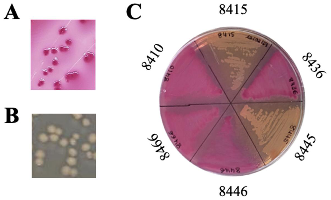
Figure 2.
E. coli clinical strains from human bacteraemia and their antimicrobial susceptibility and their extended-spectrum beta-lactamases (ESBL) profile.
Figure 2.
E. coli clinical strains from human bacteraemia and their antimicrobial susceptibility and their extended-spectrum beta-lactamases (ESBL) profile.
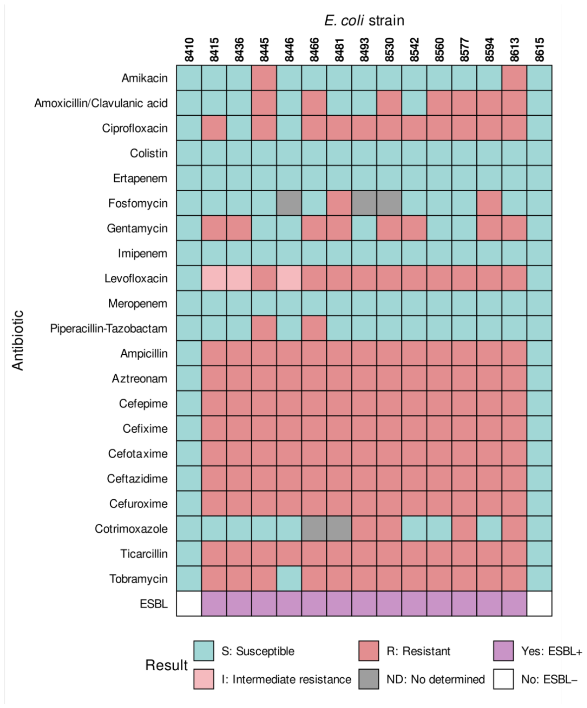
Figure 3.
Representative diversity of E. coli phage plaques isolated from water samples. Distinct lysis plaque morphologies were observed on LBA plates inoculated with the E. coli host strain after overnight incubation at 37°C, representing the phage isolates recovered from wastewater samples.
Figure 3.
Representative diversity of E. coli phage plaques isolated from water samples. Distinct lysis plaque morphologies were observed on LBA plates inoculated with the E. coli host strain after overnight incubation at 37°C, representing the phage isolates recovered from wastewater samples.
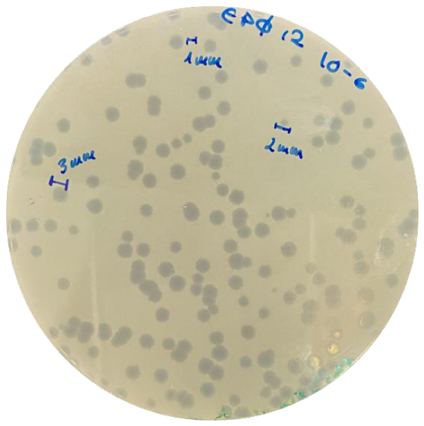
Figure 4.
Host range of E. coli water-borne phages and their extended-spectrum beta-lactamases (ESBL) profile.
Figure 4.
Host range of E. coli water-borne phages and their extended-spectrum beta-lactamases (ESBL) profile.
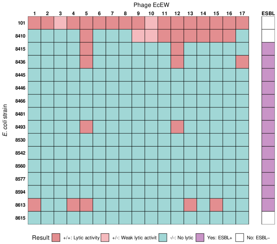
Figure 5.
Plaque morphology of E. coli phages EcEW5 (A) and EcEW12 (B). Lysis plaques were obtained on LBA plates inoculated with the E. coli clinical strain 8410, after an overnight incubation at 37°C. Details of each type of plaque are shown. In both cases, the plaques are clear, but halos can also be seen around the EcEW12 plaques.
Figure 5.
Plaque morphology of E. coli phages EcEW5 (A) and EcEW12 (B). Lysis plaques were obtained on LBA plates inoculated with the E. coli clinical strain 8410, after an overnight incubation at 37°C. Details of each type of plaque are shown. In both cases, the plaques are clear, but halos can also be seen around the EcEW12 plaques.
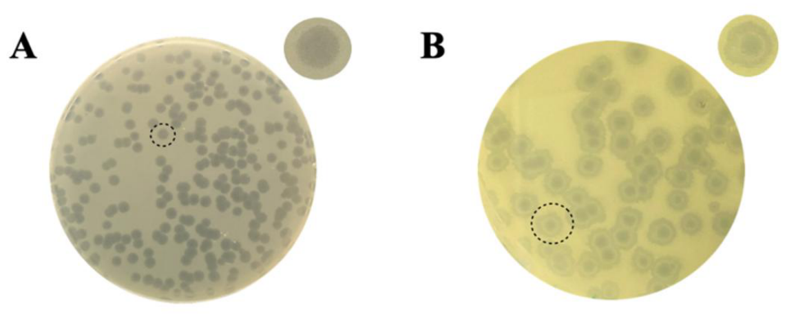
Figure 6.
Phage-mediated control of E. coli with phages EcEW5 and EcEW12 in LB broth at two bacterial concentrations. Data from co-cultures of strains 8410 (A) and 8436 (B) at 106 and 105 CFU/mL and phages EcEW5 (A) and EcEW12 (B), respectively, at 108 PFU/mL for 16 h at 37 °C. Each point represents the mean value from a triplicate experiment, and error bars indicate standard deviation. Bacterial population reductions were observed as decreases in OD600nm. Brown-Forsythe ANOVA and Dunnett's post hoc T3 analysis, p<0.05, for OD600 nm values in presence and absence of phage at two bacterial concentrations.
Figure 6.
Phage-mediated control of E. coli with phages EcEW5 and EcEW12 in LB broth at two bacterial concentrations. Data from co-cultures of strains 8410 (A) and 8436 (B) at 106 and 105 CFU/mL and phages EcEW5 (A) and EcEW12 (B), respectively, at 108 PFU/mL for 16 h at 37 °C. Each point represents the mean value from a triplicate experiment, and error bars indicate standard deviation. Bacterial population reductions were observed as decreases in OD600nm. Brown-Forsythe ANOVA and Dunnett's post hoc T3 analysis, p<0.05, for OD600 nm values in presence and absence of phage at two bacterial concentrations.
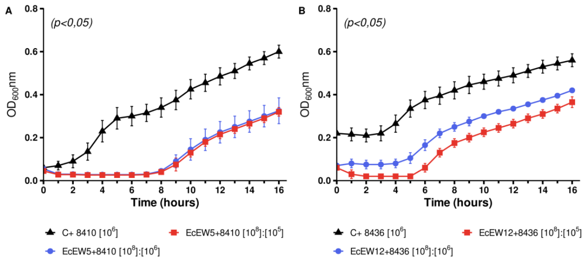
Figure 7.
Phage-mediated control of E. coli with phages EcEW5 and EcEW12 in LB broth with different bacterial strains. Data from co-cultures of strains 8410 (A), 8436 (B), 8493 (C), and 8613 (D) at 105 CFU/mL and phages EcEW5 and EcEW12 separately at 108 PFU/mL for 16 h at 37 °C. Each point represents the mean value from a triplicate experiment, and error bars indicate standard deviation. Bacterial population reductions in the presence of phages compared to bacteria controls without phages were observed as decreases in OD600 nm. Brown-Forsythe ANOVA and Dunnett's post hoc T3 analysis, p<0.05, for OD600 nm values of 8410: phage EcEW5 (A) and 8436: phage EcEW12 (B).
Figure 7.
Phage-mediated control of E. coli with phages EcEW5 and EcEW12 in LB broth with different bacterial strains. Data from co-cultures of strains 8410 (A), 8436 (B), 8493 (C), and 8613 (D) at 105 CFU/mL and phages EcEW5 and EcEW12 separately at 108 PFU/mL for 16 h at 37 °C. Each point represents the mean value from a triplicate experiment, and error bars indicate standard deviation. Bacterial population reductions in the presence of phages compared to bacteria controls without phages were observed as decreases in OD600 nm. Brown-Forsythe ANOVA and Dunnett's post hoc T3 analysis, p<0.05, for OD600 nm values of 8410: phage EcEW5 (A) and 8436: phage EcEW12 (B).
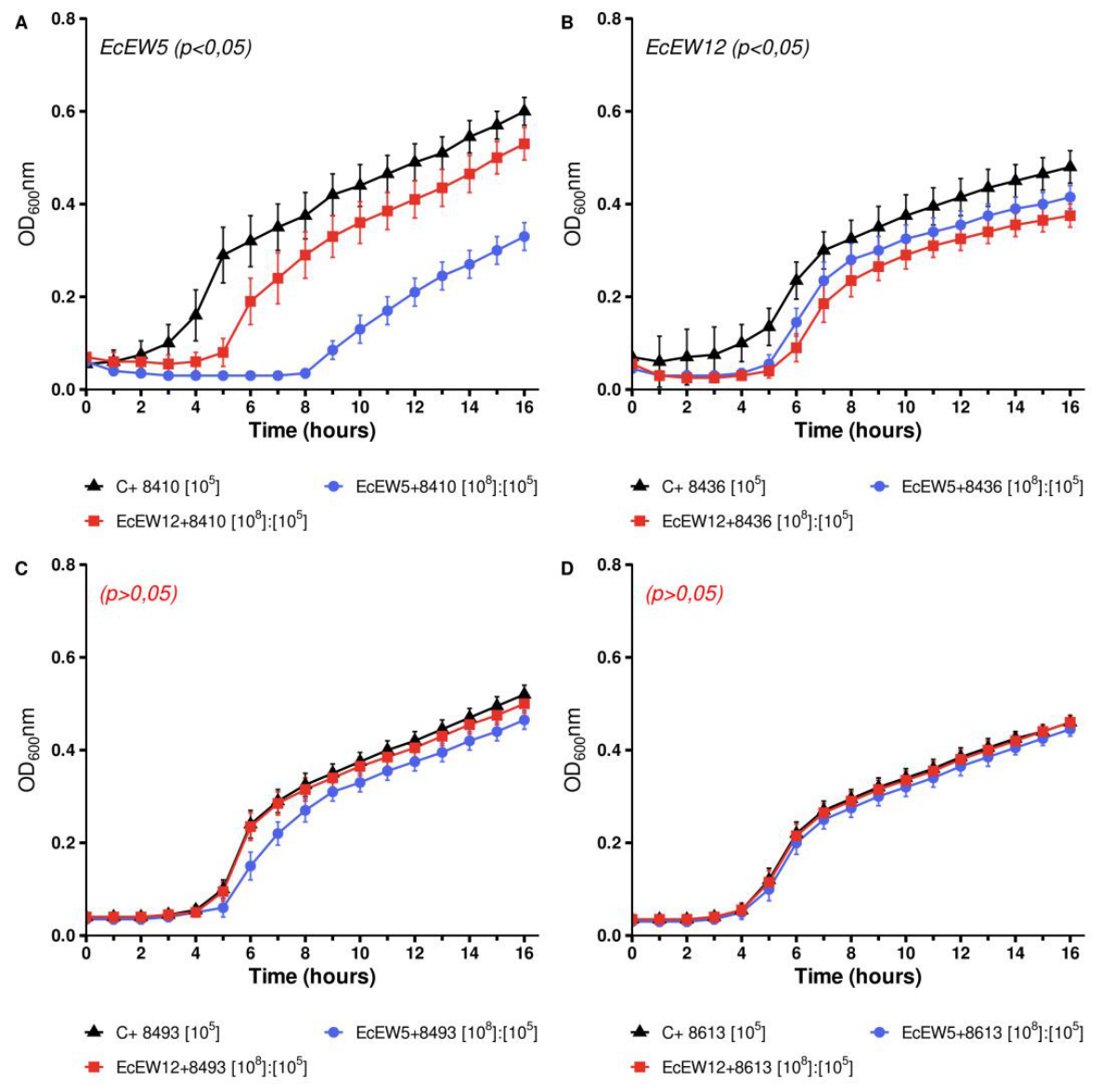
Figure 8.
Phage-mediated control of E. coli with phages EcEW5 and EcEW12 in LB broth with and without agitation (aeration). Co-cultures of strains 8410 (A) and 8436 (B) at 105 CFU/mL and phages EcEW5 and EcEW12 separately at 108 PFU/mL for 16 h at 37 °C. Each point represents the mean value from a triplicate experiment, and error bars indicate standard deviation. Bacterial population reductions were observed as decreases in OD600 nm. Brown-Forsythe ANOVA and Dunnett's post hoc T3 analysis, p<0.05, with and without agitation. Agit.: agitation, NoAgit.: absence of agitation.
Figure 8.
Phage-mediated control of E. coli with phages EcEW5 and EcEW12 in LB broth with and without agitation (aeration). Co-cultures of strains 8410 (A) and 8436 (B) at 105 CFU/mL and phages EcEW5 and EcEW12 separately at 108 PFU/mL for 16 h at 37 °C. Each point represents the mean value from a triplicate experiment, and error bars indicate standard deviation. Bacterial population reductions were observed as decreases in OD600 nm. Brown-Forsythe ANOVA and Dunnett's post hoc T3 analysis, p<0.05, with and without agitation. Agit.: agitation, NoAgit.: absence of agitation.
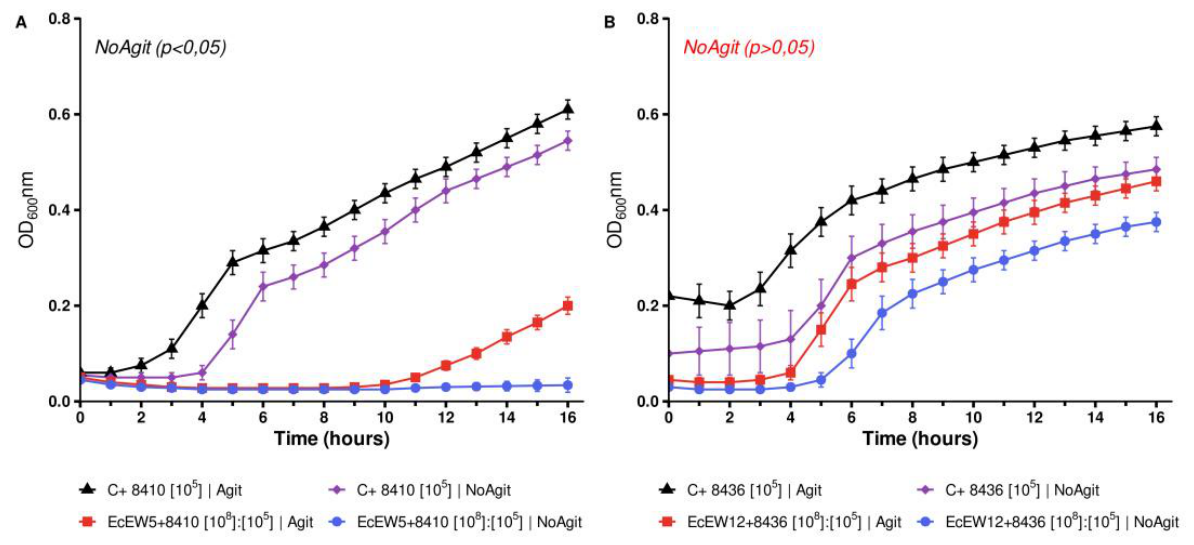
Figure 9.
Phage-mediated control of E. coli with phages EcEW5 and EcEW12 in filtered human urine. Co-cultures of strains 8410 (A) and 8436 (B) at 105 CFU/mL and the phages separately at 108 PFU/mL for 8 h at 37 °C in filtered human urine. Each point represents the mean value of an experiment performed in triplicate, with error bars indicating standard deviation. Bacterial population reductions were observed as decreases in OD600nm values. Brown-Forsythe ANOVA and Dunnett's post hoc T3 analysis (p>0.05).
Figure 9.
Phage-mediated control of E. coli with phages EcEW5 and EcEW12 in filtered human urine. Co-cultures of strains 8410 (A) and 8436 (B) at 105 CFU/mL and the phages separately at 108 PFU/mL for 8 h at 37 °C in filtered human urine. Each point represents the mean value of an experiment performed in triplicate, with error bars indicating standard deviation. Bacterial population reductions were observed as decreases in OD600nm values. Brown-Forsythe ANOVA and Dunnett's post hoc T3 analysis (p>0.05).
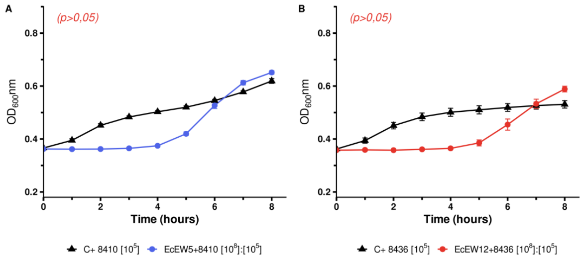
Figure 10.
Stability of E. coli phages EcEW5 and EcEW12 at different temperatures (A) and pH levels (B) in SM buffer over four weeks. The stability results are shown as the log of PFU/mL and correspond to the mean of an assay performed in duplicate with triplicate counts. Bars indicate standard deviation. Asterisks (*) indicate significant differences compared to samples stored at 4 °C (A) and at pH 7.2 (B).
Figure 10.
Stability of E. coli phages EcEW5 and EcEW12 at different temperatures (A) and pH levels (B) in SM buffer over four weeks. The stability results are shown as the log of PFU/mL and correspond to the mean of an assay performed in duplicate with triplicate counts. Bars indicate standard deviation. Asterisks (*) indicate significant differences compared to samples stored at 4 °C (A) and at pH 7.2 (B).
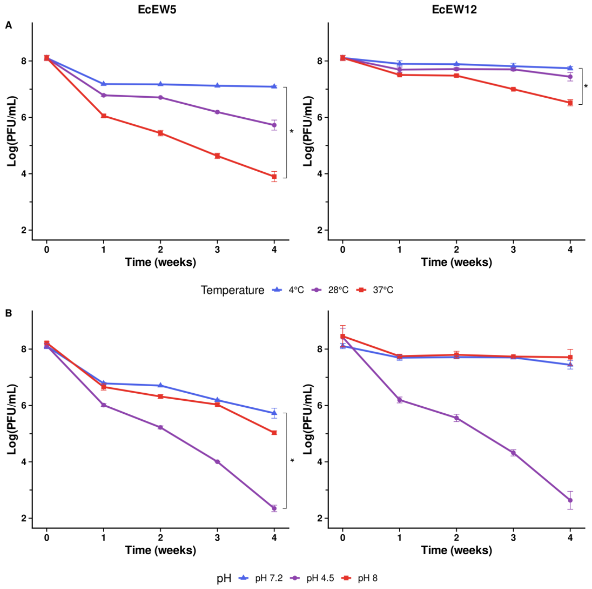
Figure 11.
Transmission electron micrographs of E. coli phages. Representative images of the virions of phages EcEW5 (A) and EcEW12 (B) after negative staining. Scale bars, 100 nm.
Figure 11.
Transmission electron micrographs of E. coli phages. Representative images of the virions of phages EcEW5 (A) and EcEW12 (B) after negative staining. Scale bars, 100 nm.
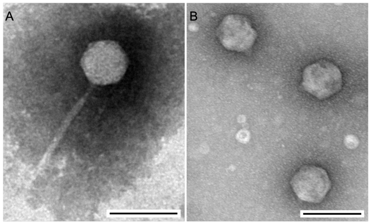
Figure 12.
Genome comparison of phage EcEW5 (A) and EcEW12 (B) with their closest phages. The coloured bars below the ORFs indicate the predicted functional categories of the encoded proteins, as defined in the corresponding legend. Grey lines connecting ORFs in different genomes represent relationships between homologous protein groups identified through MMseqs2-based protein clustering. The genomes labelled with (−) have been reverse-complemented for visualisation, in order to place homologous regions and genomic features in a consistent orientation and facilitate comparison with the reference genome. The labels (+) indicate that the original orientation of the sequence was retained.
Figure 12.
Genome comparison of phage EcEW5 (A) and EcEW12 (B) with their closest phages. The coloured bars below the ORFs indicate the predicted functional categories of the encoded proteins, as defined in the corresponding legend. Grey lines connecting ORFs in different genomes represent relationships between homologous protein groups identified through MMseqs2-based protein clustering. The genomes labelled with (−) have been reverse-complemented for visualisation, in order to place homologous regions and genomic features in a consistent orientation and facilitate comparison with the reference genome. The labels (+) indicate that the original orientation of the sequence was retained.
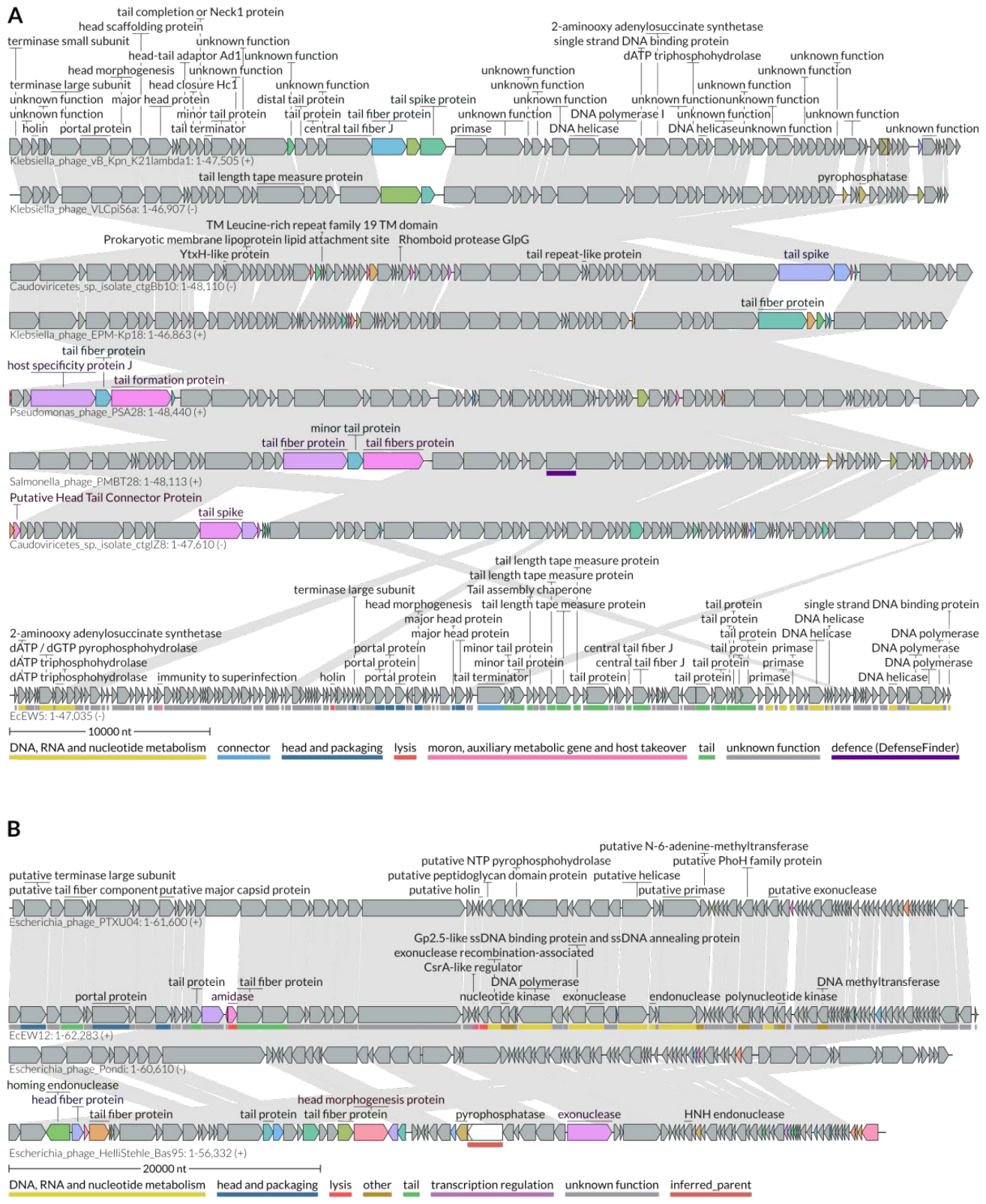
Figure 13.
Heatmaps showing pairwise intergenomic similarities between phages EcEW5 (A) and EcEW12 (B) and their closest phages, calculated with VIRIDIC using complete genome sequences. The upper triangle displays the intergenomic similarity values, expressed as percentages, with darker blue–green colours indicating higher similarity. The lower triangle reports three alignment metrics for each genome pair, shown from top to bottom: the fraction of the genome represented by the row that is aligned, the ratio of the lengths of the two genomes, and the fraction of the genome represented by the column that is aligned. The bar plots above each heatmap show the genome lengths of the analysed phages.
Figure 13.
Heatmaps showing pairwise intergenomic similarities between phages EcEW5 (A) and EcEW12 (B) and their closest phages, calculated with VIRIDIC using complete genome sequences. The upper triangle displays the intergenomic similarity values, expressed as percentages, with darker blue–green colours indicating higher similarity. The lower triangle reports three alignment metrics for each genome pair, shown from top to bottom: the fraction of the genome represented by the row that is aligned, the ratio of the lengths of the two genomes, and the fraction of the genome represented by the column that is aligned. The bar plots above each heatmap show the genome lengths of the analysed phages.
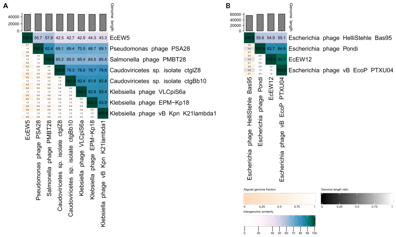
Figure 14.
ViPTree proteomic analysis of the phages EcEW5 (A) and EcEW12 (B). The trees were generated by comparing the complete phage genomes with selected prokaryotic dsDNA phage reference genomes using genome-wide tBLASTx similarities. Normalized genomic similarity scores (SG) were converted into genomic distances (1−SG) and used to construct the proteomic trees with the BIONJ algorithm. Branch lengths represent genomic distance; therefore, shorter distances indicate greater overall genome-wide similarity. The gray stars indicate phages showing the highest similarity to EcEW5 or EcEW12, respectively, as identified by BLAST analysis, whereas red stars identify the query phages EcEW5 and EcEW12.
Figure 14.
ViPTree proteomic analysis of the phages EcEW5 (A) and EcEW12 (B). The trees were generated by comparing the complete phage genomes with selected prokaryotic dsDNA phage reference genomes using genome-wide tBLASTx similarities. Normalized genomic similarity scores (SG) were converted into genomic distances (1−SG) and used to construct the proteomic trees with the BIONJ algorithm. Branch lengths represent genomic distance; therefore, shorter distances indicate greater overall genome-wide similarity. The gray stars indicate phages showing the highest similarity to EcEW5 or EcEW12, respectively, as identified by BLAST analysis, whereas red stars identify the query phages EcEW5 and EcEW12.
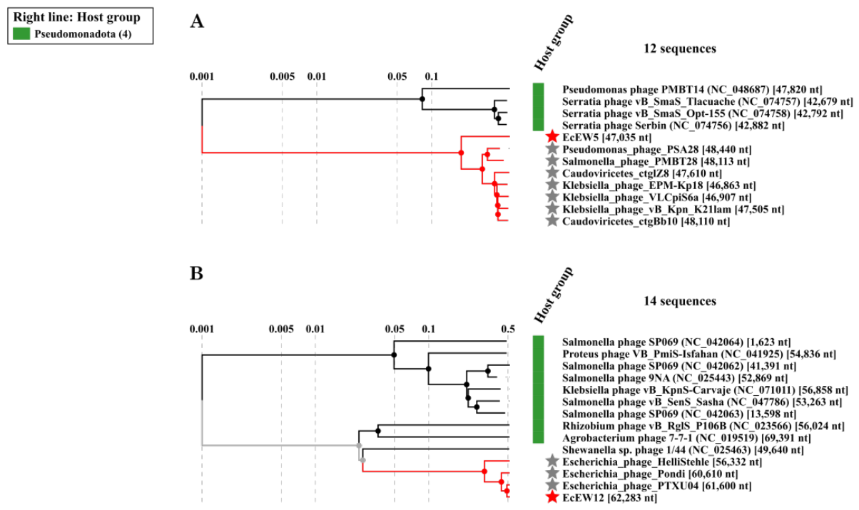

Table 1.
Genomic features of water-borne E. coli phages.
| Phage | Genome size (bp) |
GC (%) | GenBank accession |
CDSs | Hypothetical proteins |
|---|---|---|---|---|---|
| EcEW5 | 47,035 | 57.49 | PZ787841 | 144 | 104 |
| EcEW12 | 62,283 | 52.88 | PZ793367 | 98 | 73 |
Disclaimer/Publisher’s Note: The statements, opinions and data contained in all publications are solely those of the individual author(s) and contributor(s) and not of MDPI and/or the editor(s). MDPI and/or the editor(s) disclaim responsibility for any injury to people or property resulting from any ideas, methods, instructions or products referred to in the content. |
© 2026 by the authors. Licensee MDPI, Basel, Switzerland. This article is an open access article distributed under the terms and conditions of the Creative Commons Attribution (CC BY) license (http://creativecommons.org/licenses/by/4.0/).
Copyright: This open access article is published under a Creative Commons CC BY 4.0 license, which permit the free download, distribution, and reuse, provided that the author and preprint are cited in any reuse.