

Submitted:
10 September 2026
Posted:
11 September 2026
You are already at the latest version
Abstract
Background: Neutrophils play an essential role in pathogen clearance and can also contribute to pathogenesis and inflammation. Phenotypic changes of neutrophils during infection or inflammation may be associated with their functional activity. However, investigation of neutrophil phenotypes and functions in clinical studies is challenging because of their short viability ex-vivo and difficulties in effectively cryopreserving neutrophils for subsequent analyses. Methods: We isolated neutrophils from human peripheral blood and evaluated different approaches for cryopreservation. We examined labelling of cell surface receptors of interest, including CD64, CD32, CD16, CD54, CD66b, CD62L and CD14, on fresh versus cryopreserved neutrophils by flow cytometry. Trypan blue staining was used to assess cell membrane integrity. Results: We developed a neutrophil cryopreservation protocol that preserved membrane integrity and enabled subsequent investigation of phenotypes. All cell surface receptors tested could be labelled and quantified using flow cytometry. There were some differences in levels of surface staining for some receptors comparing fresh and cryopreserved cells, but these differences did not prevent the evaluation of neutrophil phenotypes. Discussion: Overall, data suggests that our neutrophil cryopreserving and thawing protocols can preserve neutrophil surface receptors relevant to functions and phenotypes and are methods suitable for use in clinical studies.
Keywords:
neutrophils
; phenotypes
; immunity
; pathogenesis
; phagocytosis
; flow cytometry
1. Introduction
Neutrophils are a group of immune cells with varying phenotypes and functions [1,2,3,4]. They are among the first line of defense during infection and have a role in modulating immune responses [5,6,7]. Neutrophils possess various effector mechanisms associated with protective immunity against pathogens. These include antibody and non-antibody mediated phagocytosis [8,9,10,11,12,13,14,15], cytokine, chemokine and granules release, reactive oxygen species (ROS) production, and neutrophil extracellular traps (NETs) formation [2,16,17,18].
Studying neutrophils is important for understanding development of disease and immunity during acute infection since they are among the early cellular immune responses. One way to investigate disease progression and immune responses is by examining and quantifying neutrophil receptor changes [19]. Studies investigating neutrophils have demonstrated that changes in surface receptor expression during inflammation and/or infection may be correlated with enhanced effector functional activity [20,21]. Surface receptors relevant to protective immunity, also referred to as ‘activation receptors’, include Fcγ receptors (FcγR) FcγRI (CD64), FcγRIIa (CD32a) and FcγRIII (CD16) [2,22,23] involved in phagocytosis and cellular cytotoxicity; and adhesive and migratory molecules CD66b (Carcinoembryonic antigen-related cell adhesion molecule 8 (CEACAM8)), CD54 (intercellular adhesion molecule 1 (ICAM-1)) and CD62L (an L-selectin or SELL) [7,19,24,25,26,27].
Investigation of phenotypic changes on neutrophils requires quantifying receptors and surface proteins by flow cytometry. In many clinical studies, access to flow cytometric analysis on freshly collected neutrophils may not be possible due to lack of access to suitable facilities, such as occurs in many clinical settings, community-based studies, and low-income or resource-constrained settings. Therefore, to investigate phenotypes, isolation of neutrophils from peripheral blood samples and cryopreservation for storage and/or shipment is necessary. Development of a granulocyte cryopreserving method has been challenging over the years due to cell fragility, short viability ex vivo and post-thaw reduced effector function [28,29]. Nevertheless, fixed leukocytes can be cryopreserved enabling post-thaw differentiation of cell population and measurement of activation receptors despite receptors despite post-fixation morphological changes [30,31].
Although some methods for immunophenotyping neutrophils in cryopreserved whole blood samples exist, neutrophil phenotyping remains a challenge [30,32]. Limitations of using cryopreserved whole blood for neutrophil immunophenotyping include alteration in neutrophil size and granularity that promote overlap with other leukocytes as well as reduced or lost antibody recognition of surface receptors due to cryopreservation-induced changes in receptor configuration [30,32]. This underscores the necessity to use purified cells when assessing immunophenotypic changes. However, limited data exists on purified cryopreserved neutrophils immunophenotyping, likely because purification and freeze-thaw steps may activate myeloid cells and alter their surface marker expression [33,34]. This restricts the capacity to study neutrophils in many clinical settings.
To address this gap, we developed a cryopreservation and thawing method that would allow us to investigate purified neutrophil phenotypes after cryopreservation from areas where no flow cytometer is available on site. Receptors of interest in our studies included the expression of FcγRs (CD64, CD32 and CD16), and receptors CD66b, CD54 and CD62L since changes in expression of these receptors are associated with an immune response during infection and/or inflammation.
2. Methods
Ethical Approval and Consent to Participate
Research involving human participants, human material, or human data, was performed in accordance with the Declaration of Helsinki. Ethics approval for collection of blood to isolate neutrophils was provided by the Human Research and Ethics Committee of Alfred Health, Australia (Protocol number HREC/497/11, date: 2012.01.30). This study was not a clinical trial. All participants gave written informed consent.
Consent to Publish Declarations
Consent to publish declarations not applicable.
Neutrophil Isolation, Cryopreservation and Thawing
The step-by-step protocol of the method is provided in the supplementary file and is published on protocols.io. DOI: dx.doi.org/10.17504/protocols.io.4r3l21723g1y/v1 (Private link for reviewers: https://www.protocols.io/private/3FB7649C735611F099340A58A9FEAC02 to be removed before publication).
Briefly, peripheral blood samples were collected from healthy adult residents in Melbourne, Australia. Whole blood was collected from 11 consenting adult Melbourne resident donors in Australia, age range 22 years – 53 years. The blood was collected in lithium heparin vacutainers (BD Vacutainer LH 170 I.U (REF 367526)) between 9 a.m. and 10 a.m. Neutrophils were isolated within 2 hours of whole blood collection, and were subsequently used fresh, or cryopreserved and then thawed, for analysis of neutrophil phenotypes. Phenotyping data was acquired using BD® LSR II flow cytometer machine.
Gating Analyses
Flow cytometry data percentages, geometric mean fluorescence Intensity (MFI), stain index (SI), and dot plots were generated using FlowJo_v10.8.1 and FlowJo_v10.10.0_CL. We gated cells on SSC-A vs FSC-A as phagocytes. Next, doublets were gated out by FSC-H vs FSC-A, FSC width (FSC-W) vs FSC-A, SSC height (SSC-H) vs SSC width (SSC-W) (Figure 1).
Optimal antibody working concentrations were ascertained by titration of staining antibodies on freshly isolated neutrophils and a subset of cryopreserved neutrophils from freshly collected cells from the same donor. Fresh cells were fixed with 2% Paraformaldehyde (PFA) after staining, while cryopreserved cells were fixed prior to staining. Positive and negative populations were gated using histograms and antibody titrations quantified by SI. We finalized our antibody panel by concatenating the serial titrations and choosing the most ideal single antibody dilutions based on the dilution that gives a good separation of positive and negative populations. The optimal antibody concentrations are highlighted in red (Figure 2). We used BD® LSR II flow cytometer machine throughout these studies to reduce variation across experiments. Ten thousand events were acquired per sample on the SSC-H vs SSC-W.
Descriptive statistics were used to report study findings, including percent of cells positive in flow cytometry for specified gating and geometric mean fluorescence intensity (MFI), and are indicated for each figure.
3. Results
Neutrophil Cryopreservation Protocol Validation
Several cryopreserving and thawing conditions were tested (see full list in Table 1 and 2, and highlights are shown in Figure S1a - c. Trypan blue exclusion indicated high cell membrane integrity when neutrophils were cryopreserved in 10% BSA + 7.5% DMSO in RPMI 1640 and then thawed and left resuspended in 10% BSA HBSS (Table 1 and 2 and Figure S1b). We compared viability based on membrane integrity of fresh and cryopreserved neutrophils resuspended in 10% BSA HBSS. We found that viability based on membrane integrity for fresh cells ranged between 98% – 99.9% (Figure S1d), while cryopreserved cell viability ranged between 90 – 99.9% (Figure S1b). Cryopreserved neutrophil viability appeared to depend to some extent on neutrophil to suspension buffer (10% BSA HBSS) ratios after thawing. Specifically, pellet with cell count approximately 1x10^7 cells/ml showed high cell membrane integrity when resuspended in 500 µl buffer. Cells with concentration more than 1 x 10^7 cells/ml required resuspension in medium range between 600 µl – 1000 µl. Larger neutrophil pellets after thawing may require extra buffer to maximise cell viability based on membrane integrity.
Forty-five minutes to an hour after thawing, neutrophil membrane integrity appeared compromised. Because cell integrity deteriorated after 45-60 minutes following thawing, cells were immediately fixed with 2% PFA final concentration. In contrast, fresh neutrophils were fixed after staining, which is the standard procedure used. Based on these studies, a step-by-step protocol was developed, which is provided in the Supplementary file.
Neutrophil Surface Receptors Are Preserved After Cryopreservation
We investigated whether neutrophil structural changes may occur following cryopreservation. We compared fresh and cryopreserved neutrophils. The fresh and cryopreserved neutrophils were obtained from the same Melbourne donor and isolated on the same day. We observed differences in the forward scatter area (FSC-A) versus side scatter area (SSC-A) between fresh and cryopreserved neutrophils during flow cytometry cell acquisition, suggesting some changes in size and granularity (Figure 1). For that reason, we acquired fresh and cryopreserved neutrophil flow cytometry data using different FSC-A and SSC-A voltages.
Neutrophils were gated on CD16 versus CD66b. We observed minor differences in fluorescence intensity distribution between cryopreserved and fresh neutrophils. Specifically, a small population of CD16dimCD66bdim neutrophils was detectable in the fresh cell preparation but absent following cryopreservation.
We then examined whether a range of cell surface receptors of interest including CD64, CD32, CD16, CD54, CD66b, CD62L and CD14 were preserved on cryopreserved neutrophils. These surface receptors were selected on the basis that they are important in neutrophil functions relevant to immunity to infectious pathogens. We compared the optimized staining of our target receptors on fresh neutrophils versus cryopreserved neutrophils from the same donor isolated on the same day. BD CompBead Anti-Mouse Ig, κ/Negative Control Particles were used to ensure signal accuracy. Our data shows that all markers detected on fresh cells were also detectable on cryopreserved neutrophils (Figure 3). The proportions of cells expressing CD64 and CD14 were somewhat higher in cryopreserved cells compared to fresh cells, but the cell surface molecules could be clearly labelled in fresh and cryopreserved neutrophils (Figure 3).
We next tested antibody staining with combinations of cell surface molecules on fresh and cryopreserved neutrophils. We gated our target cells on high FSC-A and high SSC-A. Next, doublets were excluded out by FSC-H vs FSC-A, FSC-W vs FSC-A, SSC-H vs SSC-W gates (Figure 1), neutrophils were gated on CD16 vs CD66b (Figure 4). The spread of cell surface fluorescence intensity was slightly different for cryopreserved versus fresh neutrophils, but the differences in percent of cells in the gate were small (Figure 4 panels A and B). To determine the effect of cryopreservation on receptor level expression, we tested neutrophils from two donors in technical replicates quantified by Median Fluorescence Intensity (MFI). We observed an upward trend in CD32 MFI and CD62L MFI after cryopreservation (Figure 5a - b), while CD14, CD16, CD54, and CD66b (Figure 5c - f), showed a downward trend. The changes in MFIs in frozen neutrophils were consistent across the donors. MFIs for CD64 (Figure 5g) remained unchanged between fresh and cryopreserved neutrophils, suggesting that cryopreservation did not affect receptor levels.
Overall, our data suggest that our neutrophil cryopreserving and thawing method can preserve neutrophil surface receptors and that receptors and surface proteins relevant to neutrophil functions and phenotypes can be detected. Although there were differences between cryopreserved and fresh neutrophils, the differences did not impact the neutrophil phenotype outcomes.
4. Discussion
We successfully developed and characterized neutrophil phenotypes preserved using our novel cryopreservation method. Our flow analysis revealed that the cryopreserved neutrophils could be stained, and the surface receptors distinguished. The surface receptors detected on fresh neutrophils, including FcγRs (CD16, CD32a, CD64), CD54, CD62L, CD66b and CD14 were also detectable on cryopreserved neutrophils. Differences were observed in the FSC-A versus SSC-A profiles between cryopreserved and fresh neutrophils, suggesting that cryopreservation, fixation, or both may alter cell size and granularity [30]. Additionally, the staining intensity of certain receptors varied between cryopreserved and fresh cells.
Since neutrophils are among the first immune cell responders during an infection, profiling their responses is crucial in understanding pathogenesis and immunity. Neutrophils have a short lifespan ex vivo [35], thereby requiring immediate investigation. This presents practical challenges in clinical research as assays can be laborious and time-consuming meaning that only small sample sizes can be investigated. In addition, studies in settings with limited laboratory facilities or other resource constraints may require samples to be shipped to a suitable laboratory for analysis of immune responses using neutrophil profiling. Therefore, developing a neutrophil cryopreservation method that can preserve cell structure, and phenotypes would greatly facilitate investigating neutrophil phenotypes in diverse clinical studies and populations.
Using our novel neutrophil cryopreservation method, we were still able to identify neutrophils by gating on CD16 and CD66b co-expression. There was a loss of a small subset CD16dimCD66bdim neutrophil population after cryopreservation. Alterations in neutrophil size and granularity may have increased overlap between dim and bright populations, making the dim subset appear absent [30]. Alternatively, loss of the dim population may reflect epitope masking due to receptor conformational changes causing reduced immunofluorescent detectability [34]. Additionally, the absence of dim cells in the cryopreserved cell population suggests that this subset may be preferentially lost during freezing and thawing [36,37].
All our target receptors (FcγRs, CD54, CD62L, CD66b) of interest were detectable on cryopreserved neutrophils. However, the frequency of cells expressing CD64 and CD14 receptors differed between cryopreserved and fresh cells. Non activated neutrophils in a homeostatic state are thought to express low levels of CD64 and CD14 on the surface, with the majority residing intracellularly [38,39,40]. Fixation of cells with PFA may cause cell membrane permeabilization [30,40,41]. Therefore, fixation of the cryopreserved cells before staining may have enabled some intracellular staining, thereby increasing the proportion of cells with detectable fluorescence signal compared with fresh neutrophils, which were fixed after staining. This could be considered when applying this method in future studies. CD64 expression on neutrophils may be upregulated when stimulated with inflammatory cytokines such as granulocyte colony-stimulating factor (G-CSF) and interferon gamma (IFN-γ) in vitro and in vivo [19]. CD14 can upregulate with stimulation such as lipopolysaccharide (LPS) an endotoxin [40].
Staining intensity of receptors CD14, CD16, CD54 and CD66b was reduced in cryopreserved neutrophils across biological replicates suggesting alterations (such as shedding of receptors) due to the thawing process may have occurred. There is limited data suggesting that CD32a resides intracellularly on human neutrophils in homeostatic state [2,22,38]. Therefore, it remains possible that the increase in CD32a staining intensity may have been caused by changes in membrane exposure of CD32a or potential activation of neutrophils during freezing or thawing steps [42], although we do not have evidence that this occurred. The trend toward increased CD62L MFI suggests that immediate fixation after thawing may have reduced receptor shedding; however, fixation-induced membrane permeabilization, or membrane compromise due to the freeze-thaw process, may have permitted antibody access to intracellular CD62L [27]. Despite the differences between fresh and cryopreserved neutrophils, our findings support using cryopreserved cells for phenotyping studies of neutrophils. While all the surface receptors we studied were detected on neutrophils, other relevant surface molecules should be assessed in future studies, including applications where rare or infrequent cell phenotypes are being studied. Given the observed differences in surface molecule labelling between fresh and cryopreserved neutrophils, studies involving phenotypic comparisons between subjects or groups should ensure that neutrophil samples are processed and stored consistently.
In summary, our cryopreservation and thawing method maintained neutrophil receptor expression and appeared to be suitable for analysis of neutrophil phenotypes in clinical studies.
Supplementary Materials
The following supporting information can be downloaded at the website of this paper posted on Preprints.org.
Author Contributions
S.C and J.G.B. led the conceptualization and design of the study with input from S.J.R., and R.G. S.C. conducted experiments and S.C and R.G interpreted the results; S.C. conducted data analysis with input from J.G.B., R.G., and S.J.R. S.C and J.G.B. contributed to paper preparation with input from all other co-authors. All authors approved the final manuscript.
Funding
Funding was provided by the National Health and Medical Research Council of Australia (Investigator Grant 2033320 to JGB and Infrastructure Support Scheme to Burnet Institute), an Operational Infrastructure Grant from the State Government of Victoria (to Burnet Institute) ID 2033320, Melbourne Research Scholarship (PhD) Ref: 351091, The University of Melbourne and the Mary Lugton Scholarship fund (PhD) Ref: 230818-01093826.
Data Availability Statement
Data generated in this study is available from the authors on reasonable request. All reagents used in this study are commercially available.
Acknowledgments
We would like to thank the blood donors who made this work possible. We would also like to thank ARAFLOWCORE and AMREPFLOW core facilities especially Ms Eva Orlowski-Oliver for the assistance in designing the antibody staining panel. Additionally, we would like to thank Dr Gaoqian Feng, Dr Salimeh Ebrahimnezhaddarzi, and Dr Annemarie Laumaea for their valuable advice during the development of this protocol. We acknowledge the Boon Wurrung and Wurunjeri people as the traditional custodians of the land where this work was performed.
Conflicts of Interest
The authors declare that they have no competing interests.
References
- Hassani, M.; Hellebrekers, P.; Chen, N.; van Aalst, C.; Bongers, S.; Hietbrink, F.; et al. On the origin of low-density neutrophils. J. Leukoc. Biol. 2020, 107(5), 809–18. [Google Scholar] [CrossRef] [PubMed]
- Wang, Y.; Jönsson, F. Expression, Role, and Regulation of Neutrophil Fcγ Receptors. Front Immunol. 2019, 10, 1958. [Google Scholar] [CrossRef] [PubMed]
- McKenna, E.; Mhaonaigh, A.U.; Wubben, R.; Dwivedi, A.; Hurley, T.; Kelly, L.A.; et al. Neutrophils: Need for Standardized Nomenclature. Front Immunol. 2021, 12, 602963. [Google Scholar] [CrossRef] [PubMed]
- Pillay, J.; Tak, T.; Kamp, V.M.; Koenderman, L. Immune suppression by neutrophils and granulocytic myeloid-derived suppressor cells: similarities and differences. Cell Mol. Life Sci. 2013, 70(20), 3813–27. [Google Scholar] [CrossRef] [PubMed]
- Silvestre-Roig, C.; Hidalgo, A.; Soehnlein, O. Neutrophil heterogeneity: implications for homeostasis and pathogenesis. Blood 2016, 127(18), 2173–81. [Google Scholar] [CrossRef] [PubMed]
- Mortaz, E.; Alipoor, S.D.; Adcock, I.M.; Mumby, S.; Koenderman, L. Update on Neutrophil Function in Severe Inflammation. Front Immunol. 2018, 9, 2171. [Google Scholar] [CrossRef] [PubMed]
- Rosales, C. Neutrophil: A Cell with Many Roles in Inflammation or Several Cell Types? Front Physiol. 2018, 9, 113. [Google Scholar] [CrossRef] [PubMed]
- Aitken, E.H.; Damelang, T.; Ortega-Pajares, A.; Alemu, A.; Hasang, W.; Dini, S.; et al. Developing a multivariate prediction model of antibody features associated with protection of malaria-infected pregnant women from placental malaria. Elife 2021, 10. [Google Scholar] [CrossRef] [PubMed]
- Dasari, P.; Reiss, K.; Lingelbach, K.; Baumeister, S.; Lucius, R.; Udomsangpetch, R.; et al. Digestive vacuoles of Plasmodium falciparum are selectively phagocytosed by and impair killing function of polymorphonuclear leukocytes. Blood 2011, 118(18), 4946–56. [Google Scholar] [CrossRef] [PubMed]
- Meinderts, S.M.; Oldenborg, P.A.; Beuger, B.M.; Klei, T.R.L.; Johansson, J.; Kuijpers, T.W.; et al. Human and murine splenic neutrophils are potent phagocytes of IgG-opsonized red blood cells. Blood Adv. 2017, 1(14), 875–86. [Google Scholar] [CrossRef] [PubMed]
- Zelter, T.; Strahilevitz, J.; Simantov, K.; Yajuk, O.; Adams, Y.; Ramstedt Jensen, A.; et al. Neutrophils impose strong immune pressure against PfEMP1 variants implicated in cerebral malaria. EMBO Rep. 2022, 23(6), e53641. [Google Scholar] [CrossRef] [PubMed]
- Feng, G.; Wines, B.D.; Kurtovic, L.; Chan, J.A.; Boeuf, P.; Mollard, V.; et al. Mechanisms and targets of Fcγ-receptor mediated immunity to malaria sporozoites. Nat. Commun. 2021, 12(1), 1742. [Google Scholar] [CrossRef] [PubMed]
- Garcia-Senosiain, A.; Kana, I.H.; Singh, S.; Das, M.K.; Dziegiel, M.H.; Hertegonne, S.; et al. Neutrophils dominate in opsonic phagocytosis of P. falciparum blood-stage merozoites and protect against febrile malaria. Commun. Biol. 2021, 4(1), 984. [Google Scholar] [CrossRef] [PubMed]
- Kurtovic, L.; Atre, T.; Feng, G.; Wines, B.D.; Chan, J.A.; Boyle, M.J.; et al. Multifunctional Antibodies Are Induced by the RTS,S Malaria Vaccine and Associated With Protection in a Phase 1/2a Trial. J. Infect. Dis. 2021, 224(7), 1128–38. [Google Scholar] [CrossRef] [PubMed]
- Sun, T.; Chakrabarti, C. Schizonts, merozoites, and phagocytosis in falciparum malaria. Ann. Clin. Lab. Sci. 1985, 15(6), 465–9. [Google Scholar] [PubMed]
- Mayadas, T.N.; Cullere, X.; Lowell, C.A. The multifaceted functions of neutrophils. Annu Rev. Pathol. 2014, 9, 181–218. [Google Scholar] [CrossRef] [PubMed]
- Joos, C.; Marrama, L.; Polson, H.E.; Corre, S.; Diatta, A.M.; Diouf, B.; et al. Clinical protection from falciparum malaria correlates with neutrophil respiratory bursts induced by merozoites opsonized with human serum antibodies. PLoS ONE 2010, 5(3), e9871. [Google Scholar] [CrossRef] [PubMed]
- Rodrigues, D.A.S.; Prestes, E.B.; Gama, A.M.S.; Silva, L.S.; Pinheiro, A.A.S.; Ribeiro, J.M.C.; et al. CXCR4 and MIF are required for neutrophil extracellular trap release triggered by Plasmodium-infected erythrocytes. PLoS Pathog. 2020, 16(8), e1008230. [Google Scholar] [CrossRef] [PubMed]
- Davis, B.H. Improved diagnostic approaches to infection/sepsis detection. Expert Rev. Mol. Diagn. 2005, 5(2), 193–207. [Google Scholar] [CrossRef] [PubMed]
- Nagarajan, S.; Fifadara, N.H.; Selvaraj, P. Signal-specific activation and regulation of human neutrophil Fc gamma receptors. J. Immunol. 2005, 174(9), 5423–32. [Google Scholar] [CrossRef] [PubMed]
- Witko-Sarsat, V.; Rieu, P.; Descamps-Latscha, B.; Lesavre, P.; Halbwachs-Mecarelli, L. Neutrophils: molecules, functions and pathophysiological aspects. Lab. Investig. A J. Tech. Methods Pathol. 2000, 80(5), 617–53. [Google Scholar] [CrossRef] [PubMed]
- Rosales, C. Fcγ Receptor Heterogeneity in Leukocyte Functional Responses. Front Immunol. 2017, 8, 280. [Google Scholar] [CrossRef] [PubMed]
- van Rees, D.J.; Szilagyi, K.; Kuijpers, T.W.; Matlung, H.L.; van den Berg, T.K. Immunoreceptors on neutrophils. Semin Immunol. 2016, 28(2), 94–108. [Google Scholar] [CrossRef] [PubMed]
- Patil, N.K.; Guo, Y.; Luan, L.; Sherwood, E.R. Targeting Immune Cell Checkpoints during Sepsis. Int. J. Mol. Sci. 2017, 18(11). [Google Scholar] [CrossRef] [PubMed]
- Davis, R.E.; Sharma, S.; Conceição, J.; Carneiro, P.; Novais, F.; Scott, P.; et al. Phenotypic and functional characteristics of HLA-DR(+) neutrophils in Brazilians with cutaneous leishmaniasis. J. Leukoc. Biol. 2017, 101(3), 739–49. [Google Scholar] [CrossRef] [PubMed]
- Silvestre-Roig, C.; Fridlender, Z.G.; Glogauer, M.; Scapini, P. Neutrophil Diversity in Health and Disease. Trends Immunol. 2019, 40(7), 565–83. [Google Scholar] [CrossRef] [PubMed]
- Ivetic, A. A head-to-tail view of L-selectin and its impact on neutrophil behaviour. Cell Tissue Res. 2018, 371(3), 437–53. [Google Scholar] [CrossRef] [PubMed]
- Vian, A.M.; Higgins, A.Z. Membrane permeability of the human granulocyte to water, dimethyl sulfoxide, glycerol, propylene glycol and ethylene glycol. Cryobiology 2014, 68(1), 35–42. [Google Scholar] [CrossRef] [PubMed]
- Graham-Pole, J.; Davie, M.; Willoughby, M.L. Cryopreservation of human granulocytes in liquid nitrogen. J. Clin. Pathol. 1977, 30(8), 758–62. [Google Scholar] [CrossRef] [PubMed]
- de Ruiter, K.; van Staveren, S.; Hilvering, B.; Knol, E.; Vrisekoop, N.; Koenderman, L.; et al. A field-applicable method for flow cytometric analysis of granulocyte activation: Cryopreservation of fixed granulocytes. Cytom. A 2018, 93(5), 540–7. [Google Scholar] [CrossRef] [PubMed]
- Nemes, E.; Kagina, B.M.; Smit, E.; Africa, H.; Steyn, M.; Hanekom, W.A.; et al. Differential leukocyte counting and immunophenotyping in cryopreserved ex vivo whole blood. Cytom. A 2015, 87(2), 157–65. [Google Scholar] [CrossRef] [PubMed]
- Nguyen, N.A.; Huang, X.; Cabrera, L.E.; Pekkarinen, P.T.; Nowlan, K.; Strandin, T.; et al. A comprehensive assessment of four whole blood stabilizers for flow-cytometric analysis of leukocyte populations. Cytom. A 2023, 103(4), 313–24. [Google Scholar] [CrossRef] [PubMed]
- Blanter, M.; Cambier, S.; De Bondt, M.; Vanbrabant, L.; Pörtner, N.; Abouelasrar Salama, S.; et al. Method Matters: Effect of Purification Technology on Neutrophil Phenotype and Function. Front Immunol. 2022, 13, 820058. [Google Scholar] [CrossRef] [PubMed]
- Kadić, E.; Moniz, R.J.; Huo, Y.; Chi, A.; Kariv, I. Effect of cryopreservation on delineation of immune cell subpopulations in tumor specimens as determinated by multiparametric single cell mass cytometry analysis. BMC Immunol. 2017, 18(1), 6. [Google Scholar] [CrossRef] [PubMed]
- Lahoz-Beneytez, J.; Elemans, M.; Zhang, Y.; Ahmed, R.; Salam, A.; Block, M.; et al. Human neutrophil kinetics: modeling of stable isotope labeling data supports short blood neutrophil half-lives. Blood 2016, 127(26), 3431–8. [Google Scholar] [CrossRef] [PubMed]
- Campbell, D.E.; Tustin, N.B.; Riedel, E.; Tustin, R., 3rd; Taylor, J.; Murray, J.; et al. Cryopreservation decreases receptor PD-1 and ligand PD-L1 coinhibitory expression on peripheral blood mononuclear cell-derived T cells and monocytes. Clin. Vaccine Immunol. 2009, 16(11), 1648–53. [Google Scholar] [CrossRef] [PubMed]
- Kotsakis, A.; Harasymczuk, M.; Schilling, B.; Georgoulias, V.; Argiris, A.; Whiteside, T.L. Myeloid-derived suppressor cell measurements in fresh and cryopreserved blood samples. J. Immunol. Methods 2012, 381(1-2), 14–22. [Google Scholar] [CrossRef] [PubMed]
- Fanger, M.W.; Shen, L.; Graziano, R.F.; Guyre, P.M. Cytotoxicity mediated by human Fc receptors for IgG. Immunol. Today 1989, 10(3), 92–9. [Google Scholar] [CrossRef] [PubMed]
- Kerst, J.M.; van de Winkel, J.G.; Evans, A.H.; de Haas, M.; Slaper-Cortenbach, I.C.; de Wit, T.P.; et al. Granulocyte colony-stimulating factor induces hFc gamma RI (CD64 antigen)-positive neutrophils via an effect on myeloid precursor cells. Blood 1993, 81(6), 1457–64. [Google Scholar] [CrossRef]
- Rodeberg, D.A.; Morris, R.E.; Babcock, G.F. Azurophilic granules of human neutrophils contain CD14. Infect. Immun. 1997, 65(11), 4747–53. [Google Scholar] [CrossRef] [PubMed]
- Cheng, R.; Zhang, F.; Li, M.; Wo, X.; Su, Y.W.; Wang, W. Influence of Fixation and Permeabilization on the Mass Density of Single Cells: A Surface Plasmon Resonance Imaging Study. Front Chem. 2019, 7, 588. [Google Scholar] [CrossRef] [PubMed]
- Belostocki, K.; Park, M.S.; Redecha, P.B.; Masuda, E.; Salmon, J.E.; Pricop, L. FcgammaRIIa is a target for modulation by TNFalpha in human neutrophils. Clin. Immunol. 2005, 117(1), 78–86. [Google Scholar] [PubMed]
Figure 1.
Flow cytometry hierarchy gating of fresh and cryopreserved neutrophils. Panel A represents fresh neutrophils; Panel B represents cryopreserved neutrophils. Fresh and cryopreserved neutrophils were isolated from the same donor on the same day. Fresh neutrophils were fixed after staining. Cryopreserved neutrophils were fixed after thawing and subsequently stained. Note, the SSC-A vs FSC-A gate is referred to as loose gate and not neutrophils -- even though the neutrophil isolation kit can isolate neutrophils with up to 99% purity -- for the purposes of sequential series of gating to exclude doublets and aggregates. The fresh cells panel is a representative of four experiments, and the cryopreserved panel is a representative of seven experiments. Data analysed using FlowJo_v10.10.0_CL. Values indicate the percentage of cells that fall within the specified gate defined.
Figure 1.
Flow cytometry hierarchy gating of fresh and cryopreserved neutrophils. Panel A represents fresh neutrophils; Panel B represents cryopreserved neutrophils. Fresh and cryopreserved neutrophils were isolated from the same donor on the same day. Fresh neutrophils were fixed after staining. Cryopreserved neutrophils were fixed after thawing and subsequently stained. Note, the SSC-A vs FSC-A gate is referred to as loose gate and not neutrophils -- even though the neutrophil isolation kit can isolate neutrophils with up to 99% purity -- for the purposes of sequential series of gating to exclude doublets and aggregates. The fresh cells panel is a representative of four experiments, and the cryopreserved panel is a representative of seven experiments. Data analysed using FlowJo_v10.10.0_CL. Values indicate the percentage of cells that fall within the specified gate defined.
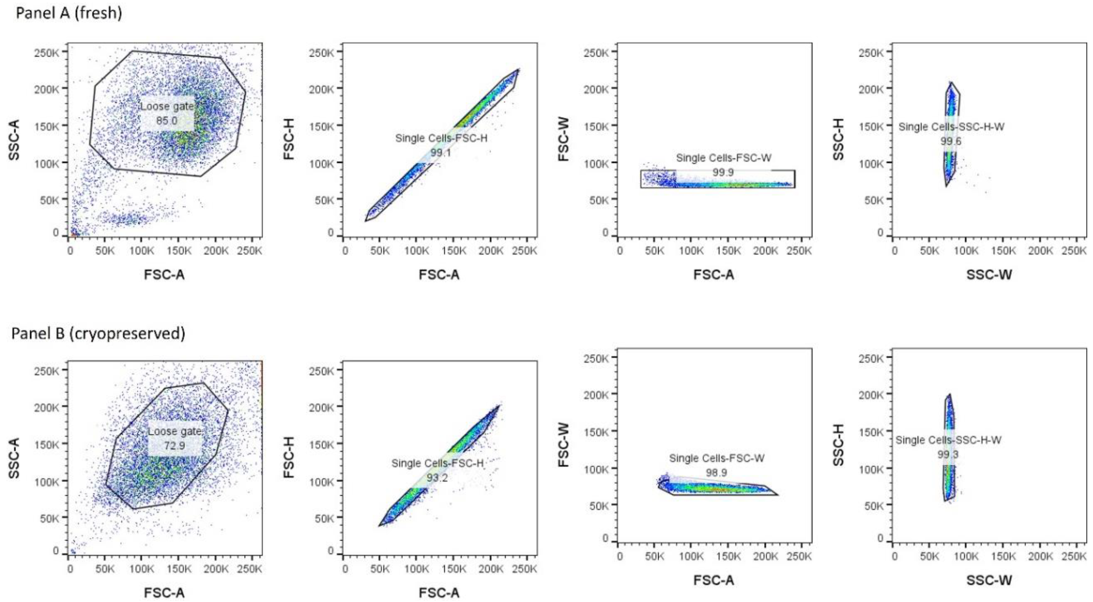
Figure 2.
Antibody serial dilutions to label surface receptors of fresh neutrophils. Red highlighted rectangles on dot plots represent optimal concentrations identified and subsequently used in our flow cytometry assays for the specific markers. CD62L and CD54 were used at 1/80 dilution. CD64, CD32, and CD16 were used at 1/40 dilution. CD66b and CD14 were used at 1/20 dilution. The figures show a representative experiment out of two experiments. FlowJo_v10.8.1 was used to analyse receptor expression. Values on the X-axis are MFI.
Figure 2.
Antibody serial dilutions to label surface receptors of fresh neutrophils. Red highlighted rectangles on dot plots represent optimal concentrations identified and subsequently used in our flow cytometry assays for the specific markers. CD62L and CD54 were used at 1/80 dilution. CD64, CD32, and CD16 were used at 1/40 dilution. CD66b and CD14 were used at 1/20 dilution. The figures show a representative experiment out of two experiments. FlowJo_v10.8.1 was used to analyse receptor expression. Values on the X-axis are MFI.
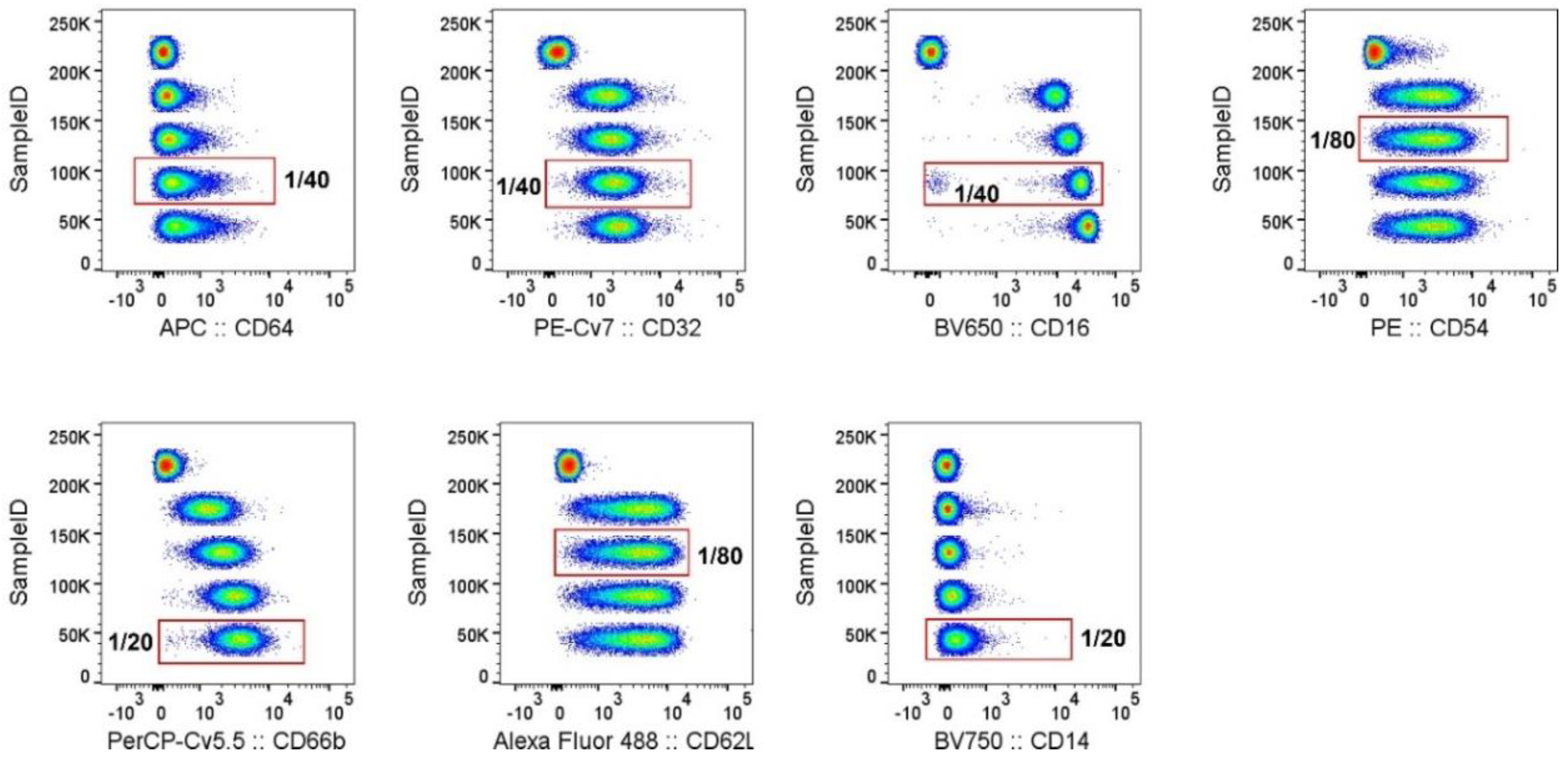
Figure 3.
Comparison of the frequency of receptor-positive cells in fresh and cryopreserved neutrophils at their respective optimal antibody dilutions. Top panel represents fresh cells while bottom panel represents cryopreserved cells, antibody staining at optimal concentrations. Neutrophils were isolated from one donor. A subset was stained immediately after isolation while the remaining neutrophils were cryopreserved and stained later. Light blue histograms represent unstained cells while red dotted histograms show stained cells. The graphs were designed in FlowJo_v10.8.1. The fresh cells panel is a representative of four experiments and the cryopreserved panel a representative of two experiments. Cells were stained for 30 minutes on ice. Data was analysed using FlowJo_v10.8.1. The percentage of cells within each gate are stated for each figure. Values on the X-axis are MFI.
Figure 3.
Comparison of the frequency of receptor-positive cells in fresh and cryopreserved neutrophils at their respective optimal antibody dilutions. Top panel represents fresh cells while bottom panel represents cryopreserved cells, antibody staining at optimal concentrations. Neutrophils were isolated from one donor. A subset was stained immediately after isolation while the remaining neutrophils were cryopreserved and stained later. Light blue histograms represent unstained cells while red dotted histograms show stained cells. The graphs were designed in FlowJo_v10.8.1. The fresh cells panel is a representative of four experiments and the cryopreserved panel a representative of two experiments. Cells were stained for 30 minutes on ice. Data was analysed using FlowJo_v10.8.1. The percentage of cells within each gate are stated for each figure. Values on the X-axis are MFI.
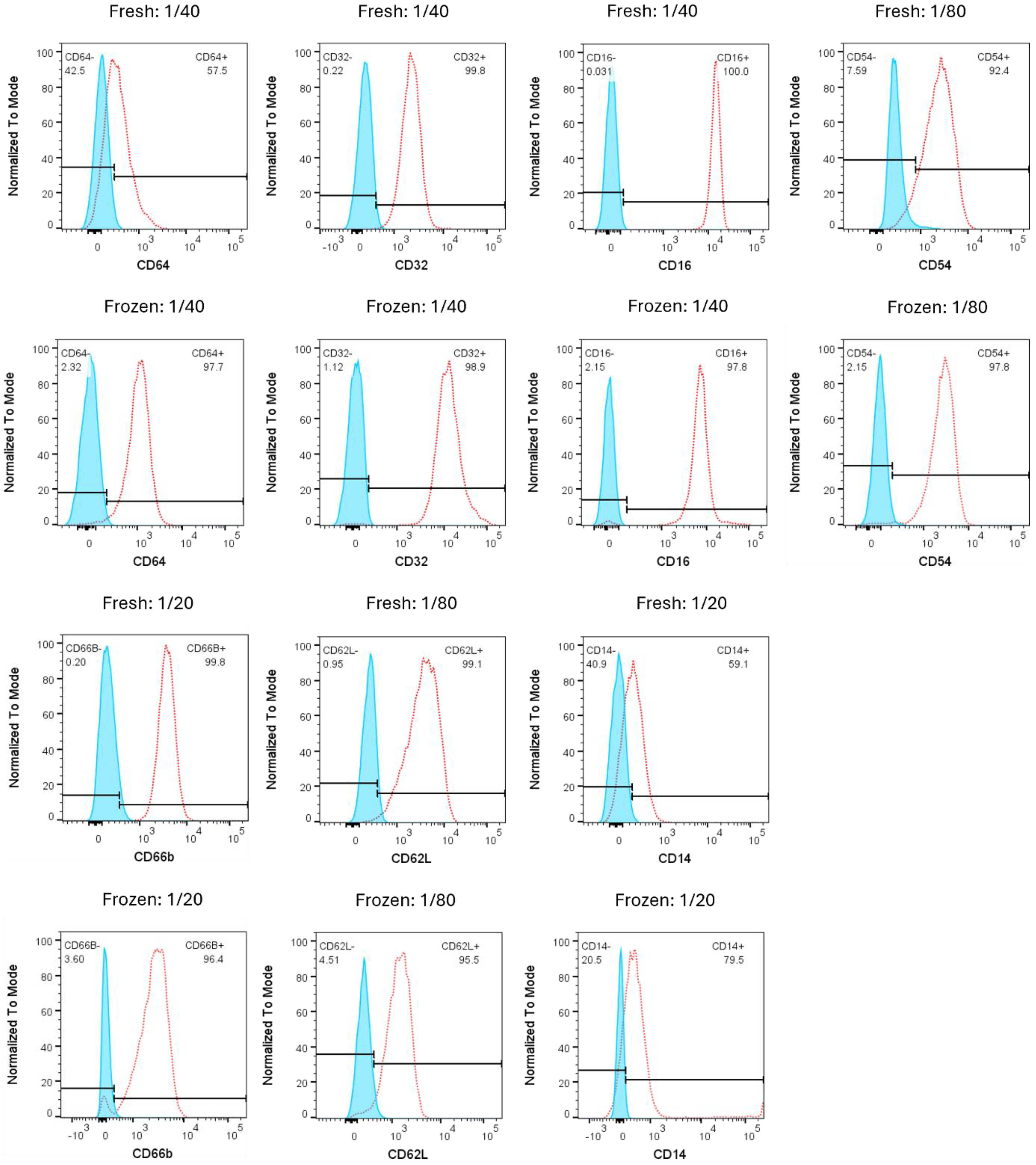
Figure 4.
Figure 4. Fresh and cryopreserved neutrophil population gating. Panel A, fresh cell gating on neutrophils from two biological replicates. Panel B, cryopreserved cells gating on neutrophils from two biological replicates. Neutrophils were gated on CD66B vs CD16. A subset of neutrophils isolated from two donors were cryopreserved and later run in duplicate. Data was analysed using FlowJo_v10.10.0_CL. The percentage of cells within each gate are stated for each figure. Values on the X-axis and Y-axis are MFI.
Figure 4.
Figure 4. Fresh and cryopreserved neutrophil population gating. Panel A, fresh cell gating on neutrophils from two biological replicates. Panel B, cryopreserved cells gating on neutrophils from two biological replicates. Neutrophils were gated on CD66B vs CD16. A subset of neutrophils isolated from two donors were cryopreserved and later run in duplicate. Data was analysed using FlowJo_v10.10.0_CL. The percentage of cells within each gate are stated for each figure. Values on the X-axis and Y-axis are MFI.
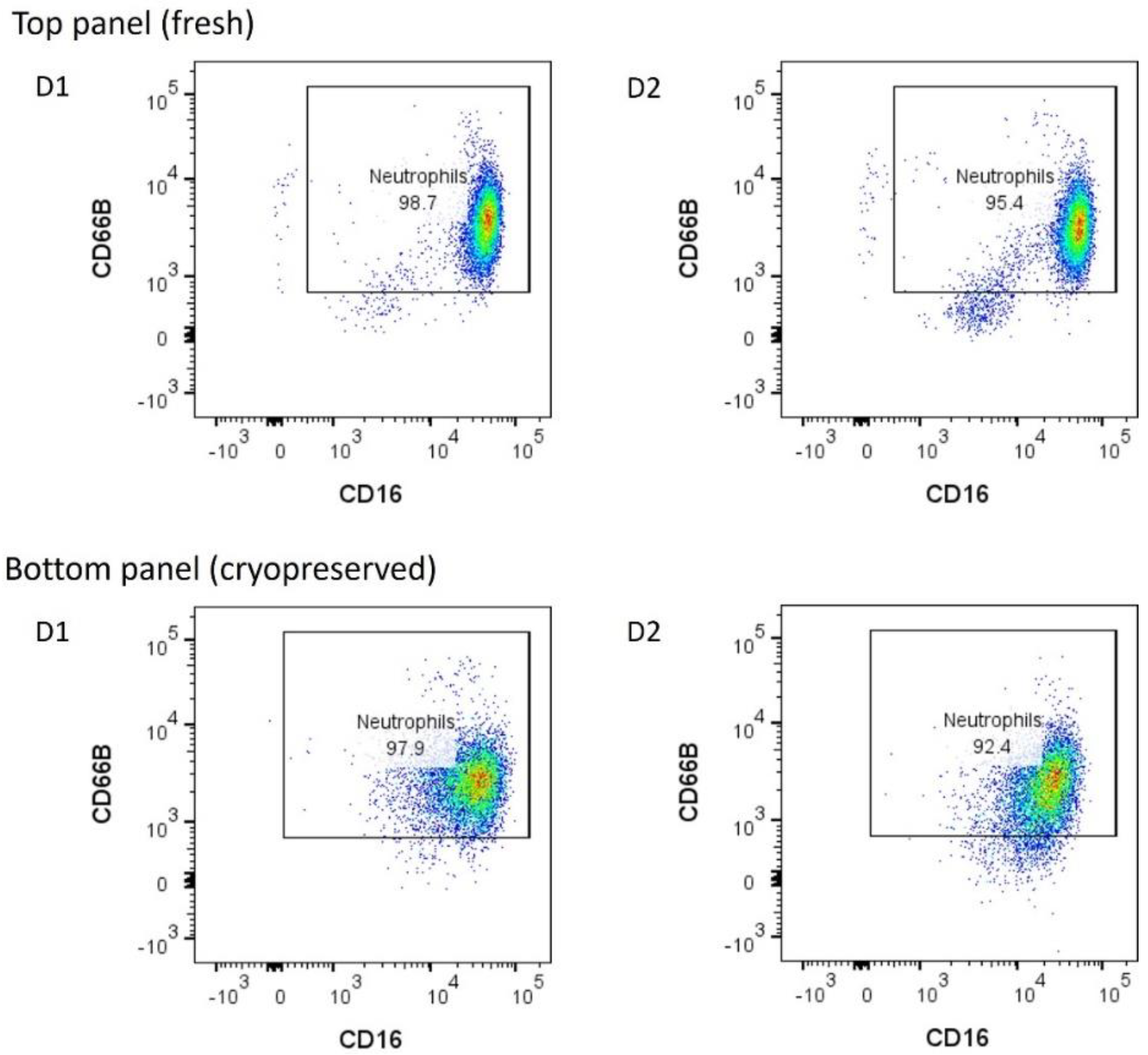
Figure 5.
Detection of neutrophil receptors on fresh versus cryopreserved neutrophils. This figure shows receptor labelling on neutrophils from two donors (tested in duplicate) from representative experiments. Receptors on fresh and frozen neutrophils were quantified by mean fluorescence intensity (MFI). Neutrophils were isolated from two donors, and a subset of cells was cryopreserved per donor. Samples were run in duplicates. Dots in the bar graph represent donor 1 and squares represent donor 2. Each dot point or square represents the average of each technical replicate. A) CD32 expression, B) CD62L expression, C) CD14 expression, D) CD16 expression, E) CD54 expression, F) CD66b expression and G) CD64 expression. Note, because CD62L for one of the donors expressed multiple peaks in the fresh cells, we excluded it from the fresh and frozen CD62L MFI analysis. Receptor staining was performed in the dark on ice for 30 minutes. Phenotype data was analysed using FlowJo_v10.10.0_CL and graphs created in GraphPad Prism Version 10.1.2 (324).
Figure 5.
Detection of neutrophil receptors on fresh versus cryopreserved neutrophils. This figure shows receptor labelling on neutrophils from two donors (tested in duplicate) from representative experiments. Receptors on fresh and frozen neutrophils were quantified by mean fluorescence intensity (MFI). Neutrophils were isolated from two donors, and a subset of cells was cryopreserved per donor. Samples were run in duplicates. Dots in the bar graph represent donor 1 and squares represent donor 2. Each dot point or square represents the average of each technical replicate. A) CD32 expression, B) CD62L expression, C) CD14 expression, D) CD16 expression, E) CD54 expression, F) CD66b expression and G) CD64 expression. Note, because CD62L for one of the donors expressed multiple peaks in the fresh cells, we excluded it from the fresh and frozen CD62L MFI analysis. Receptor staining was performed in the dark on ice for 30 minutes. Phenotype data was analysed using FlowJo_v10.10.0_CL and graphs created in GraphPad Prism Version 10.1.2 (324).


Table 1.
Preserving and thawing conditions tested.
| Cryopreserving buffer | Thawing buffer | Thawing buffer Temp | Re-suspension buffer | Proportion of cells with compromised integrity |
| 10% DMSO + 90% FBS | 50% FBS (HI), RPMI | 4˚C | 10% FBS (HI), RPMI | Over 95% cells had compromised membrane integrity |
| 10% DMSO + 90% FBS | 50% FBS (HI), RPMI | 37˚C | 10% FBS (HI), RPMI | Over 95% cells had compromised membrane integrity |
| 10% DMSO + 90% FBS | 500 ml RPMI, 50 ml FBS (HI), 5 ml (200 mM) L-Glutamine, 8.4 ml HEPES, 1 ml Gentamicin, 250 µl Minocycline (2 mg/ml) | 4˚C | R10 culture medium | Over 90% cells had compromised membrane integrity |
| 10% DMSO + 90% FBS | 500 ml RPMI, 50 ml FBS (HI), 5 ml [200 mM] L-Glutamine, 8.4 ml HEPES, 1 ml Gentamicin, 250 µl Minocycline (2 mg/ml) | 37˚C | R10 culture medium | Over 90% of cells had compromised membrane integrity |
| 10% DMSO + 90% FBS | 1XPBS (Mg2+ and Ca2+)-, 50% FBS (HI), 2 mM EDTA | 4˚C | 1XPBS (Mg2+ and Ca2+)-, 1% FBS (HI), 2mM EDTA | About 70% of the cells showed loss of membrane integrity after one hour. |
| 10% DMSO + 90% FBS | 1XPBS (Mg2+ and Ca2+)-, 50% FBS (HI) + 2 mM EDTA, 2 mg/ml Lignocaine | 4˚C | 1XPBS (Mg2+ and Ca2+)-, 1% FBS (HI), 2mM EDTA | Over 50% of the cells had compromised membrane integrity |
| 10% DMSO + 90% FBS | 1XHBSS | 4˚C | 1XHBSS | About 50% of cells had compromised membrane integrity |
| 10% DMSO + 90% FBS | 1XPBS (Mg2+ and Ca2+)-, 50% FBS (HI), 50 mM EDTA | 4˚C | 1XPBS (Mg2+ and Ca2+)-, 1% FBS (HI), 1mM EDTA | Over 50% of the cells showed compromised membrane integrity. |
| 10% DMSO + 90% FBS | 1XHBSS, RPMI | 4˚C | 1XHBSS, RPMI | Approximately 40% of cells had compromised membrane integrity. |
| 10% DMSO + 90% FBS | 1% BSA, 1XHBSS | 4˚C | 1% BSA, 1XHBSS | Approximately 40% of cells had compromised membrane integrity. |
| 10% DMSO + 90% FBS | 10% BSA, 1XHBSS | 4˚C | 10% BSA, 1XHBSS | Over 50% of cells had compromised membrane integrity. |
| 10% DMSO + 90% FBS | 10% BSA, 1XHBSS, RPMI | 4˚C | 10% BSA, 1XHBSS, RPMI | Over 50% of cells had compromised membrane integrity. |
| 10% BSA + 7.5%DMSO + RPMI | 1XHBSS | 4˚C | 1XHBSS | Approximately 30% cells had compromised membrane integrity |
| 10% BSA + 7.5%DMSO + RPMI | 1XHBSS, RPMI | 4˚C | 1XHBSS, RPMI | Approximately 30% proportion of cells had compromised membrane integrity |
| 10% BSA + 7.5%DMSO + RPMI | 1% BSA, 1XHBSS | 4˚C | 1% BSA, 1XHBSS | Over 40% cells had compromised membrane integrity |
| 10% BSA + 7.5%DMSO + RPMI | 10% BSA, 1XHBSS | 4˚C | 10% BSA, 1XHBSS | 99% population cells had intact membrane** |
| 10% BSA + 7.5%DMSO + RPMI | 10% BSA, 1XHBSS, RPMI | 4˚C | 10% BSA, 1XHBSS + RPMI | Over 10% proportion of cells had compromised membrane integrity |
Cell viability was assessed using Trypan blue exclusion, which stains dead cells.
Disclaimer/Publisher’s Note: The statements, opinions and data contained in all publications are solely those of the individual author(s) and contributor(s) and not of MDPI and/or the editor(s). MDPI and/or the editor(s) disclaim responsibility for any injury to people or property resulting from any ideas, methods, instructions or products referred to in the content. |
© 2026 by the authors. Licensee MDPI, Basel, Switzerland. This article is an open access article distributed under the terms and conditions of the Creative Commons Attribution (CC BY) license (http://creativecommons.org/licenses/by/4.0/).
Copyright: This open access article is published under a Creative Commons CC BY 4.0 license, which permit the free download, distribution, and reuse, provided that the author and preprint are cited in any reuse.