

Submitted:
10 September 2026
Posted:
11 September 2026
You are already at the latest version
Abstract
Vascular contributions to cognitive impairment and dementia (VCID) have emerged as critical determinants of age-related cognitive decline, yet they remain insufficiently recognized in both clinical practice and research. This comprehensive review synthesizes current understanding of the pathophysiological mechanisms linking chronic cerebral hypoperfusion (CCH) to vascular dementia (VaD) and related cognitive disorders. We examine the epidemiology, classification, and clinical heterogeneity of vascular cognitive impairment, emphasizing the convergence of cerebrovascular pathology on common pathogenic cascades including neurovascular unit dysfunction, white matter degeneration, neuroinflammation, and blood-brain barrier disruption. Particular attention is directed toward the selective vulnerability of specific brain regions to chronic hypoperfusion, the molecular mechanisms driving persistent neuroinflammation, and the role of peripheral immune cell infiltration in disease progression. We critically evaluate experimental models of CCH and discuss emerging therapeutic strategies including ischemic preconditioning and remote ischemic conditioning. Finally, we highlight the importance of sex differences in disease susceptibility, progression, and therapeutic response, emphasizing the need for sex-specific approaches in future research and clinical management.
Keywords:
vascular cognitive impairment
; neurovascular unit
; white matter injury
; blood–brain barrier
; neuroinflammation
; cerebral small vessel disease
; ischemic preconditioning
; sex differences
1. Introduction
Neurodegenerative diseases have emerged as one of the greatest global healthcare challenges, driven largely by increased life expectancy and population aging [1]. Among these disorders, dementia represents a major cause of disability and loss of independence, profoundly affecting patients, caregivers, and healthcare systems worldwide [2]. While Alzheimer’s disease (AD) has traditionally received considerable research attention, accumulating evidence indicates that vascular pathology plays an equally critical role in the development and progression of cognitive decline. Vascular dysfunction, impaired cerebral perfusion, and chronic cerebrovascular insufficiency are now recognized as fundamental contributors to neuronal dysfunction and the pathogenesis of VCID [3], fundamentally shifting contemporary neuroscience toward understanding the intricate relationship between the cerebral vasculature and neuronal health.
The recognition that cerebrovascular pathology contributes to cognitive impairment across the dementia spectrum has led to the development of broader conceptual frameworks including vascular cognitive impairment (VCI) and VCID. These concepts acknowledge that cognitive decline associated with vascular pathology develops progressively, often beginning long before the onset of clinically diagnosed dementia [4]. This paradigm shift has important implications for diagnosis, prevention, and therapeutic intervention, as vascular risk factors represent potentially modifiable targets for reducing the global burden of dementia. This review provides a comprehensive examination of the pathophysiological mechanisms linking chronic cerebral hypoperfusion (CCH) to vascular cognitive impairment, with particular emphasis on neurovascular dysfunction, white matter degeneration, neuroinflammation, and the potential for therapeutic interventions targeting these pathways.
2. Vascular Dementia: Clinical and Epidemiological Perspectives
2.1. Definition and Clinical Spectrum
Vascular dementia is the second most common type of dementia, resulting from cerebrovascular pathology that disrupts cerebral blood flow (CBF) and neuronal connectivity. Unlike AD, VaD arises from ischemic and hemorrhagic vascular lesions, CCH, and small vessel pathology that impair cognitive networks. Importantly, VaD should not be viewed as a single disease entity but rather as a spectrum of cognitive disorders associated with cerebrovascular injury [5]. This recognition has led to the development of the broader concept of VCI, which encompasses all forms of cognitive dysfunction attributable to cerebrovascular disease, ranging from subtle cognitive deficits to severe dementia [6,7]. VCID has emerged as a comprehensive approach for understanding vascular contributions to cognitive decline [4]. Despite the diverse cerebrovascular pathologies that can cause VaD, accumulating evidence suggests that these seemingly distinct insults converge on a common pathophysiological cascade. Vascular risk factors initiate both large- and small-vessel disease, resulting in CCH and impaired cerebrovascular function. These vascular abnormalities promote neurovascular unit dysfunction through interconnected mechanisms including endothelial injury, blood-brain barrier (BBB) breakdown, and persistent neuroinflammation. The resulting disruption of cerebral homeostasis leads to oligodendrocyte loss, demyelination, axonal degeneration, neuronal injury, and progressive white matter damage. Ultimately, these structural alterations disrupt cortical-subcortical neuronal networks responsible for memory, executive function, and information processing, culminating in cognitive decline and VaD.
2.2. Epidemiology and Global Burden
Traditionally, VaD has been regarded as a distinct clinicopathological entity resulting from cerebrovascular injury; however, contemporary evidence indicates that vascular pathology contributes more to cognitive impairment than previously appreciated. The epidemiology of VaD has undergone substantial revision over the past two decades. Earlier epidemiological studies considered VaD relatively uncommon compared with AD because diagnosis relied largely on the presence of clinically overt stroke and established dementia. Improvements in neuroimaging, neuropathological assessment, longitudinal cohort studies, and biomarker research have fundamentally altered this perception [8,9]. The burden of VaD varies considerably across geographical regions, reflecting differences in diagnostic criteria, population age structure, socioeconomic conditions, vascular risk profiles, and healthcare access. Population-based studies from Europe and North America generally report VaD accounting for approximately one-fifth of all dementia cases. In contrast, several Asian countries have historically reported higher proportions of VaD, in some regions approaching or exceeding AD, primarily because of the greater prevalence of hypertension, stroke, and uncontrolled cardiovascular risk factors. Conversely, Japanese populations demonstrate comparatively lower dementia prevalence despite advanced population aging, suggesting important interactions among lifestyle, environmental influences, and vascular health [10].
Age remains the strongest non-modifiable risk factor for VaD. The prevalence of VaD increases exponentially with advancing age, reflecting cumulative vascular injury, progressive cerebral small vessel disease, endothelial dysfunction, and increasing incidence of stroke. Epidemiological studies indicate that VaD prevalence rises from approximately 0.4% among individuals aged 60-64 years to more than 4% in those older than 85 years [8]. Neuropathological studies further demonstrate that increasing age is associated not only with greater prevalence of vascular lesions but also with increasing coexistence of vascular and neurodegenerative pathology. Autopsy-based investigations indicate that mixed dementia comprising Alzheimer’s pathology together with cerebrovascular disease becomes increasingly common beyond the ninth decade of life [11,12].
2.3. Sex-Stratified Epidemiology
Sex-related differences have been observed in the epidemiology of VaD. While AD generally occurs more frequently among women, largely because of greater longevity, VaD appears relatively more common in men. This difference is believed to reflect the higher lifetime prevalence of stroke, hypertension, coronary artery disease, and smoking among males. Nevertheless, these differences become less pronounced in advanced age, where mixed pathologies predominate and both sexes exhibit substantial cerebrovascular disease burden [10].
2.4. Stroke and Vascular Dementia
Stroke remains the single most important clinical event associated with VaD. Longitudinal cohort studies demonstrate that individuals experiencing ischemic stroke have approximately a twofold increase in long-term risk of developing dementia compared with age-matched controls, while nearly one-quarter of elderly patients hospitalized with stroke develop post-stroke dementia within the first three months after the cerebrovascular event [8,13]. Earlier estimates further suggested that approximately one-third of stroke survivors older than 65 years develop VaD within months following stroke, highlighting the enormous contribution of cerebrovascular disease to cognitive decline [14].
Beyond clinically overt stroke, increasing attention has focused on covert cerebrovascular disease as a major contributor to VCI. Silent brain infarcts, cerebral microbleeds, enlarged perivascular spaces, and extensive white matter hyperintensities are frequently detected on magnetic resonance imaging (MRI) among cognitively normal older adults and significantly increase future risk of dementia. The 2025 American Heart Association Scientific Statement estimated that in the United States alone approximately 11 million older adults harbor covert brain infarcts, another 11 million exhibit extensive white matter hyperintensities, and nearly 20 million possess cerebral microbleeds detectable by MRI. Collectively, these covert vascular lesions represent a substantial reservoir of potentially preventable cognitive impairment [9].
2.5. Epidemiological Challenges and Diagnostic Considerations
An important challenge in estimating the true epidemiological burden of VaD is the absence of universally accepted diagnostic criteria. Historically, different epidemiological studies have employed varying clinical definitions, including DSM criteria, ICD classifications, NINDS-AIREN criteria, ADDTC criteria, and Hachinski Ischemic Score-based approaches, resulting in considerable heterogeneity in prevalence estimates. Furthermore, many earlier studies classified VaD only when cognitive decline occurred immediately following a clinically apparent stroke, thereby overlooking patients with slowly progressive cognitive impairment secondary to cerebral small vessel disease, CCH, or silent ischemic lesions [8,15]. Consequently, comparisons between studies performed in different populations and different decades should be interpreted cautiously because changing diagnostic criteria have substantially influenced reported disease frequency.
The evolution of neuroimaging has significantly influenced the epidemiology of VaD. Earlier conceptions of VaD were primarily associated with large territorial infarctions or clinically evident stroke. Modern neuroimaging has demonstrated that diffuse white matter hyperintensities, cortical microinfarcts, and chronic cerebral small vessel disease are highly prevalent among elderly individuals and contribute substantially to cognitive decline. These lesions frequently accumulate silently over many years before clinical symptoms become apparent, indicating that the prevalence of vascular-related cognitive impairment is considerably higher than previously recognized.
3. Classification of Vascular Dementia
Patients with VaD exhibit considerable variability in clinical presentation, disease progression, neuroimaging findings, neuropathological features, and underlying vascular mechanisms. This heterogeneity has resulted in numerous classification systems based on anatomical location, vascular pathology, clinical phenotype, and underlying pathophysiological mechanisms.
3.1. Historical Evolution of Classification
Historically, VaD was considered synonymous with “arteriosclerotic dementia,” a broad clinical diagnosis that attributed age-related cognitive decline primarily to cerebral arteriosclerosis. During the late nineteenth and early twentieth centuries, investigators including Binswanger, Alzheimer, and Pierre Marie recognized that cerebrovascular disease could independently produce dementia. However, the absence of neuroimaging techniques limited precise clinicopathological correlations. The introduction of computed tomography (CT) and MRI during the latter half of the twentieth century fundamentally transformed understanding of VaD by revealing multiple infarcts, lacunar lesions, diffuse white matter disease, cerebral microbleeds, and silent ischemic lesions that had previously remained undetected [5,13].
3.2. Early Anatomical Classifications
The earliest modern classification divided VaD into cortical and subcortical forms depending upon the principal anatomical distribution of vascular lesions. Cortical VaD was considered to arise predominantly from large territorial infarctions affecting cortical association areas, whereas subcortical VaD resulted primarily from cerebral small vessel disease (CSVD). Although anatomically useful, this classification was unable to accommodate patients exhibiting mixed vascular lesions involving both cortical and subcortical structures, which are now recognized as common in aging populations [8,16].
3.3. Multi-Infarct Dementia
Among the earliest pathophysiological classifications, multi-infarct dementia (MID) represented the prototypical form of VaD. Introduced by Hachinski and colleagues, MID refers to progressive cognitive decline resulting from the cumulative effects of multiple ischemic infarctions occurring throughout the brain. Individual infarcts may be clinically silent; however, their cumulative burden eventually exceeds the brain’s compensatory capacity, producing executive dysfunction, memory impairment, slowed information processing, and dementia. Patients typically demonstrate a stepwise pattern of cognitive deterioration associated with recurrent ischemic strokes, making MID one of the classical VaD syndromes.
3.4. Strategic Infarct Dementia
Strategic infarct dementia results from a single infarction involving critical brain regions responsible for cognitive processing. Unlike MID, cognitive impairment in strategic infarct dementia does not depend upon the cumulative volume of ischemic injury but rather upon the functional importance of the affected anatomical location. Infarctions involving the hippocampus, thalamus, angular gyrus, caudate nucleus, internal capsule, anterior thalamic nuclei, basal forebrain, or medial frontal cortex may produce severe cognitive deficits despite relatively small lesion volumes. Consequently, strategic infarct dementia illustrates that lesion location often exerts greater influence on cognition than lesion size alone.
3.5. Subcortical Ischemic Vascular Dementia
Subcortical ischemic vascular dementia (SIVD) is currently regarded as the most prevalent subtype of VaD in elderly populations. This disorder primarily results from chronic pathological changes affecting small penetrating arteries and arterioles supplying deep white matter structures. Long-standing hypertension, diabetes mellitus, aging, endothelial dysfunction, and lipohyalinosis progressively narrow these vessels, producing CCH, lacunar infarctions, diffuse white matter rarefaction, demyelination, axonal degeneration, and disruption of fronto-subcortical neuronal circuits. Clinically, patients present predominantly with executive dysfunction, psychomotor slowing, impaired attention, gait abnormalities, urinary disturbances, and depression rather than the prominent episodic memory deficits seen in AD.
A classical manifestation of subcortical VaD is Binswanger disease, also known as subcortical arteriosclerotic encephalopathy [17,18]. This disorder is characterized neuropathologically by diffuse white matter degeneration associated with chronic ischemia secondary to hypertensive small vessel disease [19,20]. MRI typically demonstrates extensive bilateral white matter hyperintensities, multiple lacunes, enlarged perivascular spaces, ventricular enlargement, and diffuse cerebral atrophy. Histopathological examination reveals arteriolosclerosis, myelin loss, gliosis, axonal degeneration, and disruption of long association fibers connecting frontal cortical regions with subcortical nuclei.
3.6. Hemorrhagic Vascular Dementia
Hemorrhagic vascular dementia represents another important although less common subtype. Unlike ischemic VaD, cognitive impairment develops following intracerebral hemorrhage, cerebral amyloid angiopathy-related hemorrhage, multiple cerebral microbleeds, or recurrent hemorrhagic stroke [21]. The severity of cognitive dysfunction depends upon hemorrhage location, lesion volume, secondary inflammation, perihematomal edema, and associated disruption of neuronal networks. Although hemorrhagic VaD accounts for a relatively small proportion of VaD cases, increasing recognition of cerebral microbleeds using susceptibility-weighted MRI has highlighted its contribution to age-related cognitive decline [18].
3.7. Classification Based on Vascular Etiology
Based on vascular etiology, VaD can broadly be classified into large-vessel VaD, small-vessel VaD, hypoperfusion-associated VaD, hemorrhagic VaD, hereditary VaD, and mixed VaD. Large-vessel VaD develops secondary to occlusion or stenosis of major cerebral arteries, including the internal carotid, middle cerebral, anterior cerebral, and posterior cerebral arteries [27,28]. These lesions commonly produce territorial infarctions involving cortical association areas responsible for memory, language, visuospatial function, and executive processing. Because large cortical infarctions frequently produce focal neurological deficits in addition to cognitive dysfunction, patients often present with aphasia, hemiparesis, visual field defects, sensory abnormalities, and gait disturbances accompanying dementia.
Small-vessel VaD results from pathological alterations affecting penetrating arterioles, capillaries, and small intracerebral arteries. Chronic hypertension, diabetes mellitus, aging, cerebral amyloid angiopathy, endothelial dysfunction, and lipohyalinosis progressively damage these vessels, leading to diffuse white matter ischemia, lacunar infarctions, microinfarcts, microbleeds, enlarged perivascular spaces, and disruption of fronto-subcortical neuronal circuits [22,23,24,25]. Rather than producing abrupt neurological deficits, small-vessel disease usually causes slowly progressive executive dysfunction, impaired attention, reduced information-processing speed, gait abnormalities, urinary dysfunction, depression, and eventually dementia. Because CSVD frequently progresses silently over many years, it is now regarded as the most common pathological substrate underlying VCI in elderly populations [26].
Hypoperfusion-associated VaD develops following prolonged reduction in CBF without necessarily producing large infarctions [27,28,29]. Conditions such as carotid artery stenosis, chronic heart failure, severe hypotension, cerebral small vessel disease, and diffuse vascular narrowing reduce cerebral perfusion over extended periods, resulting in chronic ischemic stress within metabolically vulnerable brain regions. Experimental studies have demonstrated that sustained cerebral hypoperfusion initiates a cascade of pathological events including excitotoxicity, neuroinflammation, BBB disruption, white matter degeneration, oligodendrocyte injury, synaptic dysfunction, and neuronal loss. The recognition of CCH as an independent pathogenic mechanism has substantially expanded the concept of VaD [30,31,32,33].
3.8. Hereditary Vascular Dementia
Hereditary VaDs comprise a relatively uncommon group of monogenic disorders affecting cerebral blood vessels. The most extensively studied example is CADASIL (cerebral autosomal dominant arteriopathy with subcortical infarcts and leukoencephalopathy), which is caused by mutations in the NOTCH3 gene [34,35]. Other inherited cerebral small vessel diseases involve mutations affecting extracellular matrix proteins, collagen synthesis, or vascular integrity. Although individually rare, these disorders have substantially improved understanding of cerebral small vessel biology and demonstrate that chronic vascular dysfunction alone is sufficient to produce progressive cognitive impairment independent of Alzheimer’s pathology.
3.9. Mixed Vascular Dementia
Neuropathological investigations consistently demonstrate that isolated vascular pathology is relatively uncommon in elderly individuals. Instead, vascular lesions frequently coexist with AD, Lewy body disease, hippocampal sclerosis, or other neurodegenerative disorders. Mixed dementia therefore represents the simultaneous presence of cerebrovascular disease together with neurodegenerative pathology, both contributing significantly to cognitive decline [36,37,38,39]. Autopsy studies indicate that mixed AD and vascular pathology is considerably more prevalent than pure VaD, particularly in individuals older than 80 years.
3.10. Evolution of Conceptual Frameworks: VCI and VCID
The considerable heterogeneity observed in VaD has prompted a shift from traditional disease-specific classifications toward broader conceptual frameworks that encompass the entire spectrum of VCI. One of the most significant developments has been the introduction of VCI, which was proposed to include all forms of cognitive dysfunction attributable to cerebrovascular disease, irrespective of severity. Unlike the conventional diagnosis of VaD, which requires substantial impairment in daily functioning, VCI encompasses cognitive changes ranging from mild VCI to severe dementia, thereby facilitating earlier diagnosis and intervention. This broader concept acknowledges that vascular injury begins long before patients fulfill the diagnostic criteria for dementia and that early cognitive deficits may still be amenable to preventive treatment [40,41,42,43].
Building upon this concept, VCID has gained widespread acceptance in recent years. VCID recognizes that cerebrovascular pathology rarely exists in isolation and instead contributes to cognitive decline across multiple neurodegenerative disorders [44,45,46]. VCID considers vascular dysfunction as one of the principal biological processes that interact with amyloid deposition, tau pathology, neuroinflammation, oxidative stress, and neurodegeneration to produce progressive cognitive impairment. The diverse etiologies and clinical presentations of VaD are summarized in the classification framework provided in Figure 1.
4. Chronic Cerebral Hypoperfusion: Pathophysiology and Consequences
4.1. Definition and Clinical Significance
CCH refers to a sustained reduction in CBF that compromises the delivery of oxygen and metabolic substrates required for normal neuronal function. Given the high metabolic demands of the brain, even moderate reductions in cerebral perfusion can have profound effects on neuronal survival, synaptic activity, and cognitive performance. Over the past two decades, CCH has emerged as a critical pathological mechanism linking vascular dysfunction with cognitive impairment and neurodegeneration [47]. The consequences of CCH extend beyond reduced blood supply alone. Persistent reductions in cerebral perfusion initiate a series of pathological alterations involving neurovascular dysfunction, white matter injury, impaired BBB integrity, metabolic disturbances, and progressive neuronal damage. Prolonged hypoperfusion disrupts neurovascular homeostasis and contributes to structural and functional alterations that ultimately impair cognitive performance [48].
4.2. White Matter Injury: The Hallmark of CCH
Among the pathological consequences of CCH, white matter injury is particularly important. White matter tracts are highly vulnerable to chronic ischemic stress because of their limited vascular reserve and dependence on continuous blood flow. Prolonged reductions in cerebral perfusion contribute to demyelination, axonal degeneration, and disruption of neuronal connectivity. These alterations impair communication between cortical and subcortical regions and are strongly associated with deficits in memory, executive function, and information processing [47]. Several clinical conditions have been linked to CCH, including cerebral small vessel disease, carotid artery stenosis, chronic hypotension, heart failure, and age-related vascular dysfunction [49]. Importantly, CCH is increasingly recognized not merely as a consequence of neurological disease but as an active driver of pathological progression. The resulting cerebral hypoxia and hypoperfusion initiate a complex cascade of molecular and cellular events characterized by oxidative stress, BBB dysfunction, mitochondrial impairment, glial activation, and persistent neuroinflammation. These processes are highly interconnected and amplify one another through positive feedback mechanisms, leading to progressive disruption of neurovascular homeostasis. Consequently, CCH establishes a self-sustaining cycle of vascular dysfunction and neurodegeneration that progressively accelerates cognitive decline.
4.3. Molecular and Cellular Mechanisms of White Matter Injury
White matter injury represents one of the earliest and most consistent pathological hallmarks of VCI. Sustained reductions in oxygen and glucose availability compromise mitochondrial ATP production, increase reactive oxygen species generation, disrupt calcium homeostasis, and impair oligodendrocyte survival, ultimately leading to demyelination and axonal degeneration. Since white matter tracts are essential for communication between cortical and subcortical regions, their degeneration disrupts large-scale neuronal networks involved in memory, executive function, attention, and information processing [50]. Current evidence suggests that white matter damage is not caused solely by ischemia but rather results from the complex interaction of endothelial dysfunction, impaired cerebral autoregulation, neurovascular uncoupling, and BBB disruption. Endothelial dysfunction reduces nitric oxide (NO) bioavailability while increasing reactive oxygen species production, thereby impairing vasodilation and promoting sustained cerebral hypoperfusion. Vascular risk factors initiate endothelial injury that compromises cerebral autoregulation and neurovascular coupling, leading to chronic hypoperfusion and tissue hypoxia, which together create a permissive environment for progressive white matter degeneration.
A central mechanism underlying white matter injury is disruption of the BBB, which occurs secondary to endothelial dysfunction and chronic ischemic stress [51,52,53]. Increased BBB permeability permits extravasation of plasma proteins, including fibrinogen, into the brain parenchyma, where they activate resident microglia through Toll-like receptor (TLR)-dependent pathways. Activated microglia release additional neurotoxic mediators that amplify oxidative stress and tissue injury. Reactive astrocytes further contribute to this inflammatory cascade through cytokine secretion and altered end-foot interactions with cerebral blood vessels, thereby impairing neurovascular homeostasis. Simultaneously, oligodendrocyte precursor cells subjected to chronic hypoxia exhibit increased expression of matrix metalloproteinase-9 (MMP-9), which degrades extracellular matrix proteins and further compromises BBB integrity. These pathological processes establish a self-perpetuating cycle in which hypoperfusion enhances oxidative stress and inflammation, while inflammation further aggravates endothelial injury and cerebral hypoperfusion, ultimately accelerating white matter degeneration [50].
5. Regional Vulnerability to Chronic Cerebral Hypoperfusion
5.1. Selective Vulnerability: General Principles
Unlike acute ischemic stroke, which produces rapid neuronal death within a localized vascular territory, CCH is characterized by a sustained reduction in CBF that progressively compromises neuronal viability, white matter integrity, synaptic plasticity, and neurovascular function over months or even years. Although the reduction in cerebral perfusion may initially affect the brain globally, neuropathological and neuroimaging studies consistently demonstrate that tissue injury develops in a highly selective manner, with certain brain regions exhibiting significantly greater susceptibility to prolonged hypoperfusion than others [47,49,54]. The concept of selective vulnerability reflects the fact that the brain is anatomically and physiologically heterogeneous. Individual brain regions differ considerably in vascular architecture, collateral circulation, metabolic demand, neuronal composition, synaptic density, and capacity for adaptive vascular remodeling. Among the most consistently affected structures are the cerebral white matter, hippocampus, frontal cortex, basal ganglia, thalamus, and other regions involved in large-scale cognitive networks. Clinical neuroimaging studies demonstrate that patients with VCI frequently exhibit extensive white matter hyperintensities, lacunar infarcts, hippocampal atrophy, cortical thinning, and disruption of fronto-subcortical circuits long before the development of overt dementia [47,49].
5.2. White Matter: The Most Vulnerable Region
Among all cerebral structures affected by CCH, the cerebral white matter is widely regarded as the most susceptible region and represents the principal pathological substrate underlying VCI and VaD. Neuroimaging studies consistently identify white matter hyperintensities, lacunar infarctions, diffuse leukoaraiosis, and white matter rarefaction as the most common structural abnormalities in patients with CSVD and VaD [47,48]. Histopathological investigations similarly demonstrate extensive demyelination, axonal degeneration, oligodendrocyte loss, gliosis, and disruption of long-range axonal tracts in individuals with CCH, indicating that white matter degeneration precedes extensive neuronal loss in many forms of VaD [55]. The exceptional vulnerability of white matter primarily reflects its unique vascular architecture. Unlike the cerebral cortex, which receives a rich collateral blood supply from numerous pial arteries, the deep white matter is perfused predominantly by long medullary arteries and penetrating arterioles that possess very limited collateral circulation. Consequently, even modest reductions in cerebral perfusion pressure substantially decrease oxygen and glucose delivery to the deep white matter, making this region highly susceptible to chronic ischemic injury [56]. Furthermore, these long penetrating vessels traverse considerable distances before reaching the deep white matter, exposing distal vascular territories to greater reductions in perfusion during systemic hypotension, arterial stenosis, or CCH.
Oligodendrocytes are particularly vulnerable to chronic ischemia because of their exceptionally high metabolic requirements. These cells require continuous ATP production to maintain myelin synthesis and axonal metabolic support. During CCH, impaired mitochondrial function, reduced glucose availability, intracellular calcium overload, and excessive production of reactive oxygen species trigger oligodendrocyte apoptosis and impair differentiation of oligodendrocyte precursor cells. Consequently, remyelination becomes insufficient to compensate for ongoing myelin degeneration, leading to progressive disruption of saltatory conduction and loss of axonal integrity.
5.3. Hippocampus: A Highly Vulnerable Gray Matter Structure
The hippocampus is one of the most metabolically active and functionally important regions of the brain, playing a central role in learning, memory consolidation, spatial navigation, and cognitive flexibility. Owing to its exceptionally high metabolic demand, dense excitatory synaptic circuitry, and limited tolerance to oxygen and glucose deprivation, the hippocampus is among the earliest gray matter structures affected during CCH [57,58,59,60]. Clinical neuroimaging studies have consistently demonstrated hippocampal atrophy, reduced CBF, impaired glucose metabolism, and altered functional connectivity in patients with VCI and VaD, while experimental models of CCH reproduce progressive neuronal degeneration, synaptic dysfunction, impaired neurogenesis, and memory deficits that closely resemble the cognitive abnormalities observed in humans [47].
The marked susceptibility of the hippocampus arises from several unique anatomical and physiological characteristics. Hippocampal neurons exhibit exceptionally high rates of oxidative metabolism and maintain continuous excitatory neurotransmission required for synaptic plasticity and long-term potentiation [61,62,63]. Consequently, even modest reductions in CBF substantially impair ATP production, disrupt ionic homeostasis, and compromise synaptic transmission. Furthermore, the hippocampus possesses relatively limited vascular reserve compared with many neocortical regions, rendering it particularly sensitive to prolonged reductions in oxygen and glucose delivery [64].
Among the hippocampal subfields, CA1 is widely regarded as the region exhibiting the greatest susceptibility to CCH. Histopathological studies demonstrate pronounced neuronal shrinkage, dendritic degeneration, synaptic loss, mitochondrial swelling, and capillary damage within the CA1 pyramidal layer following prolonged reductions in CBF [64]. Several mechanisms contribute to this remarkable vulnerability. CA1 pyramidal neurons express high densities of glutamate receptors, particularly NMDA receptors, making them highly susceptible to excitotoxic injury during chronic ischemia. Sustained glutamate accumulation promotes excessive calcium influx, mitochondrial dysfunction, activation of calcium-dependent proteases, oxidative stress, and apoptotic signaling pathways that progressively impair neuronal survival.
5.4. Cerebral Cortex: Executive Dysfunction in VCI
The cerebral cortex represents one of the principal gray matter structures affected during CCH and plays a fundamental role in the development of VCI and VaD. Although white matter pathology is considered the hallmark lesion of VaD, increasing clinical and experimental evidence indicates that cortical dysfunction develops concurrently and contributes significantly to progressive cognitive decline. Chronic reductions in CBF impair neuronal metabolism, synaptic transmission, neurovascular coupling, and cortical connectivity, ultimately resulting in executive dysfunction, impaired attention, reduced processing speed, and deficits in higher cognitive functions that characterize VaD. Among cortical regions, the prefrontal cortex exhibits particular vulnerability because it is central to executive function, working memory, cognitive flexibility, planning, behavioral regulation, and decision-making. These cognitive domains are disproportionately affected in VaD and frequently deteriorate before severe episodic memory impairment becomes evident. Functional neuroimaging studies consistently demonstrate reduced cerebral perfusion and glucose metabolism within the frontal cortex of patients with VCI, reflecting impaired neuronal activity secondary to chronic vascular insufficiency [47,65].
5.5. Basal Ganglia and Thalamus: Disruption of Fronto-Subcortical Circuits
The basal ganglia and thalamus constitute integral components of the fronto-subcortical circuitry responsible for executive function, attention, behavioral regulation, motor planning, and cognitive processing. Although traditionally associated with motor control, these deep gray matter structures play equally important roles in higher cognitive functions through extensive reciprocal connections with the prefrontal cortex. The basal ganglia, comprising the caudate nucleus, putamen, globus pallidus, and associated nuclei, receive their blood supply predominantly from long, penetrating arteries arising from the middle cerebral and anterior cerebral arteries. These small perforating vessels possess limited collateral circulation and are therefore highly susceptible to chronic reductions in cerebral perfusion, hypertension, lipohyalinosis, and CSVD. Progressive narrowing of these vessels promotes lacunar infarctions, microinfarcts, diffuse ischemic injury, and degeneration of fronto-striatal pathways that are essential for executive cognitive processing [8,47]. The thalamus is another highly vulnerable deep gray matter structure because it serves as the principal relay center connecting cortical, limbic, and subcortical networks. Numerous thalamic nuclei participate in attention, memory, executive processing, emotional regulation, and sensorimotor integration. CCH compromises thalamic neuronal metabolism through persistent reductions in oxygen and glucose delivery, leading to neuronal dysfunction, synaptic degeneration, and disruption of thalamo-cortical communication [47].
6. Mechanisms of Cerebral Blood Flow Reduction
CBF is tightly regulated by the neurovascular unit, comprising endothelial cells, pericytes, astrocytes, vascular smooth muscle cells, neurons, and extracellular matrix components. Under physiological conditions, neurovascular coupling ensures that regional cerebral perfusion matches local metabolic demand. However, impairment of neurovascular regulation is increasingly recognized as a critical event in both AD and VaD, leading to CCH, reduced oxygen and glucose delivery, white matter injury, synaptic dysfunction, and progressive cognitive decline. Although both AD and VaD exhibit reduced cerebral perfusion, the underlying molecular mechanisms differ considerably. In AD, CBF reduction is predominantly driven by amyloid-β-mediated neurovascular dysfunction and inflammatory activation. Soluble amyloid-β oligomers activate microglia, leading to increased NADPH oxidase 2 activity and excessive reactive oxygen species, which stimulate endothelin-1 signaling. Endothelin-1 subsequently binds endothelin receptor A (ETA) expressed on pericytes, activating Gq-protein coupled signaling that elevates intracellular calcium primarily via IP3-mediated release from endoplasmic reticulum stores [66]. Elevated intracellular calcium activates calmodulin and myosin light-chain kinase, promoting phosphorylation of myosin and sustained pericyte contraction. Simultaneously, activation of ROCK signaling further suppresses myosin light-chain phosphatase, thereby enhancing contractility. Collectively, these events produce prolonged capillary constriction, resulting in significant reductions in CBF.
In contrast, VaD is primarily associated with chronic dysfunction of the cerebral microvasculature, particularly CSVD and hereditary arteriopathies such as CADASIL. In sporadic small vessel disease, eNOS activity and NO bioavailability are markedly reduced, resulting in impaired vasodilatory capacity and diminished autoregulation of CBF. Concurrent activation of the renin-angiotensin system increases angiotensin II signaling through angiotensin II type-1 receptors, promoting calcium-dependent pericyte contraction and persistent capillary narrowing. Chronic hypertension further exacerbates this process by enhancing vascular stiffness, endothelial injury, and arteriolar remodeling, while endothelial activation increases adhesion molecule expression, facilitating leukocyte adhesion, capillary stalling, and progressive vessel pruning. Together, these alterations culminate in sustained cerebral hypoperfusion and chronic white matter ischemia. In CADASIL, mutations in the NOTCH3 gene result in extracellular accumulation of the NOTCH3 ectodomain and deposition of granular osmiophilic material within the vessel wall, inducing extracellular matrix dysfunction, progressive pericyte degeneration, impaired vascular integrity, and cerebral microbleeds, ultimately leading to severe microvascular dysfunction and marked reductions in CBF [67,68,69].
7. Neuroinflammation in Chronic Cerebral Hypoperfusion
7.1. Microglial Activation and Innate Immune Responses
Microglia are the resident innate immune cells of the central nervous system and constitute the first line of defense against disturbances in cerebral homeostasis. Under physiological conditions, homeostatic microglia continuously survey the brain microenvironment through highly motile processes while maintaining neuronal integrity, synaptic remodeling, debris clearance, and neurovascular unit homeostasis [70]. During CCH, prolonged reductions in oxygen and glucose availability induce cellular stress within neurons, oligodendrocytes, endothelial cells, pericytes, and astrocytes, leading to the release of damage-associated molecular patterns (DAMPs), extracellular ATP, oxidized lipids, and reactive oxygen species. These endogenous danger signals are recognized by pattern-recognition receptors expressed on microglia, initiating their transition from a homeostatic phenotype to activated inflammatory states [71].
Unlike the traditional M1/M2 classification, current evidence indicates that microglial activation during VaD represents a dynamic continuum of transcriptionally distinct phenotypes regulated by persistent cerebrovascular injury. Early after cerebral hypoperfusion, activated microglia exert protective functions including phagocytosis of cellular debris, maintenance of vascular integrity, and secretion of trophic factors that support tissue repair. However, sustained hypoperfusion induces metabolic reprogramming of microglia toward chronic inflammatory phenotypes characterized by excessive cytokine production, oxidative stress, impaired phagocytosis, and neurovascular dysfunction [70]. Microglial activation is consistently demonstrated in experimental models of CCH [72] by increased expression of ionized calcium-binding adaptor molecule-1 (Iba-1), CD11b, CD68, and OX-42, with activated cells accumulating predominantly within vulnerable white matter tracts, hippocampus, frontal cortex, corpus callosum, optic tract, and striatum [71]. Increased numbers of Iba-1-positive microglia become evident approximately one week after bilateral carotid artery stenosis and persist throughout the chronic phase, indicating that microglial activation is maintained long after the initial reduction in CBF.
Recognition of hypoperfusion-induced cellular injury occurs primarily through pattern-recognition receptors expressed on microglia. Cellular damage releases DAMPs including extracellular ATP, HMGB1, heat-shock proteins, nucleic acids, and oxidized lipids, which activate Toll-like receptors, particularly TLR4, leading to recruitment of the adaptor protein MyD88. Subsequent activation of IRAK and TRAF6 stimulates the NF-κB signaling pathway, inducing transcription of numerous pro-inflammatory mediators including TNF-α, IL-1β, IL-6, inducible nitric oxide synthase, cyclooxygenase-2, and chemokines [70]. Persistent activation of the TLR4-MyD88-NF-κB axis therefore establishes a self-amplifying inflammatory circuit that perpetuates microglial activation throughout disease progression.
7.2. Cytokine and Chemokine Signaling
Interleukin-1β (IL-1β) is one of the earliest cytokines induced following CCH and functions as a master regulator of neuroinflammation. Hypoperfusion-induced cellular injury activates microglial NLRP3 inflammasomes, resulting in assembly of the NLRP3-ASC-caspase-1 complex. Activated caspase-1 cleaves inactive pro-IL-1β into mature IL-1β, which is subsequently released into the extracellular environment [73]. Mature IL-1β binds IL-1 receptor type-1 expressed on microglia, astrocytes, endothelial cells, and neurons, initiating recruitment of MyD88, activation of IRAK1/4, TRAF6, and subsequent stimulation of the NF-κB and MAPK pathways. These signaling cascades induce expression of additional inflammatory mediators including TNF-α, IL-6, CCL2, CCL5, CXCL10, COX-2, and iNOS, thereby establishing a positive inflammatory feedback loop. Persistent IL-1β signaling also suppresses oligodendrocyte maturation, enhances endothelial activation, increases BBB permeability, and promotes neuronal apoptosis, collectively contributing to white matter degeneration and cognitive impairment.
Tumor Necrosis Factor-α (TNF-α) is predominantly produced by activated microglia, infiltrating macrophages, astrocytes, and endothelial cells during CCH. TNF-α exerts its biological effects through TNF receptor-1 and TNF receptor-2, activating downstream NF-κB, JNK, and p38 MAPK signaling pathways [70]. Sustained TNF-α signaling enhances expression of endothelial adhesion molecules including VCAM-1 and ICAM-1, promotes recruitment of peripheral immune cells, stimulates reactive oxygen species production through NADPH oxidase activation, and induces MMP-9 expression, thereby aggravating BBB disruption. Furthermore, TNF-α suppresses oligodendrocyte precursor cell differentiation and inhibits remyelination, while simultaneously potentiating glutamate-mediated excitotoxicity. Clinical studies have reported elevated circulating TNF-α levels in patients with VaD, supporting its potential utility as a biomarker of chronic neuroinflammation [74].
Interleukin-6 (IL-6) represents another key mediator linking CCH with persistent neurovascular inflammation. Activated microglia, astrocytes, endothelial cells, and infiltrating macrophages secrete IL-6 in response to NF-κB activation and oxidative stress [73]. IL-6 binds the membrane-bound IL-6 receptor and gp130 co-receptor, resulting in activation of the JAK1/JAK2-STAT3 signaling pathway. Phosphorylated STAT3 translocates to the nucleus, where it regulates genes involved in astrocyte activation, inflammatory amplification, endothelial dysfunction, and gliosis. Persistent IL-6/STAT3 signaling promotes reactive astrogliosis, enhances microglial activation, suppresses oligodendrocyte differentiation, and contributes to chronic white matter injury. Elevated IL-6 concentrations have also been associated with cognitive decline and disease severity in VaD patients [74].
Chemokines coordinate the recruitment, migration, and spatial localization of immune cells within the hypoperfused brain. During CCH, activated microglia, astrocytes, endothelial cells, and infiltrating macrophages produce multiple chemokines that sustain leukocyte infiltration and chronic neuroinflammation. CCL5 (RANTES) is markedly upregulated and functions as a potent chemoattractant for monocytes, macrophages, T lymphocytes, and activated microglia. CXCL10 (interferon-γ-inducible protein-10) represents one of the principal interferon-responsive chemokines induced during vascular neuroinflammation and promotes recruitment of activated CD4⁺ and CD8⁺ T lymphocytes, monocytes, and natural killer cells into the CNS [75].
7.3. Interferon Signaling in Chronic Neuroinflammation
Interferon signaling has recently emerged as a critical regulator of chronic neuroinflammation during CCH and VCI. Beyond their classical antiviral functions, interferons modulate innate immune activation, antigen presentation, cytokine production, microglial polarization, astrocyte reactivity, and BBB integrity. Persistent activation of interferon signaling has been increasingly associated with chronic white matter injury, neuronal dysfunction, and cognitive decline observed in VaD. Experimental studies demonstrate that sustained cerebral hypoperfusion activates both type I interferon (IFN-α/IFN-β) and type II interferon (IFN-γ) pathways, leading to prolonged activation of interferon-regulated transcription factors and induction of numerous interferon-stimulated genes [73,75].
During CCH, damaged nucleic acids and oxidative stress activate innate immune sensing pathways within microglia and astrocytes, resulting in production of interferon-β and, to a lesser extent, interferon-α. Type I interferons bind to the heterodimeric interferon-α/β receptor expressed on neurons, microglia, astrocytes, endothelial cells, and oligodendrocytes. The biological effects of interferons are mediated largely through the interferon regulatory factor (IRF) family of transcription factors. Among these, IRF1, IRF3, IRF7, and IRF8 play central roles in regulating innate immune responses during CCH. Particular attention has recently focused on IRF1 as an important mediator of VCI. Experimental deletion of IRF1 significantly attenuates chronic neuroinflammation, reduces microglial activation, preserves white matter integrity, and improves cognitive performance following bilateral carotid artery stenosis, indicating that IRF1 functions upstream of multiple inflammatory pathways during CCH [76].
7.4. Astrocyte Reactivity and Glial Interactions
Reactive astrocytes play a central role in regulating extracellular glutamate concentrations. Under physiological conditions, astrocytes rapidly remove synaptically released glutamate through high-affinity transporters, thereby preventing excitotoxic neuronal injury. CCH impairs astrocytic glutamate uptake while simultaneously increasing extracellular glutamate accumulation, resulting in prolonged activation of neuronal NMDA receptors, excessive calcium influx, mitochondrial dysfunction, and activation of apoptotic signaling pathways [77]. This impairment of astrocyte-mediated neurotransmitter homeostasis significantly contributes to hippocampal and cortical neuronal dysfunction during chronic hypoperfusion.
An important regulator of reactive astrogliosis is the JAK/STAT3 signaling pathway. Cytokines such as IL-6, CNTF, and related gp130 family ligands activate Janus kinases, resulting in phosphorylation of STAT3, which subsequently translocates to the nucleus and regulates transcription of genes associated with astrocyte proliferation, glial scar formation, inflammatory signaling, and extracellular matrix remodeling [71]. Persistent STAT3 activation has been closely associated with white matter injury and chronic gliosis during cerebral hypoperfusion. Furthermore, inhibition of complement C3a receptor signaling significantly suppresses STAT3 activation and reduces astrocytic reactivity, indicating functional interaction between complement pathways and astrocytic inflammatory responses during VaD [71]. Astrocytes are also major regulators of BBB integrity through their extensive end-foot contacts surrounding cerebral microvessels. CCH disrupts astrocytic polarization and alters expression of aquaporin-4, compromising water transport, glymphatic clearance, and vascular homeostasis. Simultaneously, reactive astrocytes secrete vascular endothelial growth factor, MMP-2, and MMP-9, which degrade basement membrane components and tight junction proteins including claudin-5, occludin, and ZO-1, thereby increasing BBB permeability. The interconnected molecular mechanisms, from vascular dysfunction and oxidative stress to glial activation and inflammatory signaling that culminate in white matter injury are depicted in Figure 2.
7.5. Peripheral Immune Cell Infiltration
Under physiological conditions, the BBB tightly regulates communication between the peripheral immune system and the CNS, thereby maintaining the immune-privileged status of the brain. During CCH, persistent endothelial dysfunction, oxidative stress, and neurovascular unit disruption progressively compromise BBB integrity, allowing circulating immune cells to infiltrate the brain parenchyma. The recruitment of peripheral immune cells is initiated by activation of cerebral endothelial cells. Chronic hypoperfusion induces endothelial expression of vascular cell adhesion molecule-1, intercellular adhesion molecule-1, and endothelial selectins through activation of the NF-κB signaling pathway [73]. These adhesion molecules mediate sequential leukocyte rolling, firm adhesion, and transendothelial migration across the BBB. Increased endothelial permeability, together with degradation of tight junction proteins by MMP-2 and MMP-9, further facilitates diapedesis of circulating leukocytes into the perivascular space and subsequently into the brain parenchyma [48].
Among infiltrating leukocytes, CCR2-positive inflammatory monocytes represent one of the earliest peripheral immune populations recruited following CCH. Their migration is primarily regulated by the CCL2-CCR2 chemokine axis, whereby activated astrocytes, endothelial cells, and microglia secrete CCL2 (monocyte chemoattractant protein-1) in response to hypoxia and inflammatory cytokines. Binding of CCL2 to CCR2 expressed on circulating monocytes promotes chemotaxis toward injured cerebral vessels and facilitates their differentiation into inflammatory macrophages within the CNS [73]. These infiltrating macrophages produce large quantities of TNF-α, IL-1β, IL-6, reactive oxygen species, iNOS, and matrix metalloproteinases, thereby amplifying microglial activation and accelerating white matter injury.
8. Animal Models of Chronic Cerebral Hypoperfusion
8.1. Rationale and Requirements
CCH is widely recognized as one of the principal pathogenic mechanisms underlying VCI and VaD. In humans, CCH develops gradually as a consequence of cerebral small vessel disease, carotid artery stenosis, hypertension, diabetes mellitus, atherosclerosis, cerebral amyloid angiopathy, cardiac insufficiency, and aging. However, owing to the slow progression, heterogeneity of vascular pathology, coexistence of AD pathology, and limited availability of human brain tissue during early disease stages, elucidating the precise molecular mechanisms underlying CCH-induced VaD remains challenging in clinical settings. Therefore, experimental animal models have become indispensable tools [54,78,79].
An ideal animal model should closely reproduce the progressive reduction in CBF, chronic nature of ischemic injury, white matter pathology, neurovascular dysfunction, cognitive impairment, and molecular alterations observed in human VaD. Unlike acute stroke models, which primarily produce focal infarction, CCH models generate prolonged global or subcortical ischemia without extensive cortical infarction, thereby closely resembling the pathological features of CSVD and subcortical ischemic VaD. These models have enabled detailed investigation of temporal changes in cerebral perfusion, neurovascular remodeling, glial activation, BBB dysfunction, oxidative stress, mitochondrial injury, and progressive cognitive deterioration [80,81].
8.2. Classification of CCH Models
Based on the method used to induce CCH, available experimental models can be broadly classified into occlusive models, stenotic models, genetic or spontaneous vascular models, and combined vascular risk factor models. Occlusive models induce CCH by permanently interrupting arterial blood flow through surgical ligation or occlusion of one or more major cervical arteries. The most widely employed occlusive model is the bilateral common carotid artery occlusion (BCCAo or two-vessel occlusion; 2VO) in rats, which has served as the classical experimental model of CCH for several decades. This model produces an immediate reduction in CBF followed by partial collateral compensation, resulting in persistent cerebral hypoperfusion, hippocampal degeneration, white matter injury, metabolic impairment, and learning and memory deficits. Various surgical approaches for inducing occlusion/stenosis are detailed in Figure 3. While the rat BCCAo model is a classic paradigm, its application in mice has provided crucial insights into the global and sex-specific pathophysiology of ischemic hypoperfusion [82].
Additional occlusive paradigms include unilateral common carotid artery occlusion, three-vessel occlusion (Figure 4), and four-vessel occlusion models (Figure 5). Figure 4 illustrates 3-VO variations involving bilateral CCA, vertebral, or MCA occlusion, while Figure 5 depicts paradigms combining MCAO with ipsilateral CCA ligation or bilateral MCAO. Permanent UCCAO produces unilateral CCH with relatively low mortality and allows investigation of long-term vascular remodeling, collateral circulation, and chronic neurovascular adaptations without severe bilateral ischemia [83,84].
Stenotic models generate CCH by partially narrowing rather than completely occluding the carotid arteries, thereby producing a gradual and sustained reduction in CBF that more closely resembles human cerebrovascular disease. The most widely used model is bilateral common carotid artery stenosis using external microcoils, originally developed by Shibata and colleagues. Application of calibrated microcoils around both common carotid arteries induce reproducible chronic hypoperfusion while minimizing surgical mortality and preserving long-term survival [85]. BCAS animals develop diffuse white matter lesions, glial activation, BBB dysfunction, hippocampal metabolic abnormalities, and progressive cognitive deficits, closely recapitulating the pathological features of subcortical ischemic VaD [86]. Several modifications of the BCAS paradigm have subsequently been developed, including needle-induced carotid stenosis, ameroid constrictor models, and other external stenosis devices that allow controlled progression of vascular narrowing [78]. A more clinically relevant focal model of mild-to-moderate hypoperfusion is the transient left unilateral internal carotid artery occlusion (luICAO) in mice. This model induces region-specific ischemia, particularly affecting the striatum and hippocampus, without the extensive cortical damage seen in the more commonly used MCAO model [87,88,89].
Genetic models reproduce CCH by targeting genes involved in vascular function, endothelial integrity, or cerebral small vessel disease rather than by direct surgical manipulation. Among these, endothelial nitric oxide synthase-deficient mice exhibit spontaneous CCH resulting from impaired NO bioavailability, endothelial dysfunction, and progressive microvascular pathology. These animals develop early BBB leakage, white matter demyelination, mitochondrial dysfunction, and cognitive impairment, providing a valuable model for investigating vascular mechanisms underlying chronic hypoperfusion [90]. Other genetically modified models combine chronic hypoperfusion with specific disease-associated mutations, such as APP23 mice subjected to CCH, which demonstrate accelerated cerebrovascular remodeling, white matter injury, amyloid angiopathy, and cognitive dysfunction [91].
9. Ischemic Preconditioning in Vascular Dementia
9.1. Overview of Ischemic Preconditioning
Ischemic preconditioning is an endogenous adaptive phenomenon in which exposure to brief, non-lethal episodes of ischemia or hypoxia induce transient ischemic tolerance against subsequent prolonged ischemic insults. IPC activates a coordinated neurovascular protective program involving neurons, astrocytes, microglia, endothelial cells, oligodendrocytes, and pericytes rather than acting exclusively on neurons [92,93,94]. In the context of VaD, where CCH progressively impairs CBF, IPC represents an attractive therapeutic strategy because it targets multiple pathological processes simultaneously, including oxidative stress, mitochondrial dysfunction, neuroinflammation, endothelial injury, and BBB disruption. IPC induces widespread transcriptional and epigenetic reprogramming that enhances the brain’s intrinsic resistance to hypoperfusion-induced injury.
9.2. Mechanisms of Neuroprotection
IPC-mediated neuroprotection is orchestrated through activation of multiple interconnected signaling pathways. Hypoxic stress stabilizes HIF-1α, leading to increased expression of VEGF, erythropoietin, GLUT1, GLUT3, and glycolytic enzymes that improve oxygen utilization and cerebral energy metabolism [84,93]. Mitochondrial preservation represents another major protective mechanism, involving maintenance of mitochondrial membrane potential, inhibition of mitochondrial permeability transition pore opening, reduced cytochrome-c release, decreased caspase-3 activation, and increased expression of anti-apoptotic proteins such as Bcl-2 while suppressing Bax-mediated apoptosis. IPC additionally enhances autophagy, preserves ATP production, reduces ROS generation, and promotes eNOS activation, thereby maintaining neurovascular integrity.
9.3. Ischemic Preconditioning in CCH and VaD
Recent evidence indicates that IPC is particularly relevant for CCH-induced VaD because it directly counteracts the principal pathological mechanisms underlying disease progression. In experimental CCH models, IPC improves CBF, attenuates hippocampal neuronal loss, reduces white matter degeneration, suppresses microglial activation, preserves BBB integrity, and improves spatial learning and memory [84,95]. One important mechanism involves activation of the eNOS-NO pathway, which enhances angiogenesis, increases cerebral perfusion, and improves hippocampal vascular density [96]. IPC also enhances cerebral glucose metabolism through activation of the AMPK-GLUT1/GLUT3 signaling pathway, increasing glucose uptake and ATP availability while improving cognitive performance in CCH models. Several IPC paradigms have been developed to investigate endogenous neuroprotection in cerebrovascular disease. Direct ischemic preconditioning involves brief, transient occlusion of cerebral arteries before prolonged ischemia, whereas hypoxic preconditioning exposes animals to controlled episodes of reduced oxygen tension to activate adaptive hypoxia-responsive pathways. The most clinically applicable strategy is remote ischemic preconditioning (RIPC), in which repeated cycles of transient ischemia and reperfusion are induced in a distant organ, typically the upper or lower limb using an inflatable blood-pressure cuff. Experimental RIPC protocols generally consist of 3-5 cycles of 5-minute ischemia followed by 5-minute reperfusion, producing systemic protection without direct cerebral ischemia. RIPC activates humoral mediators, neural pathways, extracellular vesicles, NO signaling, and circulating anti-inflammatory factors that subsequently protect the brain against chronic hypoperfusion-induced injury.
9.4. Translational Potential in Vascular Dementia
Among all conditioning strategies, remote ischemic conditioning has demonstrated the greatest translational potential for VaD because it is non-invasive, inexpensive, repeatable, and safe for long-term administration. Preclinical studies consistently show that RIC increases CBF, reduces BBB disruption, suppresses neuroinflammation, decreases MMP-2 and MMP-9, preserves tight junction proteins (ZO-1, occludin, claudin-5), and promotes angiogenesis through eNOS-NO signaling [97]. Importantly, a randomized clinical study in patients with subcortical ischemic vascular dementia demonstrated that long-term RIC significantly improved cognitive performance, reduced circulating high-sensitivity C-reactive protein, and showed trends toward decreased white matter lesion progression, supporting its therapeutic potential in VCI [98]. These findings suggest that ischemic preconditioning, particularly remote ischemic conditioning, represents a promising multi-target therapeutic strategy capable of simultaneously improving cerebral perfusion, vascular integrity, neuroinflammation, metabolism, and cognitive function in CCH and VaD.
10. Sex Differences in Vascular Dementia
10.1. Epidemiological and Clinical Sex Differences
Sex is increasingly recognized as an important biological variable influencing the incidence, pathophysiology, clinical presentation, and progression of VCI and VaD. These differences arise from complex interactions among sex hormones, genetic factors, cerebrovascular physiology, immune responses, vascular risk factors, and aging. While men generally develop VaD at an earlier age because of a higher burden of hypertension, atherosclerosis, smoking, and stroke, women experience a marked increase in disease susceptibility after menopause owing to the loss of estrogen-mediated neurovascular protection [99,100].
Epidemiological studies demonstrate clear sex-dependent differences in VaD, although these differences vary across populations and age groups. A global meta-analysis reported that the overall incidence of VaD does not differ markedly between men and women after adjustment for age; however, men generally develop VaD earlier, whereas women’s risk increases substantially after menopause and in advanced age [101]. Similarly, analyses from the UK Biobank demonstrated that cardiovascular diseases confer different risks of dementia between the sexes. Women with coronary heart disease or heart failure exhibited a greater relative risk of AD, whereas men showed a higher risk of VaD following heart failure [102]. Furthermore, elevated midlife blood pressure demonstrated a stronger association with future dementia in women than in men, suggesting that hypertension may exert sex-specific effects on cerebrovascular pathology [103]. Clinical manifestations also differ between the sexes. Women with mild VaD are more likely to exhibit depression, hallucinations, and delusions, whereas men more frequently develop apathy during later disease stages [104]. Longitudinal population studies indicate that hypertension contributes more strongly to memory decline in women, while stroke has a greater impact on cognitive decline in men. These findings indicate that sex influences not only disease susceptibility but also cognitive and neuropsychiatric phenotypes [105].
10.2. Sex Differences in Cerebrovascular Physiology
Sex-dependent regulation of cerebrovascular physiology contributes substantially to differential susceptibility to CCH. Estrogen enhances eNOS activity, increases NO bioavailability, suppresses vascular oxidative stress, improves endothelial function, and preserves cerebral autoregulation [99]. These mechanisms promote vasodilation, maintain CBF, and protect the neurovascular unit during ischemic stress. Following menopause, declining estrogen levels reduce NO production, increase vascular stiffness, impair endothelial function, and enhance susceptibility to cerebral hypoperfusion and small vessel disease. Female animals generally exhibit reduced white matter injury, improved metabolic adaptation, and greater preservation of cognitive function compared with males. However, induction of menopause markedly increases glucose intolerance, visceral adiposity, cognitive impairment, and myelin-related abnormalities in UCCAo models, emphasizing the critical role of ovarian hormones in maintaining cerebrovascular and metabolic homeostasis [106,107]. Similarly, high-fat diet-induced metabolic dysfunction interacts with CCH in a sex-dependent manner, producing greater cognitive deficits and white matter abnormalities in females, whereas males exhibit greater microglial activation following metabolic stress [107].
10.3. Molecular Basis of Sex Differences
At the molecular level, sex differences in VaD arise through coordinated regulation of endothelial signaling, mitochondrial function, oxidative stress, neuroinflammation, and cellular metabolism. Estrogen receptor signaling (ERα, ERβ, and GPER) modulates PI3K/Akt, eNOS, ERK1/2, and antioxidant pathways, thereby suppressing NF-κB activation, reducing TNF-α, IL-1β, and IL-6 production, and limiting reactive oxygen species generation [99]. Conversely, loss of estrogen enhances endothelial dysfunction, BBB instability, mitochondrial impairment, and chronic neuroinflammation, increasing vulnerability to white matter degeneration and cognitive decline. Recent experimental studies further reveal that males and females activate distinct inflammatory and metabolic pathways during CCH. Male mice demonstrate greater Iba1⁺CD68⁺ microglial activation following metabolic stress, whereas females exhibit altered expression of pro-inflammatory cytokines, pro-resolving mediators, and myelin-associated genes [101].
10.4. Sex Differences in White Matter Injury and Myelin Pathology
White matter degeneration is the pathological hallmark of VaD, yet the extent and mechanisms of white matter injury differ between males and females. Experimental models of VCID demonstrate that CCH induces greater white matter damage in males, whereas metabolic dysfunction exerts a more pronounced effect in females [101]. In females, increasing metabolic impairment correlates negatively with myelin-associated markers, indicating greater susceptibility of oligodendrocytes and myelin maintenance pathways to systemic metabolic stress. In contrast, males exhibit more pronounced microglial activation associated with white matter injury, suggesting that inflammatory mechanisms contribute more substantially to demyelination in males. Menopause further exacerbates white matter pathology during CCH. Experimental induction of ovarian failure significantly reduces myelin basic protein gene expression within the corpus callosum and aggravates cognitive impairment despite relatively preserved cortical blood flow [106]. These findings indicate that estrogen deficiency compromises oligodendrocyte function and myelin maintenance independently of large changes in cerebral perfusion, thereby increasing susceptibility to white matter degeneration following chronic hypoperfusion.
11. Future Directions and Therapeutic Implications
The recognition that vascular pathology plays a fundamental role in cognitive impairment across the dementia spectrum has important implications for prevention, diagnosis, and treatment. Unlike neurodegenerative pathology, which remains largely irreversible, vascular risk factors are potentially modifiable through lifestyle interventions, blood pressure control, glycemic management, lipid-lowering therapy, and other cardiovascular risk reduction strategies. Current therapeutic approaches for VaD primarily focus on managing vascular risk factors and symptomatic treatment. However, emerging evidence suggests that targeting the underlying pathophysiological mechanismsm including neuroinflammation, BBB dysfunction, white matter injury, and CCH may offer more effective therapeutic strategies. The identification of specific molecular pathways, such as interferon signaling, complement activation, and chemokine-mediated leukocyte recruitment, provides novel targets for pharmacological intervention.
Ischemic preconditioning, particularly remote ischemic conditioning, represents a promising non-pharmacological approach that addresses multiple pathological mechanisms simultaneously. The demonstration of cognitive benefits in clinical trials supports further investigation of RIC as an adjunctive therapy in VCID. However, optimal dosing regimens, patient selection criteria, and mechanisms of action require further elucidation. Sex differences in disease susceptibility and progression highlight the importance of considering biological sex in both research and clinical practice. Future studies should be adequately powered to examine sex-specific effects and should consider hormonal status, particularly in postmenopausal women, as a critical variable affecting disease progression and therapeutic response.
12. Conclusions
Vascular contributions to cognitive impairment and dementia represent a major public health challenge that has been historically underestimated. The convergence of cerebrovascular pathology on common pathogenic cascades including neurovascular unit dysfunction, white matter degeneration, BBB disruption, and persistent neuroinflammation provides a unified framework for understanding the progression from vascular risk factors to cognitive decline. Chronic cerebral hypoperfusion emerges as a central pathogenic mechanism linking vascular dysfunction with neuronal injury, with regional selectivity that reflects the unique vascular architecture and metabolic demands of vulnerable brain regions. The development of experimental models that faithfully reproduce the chronic, progressive nature of CCH has enabled detailed investigation of disease mechanisms and has facilitated the identification of potential therapeutic targets. Emerging evidence supporting the neuroprotective effects of ischemic preconditioning offers hope for novel therapeutic strategies that enhance the brain’s intrinsic resistance to hypoperfusion-induced injury. The recognition of sex differences in disease susceptibility, progression, and therapeutic response underscores the importance of personalized approaches in both research and clinical management. Moving forward, continued integration of clinical, neuroimaging, and molecular investigations will be essential for translating mechanistic insights into effective interventions for VCID.:
Abbreviations.
AD: Alzheimer’s disease.
BBB: Blood-brain barrier.
BCAS: Bilateral common carotid artery stenosis.
BCCAo: Bilateral common carotid artery occlusion.
CBF: Cerebral blood flow.
CCH: Chronic cerebral hypoperfusion.
CSVD: Cerebral small vessel disease.
eNOS: Endothelial nitric oxide synthase.
IL-1β: Interleukin-1β.
IL-6: Interleukin-6.
IPC: Ischemic preconditioning.
MRI: Magnetic resonance imaging.
NO: Nitric oxide.
RIC: Remote ischemic conditioning.
RIPC: Remote ischemic preconditioning.
ROS: Reactive oxygen species.
TLR: Toll-like receptor.
TNF-α: Tumor necrosis factor-α.
VaD: Vascular dementia.
VCAM-1: Vascular cell adhesion molecule-1.
VCI: Vascular cognitive impairment.
VCID: Vascular contributions to cognitive impairment and dementia
Funding
This work was supported by SERB-POWER Fellowship (SPF/2021/000045) to SC. SP and RK wish to acknowledge CSIR India for their doctoral fellowships.
CRediT Author Statement
Shashikant Patel: Conceptualization, Data Curation, Writing - Original Draft, Visualization, Writing - Review & Editing; Roli Kushwaha: Conceptualization, Data Curation, Writing - Original Draft, Visualization, Writing - Review & Editing; Arvind Kumar: Conceptualization, Data Curation, Supervision, Resources, Project administration, Funding acquisition, Writing - Review & Editing; Sumana Chakravarty: Conceptualization, Data Curation, Supervision, Resources, Project administration, Funding acquisition, Writing - Review & Editing.
Data Availability
Not Applicable.
Acknowledgments
KIM Department of CSIR-IICT is acknowledged for generating the institutional publication number. The authors acknowledge the use of BioRender.com for the creation of scientific illustrations included in this manuscript.
Conflicts of Interest
The authors declare no conflict of interest.
References
- Wang, S.; Jiang, Y.; Yang, A.; Meng, F.; Zhang, J. The Expanding Burden of Neurodegenerative Diseases: An Unmet Medical and Social Need. Aging Dis. 2024. [Google Scholar] [CrossRef] [PubMed]
- Wimo, A.; Guerchet, M.; Ali, G.C.; Wu, Y.T.; Prina, A.M.; Winblad, B.; Jönsson, L.; Liu, Z.; Prince, M. The Worldwide Costs of Dementia 2015 and Comparisons with 2010. Alzheimer’s Dement. 2017, 13. [Google Scholar] [CrossRef] [PubMed]
- Toth, P.; Tarantini, S.; Csiszar, A.; Ungvari, Z. Functional Vascular Contributions to Cognitive Impairment and Dementia: Mechanisms and Consequences of Cerebral Autoregulatory Dysfunction, Endothelial Impairment, and Neurovascular Uncoupling in Aging. Am. J. Physiol. Heart Circ. Physiol. 2017, 312. [Google Scholar] [CrossRef] [PubMed]
- Mok, V.C.T.; Cai, Y.; Markus, H.S. Vascular Cognitive Impairment and Dementia: Mechanisms, Treatment, and Future Directions. Int. J. Stroke 2024, 19. [Google Scholar] [CrossRef] [PubMed]
- Yang, H.M. Vascular Dementia: From Pathophysiology to Therapeutic Frontiers. J. Clin. Med. 2025, 14. [Google Scholar] [CrossRef] [PubMed]
- Kalaria, R.N.; Akinyemi, R.O.; Paddick, S.M.; Ihara, M. Current Perspectives on Prevention of Vascular Cognitive Impairment and Promotion of Vascular Brain Health. Expert Rev. Neurother. 2024, 24. [Google Scholar] [CrossRef] [PubMed]
- Rundek, T.; Tolea, M.; Ariko, T.; Fagerli, E.A.; Camargo, C.J. Vascular Cognitive Impairment (VCI). Neurotherapeutics 2022, 19. [Google Scholar] [CrossRef] [PubMed]
- Ng, S.; Hornblass, A.; Habibi, P.; Ikramuddin, S.; Chen, J.; Feng, W.; Cai, D. Updates on Vascular Dementia. Stroke Vasc. Neurol. 2025, 10. [Google Scholar] [CrossRef] [PubMed]
- Smith, E.E.; Aparicio, H.J.; Gottesman, R.F.; Goyal, M.S.; Greenberg, S.M.; Schneider, J.A.; Sorond, F.A.; Wright, C.B. Vascular Contributions to Cognitive Impairment and Dementia in the United States: Prevalence and Incidence: A Scientific Statement From the American Heart Association. Stroke 2025. [Google Scholar] [CrossRef] [PubMed]
- Rizzi, L.; Rosset, I.; Roriz-Cruz, M. Global Epidemiology of Dementia: Alzheimer’s and Vascular Types. BioMed Res. Int. 2014, 2014. [Google Scholar] [CrossRef] [PubMed]
- De Reuck, J.; Maurage, C.A.; Deramecourt, V.; Pasquier, F.; Cordonnier, C.; Leys, D.; Bordet, R. Aging and Cerebrovascular Lesions in Pure and in Mixed Neurodegenerative and Vascular Dementia Brains: A Neuropathological Study. Folia Neuropathol. 2018, 56. [Google Scholar] [CrossRef] [PubMed]
- Jellinger, K.A.; Attems, J. Prevalence and Pathology of Vascular Dementia in the Oldest-Old. J. Alzheimer’s Dis. 2010, 21. [Google Scholar] [CrossRef] [PubMed]
- Leys, D.; Pasquier, F.; Parnetti, L. Epidemiology of Vascular Dementia. Haemostasis 1998, 28. [Google Scholar] [CrossRef] [PubMed]
- Román, G.C. Vascular Dementia May Be the Most Common Form of Dementia in the Elderly. Proc. J. Neurol. Sci. 2002, Vol. 203–204. [Google Scholar]
- Prajjwal, P.; Marsool, M.D.M.; Inban, P.; Sharma, B.; Asharaf, S.; Aleti, S.; Gadam, S.; Al Sakini, A.S.; Hadi, D.D. Vascular Dementia Subtypes, Pathophysiology, Genetics, Neuroimaging, Biomarkers, and Treatment Updates along with Its Association with Alzheimer’s Dementia and Diabetes Mellitus. Disease-a-Month 2023, 69. [Google Scholar] [CrossRef] [PubMed]
- Konno, S.; Meyer, J.S.; Terayama, Y.; Margishvili, G.M.; Mortel, K.F. Classification, Diagnosis and Treatment of Vascular Dementia. Drugs Aging 1997, 11. [Google Scholar] [CrossRef] [PubMed]
- Jellinger, K.A. Pathology and Pathogenesis of Vascular Cognitive Impairment-a Critical Update. Front. Aging Neurosci. 2013, 5. [Google Scholar] [CrossRef] [PubMed]
- Tomimoto, H.; Akiguchi, I.; Ohtani, R.; Yagi, H.; Kanda, M.; Shibasaki, H.; Yamamoto, Y. The Coagulation-Fibrinolysis System in Patients with Leukoaraiosis and Binswanger Disease. Arch. Neurol. 2001, 58. [Google Scholar] [CrossRef] [PubMed]
- Libon, D.J.; Price, C.C.; Giovannetti, T.; Swenson, R.; Bettcher, B.M.; Heilman, K.M.; Pennisi, A. Linking MRI Hyperintensities with Patterns of Neuropsychological Impairment: Evidence for a Threshold Effect. Stroke 2008, 39. [Google Scholar] [CrossRef] [PubMed]
- Rosenberg, G.A. Binswanger’s Disease: Biomarkers in the Inflammatory Form of Vascular Cognitive Impairment and Dementia. J. Neurochem. 2018, 144. [Google Scholar] [CrossRef] [PubMed]
- Benisty, S. Current Concepts in Vascular Dementia. Gériatrie Et. Psychol. Neuropsychiatr. Du. Viellissement 2019, 11. [Google Scholar] [CrossRef] [PubMed]
- De Carolis, A.; Cipollini, V.; Donato, N.; Sepe-Monti, M.; Orzi, F.; Giubilei, F. Cognitive Profiles in Degenerative Dementia without Evidence of Small Vessel Pathology and Small Vessel Vascular Dementia. Neurol. Sci. 2017, 38. [Google Scholar] [CrossRef] [PubMed]
- Lambert, C.; Zeestraten, E.; Williams, O.; Benjamin, P.; Lawrence, A.J.; Morris, R.G.; Mackinnon, A.D.; Barrick, T.R.; Markus, H.S. Identifying Preclinical Vascular Dementia in Symptomatic Small Vessel Disease Using MRI. Neuroimage Clin. 2018, 19. [Google Scholar] [CrossRef] [PubMed]
- Saks, D.G.; Sachdev, P.S. Monogenic Causes of Cerebral Small Vessel Disease- Models for Vascular Cognitive Impairment and Dementia? Curr. Opin. Psychiatry 2025, 38. [Google Scholar] [CrossRef] [PubMed]
- Srinivasan, V.; Braidy, N.; Xu, Y.H.; Xie, P.; Kancherla, K.; Chandramohan, S.; Chan, E.K.W.; Chan, D.K.Y. Association of Genetic Polymorphisms of Claudin-1 with Small Vessel Vascular Dementia. Clin. Exp. Pharmacol. Physiol. 2017, 44. [Google Scholar] [CrossRef] [PubMed]
- Altahrawi, A.Y.; James, A.W.; Shah, Z.A. The Role of Oxidative Stress and Inflammation in the Pathogenesis and Treatment of Vascular Dementia. Cells 2025, 14. [Google Scholar] [CrossRef] [PubMed]
- Duncombe, J.; Kitamura, A.; Hase, Y.; Ihara, M.; Kalaria, R.N.; Horsburgh, K. Chronic Cerebral Hypoperfusion: A Key Mechanism Leading to Vascular Cognitive Impairment and Dementia. Closing the Translational Gap between Rodent Models and Human Vascular Cognitive Impairment and Dementia. Clin. Sci. 2017, 131. [Google Scholar] [CrossRef] [PubMed]
- Poh, L.; Fann, D.Y.; Wong, P.; Lim, H.M.; Foo, S.L.; Kang, S.W.; Rajeev, V.; Selvaraji, S.; Iyer, V.R.; Parathy, N.; et al. AIM2 Inflammasome Mediates Hallmark Neuropathological Alterations and Cognitive Impairment in a Mouse Model of Vascular Dementia. Mol. Psychiatry 2021, 26. [Google Scholar] [CrossRef] [PubMed]
- Chami, A.A.; Gravel, C.; Weng, Y.C.; Kriz, J.; Julien, J.P. Virus-Mediated Delivery of Single-Chain Antibody Targeting TDP-43 Protects against Neuropathology, Cognitive Impairment and Motor Deficit Caused by Chronic Cerebral Hypoperfusion. Exp. Neurol. 2025, 385. [Google Scholar] [CrossRef] [PubMed]
- Yu, W.; Li, Y.; Hu, J.; Wu, J.; Huang, Y. A Study on the Pathogenesis of Vascular Cognitive Impairment and Dementia: The Chronic Cerebral Hypoperfusion Hypothesis. J. Clin. Med. 2022, 11. [Google Scholar] [CrossRef] [PubMed]
- Gupta, S.; Singh, P.; Sharma, B. Neuroprotective Effects of Nicorandil in Chronic Cerebral Hypoperfusion-Induced Vascular Dementia. J. Stroke Cerebrovasc. Dis. 2016, 25. [Google Scholar] [CrossRef] [PubMed]
- Dinh, Q.N.; Arumugam, T. The Bilateral Carotid Artery Stenosis (BCAS) Model of Vascular Dementia. Methods Mol. Biol. 2024, 2746. [Google Scholar] [CrossRef] [PubMed]
- Fu, P.; Chen, Y.; Wu, M.; Bao, B.; Yin, X.; Chen, Z.; Zhang, M. Effect of Ferroptosis on Chronic Cerebral Hypoperfusion in Vascular Dementia. Exp. Neurol. 2023, 370. [Google Scholar] [CrossRef] [PubMed]
- Kashyap, P.V.; Bhat, S.J.; Bhatt, S.; Dhasmana, M. Cerebral Autosomal Dominant Arteriopathy with Subcortical Infarcts and Leukoencephalopathy (CADASIL). J. Assoc. Physicians India 2019, 67. [Google Scholar] [CrossRef] [PubMed]
- Haile, S.; Balzer, B.C.; Egan, E.; Jorcyk, C.L.; Ali, N. Cerebral Autosomal Dominant Arteriopathy with Subcortical Infarcts and Leukoencephalopathy (CADASIL): Potential Therapeutic Approaches. Nucleus 2025, 68. [Google Scholar] [CrossRef]
- Meguro, K.; Tanaka, N.; Nakatsuka, M.; Nakamura, K.; Satoh, M. Vascular Lesions in Mixed Dementia, Vascular Dementia, and Alzheimer Disease with Cerebrovascular Disease: The Kurihara Project. J. Neurol. Sci. 2012, 322. [Google Scholar] [CrossRef] [PubMed]
- Bombois, S.; Debette, S.; Bruandet, A.; Delbeuck, X.; Delmaire, C.; Leys, D.; Pasquier, F. Vascular Subcortical Hyperintensities Predict Conversion to Vascular and Mixed Dementia in MCI Patients. Stroke 2008, 39. [Google Scholar] [CrossRef] [PubMed]
- Denver, P.; D’adamo, H.; Hu, S.; Zuo, X.; Zhu, C.; Okuma, C.; Kim, P.; Castro, D.; Jones, M.R.; Leal, C.; et al. A Novel Model of Mixed Vascular Dementia Incorporating Hypertension in a Rat Model of Alzheimer’s Disease. Front. Physiol. 2019, 10. [Google Scholar] [CrossRef] [PubMed]
- Ramli, Y. Clinical Approach to Probable Mixed Vascular Dementia and Progressive Supranuclear Palsy. Romanian J. Neurol./ Rev. Romana De Neurol. 2024, 23. [Google Scholar] [CrossRef]
- Nguyen, D.H.; Cunningham, J.T.; Sumien, N. Estrogen Receptor Involvement in Vascular Cognitive Impairment and Vascular Dementia Pathogenesis and Treatment. Geroscience 2021, 43. [Google Scholar] [CrossRef] [PubMed]
- Kakae, M.; Kawashita, A.; Onogi, H.; Nakagawa, T.; Shirakawa, H. Bilateral Common Carotid Artery Stenosis in Mice: A Model of Chronic Cerebral Hypoperfusion-Induced Vascular Cognitive Impairment. Bio. Protoc. 2024, 14. [Google Scholar] [CrossRef] [PubMed]
- Torres-Simón, L.; Doval, S.; Nebreda, A.; Llinas, S.J.; Marsh, E.B.; Maestú, F. Understanding Brain Function in Vascular Cognitive Impairment and Dementia with EEG and MEG: A Systematic Review. Neuroimage Clin. 2022, 35. [Google Scholar] [CrossRef] [PubMed]
- Bai, L.; Cheng, H.L.; Zhang, W.D.; Hu, P.J.; Xu, F.Y.; Liu, Y.Q.; Dai, F.; Zhang, L.Y. Effect of Acupuncture on Vascular Cognitive Impairment (VCI): A Randomized Controlled Trial. Medicine 2025, 104. [Google Scholar] [CrossRef] [PubMed]
- Corriveau, R.A.; Bosetti, F.; Emr, M.; Gladman, J.T.; Koenig, J.I.; Moy, C.S.; Pahigiannis, K.; Waddy, S.P.; Koroshetz, W. The Science of Vascular Contributions to Cognitive Impairment and Dementia (VCID): A Framework for Advancing Research Priorities in the Cerebrovascular Biology of Cognitive Decline. Cell. Mol. Neurobiol. 2016, 36. [Google Scholar] [CrossRef] [PubMed]
- Hainsworth, A.H.; Yeo, N.E.; Weekman, E.M.; Wilcock, D.M. Homocysteine, Hyperhomocysteinemia and Vascular Contributions to Cognitive Impairment and Dementia (VCID). Biochim. Biophys. Acta Mol. Basis Dis. 2016, 1862. [Google Scholar] [CrossRef] [PubMed]
- Duong, M.T.; Nasrallah, I.M.; Wolk, D.A.; Chang, C.C.Y.; Chang, T.Y. Cholesterol, Atherosclerosis, and APOE in Vascular Contributions to Cognitive Impairment and Dementia (VCID): Potential Mechanisms and Therapy. Front. Aging Neurosci. 2021, 13. [Google Scholar] [CrossRef] [PubMed]
- Rajeev, V.; Chai, Y.L.; Poh, L.; Selvaraji, S.; Fann, D.Y.; Jo, D.G.; De Silva, T.M.; Drummond, G.R.; Sobey, C.G.; Arumugam, T. V.; et al. Chronic Cerebral Hypoperfusion: A Critical Feature in Unravelling the Etiology of Vascular Cognitive Impairment. Acta Neuropathol. Commun. 2023, 11. [Google Scholar] [CrossRef] [PubMed]
- Rajeev, V.; Fann, D.Y.; Dinh, Q.N.; Kim, H.A.; De Silva, T.M.; Lai, M.K.P.; Chen, C.L.H.; Drummond, G.R.; Sobey, C.G.; Arumugam, T. V. Pathophysiology of Blood Brain Barrier Dysfunction during Chronic Cerebral Hypoperfusion in Vascular Cognitive Impairment. Theranostics 2022, 12. [Google Scholar] [CrossRef] [PubMed]
- Ciacciarelli, A.; Sette, G.; Giubilei, F.; Orzi, F. Chronic Cerebral Hypoperfusion: An Undefined, Relevant Entity. J. Clin. Neurosci. 2020, 73. [Google Scholar] [CrossRef] [PubMed]
- Iadecola, C. The Pathobiology of Vascular Dementia. Neuron 2013, 80. [Google Scholar] [CrossRef] [PubMed]
- Rosenberg, G.A. Extracellular Matrix Inflammation in Vascular Cognitive Impairment and Dementia. Clin. Sci. 2017, 131. [Google Scholar] [CrossRef] [PubMed]
- Ueno, M.; Chiba, Y.; Matsumoto, K.; Murakami, R.; Fujihara, R.; Kawauchi, M.; Miyanaka, H.; Nakagawa, T. Blood-Brain Barrier Damage in Vascular Dementia. Neuropathology 2016, 36. [Google Scholar] [CrossRef] [PubMed]
- Ding, R.; Hase, Y.; Ameen-Ali, K.E.; Ndung’u, M.; Stevenson, W.; Barsby, J.; Gourlay, R.; Akinyemi, T.; Akinyemi, R.; Uemura, M.T.; et al. Loss of Capillary Pericytes and the Blood–Brain Barrier in White Matter in Poststroke and Vascular Dementias and Alzheimer’s Disease. Brain Pathol. 2020, 30. [Google Scholar] [CrossRef] [PubMed]
- Farkas, E.; Luiten, P.G.M.; Bari, F. Permanent, Bilateral Common Carotid Artery Occlusion in the Rat: A Model for Chronic Cerebral Hypoperfusion-Related Neurodegenerative Diseases. Brain Res. Rev. 2007, 54. [Google Scholar] [CrossRef] [PubMed]
- Zhao, Y.; Gong, C.-X. From Chronic Cerebral Hypoperfusion to Alzheimer-like Brain Pathology and Neurodegeneration. Cell. Mol. Neurobiol. 2015, 35, 101–110. [Google Scholar] [CrossRef] [PubMed]
- Moody, D.M.; Bell, M.A.; Challa, V.R. Features of the Cerebral Vascular Pattern That Predict Vulnerability to Perfusion or Oxygenation Deficiency: An Anatomic Study. Am. J. Neuroradiol. 1990, 11. [Google Scholar] [PubMed]
- Bian, R.X.; Han, J.Y.; Kim, J.K.; Choi, I.S.; Lee, S.G.; Park, J.S.; Jung, Y. Do The Effect of Chronic Formaldehyde Exposure on the Hippocampus in Chronic Cerebral Hypoperfusion Rat Model. Toxicol. Environ. Chem. 2012, 94. [Google Scholar] [CrossRef]
- Lee, J.I.; Lim, J.S.; Hong, J.H.; Kim, S.; Lee, S.W.; Ji, H.D.; Won, K.S.; Song, B. Il; Kim, H.W. Selective Neurodegeneration of the Hippocampus Caused by Chronic Cerebral Hypoperfusion: F-18 FDG PET Study in Rats. PLoS ONE 2022, 17. [Google Scholar] [CrossRef] [PubMed]
- Zhang, X.; Huang, X.; Fang, C.; Li, Q.; Cui, J.; Sun, J.; Li, L. MiR-124 Regulates the Expression of BACE1 in the Hippocampus Under Chronic Cerebral Hypoperfusion. Mol. Neurobiol. 2017, 54. [Google Scholar] [CrossRef] [PubMed]
- Martini, A.P.R.; Hoeper, E.; Pedroso, T.A.; Carvalho, A.V.S.; Odorcyk, F.K.; Fabres, R.B.; Pereira, N. de S.C.; Netto, C.A. Effects of Acrobatic Training on Spatial Memory and Astrocytic Scar in CA1 Subfield of Hippocampus after Chronic Cerebral Hypoperfusion in Male and Female Rats. Behav. Brain Res. 2022, 430. [Google Scholar] [CrossRef] [PubMed]
- Li, S.; Sheng, Z.H. Energy Matters: Presynaptic Metabolism and the Maintenance of Synaptic Transmission. Nat. Rev. Neurosci. 2022, 23. [Google Scholar] [CrossRef] [PubMed]
- Massaad, C.A.; Klann, E. Reactive Oxygen Species in the Regulation of Synaptic Plasticity and Memory. Antioxid. Redox Signal. 2011, 14. [Google Scholar] [CrossRef] [PubMed]
- Miyamoto, E. Molecular Mechanism of Neuronal Plasticity: Induction and Maintenance of Long-Term Potentiation in the Hippocampus. J. Pharmacol. Sci. 2006, 100. [Google Scholar] [CrossRef] [PubMed]
- De Jong, G.I.; Farkas, E.; Stienstra, C.M.; Plass, J.R.M.; Keijser, J.N.; De La Torre, J.C.; Luiten, P.G.M. Cerebral Hypoperfusion Yields Capillary Damage in the Hippocampal CA1 Area That Correlates with Spatial Memory Impairment. Neuroscience 1999, 91. [Google Scholar] [CrossRef] [PubMed]
- Park, J.H.; Hong, J.H.; Lee, S.W.; Ji, H.D.; Jung, J.A.; Yoon, K.W.; Lee, J.I.; Won, K.S.; Song, B. Il; Kim, H.W. The Effect of Chronic Cerebral Hypoperfusion on the Pathology of Alzheimer’s Disease: A Positron Emission Tomography Study in Rats. Sci. Rep. 2019, 9. [Google Scholar] [CrossRef] [PubMed]
- Anderle, S.; Dixon, M.; Quintela-Lopez, T.; Sideris-Lampretsas, G.; Attwell, D. The Vascular Contribution to Cognitive Decline in Ageing and Dementia. Nat. Rev. Neurosci. 2025, 26. [Google Scholar] [CrossRef] [PubMed]
- You, T.-Y.; Dong, Q.; Cui, M. Emerging Links between Cerebral Blood Flow Regulation and Cognitive Decline: A Role for Brain Microvascular Pericytes. Aging Dis. 2022. [Google Scholar] [CrossRef] [PubMed]
- Stanimirovic, D.B.; Friedman, A. Pathophysiology of the Neurovascular Unit: Disease Cause or Consequence. J. Cereb. Blood Flow Metab. 2012, 32. [Google Scholar] [CrossRef] [PubMed]
- Uemura, M.T.; Maki, T.; Ihara, M.; Lee, V.M.Y.; Trojanowski, J.Q. Brain Microvascular Pericytes in Vascular Cognitive Impairment and Dementia. Front. Aging Neurosci. 2020, 12. [Google Scholar] [CrossRef] [PubMed]
- Shukla, M.; Li, J.; Sun, Y.; Jin, R.; Li, G. Microglial Plasticity in Vascular Dementia: Mechanisms and Therapeutic Reprogramming. Int. J. Mol. Sci. 2026, 27. [Google Scholar] [CrossRef] [PubMed]
- Tian, Z.; Ji, X.; Liu, J. Neuroinflammation in Vascular Cognitive Impairment and Dementia: Current Evidence, Advances, and Prospects. Int. J. Mol. Sci. 2022, 23. [Google Scholar] [CrossRef] [PubMed]
- Cechetti, F.; Pagnussat, A.S.; Worm, P. V.; Elsner, V.R.; Ben, J.; da Costa, M.S.; Mestriner, R.; Weis, S.N.; Netto, C.A. Chronic Brain Hypoperfusion Causes Early Glial Activation and Neuronal Death, and Subsequent Long-Term Memory Impairment. Brain Res. Bull. 2012, 87. [Google Scholar] [CrossRef] [PubMed]
- Mizutani, A.; Parra-Pérez, C.; Shindo, A.; Lizasoain, I.; Moro, M.Á.; Arai, K. Neuroinflammation in Vascular Cognitive Impairment and Dementia: Insights from Clinical Biomarkers and Experimental Models. J. Cereb. Blood Flow Metab. 2026, 0271678X261437523. [Google Scholar] [CrossRef] [PubMed]
- Schmitz, M.; Hermann, P.; Oikonomou, P.; Stoeck, K.; Ebert, E.; Poliakova, T.; Schmidt, C.; Llorens, F.; Zafar, S.; Zerr, I. Cytokine Profiles and the Role of Cellular Prion Protein in Patients with Vascular Dementia and Vascular Encephalopathy. Neurobiol. Aging 2015, 36. [Google Scholar] [CrossRef] [PubMed]
- Iadecola, C.; Anrather, J. The Immunology of Stroke and Dementia. Immunity 2025, 58. [Google Scholar] [CrossRef] [PubMed]
- Mogi, M.; Iwanami, J.; Wang, X.L.; Tsukuda, K.; Kan-no, H.; Bai, H.Y.; Shan, B.S.; Higaki, A.; Min, L.J.; Horiuchi, M. Deletion of Interferon-Regulatory Factor-1 Results in Cognitive Impairment. Hypertens. Res. 2018, 41. [Google Scholar] [CrossRef] [PubMed]
- Du, S.Q.; Wang, X.R.; Xiao, L.Y.; Tu, J.F.; Zhu, W.; He, T.; Liu, C.Z. Molecular Mechanisms of Vascular Dementia: What Can Be Learned from Animal Models of Chronic Cerebral Hypoperfusion? Mol. Neurobiol. 2017, 54. [Google Scholar] [CrossRef] [PubMed]
- Gooch, J.; Wilcock, D.M. Animal Models of Vascular Cognitive Impairment and Dementia (VCID). Cell. Mol. Neurobiol. 2016, 36. [Google Scholar] [CrossRef] [PubMed]
- Jiwa, N.S.; Garrard, P.; Hainsworth, A.H. Experimental Models of Vascular Dementia and Vascular Cognitive Impairment: A Systematic Review. J. Neurochem. 2010, 115. [Google Scholar] [CrossRef] [PubMed]
- Kimura, S.; Iwata, M.; Takase, H.; Lo, E.H.; Arai, K. Oxidative Stress and Chronic Cerebral Hypoperfusion: An Overview from Preclinical Rodent Models. J. Cereb. Blood Flow Metab. 2025, 45. [Google Scholar] [CrossRef] [PubMed]
- Ishikawa, H.; Shindo, A.; Mizutani, A.; Tomimoto, H.; Lo, E.H.; Arai, K. A Brief Overview of a Mouse Model of Cerebral Hypoperfusion by Bilateral Carotid Artery Stenosis. J. Cereb. Blood Flow Metab. 2023, 43. [Google Scholar] [CrossRef] [PubMed]
- Sinha, D.; Kushwaha, R.; Patel, S.; Dhaygude, S.S.; Radhakrishnan, M.; Basuthakur, P.; Kumar, A.; Chakravarty, S. Sex-Dependent Regional Vulnerability and Recovery Mechanisms in a Mouse Model of Transient Global Cerebral Ischemia. Neuromolecular Med. 2025, 27, 67. [Google Scholar] [CrossRef] [PubMed]
- Struys, T.; Govaerts, K.; Oosterlinck, W.; Casteels, C.; Bronckaers, A.; Koole, M.; Van Laere, K.; Herijgers, P.; Lambrichts, I.; Himmelreich, U.; et al. In Vivo Evidence for Long-Term Vascular Remodeling Resulting from Chronic Cerebral Hypoperfusion in Mice. J. Cereb. Blood Flow Metab. 2017, 37. [Google Scholar] [CrossRef] [PubMed]
- Kushwaha, R.; Patel, S.; Yuvaraj, K.S.; Sharma, P.; Kumar, A.; Chakravarty, S. Investigating Molecular Mechanisms in Ischemic Preconditioning-Induced Resiliency to Severe Acute Global Cerebral Ischemia Using a Mouse Model of Chronic Cerebral Hypoperfusion. Cell. Mol. Neurobiol. 2025, 45, 27. [Google Scholar] [CrossRef] [PubMed]
- Shibata, M.; Ohtani, R.; Ihara, M.; Tomimoto, H. White Matter Lesions and Glial Activation in a Novel Mouse Model of Chronic Cerebral Hypoperfusion. In Proceedings of the Stroke, 2004; Vol. 35. [Google Scholar]
- Nishio, K.; Ihara, M.; Yamasaki, N.; Kalaria, R.N.; Maki, T.; Fujita, Y.; Ito, H.; Oishi, N.; Fukuyama, H.; Miyakawa, T.; et al. A Mouse Model Characterizing Features of Vascular Dementia with Hippocampal Atrophy. Stroke 2010, 41. [Google Scholar] [CrossRef] [PubMed]
- Radhakrishnan, M.; Vijay, V.; Supraja Acharya, B.; Basuthakur, P.; Patel, S.; Soren, K.; Kumar, A.; Chakravarty, S. Uncovering Sex-Specific Epigenetic Regulatory Mechanism Involving H3k9me2 in Neural Inflammation, Damage, and Recovery in the Internal Carotid Artery Occlusion Mouse Model. Neuromolecular Med. 2024, 26, 3. [Google Scholar] [CrossRef] [PubMed]
- Radhakrishnan, M.; Undru, A.; Patel, S.; Sharma, P.; Kumar, A.; Chakravarty, S. Transcriptomic Profiling Reveals Sex-Specific Epigenetic Dynamics Involving Kdm6b and H3K27 Methylation in Cerebral Ischemia-Induced Neurogenesis and Recovery. Neuromolecular Med. 2024, 26, 49. [Google Scholar] [CrossRef] [PubMed]
- Chakravarty, S.; Patel, S.; Radhakrishnan, M.; Kushwaha, R.; Jhelum, P.; Kumar, A. Stroke Modeling and Assessment in Mice Using Internal Carotid Artery Occlusion. Curr. Protoc. 2025, 5. [Google Scholar] [CrossRef] [PubMed]
- Chen, X.; Chen, L.; Lin, G.; Wang, Z.; Kodali, M.C.; Li, M.; Chen, H.; Lebovitz, S.G.; Ortyl, T.C.; Li, L.; et al. White Matter Damage as a Consequence of Vascular Dysfunction in a Spontaneous Mouse Model of Chronic Mild Chronic Hypoperfusion with ENOS Deficiency. Mol. Psychiatry 2022, 27. [Google Scholar] [CrossRef] [PubMed]
- Zhai, Y.; Yamashita, T.; Nakano, Y.; Sun, Z.; Shang, J.; Feng, T.; Morihara, R.; Fukui, Y.; Ohta, Y.; Hishikawa, N.; et al. Chronic Cerebral Hypoperfusion Accelerates Alzheimer’s Disease Pathology with Cerebrovascular Remodeling in a Novel Mouse Model. J. Alzheimer’s Dis. 2016, 53. [Google Scholar] [CrossRef] [PubMed]
- Tang, X.; Ke, J.; Chen, F.; Lin, Q.; You, Y.; Zheng, N.; Gong, Z.; Han, X.; Zhuang, Y.; Chen, F. Hypoxic Preconditioned Mesenchymal Stem Cells Ameliorate Rat Brain Injury after Cardiopulmonary Resuscitation by Suppressing Neuronal Pyroptosis. J. Cell. Mol. Med. 2023, 27. [Google Scholar] [CrossRef] [PubMed]
- Liu, J.; Gu, Y.; Guo, M.; Ji, X. Neuroprotective Effects and Mechanisms of Ischemic/Hypoxic Preconditioning on Neurological Diseases. CNS Neurosci. Ther. 2021, 27. [Google Scholar] [CrossRef] [PubMed]
- Gidday, J.M. Cerebral Preconditioning and Ischaemic Tolerance. Nat. Rev. Neurosci. 2006, 7. [Google Scholar] [CrossRef] [PubMed]
- Ma, X.; Ji, C. Remote Ischemic Conditioning: A Potential Treatment for Chronic Cerebral Hypoperfusion. Eur. Neurol. 2022, 85. [Google Scholar] [CrossRef] [PubMed]
- Ren, C.; Li, N.; Li, S.; Han, R.; Huang, Q.; Hu, J.; Jin, K.; Ji, X. Limb Ischemic Conditioning Improved Cognitive Deficits via ENOS-Dependent Augmentation of Angiogenesis after Chronic Cerebral Hypoperfusion in Rats. Aging Dis. 2018, 9. [Google Scholar] [CrossRef] [PubMed]
- Chen, Z. zhen; Gong, X.; Lei, J. The Combination of Ling Jiao Gou Teng Decoction and Ischemic Preconditioning Protect against Cerebral Ischemic Reperfusion Injury in Rats by Protecting the Blood Brain Barrier. Pharmacol. Res.-Mod. Chin. Med. 2022, 3. [Google Scholar] [CrossRef]
- Liao, Z.; Bu, Y.; Li, M.; Han, R.; Zhang, N.; Hao, J.; Jiang, W. Remote Ischemic Conditioning Improves Cognition in Patients with Subcortical Ischemic Vascular Dementia. BMC Neurol. 2019, 19. [Google Scholar] [CrossRef] [PubMed]
- Akhter, F.; Persaud, A.; Zaokari, Y.; Zhao, Z.; Zhu, D. Vascular Dementia and Underlying Sex Differences. Front. Aging Neurosci. 2021, 13. [Google Scholar] [CrossRef] [PubMed]
- Rocca, W.A.; Mielke, M.M.; Vemuri, P.; Miller, V.M. Sex and Gender Differences in the Causes of Dementia: A Narrative Review. Maturitas 2014, 79. [Google Scholar] [CrossRef] [PubMed]
- Huque, H.; Eramudugolla, R.; Chidiac, B.; Ee, N.; Ehrenfeld, L.; Matthews, F.E.; Peters, R.; Anstey, K.J. Global Distribution of Sex Differences in the Incidence and Prevalence of Any Dementia, Alzheimer’s Disease, and Vascular Dementia: A Systematic Review and Meta-Analysis. SSRN Electron. J. 2021. [Google Scholar] [CrossRef]
- Dong, C.; Zhou, C.; Fu, C.; Hao, W.; Ozaki, A.; Shrestha, N.; Virani, S.S.; Mishra, S.R.; Zhu, D. Sex Differences in the Association between Cardiovascular Diseases and Dementia Subtypes: A Prospective Analysis of 464,616 UK Biobank Participants. Biol. Sex. Differ. 2022, 13. [Google Scholar] [CrossRef] [PubMed]
- Gong, J.; Harris, K.; Peters, S.A.E.; Woodward, M. Sex Differences in the Association between Major Cardiovascular Risk Factors in Midlife and Dementia: A Cohort Study Using Data from the UK Biobank. BMC Med. 2021, 19. [Google Scholar] [CrossRef] [PubMed]
- Xing, Y.; Wei, C.; Chu, C.; Zhou, A.; Li, F.; Wu, L.; Song, H.; Zuo, X.; Wang, F.; Qin, W.; et al. Stage-Specific Gender Differences in Cognitive and Neuropsychiatric Manifestations of Vascular Dementia. Am. J. Alzheimers Dis. Other Demen. 2012, 27. [Google Scholar] [CrossRef] [PubMed]
- Anstey, K.J.; Peters, R.; Mortby, M.E.; Kiely, K.M.; Eramudugolla, R.; Cherbuin, N.; Huque, M.H.; Dixon, R.A. Association of Sex Differences in Dementia Risk Factors with Sex Differences in Memory Decline in a Population-Based Cohort Spanning 20–76 Years. Sci. Rep. 2021, 11. [Google Scholar] [CrossRef] [PubMed]
- Gannon, O.J.; Naik, J.S.; Riccio, D.; Mansour, F.M.; Abi-Ghanem, C.; Salinero, A.E.; Kelly, R.D.; Brooks, H.L.; Zuloaga, K.L. Menopause Causes Metabolic and Cognitive Impairments in a Chronic Cerebral Hypoperfusion Model of Vascular Contributions to Cognitive Impairment and Dementia. Biol. Sex. Differ. 2023, 14. [Google Scholar] [CrossRef] [PubMed]
- Abi-Ghanem, C.; Salinero, A.E.; Kordit, D.; Mansour, F.M.; Kelly, R.D.; Venkataganesh, H.; Kyaw, N.R.; Gannon, O.J.; Riccio, D.; Fredman, G.; et al. Sex Differences in the Effects of High Fat Diet on Underlying Neuropathology in a Mouse Model of VCID. Biol. Sex. Differ. 2023, 14. [Google Scholar] [CrossRef] [PubMed]
Figure 1.
This schematic illustrates the heterogeneous nature of vascular dementia (VaD) through four complementary classification systems. The anatomical approach distinguishes cortical VaD from subcortical VaD, primarily associated with cerebral small vessel disease (CSVD). The etiological classification categorizes VaD based on underlying pathology, including hypoperfusion, large-vessel occlusion, small-vessel disease, hemorrhagic lesions, hereditary arteriopathies (such as CADASIL), and mixed etiologies. Clinically, VaD presents as multi-infarct dementia (MID) with stepwise decline, strategic infarct dementia resulting from single critical lesions affecting regions such as the thalamus, hippocampus, basal ganglia, or angular gyrus, and Binswanger disease characterized by diffuse subcortical leukoencephalopathy. Finally, modern conceptual frameworks including vascular cognitive impairment (VCI) and vascular contributions to cognitive impairment and dementia (VCID) encompass the full spectrum of cerebrovascular-related cognitive dysfunction, from mild deficits to severe dementia, acknowledging the frequent coexistence of vascular and neurodegenerative pathology in aging populations.
Figure 1.
This schematic illustrates the heterogeneous nature of vascular dementia (VaD) through four complementary classification systems. The anatomical approach distinguishes cortical VaD from subcortical VaD, primarily associated with cerebral small vessel disease (CSVD). The etiological classification categorizes VaD based on underlying pathology, including hypoperfusion, large-vessel occlusion, small-vessel disease, hemorrhagic lesions, hereditary arteriopathies (such as CADASIL), and mixed etiologies. Clinically, VaD presents as multi-infarct dementia (MID) with stepwise decline, strategic infarct dementia resulting from single critical lesions affecting regions such as the thalamus, hippocampus, basal ganglia, or angular gyrus, and Binswanger disease characterized by diffuse subcortical leukoencephalopathy. Finally, modern conceptual frameworks including vascular cognitive impairment (VCI) and vascular contributions to cognitive impairment and dementia (VCID) encompass the full spectrum of cerebrovascular-related cognitive dysfunction, from mild deficits to severe dementia, acknowledging the frequent coexistence of vascular and neurodegenerative pathology in aging populations.
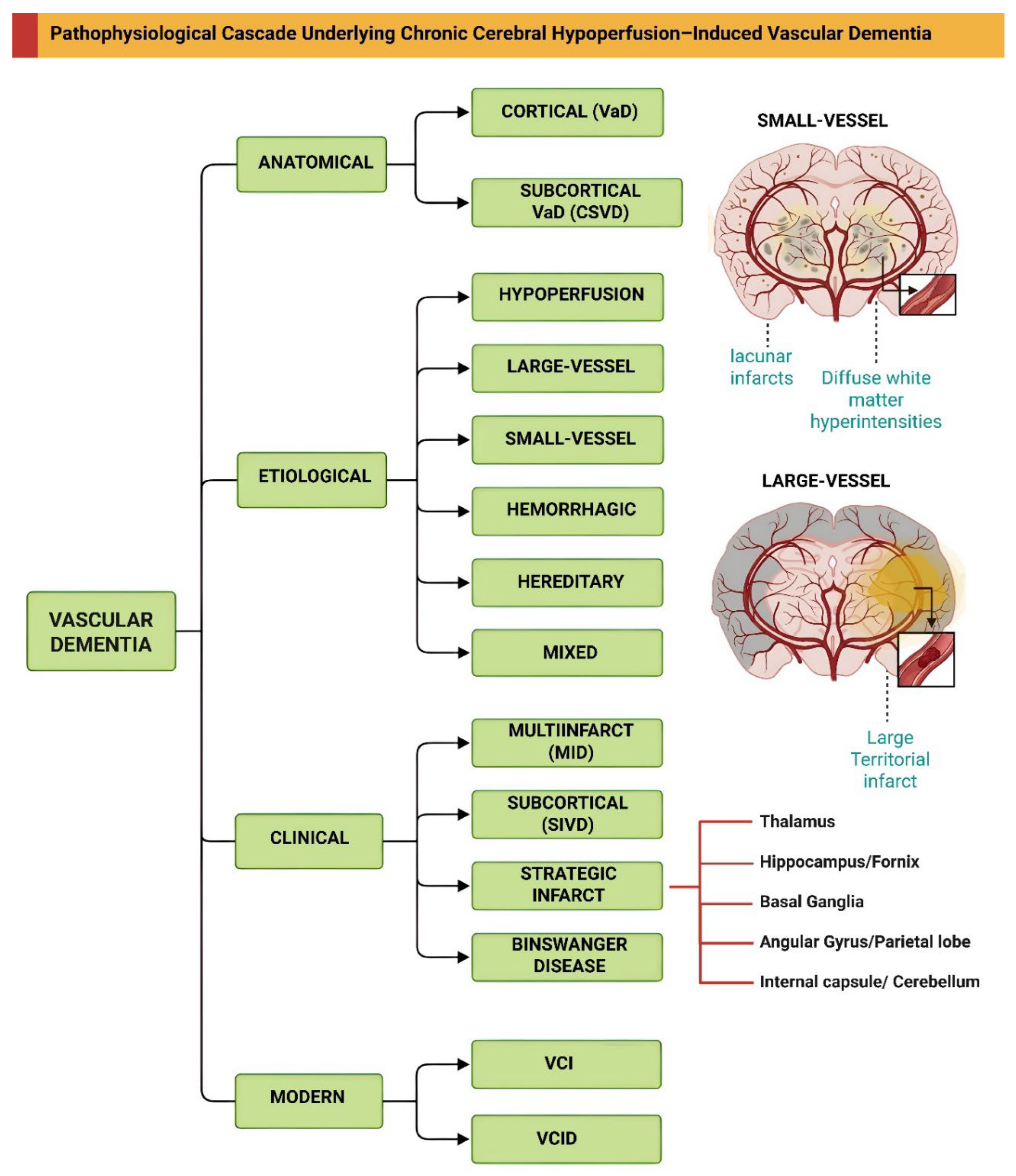
Figure 2.
Pathophysiological cascade underlying chronic cerebral hypoperfusion-induced vascular dementia. Vascular risk factors, including hypertension, diabetes, atherosclerosis, and aging, converge to initiate chronic cerebral hypoperfusion (CCH), characterized by sustained reductions in cerebral blood flow, tissue hypoxia, and metabolic stress. This hypoperfusion drives progressive neurovascular unit dysfunction, wherein endothelial dysfunction, marked by reduced endothelial nitric oxide synthase (eNOS) activity and nitric oxide (NO) bioavailability, leads to impaired vasodilation. Concurrently, elevated endothelin-1 (ET-1) signaling promotes calcium-dependent pericyte constriction, further compromising capillary perfusion. These vascular alterations directly precipitate blood–brain barrier (BBB) breakdown through downregulation of tight junction proteins such as claudin-5 and occludin, allowing extravasation of plasma components into the brain parenchyma. BBB disruption, in conjunction with oxidative stress and cellular damage, triggers robust glial activation. Astrocytes undergo reactive gliosis via JAK/STAT3 signaling, while microglia transition from a resting to an activated state through TLR4/MyD88-dependent NF-κB activation, releasing pro-inflammatory cytokines including TNF-α, IL-1β, and IL-6. This inflammatory milieu is amplified by chemokine-mediated recruitment of peripheral immune cells (CCL2–CCR2 axis, VCAM-1/ICAM-1) and by sustained interferon signaling involving IRF1 and interferon-stimulated genes (ISGs). Oligodendrocytes exhibit reduced myelin basic protein (MBP) expression and increased matrix metalloproteinase-9 (MMP-9) secretion, which degrades extracellular matrix and further compromises BBB integrity. These interconnected pathological events culminate in demyelination, axonal degeneration, neuronal loss and progressive white matter injury, which disrupts cortical–subcortical connectivity and drives the cognitive decline characteristic of vascular dementia.
Figure 2.
Pathophysiological cascade underlying chronic cerebral hypoperfusion-induced vascular dementia. Vascular risk factors, including hypertension, diabetes, atherosclerosis, and aging, converge to initiate chronic cerebral hypoperfusion (CCH), characterized by sustained reductions in cerebral blood flow, tissue hypoxia, and metabolic stress. This hypoperfusion drives progressive neurovascular unit dysfunction, wherein endothelial dysfunction, marked by reduced endothelial nitric oxide synthase (eNOS) activity and nitric oxide (NO) bioavailability, leads to impaired vasodilation. Concurrently, elevated endothelin-1 (ET-1) signaling promotes calcium-dependent pericyte constriction, further compromising capillary perfusion. These vascular alterations directly precipitate blood–brain barrier (BBB) breakdown through downregulation of tight junction proteins such as claudin-5 and occludin, allowing extravasation of plasma components into the brain parenchyma. BBB disruption, in conjunction with oxidative stress and cellular damage, triggers robust glial activation. Astrocytes undergo reactive gliosis via JAK/STAT3 signaling, while microglia transition from a resting to an activated state through TLR4/MyD88-dependent NF-κB activation, releasing pro-inflammatory cytokines including TNF-α, IL-1β, and IL-6. This inflammatory milieu is amplified by chemokine-mediated recruitment of peripheral immune cells (CCL2–CCR2 axis, VCAM-1/ICAM-1) and by sustained interferon signaling involving IRF1 and interferon-stimulated genes (ISGs). Oligodendrocytes exhibit reduced myelin basic protein (MBP) expression and increased matrix metalloproteinase-9 (MMP-9) secretion, which degrades extracellular matrix and further compromises BBB integrity. These interconnected pathological events culminate in demyelination, axonal degeneration, neuronal loss and progressive white matter injury, which disrupts cortical–subcortical connectivity and drives the cognitive decline characteristic of vascular dementia.
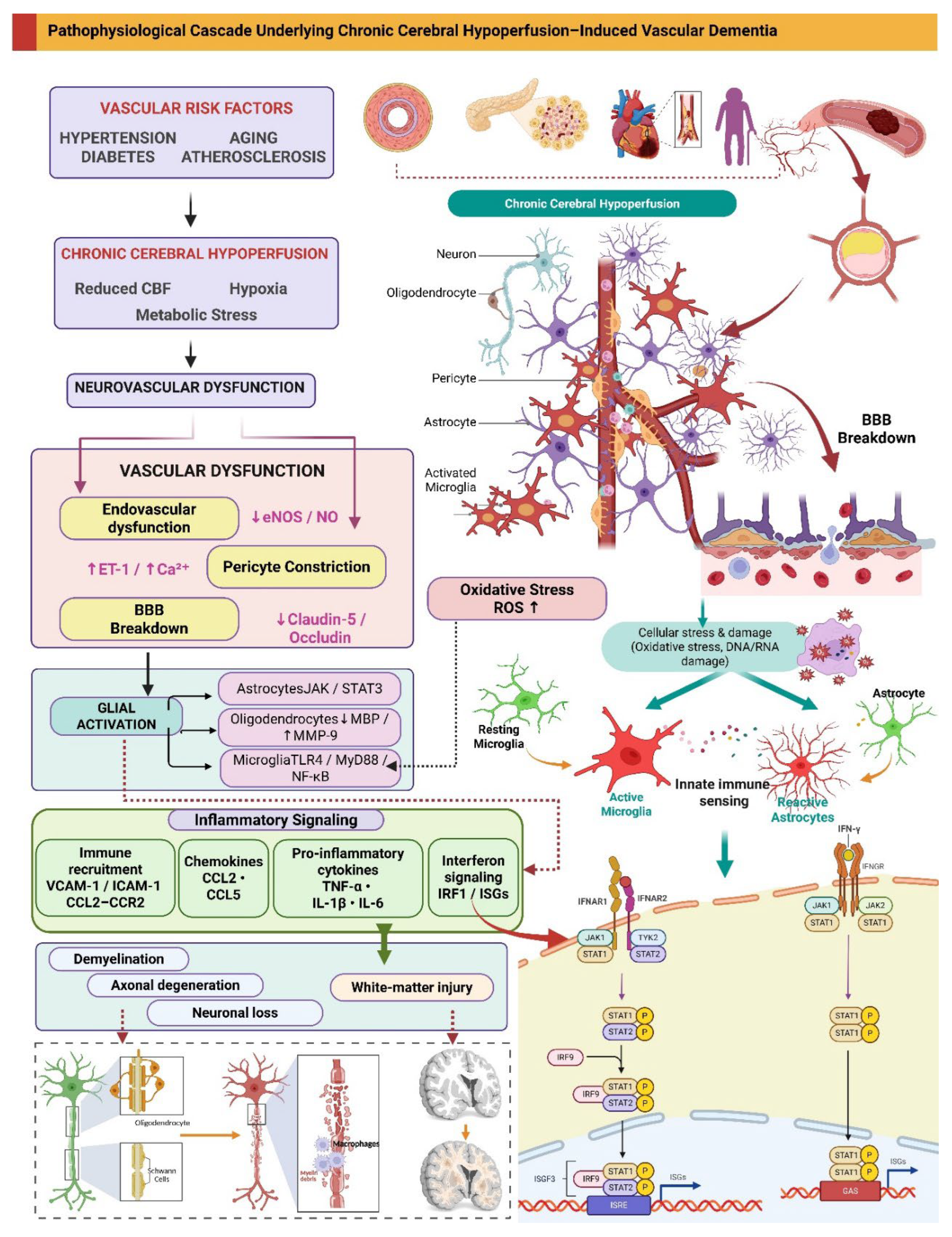
Figure 3.
Rat cerebrovascular anatomy and experimental strategies for inducing chronic cerebral hypoperfusion. (A) provides a schematic representation of normal rat cerebrovascular anatomy, highlighting the Circle of Willis, common, internal, and external carotid arteries (CCA, ICA, ECA), basilar artery (BA), and major cerebral branches. Panels (B–H) depict various surgical approaches for inducing chronic cerebral hypoperfusion (CCH). Panel (B) shows classical bilateral common carotid artery occlusion (BCCAo/2VO), which produces acute severe hypoperfusion. Panel (C) illustrates bilateral common carotid artery stenosis (BCAS) using microcoils, which induces gradual sustained flow reduction closer to human cerebrovascular disease. Panel (D) displays a variation of BCCAo involving distal transection of the common carotid arteries proximal to the bifurcation. Panel (E) demonstrates unilateral internal carotid artery occlusion (UICAO), while Panel (F) shows bilateral internal carotid artery occlusion (BICAO), both providing more direct upstream flow restriction. Panel (G) depicts unilateral common carotid artery occlusion (UCCAO), a mild model suitable for studying long-term vascular remodeling. Finally, Panel (H) illustrates a three-vessel occlusion (3VO) strategy combining bilateral CCA ligation with basilar artery occlusion; this approach eliminates the primary posterior collateral pathway via the Circle of Willis, producing a more severe and extensive global ischemic insult than 2VO alone. Note: In all illustrated models, occlusions are representative of the target vessels. While common carotid and vertebral arteries are typically directly ligated or clipped, middle cerebral artery (MCA) involvement is experimentally achieved via intraluminal suture filament insertion rather than direct transection, as depicted schematically.
Figure 3.
Rat cerebrovascular anatomy and experimental strategies for inducing chronic cerebral hypoperfusion. (A) provides a schematic representation of normal rat cerebrovascular anatomy, highlighting the Circle of Willis, common, internal, and external carotid arteries (CCA, ICA, ECA), basilar artery (BA), and major cerebral branches. Panels (B–H) depict various surgical approaches for inducing chronic cerebral hypoperfusion (CCH). Panel (B) shows classical bilateral common carotid artery occlusion (BCCAo/2VO), which produces acute severe hypoperfusion. Panel (C) illustrates bilateral common carotid artery stenosis (BCAS) using microcoils, which induces gradual sustained flow reduction closer to human cerebrovascular disease. Panel (D) displays a variation of BCCAo involving distal transection of the common carotid arteries proximal to the bifurcation. Panel (E) demonstrates unilateral internal carotid artery occlusion (UICAO), while Panel (F) shows bilateral internal carotid artery occlusion (BICAO), both providing more direct upstream flow restriction. Panel (G) depicts unilateral common carotid artery occlusion (UCCAO), a mild model suitable for studying long-term vascular remodeling. Finally, Panel (H) illustrates a three-vessel occlusion (3VO) strategy combining bilateral CCA ligation with basilar artery occlusion; this approach eliminates the primary posterior collateral pathway via the Circle of Willis, producing a more severe and extensive global ischemic insult than 2VO alone. Note: In all illustrated models, occlusions are representative of the target vessels. While common carotid and vertebral arteries are typically directly ligated or clipped, middle cerebral artery (MCA) involvement is experimentally achieved via intraluminal suture filament insertion rather than direct transection, as depicted schematically.
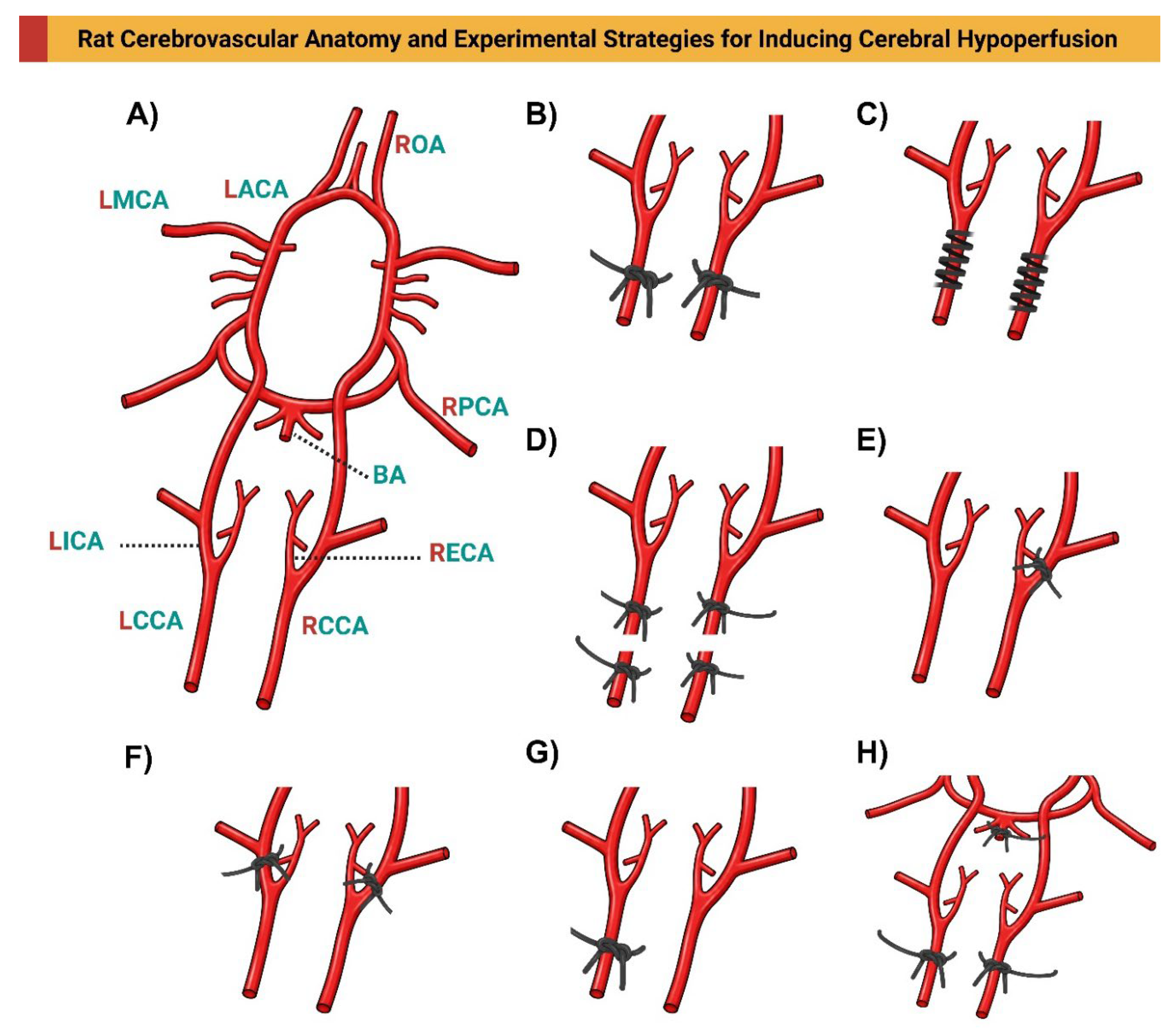
Figure 4.
Experimental strategies for inducing chronic cerebral hypoperfusion using three-vessel occlusion. This schematic illustrates three variations of the three-vessel occlusion (3-VO) model, each designed to produce global cerebral hypoperfusion by disrupting both anterior and posterior collateral blood flow. Panel (A) depicts the classical 3-VO approach, involving permanent ligation of the bilateral common carotid arteries (LCCA and RCCA) combined with occlusion of the basilar artery (BA). By eliminating the primary posterior circulation supply via the vertebrobasilar system, this strategy severely restricts compensatory flow through the Circle of Willis, resulting in significant ischemia in the hippocampus, thalamus, and white matter tracts. Panel (B) demonstrates an alternative 3-VO configuration wherein bilateral vertebral arteries (LVA and RVA) are occluded in conjunction with ligation of the right common carotid artery (RCCA). This strategy preserves some anterior circulation through the left common carotid artery while severely compromising posterior blood supply. (C) illustrates a third variation combining bilateral common carotid artery occlusion with occlusion of the right middle cerebral artery (RMCA). This strategy superimposes focal cortical ischemia upon global hypoperfusion, modeling the clinical scenario of chronic small-vessel disease complicated by acute territorial infarction. Each model’s specific vascular occlusion pattern critically determines the regional distribution and severity of hypoperfusion-induced brain injury. Note: The occlusions shown are schematic representations of target vessels. While the common carotid arteries are directly ligated, middle cerebral artery (MCA) occlusion is experimentally performed using the intraluminal suture filament method, rather than direct transection of the artery itself.
Figure 4.
Experimental strategies for inducing chronic cerebral hypoperfusion using three-vessel occlusion. This schematic illustrates three variations of the three-vessel occlusion (3-VO) model, each designed to produce global cerebral hypoperfusion by disrupting both anterior and posterior collateral blood flow. Panel (A) depicts the classical 3-VO approach, involving permanent ligation of the bilateral common carotid arteries (LCCA and RCCA) combined with occlusion of the basilar artery (BA). By eliminating the primary posterior circulation supply via the vertebrobasilar system, this strategy severely restricts compensatory flow through the Circle of Willis, resulting in significant ischemia in the hippocampus, thalamus, and white matter tracts. Panel (B) demonstrates an alternative 3-VO configuration wherein bilateral vertebral arteries (LVA and RVA) are occluded in conjunction with ligation of the right common carotid artery (RCCA). This strategy preserves some anterior circulation through the left common carotid artery while severely compromising posterior blood supply. (C) illustrates a third variation combining bilateral common carotid artery occlusion with occlusion of the right middle cerebral artery (RMCA). This strategy superimposes focal cortical ischemia upon global hypoperfusion, modeling the clinical scenario of chronic small-vessel disease complicated by acute territorial infarction. Each model’s specific vascular occlusion pattern critically determines the regional distribution and severity of hypoperfusion-induced brain injury. Note: The occlusions shown are schematic representations of target vessels. While the common carotid arteries are directly ligated, middle cerebral artery (MCA) occlusion is experimentally performed using the intraluminal suture filament method, rather than direct transection of the artery itself.
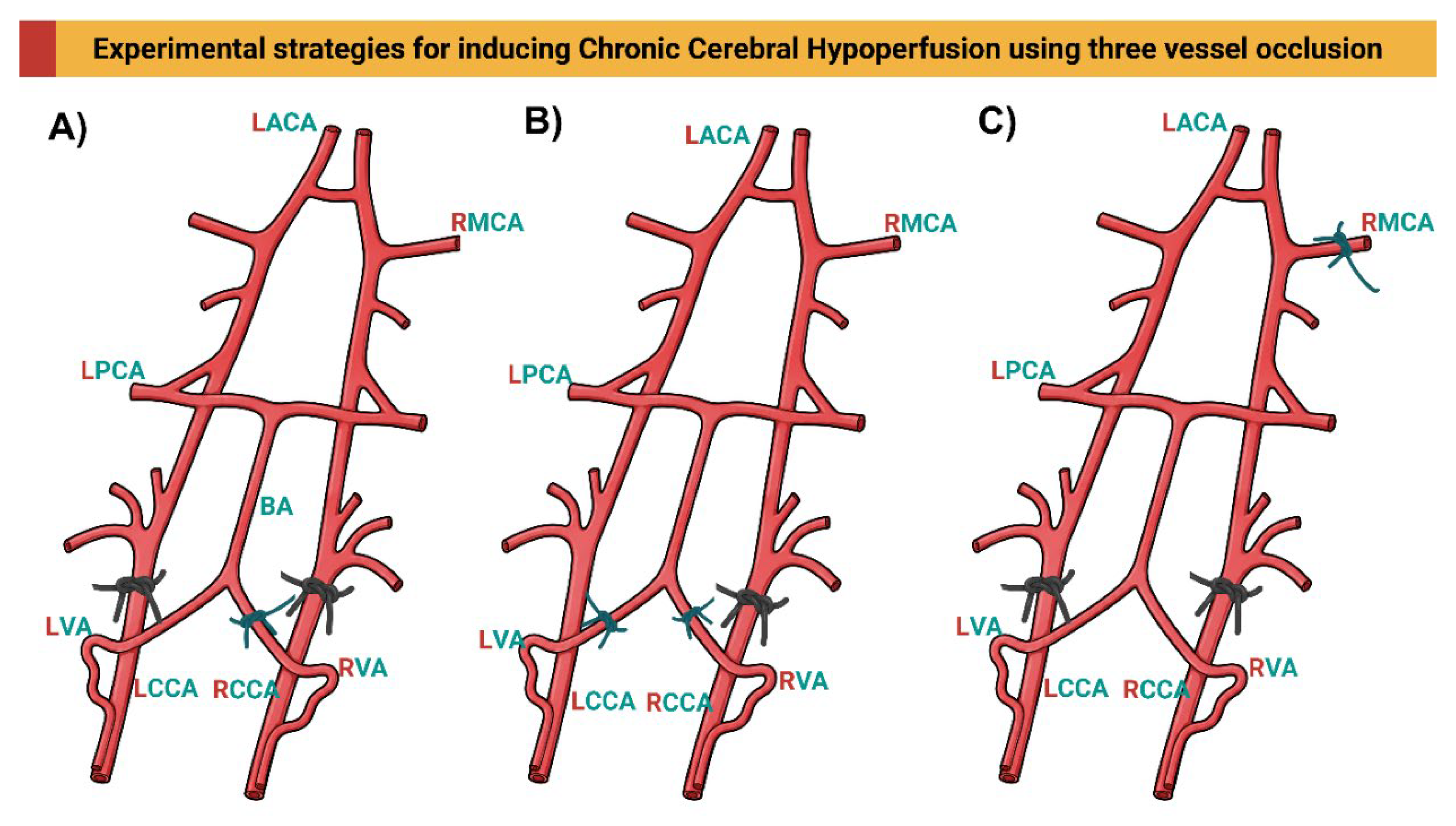
Figure 5.
Experimental strategies for inducing chronic cerebral hypoperfusion involving middle cerebral artery occlusion. This schematic illustrates three surgical paradigms that incorporate middle cerebral artery (MCA) occlusion, either alone or in combination with other major vessels, to model distinct patterns of ischemic injury. Panel (A) depicts the four-vessel occlusion (4-VO) model, wherein bilateral common carotid arteries (LCCA and RCCA) are permanently ligated in combination with occlusion of both the left and right vertebral arteries (LVA and RVA). This represents the most severe hypoperfusion insult among the models illustrated. Panel (B) demonstrates a combined two-vessel occlusion approach involving unilateral middle cerebral artery occlusion (RMCA) together with ipsilateral common carotid artery ligation (RCCA), i.e., on the same side as the MCAO. This configuration eliminates antegrade flow through the ipsilateral carotid system while preserving contralateral circulation, thereby reducing collateral compensation and producing significant unilateral cortical infarction with additional hemispheric hypoperfusion. Panel (C) illustrates isolated middle cerebral artery occlusion (MCAO) without concomitant carotid artery ligation. While this model primarily produces focal cortical infarction within the MCA territory, it also generates peri-infarct hypoperfusion and secondary neuroinflammatory changes that mimic acute ischemic stroke. Note: The occlusions shown are schematic representations of target vessels. While the common carotid arteries are directly ligated, middle cerebral artery (MCA) occlusion is experimentally performed using the intraluminal suture filament method, rather than direct transection of the artery itself. In practice, the 4-VO model is usually performed by permanent electrocautery of the bilateral vertebral arteries followed by transient clamping of the bilateral CCAs.
Figure 5.
Experimental strategies for inducing chronic cerebral hypoperfusion involving middle cerebral artery occlusion. This schematic illustrates three surgical paradigms that incorporate middle cerebral artery (MCA) occlusion, either alone or in combination with other major vessels, to model distinct patterns of ischemic injury. Panel (A) depicts the four-vessel occlusion (4-VO) model, wherein bilateral common carotid arteries (LCCA and RCCA) are permanently ligated in combination with occlusion of both the left and right vertebral arteries (LVA and RVA). This represents the most severe hypoperfusion insult among the models illustrated. Panel (B) demonstrates a combined two-vessel occlusion approach involving unilateral middle cerebral artery occlusion (RMCA) together with ipsilateral common carotid artery ligation (RCCA), i.e., on the same side as the MCAO. This configuration eliminates antegrade flow through the ipsilateral carotid system while preserving contralateral circulation, thereby reducing collateral compensation and producing significant unilateral cortical infarction with additional hemispheric hypoperfusion. Panel (C) illustrates isolated middle cerebral artery occlusion (MCAO) without concomitant carotid artery ligation. While this model primarily produces focal cortical infarction within the MCA territory, it also generates peri-infarct hypoperfusion and secondary neuroinflammatory changes that mimic acute ischemic stroke. Note: The occlusions shown are schematic representations of target vessels. While the common carotid arteries are directly ligated, middle cerebral artery (MCA) occlusion is experimentally performed using the intraluminal suture filament method, rather than direct transection of the artery itself. In practice, the 4-VO model is usually performed by permanent electrocautery of the bilateral vertebral arteries followed by transient clamping of the bilateral CCAs.
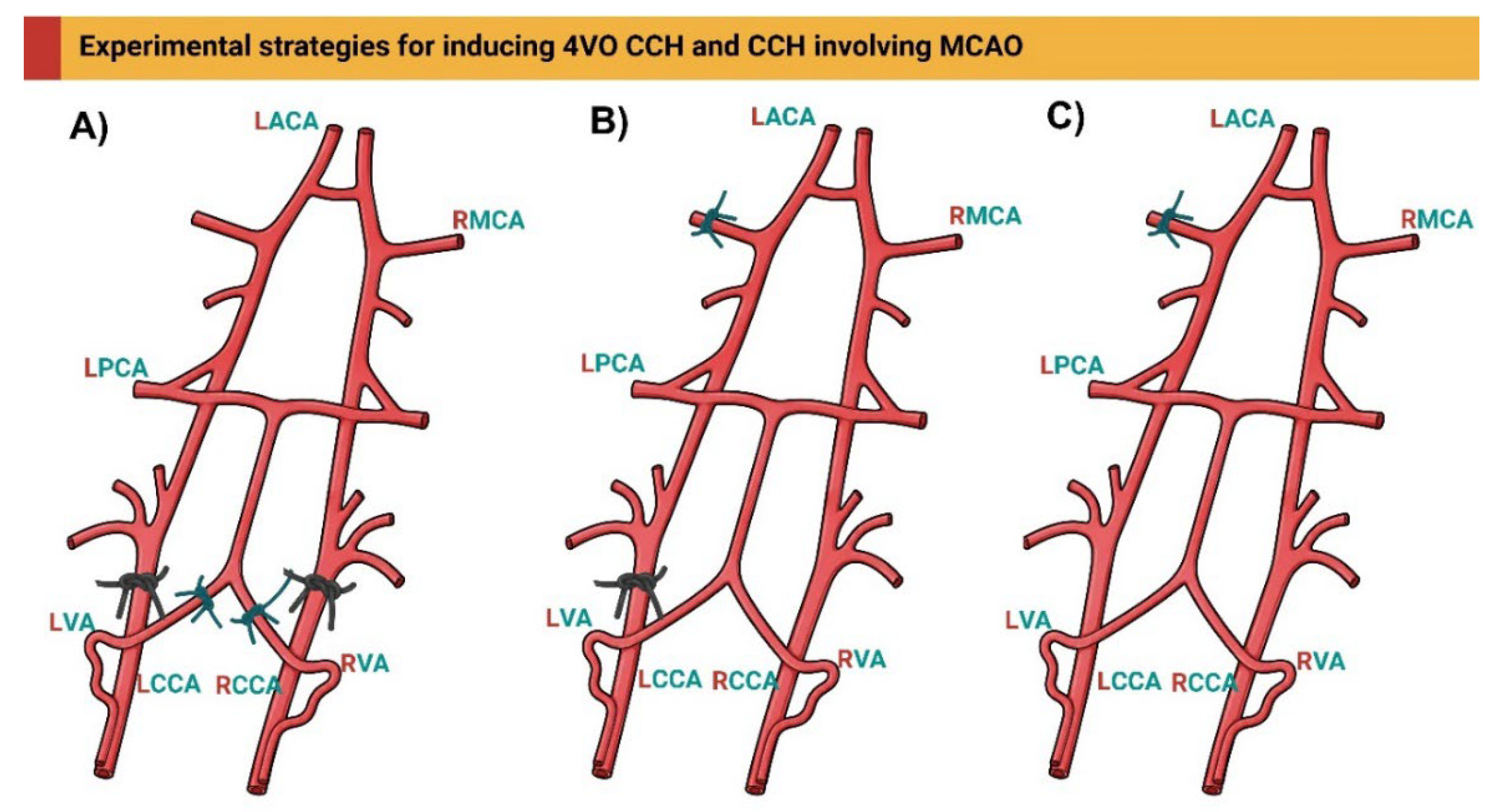
Disclaimer/Publisher’s Note: The statements, opinions and data contained in all publications are solely those of the individual author(s) and contributor(s) and not of MDPI and/or the editor(s). MDPI and/or the editor(s) disclaim responsibility for any injury to people or property resulting from any ideas, methods, instructions or products referred to in the content. |
© 2026 by the authors. Licensee MDPI, Basel, Switzerland. This article is an open access article distributed under the terms and conditions of the Creative Commons Attribution (CC BY) license (http://creativecommons.org/licenses/by/4.0/).
Copyright: This open access article is published under a Creative Commons CC BY 4.0 license, which permit the free download, distribution, and reuse, provided that the author and preprint are cited in any reuse.