
Submitted:
09 September 2026
Posted:
11 September 2026
You are already at the latest version
Abstract
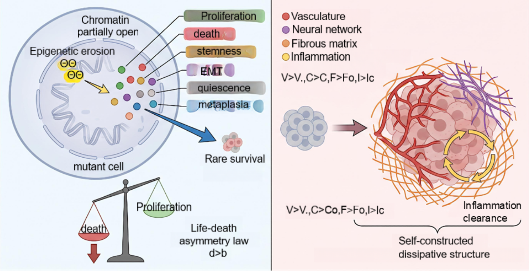
Tumors, especially malignant tumors, are diseases of uncontrolled proliferation. The most fundamental properties of malignant tumors, pervasive nongenetic heterogeneity, the necessity of supporting tissues, rarity of occurrence, and inevitable therapy resistant relapses, remain unexplained by mutation-centric models. Here, we provide a multiscale tumor model rooted in first principles. The central premise of the model is that globally arbitrary accesses of chromatins in a cell unleash a repertoire of stochastic gene expression modules, including proliferation, death, stemness, EMT, angiogenesis, neurogenesis, fibrogenesis, and inflammation, that compete for finite transcriptional resources and are antagonized by the differentiation program that drive the tumor cell along its differentiation pathway from what it originates. A tumor can only become clinically diseases when it successfully self-organizes a complete supporting stroma of blood vessels, nerves, connective tissues, and an inflammation-driven clearance cycle, thereby locking into a self-constructed, noise-driven, evolvable dissipative structure. The model visualizes extremely low probability of tumor formation across pediatric and adult contexts, and the post-treatment recurrence with progressive dedifferentiation and can display testable predictions, such as prognostically favorable and poor tumor cell subpopulations and therapeutic taxic. By providing a unified physical theory of tumors, the model transforms tumor biology from a descriptive science into a predictive one.
Keywords:
tumors
; globally arbitrary accesses of chromatins
; dissipative structure
; multiscale tumor model
; prognostically favorable tumor cell subpopulations
; prognostically poor tumor cell subpopulations
Introduction
Tumor tissues, pathological lesions in human body, consist of tumor cells and supporting tissues including blood vessels, neuro tissue, connective tissue, inflammatory cells and are the subjects of experimental and clinical practices 1-4. Tumor cells initiate and maintain the formation of tumor tissues in primary tissues and at remote sites via metastasis. Supporting tissues are essential for the formation of tumor tissues3-6. Lack of any elements in supporting tissues can halt tumor formation or inhibits development and progression of tumors. Accumulated data includes vast molecular details and clinical observations of tumors7,8. The data show, on one hand, that the transformation of a normal somatic cell into a tumor cell is an extremely rare event. On the other hand, once established, the tumor displays remarkable heterogeneity. Clinically, therapies can induce temporary remission of tumors but almost invariably leaves behind a minimal population of drug-tolerant residual cells that re-initiate tumor growth after a latency period 9,10. Strikingly, the relapsed tumors are typically less differentiated and more heterogeneous than the primary ones. After multiple cycles relapsed tumors may evolve into a completely undifferentiated, anaplastic, and ultimately lethal malignancy. Up to date, these observations are the results of cross-sectional, cellularly and molecularly phenotypic investigations and often remain dispersed in different contexts without unified logic conceptualizations3,7,8.
Gene expression plays key roles in a wide variety of core biological processes in tumor cells, ranging from tumor tissue development, homeostasis, and tumor cell specialization to cellular stress response 11. Previously, we analyzed single-cell sequencing datasets of human tumor tissues and identified that the number of genes expressed in a tumor cell delineates mRNA copy number, as well as the expression of oncogenes and tumor suppressor genes 3,12,13. Gene expression in tumor cells consists of both default networks and incidental networks. There are two kinds of default gene expression networks existing in a human tumor cell. One is the expression networks of housekeeping genes for surviving. For instance, the gene expression networks of energy production are key programs for cell survival. We identify that there is steady gene expression involved in tricarboxylic acid (TCA) cycle and ATP production, despite the alteration of the gene expressing activation and the gene expression networks responsible for glycolysis and oxidative phosphorylation among tumor cells in a tumor sample 12,13. The most characteristic default gene expression networks are the expression of differentiation genes that drive the tumor cell along its differentiation pathway from what it originates to become a mature cell 12,13. The incidental gene expression networks provide tumor cells with a variety of cellular states and response traits that coexist within one single tumor cell. These multiple cellular states include proliferation, apoptosis, autophagy, stemness, migration, senescence, quiescence. This coexistence of multiple states also allows tumor cells to organize and remodel supporting tissues, enabling processes such as epithelial-mesenchymal transition, neurogenesis, angiogenesis, connective tissue formation and remodeling, inflammatory responses, immune evasion, and metastasis 12,13.
Based on the transcriptional phenotypes in a tumor cell, we postulate that total gene expression in a tumor cell is the sum of two programs, a deterministic spectrum including differentiation program that preserves the memory of the cell’s origin and housekeeping gene expressing patterns, and a stochastic spectrum composed of multiple functional modules that are aberrantly activated due to globally arbitrary accesses of chromatins caused by genomic and epigenetic mutations and alterations, including single-nucleotide variants, small insertions or deletions, somatic mutations, gene copy number alterations, chromatin structural variants, chromatin regulator mutations, DNA methylation changes, and altered compositions of enhancers and repressors, and mutations in protein that modulate chromatin structure and activity of enhancers or repressors crossing genomes 14-17. We formalized the observations into a comprehensive model and illustrated tumors from globally arbitrary accesses of chromatins in a cell to therapeutic responses. The model of tumors yields a unified, quantitative account of tumor incidence, progression, therapeutic response, and relapses, thereby transforming tumor biology from a descriptive science to physical ones.
Results
Two biological inputs were used to build up a physical model of tumors. First, the total expressing genes determines total mRNA content in a tumor cell12. Secondly, the transcriptome in a tumor cell consists of a deterministic spectrum (the expression of housekeeping and differentiation genes) and a stochastic spectrum of genes that randomly activated functional modules via globally arbitrary accesses of chromatins12. The core of the model was a set of stochastic partial differential equations for the tumor volume fraction , a deterministic differentiation potential , and multiple independent incidental fields representing proliferation (prolif), death, stemness (stem), epithelial to mesenchymal transition (EMT), migration (mig), quiescence (quies), senescence (senes), neurogenesis (neuro), angiogenesis (ang), fibrogenesis (fibro), and inflammation (inflam). All kinetic rates, proliferation, death, metabolic consumption, and motility, were derived from the free-energy landscape, not imposed phenomenologically (Figure 1). The local state of the tumor tissue at position and time was described by the fields including : volume fraction of tumor cells,: differentiation potential encoding the degree to which tumor cells retain the differentiation program of their origin, : neurogenic differentiation potential of tumor cell,: endothelial differentiation potential of tumor cell,: a set of independent incidental functional fields, each , representing the abnormal stochastic activation of specific gene expression modules, the set ,: total mRNA copies of a tumor cell, : concentration of a generic nutrient (e.g., oxygen or glucose),: concentration of necrotic debris. target value depends on local nutrient concentration and stress . We then embed these elements into a thermodynamically self-consistent framework using conservation laws, a free-energy functional, and the fluctuation-dissipation theorem (Supplementary methods, table s1, s2).
Fundamental Model Construction
The total transcriptional potential of a tumor cell was defined as
where is the spatially uniform housekeeping potential (e.g. TCA cycle, ATP synthesis), is the differentiation potential (diff) pointing toward the cell of origin, and are potentials for neurogenic (ND) and endothelial (ED) differentiation, and are independent incidental fields of gene expression networks, each . The diff, ND, ED potentials have prescribed target values (, etc.) that depend on local micro-environmental conditions (e.g. nutrient , stress ), while the incidental fields have no target value (their free-energy minimum is zero) and are driven solely by internal noise. The total free energy of the system is
with the bulk free-energy density
The competition terms () enforce the observed inverse correlation between gene expression networks of differentiation and gene expression of incidental fields (Figure 2). High gene expression networks of differentiation suppress noise. Under low differentiation, noise is liberated. From the variational derivatives , we applied Onsager's principle and the fluctuation-dissipation theorem to obtain the incidental evolution equations. For example, the tumor volume fraction evolves as
and each incidental field satisfies
with noise correlations and . Effective noise temperatures quantify the intensity of transcriptional bursting for each module (Note S1). Thus, a model of tumors, the self-constructed, noise-driven, evolvable dissipative structure of tumors (SNEDS-T), is built up via mathematical and physical approaches.
Entropy Production in SNEDS-T
The model's thermodynamic consistency is rigorously established. We derived the local entropy production rate for the full system, showed that it decomposed into a sum of positive-definite contributions from diffusive transport and chemical reactions, and verified that it was evaluated without introducing any new parameters. The thermodynamic consistency of SNEDS-T follows as a direct consequence of its Onsager structure and the free-energy-grounded form of the reaction rates. This guarantees that SNEDS-T obeys the Second Law of Thermodynamics in every solution (Note S2).
The Life-Death Asymmetry Principle
SNEDS-T shows that the incidental gene expression networks of death module possess an inherent structural advantage. The promoters of genes that control death are CpG-poor and more permissive, leading to a higher derepressing rate and lower re-silencing rate. Moreover, the functional threshold for cellular death is lower than that for cell-cycle entry. Across almost all of phenotype space, the effective death rate exceeds the proliferation rate. Thus, the proliferation rate is derived from the free-energy barrier picture as a linear function of the fields that promote cell-cycle entry.
In contrast, the death rate receives super linear contributions from the stress-inducing incidental fields.
The super linearity of death arises because the energy barrier for death decreases as the squared amplitude of stress fields increases (Kramers' theory). Consequently, for sufficiently large total stochastic activity , the net growth rate is always negative. It is a principle of the life-death asymmetry in SNEDS-T (Figure 3). Intrinsic noise is far more likely to kill a cell than to make it divide (Note S3).
Probability Barriers of Tumor Formation
For a mutated cell to establish a growing tumor, it must simultaneously achieve such as , (to drive proliferation), or (to build blood vessels), , (to avoid suicide). Because the incidental fields are independent and their fluctuations are governed by the Boltzmann distribution , the joint probability is the product of individual tail probabilities
Tumor formation is not a single stochastic event but a sequential traversal of nested probability barriers (Figure 4). Each barrier represents a distinct bottleneck that a mutated cell and its progeny must overcome to give rise to clinically manifest disease.
The single-cell survival filter is the first and most fundamental barrier. Immediately after an oncogenic mutation, the mutated cell must survive its own intrinsic instability caused by the mutation-induced activation of random gene expression. In SNEDS-T, oncogenic mutation is modeled as a sharp increase in the effective noise temperatures for multiple random fields, coupled with a weakening of the restoring forces . The cell's total stochastic activity surges. By the life-death asymmetry principle, the death rate responds as
The probability that a single mutated cell survives this initial crisis is the probability that its net growth rate remains positive. This requires a rare, favorable combination of random field values, in which proliferation and stemness fields moderately elevate while death and inflammation fields remain low. This is a classic tail event of a high-dimensional Boltzmann distribution.
The vast majority of mutated cells, estimates suggest over 99.9%, are eliminated by this barrier alone. The cell's own transcriptional instability, not immune surveillance, is the first and most potent tumor suppressor mechanism.
Secondly, a single surviving cell that begins to proliferate forms a micro-aggregate of a few hundred cells. At this scale, the cellular aggregate is avascular and must rely on passive diffusion for nutrient supply. Simultaneously, the aberrant proliferation and incipient genomic instability trigger local innate immune responses. The nascent micro-aggregate lacks any self-constructed microenvironment. In the nutrient equation, the supply terms are limited to basal vascular diffusion.
Without angiogenic (), neurogenic (), or fibrotic () fields, the aggregate cannot sustain nutrient influx as it grows. Central nutrient depletion triggers a collapse of the differentiation potential , which in turn releases the suppression on death and inflammation fields. Simultaneously, the aggregate's limited size and lack of immunosuppressive stroma leave it highly vulnerable to immune attack by immune cells. The probability of surviving this stage is the joint probability that the aggregate remains below the immune detection threshold long enough to develop protective mechanisms, while also avoiding lethal inflammation.
This barrier is particularly potent in immunocompetent hosts and explains the dramatically increased cancer incidence in immunosuppressed patients. It also explains why tumors that do emerge have already been immune edited to be less immunogenic. This barrier is less stringent in immunologically privileged sites and in hereditary cancer syndromes where immune surveillance may be compromised.
At third stage, to grow beyond the diffusion-limited size of approximately 1-2 mm in diameter, the micro-tumor must acquire the ability to construct its own supportive microenvironment. This includes angiogenesis ( and/or ), fibrosis (), neurogenesis ( and/or ), and inflammation-mediated clearance of necrotic debris (). It is the most stringent of the barriers in tumor formation. The tumor must simultaneously co-activate multiple independent, functionally distinct random fields, each of which is a rare stochastic event. The probability is the product of independent tail probabilities.
Each field's activation requires a fluctuation of its amplitude several standard deviations above its mean (activation ratio ). With four fields, the combined probability is as SNEDS-T further predicts that without this barrier being traversed, the nascent tumor inevitably undergoes spontaneous regression, consistent with the phenomenon of stage 4S neuroblastoma and the clinical and experimental observations that avascular tumors cannot grow beyond microscopic size. It also explains why therapies targeting a single microenvironmental pathway often fail.
At stage four, the somatic mutation accumulation filter applies specifically to sporadic adult tumors and is the only barrier that is not a single stochastic event but a cumulative process occurring over decades. It represents the time required for a cell to accumulate the requisite set of somatic mutations that collectively raise the effective noise temperatures and weaken the restoring forces . In the SNEDS-T model, the probability of tumor formation is an explicit function of the noise parameters.
Each successive oncogenic mutation incrementally raises or lowers . The probability of traversing Stages 1-3 remains negligible until the cumulative mutational burden crosses a critical threshold.
The time to accumulate this threshold number of mutations is determined by the spontaneous somatic mutation rate per cell division, the number of stem cell divisions in the tissue of origin, the selective advantage conferred by each intermediate mutation, and the efficiency of DNA repair mechanisms. This is the only barrier that operates on a timescale of decades, providing a natural explanation for the age-dependent incidence of most sporadic carcinomas. The barrier explains the dramatic difference in tumor probability between childhood and adult cancers. Childhood tumors often arise from congenital mutations (germline or very early somatic mutations) that effectively bypass Stage 4, already having elevated at birth. Adult tumors require the slow, stochastic accumulation of somatic mutations over 40-70 years to reach the same threshold. The phenotype also explains the exponential increase in cancer incidence with age, a relationship that has been empirically observed across all human populations.
The overall probability of a single mutated cell giving rise to a clinical tumor is the product of the probability of traversing each barrier The central estimates from the ranges above are as For a childhood tumor arising from a cell with pre-existing elevated noise temperatures. For an adult tumor requiring decades of mutation accumulation
These numbers are in remarkable agreement with clinical observations. For childhood congenital tumors, inherited mutations reduce the first barrier and developing tissues provide partial stromal support, giving. For adult familial cancers and genetically engineered mice that must build all stroma autonomously, giving. These numbers quantitatively match the observed incidence rates and explain why hundreds of millions of mutated cells typically yield a single clinical tumor in human bodies (Note S4).
Tumors as Diseases of Aberrant Tissue Formation
SNEDS-T suggests that cancer is not primarily a disease of uncontrolled proliferation, but a disease of aberrant tissue formation. The model identifies the true bottleneck for malignant progression as the capacity filter of tissue formation. A mutated cell can acquire all the classical hallmarks of cancer, such as uncontrolled proliferation, evasion of apoptosis, metabolic reprogramming. If a tumor cannot construct a supportive tissue microenvironment, its progeny will inevitably regress through the nutrient crisis, differentiation collapse, death surge cascade described in the previous sections. A tumor is not merely a collection of mutated cells, but a pathological organoid that must be actively built, maintained, and remodeled through the stochastic co-activation of tissue formation programs. The model identifies essential construction programs, each represented by a specific random field, that must be co-activated for a tumor to establish itself as a stable, macroscopic organ-like structure (Figure 5).
The construction of a dedicated vascular network is the most fundamental requirement for tumor growth beyond the diffusion-limited size. In SNEDS-T, it is captured by the nutrient supply terms Without these terms, the nutrient equation lacks positive feedback, and the tumor is trapped in the avascular regime where regression is thermodynamically mandated. The stochastic activation of the angiogenic field is thus not merely an adaptive response to hypoxia but a prerequisite for crossing the phase boundary between a transient cellular aggregate and a stable tissue mass.
The construction of a fibrotic stroma serves multiple functions that are essential for tumor survival and progression. In SNEDS-T, the fibrotic field modulates tissue mechanics through permeability and active stress terms.
Without adequate fibrotic construction (), the tumor is mechanically fragile and immunologically exposed, significantly lowering its probability of survival.
The construction of a tumor-associated neural network is the most recently appreciated and perhaps most insidious of the construction programs. SNEDS-T captures this through the neurogenic random field and the deterministic neurogenic differentiation potential . These fields contribute to nutrient supply () and modulate tissue permeability (). The stochastic activation of the neurogenic program is particularly harmful for hosts because the neural processes create a positive feedback loop of neural reactions. Neural activity stimulates tumor growth, which in turn promotes further nerve infiltration, progressively integrating the tumor into the host's nervous system.
An essential program of tissue formation is the ability to manage the consequences of the tumor's own high cell turnover. Necrotic cell death generates debris that must be cleared to prevent toxic accumulation and destructive inflammation. SNEDS-T captures this through the debris clearance term.
Without adequate inflammatory clearance (), necrotic debris accumulates, generating osmotic stress () and releasing damage-associated molecular patterns that trigger uncontrolled inflammatory death. This creates the positive feedback loop that drives construction-deficient aggregates toward regression. However, the inflammatory program is a double-edged sword. Excessive can itself be destructive, while the appropriate level of inflammation remodels the immune microenvironment into a tumor-promoting state. SNEDS-T predicts that successful tumors achieve a delicate balance of inflammation reactions. Sufficient inflammatory activities clear debris and remodel the stroma, but not so much trigger immune rejection or destructive inflammation. This balance is reflected in the chronic, smoldering inflammation that are characterized in most tumors.
Spontaneous Regression Without Microenvironmental Self-Construction
In the absence of sufficient angiogenic (), neurogenic (), and fibrogenic () activity, the nutrient equation lacks the positive feedback terms . As the tumor volume fraction grows, the diffusive supply cannot keep pace with consumption, and the central drops. Low reduces the differentiation target , causing to reduce toward zero. This removes the competitive suppression () on all incidental fields, which then explode under their intrinsic noise. The death rate is vastly expended, driving and rapid regression of the cell clusters. This mechanism explains the spontaneous regression of stage 4S neuroblastoma and the clinical observation that avascular tumors rarely exceed 1-2 mm in diameter. It also implies that the acquisition of microenvironmental remodeling capacity is the true bottleneck for malignant progression of tumors.
Irreversible Dedifferentiation Under Therapy
SNEDS-T predicts that chemotherapy and radiotherapy can be modeled as perturbations that increase the effective noise temperatures and/or weaken the restoring forces (e.g., by causing DNA damage, proteotoxic stress, and epigenetic scrambling, etc.). The immediate effect is a dramatic increase in , , and other stress fields, killing the vast majority of tumor cells (, tumor shrinkage). However, tiny subsets of tumor cells may, by the stochastic combination of their fields, express high (low proliferation, hence resistance to antimitotic agents), significant contents of (survival and plasticity), and carry a varied degree of (from undifferentiation to partial differentiation, not fully committed). These cell sets survive because their (balanced proliferation and death). After treatment cessation, these cells can stochastically reactivate and microenvironmental fields, repopulating the cell masses into tumors. Crucially, because they were selected from the low- tail of the original distribution, the new tumor has a lower average . The competition terms determine that this lower differentiation is self-reinforcing. Each cycle of therapy and relapse causes a monotonic decrease in the differentiation potential, driving the system . This is the physical origin of anaplastic progression and the increasing malignancy observed in relapsed cancers (Figure 6).
Poor and Favorable Prognosis of Tumor Subpopulations
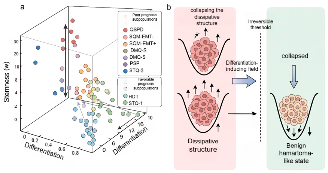
SNEDS-T rigorously predicts the existence of multiple distinct subpopulations of tumor cells associated with poor and favorable prognosis. The populations of tumor cells share a common physical origin and occupy specific local minima (attractors) in the free-energy landscape that corresponds to undifferentiated or partially differentiated states with elevated stochastic functional fields. In the model’s free-energy landscape, two global minimums exist. The terminally differentiated attractor: , all . Tumor cells are functional, non-proliferative, non-invasive and are associated with favorable prognosis. The undifferentiated attractor: , multiple significantly non-zero (, etc.). Tumor cells are malignant and are associated with poor prognosis of patients. Between these two extremes lie multiple metastable partially differentiated attractors, each defined by a specific combination of elevated random fields. Tumor cells occupying these attractors possess key deleterious capabilities (quiescence, stemness, lineage plasticity, inflammation) and are strongly associated with poor prognosis. The model further predicts that the subpopulation of tumor cells directly correlates with disease-free survival, making them a mechanistic biomarker for poor or favorable prognosis (Figure 7a) (Note S5-7).
Quiescent Stem-like Partially Differentiated (QSPD) Cells
QSPD cells are defined by high stemness, high quiescence, partial differentiation, low proliferation, and no EMT or metaplasia. They arise when quiescence and stemness are simultaneously activated against a background of incomplete differentiation. Stemness suppresses apoptosis; quiescence blocks cell-cycle entry. They survive conventional therapies because of their non-proliferative state and anti-apoptotic protection. QSPD cells constitute a reservoir for late local or regional relapse. Their prognostic effect may be masked in the short term by dormancy but becomes apparent at intermediate-to-long follow-up.
Quiescent Stem-like Metaplastic Cells Without EMT (SQM-EMT-Negative)
These cells combine high stemness, high quiescence, metaplasia, and no EMT programs. Metaplasia adds lineage instability and phenotypic plasticity. Without EMT, the cells remain constrained within the epithelial compartment, which delays distant dissemination but does not eliminate malignant potential. They are associated with treatment resistance and local recurrence, with a tendency toward late relapses. Their short-term prognostic effect may be weak or absent because their risk is released only after awakening and possibly after EMT acquisition.
Quiescent Stem-like Metaplastic Cells with EMT (SQM-EMT-Positive)
These cells are defined by high stemness, high quiescence, metaplasia, and active EMT programs. Simultaneous activation of stemness, metaplasia, and EMT creates a highly plastic, dissemination-competent, dormant-like state. EMT confers invasiveness and resistance to anoikis, enabling early distant spread even while proliferation is low. SQM-EMT-positive cells are predicted to be the strongest poor-prognosis population. Their presence correlates with early metastatic relapses, multiorgan dissemination, and markedly reduced overall survival.
True Malignant Proliferative Stem-like Partially Differentiated (PSP) Cells
True malignant PSP cells combine high stemness with self-locking, partial differentiation, active proliferation, and no EMT or quiescence. When the stemness module becomes self-sustaining and the differentiation potential is flattened, cells can simultaneously renew and proliferate. The cells are able to produce an expanding pool of malignant progenitors that continuously generate more tumor cells and serve as a precursor to more aggressive states. Their presence indicates active tumor evolution and is associated with poor disease-free survival and risk of phenotypic transition to more aggressive states.
Inflammatory Metaplastic Tumor (IMT) Cells
Inflammatory metaplastic tumor cells are defined by high inflammation, metaplasia, and variable stemness, EMT, and proliferation. They form four subtypes. Inflammatory metaplastic proliferative cells combine inflammation, metaplasia, stemness, and proliferation without EMT and can drive rapid local progression. Inflammatory metaplastic EMT cells combine inflammation, metaplasia, stemness, and EMT, representing the most invasive and metastatic combination. Inflammatory metaplastic quiescent cells combine inflammation, metaplasia, and quiescence without proliferation and are short-term indolent but long-term dangerous. Inflammatory metaplastic high-differentiation cells combine inflammation with metaplasia toward terminal differentiation and low stemness and are the only IMT subtype with relatively favorable prognosis.
Differentiated Metaplastic Quiescent Stem-like (DMQ-S) Cells
These cells combine high differentiation, metaplasia, high quiescence, and high stemness, with variable EMT. They represent a triple-locked state of stemness, metaplasia, and quiescence. DMQ-S EMT-negative cells are constrained to the epithelial compartment and act as deep reservoirs for late relapses. DMQ-S EMT-positive cells additionally possess invasive capacity and are among the most dangerous cell types, driving early metastasis and treatment resistance. Differentiation markers do not mitigate the risk conferred by stemness and metaplasia. Instead, they may cause underestimation of malignant potential in routine pathology.
High-Differentiation Terminal-like (HDT) Cells
HDT cells are defined by high differentiation past an irreversible threshold, low stemness, low proliferation, no EMT, no metaplasia, and low stochastic activity. These cells exit the reversible phenotypic space and no longer contribute to tumor growth or evolution. They represent the principal good-prognosis population. A tumor composed predominantly of HDT cells is approaching a benign, hamartoma-like state.
Microenvironment-Normalizing Cells
These cells have intermediate differentiation, low stemness, low EMT, and low proliferation, but actively secrete normalizing extracellular matrix components and anti-angiogenic or immunoregulatory factors. They act as endogenous stabilizers that pull stromal fields below critical thresholds and destabilize the malignant dissipative structure. Their presence correlates with lower vascular density, reduced fibrosis, and improved prognosis.
Stable Transdifferentiated Quiescent (STQ-1) Cells
STQ-1 cells are defined by high differentiation, stable mature transdifferentiation, no stemness, no EMT, and quiescence. They complete transdifferentiation toward a mature, functional epithelial lineage and then exit the cell cycle. Their quiescence is the natural consequence of terminal differentiation. They are associated with good prognosis, although their protective effect is weaker than that of HDT cells because the history of lineage reprogramming implies residual plasticity.
Aberrant Transdifferentiated Quiescent (STQ-2) Cells
STQ-2 cells display high differentiation, aberrant or incomplete transdifferentiation, no stemness, and quiescence. They carry the memory of an incomplete lineage switch, often with mixed or non-physiological marker expression. Their quiescence is partly natural and partly stress induced. They have residual phenotypic reversibility and may re-enter the cell cycle when the local microenvironment changes. They are associated with intermediate prognosis and represent a potential source of late recurrence, particularly in chronically inflamed or previously treated tissues.
Stress-Induced Transdifferentiated Quiescent (STQ-3) Cells
STQ-3 cells are characterized by high differentiation, stress-induced or therapy-induced transdifferentiation, no stemness, and quiescence. These cells arise as an escape response to therapy, hypoxia, or metabolic stress. They upregulate anti-apoptotic proteins and enter a protective dormant state. Although they lack stemness markers, they retain the ability to re-activate proliferation and to acquire stemness or EMT features upon awakening. Their presence is a strong predictor of late relapse after treatment and secondary therapeutic resistance. Because they do not express conventional stemness markers, STQ-3 cells are easily missed by standard biomarker panels, representing a diagnostically silent but clinically dangerous population.
Toxic Reactions and Curative Strategy of Tumor Therapies
In SNEDS-T, normal tissue is characterized by high deterministic differentiation potential (fully differentiated state), near-zero incidental fields for all , low effective noise temperatures and strong restoring forces . Host toxicity of tumor therapies is defined as any perturbation that destabilizes the normal constitution of hosts. It raises the effective noise temperatures , weakens the restoring forces , or lowers the differentiation target in normal cells. Such perturbation increases the probability of stochastic activation of stress fields (), causing death of healthy cells, tissue damage, or aberrant transdifferentiation. Thus, the toxicity potential of a therapy can be quantified by the magnitude of and inducing in normal tissue compartments. Therapies that act by elevating noise are predicted to cause severe host damage such as cytotoxic chemotherapy (e.g., DNA-damaging agents). They massively increase the effective noise temperature for death and stress modules in both tumor and normal cells, leading to apoptosis of rapidly dividing normal cell populations. Radiation therapies similarly raise in the irradiated field, damaging normal tissue barriers. Mathematically, the death rate in normal tissue under such therapies becomes as follow.
where is driven upward by the elevated . This causes dose-limiting toxicity (Note S8). The predictions of tumor therapies that cause highly toxic reactions to hosts are completely in agreement with clinical observations 18-21.
SNEDS-T predicts that the ideal curative strategy with minimal host toxicity is differentiation therapy to remodel the free-energy landscape of tumor tissues so that the terminally differentiated state ( high, all ) becomes the unique, globally stable attractor for tumor cells, without raising the noise levels in normal tissues. The strategy is achieved by using transient, targeted perturbations that raise the free energy of the low-, high-noise state without affecting the landscape of normal tissues in hosts. All the approaches of differentiation therapies are required to reinforce the differentiation potential to increase the restoring force and/or the target specifically in tumor cells, to lower the effective noise temperatures for proliferation, stemness, and death modules through chromatin stabilization or metabolic normalization, applied selectively or at low doses, and to eliminate the undifferentiated attractor. Because normal cells already reside in the high-differentiation attractor, they are thermodynamically unresponsive to such interventions. Their is already at the target, and their noise is already minimal. Therefore, differentiation therapies cause negligible toxicity while irreversibly driving tumor cells into a benign, post-mitotic cellular state (the clinical equivalent of a cured state or a benign hamartoma) (Figure 7b) (Note S8).
Discussion
Up to date, many mathematical and physical models have dissected cancer complexity, visualizing spatial and temporal levels from subcellular signaling in a tumor cell to tumor tissues 22,23. For instance, reaction-diffusion equations are used to describe avascular tumor growth for nutrient-limited proliferation and cell motility 24 and invasion of malignant cells that acid-mediated microenvironmental remodeling can produce a traveling wave of malignant cells 25. Carcinoma growth in the nonlinear regime using Phase-field simulations demonstrate that critical conditions exist for which the tumor evolves to nontrivial dormant states or grows self-similarly, leading to the possibility of shape control and of controlling the release of tumor angiogenic factors by restricting the tumor volume-to-surface-area ratio 26, extending by coupling tumor cell dynamics with angiogenic factors and nutrients 27,28. Phase-field models are also used to resolve cell-cell adhesion and interfacial tension for the transition from compact spheroids to fingering, invasive patterns of tumor cells. A macroscopic mechanical model of solid tumor can account for several complex interactions. How tumor growth can be influenced by stress, how and where it can generate cell reorganization to release stress levels, how it can lead to capsule formation and compression of the surrounding tissue 29. Nonlinear numerical simulations showed that the range of morphological responses can be placed in three categories that depend primarily upon the tumor microenvironment. The qualitative behavior of the tumor morphologies was similar across a broad range of parameters that govern the tumor genetic characteristics 30. Those physical models are used to underline that mechanical confinement is not merely a passive consequence but an active driver of invasion and therapy resistance.
When cellular heterogeneity and individual behavior of tumor are major points concerned, many models are developed to solve the heterogeneity of tumor tissues. A three-dimensional cellular automaton model simulates Gompertzian growth for brain tumor growing over nearly three orders of magnitude in radius and demonstrates the flexibility of the model by showing the emergence, and eventual dominance, of a second tumor clone with a different genotype 31,32. Agent-based models and simulations recapitulate the dynamics of neoplasia and tumor - host interactions and leverage high-fidelity simulations of multi-scale, or multi-level, cancer models with a focus on verification approached as simulation calibration 33. An open-source physics-based cell simulator is used to incorporate mechanics-driven cell motion, cell-cell adhesion, and substrate consumption in tumor tissues 34. A hybrid mathematical model of the invasion of healthy tissue by a solid tumor is used to examine how individual-based cell interactions can affect the tumor shape, linking an individual-based cell layer with reaction-diffusion equations for matrix-degrading enzymes and nutrients for invasion of tumor cells 35. This paradigm has been expanded into large-scale systems that simultaneously simulate angiogenesis, immune infiltration, and therapy responses 36,37. Cancer evolves under selection pressures, a process matching the evolutionary game theory 38. These game-theoretic models and many other models including population dynamic models, hylogenetic methods and probabilistic graphical models, help to understand how tumors arise, play an increasingly important prognostic role in predicting disease progression and the outcome of medical interventions, such as targeted therapy and provide a theoretical foundation for evolution-informed oncology 39.
A continuous and discrete mathematical model describes the formation of the capillary sprout network in response to chemical stimuli supplied by a solid tumor and can track individual endothelial cells at the sprout tips and incorporate anastomosis, mitosis and branching explicitly into the model40. The theoretical capillary networks generated by computer simulations of the discrete model are compared with the morphology of capillary networks in vivo experiments 40. Later hybrid models are used to integrate the angiogenesis scheme with continuum descriptions of tumor growth, oxygen transport, and vessel remodeling, enabling simulation of tumor-vascular co-evolution35,36. These models reveal how vascular architecture shapes intratumoral hypoxia and influences treatment delivery in tumor tissues. There are also many mathematical and physical models to describe the interaction between the immune cells and tumor cells. For example, the interplay between tumor cells and the immune system has been captured by ordinary differential equation models that describe effector cell recruitment, tumor kill, and immune suppression 41-43. Taken together, the mathematical and physical modeling of cancer has evolved from simple logistic growth laws to sophisticated multiscale, multiphysics frameworks. However, the models could not unify the origin of heterogeneity, the inevitability of death in tumor tissues, the imperative of stroma co-construction, and the therapeutic collapse. They describe what a tumor is, but not why it must be so, nor how it can be physically done.
In present work, we show a first-principles physical tumor model, SNEDS-T that unifies tumor initiation, heterogeneity, microenvironmental dependence, therapeutic response, and malignant progression. By grounding SNEDS-T in conservation laws, a free-energy functional, and the fluctuation-dissipation theorem, we eliminate the needs of arbitrary growth laws or ad hoc cell-state classifications. The life-death asymmetry is the engine of both tumor suppression and therapeutic vulnerability. The same noise that generates phenotypic diversity overwhelmingly drives cell death. Only cells that manage to construct a supportive microenvironment, via stochastic activation of angiogenesis, neurogenesis, and fibrogenesis, can overcome this intrinsic death bias and establish a macroscopic tumor. SNEDS-T reframes the hallmarks of cancer not as independent acquired capabilities, but as necessarily co-activated rare events required to escape the default fates of regression.
SNEDS-T explores intertumoral and intratumor heterogeneity, microenvironmental dependency, probabilistic onset, and therapy-resistant relapses in a self-constructed, noise-driven, evolvable dissipative structure. SNEDS-T does not rely on free parameters. All quantities of SNEDS-T are experimentally measurable. Gene-switch rates, transcriptional burst sizes, diffusion coefficients, and secretion rates in tumor make the model fully falsifiable. For instance, SNEDS-T predicts many novel cell populations in tumor tissues, such as tumor cell populations carrying markers of stemness, differentiation, transdifferentiations and cell cycle quiescence. SNEDS-T also predicts that tumor cell populations with identical core markers and with different EMT programs are predicted to be associated with different outcomes of patients. In addition, SNEDS-T predicts prognostically favorable tumor cell subpopulations in tumor tissues. Analysis of epithelial cancer cells in colorectal carcinoma shows that a couple of epithelial cancer cell subpopulations are prognostically favorable or poor for patients, match the model predictions, and provide evidence to confirm the model (manuscript in preparation). SNEDS-T predictions of highly toxic reactions to hosts by tumor therapies, intertumoral and intratumor heterogeneity, microenvironmental dependency, probabilistic onset, and therapy-resistant relapses and cellular death of tumor tissues are completely in agreement with clinical and experimental observations. Thus, all the data supports the predictions of SNEDS-T and indicates that the tumor model is a deep-rooted physical framework for understanding cancer. The data that predictions are consistent with experimental and clinical observations establishes the model’s validity and utility.
The SNEDS-T is an approximate model of tumors. Real tumor cells may engage in non-gradient dynamics, including chemotaxis along external gradients, active cytoskeletal migration of tumor cells, or metabolic-proliferative coupling with non-zero circulatory fluxes in the state space of tumor cells. In the mathematical language of stochastic thermodynamics, such processes correspond to a non-zero probability current in the stationary state, which violates detailed balance. Crucially, SNEDS-T framework is structurally open to incorporating such non-gradient forces. SNEDS-T shows stochastic gene expressions as Gaussian white noise whose amplitude is constrained by the fluctuation-dissipation theorem. This is a minimal assumption that guarantees thermodynamic consistency. In the absence of external driving, the system relaxes to the Boltzmann distribution defined by . However, transcriptional bursting in eukaryotic cells often exhibits non-Gaussian statistics with heavy tails and intermittent large-amplitude pulses that cannot be fully captured by an effective temperature. Future extensions of SNEDS-T could incorporate non-Gaussian noise sources for specific random fields.
We do not present SNEDS-T as a final, closed model of tumors. The core structure, including the state vector , the free-energy functional , the life-death asymmetry, the fluctuation, and dissipation constraints, of the model is capable to accommodate systematic extensions as knowledge of tumors intensifies. The distinct identification of the theory's current boundaries is intended to guide, rather than prevent, future experimental and theoretical work that will refine, extend, or, where necessary, correct the assumptions made in the model. Here, a defining feature of SNEDS-T is the internalization of what are traditionally external boundary conditions. Nutrient supply, tissue permeability, and mechanical stress are expressed as functions of the internal state variables . This is a deliberate methodological choice. We aim to describe the tumor as a closed, self-consistent dynamical system. However, the host vasculature, immune system, and surrounding normal tissue impose boundary conditions that are not fully determined by the tumor's internal state. These represent true external constraints that are absorbed into effective parameters (, etc.). Future work could explicitly model the host-tumor interface by coupling SNEDS-T equations for the tumor compartment to separate dynamical equations for the host elements such as immune, vascular, and stromal compartments.
In brief, our works show that SNEDS-T can be encapsulated by an effective field-theoretic equation of the form
where denotes the multi-scale state fields, captures relaxation toward the differentiation landscape, represents the non-equilibrium probability currents and reaction sources that break detailed balance, and denotes stochastic fluctuations. This compact form highlights the three physical ingredients, gradient relaxation, non-equilibrium driving, and noise, essential to the theory.
SNEDS-T is open and extensible. New modules, e.g. immune checkpoints, transposable elements, host-tumor interface, and additional cell types, such as immune cells, fibroblasts, can be incorporated as extra dimensions or fields and coupling terms, without altering the fundamental structure. This makes SNEDS-T adaptable to the rapidly expanding catalog of cancer-associated gene-expression programs. From a theoretical standpoint, SNEDS-T can provide a rigorous foundation for physical oncology, transforming cancer biology from a descriptive to a predictive science. It also opens a series of novel mathematical and physical problems, from large-deviation proofs to the classification of mixed universality classes, waiting contributions of mathematical and physical fields. We anticipate that this work will activate new interdisciplinary research areas aimed at understanding and controlling nonequilibrium dissipative structures in living biological entities.
Methods
Model Derivation
The model was derived axiomatically from the conservation of mass and momentum, the minimization of a free-energy functional, and the fluctuation-dissipation theorem. The full system of stochastic partial differential equations was closed with constitutive relations for the free energy, chemical potentials, and reaction rates, all derived from the free-energy landscape. Parameter symbols and their physical interpretations are summarized in Table S1. The predictions are qualitative and require no specific numerical parameterization, though quantitative parameterization is possible using single-cell transcriptomic and proteomic data.
Entropy Production and Thermodynamic Consistency
We verified that the SFTM satisfies the Second Law of Thermodynamics identically. The local entropy production rate is the sum of a diffusive part and a reactive part:
where runs over all state fields , and , are the net reaction rates and affinities. The diffusive term is positive definite because all mobilities are strictly positive. The reaction terms are non-negative because the rate laws derived from the free-energy barrier (Kramers form) ensure that each net rate has the same sign as its conjugate affinity. The total entropy production follows directly. Every quantity entering -mobilities, free-energy derivatives, rates, affinities—is constructed from the fields and parameters already listed in Table S1. No new parameters are required. A detailed derivation is given in Supplemental Note S2.
Life-Death Asymmetry Proof
The super linearity of the death rate and linearity of the proliferation rate were formally derived from Kramers' escape rate theory and mass-action kinetics, respectively (Supplemental Note S3).
Tumor Formation Probability
The rare-event probability was estimated using the stationary Boltzmann distribution of the random fields, yielding – for realistic activation thresholds (Supplemental Note S4).
Mathematical Formalization
The mathematical structure of the model (free-energy functional, variational derivatives, Onsager relations, fluctuation-dissipation relations, and the unified master equation) was developed through an iterative process between the authors and AI tool (DeepSeek, a large language model). The authors provided all biological inputs and specified the required mathematical framework. The AI tool was then tasked with formalizing these constraints into a thermodynamically self-consistent free-energy landscape and deriving the corresponding evolution equations. All outputs were critically evaluated by the authors against biological plausibility and known clinical observations. The final equations represent a synthesis of the authors' biological conceptualization and the AI's formal mathematical translation.
Quantification and Statistical Analysis
All statistical analyses were performed using R. Survival analyses used Cox proportional hazards models. Differences in continuous variables were assessed by Wilcoxon rank-sum tests. Adjustments for multiple comparisons used the Benjamini-Hochberg method.
Supplementary Materials
The following supporting information can be downloaded at the website of this paper posted on Preprints.org. Supplementary Methods: Methods in detail. Table S1: Model Parameters, Symbols, Units, and Biological/Physical Interpretations, Related to Methods. Table S2: mathematical and physical symbols. Note S1: Fundamental construction of the tumor model. Note S2: Entropy production and the second law. Note S3: Detailed derivation of the life-death asymmetry from Kramers' rate theory. Note S4: Estimation of the tumor formation probability. Note S5: Predicting quiescent stem like partially differentiated (QSPD) tumor cells. Note S6. Differentiated metaplastic quiescent non-Stem (DMQNS) tumor cells. Note S7. Prognostically favorable tumor cell subpopulations predicted by the non-equilibrium stochastic field model of tumors. Note S8: Predicting host toxicities of tumor therapies.
Author Contributions
X.M., assisted by H.X, conceived SNEDS-T concepts, conceived biological inputs, formulated the core hypotheses, provided all experimental observations, directed the theoretical development, specified the mathematical framework, guided the derivation of the unified master equation, and wrote the manuscript.
Funding
This work was supported by the 1.3.5 project for disciplines of excellence of West China Hospital (ZYGD23026).
Acknowledgments
The authors acknowledge the use of DeepSeek, an AI-assisted tool developed by DeepSeek Company, for its technical assistance during the development of this work. The AI- tool was used to act as a mathematical translation tool under the author's direct supervision and iterative refinement and to assist in the systematic derivation of the multiscale non-equilibrium statistical mechanics framework, the compilation and LaTeX formatting of mathematical and physical formulas, the structured organization of model parameters and the primary mathematical and physical drafts of this manuscript. The AI tool did not participate in the proposal of any scientific concepts, the construction of theoretical hypotheses, the derivation of physical mechanisms, or the drawing of scientific conclusions. The authors conceived biological inputs, formulated the core hypotheses, provided all experimental observations that form the empirical foundation of the model, directed the theoretical development, specified the mathematical framework, and guided the derivation of the unified master equation. The authors assume full academic responsibility for the originality, accuracy, and integrity of all content in this manuscript and have written, reviewed and verified the accuracy of the manuscript.
Conflicts of Interest
The authors declare that the research was conducted in the absence of any commercial or financial relationships that could be construed as a potential conflict of interest.
Data and Code Availability
All equations and computational scripts to reproduce the simulations are available upon request. No new experimental data was generated for this theoretical study.
Consent for Publication
The authors are consented to publishing the manuscript.
References
- Farc, O., and Cristea, V. (2021). An overview of the tumor microenvironment, from cells to complex networks (Review). Exp Ther Med 21, 96. [CrossRef]
- Fang, J., Lu, Y., Zheng, J., Jiang, X., Shen, H., Shang, X., Lu, Y., and Fu, P. (2023). Exploring the crosstalk between endothelial cells, immune cells, and immune checkpoints in the tumor microenvironment: new insights and therapeutic implications. Cell Death Dis 14, 586. [CrossRef]
- Mo, X. (2026 ). Characteristics of tumor tissues and strategies for tumor diagnosis and treatment. J Sichuan Univ: Nat Sci Ed 63, 1-10. [CrossRef]
- de Visser, K.E., and Joyce, J.A. (2023). The evolving tumor microenvironment: From cancer initiation to metastatic outgrowth. Cancer Cell 41, 374-403. [CrossRef]
- Winkler, F., Venkatesh, H.S., Amit, M., Batchelor, T., Demir, I.E., Deneen, B., Gutmann, D.H., Hervey-Jumper, S., Kuner, T., Mabbott, D., et al. (2023). Cancer neuroscience: State of the field, emerging directions. Cell 186, 1689-1707. [CrossRef]
- Hanahan, D., and Monje, M. (2023). Cancer hallmarks intersect with neuroscience in the tumor microenvironment. Cancer Cell 41, 573-580. [CrossRef]
- Cell Editorial, T. (2026). The Hallmarks of Cancer: 25 years guiding discovery and therapy. Cell 189, 2195-2196. [CrossRef]
- Hanahan, D. (2026). Hallmarks of cancer-Then and now, and beyond. Cell 189, 2254-2277. [CrossRef]
- Agudo, J., Aguirre-Ghiso, J.A., Bhatia, M., Chodosh, L.A., Correia, A.L., and Klein, C.A. (2024). Targeting cancer cell dormancy. Nat Rev Cancer 24, 97-104. [CrossRef]
- Klein, S., McCormick, F., and Levitzki, A. (2005). Killing time for cancer cells. Nat Rev Cancer 5, 573-580. [CrossRef]
- Figueiredo, R.Q., Del Ser, S.D., Raschka, T., Hofmann-Apitius, M., Kodamullil, A.T., Mubeen, S., and Domingo-Fernández, D. (2022). Elucidating gene expression patterns across multiple biological contexts through a large-scale investigation of transcriptomic datasets. BMC Bioinformatics 23, 231. [CrossRef]
- Lv, Y., Chen, Y., Li, X., Li, S., Huang, Q., Lu, R., Ye, J., Meng, W., Chen, X., and Mo, X. (2024). The uncertainties and certainties of gene transcription in a human tumor cell. Heliyon 10, e35529. [CrossRef]
- Zhao, J., Lu, R., Jin, C., Li, S., Chen, Y., Huang, Q., Li, X., Meng, W., Wu, H., Wen, T., and Mo, X. (2023). Gene expression networks involved in multiple cellular programs coexist in individual hepatocellular cancer cells. Heliyon 9, e18305. [CrossRef]
- Dentro, S.C., Leshchiner, I., Haase, K., Tarabichi, M., Wintersinger, J., Deshwar, A.G., Yu, K., Rubanova, Y., Macintyre, G., Demeulemeester, J., et al. (2021). Characterizing genetic intra-tumor heterogeneity across 2,658 human cancer genomes. Cell 184, 2239-2254 e2239. [CrossRef]
- Nebbioso, A., Tambaro, F.P., Dell'Aversana, C., and Altucci, L. (2018). Cancer epigenetics: Moving forward. PLoS Genet 14, e1007362. [CrossRef]
- Gerstung, M., Jolly, C., Leshchiner, I., Dentro, S.C., Gonzalez, S., Rosebrock, D., Mitchell, T.J., Rubanova, Y., Anur, P., Yu, K., et al. (2020). The evolutionary history of 2,658 cancers. Nature 578, 122-128. [CrossRef]
- Li, Y., Roberts, N.D., Wala, J.A., Shapira, O., Schumacher, S.E., Kumar, K., Khurana, E., Waszak, S., Korbel, J.O., Haber, J.E., et al. (2020). Patterns of somatic structural variation in human cancer genomes. Nature 578, 112-121. [CrossRef]
- Verginadis, II, Citrin, D.E., Ky, B., Feigenberg, S.J., Georgakilas, A.G., Hill-Kayser, C.E., Koumenis, C., Maity, A., Bradley, J.D., and Lin, A. (2025). Radiotherapy toxicities: mechanisms, management, and future directions. Lancet 405, 338-352. [CrossRef]
- Griesinger, F., Korol, E.E., Kayaniyil, S., Varol, N., Ebner, T., and Goring, S.M. (2019). Efficacy and safety of first-line carboplatin-versus cisplatin-based chemotherapy for non-small cell lung cancer: A meta-analysis. Lung Cancer 135, 196-204. [CrossRef]
- Morad, G., Helmink, B.A., Sharma, P., and Wargo, J.A. (2022). Hallmarks of response, resistance, and toxicity to immune checkpoint blockade. Cell 185, 576. [CrossRef]
- Wang, S.J., Dougan, S.K., and Dougan, M. (2023). Immune mechanisms of toxicity from checkpoint inhibitors. Trends Cancer 9, 543-553. [CrossRef]
- Rockne, R.C., Hawkins-Daarud, A., Swanson, K.R., Sluka, J.P., Glazier, J.A., Macklin, P., Hormuth, D.A., Jarrett, A.M., Lima, E., Tinsley Oden, J., et al. (2019). The 2019 mathematical oncology roadmap. Phys Biol 16, 041005. [CrossRef]
- Pradelli, F., Strobl, M., Marzban, S., de Kermenguy, F., Barnett, A., Ganesan, K., Lorenzo, G., Hormuth, D.A., 2nd, Hamis, S., Bhaskar, D., et al. (2026). 75 Years of Mathematical Oncology. bioRxiv. [CrossRef]
- Byrne, H.M., and Chaplain, M.A. (1995). Growth of nonnecrotic tumors in the presence and absence of inhibitors. Math Biosci 130, 151-181. [CrossRef]
- Gatenby, R.A., and Gawlinski, E.T. (1996). A reaction-diffusion model of cancer invasion. Cancer Res 56, 5745-5753.
- Cristini, V., Lowengrub, J., and Nie, Q. (2003). Nonlinear simulation of tumor growth. J Math Biol 46, 191-224. [CrossRef]
- Frieboes, H.B., Jin, F., Chuang, Y.L., Wise, S.M., Lowengrub, J.S., and Cristini, V. (2010). Three-dimensional multispecies nonlinear tumor growth-II: Tumor invasion and angiogenesis. J Theor Biol 264, 1254-1278. [CrossRef]
- Lowengrub, J.S., Frieboes, H.B., Jin, F., Chuang, Y.L., Li, X., Macklin, P., Wise, S.M., and Cristini, V. (2010). Nonlinear modelling of cancer: bridging the gap between cells and tumours. Nonlinearity 23, R1-R9. [CrossRef]
- Preziosi, L., Ambrosi, D., and Verdier, C. (2010). An elasto-visco-plastic model of cell aggregates. J Theor Biol 262, 35-47. [CrossRef]
- Macklin, P., and Lowengrub, J. (2007). Nonlinear simulation of the effect of microenvironment on tumor growth. J Theor Biol 245, 677-704. [CrossRef]
- Kansal, A.R., Torquato, S., Chiocca, E.A., and Deisboeck, T.S. (2000). Emergence of a subpopulation in a computational model of tumor growth. J Theor Biol 207, 431-441. [CrossRef]
- Kansal, A.R., Torquato, S., Harsh, G.I., Chiocca, E.A., and Deisboeck, T.S. (2000). Simulated brain tumor growth dynamics using a three-dimensional cellular automaton. J Theor Biol 203, 367-382. [CrossRef]
- Cogno, N., Axenie, C., Bauer, R., and Vavourakis, V. (2024). Agent-based modeling in cancer biomedicine: applications and tools for calibration and validation. Cancer Biol Ther 25, 2344600. [CrossRef]
- Ghaffarizadeh, A., Heiland, R., Friedman, S.H., Mumenthaler, S.M., and Macklin, P. (2018). PhysiCell: An open source physics-based cell simulator for 3-D multicellular systems. PLoS Comput Biol 14, e1005991. [CrossRef]
- Anderson, A.R. (2005). A hybrid mathematical model of solid tumour invasion: the importance of cell adhesion. Math Med Biol 22, 163-186. [CrossRef]
- Rejniak, K.A., and Anderson, A.R. (2011). Hybrid models of tumor growth. Wiley Interdiscip Rev Syst Biol Med 3, 115-125. [CrossRef]
- Wang, Z., Butner, J.D., Kerketta, R., Cristini, V., and Deisboeck, T.S. (2015). Simulating cancer growth with multiscale agent-based modeling. Semin Cancer Biol 30, 70-78. [CrossRef]
- Archetti, M., and Pienta, K.J. (2019). Cooperation among cancer cells: applying game theory to cancer. Nat Rev Cancer 19, 110-117. [CrossRef]
- Beerenwinkel, N., Schwarz, R.F., Gerstung, M., and Markowetz, F. (2015). Cancer evolution: mathematical models and computational inference. Syst Biol 64, e1-25. [CrossRef]
- Anderson, A.R., and Chaplain, M.A. (1998). Continuous and discrete mathematical models of tumor-induced angiogenesis. Bull Math Biol 60, 857-899. [CrossRef]
- Kirschner, D., and Panetta, J.C. (1998). Modeling immunotherapy of the tumor-immune interaction. J Math Biol 37, 235-252. [CrossRef]
- Satani, S., Suri, A., and Dobrovolny, H. (2025). Mathematical modelling of oncolytic vaccinia virus therapy highlights neutrophil impact on tumour suppression. J R Soc Interface 22. [CrossRef]
- Shariatpanahi, S.P., Shariatpanahi, S.P., Madjidzadeh, K., Hassan, M., and Abedi-Valugerdi, M. (2018). Mathematical modeling of tumor-induced immunosuppression by myeloid-derived suppressor cells: Implications for therapeutic targeting strategies. J Theor Biol 442, 1-10. [CrossRef]
- Cross, M.C., Hohenberg, P.C. (1993). Pattern formation outside of equilibrium. Reviews of Modern Physics 65, 851–1112. [CrossRef]
- Hohenberg, P.C., Halperin, B.I. (1977). Theory of dynamic critical phenomena. Reviews of Modern Physics 49, 435–479. [CrossRef]
Figure 1.
Schematic representation of the deterministic spectrum (housekeeping and differentiation modules) and the stochastic spectrum (proliferation, death, stemness, EMT, quiescence, senescence, neurogenesis, angiogenesis, fibrogenesis, inflammation, and metaplasia modules) of transcription activations in a tumor cell.
Figure 1.
Schematic representation of the deterministic spectrum (housekeeping and differentiation modules) and the stochastic spectrum (proliferation, death, stemness, EMT, quiescence, senescence, neurogenesis, angiogenesis, fibrogenesis, inflammation, and metaplasia modules) of transcription activations in a tumor cell.
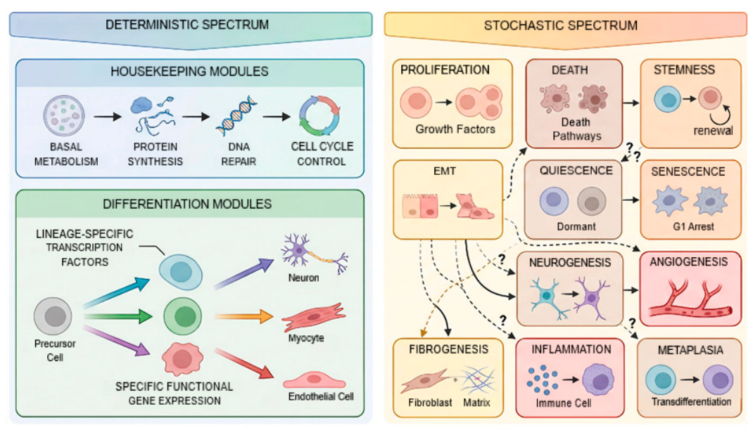
Figure 2.
The mutual inhibition between the differentiation order parameter and total stochastic activity produces a broad, unimodal joint distribution in a tumor cell.
Figure 2.
The mutual inhibition between the differentiation order parameter and total stochastic activity produces a broad, unimodal joint distribution in a tumor cell.
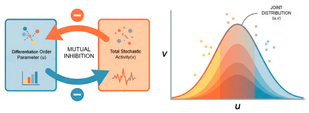
Figure 3.
Effective birth rate and death rate as functions of stemness . The death-dominant default state () prevails across nearly all of phenotype space. Only rare fluctuations allow net proliferation.
Figure 3.
Effective birth rate and death rate as functions of stemness . The death-dominant default state () prevails across nearly all of phenotype space. Only rare fluctuations allow net proliferation.
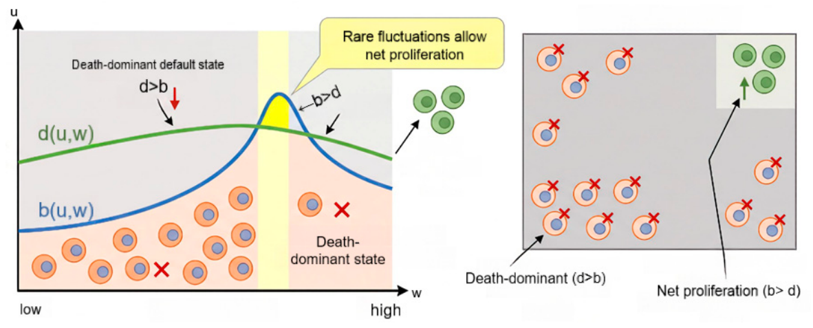
Figure 4.
Tumor formation as a multistep nonequilibrium rare event. a. Four sequential probability barriers that a mutant cell must traverse to a clinical tumor. Barrier 1: single mutant cell survival. Barrier 2: supply passive diffusion and immune escape. Barrier 3: microenvironment construction. Barrier 4: somatic mutation accumulation. b. Cumulative tumor formation probability as a function of barriers traversed by a mutant cell. Each barrier reduces the probability by 1-4 orders of magnitude. Childhood congenital tumors: 10-7-10-9per cell. Adult sporadic tumors: 10-13-10-16 per cell (red).
Figure 4.
Tumor formation as a multistep nonequilibrium rare event. a. Four sequential probability barriers that a mutant cell must traverse to a clinical tumor. Barrier 1: single mutant cell survival. Barrier 2: supply passive diffusion and immune escape. Barrier 3: microenvironment construction. Barrier 4: somatic mutation accumulation. b. Cumulative tumor formation probability as a function of barriers traversed by a mutant cell. Each barrier reduces the probability by 1-4 orders of magnitude. Childhood congenital tumors: 10-7-10-9per cell. Adult sporadic tumors: 10-13-10-16 per cell (red).
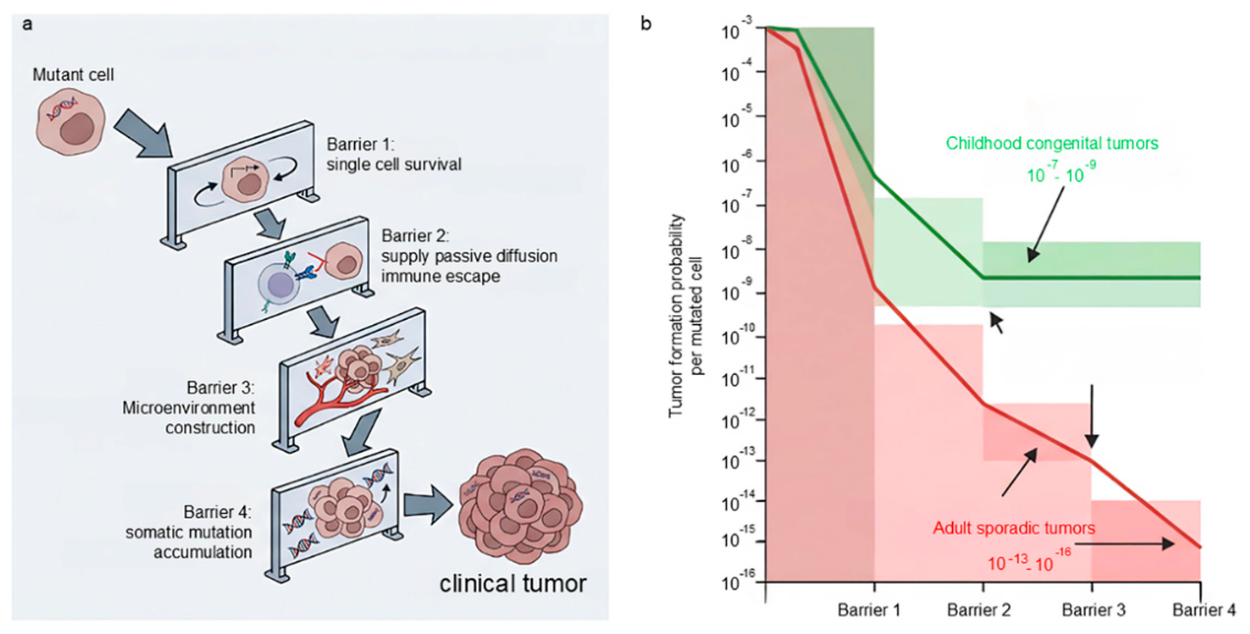
Figure 5.
Tumors as diseases of aberrant tissue formation. A tumor is pathological organoid that must be actively built and maintained through the stochastic coactivation of tissue construction programs including vascular network, fibrotic stroma, neural network, and inflammatory clearance. All the fields must be simultaneously active. Failure of any program leads to spontaneous regression.
Figure 5.
Tumors as diseases of aberrant tissue formation. A tumor is pathological organoid that must be actively built and maintained through the stochastic coactivation of tissue construction programs including vascular network, fibrotic stroma, neural network, and inflammatory clearance. All the fields must be simultaneously active. Failure of any program leads to spontaneous regression.
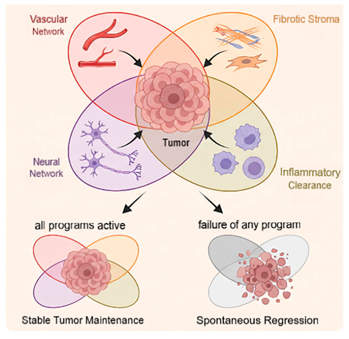
Figure 6.
The irreversible dedifferentiation ratchet under therapy. a. The free-energy landscape with two attractor basins. The differentiated attractor (green, right) represents terminal differentiation; the undifferentiated attractor (red, left) represents malignancy. The competition term is the physical basis of the ratchet: high differentiation suppresses stochastic fields, while low differentiation liberates them. b. The distribution of differentiation potential across the tumor cell population, shown by the pre-treatment state and four successive relapse cycles. Each cycle shifts the distribution peak monotonically toward (complete undifferentiation), because therapy preferentially eliminates high-differentiation cells while sparing low-differentiation persister cells. c. Clinical-pathological correlation is shown from well-differentiated initial tumor (green) through moderately differentiated first relapse (orange) to undifferentiated anaplastic late relapse (red). This progressive loss of differentiation is the physical origin of anaplastic progression.
Figure 6.
The irreversible dedifferentiation ratchet under therapy. a. The free-energy landscape with two attractor basins. The differentiated attractor (green, right) represents terminal differentiation; the undifferentiated attractor (red, left) represents malignancy. The competition term is the physical basis of the ratchet: high differentiation suppresses stochastic fields, while low differentiation liberates them. b. The distribution of differentiation potential across the tumor cell population, shown by the pre-treatment state and four successive relapse cycles. Each cycle shifts the distribution peak monotonically toward (complete undifferentiation), because therapy preferentially eliminates high-differentiation cells while sparing low-differentiation persister cells. c. Clinical-pathological correlation is shown from well-differentiated initial tumor (green) through moderately differentiated first relapse (orange) to undifferentiated anaplastic late relapse (red). This progressive loss of differentiation is the physical origin of anaplastic progression.
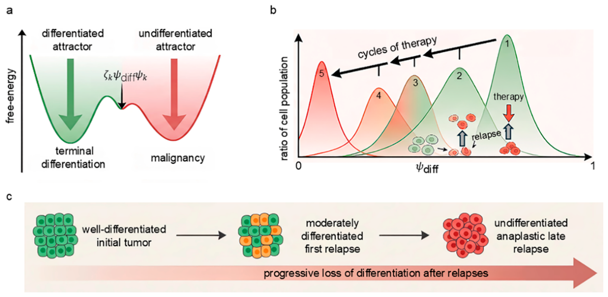
Figure 7.
Prognostic cell subpopulations and the curative strategy of tumor therapies. a. The phenotypic landscape spanned by stemness, differentiation, and EMT, showing the positions of poor-prognosis subpopulations (QSPD, SQM-EMT⁻, SQM-EMT⁺, DMQ-S, PSP, STQ-3) and good-prognosis subpopulations (HDT, STQ-1). b. The forced-differentiation curative strategy. The differentiation-inducing field drives cells past the irreversible threshold, collapsing the dissipative structure into a benign hamartoma-like state.
Figure 7.
Prognostic cell subpopulations and the curative strategy of tumor therapies. a. The phenotypic landscape spanned by stemness, differentiation, and EMT, showing the positions of poor-prognosis subpopulations (QSPD, SQM-EMT⁻, SQM-EMT⁺, DMQ-S, PSP, STQ-3) and good-prognosis subpopulations (HDT, STQ-1). b. The forced-differentiation curative strategy. The differentiation-inducing field drives cells past the irreversible threshold, collapsing the dissipative structure into a benign hamartoma-like state.
Disclaimer/Publisher’s Note: The statements, opinions and data contained in all publications are solely those of the individual author(s) and contributor(s) and not of MDPI and/or the editor(s). MDPI and/or the editor(s) disclaim responsibility for any injury to people or property resulting from any ideas, methods, instructions or products referred to in the content. |
© 2026 by the authors. Licensee MDPI, Basel, Switzerland. This article is an open access article distributed under the terms and conditions of the Creative Commons Attribution (CC BY) license.
Copyright: This open access article is published under a Creative Commons CC BY 4.0 license, which permit the free download, distribution, and reuse, provided that the author and preprint are cited in any reuse.