

Submitted:
09 September 2026
Posted:
10 September 2026
You are already at the latest version
Abstract
Soccer is one of the most widely practiced and studied sports worldwide. The increasing professionalization of women’s soccer has raised interest in bone health. This study aimed to analyze the mediating role of lean body mass in the association between muscle strength, jump height, and bone mineral density (BMD) in elite female soccer players. A cross-sectional study was conducted with 28 players from the Spanish national soccer team. Muscle strength was assessed using estimated 1RM in the deadlift, jump height through the countermovement jump (CMJ) test, and BMD using dual-energy X-ray absorptiometry (DXA). Lean body mass significantly mediated the association between muscle strength and BMD at the lumbar spine (B = 0.0020, 95% CI [0.0001–0.0049]), femoral neck (B = 0.0020, 95% CI [0.0003–0.0052]), and whole body (B = 0.0019, 95% CI [0.0005–0.0038]). These results suggest that muscle strength alone may not fully explain inter-individual differences in bone mass and that lean body mass may play an important role in this relationship Combining soccer practice with appropriate strength training may contribute to optimizing bone health, preventing injuries and fractures, and reducing future age-related bone loss. Longitudinal studies are needed to confirm these findings.
Keywords:
bone health
; body composition
; football players
; physical fitness
; female athlete
; muscle function
1. Introduction
Soccer is characterized as a high-intensity, intermittent, multidirectional sport that demands physical, technical, and tactical proficiency [1]. It is not only the sport with the largest number of participants worldwide, but also the most studied, with nearly 14,000 citations in PubMed—nearly 60% more than the next most studied sport—and exponential growth since 1980 [2].
Although women’s soccer has grown considerably in terms of participation, professionalism, and scientific interest in recent decades [3,4], female soccer players, especially elite ones, remain underrepresented in the current literature. The increasing physical demands inherent in playing this sport at the highest level make it necessary to focus on factors that influence the long-term health and performance of female athletes. Bone health is a key component among these factors because optimal bone mineral density (BMD) contributes to skeletal integrity throughout life and reduces the risk of stress-related injuries during sports [5,6].
The acquisition and maintenance of bone mass are influenced by multiple factors, including genetics, hormonal status, nutritional habits, body composition, and mechanical loading [7]. In female athletes, bone health has received particular attention due to the potential adverse effects of low energy availability and menstrual dysfunction, conditions associated with impaired bone metabolism and reduced BMD [6,8,9,10]. Given these factors, as well as the high incidence of injury among female soccer players [11], it is of significant practical and clinical importance to understand the determinants of bone health in this population.
Participation in soccer has consistently been associated with a positive effect on women’s bone health [8,12,13,14]. The high-impact nature of this sport, characterized by repeated accelerations, decelerations, changes of direction, jumps, and landings, provides an osteogenic stimulus capable of promoting bone formation and maintenance [14,15]. Previous studies have shown that female soccer players have higher BMD values compared to non-athletic women and even to athletes in sports with lower bone-loading demands, such as water sports [13,16,17,18,19].
Beyond sport participation itself, the relationship between muscle and bone has emerged as a major determinant of skeletal health. This interaction could be explained by mechanical mechanisms derived from the forces generated during muscle contraction and biochemical mechanisms related to the release of myokines involved in regulating bone metabolism [20]. Consequently, individuals with greater muscular size and strength are generally exposed to higher mechanical loads, which may stimulate bone formation and contribute to greater BMD. In athletic populations, lean body mass has repeatedly been identified as one of the strongest predictors of bone mass, suggesting that muscle-related factors may partly explain the beneficial skeletal adaptations observed in soccer players [21,22,23]. Similarly, muscle strength has also been considered a marker of bone health in recent literature [14,24,25]. However, whether this relationship is primarily attributable to the direct mechanical effects of force production or is largely explained by the greater lean body mass typically observed in stronger individuals remains unclear. This distinction is particularly relevant in female soccer players, in whom both muscular strength and lean body mass are highly developed and may contribute simultaneously to bone adaptation. Furthermore, it is possible that the specific demands of each playing position—associated with different anthropometric and neuromuscular profiles—could potentially affect bone-related outcomes [26,27]. However, there is little evidence regarding these issues among female soccer players, particularly elite players competing at the highest level.
Therefore, the aims of this study were: (i) to examine the relationships among muscular fitness, lean mass and BMD in elite Spanish female soccer players; (ii) to determine whether lean body mass mediates the association of muscle strength and jump height with bone mineral density; and (iii) to explore potential differences in bone-related variables according to playing position. It was hypothesized that muscle strength would be positively associated with BMD, that lean body mass would significantly mediate this relationship, and that positional differences would be observed in body composition and bone-related outcomes.
2. Materials and Methods
2.1. Subjects and Study Design
The study adopted a cross-sectional design. This involved assessing the body composition and sports-related performance of elite female soccer players from the Spanish senior national team in June 2022. The study included all professional female players on the Spanish senior national team who were training at the Ciudad del Fútbol in Las Rozas. All participants met the inclusion criteria, which consisted of being adults and not being pregnant or having any injuries.
All participants signed a written informed consent form prior to taking part in the study. All procedures were approved by the Human Ethics Committee of Universidad Autónoma de Madrid (CEI-124 2528), in accordance with the Declaration of Helsinki and the principles of the European Code of Conduct for Research Integrity [28], as well as the provisions of the EU General Data Protection Regulation (GDPR) 2016/679 [29].
2.2. Anthropometric Measurements
Anthropometric measurements were obtained on each subject immediately before DXA assessment. Both measurements were performed in the upright position, in underwear and barefoot. Height was measured in the Frankfort plane on a stadiometer with a precision of 1 mm (Seca 711, Hamburg, Germany). Body mass was determined using a balance with a 100 g precision (Seca 711, 120 Hamburg, Germany). Body mass index (BMI) was calculated as body mass divided by height squared (kg·m2).
2.3. Bone Measurements and Body Composition
Dual-Energy X-ray Absorptiometry (DXA) scans were undertaken to assess BMD of whole body, lumbar spine (L1–L4), and proximal region of the femur (femoral neck) using a Hologic Horizon W system (Hologic Inc., Bedford, MA, USA) [30]. Whole body fat mass (kg), lean mass (bone free) (kg) and percentage body fat mass were also obtained from the total body DXA scan. All DXA scan tests were analyzed using the Physician’s Viewer, APEX System Software Version 3.1.2. (Bedford, USA). Scans were made in a supine position, wearing light clothing with no metal and no shoes or jewelry. DXA equipment was calibrated using a lumbar spine phantom and following the Hologic guidelines. The bone T-scores were calculated in each participant for whole body, femoral neck and lumbar spine to classify by bone health status. Femoral neck values were calculated as the mean of measurements obtained from both hips.
2.4. Countermovement Jump
CMJ height (cm) was assessed using a force plate (ForceDecks, London, United Kingdom) with a sampling rate of 1000Hz in accordance with previously described procedures [31]. Participants stepped onto the force plates and remained standing upright and motionless for at least 1 s to allow body weight to be determined prior to movement initiation. All jumps were performed with the hands placed on the hips to minimise upper-limb contribution. Athletes were instructed to jump “as fast and as high as possible” while maintaining lower-limb extension during flight and landing. Following a standardized warm-up and familiarization, each participant performed five maximal CMJ trials separated by ~60 s of passive recovery, and the trial with the greatest jump height was selected for analysis. Jump height was calculated as the highest displacement of the centre of mass calculated from vertical velocity at take-off following procedures published elsewhere [32].
2.5. One-Repetition Maximum Estimation in the Deadlift Exercise
All elite soccer players had prior experience in resistance training with maximal intended velocity and deadlift execution, ensuring the familiarization with the evaluation protocol. The one-repetition maximum (1RM) was estimated using a submaximal load-velocity profile rather than a direct maximal test. As highlighted in previous literature, this approach minimizes fatigue, reduces the risk of injury, is highly practical, and relies on the strong linear load-velocity relationship [33].
The exercise was performed in a free-weight modality using a standard 20-kg barbell, with extra load added via sliding weight disc according to previously procedures in women [34]. For all attempts, participants adopted a self-selected grip and foot stance. Following a standardized warm-up, participants performed a submaximal progressive loading test consisting of two incremental loads reaching ~60% of their 1RM, using lifting velocity as a parameter for load progression. This moderate threshold of intensity was chosen because previous research on the load-velocity relationship demonstrates that submaximal loads are highly reliable for accurately estimating the 1RM without the need to expose participants to fatigue and several loads [35]. During execution, the concentric phase of all repetitions was performed at maximally intended velocity throughout the full range of motion. For each load, participants performed 2–4 repetitions, and only the fastest repetition (i.e., the one with the highest mean concentric velocity) was considered for subsequent analysis. A pause was imposed between repetitions to eliminate the stretch-shortening cycle and increase the reliability of assessments [36]. A 3-minute rest was conducted between loads. Mean velocity during the concentric phase of each repetition was determined using a linear position transducer (Chronojump, Boscosystem, Barcelona, Spain). The cable of the device was attached to the barbell in a manner that ensured a strictly vertical displacement during the lift [37].
Finally, the 1RM was estimated using the specific load-velocity equation for the deadlift proposed by Nieto-Acevedo et al. [34] with the load and velocity attained at ~60%1RM, as this formula was developed following this exact execution methodology in a cohort of women with resistance training experience.
2.6. Statistical Analyses
Means, standard deviations, minimum and maximum values were used for the basic description. Normality was assessed using the Shapiro-Wilk test. Levene’s test was used to analyse homoscedasticity. A one-way ANOVA was used to assess differences by playing position in variables with a normal distribution and homogeneous variances. Where homogeneity of variances was not met, Welch’s ANOVA was used as an alternative. When variables did not follow a normal distribution, the non-parametric Kruskal–Wallis H test was used. For post-hoc analysis, Tukey’s or Games-Howell’s tests were used following ANOVA, depending on the homogeneity of variances, and Dunn’s test was used as a post-hoc analysis following the Kruskal-Wallis test. Partial correlations were calculated, adjusting for age and height, to analyze the relationship between the players’physical fitness, bone measurements and body composition.
Six mediation analyses were performed to examine the mediating role of lean body mass in the association between physical fitness and BMD using the PROCESS macro (version 5.0; Model 4) for SPSS (IBM Corp., Armonk, NY, USA), developed by Andrew F. Hayes [38]. Three models were conducted with 1RM as the independent variable and lumbar spine, femoral neck, and whole-body BMD as the dependent variables. Another three models were conducted with CMJ height as the independent variable and the same BMD outcomes. For each model, the total effect (c), direct effect (c′), and indirect effect (a × b) were estimated. Path (a) represents the association between the independent variable and the mediator (lean body mass), whereas path (b) represents the association between the mediator and BMD after controlling for the independent variable. Age and height were included as covariates in all mediation models.
Indirect effects were tested using a non-parametric bootstrapping procedure with 10,000 resamples, as recommended by Preacher and Hayes [39]. Bias-corrected 95% confidence intervals (CI) were computed for the indirect effect. Mediation was considered statistically significant when the CI did not include zero.
All analyses were conducted using SPSS Statistics (version 27.0; IBM Corp.). Statistical significance was set at a two-tailed alpha level of p ≤ 0.05.
3. Results
The final sample comprised twenty-eight athletes aged between 18 and 31: three goalkeepers, nine defenders, seven midfielders and nine forwards. Table 1 summarises the characteristics of the total sample of players by playing position. Statistically significant differences were observed in fat mass expressed in kilograms (p = 0.041) and as a percentage (p = 0.015) between defenders and midfielders, as well as in the percentage of fat mass (p = 0.035) between defenders and forwards. No statistically significant differences were observed between the various positions with regard to the other variables assessed.
The players had positive average T-scores for BMD in the lumbar region (1.48 ± 0.98), the average femoral neck (2.34 ± 1.05), and the whole body (2.42 ± 0.86); no statistically significant differences in these values were observed among the different playing positions. None of the players had bone T-scores below -1 in any body region (Figure 1).
Figure 2 shows the partial correlations between the analysed variables after adjusting for age and height. Total body mass and lean body mass were positively correlated with muscle strength and all BMD variables (all p ≤ 0.05). Fat mass was negatively correlated with CMJ height (p = 0.049). No significant associations were found among the remaining variables.
Figure 3 presents the mediation models in which significant indirect effects were observed after adjustment for age and height. In first stage of the analyses (path a), deadlift 1RM was positively associated with lean body mass (B = 0.1103, p ≤ 0.01), and the model explained 60.93% of the variance in lean body mass (R² = 0.6093). In the total effect models (path c), deadlift 1RM was not significantly associated with lumbar spine, femoral neck, or whole-body BMD (all p > 0.05). When lean body mass was included as a mediator, significant positive associations were observed between lean body mass and lumbar spine BMD and femoral neck BMD (path b; both p ≤ 0.05), as well as whole-body BMD (p ≤ 0.01). In contrast, the direct effects of deadlift 1RM on BMD (path c′) were no longer significant across all skeletal sites. Bootstrap analyses revealed significant indirect effects of deadlift 1RM on lumbar spine BMD, femoral neck BMD, and whole-body BMD, as the 95% confidence intervals did not include zero. These findings indicate that lean body mass mediates the association between deadlift strength and BMD across all skeletal sites examined.
Unlike deadlift 1RM, no significant indirect effects were observed for CMJ height on lumbar spine (B = -0.0036, 95% CI [-0.0208, 0.0026]), femoral neck (B = -0.0046, 95% CI [0.0091, 0.0210]), or whole-body BMD (B = -0.0032, 95% CI [-0.0165, 0.0021]), as all 95% confidence intervals for the indirect effects included zero.
4. Discussion
The primary objective of this study was to determine if lean body mass mediates the relationship between physical fitness, specifically maximum deadlift strength, and BMD in players on the Spanish women’s national soccer team. To our knowledge, this is the first study to analyze the relationship among these three factors in elite female soccer players. The results revealed that lean body mass statistically mediated the relationship between maximum deadlift strength and BMD in the lumbar region, femoral neck, and whole body of the players. Additionally, differences in fat mass, were observed among athletes in different playing positions.
On average, elite female soccer players had BMD values higher than the average for a healthy young adult. These results are consistent with previous evidence indicating higher BMD values in female soccer players [14,17,18,40,41]. These findings reinforce the protective role of soccer on women’s bone mass compared to other lower-impact sports, such as water sports [14,17,18]. This is due to the osteogenic response to the mechanical loads and ground reaction forces characteristic of high-impact sports, such as soccer [42,43]. Combining this type of exercise with appropriate strength training could help prevent the injuries, bone fractures, and health problems that are common among female athletes in this sport [11,42,44]. It could also serve as an important protective strategy against age-related bone loss for these athletes in the future [43,45,46,47]. Long-term studies could help determine the optimal training loads to maximize benefits while preventing future adverse effects on the bone health of female soccer players.
Our study clarified the role of lean body mass as a mediator in the relationship between muscle strength and BMD in different body regions of elite female soccer players. Since no statistically significant associations were observed between 1RM deadlift and BMD in any skeletal region, these results suggest that maximum force production alone may not be a decisive factor in explaining interindividual differences in bone mass among elite female soccer players [13,15]. However, muscle strength’s indirect influence on lean body mass variability could play a significant role in this regard. Accordingly, structural characteristics of the musculoskeletal system, such as lean body mass, should be considered a mediating variable rather than a confounding variable in this relationship between muscle strength and bone mass [48]. Additionally, these relationships are likely mediated by more than one variable. Since muscle strength has been associated with bone mass in anatomical locations distant from the muscle group’s site of action, this association may not be solely due to local biomechanical factors but may also involve environmental, endocrine, growth, or genetic factors [48]. Among these factors, body composition plays a particularly significant role in female soccer players [49]. Future studies using structural equation modeling could clarify the potential mediating role of each factor more specifically.
Furthermore, differences in body fat were observed only among defenders compared to midfielders and forwards among the different playing positions. These findings contrast with previous studies in which midfielders had lower body fat levels [50], and studies in which no differences in body composition were observed across playing positions in diverse samples of female soccer players [51,52,53]. The small sample size in some positions may have limited the observation of significant differences in certain analyzed variables. Given its influence on the relationship between specific motor skills and body composition, playing position must be considered. This allows coaches to program and plan the training process with greater precision and facilitates the selection of the most appropriate position for a player based on body composition characteristics [54,55]. For example, players with more offensive roles, such as forwards and wingers, face greater physical demands during competition and require different considerations regarding these aspects [55]. Therefore, periodically assessing body composition across playing positions while considering various methods and their differences may be a valuable strategy for this group of athletes [56,57].
This study has some limitations. First, the cross-sectional design prevents causal inference. Although the sample size was small—especially after stratifying by playing position—it is important to note that this study included 100% of the women selected for a world-class elite competition. Therefore, this limitation should be interpreted within the context of this unique athletic population [58]. Finally, this study does not account for important control variables, such as prior calcium or vitamin D intakes, menstrual status, energy availability and hormonal contraceptive use [59]. Future studies that take these variables into account are necessary.
5. Conclusions
Lean body mass was found to significantly mediate the relationship between maximum deadlift strength and BMD in elite female soccer players. These findings suggest that muscle strength alone may not fully explain inter-individual differences in bone mass in this population, and that lean body mass may represent an important underlying factor linking strength and bone health. Furthermore, elite female athletes had average BMD values higher than the average values for healthy young adults, with no significant differences observed between the different playing positions.
From a practical perspective, these results may help inform training strategies aimed at optimizing both physical performance and skeletal health in female soccer players. Future longitudinal studies are warranted to clarify the temporal relationships among these variables and to determine whether these findings can be generalized to broader populations of female soccer players.
Author Contributions
Conceptualization, N.P.G.-A. and I.A; methodology, B.R.-M., I.A and N.P.G.-A; validation, B.R.-M. and C.V.C.; formal analysis, A.M.-F. and I.R.-G.; investigation, B.R.-M., C.V.C and N.P.G.-A.; resources, B.R.-M., C.V.C and N.P.G.-A.; data curation, I.R.-G and A.B.-R..; writing—original draft preparation, A.M.-F.; writing—review and editing, N.P.G.-A., I.R.-G., C.V.C., B.R.-M., A.B.-R and I.A; supervision, I.R.-G and A.B.-R.; project administration, N.P.G.-A. and I.A. All authors have read and agreed to the published version of the manuscript.
Funding
A.M.-F. is supported by a postgraduate training fellowship granted by the Consejo Superior de Deportes (CSD) at the Centro de Medicina del Deporte (055078_2025). I.R.-G., Á.B.-R. and I.A. are supported by CIBERFES (CB16/10/00477), Plan Propio de Investigación of the Universidad de Castilla-La Mancha and FEDER funds from the European Union (2025-GRIN-38408) and the Instituto de Investigación Sanitaria de Castilla-La Mancha-IDISCAM (TEC2022-007). ÁB-R is supported by a postdoctoral contract granted by Spanish Ministry of Science and Innovation (JDC2023-052593-I, funded by MCIU/AEI/10.13039/501100011033).
Institutional Review Board Statement
The study was conducted in accordance with the Declaration of Helsinki, and approved by the Institutional Review Board (or Ethics Committee) of Universidad Autónoma de Madrid (CEI-124 2528).
Informed Consent Statement
Informed consent was obtained from all subjects involved in the study.
Data Availability Statement
The data presented in this study are available on request from the corresponding author due to the public profile of the participants and the potential risk of identification, the data are not publicly available.
Acknowledgments
We would like to sincerely thank all the football players who participated in this study for their time, commitment, and valuable contribution to this research.
Conflicts of Interest
The authors declare no conflicts of interest.
Abbreviations
The following abbreviations are used in this manuscript:
| BMD | Bone Mineral Density |
| CMJ | Countermovement Jump |
| DXA | Dual-Energy-X-Ray Absorptiometry |
| 1RM | One-repetition maximum |
| CI | Confidence Intervals |
| BMI | Body Mass Index |
| WB | Whole Body |
| L1-L4 | Lumbar Spine |
| FN | Femoral Neck |
References
- Manson, S. A.; Brughelli, M.; Harris, N. K. Physiological Characteristics of International Female Soccer Players. J. Strength Cond. Res. 2014, 28(2), 308–318. [Google Scholar] [CrossRef] [PubMed]
- Kirkendall, D. T. Evolution of Soccer as a Research Topic. Prog. Cardiovasc. Dis. 2020, 63(6), 723–729. [Google Scholar] [CrossRef] [PubMed]
- Bandyopadhyay, K. Introduction: Soccer, Women and Society. Soccer Soc. 2026, 27(2–3), 195–206. [Google Scholar] [CrossRef]
- Paul, R. W.; Sonnier, J. H.; Johnson, E. E.; Hall, A. T.; Osman, A.; Connors, G. M.; Freedman, K. B.; Bishop, M. E. Inequalities in the Evaluation of Male Versus Female Athletes in Sports Medicine Research: A Systematic Review. Am. Jorunal Sports Med. 2023, 51(12), 3335–3342. [Google Scholar] [CrossRef] [PubMed]
- Randell, R. K.; Clifford, T.; Drust, B.; Moss, S. L.; Unnithan, V. B.; De Ste Croix, M. B. A.; Datson, N.; Martin, D.; Mayho, H.; Carter, J. M.; Rollo, I. Physiological Characteristics of Female Soccer Players and Health and Performance Considerations: A Narrative Review. Sports Med. 2021, 51(7), 1377–1399. [Google Scholar] [CrossRef] [PubMed]
- Sundgot-Borgen, J.; Torstveit, M. K. The Female Football Player, Disordered Eating, Menstrual Function and Bone Health. Br. J. Sports Med. 2007, 41 (Supplement 1), i68–i72. [Google Scholar] [CrossRef] [PubMed]
- Weaver, C. M.; Gordon, C. M.; Janz, K. F.; Kalkwarf, H. J.; Lappe, J. M.; Lewis, R.; O’Karma, M.; Wallace, T. C.; Zemel, B. S. The National Osteoporosis Foundation’s Position Statement on Peak Bone Mass Development and Lifestyle Factors: A Systematic Review and Implementation Recommendations. Osteoporis Int. 2016, 27(4), 1281–1386. [Google Scholar] [CrossRef] [PubMed]
- Skarakis, N. S.; Mastorakos, G.; Georgopoulos, N.; Goulis, D. G. Energy Deficiency, Menstrual Disorders, and Low Bone Mineral Density in Female Athletes: A Systematic Review. Hormones 2021, 20(3), 439–448. [Google Scholar] [CrossRef] [PubMed]
- Matzkin, E.; Curry, E. J.; Whitlock, K. Female Athlete Triad: Past, Present, and Future. JAAOS—Journal Am. Acad. Orthop. Surg. 2015, 23(7), 424–432. [Google Scholar] [CrossRef] [PubMed]
- Thein-Nissenbaum, J. Long Term Consequences of the Female Athlete Triad. Maturitas 2013, 75(2), 107–112. [Google Scholar] [CrossRef] [PubMed]
- Giza, E.; Mithöfer, K.; Farrell, L.; Zarins, B.; Gill, T. Injuries in Women’s Professional Soccer. Br. J. Sports Med. 2005, 39(4), 212–216. [Google Scholar] [CrossRef] [PubMed]
- Nichols, D. L.; Sanborn, C. F.; Essery, E. V. Bone Density and Young Athletic Women. Sports Med. 2007, 37(11), 1001–1014. [Google Scholar] [CrossRef] [PubMed]
- Alfredson, H.; Nordström, P.; Lorentzon, R. Total and Regional Bone Mass in Female Soccer Players. Calcif. Tissue Int. 1996, 59(6), 438–442. [Google Scholar] [CrossRef] [PubMed]
- Seabra, A.; Marques, E.; Brito, J.; Krustrup, P.; Abreu, S.; Oliveira, J.; Rêgo, C.; Mota, J.; Rebelo, A. Muscle Strength and Soccer Practice as Major Determinants of Bone Mineral Density in Adolescents. Jt. Bone Spine 2012, 79(4), 403–408. [Google Scholar] [CrossRef] [PubMed]
- Söderman, K.; Bergström, E.; Lorentzon, R.; Alfredson, H. Bone Mass and Muscle Strength in Young Female Soccer Players. Calcif. Tissue Int. 2000, 67(4), 297–303. [Google Scholar] [CrossRef] [PubMed]
- Düppe, H.; Gärdsell, P.; Johnell, O.; Ornstein, E. Bone Mineral Density in Female Junior, Senior and Former Football Players. Osteoporos. Int. 1996, 6(6), 437–441. [Google Scholar] [CrossRef] [PubMed]
- Bellew, J. W.; Gehrig, L. A Comparison of Bone Mineral Density in Adolescent Female Swimmers, Soccer Players, and Weight Lifters. Pediatr. Phys. Ther. 2006, 18(1), 19–22. [Google Scholar] [CrossRef] [PubMed]
- Bellver, M.; Del Rio, L.; Jovell, E.; Drobnic, F.; Trilla, A. Bone Mineral Density and Bone Mineral Content among Female Elite Athletes. Bone 2019, 127, 393–400. [Google Scholar] [CrossRef] [PubMed]
- Hagman, M.; Helge, E. W.; Fristrup, B.; Jørgensen, N. R.; Helge, J. W.; Krustrup, P. High Bone Mineral Density in Lifelong Trained Female Team Handball Players and Young Elite Football Players. Eur. J. Appl. Physiol. 2021, 121(10), 2825–2836. [Google Scholar] [CrossRef] [PubMed]
- Guo, B.; Zhang, Z.-K.; Liang, C.; Li, J.; Liu, J.; Lu, A.; Zhang, B.-T.; Zhang, G. Molecular Communication from Skeletal Muscle to Bone: A Review for Muscle-Derived Myokines Regulating Bone Metabolism. Calcif. Tissue Int. 2017, 100(2), 184–192. [Google Scholar] [CrossRef] [PubMed]
- Pelegrini, A.; Bim, M. A.; Alves, A. D.; Scarabelot, K. S.; Claumann, G. S.; Fernandes, R. A.; de Angelo, H. C. C.; de Araújo Pinto, A. Relationship between Muscle Strength, Body Composition and Bone Mineral Density in Adolescents. J. Clin. Densitom. 2022, 25(1), 54–60. [Google Scholar] [CrossRef] [PubMed]
- Bevier, W. C.; Wiswell, R. A.; Pyka, G.; Kozak, K. C.; Newhall, K. M.; Marcus, R. Relationship of Body Composition, Muscle Strength, and Aerobic Capacity to Bone Mineral Density in Older Men and Women. J. Bone Miner. Res. 1989, 4(3), 421–432. [Google Scholar] [CrossRef] [PubMed]
- Sioen, I.; Lust, E.; De Henauw, S.; Moreno, L. A.; Jiménez-Pavón, D. Associations Between Body Composition and Bone Health in Children and Adolescents: A Systematic Review. Calcif. Tissue Int. 2016, 99(6), 557–577. [Google Scholar] [CrossRef] [PubMed]
- Torres-Costoso, A.; López-Muñoz, P.; Martínez-Vizcaíno, V.; Álvarez-Bueno, C.; Cavero-Redondo, I. Association Between Muscular Strength and Bone Health from Children to Young Adults: A Systematic Review and Meta-Analysis. Sports Med. 2020, 50(6), 1163–1190. [Google Scholar] [CrossRef] [PubMed]
- Riviati, N.; Darma, S.; Reagan, M.; Iman, M. B.; Syafira, F.; Indra, B. Relationship between Muscle Mass and Muscle Strength with Bone Density in Older Adults: A Systematic Review. Ann. Geriatr. Med. Res. 2025, 29(1), 1–14. [Google Scholar] [CrossRef] [PubMed]
- Sebastiá-Rico, J.; Martínez-Sanz, J. M.; González-Gálvez, N.; Soriano, J. M. Differences in Body Composition between Playing Positions in Men’s Professional Soccer: A Systematic Review with Meta-Analysis. Appl. Sci. 2023, 13(8), 4782. [Google Scholar] [CrossRef]
- Sutton, L.; Scott, M.; Wallace, J.; Reilly, T. Body Composition of English Premier League Soccer Players: Influence of Playing Position, International Status, and Ethnicity. J. Sports Sci. 2009, 27(10), 1019–1026. [Google Scholar] [CrossRef] [PubMed]
- ALLEA (All European Academies). The European Code of Conduct for Research Integrity—Revised Edition 2023; ALLEA: Berlin, 2023. [Google Scholar] [CrossRef]
- Regulation (EU) 2016/679 of the European Parliament and of the Council of 27 April 2016 on the Protection of Natural Persons with Regard to the Processing of Personal Data and on the Free Movement of Such Data, and Repealing Directive 95/46/EC (General Data Protection Regulation). Off. J. Eur. Union 2016, L 119, 1–88.
- Rodríguez-Gómez, I.; Mañas, A.; Losa-Reyna, J.; Rodríguez-Mañas, L.; Chastin, S. F. M.; Alegre, L. M.; García-García, F. J.; Ara, I. Associations between sedentary time, physical activity and bone health among older people using compositional data analysis. PLoS ONE 2018, 13(10), e0206013. [Google Scholar] [CrossRef] [PubMed]
- Badby, A. J.; Comfort, P.; Ripley, N. J.; Mundy, P. D.; Soriano, M. A.; Robles-Palazón, F. J.; Fahey, J.; Sindall, P.; Bramah, C.; McMahon, J. J. Normative Data and Objective Benchmarks for Selected Force Plate Tests for Professional and Youth Soccer Players in the English Football League. J. Sports Sci. 2025, 43(20), 2306–2323. [Google Scholar] [CrossRef] [PubMed]
- Romero-Moraleda, B.; Morencos-Martínez, E.; Varón, P.; Lara, B.; Jiménez-Ormeño, E.; Peinado, A. B.; González-García, J. The Influence of Menstrual Cycle Phase on Neuromuscular Performance and Subjective Perception of Effort in Elite Football Players. Biol. Sport 2025, 42(3), 89–98. [Google Scholar] [CrossRef] [PubMed]
- McBurnie, A. J.; Allen, K. P.; Garry, M.; Martin, M.; Thomas, D.; Jones, P. A.; Comfort, P.; McMahon, J. J. The Benefits and Limitations of Predicting One Repetition Maximum Using the Load-Velocity Relationship. Strength Cond. J. 2019, 41(6), 28–40. [Google Scholar] [CrossRef]
- Nieto-Acevedo, R.; Romero-Moraleda, B.; Montalvo-Pérez, A.; Valdés-Álvarez, A.; García-Sánchez, C.; Mon-López, D. Should We Use the Men Load–Velocity Profile for Women in Deadlift and Hip Thrust? Int. J. Environ. Res. Public Health 2023, 20(6), 4888. [Google Scholar] [CrossRef] [PubMed]
- Garcia-Ramos, A.; Jaric, S. Two-Point Method: A Quick and Fatigue-Free Procedure for Assessment of Muscle Mechanical Capacities and the 1 Repetition Maximum. Strength Cond. J. 2018, 40(2), 54–66. [Google Scholar] [CrossRef]
- Pallarés, J. G.; Sánchez-Medina, L.; Pérez, C. E.; De La Cruz-Sánchez, E.; Mora-Rodriguez, R. Imposing a Pause between the Eccentric and Concentric Phases Increases the Reliability of Isoinertial Strength Assessments. J. Sports Sci. 2014, 32(12), 1165–1175. [Google Scholar] [CrossRef] [PubMed]
- Hernández-Belmonte, A.; Buendía-Romero, Á.; Pallares, J. G.; Martínez-Cava, A. Velocity-Based Method in Free-Weight and Machine-Based Training Modalities: The Degree of Freedom Matters. J. Strength Cond. Res. 2023, 37(9), e500–e509. [Google Scholar] [CrossRef] [PubMed]
- Bolin, J. H.; Hayes, A. F. Introduction to Mediation, Moderation, and Conditional Process Analysis: A Regression-Based Approach. New York, NY: The Guilford Press. Journal of Educational Measurement 2013, 51(3), 335–337. [Google Scholar] [CrossRef]
- Preacher, K. J.; Hayes, A. F. Asymptotic and Resampling Strategies for Assessing and Comparing Indirect Effects in Multiple Mediator Models. Behav. Res. Methods 2008, 40(3), 879–891. [Google Scholar] [CrossRef] [PubMed]
- Rocha-Rangel, J.; Liang, M. T. C.; Tsai, A. H.; Auslander, A. T.; Robles, P.; Kwoh, Y.-L.; Arnaud, S. B. Bone Bending Strength and BMD of Female Athletes in Volleyball, Soccer, and Long-Distance Running. Eur. J. Appl. Physiol. 2023, 123(10), 2213–2223. [Google Scholar] [CrossRef] [PubMed]
- Jeon, W.; Harrison, J. M.; Stanforth, P. R.; Griffin, L. Bone Mineral Density Differences Across Female Olympic Lifters, Power Lifters, and Soccer Players. J. Strength Cond. Res. 2021, 35(3), 638–643. [Google Scholar] [CrossRef] [PubMed]
- Lynch, K. R.; Kemper, H. C. G.; Turi-Lynch, B.; Agostinete, R. R.; Ito, I. H.; Luiz-De-Marco, R.; Rodrigues-Junior, M. A.; Fernandes, R. A. Impact Sports and Bone Fractures among Adolescents. J. Sports Sci. 2017, 35(24), 2421–2426. [Google Scholar] [CrossRef] [PubMed]
- Guadalupe-Grau, A.; Fuentes, T.; Guerra, B.; Calbet, J. A. L. Exercise and Bone Mass in Adults. Sports Med. 2009, 39(6), 439–468. [Google Scholar] [CrossRef] [PubMed]
- Kohrt, W. M.; Bloomfield, S. A.; Little, K. D.; Nelson, M. E.; Yingling, V. R. Physical Activity and Bone Healt. Med. Sci. Sports Exerc. 2004, 36(11), 1985–1996. [Google Scholar] [CrossRef] [PubMed]
- Dasarathy, J.; Labrador, H. Bone Health in Women. Prim. Care Clin. Off. Pract. 2018, 45(4), 643–657. [Google Scholar] [CrossRef] [PubMed]
- Mosekilde, L. Age-Related Changes in Bone Mass, Structure, and Strength—Effects of Loading. Z. Rheumatol. 2000, 59(1), I1–I9. [Google Scholar] [CrossRef] [PubMed]
- Lupsa, B. C.; Insogna, K. Bone Health and Osteoporosis. Endocrinol. Metab. Clin. North Am. 2015, 44(3), 517–530. [Google Scholar] [CrossRef] [PubMed]
- Torres-Costoso, A.; Gracia-Marco, L.; Sánchez-López, M.; García-Prieto, J. C.; García-Hermoso, A.; Díez-Fernández, A.; Martínez-Vizcaíno, V. Lean Mass as a Total Mediator of the Influence of Muscular Fitness on Bone Health in Schoolchildren: A Mediation Analysis. J. Sports Sci. 2015, 33(8), 817–830. [Google Scholar] [CrossRef] [PubMed]
- Baker, B. S.; Chen, Z.; Larson, R. D.; Bemben, M. G.; Bemben, D. A. Sex Differences in Bone Density, Geometry, and Bone Strength of Competitive Soccer Players. J. Musculoskelet. Neuronal Interact. 2020, 20(1), 62–76. [Google Scholar] [PubMed]
- Hernandez-Martinez, J.; Perez-Carcamo, J.; Canales-Canales, S.; Coñapi-Union, B.; Cid-Calfucura, I.; Herrera-Valenzuela, T.; Branco, B. H. M.; Valdés-Badilla, P. Body Composition and Physical Performance by Playing Position in Amateur Female Soccer Players. Appl. Sci. 2024, 14(13), 5665. [Google Scholar] [CrossRef]
- Mora-Fernandez, A.; Rojas, J. P.; Gimenez-Blasi, N.; Conde-Pipó, J.; Latorre, J. A.; Mariscal-Arcas, M. Relationship of Vitamin D Status with Biomarkers of Muscle Damage and Body Composition in Spanish Elite Female Football Players: A Cross-Sectional Study. Appl. Sci. 2024, 14(14), 6349. [Google Scholar] [CrossRef]
- Toro-Román, V.; Grijota, F. J.; Muñoz, D.; Maynar-Mariño, M.; Clemente-Gil, S.; Robles-Gil, M. C. Anthropometry, Body Composition, and Physical Fitness in Semi-Professional Soccer Players: Differences between Sexes and Playing Position. Appl. Sci. 2023, 13(3), 1249. [Google Scholar] [CrossRef]
- Barrera, J.; Valenzuela, L.; Sarmento, H.; Zurita, E.; Pastor, A.; Villaseca-Vicuña, R. Comparison of Aerobic Performance and Body Composition According to Game Position and Its Relationship between Variables in Professional Women’s Soccer Players. J. Phys. Educ. Sport 2022, 22(10), 2281–2288. [Google Scholar] [CrossRef]
- Stanković, M.; Čaprić, I.; Đorđević, D.; Đorđević, S.; Preljević, A.; Koničanin, A.; Maljanović, D.; Nailović, H.; Muković, I.; Jelaska, I.; Sporiš, G. Relationship between Body Composition and Specific Motor Abilities According to Position in Elite Female Soccer Players. Int. J. Environ. Res. Public Health 2023, 20(2), 1327. [Google Scholar] [CrossRef] [PubMed]
- Sánchez-Abselam, O.; González-Fernández, F. T.; Figueiredo, A.; Castillo-Rodríguez, A.; Onetti-Onetti, W. Effect of the Role, Playing Position and the Body Characteristics on Physical Performance in Female Soccer Players. Heliyon 2024, 10(7), e29240. [Google Scholar] [CrossRef] [PubMed]
- Erselcan, T.; Candan, F.; Saruhan, S.; Ayca, T. Comparison of Body Composition Analysis Methods in Clinical Routine. Ann. Nutr. Metab. 2000, 44(5–6), 243–248. [Google Scholar] [CrossRef] [PubMed]
- Larson-Meyer, D. E.; Woolf, K.; Burke, L. Assessment of Nutrient Status in Athletes and the Need for Supplementation. Int. J. Sport Nutr. Exerc. Metab. 2018, 28(2), 139–158. [Google Scholar] [CrossRef] [PubMed]
- McKay, A. K. A.; Stellingwerff, T.; Smith, E. S.; Martin, D. T.; Mujika, I.; Goosey-Tolfrey, V. L.; Sheppard, J.; Burke, L. M. Defining Training and Performance Caliber: A Participant Classification Framework. Int. J. Sports Physiol. Perform. 2021, 17(2), 317–331. [Google Scholar] [CrossRef] [PubMed]
- Sale, C.; Elliott-Sale, K. J. Nutrition and Athlete Bone Health. Sports Med. 2019, 49(2), 139–151. [Google Scholar] [CrossRef] [PubMed]
Figure 1.
BMD T-scores by playing position.
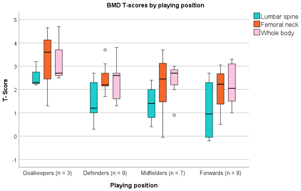
Figure 2.
Bivariate partial correlations between variables adjusted for age and height. BMD: Bone mineral density; WB: Whole body; L1-L4: Lumbar spine; FN: Femoral neck; CMJ: Countermovement jump; 1RM: one-repetition maximum. *p ≤ 0.05; **p ≤ 0.01.
Figure 2.
Bivariate partial correlations between variables adjusted for age and height. BMD: Bone mineral density; WB: Whole body; L1-L4: Lumbar spine; FN: Femoral neck; CMJ: Countermovement jump; 1RM: one-repetition maximum. *p ≤ 0.05; **p ≤ 0.01.
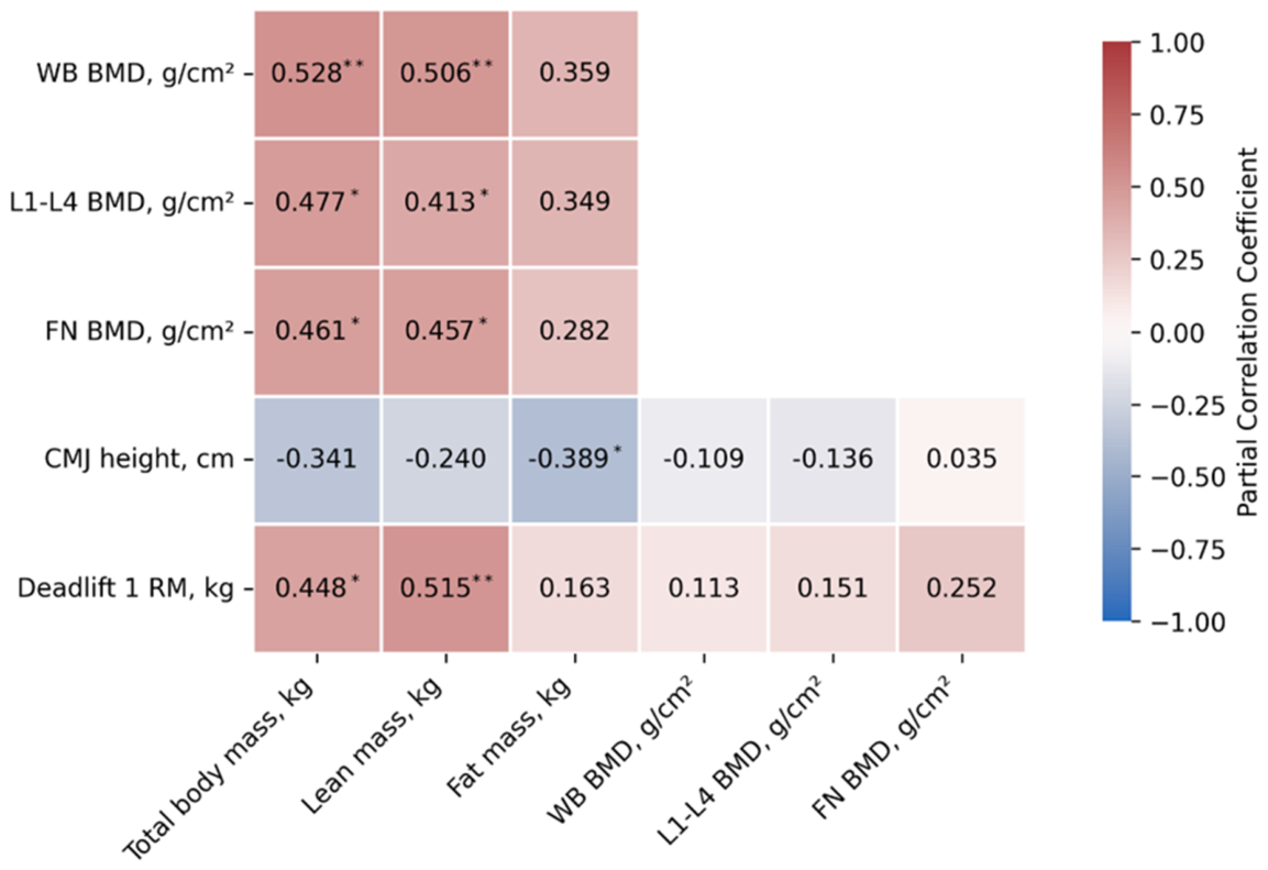
Figure 3.
Mediation models examining the mediating role of lean body mass in the association between deadlift 1RM and BMD at different skeletal sites. (a) Lumbar spine BMD; (b) femoral neck BMD; (c) whole-body BMD. Unstandardized regression coefficients are presented. BMD, bone mineral density; WB, whole body; L1–L4, lumbar spine; FN, femoral neck; 1RM, one-repetition maximum; a= effect of the independent variable on the mediator; b= effect of the mediator on the dependent variable after controlling for the independent variable; c′= direct effect of the independent variable on the dependent variable; c= total effect of the independent variable on the dependent variable. Models were adjusted for age and height. *p ≤ 0.05; **p ≤ 0.01.
Figure 3.
Mediation models examining the mediating role of lean body mass in the association between deadlift 1RM and BMD at different skeletal sites. (a) Lumbar spine BMD; (b) femoral neck BMD; (c) whole-body BMD. Unstandardized regression coefficients are presented. BMD, bone mineral density; WB, whole body; L1–L4, lumbar spine; FN, femoral neck; 1RM, one-repetition maximum; a= effect of the independent variable on the mediator; b= effect of the mediator on the dependent variable after controlling for the independent variable; c′= direct effect of the independent variable on the dependent variable; c= total effect of the independent variable on the dependent variable. Models were adjusted for age and height. *p ≤ 0.05; **p ≤ 0.01.
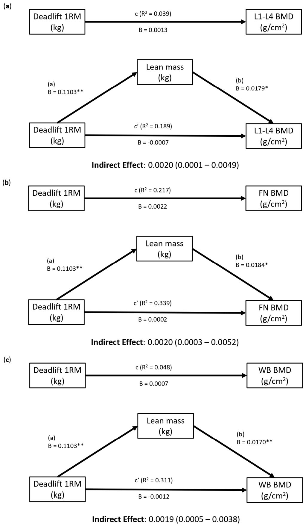

Table 1.
Characteristics of athletes by playing positions.
| Variable | Total (n=28) | Goalkeeper (n=3) | Defenders (n=9) | Midfielders (n=7) | Forwards (n=9) | p |
|---|---|---|---|---|---|---|
| Age, yrs | 24.96 (3.47) | 26.67 (4.04) | 25.89 (3.18) | 25.14 (3.29) | 23.33 (3.64) | 0.331 |
| Weight, kg | 59.86 (5.45) | 63.34 (5.50) | 58.45 (4.53) | 61.86 (6.79) | 58.56 (5.06) | 0.367 |
| Height, m | 1.67 (0.05) | 1.72 (0.03) | 1.67 (0.06) | 1.66 (0.05) | 1.65 (0.05) | 0.191 |
| BMI, kg/m2 | 21.55 (1.71) | 21.44 (2.29) | 20.94 (1.54) | 22.38 (1.99) | 21.55 (1.49) | 0.443 |
| Lean mass, kg | 43.98 (3.73) | 45.45 (3.15) | 44.40 (3.60) | 44.43 (4.23) | 42.72 (3.90) | 0.658 |
| Fat mass, kg | 13.13 (2.73) | 14.83 (2.03) | 11.16 (1.57)† | 14.63 (3.45) | 13.36 (2.28) | 0.034 |
| Fat mass, % | 21.80 (3.10) | 23.37 (1.94) | 19.12 (1.97)†‡ | 23.43 (3.03) | 22.70 (2.93) | 0.009 |
| WB BMD, g/cm2 | 1.32 (0.08) | 1.39 (0.12) | 1.31 (0.07) | 1.31 (0.07) | 1.30 (0.08) | 0.340 |
| L1-L4 BMD, g/cm2 | 1.21 (0.11) | 1.33 (0.06) | 1.21 (0.10) | 1.20 (0.08) | 1.16 (0.13) | 0.142 |
| FN BMD, g/cm2 | 1.11 (0.12) | 1.17 (0.24) | 1.12 (0.07) | 1.09 (0.15) | 1.08 (0.11) | 0.712 |
| CMJ height, cm | 33.07 (2.96) | 33.52 (2.01) | 32.56 (3.31) | 32.88 (3.31) | 33.57 (2.95) | 0.905 |
| Deadlift 1RM, kg | 101.43 (15.55) | 111.27 (13.41) | 101.99 (17.96) | 100.20 (14.74) | 98.54 (15.43) | 0.544 |
Means and Standard Deviations (SD). BMI: Body Mass Index; BMD: Bone mineral density; WB: Whole body; L1-L4: Lumbar spine; FN: Femoral neck; CMJ: Countermovement jump; 1RM: one-repetition maximum. †p ≤ 0.05 between defenders and midfielders. ‡p ≤ 0.05 between defenders and forwards.
Disclaimer/Publisher’s Note: The statements, opinions and data contained in all publications are solely those of the individual author(s) and contributor(s) and not of MDPI and/or the editor(s). MDPI and/or the editor(s) disclaim responsibility for any injury to people or property resulting from any ideas, methods, instructions or products referred to in the content. |
© 2026 by the authors. Licensee MDPI, Basel, Switzerland. This article is an open access article distributed under the terms and conditions of the Creative Commons Attribution (CC BY) license.
Copyright: This open access article is published under a Creative Commons CC BY 4.0 license, which permit the free download, distribution, and reuse, provided that the author and preprint are cited in any reuse.