

Submitted:
03 September 2026
Posted:
09 September 2026
You are already at the latest version
Abstract
Sleep is essential for memory consolidation, immune homeostasis, and the maintenance of brain function. Emerging evidence indicates that these processes are closely linked to the gut microbiota through bidirectional neural, endocrine, metabolic, and immune pathways. This review integrates current knowledge on the complementary roles of slow-wave and rapid eye movement sleep in the stabilization, reorganization, and integration of memories, while examining how sleep disruption impairs cognition through neuroinflammation, altered synaptic plasticity, and disturbed hippocampal–neocortical communication. It also summarizes the major categories of sleep disorders and their potential effects on memory-related processes. Particular emphasis is placed on the microbiota–gut–brain axis as a mechanistic interface connecting sleep physiology with immune regulation and cognitive performance. Sleep loss and circadian disruption can increase intestinal permeability, promote microbial dysbiosis, facilitate systemic exposure to bacterial products, and activate inflammatory pathways that compromise hippocampal function. Conversely, microbiota-derived metabolites, including short-chain fatty acids and tryptophan-derived compounds, may influence sleep architecture, blood–brain barrier integrity, microglial activity, and neuroplasticity. Evidence from experimental models further suggests that microbiota alterations induced by sleep deprivation can contribute directly to memory deficits and that microbiota-targeted interventions may partially restore cognitive function. Collectively, these findings support an integrated sleep–microbiota–immunity framework in which disruption of any component can amplify dysfunction across the others, with important implications for understanding and treating cognitive impairment associated with sleep disorders.
Keywords:
sleep
; microbiota–gut–brain axis
; memory consolidation
1. Introduction
Sleep is a fundamental biological process that plays a critical role in learning and memory. Over the past two decades, converging evidence from behavioral, electrophysiological, and neuroimaging studies has demonstrated that sleep actively contributes to memory consolidation rather than merely protecting newly acquired information from interference. In particular, slow-wave sleep (SWS) and rapid eye movement (REM) sleep support complementary aspects of memory processing, including the stabilization, integration, and reorganization of newly encoded memories. The hippocampal–neocortical dialogue that occurs during SWS, characterized by the coordinated interaction of cortical slow oscillations, thalamocortical sleep spindles, and hippocampal sharp-wave ripples, is considered a central mechanism underlying declarative memory consolidation (Diekelmann and Born, 2010; Rasch and Born, 2013).
Beyond its effects on neural circuits, sleep exerts profound regulatory effects on immune function. Sleep and the immune system engage in a bidirectional relationship in which immune mediators influence sleep architecture, whereas sleep modulates both innate and adaptive immune responses. Experimental sleep deprivation increases circulating levels of pro-inflammatory cytokines, including interleukin (IL)-1β, IL-6, and tumor necrosis factor-α (TNF-α), whereas adequate sleep promotes immune homeostasis and facilitates the development of immunological memory (Besedovsky et al., 2019; Irwin, 2019). Notably, several cytokines involved in immune regulation also influence synaptic plasticity, long-term potentiation, and memory formation, suggesting that immune signaling pathways may directly contribute to sleep-dependent cognitive processes.
Increasing evidence also indicates that the gut microbiota is an important regulator of both sleep physiology and brain function. Through the microbiota–gut–brain axis, intestinal microorganisms communicate with the central nervous system via neural, endocrine, metabolic, and immune pathways. Microbiota-derived metabolites, including short-chain fatty acids, indole derivatives, and neurotransmitter precursors, influence sleep architecture, circadian rhythms, neuroinflammation, and synaptic plasticity. Conversely, sleep disruption can alter microbial composition and diversity, thereby promoting intestinal dysbiosis and systemic inflammation (Wagner-Skacel et al., 2020; Sun et al., 2023). These observations support a reciprocal relationship between sleep and the gut microbiome. Recent studies have begun to integrate these previously separate fields, suggesting that immune signaling may serve as a critical mechanistic bridge linking the gut microbiota to sleep-dependent memory consolidation. Alterations in microbial communities can modulate peripheral and central cytokine production, microglial activity, blood–brain barrier integrity, and hippocampal plasticity, all of which influence learning and memory. Experimental models have shown that sleep deprivation-induced dysbiosis is associated with increased production of inflammatory cytokines and impaired cognitive performance, whereas microbiota-targeted interventions can improve both sleep quality and memory-related outcomes (Zhang et al., 2023).
Taken together, these findings support a conceptual framework in which sleep, immunity, and the gut microbiota function as components of an integrated biological network that regulates memory formation and long-term cognitive function. This review critically examines the evidence supporting the role of sleep in memory consolidation and explores how immune mechanisms and microbiota-derived signals influence this process. Particular emphasis is placed on the molecular and cellular pathways linking sleep physiology, neuroimmune regulation, and microbiota–gut–brain communication.
2. Major Types of Sleep Disorders
Sleep disorders comprise a heterogeneous group of conditions that affect sleep quality, duration, timing, or continuity, with substantial consequences for physical health, cognitive performance, and emotional well-being. The International Classification of Sleep Disorders, Third Edition (ICSD-3), organizes these conditions into six major categories: insomnia disorders, sleep-related breathing disorders, central disorders of hypersomnolence, circadian rhythm sleep–wake disorders, parasomnias, and sleep-related movement disorders (Ferri et al., 2014).
Insomnia Disorders. Insomnia is the most prevalent sleep disorder worldwide, affecting approximately 10% of adults in its chronic form. It is characterized by persistent difficulty initiating or maintaining sleep, or by an inability to obtain restorative sleep, despite adequate opportunity and appropriate environmental conditions. The ICSD-3 consolidated several previously distinct phenotypes into the diagnosis of chronic insomnia disorder, which requires symptoms to occur at least three times per week for a minimum of three months (Ferri et al., 2014). Beyond impaired sleep quality, chronic insomnia is associated with deficits in attention, executive function, emotional regulation, and memory consolidation, underscoring its broad effects on brain function.
Sleep-Related Breathing Disorders. Sleep-related breathing disorders encompass conditions characterized by abnormalities in respiratory airflow or ventilation during sleep. Obstructive sleep apnea (OSA), the most common disorder in this category, is characterized by recurrent upper-airway collapse, resulting in intermittent hypoxia and repeated sleep fragmentation. Other conditions include central sleep apnea syndromes, in which impaired neural signaling to the respiratory muscles disrupts normal breathing patterns, and sleep-related hypoventilation disorders (Ferri et al., 2014). OSA has received particular attention because intermittent hypoxia and sleep fragmentation promote oxidative stress, neuroinflammation, and glymphatic dysfunction, all of which contribute to cognitive impairment and may increase susceptibility to neurodegenerative diseases (Deyang et al., 2024).
Central Disorders of Hypersomnolence. Central disorders of hypersomnolence are characterized primarily by excessive daytime sleepiness that cannot be explained by insufficient sleep, circadian misalignment, or other medical conditions. This category includes narcolepsy type 1, narcolepsy type 2, idiopathic hypersomnia, and the rare Kleine–Levin syndrome (Ferri et al., 2014). Narcolepsy type 1 is strongly associated with hypocretin (orexin) deficiency and is frequently accompanied by cataplexy. Individuals with central hypersomnolence disorders often exhibit impairments in sustained attention, executive function, processing speed, and emotional regulation, illustrating the critical role of sleep–wake regulatory systems in cognitive performance (Filardi et al., 2021).
Circadian Rhythm Sleep–Wake Disorders. Circadian rhythm sleep–wake disorders arise from a mismatch between the endogenous circadian timing system and external environmental or social schedules. Common examples include shift work disorder and jet lag disorder, both of which cause sleep disturbances and excessive sleepiness at biologically inappropriate times (Ferri et al., 2014). Persistent circadian misalignment has been shown to impair hippocampal neurogenesis and memory formation, producing cognitive deficits that may persist even after restoration of a normal sleep schedule (Gibson et al., 2010). These findings emphasize the importance of circadian synchronization for optimal brain function and cognitive health.
Parasomnias. Parasomnias comprise a group of disorders characterized by abnormal behaviors, motor activity, emotions, perceptions, or autonomic responses occurring during sleep or during transitions between sleep and wakefulness. They are broadly classified as non-rapid eye movement (NREM) parasomnias, including sleepwalking and night terrors, and rapid eye movement (REM) parasomnias. Among the latter, REM sleep behavior disorder (RBD) is of particular clinical importance because it is strongly associated with the subsequent development of α-synucleinopathies, including Parkinson’s disease, dementia with Lewy bodies, and multiple system atrophy (Ferri et al., 2014). Consequently, RBD is increasingly recognized as a prodromal marker of neurodegeneration.
Sleep-Related Movement Disorders. Sleep-related movement disorders are characterized by relatively simple, repetitive, involuntary movements that disrupt sleep continuity. The most common condition in this category is restless legs syndrome (RLS), which is defined by uncomfortable sensations and an irresistible urge to move the limbs, particularly during periods of rest or inactivity. Periodic limb movement disorder (PLMD) is another common condition that causes repetitive limb movements during sleep, leading to frequent microarousals and sleep fragmentation (Ferri et al., 2014). Although often overlooked, these disorders can substantially impair sleep quality and contribute to daytime fatigue, cognitive dysfunction, and reduced quality of life.
Collectively, these disorders demonstrate that sleep disturbances extend beyond simple reductions in sleep duration. Each category disrupts distinct neurobiological pathways involved in memory consolidation, synaptic plasticity, immune regulation, and metabolic homeostasis, thereby contributing to a broad spectrum of neurological and systemic consequences (Khan, 2023).
3. The Gut–Brain Axis: Bidirectional Communication Between the Microbiota and Sleep
The relationship between the gut microbiota and sleep regulation is mediated by a bidirectional communication network within the gut–brain axis (Figure 1). This system enables microbial signals to modulate the central nervous system (CNS), while the host’s sleep–wake rhythms simultaneously shape the composition and dynamics of the intestinal ecosystem (Oriquat et al., 2026).
When sleep disturbances such as insomnia or sleep apnea occur, this circadian synchrony is disrupted, promoting marked dysbiosis (Lin et al., 2024).
At the cellular and molecular levels, sleep deprivation increases intestinal mucosal permeability by disrupting tight junction proteins. This alteration facilitates the translocation of bacterial components, such as lipopolysaccharide (LPS), into the systemic circulation (Figure 1). Endotoxins in the bloodstream activate myeloid cells and induce the release of pro-inflammatory cytokines, primarily IL-1β, IL-6, and TNF-α (Pan et al., 2025).
These cytokines can subsequently cross the blood–brain barrier or activate vagal afferent pathways, thereby promoting systemic inflammation and neuroinflammation. This process may culminate in microglial overactivation within the hippocampus and disruption of normal sleep–wake patterns through interference with hypothalamic control centers (Pan et al., 2025).
Consistent with these findings, numerous studies have identified distinct microbiome signatures associated with specific sleep disorders. The following table summarizes the characteristic alterations in the gut microbiota associated with the major categories of sleep disorders described above:
In turn, dysbiosis may further exacerbate poor sleep quality by compromising intestinal barrier integrity and reducing the production of essential microbial metabolites, such as gamma-aminobutyric acid (GABA) and short-chain fatty acids (SCFAs). This metabolic deficiency intensifies neuroinflammation and increases the host’s reactivity to stress. Consequently, a pathological feedback loop emerges in which dysfunction in one system accelerates deterioration in the other, perpetuating both microbial imbalance and disruption of sleep architecture (Oriquat et al., 2026).
4. Influence of the Gut Microbiota on Cognitive Performance and Learning
In addition to altering sleep architecture and quality, the gut microbiota has been shown to influence multiple forms of learning. Studies across diverse experimental models have identified specific microbial signatures associated with cognitive performance. The predominance of certain bacterial taxa has been negatively correlated with learning, an association that appears to be mediated by increased intestinal permeability (Xu et al., 2023; Gupta, 2024). Impairment of the intestinal barrier facilitates the translocation of lipopolysaccharide (LPS) into the circulation, resulting in systemic endotoxemia (Gupta, 2024), which may subsequently trigger neuroinflammation in brain regions critical for cognition, including the hippocampus and cerebral cortex (Zhao et al., 2026).
By contrast, several bacterial populations have been associated with cognition-supporting and neuroprotective effects at both metabolic and structural levels (Zhao et al., 2026). Metabolically, the production of short-chain fatty acids (SCFAs) helps restore intestinal barrier integrity and suppress inflammatory pathways (Gupta, 2024; Zhao et al., 2026). Structurally, these effects may preserve white matter integrity and prevent gray matter atrophy in diencephalic regions such as the thalamus (Salimi et al., 2026; Zhao et al., 2026). The intestinal ecosystem also modulates the expression of brain-derived neurotrophic factor (BDNF), a key regulator of neurogenesis (Salami and Soheili, 2022; Xu et al., 2023), and reduces oxidative stress in the prefrontal cortex, thereby protecting neurons from apoptosis (Salami and Soheili, 2022; Zhao et al., 2022).
This signaling network is mediated by endocrine factors and vagal pathways. The vagus nerve constitutes a major physical and sensory route connecting the gastrointestinal tract to the brainstem (Yue et al., 2026). Through this parasympathetic pathway, vagal afferent terminals detect immunological and metabolic signals derived from the gut microbiota and relay this information to brain regions involved in cognition (Yue et al., 2026). Given these contrasting effects, microbial taxa may be functionally categorized according to their detrimental or beneficial associations with cognitive performance. Table 1 summarizes these microbial signatures and their reported effects on different domains of learning.
Across the studies summarized, specific taxa repeatedly emerge as either detrimental or beneficial modulators of different learning domains. Among the microorganisms associated with cognitive impairment, the Enterobacteriaceae and Helicobacteraceae families are particularly prominent. Both exhibit strongly pro-inflammatory profiles and include well-known pathogenic genera, such as Salmonella and Shigella within Enterobacteriaceae and Helicobacter within Helicobacteraceae (Zhao et al., 2022; Shi et al., 2023; Zhang et al., 2023).
Conversely, a recurring group of taxa appears to support learning across multiple cognitive domains. Particularly notable are the genera Lactobacillus and Bifidobacterium, which are widely recognized as classical probiotics. These microorganisms are characterized by potent anti-inflammatory activity and robust production of short-chain fatty acids (SCFAs) (Salami and Soheili, 2022; Shi et al., 2023; Gupta, 2024; Salimi et al., 2026).
These findings are consistent with the evidence discussed in the section on sleep disorders. As noted above, sleep architecture and quality are bidirectionally related to the gut microbiota, creating a pathological feedback loop when sleep is disrupted (Oriquat et al., 2026). The association of cognitive deficits with specific microbial signatures, together with improvements in cognitive performance following restoration of key bacterial populations, strongly supports the existence of a broader systemic interaction.
Taken together, these observations support a complex tripartite network in which sleep quality, gut microbiome homeostasis, and learning consolidation reciprocally influence one another.
The Sleep–Microbiota–Cognition Triad
Sleep disorders markedly impair cognitive function, particularly declarative, non-declarative, and working memory, through neurochemical and electrophysiological alterations (Brem et al., 2013; Ratcliff and Van Dongen, 2018).
Sleep deprivation and the consequent reduction in slow-wave sleep (SWS) impair the clearance of synaptic noise, thereby disrupting both the consolidation of recently acquired memories and the encoding of new information (Brem et al., 2013; Ratcliff and Van Dongen, 2018). More recent evidence suggests that this neurocognitive phenotype is strongly influenced by the gut–brain axis. In 2023, Wang et al. demonstrated that fecal microbiota transplantation from sleep-deprived mice was sufficient to induce cognitive impairment in healthy rodents (Li et al., 2023). In complementary experiments, treatment with short-chain fatty acids (SCFAs) reversed these deficits, strongly suggesting that intestinal dysbiosis contributes to sleep deprivation-induced memory impairment (Figure 1) (Li et al., 2023). This systemic hypothesis is further supported by integrating microbiological findings from sleep disorders with bacterial signatures associated with impaired learning. A consistent overlap emerges among specific taxa acting through well-defined neuroimmune and metabolic pathways:
Phylum Proteobacteria and the Enterobacteriaceae Family
These taxa are enriched under conditions of circadian disruption, such as jet lag and shift work (Table 2), and are also associated with impairments in hippocampus-dependent memory domains, including spatial, verbal, working, and associative memory (Table 1). Their expansion provides a direct immunological link because, as Gram-negative bacteria, they substantially increase the intestinal burden of lipopolysaccharide (LPS) (Cani et al., 2008). Translocation of LPS into the bloodstream induces metabolic endotoxemia and, when sustained, chronically activates hippocampal microglia and disrupts synaptic plasticity (Sparkman and Johnson, 2008). This microbial signature may therefore provide a biochemical explanation for the “brain fog” commonly experienced by individuals with circadian rhythm disturbances (Figure 2) (Dantzer, 2009; Thaiss et al., 2014).
Phylum Bacteroidetes
Frequently elevated in acute insomnia (Table 2), the overgrowth of genera such as Alistipes and Prevotella may act as a metabolic sink that sequesters essential amino acids, particularly dietary tryptophan (Agus et al., 2018; Valles-Colomer et al., 2019). By diverting tryptophan metabolism, these microorganisms reduce its availability for serotonin and melatonin synthesis, potentially exacerbating insomnia, while redirecting metabolism toward the kynurenine pathway or excessive indole production (Kennedy et al., 2017). Metabolites generated through this pathway, including quinolinic acid and kynurenic acid, are highly neuroactive and, at elevated concentrations, can interfere with NMDA receptor signaling in the central nervous system. These receptors are critically involved in the consolidation of spatial and working memory (Figure 2) (Schwarcz et al., 2012).
Phylum Firmicutes
Although this phylum includes numerous beneficial bacterial species, its disproportionate expansion may become detrimental. An excess of fermentative bacteria can promote abnormal D-lactate production or an imbalanced increase in specific SCFAs, thereby altering the pH of the local microenvironment (Kowlgi and Chhabra, 2015). The human brain is particularly sensitive to D-lactate because of its limited capacity to metabolize this neurotoxic compound. Subclinical D-lactate accumulation can cause spatial disorientation, lethargy, and pronounced brain fog (Uribarri et al., 1998).
Converging evidence regarding the energetic sensitivity of the basal ganglia and their central role in procedural learning suggests that D-lactate-induced metabolic disruption may provide a plausible mechanistic explanation for impairment in this form of learning. Procedural learning requires precise energetic and motor coordination, which may be compromised when these bacterial populations dominate the intestinal ecosystem (Figure 2) (Cross and Callaway, 1984; Graybiel, 2008).
Overall, current evidence supports a systemic model in which sleep architecture, gut microbiome homeostasis, and cognitive processes form a tripartite, interdependent regulatory network. Alterations in the sleep–wake cycle not only compromise memory consolidation at the electrophysiological level but also act as potent drivers of intestinal dysbiosis. This microbial disruption may perpetuate cognitive impairment through specific neuroimmune and metabolic pathways, including LPS-mediated endotoxemia, altered production of short-chain fatty acids (SCFAs), and the pathological diversion of neuroactive precursors. Understanding the complexity of the gut–brain axis is therefore essential because it underscores that memory impairment associated with sleep disorders is not exclusively neurological but may instead reflect broader multisystem metabolic and immunological dysfunction.
5. Conclusions
In conclusion, the available evidence supports a tightly interconnected relationship among sleep, immune regulation, the gut microbiota, and memory consolidation. Sleep disruption can impair hippocampal–neocortical communication and synaptic plasticity while simultaneously promoting intestinal dysbiosis, barrier dysfunction, systemic inflammation, and neuroimmune activation. In turn, altered microbial communities and their metabolites may further disturb sleep architecture and cognitive performance, establishing self-reinforcing pathological loops. Although several microbial taxa and metabolic pathways have been associated with beneficial or detrimental cognitive effects, much of the evidence remains correlative, and causal mechanisms are not yet fully resolved. Future studies should combine longitudinal human cohorts with mechanistically rigorous experimental models to determine how specific microorganisms, metabolites, immune mediators, and neural pathways interact across different sleep disorders and stages of memory processing. Such work will be essential for identifying reliable biomarkers and for developing microbiota- or immune-targeted interventions capable of improving both sleep quality and cognitive outcomes.
Funding
This work was supported by the grants from Agencia Nacional de Investigación y Desarrollo: “Financiamiento Basal para Centros Científicos y Tecnológicos de Excelencia” Centro Ciencia & Vida [FB210008] (to Fundación Ciencia & Vida), and FONDECYT [1250021] (to R.P.).
Conflicts of Interest
The authors declare that they have no conflicts of interest.
References
- Agus A, Planchais J, Sokol H (2018) Gut Microbiota Regulation of Tryptophan Metabolism in Health and Disease. Cell Host Microbe 23:716–724.
- Besedovsky L, Lange T, Haack M (2019) The Sleep-Immune Crosstalk in Health and Disease. Physiological reviews 99:1325–1380.
- Brem AK, Ran K, Pascual-Leone A (2013) Learning and memory. Handb Clin Neurol 116:693–737.
- Cani PD, Bibiloni R, Knauf C, Waget A, Neyrinck AM, Delzenne NM, Burcelin R (2008) Changes in gut microbiota control metabolic endotoxemia-induced inflammation in high-fat diet-induced obesity and diabetes in mice. Diabetes 57:1470–1481.
- Cross SA, Callaway CW (1984) D-Lactic acidosis and selected cerebellar ataxias. Mayo Clin Proc 59:202–205.
- Dantzer R (2009) Cytokine, sickness behavior, and depression. Immunol Allergy Clin North Am 29:247–264.
- Deyang T, Baig MAI, Dolkar P, Hediyal TA, Rathipriya AG, Bhaskaran M, PandiPerumal SR, Monaghan TM, Mahalakshmi AM, Chidambaram SB (2024) Sleep apnoea, gut dysbiosis and cognitive dysfunction. FEBS J 291:2519–2544.
- Diekelmann S, Born J (2010) The memory function of sleep. Nature reviews Neuroscience 11:114–126.
- Ferri R, Cosentino FI, Manconi M, Rundo F, Bruni O, Zucconi M (2014) Increased electroencephalographic high frequencies during the sleep onset period in patients with restless legs syndrome. Sleep 37:1375–1381.
- Filardi M, D’Anselmo A, Agnoli S, Rubaltelli E, Mastria S, Mangiaruga A, Franceschini C, Pizza F, Corazza GE, Plazzi G (2021) Cognitive dysfunction in central disorders of hypersomnolence: A systematic review. Sleep Med Rev 59:101510.
- Gibson EM, Wang C, Tjho S, Khattar N, Kriegsfeld LJ (2010) Experimental ‘jet lag’ inhibits adult neurogenesis and produces long-term cognitive deficits in female hamsters. PloS one 5:e15267.
- Graybiel AM (2008) Habits, rituals, and the evaluative brain. Annu Rev Neurosci 31:359–387.
- Gupta PC, S. (2024) THE ROLE OF THE GUT MICROBIOME IN LEARNING AND MEMORY. In: Futuristic Trends in Pharmacy & Nursing.
- Irwin MR (2019) Sleep and inflammation: partners in sickness and in health. Nat Rev Immunol 19:702–715.
- Kennedy PJ, Cryan JF, Dinan TG, Clarke G (2017) Kynurenine pathway metabolism and the microbiota-gut-brain axis. Neuropharmacology 112:399–412.
- Khan MAA-J, H. (2023) The consequences of sleep deprivation on cognitive performance. Neurosciences 28:91–99.
- Kowlgi NG, Chhabra L (2015) D-lactic acidosis: an underrecognized complication of short bowel syndrome. Gastroenterol Res Pract 2015:476215.
- Lecomte A, Barateau L, Pereira P, Paulin L, Auvinen P, Scheperjans F, Dauvilliers Y (2020) Gut microbiota composition is associated with narcolepsy type 1. Neurol Neuroimmunol Neuroinflamm 7.
- Li N, Tan S, Wang Y, Deng J, Wang N, Zhu S, Tian W, Xu J, Wang Q (2023) Akkermansia muciniphila supplementation prevents cognitive impairment in sleep-deprived mice by modulating microglial engulfment of synapses. Gut Microbes 15:2252764.
- Li Y, Zhang B, Zhou Y, Wang D, Liu X, Li L, Wang T, Zhang Y, Jiang M, Tang H, Amsel LV, Fan F, Hoven CW (2020) Gut Microbiota Changes and Their Relationship with Inflammation in Patients with Acute and Chronic Insomnia. Nat Sci Sleep 12:895–905.
- Lin Z, Jiang T, Chen M, Ji X, Wang Y (2024) Gut microbiota and sleep: Interaction mechanisms and therapeutic prospects. Open Life Sci 19:20220910.
- Liu Z, Wei ZY, Chen J, Chen K, Mao X, Liu Q, Sun Y, Zhang Z, Zhang Y, Dan Z, Tang J, Qin L, Chen JH, Liu X (2020) Acute Sleep-Wake Cycle Shift Results in Community Alteration of Human Gut Microbiome. mSphere 5.
- Montini A, Pellegrini C, Loddo G, Ravaioli F, Baldelli L, Mainieri G, Pirazzini C, Mazzotta E, Carano F, Sala C, De Fanti S, Bacalini MG, Provini F (2026) Analysis of gut microbiota in Restless Legs Syndrome: searching for a metagenomic signature. Sleep 49.
- Moreno-Indias I, Torres M, Montserrat JM, Sanchez-Alcoholado L, Cardona F, Tinahones FJ, Gozal D, Poroyko VA, Navajas D, Queipo-Ortuno MI, Farre R (2015) Intermittent hypoxia alters gut microbiota diversity in a mouse model of sleep apnoea. Eur Respir J 45:1055–1065.
- Oriquat G, Al-Hasnaawei S, Mousa HM, Malathi H, Singla S, Sahoo S, Arora V, Chauhan AS, Nourizadeh M (2026) Gut Microbiome-Sleep Crosstalk: Mechanistic Pathways, Dysbiosis Signatures, and Microbiome-Based Interventions. Brain Behav 16:e71525.
- Pan D, Li J, Chen S, Gu S, Jiang M, Xu Q (2025) Microbiota-gut-brain axis pathogenesis and targeted therapeutics in sleep disorders. Frontiers in neurology 16:1721606.
- Rasch B, Born J (2013) About sleep’s role in memory. Physiological reviews 93:681–766.
- Ratcliff R, Van Dongen HPA (2018) The effects of sleep deprivation on item and associative recognition memory. J Exp Psychol Learn Mem Cogn 44:193–208.
- Salami M, Soheili M (2022) The microbiota-gut- hippocampus axis. Front Neurosci 16:1065995.
- Salimi Y, Namdarzadeh B, Dehghani-Arani F, Vahabie AH, Rezayat E (2026) Investigating the effects of synbiotic intervention on working memory, attention, and inhibitory control in healthy young women. Behav Brain Res 513:116349.
- Schwarcz R, Bruno JP, Muchowski PJ, Wu HQ (2012) Kynurenines in the mammalian brain: when physiology meets pathology. Nature reviews Neuroscience 13:465–477.
- Shi J, Zhao Y, Chen Q, Liao X, Chen J, Xie H, Liu J, Sun J, Chen S (2023) Association Analysis of Gut Microbiota and Prognosis of Patients with Acute Ischemic Stroke in Basal Ganglia Region. Microorganisms 11.
- Sparkman NL, Johnson RW (2008) Neuroinflammation associated with aging sensitizes the brain to the effects of infection or stress. Neuroimmunomodulation 15:323–330.
- Sun J, Fang D, Wang Z, Liu Y (2023) Sleep Deprivation and Gut Microbiota Dysbiosis: Current Understandings and Implications. Int J Mol Sci 24.
- Thaiss CA, Zeevi D, Levy M, Zilberman-Schapira G, Suez J, Tengeler AC, Abramson L, Katz MN, Korem T, Zmora N, Kuperman Y, Biton I, Gilad S, Harmelin A, Shapiro H, Halpern Z, Segal E, Elinav E (2014) Transkingdom control of microbiota diurnal oscillations promotes metabolic homeostasis. Cell 159:514–529.
- Uribarri J, Oh MS, Carroll HJ (1998) D-lactic acidosis. A review of clinical presentation, biochemical features, and pathophysiologic mechanisms. Medicine 77:73–82.
- Valles-Colomer M, Falony G, Darzi Y, Tigchelaar EF, Wang J, Tito RY, Schiweck C, Kurilshikov A, Joossens M, Wijmenga C, Claes S, Van Oudenhove L, Zhernakova A, Vieira-Silva S, Raes J (2019) The neuroactive potential of the human gut microbiota in quality of life and depression. Nat Microbiol 4:623–632.
- Wagner-Skacel J, Dalkner N, Moerkl S, Kreuzer K, Farzi A, Lackner S, Painold A, Reininghaus EZ, Butler MI, Bengesser S (2020) Sleep and Microbiome in Psychiatric Diseases. Nutrients 12.
- Xu X, Zhuo L, Zhang L, Peng H, Lyu Y, Sun H, Zhai Y, Luo D, Wang X, Li X, Li L, Zhang Y, Ma X, Wang Q, Li Y (2023) Dexmedetomidine alleviates host ADHD-like behaviors by reshaping the gut microbiota and reducing gut-brain inflammation. Psychiatry Res 323:115172.
- Yue CB, Luan WW, Qiu D, Ding X, Gu HW, Liu PM, Hashimoto K, Yang JJ, Wang XM (2026) A vagus-dependent gut microbiota-metabolite axis drives chronic inflammatory pain and working-memory deficits in mice. Brain Res Bull 234:111702.
- Zhang M, Zhang M, Kou G, Li Y (2023) The relationship between gut microbiota and inflammatory response, learning and memory in mice by sleep deprivation. Front Cell Infect Microbiol 13:1159771.
- Zhao W, Zhou X, Cai H, Tang T, Shen Y, Sun Z, Zhu J, Yu Y (2026) The relation between gut microbiota, brain structure and cognitive function in metabolic syndrome. Brain, behavior, and immunity 132:106209.
- Zhao Z, Cui D, Wu G, Ren H, Zhu X, Xie W, Zhang Y, Yang L, Peng W, Lai C, Huang Y, Li H (2022) Disrupted gut microbiota aggravates working memory dysfunction induced by high-altitude exposure in mice. Front Microbiol 13:1054504.
Figure 1.
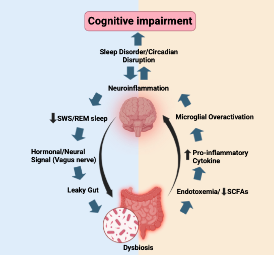
Pathological feedback loop linking intestinal alterations, neuroinflammation, and cognitive decline. The flowchart illustrates the temporal and mechanistic progression from increased intestinal permeability to neurological dysfunction through interconnected immune and neural pathways. Gut dysbiosis promotes endotoxin translocation and disrupts the balance of short-chain fatty acids (SCFAs), generating aberrant signals that can be transmitted to the central nervous system, including via the vagus nerve. The convergence of these peripheral signals promotes a pro-inflammatory cytokine cascade and sustained microglial activation, ultimately leading to chronic neuroinflammation. This neuroinflammatory state disrupts normal sleep architecture, particularly slow-wave sleep (SWS) and rapid eye movement (REM) sleep, thereby contributing to circadian dysregulation and cognitive decline. These alterations may, in turn, further exacerbate intestinal dysbiosis, perpetuating the pathological feedback loop.
Figure 1.
Pathological feedback loop linking intestinal alterations, neuroinflammation, and cognitive decline. The flowchart illustrates the temporal and mechanistic progression from increased intestinal permeability to neurological dysfunction through interconnected immune and neural pathways. Gut dysbiosis promotes endotoxin translocation and disrupts the balance of short-chain fatty acids (SCFAs), generating aberrant signals that can be transmitted to the central nervous system, including via the vagus nerve. The convergence of these peripheral signals promotes a pro-inflammatory cytokine cascade and sustained microglial activation, ultimately leading to chronic neuroinflammation. This neuroinflammatory state disrupts normal sleep architecture, particularly slow-wave sleep (SWS) and rapid eye movement (REM) sleep, thereby contributing to circadian dysregulation and cognitive decline. These alterations may, in turn, further exacerbate intestinal dysbiosis, perpetuating the pathological feedback loop.
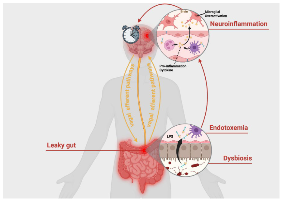
Figure 2.
Impact of microbiota-derived metabolites on synaptic plasticity and memory consolidation. The figure illustrates the cellular and molecular mechanisms through which gut dysbiosis and endotoxaemia can alter the neuronal microenvironment. Dysbiosis promotes the systemic release of bacterial components and metabolites, including lipopolysaccharide (LPS; left panel), indoles and disturbances in tryptophan (Trp) metabolism (midle panel), and D-lactate (right panel), which may reach and influence the central nervous system. These signals may disrupt glutamatergic neurotransmission, including NMDA receptor-dependent signalling, thereby impairing the structural and functional synaptic plasticity required for effective memory consolidation. Such alterations may ultimately contribute to cognitive impairment, including difficulties commonly described as brain fog.
Figure 2.
Impact of microbiota-derived metabolites on synaptic plasticity and memory consolidation. The figure illustrates the cellular and molecular mechanisms through which gut dysbiosis and endotoxaemia can alter the neuronal microenvironment. Dysbiosis promotes the systemic release of bacterial components and metabolites, including lipopolysaccharide (LPS; left panel), indoles and disturbances in tryptophan (Trp) metabolism (midle panel), and D-lactate (right panel), which may reach and influence the central nervous system. These signals may disrupt glutamatergic neurotransmission, including NMDA receptor-dependent signalling, thereby impairing the structural and functional synaptic plasticity required for effective memory consolidation. Such alterations may ultimately contribute to cognitive impairment, including difficulties commonly described as brain fog.
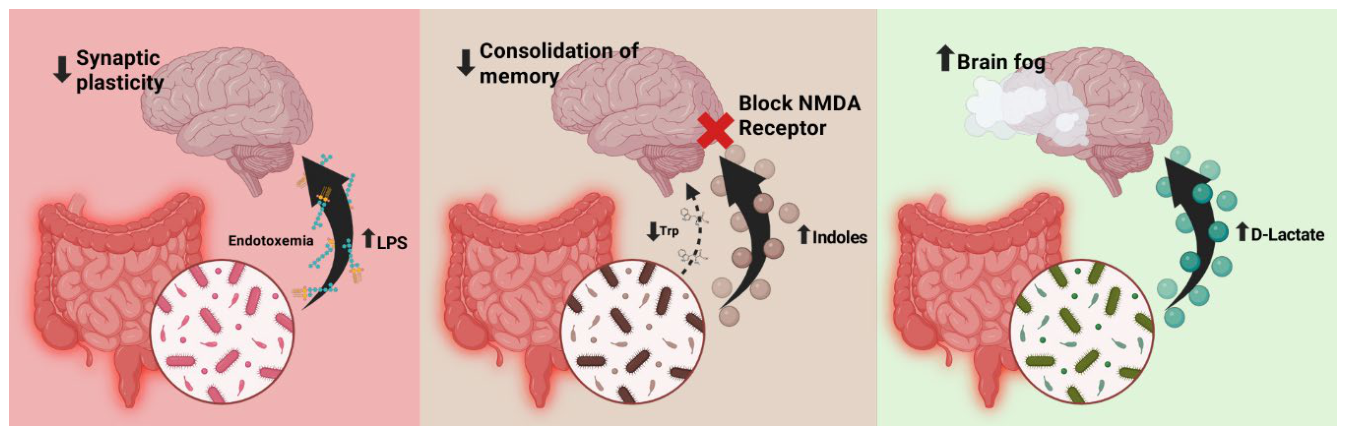

Table 1.
Gut microbial signatures and their reported effects on cognitive performance.
| Learning Type1 | Detrimental Microbial Taxa | Beneficial Microbial Taxa | Scientific interpretation/reference | |
| Working/Short-Term Memory | Helicobacteraceae, Enterobacteriaceae, Porphyromonadaceae, and Enterococcaceae | Lactobacillus, Bifidobacterium, Butyricimonas, Parasutterella, and Ruminiclostridium | Microbiota disruption aggravates working memory dysfunction by increasing lipopolysaccharide (LPS)-induced neuroinflammation, whereas taxa like Lactobacillus and Bifidobacterium protect intestinal barrier integrity through the production of short-chain fatty acids (Zhao et al., 2022; Xu et al., 2023; Salimi et al., 2026) | |
| Declarative Learning | Spatial Memory | Tannerellaceae, Rhodospirillales, Alistipes, Parabacteroides, Burkholderiaceae, Enterobacteriaceae, and Erysipelotrichaceae | Lactobacillus, Bifidobacterium, and Akkermansia muciniphila | The overgrowth of genera such as Alistipes diverts tryptophan metabolism toward the kynurenine pathway, generating metabolites that interfere with NMDA receptor signaling, which is critical for spatial memory consolidation (Zhang et al., 2023) |
| Verbal Memory | Fusobacterium, Streptococcus, and the Enterobacteriaceae family | Lactobacillus, Bifidobacterium, Lachnospira, Clostridium XIVa, Kineothrix, and Acetivibrio | An excess of bacteria with pro-inflammatory profiles, such as the Enterobacteriaceae family, promotes neuroinflammation that negatively affects brain structures and hippocampus-dependent memory domains (Zhao et al., 2026) | |
| Non-Declarative Learning | Procedural Learning | Acetanaerobacterium, Parascardovia, Frisingicoccus, Proteus, Staphylococcus, Robinsoniella, Catabacter, and the Clostridium innocuum group | Lactobacillus, Bifidobacterium, Romboutsia, and Fusicatenibacter | Procedural learning relies on the basal ganglia, whose motor and energetic coordination may be compromised by metabolic disruption induced by the neurotoxic accumulation of D-lactate stemming from an excess of fermentative bacteria (Shi et al., 2023) |
| Associative and Emotional Learning | Helicobacteraceae, Frisingicoccus, Alistipes, and Enterobacteriaceae | Lactobacillus, Bifidobacterium, and Bacteroides | The systemic translocation of LPS mediated by pathogenic families like Helicobacteraceae chronically activates microglia and disrupts synaptic plasticity, affecting associative memory (Gupta, 2024) | |
1Sensory learning was excluded from the table because there is currently insufficient evidence linking these two concepts.
Table 2.
Reported microbiome, inflammatory, and metabolic alterations across selected categories of sleep disorders.
Table 2.
Reported microbiome, inflammatory, and metabolic alterations across selected categories of sleep disorders.
| Sleep-disorder category1 | Study population/model | Microbiota alterations reported | Inflammatory/metabolic correlates | Scientific interpretation/reference |
| Insomnia | Humans; acute and chronic insomnia | Chronic insomnia: increased Blautia, Eubacterium hallii and Actinobacteria; decreased Faecalibacterium, Prevotella and Roseburia. Acute insomnia: increased Bacteroides; decreased Lachnospira and Firmicutes. | Increased plasma IL-1β in acute and chronic insomnia; trends towards higher IL-6 and TNF-α in chronic insomnia. | Supports an association among insomnia symptoms, gut microbiota and systemic inflammation; causality remains unresolved (Li et al., 2020). |
| Sleep-related breathing disorders / obstructive sleep apnoea model | C57BL/6 mice exposed to chronic intermittent hypoxia | Increased Prevotella, Paraprevotella, Desulfovibrio, Lachnospiraceae and Firmicutes; decreased Bacteroides, Odoribacter, Turicibacter and Erysipelotrichaceae. | Increased LBP, consistent with enhanced endotoxin exposure. | Intermittent hypoxia can remodel gut microbial communities and may contribute to inflammatory or metabolic consequences of obstructive sleep apnoea (Moreno-Indias et al., 2015). |
| Central disorders of hypersomnolence | Humans with narcolepsy type 1 (NT1) | Reported enrichment of Flavonifractor and Bacteroidetes and reduced Bacteroides in NT1-associated microbiota profiles. | Inflammatory markers were not the main endpoint of the cited microbiome study. | Suggests an association between NT1 and gut microbiota composition, but replication and mechanistic work are required (Lecomte et al., 2020). |
| Circadian rhythm sleep-wake disorders / circadian misalignment | Humans exposed to an acute sleep-wake cycle shift | Acute sleep-wake misalignment altered gut microbial community structure and functional profiles, including changes in taxa involved in carbohydrate and amino-acid metabolism. | Inflammatory markers were not consistently assessed in the cited acute-shift microbiome study. | Supports the concept that circadian desynchrony can rapidly alter the human gut microbiome; longer longitudinal studies are needed (Liu et al., 2020). |
| Sleep-related movement disorders | Humans with restless legs syndrome (RLS) | Reported RLS-associated increase in Eubacterium and reduction in Lachnoclostridium, Flavonifractor and Eggerthellaceae. | Direct inflammatory-marker profiling was not central to the cited microbiome analysis; inflammatory involvement in RLS remains biologically plausible but requires integrated microbiome-cytokine studies. | Provides an initial metagenomic signature for RLS, but findings require validation in larger and better-controlled cohorts (Montini et al., 2026). |
1Parasomnias, narcolepsy type 2 and idiopathic hypersomnia were not included because the available microbiome evidence remains sparse or insufficient for a robust synthesis. GABA, gamma-aminobutyric acid; LBP, lipopolysaccharide-binding protein; LPS, lipopolysaccharide; NT1, narcolepsy type 1; OSA, obstructive sleep apnoea; RLS, restless legs syndrome; SCFAs, short-chain fatty acids.
Disclaimer/Publisher’s Note: The statements, opinions and data contained in all publications are solely those of the individual author(s) and contributor(s) and not of MDPI and/or the editor(s). MDPI and/or the editor(s) disclaim responsibility for any injury to people or property resulting from any ideas, methods, instructions or products referred to in the content. |
© 2026 by the authors. Licensee MDPI, Basel, Switzerland. This article is an open access article distributed under the terms and conditions of the Creative Commons Attribution (CC BY) license (http://creativecommons.org/licenses/by/4.0/).
Copyright: This open access article is published under a Creative Commons CC BY 4.0 license, which permit the free download, distribution, and reuse, provided that the author and preprint are cited in any reuse.