

Submitted:
14 August 2026
Posted:
09 September 2026
You are already at the latest version
Abstract
Chronic metabolic diseases, including Type 2 Diabetes, hypertension, non-alcoholic fatty liver disease (NAFLD), prostate enlargement, musculoskeletal joint friction, cardiac arrhythmias, neurodegenerative waste accumulation (Alzheimer's), and elevated intraocular hydraulic pressure (glaucoma), are frequently treated as distinct, unavoidable consequences of chronological aging. However, from a systems engineering and thermodynamic perspective, the human body is an open system whose homeostatic equilibrium is dictated entirely by its input parameters. This article analyses the mechanical and biochemical pathways of human nutrition, specifically examining how dietary choices past the age of 50 modulate our master hormonal control valve: insulin. By evaluating comparative gastrointestinal architecture, this paper establishes that our physiology is evolutionarily optimized for nutrient-dense, low-entropy structural proteins and stable lipids, rather than volatile carbohydrate loads. Chronic carbohydrate influx triggers signal attenuation (insulin resistance), initiating a destructive positive feedback loop. Our liver acts as an emergency overflow reservoir, converting excess glucose into triglycerides, leading to visceral adiposity, which systematically suffocates our pancreas and gallbladder. This metabolic backlog degrades cardiovascular fluid dynamics via the suppression of endothelial nitric oxide and the permanent activation of epithelial sodium channels (ENaC) in the kidneys, causing chronic hypertension. Furthermore, this systemic signal failure acts as a potent growth accelerator in the prostate matrix, starves chondrocytes and spinal discs of nutrients to trigger musculoskeletal wear, generates disruptive electrophysiological noise in the cardiac grid, induces central nervous system energy failure (metabolic crisis) by overwhelming amyloid-clearing enzymes (amyloid is an abnormal misfolded protein), and clogs the ocular trabecular filtration sieve (optic nerve damage in glaucoma). To reverse this systemic decay, this paper outlines a practical framework utilizing the physics of gastric emptying, circadian chrononutrition, and zero-input windows (16:8 intermittent fasting). These targeted interventions leverage hormetic stress and mitophagy to flush out accumulated oxidized lipids, eliminate senescent cells, and restore high-yield mitochondrial efficiency, providing a hypothesis-driven framework for senior metabolic rejuvenation.
Keywords:
insulin resistance
; metabolic thermodynamics
; intermittent fasting
; lipid peroxidation
; chrononutrition
; senior metabolic health
1. Introduction: The Body as an Open Thermodynamic System.
To a physician or a nutritionist, the human body is a collection of distinct biological tissues, blood vessels, and metabolic organs governed by medical directives. To us as physicists, however, our body is best understood as a highly complex, automated, open thermodynamic system. An open system is defined by its constant exchange of matter, energy, and information with the surrounding environment [1].
In physics, a state of perfect health corresponds to homeostasis i.e., a dynamic, self-regulating equilibrium where our internal system minimizes its internal entropy (disorder) and maintains structural integrity despite the chaotic variables of the outside world. Our human engine requires a continuous throughput of external fuel to drive its metabolic pumps, maintain its electrical gradients, and repair its physical architecture against the relentless drag of the Second Law of Thermodynamics. We use these physical and engineering terms like entropy, open systems, control valves, hydraulic pressure, as structured analogies to organize established physiological mechanisms into an intuitive framework, not as literal, quantitatively derived physical models. Every biological mechanism described in this paper is grounded in the cited experimental literature; the physics vocabulary is a pedagogical lens for a general audience, not a claim of a first-principles thermodynamic derivation of human metabolism.
As this biological machine passes our 50-year operational milestone, our baseline repair efficiency and signal processing capabilities naturally degrade. Over decades, if we feed our system incorrectly, with highly volatile inputs, our regulatory mechanisms begin to fail.
Modern chronic conditions, such as skyrocketing blood pressure, expanding waistlines, and elevated blood sugar are not random, isolated biological "bugs". Nor are age-related phenomena like prostate matrix expansion, structural joint degradation, erratic cardiac rhythm failures, cognitive decline, or rising intraocular pressure. They are entirely predictable, cascading system failures caused by prolonged input mismanagement that de-calibrates our master metabolic signalling infrastructure.
It is important to state clearly that we do not claim chronic hyperinsulinemia is the sole, or even primary, cause of every condition discussed in this article. Prostate hypertrophy, osteoarthritis, glaucoma, Alzheimer's disease, and cardiovascular disease are each multifactorial conditions shaped by genetics, mechanical wear, immune function, and other pathways independent of insulin signalling. What we argue is narrower: that insulin signalling is a well-documented, modifiable accelerant common to all of these pathways, and that recalibrating it offers a scientifically grounded lever for slowing their progression and not a claim that it is the singular root cause, or that correcting it alone ‘solves’ aging.
This article aims to strip away the complex, often contradictory jargon of conventional dietary advice. Instead, we will trace the exact physical pathways, fluid dynamics, electrophysiological grids, and hydraulic pressure valves (biological floodgates that regulate fluid flow) that govern our metabolism. By treating our nutrition not as a moral choice, but as an act of deliberate system engineering, we can close the destructive feedback loops of chronic disease. We will analyse how managing our master insulin control valve allows us to eliminate destructive signal noise in our cardiac electrical grid, clear high-entropy waste products clogging our neural pathways, reduce the hydraulic friction which wear down our musculoskeletal joints, and open up the clogged filters of our ocular drainage networks. By doing so, we can drain our visceral fat reservoirs, restore our biological subsystems, and return our biological engine to peak thermodynamic efficiency. The author, being a fellow senior who personally practices this protocol and has benefited significantly by understanding the scientific principles behind metabolic rejuvenation, felt it was imperative to share these exact scientific mechanisms, so that we, as seniors, can take active control of our food processing systems and systematically undo the entropic damage done in our younger decades.
2. Are We Built for Plants or Prey?
2.1. Anatomy of the Engine and Energy Generation.
We know that to understand how a machine processes fuel, an engineer will first look at its internal structure of the engine. Whereas, in biology, this is the gastrointestinal (digestive) system. By evaluating the structural design and parameters of the human gastrointestinal (GI) system against strict herbivores (like cows) and strict carnivores (like lions), we can deduce what type of input fuel our engine is optimized to burn [2,3]. Below, in Figure 1, we show schematically the gastrointestinal (digestive) system of a human and label its parts (organs).
Humans mainly consume proteins, fats and carbohydrates. Proteins are the literal building blocks of our human body machine. When broken down by the digestive system into amino acids, they repair cellular walls, manufacture enzymes, construct muscle tissue, and maintain organs. Furthermore, since there is no mechanism in the human body to store amino acids for later structural use, and if we do not consume proteins, the body must break down its own infrastructure to get those essential elements, leading to muscle loss (sarcopenia) that systematically affects our nervous system and metabolic organs. Fats, on the other hand, serve a dual role as a structural and an energy source. Structurally, every cell membrane in our body is composed of a phospholipid (fatty) bilayer; our brain is roughly 60% fat by dry weight (excluding water). Also, chemically, fat is an exceptionally stable, high-density energy source yielding 9 calories per gram compared to the 4 calories per gram found in carbohydrates.
In comparison, carbohydrates (starches and sugars) provide kinetic fuel, they are also crucial architectural components used to build and protect our body's tissues. Carbohydrate chains physically bonded to proteins (Glycoproteins) or fats (Glycolipids) on the surface of our cell membranes act like microscopic cellular ID tags. They allow our immune system to distinguish our own healthy cells from invading bacteria (Cell Identity & Communication). Carbohydrates are used for Joint and Tissue Architecture (Proteoglycans), Joint Lubrication (Glycosaminoglycans), the Backbone of DNA and RNA (Genetic Structure), Protective Barriers (Mucins) protecting our stomach lining from being digested by its own harsh gastric acid. Seniors can get their essential, complex carbohydrates from unrefined, nutrient-dense whole foods that do not cause rapid insulin spikes (this aspect will discuss as we proceed). Since cells require energy to live, which can also be procured from fat instead of carbohydrates. Thus, protein and fat are essential for building the body than carbohydrates because proteins provide the structural building blocks for cells and fat is a more energy-dense source of fuel which will be explained as we proceed. Looking at the image (Figure 1), we can see the digestive organs work together to process these vital nutrients. At the top right, the stomach begins breaking down proteins using gastric acid. To the left of the stomach, the large liver and the small gallbladder underneath it are jointly essential for fat digestion, as the liver produces digestive juices called bile and the gallbladder stores bile to help dissolve fats. Bile acts as an emulsifier which breaks large fat globules into smaller droplets to create larger surface area. Below these is shown the pancreas. The pancreas releases enzymes, including lipase, to digest fats in tandem with bile from the liver. Together, they break down dietary fats (triglycerides) into fatty acids and monoglycerides. These molecules are small enough to be absorbed by intestinal cells, where they are packaged into chylomicrons and released into the lymphatic system before entering the bloodstream (readers are advised to check the terms used here on the internet for better understanding, but one can read it literally without getting boggled with the terminological names).
The pancreas also releases the amylase enzyme through the pancreatic duct directly into the duodenum (the first section of the small intestine) to digest carbohydrates by breaking down complex carbohydrates and starches into simpler sugars like maltose. Then, brush border enzymes present in the intestinal wall finish the final clipping of carbohydrates into single sugars and the body uses specialized transport proteins help transport these sugars across the intestinal membrane, because these sugars cannot freely pass through the fatty cell walls and into the bloodstream. The pancreas also releases proteases which break proteins into amino acids. They are secreted in an inactive form (zymogens like trypsinogen and chymotrypsinogen) to prevent the pancreas from digesting itself. They are enzymatically cleaved and activated in the small intestine (the activated forms being trypsin, chymotrypsin, and carboxypeptidase) to digest proteins so that they can be absorbed by the bloodstream, and the pancreas also secretes nuclease enzymes like ribonuclease and deoxyribonuclease, which break down nucleic acids (Ribonucleic Acid/RNA and Deoxyribonucleic Acid/DNA) to be reused as building blocks for RNA and DNA and get transferred directly into the bloodstream by the transport proteins.
In addition to these enzymes, the pancreas also secretes sodium bicarbonate. This alkaline substance neutralizes the highly acidic food mixture (chyme) exiting our low-pH stomach, protecting the lining of the small intestine, and providing a proper pH environment for these enzymes to function. Thus, the pancreas plays a major role in digestion. Further, in the centre and bottom of the image, we see the long-coiled small intestine is where the most nutrient absorption occurs, providing the body with the materials it needs for growth. But the digestive system is not complete without talking of the bacteria present in our digestive (GI) system.
Digestive bacteria are primarily located in the large intestine (colon), where they break down complex fibres, synthesize essential vitamins, and regulate the immune system. While they exist throughout the entire gastrointestinal (GI) tract, their population density increases dramatically as food moves from the stomach toward the colon and the bacterial population is collectively known as the gut microbiota, with distinct zones. In the stomach, a very low number (under 1,000 cells per gram) is present. The extreme acidity kills most bacteria. The small intestine has moderate numbers (10,000 to 10 million cells per gram) because of the fast transit times and bile acids limit growth. Whereas, the large intestine (Colon) is the primary reservoir (over 100 billion cells per gram). The low-oxygen environment and low movements allow a massive bacterial ecosystem to thrive in the colon.
The blood transports these nutrients to each cell for maintenance and producing energy. Within our cells, the mitochondria (a bacteria living symbiotically within the cells except the mature red blood cells, which completely lack them) produce energy. Mitochondria primarily produce energy in the form of a chemical molecule called ATP (adenosine triphosphate). Since these three phosphates have like charges hence, they repel each other, thus, these three groups hate being next to each other contributing to the high energy potential of ATP. They push apart with great force creating an unstable bond, the bonds holding the second and third phosphates together are highly strained, acting like a tightly coiled metal spring ready to snap. Water acts as the key i.e., when a cell needs energy, it introduces a molecule of water to the ATP molecule present in the matrix of the mitochondria, a specialized enzyme assists the water molecule in breaking (cleaving) the bond, causing the third phosphate group to snap off instantly. This chemical reaction changes ATP (Adenosine Triphosphate) into ADP (Adenosine Diphosphate), and thus a free lone phosphate group along with energy is released. While ATP provides the direct fuel for bodily functions, about 60% of the energy released during this process naturally dissipates as heat, which maintains our body temperature and keeps cellular molecules in motion.
Mitochondria use Acetyl-CoA to make ATP, and production of Acetyl-CoA can be achieved from the fat or carbohydrates taken as food. Long fatty acid chains from food or stored fat are transported into the mitochondrial matrix using a special shuttle called carnitine. Inside the mitochondrial matrix, enzymes repeatedly chop two carbon atoms at a time from the fatty acid chain and each chop converts those carbons into Acetyl-CoA. Whereas in the case of carbohydrates, glucose (sugar) absorbed from the blood is first broken down outside the mitochondria in the cell's fluid (cytoplasm) through a process called glycolysis which splits glucose into a smaller molecule called pyruvate (see Figure 2) and then pyruvate enters the mitochondria and is converted directly into Acetyl-CoA. Acetyl-CoA itself is chemically identical whether it comes from fat or carbohydrates, and a single molecule of Acetyl-CoA yields the exact same amount of ATP in the process called the citric acid cycle. In fig.2 we show the carbohydrate splitting cascade.
Here we would like to point out as an example that one molecule of glucose (from carbohydrate) gives up to 30 to 32 ATP and one molecule of palmitic acid (from fat) gives up to 106 ATP. However, while various carbohydrates including glucose, fructose, and starch, and on the other hand diverse fatty acids, such as palmitic, stearic, and oleic acids all converge into the mitochondria to form Acetyl-CoA and generate ATP, but biochemical calculations show that fat contains more chemical energy per gram than carbohydrate. This is so because fatty acids are packed with a much higher density of hydrogen atoms and contain very little oxygen compared to carbohydrates, here oxygen represents "spent" or already-burned energy, while hydrogen represents "unlocked" or harvestable energy. Thus, fatty acids chemical bonds hold far more harvestable energy. In other words, oxygen makes carbohydrates a partially oxidized fuel, forcing the cell to discard carbon atoms (carbon loss) as carbon-dioxide waste which we breathe out, resulting in a lower production of Acetyl-CoA molecules. On the other hand, the abundant hydrogen keeps fats highly reduced, allowing the carbon chain to be converted into acetyl-CoA units without direct carbon loss during beta-oxidation; carbon is subsequently released as carbon-dioxide () during the citric acid cycle. Also, mathematical and thermodynamical analyses show that fat provides more than twice the caloric energy per gram of carbohydrate, and complete oxidation of a fatty-acid molecule can generate substantially more ATP than oxidation of a single glucose molecule. This high energy density makes fat an important and efficient storage fuel, although the relative usefulness of fat and carbohydrate depends on the tissue and the level of energy demand. [2,3].
2.2. The Engineering Blueprints: Herbivore vs. Carnivore vs. Human.

Table 1.
Comparison of digestive system among Herbivore vs. Carnivore vs. Human.
| Anatomical Metric | Herbivore (e.g., Ruminant/Sheep) | Carnivore (e.g., Cat/Lion) | Human Engine |
|---|---|---|---|
| Typical fasting gastric pH (approx.)* | 4.0 to 7.0 (Mildly acidic to neutral) | 1.0 to 2.0 (Highly acidic) | 1.5 to 2.0 (Highly acidic) |
| Relative Small - Intestine Length | Very long (Massive absorption surface) | Short (Quick absorption) | Moderate length (Intermediate; shorter colon offsets a proportionally long small intestine) |
| Cecum/Colon Capacity | Large (Fermentation vat for cellulose fibre) | Extremely small/vestigial | Small (Reduced vs. herbivores, but retains meaningful microbial fermentation capacity) |
| Primary Structural Fuel | Short-chain fatty acids (from plant fermentation) | Fatty acids and amino acids | Fatty acids and amino acids |
*Note: Values are approximate and vary with species and physiological state.
Herbivores are specialized bioreactors designed to ferment low-nutrient plant matter (cellulose) into usable energy. This requires massive fermentation vats and an incredibly long transit time. Carnivores, conversely, ingest high-nutrient density food (fat and protein) that must be absorbed rapidly before it putrefies. When we look at the human digestive architecture, the engineering metrics reveal that the human digestive system differs substantially from that of specialized herbivores and also from that of strict carnivores (see table 1) [2,3]. It is adapted to process a mixed diet containing animal and plant foods.
2.2.1. The Stomach Chemical Barrier (pH)
A striking metric is stomach acidity. Herbivores maintain a weak acid or neutral stomach because their food is safe from a pathogen perspective and requires bacterial fermentation. Carnivores maintain a highly acidic stomach (pH 1–2) designed to kill pathogens found in raw meat and scavenged carcasses, and to denature complex protein structures. Whereas, the human stomach operates at a fasting pH of 1.5 to 2.0. This indicates that the human digestive system is highly optimized to handle dense protein structures and shield itself from biological pathogens, showing that humans possess a strongly acidic stomach capable of processing protein-rich foods [2,3].
2.2.2. Total Volumetric Architecture
In pure herbivores, the foregut (stomach) and hindgut (colon) account for up to 70% of the total GI tract volume to allow room for microbial fermentation of grass and leaves; whereas, in humans, the small intestine dominates over 60% of the total gut volume, while the colon is drastically shrunk. The small intestine is where proteins and fats are enzymatically broken down and absorbed as mentioned earlier. Our structural volume indicates that we are well suited to enzymatic digestion and absorption of concentrated nutrients, while we also retain the ability to process dietary fibre through the large intestine and gut microbiota.
2.2.3. Recapitulating the Cellular Power Plant:
A common misconception is that the body needs carbohydrates for energy production. Looking at the cellular level, the mitochondria (the power plants of our cells) are highly adept at utilizing fat and protein inputs (as mentioned below) to generate ATP, the body's universal energy currency.
The Cellular Power Plants (Mitochondria): When carbohydrates enter the system, they are broken down into glucose. Glucose undergoes glycolysis in the cell cytoplasm, creating pyruvate, which then enters the mitochondria to produce ATP. This pathway is fast but volatile, glucose metabolism can increase electron flow through the mitochondrial energy-producing pathways, and under some conditions this can increase the production of reactive oxygen species (ROS) (biological sparks i.e., electrons ejecting from the mitochondria), highly reactive, unstable oxygen molecules that damage cellular components.
In high-carb diet in idling conditions a wave of glucose floods the blood and enters the cells. Because we are resting, the cells do not need ATP. When energy supply exceeds immediate cellular demand, changes in mitochondrial electron flow can increase the generation of ROS. When ATP-synthase turbines stop spinning and the electrons have nowhere to go, so they escape the mitochondrial tracks designed to run the ATP-synthase turbine, leaking out in massive quantities, reacting with oxygen to create a flood of Reactive Oxygen Species (ROS). This creates severe chronic oxidative stress that damages the blood vessels and tissues, acting exactly like an engine that is being raced to its limit while stuck in "Park." This is similar to overheating of engine.
Antioxidants act like engine coolant to neutralize these sparks before they cause damage. Body views this as an emergency and responds by triggering DNA to manufacture a massive wave of its own internal antioxidants. Paradoxically, exercise actually creates a massive amount of ROS while we are exercising, but it is the absolute best way to reduce our baseline ROS in the long run. How it happens? Exercise forces cells to destroy damaged, leaky mitochondria (a mechanical clean-up process called mitophagy) and replace them with brand-new, highly efficient ones (mitochondrial biogenesis, the duplication of fresh power units). New mitochondria have tightly sealed membranes that rarely let electrons slip out, drastically lowering our everyday ROS production by exercising. But, fats follow a different metabolic path that generally avoids this sudden ‘overheating.’
The Fat Pathway (Beta-Oxidation): When carbohydrate inputs are low, the body mobilizes dietary fats or stored body fat (See Figure 3). These fatty acids are transported into the mitochondria and broken down through a process called “Beta-Oxidation” into Acetyl-CoA as discussed earlier, which directly feeds the Krebs cycle to produce massive quantities of ATP.
The Protein Safeguard (Gluconeogenesis): Even though certain specialized cells (like some portion of the brain and red blood cells RBC) strictly require glucose, the body does not need us to eat carbs to feed them. Through a metabolic pathway called gluconeogenesis, the liver can easily convert glycerol (from fats) and specific amino acids (from proteins) into the required amount of glucose the system requires to maintain blood glucose equilibrium. RBCs are completely restricted to anaerobic glycolysis, a process that happens in their cytoplasm and exclusively utilizes glucose to manufacture ATP. Because mature red blood cells lack mitochondria, they cannot use oxidative phosphorylation and rely on anaerobic glycolysis for their own ATP production. Haemoglobin binds oxygen in the lungs and releases it according to local oxygen and carbon-dioxide conditions, allowing oxygen to reach tissues where it is used by mitochondria in other cells. For the sake of further understanding, when cells or muscle tissues are actively working, their mitochondria burn glucose and oxygen for energy. As a direct byproduct of this cellular metabolism, they generate large amounts of carbon-dioxide () gas. This gas diffuses out of the tissue cells and into the surrounding capillary blood. In the lungs, where is low, haemoglobin releases . RBCs never consume the oxygen which they are actively carrying to the rest of the body. Our body uses haemoglobin inside red blood cells to transport that oxygen, traveling through the bloodstream, but weak enough to release it once it reaches oxygen-starved tissues so that the released oxygen can act as the final electron acceptor in the mitochondria, which keeps the cellular power grid running for other cells.
To remind, the movement of electrons down the Electron Transport Chain (ETC) on the surface membrane of the mitochondria provides the energy needed to pump hydrogen ions across the mitochondrial membrane, creating a high-pressure "dam" of protons. This dammed-up hydrogen flows back through a microscopic turbine called ATP-Synthase, spinning it to generate bulk ATP. When the electron traffic jam happens due to a lack of oxygen, the pumping stops. The hydrogen dam collapses, and ATP Synthase grinds to a halt.
2.3. How the Cellular Engine Selects Fuel
Our body constantly blends fuel, but it shifts the dominant fuel source based on how fast the "engine" is spinning (Engine in hybrid mode). In Figure 4 we show how macronutrients are broken down into fats, proteins and carbohydrates used as fuel.
- At Rest / Low Intensity (Fat Dominant): The engine ticks over slowly. Oxygen is abundant. The cells choose fats because they yield the highest energy per molecule, and the slow processing time is not an issue.
- Moderate Intensity (The Hybrid Zone): As we pick up the pace, fat breakdown cannot keep up with the energy demand. The cell begins mixing in carbohydrates to speed up production.
- High Intensity (Greater Carbohydrate Contribution): The engine is redlining. There is no time to process fat. The cell locks onto carbohydrate which can provide energy rapidly and therefore becomes increasingly important as exercise intensity rises. without waiting for complex processing.
The Non-Carb Adaptation: On a low-carb diet, the engine still runs on glucose during a sprint, but the fuel source shifts. The liver runs gluconeogenesis at a constant, steady baseline while resting or sleeping. It manufactures glucose from dietary protein (amino acids) and fat (glycerol) to slowly pack the muscle glycogen tanks. Thus, an athlete following a very-low-carbohydrate diet uses glucose stored in their muscles, but it was manufactured yesterday, not from a meal eaten an hour ago.
The Hybrid Advantage: Through natural keto-adaptation, the athlete's cells become adapted to use fat more extensively as a fuel. They can rely on fat for fuel at much higher speeds and intensities than a standard runner could manage.
The "Interval" Caution: Because gluconeogenesis is exceptionally slow, the liver cannot convert fats or proteins into glucose fast enough to meet the instant, explosive demands of back-to-back high-intensity intervals. If an athlete drains their muscle glycogen tanks rapidly, the body cannot refill them in time for the next round, resulting in severe engine fatigue and a drop in power output.
The Hybrid Workaround: Strategically, athletes can consume easily digestible carbohydrate sources may be useful before or after demanding high-intensity exercise, depending on the individual's training needs. This instantly tops up the high-octane fuel tank without losing the long-term metabolic benefits of their protein (meat)-based baseline diet.
Not All Carbohydrates Are Equal: The caution in this article is directed at refined, fibre-stripped carbohydrates (white bread, pastries, sugary drinks, polished rice) and not at whole fruits and vegetables. Intact fibre and cellular structure slow sugar release into the bloodstream, producing a far gentler insulin response than the rapid spikes driven by refined starches and sugars.
2.4. The Metabolic Conclusion
Shifting the senior diet away from excessive refined carbohydrate intake and toward adequate protein and appropriate sources of dietary fat does not starve the engine. Instead, it can shift the body's relative use of stored fat and carbohydrate as fuels to a highly efficient, steady-state fat-burning system, allowing the body to naturally draw down its backup fuel reserves from visceral fat deposits while sparing structural muscle mass.
3: The Control Valve:Insulin and the Cascade of System Failure
Now that we have established that the human engine is optimized for structural proteins and high-density fats, we must look at the master control valve that regulates how these fuels are distributed: ‘Insulin’ comes to play.
In any complex hydraulic or mechanical system, a single faulty valve can cause an error accumulation that destroys surrounding subsystems. In the human body over 50 years of age, an important control problem is chronic insulin resistance and persistently elevated insulin signalling.
3.1. The Microscopic Signalling View: Signal Attenuation (Insulin Resistance)
From a physics perspective, insulin is a chemical signal meant to decrease entropy by packing floating glucose into storage cells packing neatly into structured arrays like glycogen or fat. When carbohydrates (starches and sugars) enter the bloodstream, they are broken down into glucose, causing a rapid spike in blood sugar. The pancreas reacts by opening the valve and releasing a wave of insulin. Insulin binds to its receptor and activates signalling pathways that increase glucose uptake in tissues such as skeletal muscle and adipose tissue. When our system is continuously bombarded with a high-intensity signal (by a high-carbohydrate diet), it protects itself through signal attenuation (downregulation). When insulin signalling remains chronically elevated in the setting of excess energy availability and other metabolic stresses, cells can become less responsive to insulin. This state is known as insulin resistance (IR). Because the cells are locked, glucose accumulates in the bloodstream, causing a system-wide traffic jam. Below we show the flow of the consequences:
Excess Energy / Frequent Refined-Carbohydrate Intake → Higher Insulin Demand → Reduced Insulin Sensitivity → Impaired Glucose Regulation → Higher Blood Glucose.
3.2. The Macroscopic Engine View: Fuel Overload and Visceral Fat Accumulation.
With the standard cellular doorways locked by insulin resistance, the homeostatic system faces an emergency because high blood glucose is toxic to blood vessels. The liver is forced to act as an emergency overflow reservoir (excess glucose collector). When energy intake exceeds immediate requirements and glycogen storage capacity, the liver can convert some excess carbohydrate into fatty acids and triglycerides.
The Liver (De Novo Lipogenesis): The liver takes up some of the excess circulating glucose and chemically forces it into a storage form called Triglycerides. Because the liver's cellular glycogen storage capacity is limited, and when in excess, it begins packing these as fat (triglycerides) directly into its own structural tissue, leading to Non-Alcoholic Fatty Liver Disease (NAFLD).
The Pancreas and Gallbladder Cascade: Excess fat can accumulate in and around metabolic organs, including the liver and pancreas, and this ectopic fat may impair normal metabolic function. Fat in the pancreas destroys the insulin producing cells’ (beta-cells) ability to sense glucose correctly, hence, leading to erratic insulin secretion worsening the cycle as shown below:
Note that lipotoxicity and glucolipotoxicity are the precise biological mechanisms driving this organ suffocation. Visceral fat does not simply act through physical pressure; lipid intermediates can also interfere with cellular function; it floods the pancreatic beta-cells with toxic lipid intermediates (like ceramides) that physically de-calibrate their glucose-sensing channels [4,5].
It is a runaway positive feedback loop that destabilizes the entire machine. Meanwhile, fat accumulation around the gallbladder impairs its ability to store and contract bile efficiently, leading to stagnation, cholesterol crystallization, and gallbladder stones.
3.3. The Molecular Mathematics of Blood Triglycerides
This is where we introduce the paradox that confuses non-physicists: High blood triglycerides can be influenced strongly by excess refined carbohydrate and energy intake, although dietary fat composition and overall energy balance also matter.
When insulin is chronically elevated, it activates a master brake on fat-burning by coenzyme molecule Malonyl-CoA which inhibits CPT1 (Carnitine Palmitoyltransferase 1), an important component of the transport system that allows long-chain fatty acids to enter mitochondria. To burn fat, long-chain fatty acids must physically cross the inner membrane of the mitochondria to undergo Beta-Oxidation. However, fat molecules cannot pass through this membrane on their own. The carnitine shuttle (specifically an enzyme) acts as a mechanical transport vehicle that binds to the fat, carries it across into the mitochondrial furnace, and drops it off. When insulin is high Malonyl-CoA is activated, it binds to and locks this vehicle (carnitine shuttle) shutting down the shuttle and stranding fat outside the mitochondria. Now this strongly suppresses the entry of long-chain fatty acids into mitochondria for beta-oxidation (fat-burning). Malonyl-CoA functions as a literal master brake pedal inside the cell. Its primary job is to tell the cell: "We have plenty of energy coming from carbohydrates, so immediately stop burning fat for fuel." Therefore, any dietary fat we eat, combined with the triglycerides newly manufactured by the liver from starches, has nowhere to go. It accumulates in the bloodstream as high serum triglycerides, and coating internal organs as visceral belly fat.
4. The Fluid Dynamics of Hypertension: From Insulin to Arteries.
4.1. The Hydraulic Equation: Ohm’s Law of Blood Pressure.
To a physicist, blood pressure is governed by classic fluid mechanics, specifically a biological variant of Ohm’s Law (V = IR), expressed as: BP = CO x TPR, where BP is Blood Pressure, CO is Cardiac Output (fluid flow rate), and TPR is Total Peripheral Resistance (the friction and diameter of the pipes). Hypertension occurs when either the volume of fluid running through the system spikes, or the pipes become rigid and narrow, drastically increases the resistance. In a healthy body, insulin is actually a vasodilator (it widens pipes). However, when we develop high insulin levels leading to insulin resistance, this balance can become impaired. When our system is flooded with insulin, the widening pathway (called the PI3K pathway, which tells the body to produce Nitric Oxide (NO) to relax the muscle around arteries for widening the pipes) can become impaired. This causes resistance to increase further, due to localized insulin resistance in the endothelial cells of the blood vessels. However, the narrowing pathway called the MAPK which stands for the Mitogen-Activated Protein Kinase pathway may remain relatively preserved or become disproportionately active, producing a powerful chemical called Endothelin-1 (ET-1) which forces the smooth muscle to constrict. As a result, vascular endothelium (because of high insulin) continuously pumps out Endothelin-1 without any Nitric Oxide to balance it. The smooth muscle rings around our arteries get squeezed tightly, shrinking the pipe's internal diameter. Because the fluid is forced through narrower tubes, friction skyrockets, directly increasing Total Peripheral Resistance (TPR). (Note: In biology, a ‘pathway’ is simply a cellular domino effect. For example, when a hormone like insulin lands on a cell, it doesn't enter the cell itself; instead, it knocks over the first domino on the outside, which triggers a chain reaction of proteins inside the cell to deliver a specific command to the cell's nucleus by altering gene expression.)
In the high insulin case, insulin easily crosses the blood-brain barrier and directly stimulates the brain’s autonomic control centre. Elevated insulin and insulin resistance may contribute to increased Sympathetic Nervous System (SNS) activity which is the "fight-or-flight" system. This SNS surge releases adrenaline and noradrenaline, which physically strike the heart muscle. It forces the heart to beat faster (higher heart rate) and squeeze with much greater force (higher stroke volume) and this also increases Cardiac Output (CO). Insulin also acts directly on the kidneys, can promote renal sodium reabsorption rather than flushing it out in urine. Because water always follows salt, the body holds onto excess water to dilute the sodium instead of letting it be flushed out as urine. This extra fluid is dumped straight into the bloodstream, increasing the total blood volume. With more fluid filling the bloodstream, a larger volume of blood returns to the heart, forcing the pump to eject more fluid with every single beat.
To understand the above mechanism, we probe a bit further in. When insulin levels are high, insulin binds to receptors on the kidney's tubular cells. It activates a specific chemical pathway (the PI3K/SGK1 pathway), where PI3K (Phosphoinositide 3-kinase) and SGK1 (Serum and Glucocorticoid-regulated Kinase 1) are simply the names of specific signalling proteins (enzymes) that live inside our kidney cells. They act like relay runners passing a baton that targets the Epithelial Sodium Channel (ENaC) located at the very end of the kidney tubes, which act as gates specialised in absorbing salt. Insulin chemically alters and prevents these ENaC gates from being degraded or pulled away from the tube walls. With more ENaC gates open, the kidney cells aggressively pull sodium out of the fluid which was destined to become urine, and now pump it directly back into the body’s tissues and blood vessels.
Actually, an optimal amount of sodium is required to keep the body hydrated. Our ancestors evolved in environments where dietary sodium was incredibly scarce, but staying hydrated was a matter of daily survival. If our kidneys blindly flushed out all the salt we filtered, our blood volume would plummet within hours, our blood circulating system would collapse, and we would go into fatal circulatory shock. ENaC is our evolutionary survival mechanism; it ensures that even if we consume very little salt, our body can hoard enough of it to hold onto water and maintain stable blood pressure. ENaC is an important component of the kidney's system for regulating sodium balance and extracellular fluid volume. Its activity therefore contributes to the regulation of sodium balance, fluid volume and blood pressure. ENaC is important, but it is not the body's single ‘ultimate volume control knob.’ There are also multiple renal and hormonal systems involved.
Actually, the full story is that when insulin binds to the kidney cell, switching on PI3K, PI3K in turn turns on SGK1, now, SGK1 has a specific target that goes over and disables a protein called Nedd4-2, which under normal conditions acts like a trash collector, meaning its job is to dislodge the ENaC sodium gates off the tube wall and destroy them so our body doesn't absorb too much salt. Thus, since SGK1 has disabled the trash collector (Nedd4-2), the ENaC sodium gates cannot be removed and hence stay permanently locked wide open on the kidney tube walls pulling and retaining salt out of the urine line and dumping it back into the bloodstream. Thus, high insulin results in salt retention instead of flushing it as urine which increases the blood pressure as explained here [6,7,8]. Thus, insulin signalling can influence renal sodium handling (although dietary sodium intake), kidney function, blood pressure regulation and individual susceptibility all contribute to hypertension.
4.2. The Volumetric Spike: Water Retention and Osmotic Pressure.
To reiterate, the kidneys act as the primary filtration and pressure-regulation valves of the human machine. Under a low-carbohydrate baseline, lower insulin levels can be associated with increased sodium and water excretion, but when the body experiences chronic insulin signalling, it influences sodium handling along several segments of the renal tubule.
Insulin commands the kidneys to reabsorb sodium rather than flush it out. By basic rules of osmotic pressure, the inside of the kidney cell is negatively charged, which strongly attracts positively charged sodium ions , The fluid inside the kidney tube (destined to be urine) has a very high concentration of sodium, while the inside of the kidney cell has a very low concentration of sodium. Sodium moves down its electrochemical gradient through appropriate transport pathways to get inside the cell. However, it cannot pass through the fatty cell membrane. ENaC is the specific keyhole that opens up. The moment ENaC gates open on the tube wall, sodium naturally cascades through them into the cell, which then passes it right into our bloodstream and water follows sodium. This significantly expands the total blood volume (increasing CO). Pumping a higher volume of fluid through a closed loop of pipes immediately drives up the baseline hydraulic pressure.
4.3. Stiffening the Pipes: Endothelial Dysfunction and Nitric Oxide Suppression.
To reiterate another point discussed in this section: Healthy blood vessels are elastic; they expand and contract dynamically to absorb changes in fluid velocity. As discussed earlier, this elasticity is regulated by a thin inner lining called the endothelium, that lines the entire interior surface of our circulatory system, which manufactures Nitric Oxide (NO) which is a chemical signalling molecule that acts as a physical dilator to relax the smooth muscle walls of the arteries. When blood glucose accumulates, creating a backlog that triggers a massive storm of Reactive Oxygen Species (ROS) (as described in our engine-idling analogy), these "biological sparks" destroy nitric oxide molecules instantly. Lacking nitric oxide, the arteries cannot dilate. Furthermore, high insulin causes the smooth muscle cells around the arteries to thicken and stiffen the pipe walls. The system is now forcing an expanded volume of fluid through rigid, narrowed pipes, causing chronic Hypertension.
4.4. Destruction of the Filters: Diabetic Nephropathy.
The kidneys filter metabolic waste through millions of microscopic capillary loops called nephrons. These nephrons are delicate structural membranes. When a senior citizen maintains high blood pressure alongside high blood sugar, these filters are hit by a double-destructive force:
Mechanical Shear Stress: The sheer hydraulic pressure tears and scars the delicate nephron walls.
Advanced Glycation End-products (AGEs): Excess glucose in the blood binds to the structural proteins of the kidney filters, effectively forming cross-links and altering the structure and function of proteins in the filtration system caramelising and stiffening the filtration mesh.
As the filters degrade, the kidneys lose their ability to regulate fluid balance and blood pressure correctly, creating yet another devastating positive feedback loop that destroys kidney function and forces blood pressure even higher.
5. Macro-Inputs: The Structural Necessity of Protein and Fat vs. Carbohydrates.
From a classical mechanics and materials science perspective, any structure subjected to continuous operational wear requires a steady input of specific raw materials for structural maintenance and component replacement. As our human engine passes the 50-year milestone, its internal repair efficiency drops, making the choice of these macro-inputs critical. To a physicist, macronutrients are not just generalized ‘calories.’ They are specific molecular vectors of mass, structure, and information.
5.1. Fighting the Entropy of Aging: The Physics of Sarcopenia.
In the engineering analogy used in this article, the Second Law of Thermodynamics dictates that closed and even unmaintained open systems naturally progress toward a state of maximum disorder and decay (entropy increases). In our aging human body, this entropic degradation manifests physically as Sarcopenia, the progressive and involuntary loss of skeletal muscle mass, quality, and strength. Skeletal muscle is not just a kinetic apparatus for locomotion; it is the body's primary structural sink for glucose disposal and one of the body's largest reservoirs of amino acids. As we age, our cells develop anabolic resistance which means in our mechanical engine analogy, anabolic resistance is the biological equivalent of a rusted, unresponsive accelerator pedal. No matter how much high-quality fuel (protein) we feed our engine, or how hard we press down on the gas (by doing exercises), our cellular machinery refuses to rev up and build new muscle tissue. From engineering point of view and biologically, it is a state where our muscle cells become resistant to the signals that trigger muscle protein synthesis (building muscle).
In a healthy body, building muscle requires a spark plug called mTOR (a master growth protein inside cells). How do we flip this mTOR switch to "ON"? Specifically, an amino acid called leucine found in dietary protein and insulin act as helper hormones to deliver nutrients into the muscle and also by mechanical stress (by lifting weights or resistance exercises). In our younger or healthier days, eating a steak or lifting weights triggers an increase in mTOR signalling, causing the body to aggressively repair and grow muscle tissue. When we experience anabolic resistance, the exact same dose of protein or exercise triggers a weak, blunted mTOR response. Our cellular signal gets lost in translation. As a result, our body cannot repair muscle damage efficiently. Over time, muscle protein breakdown outpaces muscle protein synthesis, leading to progressive muscle wasting (sarcopenia).
This anabolic resistance "rusted pedal" effect is primarily driven by four interconnected lifestyle and biological factors:
(1) Ageing (Sarcopenic Obesity): As we age, cellular signalling naturally becomes less sensitive. Older adults may require a larger protein dose to achieve a comparable muscle protein synthesis response. It requires a much higher concentration of leucine in our blood to flip the mTOR switch compared to a young adult.
(2) Physical Inactivity: If a muscle engine sits idling for too long (sedentary lifestyle or bed rest), the cells actively dismantle their growth signalling pathways. Our muscle literally forgets how to respond to protein.
(3) Chronic Inflammation & Oxidative Stress: As we discussed earlier, an over-revved, un-exercised, or high-carb engine emits out a flood of ROS. This chronic, low-grade inflammation acts like rust on our mTOR machinery, physically blocking the growth signals.
(4) Insulin Resistance: Insulin contributes to the regulation of muscle protein metabolism, but muscle protein synthesis is strongly influenced by amino-acid availability and mechanical loading. When our muscle cells become resistant to insulin, they lose a massive portion of their overall anabolic (growth) drive.
How to Fix Anabolic Resistance (Overcoming the Rust)? We cannot fix anabolic resistance by simply eating a little bit more food. We have to change the concentration and intensity of the inputs to force the rusted pedal to trigger for restoring sensitivity and the requirements are:
(1) The Leucine Trigger: Anabolic resistant individuals need more protein per meal (roughly 35–40 grams of high-quality protein per meal may be useful for some older adults, depending on body size, protein quality and individual needs.) to provide a sufficient leucine-rich protein stimulus to wake up the stubborn mTOR pathway [9,10,11].
(2) Resistance Training: Resistance exercise stimulates muscle protein synthesis through mechanical and intracellular signalling pathways that can operate partly independently of insulin signalling, forcing our muscle engine to open up and synthesize protein.
(3) Lowering Baseline Inflammation: Reducing processed carbs and managing baseline oxidative stress cleans the cellular "rust" off our engine block, restoring natural sensitivity [9].
If we consume a low-protein diet as seniors, our system faces a critical structural deficit. To maintain essential metabolic functions, our body is forced to scavenge its own infrastructure, breaking down skeletal muscle to harvest missing amino acids. To halt this entropic slide past the age of 50 requires a significantly higher density of high-quality protein inputs per meal than younger systems just to trigger the same level of muscle maintenance for the reason mentioned above and flow chart of strategies for supporting muscle preservation After 50 (Sarcopenia Reversal) is shown in Figure 5.
5.2. Proteins: The System Infrastructure and the mTOR Switch.
Proteins are assemblies of amino acids that serve as the literal machinery of life-forming enzymes, structural cellular scaffolding, and contractile muscle tissues. When we ingest a clean source of protein, it breaks down into constituent amino acids, causing a localized concentration spike in the bloodstream. A specific essential amino acid the leucine acts as a digital physical switch that triggers a master nutrient-sensing network the mTOR (mammalian target of rapamycin). Think of mTOR as a construction foreman. When the concentration of leucine hits a specific threshold (typically requiring a sufficient dose of high-quality protein, 35 to 40 grams in a single sitting for seniors, particularly one rich in leucine), stimulates mTOR signalling and muscle protein synthesis [9,10,11]), the foreman flips the switch to "On," signalling the cell to begin building and repairing structural proteins.
The Satiety Mechanism: Protein has a high thermic effect of food (TEF), means that protein is an incredibly inefficient fuel that requires our engine to burn massive amounts of its own energy just to process it. It represents the energy tax our body must pay to digest, absorb, transport, and store the nutrients we eat. We can think of TEF as the "refinery cost" of fuel. Unlike simple sugars, proteins are massive, tightly folded chains of amino acids. Breaking these sturdy peptide bonds requires our stomach and pancreas to secrete highly concentrated acids and heavy-duty digestive enzymes. Burning protein for energy requires stripping away its nitrogen component. This creates toxic ammonia. Our liver has to work overtime, expending heavy amounts of ATP (cellular energy) to convert that ammonia into harmless urea so our kidneys can flush it out. Once absorbed, our body doesn't just store amino acids as easily as fat. It aggressively uses them to rebuild organs, skin, and muscles via the mTOR pathway, which is an incredibly energy-demanding construction process.
Different fuels require different amounts of energy to be broken down. Protein demands significantly more processing energy than carbohydrates or fats. Fats require 0% to 3% TEF (Extremely easy to process; highly efficient storage), carbohydrates require 5% to 10% TEF (Moderate processing cost) and protein requires 20% to 30% TEF (Massive processing cost) just to break its peptide bonds [12]. Furthermore, protein stimulates the release of satiety (satisfied) hormones like Peptide YY (PYY) and Cholecystokinin (CCK), sending a strong feedback signal to our brain that the system's structural requirements have been satisfied, naturally shutting down our drive to overeat. Caution: A condition where we feel completely full after eating very little, it can be a symptom of underlying medical issues like ulcer, gastroparesis, or gastric obstruction.
5.3. Fats: Structural Bilayers and the Lipid Energy Matrix.
Dietary fats (lipids) are often falsely classified by non-physicists as simple energy storage. In reality, they are primary structural components. Every single cell in our body is encapsulated by a phospholipid bilayer which is a fluid, dynamic membrane that controls cellular transport and housing receptor sites (including our insulin receptors). Our human brain is roughly 60% fat by dry weight, and myelin sheaths (the insulating blankets wrapping our neurological wiring) are entirely lipid-based (Note: every fat is a lipid, but not every lipid is a fat, the lipid family includes fats, oils, waxes, certain vitamins (A, D, E, K), hormones, and cell membranes, they all are hydrophobic). When fat is used as a fuel source via mitochondrial Beta-Oxidation, fats represent an exceptionally stable, low-volatility input. As discussed earlier fat molecules yield 9 kcal/gram more than double the energy density of carbohydrates (4 kcal/g). This allows our body to run smoothly on lower volumetric inputs of food. Unlike carbohydrates, dietary fats generally produce a smaller immediate insulin response than carbohydrate. Ingesting healthy fats keeps our master control valve (pancreas – insulin response) completely low, preventing the hormonal cascades that drive fat storage and arterial degradation.
5.4. The Biological Super-Fuel: Ketone Bodies
When carbohydrate inputs are strictly restricted and our liver glycogen stores are depleted, the liver transitions into a unique metabolic state: it begins converting fatty acids into water-soluble energy molecules called Ketone Bodies (Acetoacetate, Beta-Hydroxybutyrate, and Acetone). Ketones are an incredibly elegant, clean-burning alternative fuel source for our human machine. Unlike glucose, which must undergo glycolysis in the cell cytoplasm to produce pyruvate which then enters the mitochondria to convert into Acetyl-CoA, ketone bodies are converted within cells into acetyl-CoA, which can then enter mitochondrial energy metabolism. For our aging brain, this is a game-changer. As we seniors develop insulin resistance, the brain's ability to import and utilize glucose drops, entering a state sometimes informally termed 'Type 3 Diabetes' (see Section 12 for important caveats on this term) which is a brain-specific metabolic power failure leading to metabolic cognitive decline. Ketones effortlessly cross our blood-brain barrier via monocarboxylate transporters, providing the brain with an alternative fuel that can be used when carbohydrate availability or glucose utilization is reduced. This provides our aging neurological engine with a steady, high-yielding energy flux that generates significantly fewer Reactive Oxygen Species (ROS) "sparks" than glucose, preserving cognitive function and lowering systemic oxidative stress.
6. Tribology and Fuel Quality: The Role of Dietary Oils.
In mechanical engineering, tribology is the science of friction, wear, and lubrication between interacting surfaces in relative motion. If we put low-grade, highly volatile oil into our high-performance mechanical engine, it breaks down under heat, gums up the valves, creates friction, and ultimately causes mechanical failure.
In our human machine, dietary fats and oils serve as the primary lubricants and structural materials for our cell membranes. Consuming the wrong type of oil alters the physical chemistry of our cell membrane, inducing friction, inflammation, and structural breakdown at the molecular level.
6.1. The Physics of Molecular Stability: Saturated vs. Unsaturated Bonds.
To understand why certain oils, destroy our biological machinery of our cell membrane, we must look at their molecular architecture and bond thermodynamics. The stability of a fat molecule depends entirely on the chemical bonds connecting its carbon atoms:
Figure 6.
Structural Saturation Map of Fatty Acids. Saturated Fat (No Double Bonds = Straight, Rigid, Thermally Stable Structure), Monounsaturated Fat / MUFA (Single Double Bonds = Slight Kinked, Less Rigid, Mild Reactive/ Moderately Stable). Polyunsaturated Fat / PUFA (Multiple Double Bonds = Kinked, Flexible, More susceptible to oxidation.) [Conceptual model].
Figure 6.
Structural Saturation Map of Fatty Acids. Saturated Fat (No Double Bonds = Straight, Rigid, Thermally Stable Structure), Monounsaturated Fat / MUFA (Single Double Bonds = Slight Kinked, Less Rigid, Mild Reactive/ Moderately Stable). Polyunsaturated Fat / PUFA (Multiple Double Bonds = Kinked, Flexible, More susceptible to oxidation.) [Conceptual model].
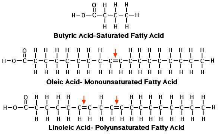
- Saturated Fats (Single Bonds): In saturated fats (found in butter, ghee, coconut oil, and animal fats), every carbon atom is fully saturated with hydrogen atoms. There are no double bonds. This creates a straight, tightly packed molecular chain that is highly stable, rigid, and resistant to thermal breakdown or chemical attack (see Figure 6).
- Monounsaturated Fats (One Double Bond): Oils like olive oil and avocado oil contain a single double bond. This introduces a slight bend in the molecular chain but remain reasonably stable under moderate conditions (see fig.6).
- Polyunsaturated Fatty Acids / PUFAs (Multiple Double Bonds): Industrial seed oils (like soybean, corn, sunflower, canola, and cottonseed oils) contain multiple double bonds. The spaces between these double bonds harbour highly vulnerable hydrogen atoms. The electron clouds around these double bonds are loosely held, making them prime targets for oxygen attacks (see Figure 6).
Molecular oxygen is a stable gas molecule, but has two completely unpaired electrons with parallel spins, and is looking out desperately to pair up those two lone, spinning electrons. An aggressive, highly reactive oxygen "spark" already loose and floating in our body, most notably the Hydroxyl Radical generated from mitochondrial leaks attacks the weak carbon-hydrogen bonds of the PUFA. It violently rips away a hydrogen atom. With its hydrogen stolen, the PUFA is left with a lonely, exposed electron. It rearranges its double bonds to try and stabilize itself, turning into an incredibly reactive Lipid Radical .
Now that the lipid chain has an open, unzipped electron, the sluggish molecular floating nearby is instantly attracted to it. The snaps onto the lipid radical effortlessly, transforming into a Lipid Peroxyl Radical . Thus, once a stray free hydroxyl radical creates that first "spark" () our body heat provides the perfect, warm environment for the fire to spread. At 37°C, molecules are bouncing around rapidly. This heat energy forces the newly formed lipid peroxyl radical to crash into neighbouring healthy fats. It steals their hydrogens, creating a self-sustaining, non-enzymatic chain reaction that can damage membrane lipids and generate reactive lipid products without needing any extra energy.
Our intestinal wall packs these absorbed fats along with PUFA into delivery vehicles called chylomicrons/lipoproteins and releases them into the bloodstream. Because our cells constantly need fresh fats to repair their outer walls, our cell membranes willingly grab these incoming fats and stitch them directly into their fluid lipid bilayer. Once there, their incorporation can influence membrane composition and fluidity under stress.
But our body knows that free radicals and oxidized fats are highly dangerous, so it does not let them roam free. It deploys an elegant tactical defence system. In the small intestine, our liver bile wraps the fats tightly into microscopic bubbles called micelles. This isolates the oxidized fats, keeping them surrounded by water so they cannot directly touch or damage our delicate intestinal tissue. Our digestive system absorbs dietary Vitamin E (a fat-soluble antioxidant) alongside the fats. Vitamin E travels inside the exact same lipid delivery particles. It acts like a bomb disposal technician, absorbing the unstable electron energy of the peroxyl radicals to halt the destructive domino effect before it hits our cell walls. Inside our gut cells and liver, an enzyme called Glutathione Peroxidase acts as a chemical cleaner. It forces the dangerous, oxygen-fused lipid radicals to react with glutathione, converting the toxic, volatile peroxide into a harmless, perfectly stable alcohol molecule that our body can easily process or discard.
Thus, when unstable PUFAs are exposed to heat, light, or oxygen either during industrial processing, cooking, or inside the warm oxygen-rich environment of the human body, they undergo a catastrophic chemical degradation process called Lipid Peroxidation. This is a self-propagating free-radical chain reaction that operates precisely like a nuclear fission event or a spreading fire. The breakdown of these fats leaves behind highly toxic, cytotoxic aldehydes, most notably 4-Hydroxynonenal (4-HNE) and Malondialdehyde (MDA). Inside our senior citizen's body, these toxic aldehydes act like reactive products that can modify proteins, lipids and other cellular components. They physically bind to and mutate DNA, cross-link vital proteins, and disable the mitochondrial enzymes responsible for producing ATP [13,14]. However, these pathways protect the system to a large extent if defensive mechanisms like liver bile, vitamin E and Glutathione Peroxidase are at optimum level.
6.2. Altering Membrane Fluidity and Signal Transduction
Every cell in the human body is wrapped in a dynamic phospholipid bilayer. The fluidity and elasticity of this membrane dictate how well the cell communicates with the rest of the body.
When our diet is rich in industrial seed oils, our body is forced to build its cell membranes using these fragile, highly reactive PUFAs. As these fats oxidize within the cell wall, the membrane loses its structural integrity. It becomes rigid, leaky, and inflamed (see Figure 7).
Changes in membrane lipid composition and lipid oxidation may influence membrane signalling, including insulin signalling. This structural degradation directly damages the membrane-bound insulin receptors. When the cell wall is distorted and inflamed by oxidized oils, changes in membrane composition may alter the environment in which insulin receptor function is impaired, our insulin key can no longer physically fit into the receptor lock properly. This introduces an entirely separate, structurally driven layer of Insulin Resistance, compounding the damage already caused by excess carbohydrate intake.
6.3. Engine Sludge: The Danger of Trans Fats and Interesterified Fats
While natural saturated fats are clean-burning fuels, chemically modified fats represent a severe threat to human tribology:
- Trans Fats: Created by forcing hydrogen gas into vegetable oils at extreme pressure and heat (partial hydrogenation). Trans fats alter the physical geometry of the fat molecule, turning a flexible chain into an unnaturally rigid block, causing it to accumulate like un-degradable sludge in our arterial walls (can alter lipid metabolism and increase cardiovascular risk) and liver tissue.
- Interesterified Fats: Often used by manufacturers to replace banned trans fats, these oils rearrange the fatty acids on a glycerol molecule using chemical or enzymatic catalysts. Because these configurations do not match natural evolutionary lipid profiles, they disrupt normal hepatic fat processing, accelerating visceral fat deposition and raising our systemic inflammation markers. We note that the relationship between dietary saturated fat, blood lipid markers (LDL-C, ApoB), and cardiovascular risk remains an actively debated area of nutritional science; major bodies such as the American Heart Association and WHO continue to recommend limiting saturated fat intake, and individual lipid responses to saturated fat vary considerably. Our recommendation to prioritize stable natural fats over industrially processed seed oils rests specifically on their relative oxidative stability (a tribological argument), not on a claim that saturated fat is universally cardioprotective. Readers with elevated LDL-C or existing cardiovascular disease should have their lipid response monitored by a physician before increasing saturated fat intake.
To protect the structural integrity of the senior engine, we recommend limiting highly processed sources of industrial oils as part of an overall dietary pattern. The machine should instead be lubricated with stable, time-tested natural fats such as ghee, butter, and extra virgin olive oil, selected according to the individual's overall dietary needs and cooking method. These maintain their molecular structure under heat and keep cellular communication channels fluidic and responsive.
To reduce the amount of linoleic acid stored in body tissues, the body must gradually remodel and replace tissue lipids over time to rebuild. While liver bile, Vitamin E, and Glutathione Peroxidase are brilliant defence mechanisms, they cannot eliminate these oils out of our system once they are already stored. They only protect us at the front door. Liver bile, Vitamin E, and Glutathione Peroxidase are defensive bodyguards, not garbage trucks. They prevent immediate damage, but they do not eliminate the fats we have already incorporated into our body tissue.
Liver bile is only active inside our digestive tract. While liver bile still cannot go out into our muscles or fat cells to vacuum out the oils stored inside our tissues, it actually plays a massive, direct role in flushing newly ingested or circulating oxidized fats out of our body before they cause harm. Our liver is the primary filtration plant of the body. When we absorb damaged, oxidized seed oils, they eventually pass through the liver. Many of the toxic byproducts of burnt oils (like aldehydes and oxidized lipids) are fat-soluble. Our kidneys can only flush out water-soluble waste (urine). They cannot flush out fat-soluble toxins.
To solve this, the liver processes these fat-soluble toxins and dumps them directly into liver bile. Bile is effectively the liver’s "garbage truck" fluid. Once the liver fills the bile with these toxic, oxidized fat byproducts, it squirts the bile into our intestines. From here, our body tries to throw the garbage away. The bile carries the oxidized waste down our intestinal tract so it can be flushed out of the body through our stool but does not flush stored seed oils or toxic aldehydes from tissues.
Our body is incredibly thrifty. Under normal conditions, our intestines reabsorb about 95% of our liver bile at the very end of the digestive tract and send it back to the liver to be reused. Because most bile acids are normally reabsorbed, soluble fibre can increase faecal loss of some bile acids by reducing their reabsorption. If we reabsorb the bile, we also reabsorb the toxic, oxidized fat byproducts it was trying to flush out, forcing them back into our circulation! To stop our body from recycling this toxic bile and force it to exit the system, we can do this naturally by consuming soluble fibre (found in vegetables, avocados, berries, and beans). The dietary fibre acts as a powerful structural shield and chemical sponge to neutralise, block, and flush out toxic fats like trans fats and oxidised industrial oils before they can enter our bloodstream and damage our arterial walls.
When we eat fibre alongside healthy meals, the fibre acts like a sponge inside our intestines. It physically binds to the toxic liver bile, locking it up so our intestines cannot reabsorb it. The fibre forces the contaminated bile to stay in the digestive tract, successfully flushing it completely out of our system in our bowel movements. The flow chart of this process is shown in Figure 8. Now because our body just lost that bile, the liver is forced to grab fresh cholesterol and fats from our bloodstream to manufacture brand-new, clean liver bile, effectively lowering our body's toxic fat load.
Remember that once a seed oil passes through our intestinal wall and enters our blood, bile can never touch it again. Same with Vitamin E which sits inside the cell membrane. It can only absorb the dangerous electron sparks from a radical attack. It stops a rancid PUFA from destroying its neighbouring fats, but it leaves the PUFA exactly where it is inside the cell wall. And Glutathione Peroxidase, this enzyme chemically neutralizes volatile lipid peroxides into stable, harmless alcohols. It douses the chemical fire, but it does not remove the unburnt, volatile oil bricks from our membrane architecture.
The hard truth is that once we consume industrial seed oils, our body stores them in two main places: our cell membranes and our adipose tissue (body fat storage tanks). According to lipid biochemistry research, the half-life of linoleic acid in human adipose tissue is roughly 600 days [15]. This means if we completely stop consuming industrial seed oils today, it will take nearly two years for our body to naturally burn off and replace just half of the stored seed oils in our fat tissue. To entirely purge these volatile oils from our system, we have to force our body to systematically dismantle its old, fragile "bricks" and replace them with stable ones. This requires a three-phase lifestyle shift:
Phase 1: We must completely stop adding new volatile fuel to the engine. This means eliminating processed foods, fast food, and limiting industrially processed oils (canola, soybean, corn, cottonseed) where appropriate. If we keep consuming them, our body will continuously prioritize storing them over clearing them.
Phase 2: Force the Engine to burn stored fuel (Beta-Oxidation): To get the stored linoleic acid out of our fat tissues, we have to force our body to open its storage tanks and burn that fat for energy. As we discussed, keeping insulin low (by reducing refined carbs) opens up our fat cells, allowing stored fatty acids to escape into the bloodstream. Also, engaging in steady, low-to-moderate intensity exercise (like walking, cycling, or jogging) forces our mitochondrial engine into greater reliance on fat oxidation, meaning it cleanly burns through stored fats for hours.
Phase 3: Accelerate Mitophagy (Cellular Rebuilding): To clear the fragile oils out of our cell membranes, we need our body to recycle old, damaged cells and build fresh ones. Periods of intermittent fasting and exercise can activate cellular stress-response and recycling pathways triggering cellular cleanup processes where our body purposefully destroys leaky, damaged mitochondria and worn-out cell membranes (Mitophagy & Autophagy), although the magnitude and timing of these responses in humans vary. When our body creates these brand-new cells, we must supply them with stable building blocks. Eating stable monounsaturated fats (Oleic acid from Olive Oil) and clean polyunsaturated fats (obtained from whole foods or minimally processed sources) which ensures our new cell membranes are built with tough, oxygen-resistant bricks. Because our body views linoleic acid (polyunsaturated fatty acid) as a structural building block, it does not just flush it out through urine or sweat. Instead, it must be burned for energy through Beta-Oxidation or cleared out when a cell naturally dies.
6.4. The Mechanical Digestion Matrix: Whole Raw Seeds and Nuts.
However, whole raw seeds and nuts also contain linoleic acid (the omega-6 polyunsaturated fatty acid). However, they should not be restricted in our diet. There is a profound biological difference between eating linoleic acid in its natural, matrix-bound form (whole nuts and seeds) versus consuming it as an isolated, industrially processed liquid (seed oils). It seems logical to fear nuts and seeds. But whole foods come with built-in biological armour that can substantially change how they are digested and absorbed.
In an industrial seed oil, factory processing heat strips away natural antioxidants. In contrast, a raw walnut or sunflower seed is packed with massive amounts of Vitamin E and polyphenols. As we discussed earlier, Vitamin E acts as a literal bodyguard inside the fat. Since the linoleic acid in a nut is surrounded by its natural antioxidant shield, it resists oxidation inside our digestive tract.
Our digestive system has to work for hours to slowly break down the fibre and extract the fat. This slow-motion release prevents the metabolic "traffic jams" and sudden mitochondrial electron leaks that cause heavy oxidative stress. As we discussed earlier with liver bile reabsorption, whole nuts and seeds provide the required tool needed to clear waste. The fibre matrix from the chewed nuts binds tightly to circulating liver bile in our intestines, ensuring that some oxidized lipid waste is trapped and flushed out through our stool rather than being reabsorbed.
We can enjoy whole, raw, or lightly sprouted nuts and seeds as part of a healthy diet. The only caution is to avoid nuts and seeds that have been commercially roasted in seed oils (always check the ingredient label for if any vegetable oil is being used). Note: The only way to flush volatile oils from the system is through time and also by lowering insulin to access stored fats, and consistently supplying stable fats to rebuild our cellular architecture over a 2-to-3-year window.
7. Meal Mechanics: The Importance of Phase, Timing, and Sequence.
In any industrial assembly line or chemical process, the efficiency of the output depends heavily on two critical variables: the sequence of operations and the throughput timing. If we dump raw materials into a reactor in the wrong order, or if we flood the conveyor belt without giving the machinery time to clear the previous batch, the entire system clogs.
For us as seniors, managing nutrition is not merely about managing what enters the machine, but optimizing the mechanical phase, timing, and sequence of entry to smooth out metabolic spikes and allow the system to self-clean.
7.1. The Physics of Phase Separation: Gastric Emptying and the Glucose Dampening Curve.
When our mixed meal enters our stomach, it does not pass into the small intestine all at once. It follows a rate-limiting mechanical process known as gastric emptying. By intentionally altering the sequence in which we consume food groups during a single meal, the meal composition and sequence can influence the rate and magnitude of the post-meal glucose response.
- The Wrong Order (Carbohydrates First): If starches or sugars hit our empty stomach, they are rapidly liquefied into a low-viscosity fluid. The stomach flushes this fluid into the small intestine almost immediately. This triggers a high-velocity influx of glucose into the bloodstream, causing a sharp, volatile insulin spike and a storm of oxidative "sparks" (ROS) in our mitochondria.
- The Right Order (Fiber → Protein & Fats → Carbs Last): When we consume non-starchy fibrous vegetables first, followed by structural proteins and fats, we alter the physical state of the stomach contents. The fibre mixes with gastric juices to form a highly viscous, gelatinous mesh. The proteins and fats trigger the release of cholecystokinin (CCK), which physically slows down the stomach’s muscular pumping action.
When carbohydrates are finally eaten at the very end of the meal, they become trapped inside this viscous fibre-and-protein mesh. This slows down enzymatic breakdown and stretches out glucose absorption over a much longer time domain. The result is a smooth, low-amplitude glucose curve that may reduce the magnitude of the post-meal glucose and insulin response.
7.2. Throughput Control: The 16:8 Intermittent Fasting Window
In systems engineering, if a manufacturing facility is overloaded with a backlog of unfinished inventory, the first step is to implement a zero-input window. We must temporarily halt incoming raw materials so the factory can clear the floor and process the backlog. For us past the age of 50 with a protruding belly, that backlog is visceral fat.
The 16:8 Intermittent Fasting protocol establishes a daily 16-hour window of zero thermodynamic input that is, no caloric intake, leaving an 8-hour window for eating. During the 8-hour feeding phase, our body naturally secretes insulin to handle incoming nutrients. However, during the 16-hour fasting window, the insulin levels generally fall during fasting, and circulating insulin generally falls substantially during fasting.
Once insulin is out of the way, Malonyl-CoA levels can fall, reducing inhibition of fatty-acid entry into mitochondria, and the mitochondrial transport elevator (the carnitine shuttle) reactivates. Our body can now freely access its deep, internal storage tanks increasing reliance on stored fat as an energy source, burning off toxic visceral belly fat from around our liver and pancreas to meet its baseline kinetic energy requirements.
7.3. Chrononutrition: Circadian Rhythms and Metabolic Sensitivity.
A physical system's efficiency is not static; it varies based on a master internal clock. In human physiology, this clock is driven by the circadian rhythm, regulated by the brain and metabolic organs like the liver. Due to evolutionary programming, our human engine exhibits higher insulin sensitivity and metabolic efficiency in the earlier phases of the solar cycle (morning and afternoon) compared to the dark phase (late evening and night).
- Daytime Processing: During daylight hours, insulin sensitivity and glucose handling are generally better earlier in the day than late at night, and skeletal muscle tissue is primed to absorb glucose efficiently.
- Nighttime Backlog: As night falls and our body begins synthesizing melatonin to prepare for sleep, insulin secretion naturally degrades, and our liver becomes resistant to storing nutrients. Eating a heavy, carbohydrate-dense meal late at night forces our sluggish system to process high-entropy inputs when our metabolic processing is less well aligned with a large late-night meal. This causes blood glucose to remain elevated for hours during sleep, disrupting cellular repair, accelerating visceral fat deposition, and keeping systemic inflammation high.
The Mechanical Timing Protocol: To maximize the efficiency of our human engine, we as seniors should prioritize eating their meals in a structured sequence (fibre and protein first), restrict their intake to a clean 16:8 zero-input window, and front-load their nutritional inputs earlier in the day to align with the body's natural thermodynamic peak.
8. Hormetic Stress: System Calibration via Controlled Perturbation.
In materials science and structural engineering, systems often require controlled physical challenges to maintain their resilience. Metals can be hardened through controlled thermal tempering, and mechanical bridges are engineered to withstand shifting dynamic loads. If our mechanical system is kept in a completely static, stress-free environment, it undergoes rust, stagnation, and structural decay.
In our human machine, this type of adaptive response is commonly described as hormesis. Hormesis is an evolutionary phenomenon where a brief, non-destructive dose of physical stress triggers an over-compensatory adaptive response, forcing our biological system to calibrate, repair, and optimize its internal machinery.
8.1. The Physics of Autophagy: Reducing Systemic Entropy.
From a thermodynamics perspective, a major hallmark of aging past 50 is the accumulation of cellular entropy, disorder and structural degradation. Over decades of operational wear, our cells accumulate damaged organelles, misfolded proteins, and dysfunctional mitochondria that can no longer produce ATP efficiently. These worn-out parts act like broken gears in an engine, generating excessive friction and leaking destructive ROS "sparks" into our cell. When our body undergoes a temporary period of severe nutrient scarcity, such as during the latter hours of a 16-hour fast, it activates Autophagy (literally, "self-eating"). Thus, nutrient restriction and fasting can activate autophagy-related cellular recycling pathways, although the precise timing and magnitude of autophagy in humans are not established as a simple 16-hour threshold
Autophagy is our system’s internal recycling program. During nutrient restriction, cellular signalling can shift toward greater recycling and maintenance activity, specialized cellular trash compactors called lysosomes sweep through our cell, engulfing high-entropy cellular debris, broken proteins, and exhausted mitochondria. Our cells break this biological garbage down into raw amino acids and lipids, using them to manufacture brand-new, low-entropy, high-functioning cellular components. Autophagy is one important mechanism by which cells recycle damaged or unnecessary components.
8.2. Eliminating the "Zombie" Backlog: Cellular Senescence.
As our human machine ages, certain cells reach their replication limit or sustain too much genetic damage to function. Instead of dying quietly through programmed cell death (apoptosis), these damaged units turn into Senescent Cells, often referred to as Zombie Cells. Senescent cells refuse to divide, yet they remain metabolically active. They anchor themselves inside our vital organs, our liver, pancreas, and arterial walls, where they secrete a toxic blend of pro-inflammatory signalling proteins known as the Senescence-Associated Secretory Phenotype (SASP). This chronic chemical leakage pollutes the local cellular microenvironment, damaging neighbouring healthy cells and accelerating the onset of insulin resistance and tissue degeneration.
Hormetic stressors act as a targeted filtration system against this zombie cell backlog. The acute energy crisis brought on by fasting, combined with the metabolic demands of intense exercise, places senescent cells under severe structural strain. Because these cells are already unstable and fragile, the added hormetic pressure pushes them over the edge into apoptosis, clearing them out of our tissue matrix and making physical room for young, high-functioning cells to take their place. Exercise and metabolic stress (nutrient restriction and fasting) may influence pathways involved in cellular senescence and tissue maintenance, but the extent to which fasting or exercise selectively removes senescent cells in humans remains an area of active research.
8.3. Mitohormesis: Forging High-Yield Cellular Power Plants.
Just as a mechanical engine can be tuned to burn fuel more efficiently, our cellular power plants, the mitochondria, can be upgraded through a process called Mitohormesis. When we push our muscles through intense exercise or restrict glucose via a low-carbohydrate lifestyle, we subject our mitochondria to an acute, localized burst of oxidative stress. This transient spike in ROS acts as a critical calibration signal. Our cell responds by triggering Mitochondrial Biogenesis leading to literal duplication and creation of fresh, low-entropy mitochondria.
Simultaneously, our cell uses a specialized version of autophagy called mitophagy to identify and destroy older, leaky power plants (mitochondria). By systematically replacing inefficient, high-wear mitochondria with a dense network of tightly packed, high-yield power units, mitohormesis may improve mitochondrial function and the body's ability to manage oxidative stress of our senior body, allowing our engine to generate massive amounts of clean ATP currency without producing damaging cellular sparks.
9. Cellular Hypertrophy and Mass Accumulation: The Proliferative Signal of Insulin on the Prostate Gland.
In our biological machinery, insulin can influence growth-related signalling pathways in prostate tissue, it can act as a primary mitogenic driver (induces mitosis - cell division) and may contribute to prostate growth-related signalling in the enlargement of the prostate gland. Chronically high levels of insulin (hyperinsulinemia) typically caused by insulin resistance, metabolic syndrome, or type 2 diabetes act as a powerful biological trigger for prostate tissue growth. While male sex hormones (androgens) are the primary driver of Benign Prostatic Hyperplasia (BPH), insulin acts as a potent growth accelerator. High circulating insulin levels expand the prostate gland through several intersecting biological pathways (see Figure 9):
- The Growth Factor Mimicry: Insulin is structurally similar to major cellular growth factors. When our bloodstream is consistently flooded with insulin, it binds directly to insulin receptors on our prostate epithelial and stromal (connective tissue) cells, sending pro-survival and pro-proliferative signals that can promote cell survival and growth-related signalling.
- The Liver IGF-1 Feedback Loop: Hyperinsulinemia forces our liver to produce more Insulin-like Growth Factor 1 (IGF-1). Insulin also lowers the production of binding proteins that normally keep IGF-1 inactive. This leaves an immense amount of free, hyperactive IGF-1 circulating inside our system, which directly binds to our prostate tissue and accelerates cellular hypertrophy (enlargement) [16,17].
- Enzymatic Shifting of Testosterone: Insulin fundamentally alters how our prostate handles testosterone. It increases the transcription of the enzyme 5-alpha reductase, which converts standard testosterone into Dihydrotestosterone (DHT). DHT is an important androgenic signal involved in prostate growth.
- Hydraulic Pressure and Smooth Muscle Tension: High insulin levels overstimulate our sympathetic nervous system (our "fight-or-flight" baseline). This hyper-activation causes the smooth muscles within our prostate and the bladder neck to contract tightly. This muscle tension increases urinary resistance, compounding the physical urinary symptoms (like a weak stream or frequent night-time urination) caused by the physical size of the gland.
Some clinical and observational studies have reported an association between hyperinsulinemia, metabolic dysfunction and prostate enlargement [16,17]. Managing our metabolic health through diet, exercise, and correcting insulin resistance is now recognized as a vital pillar in preventing and managing an enlarged prostate, although BPH remains primarily a multifactorial condition.
10. Structural Wear and Tensile Breakdown: How Metabolic Signal Resistance Drives Musculoskeletal Friction and Joint Pain
Insulin resistance and metabolic dysfunction may influence musculoskeletal health, including processes relevant to arthritis and degenerative spinal conditions. Chronically high insulin levels heavily impact arthritis, slip discs, and overall joint and body pain (see Figure 10). While we as seniors often attribute joint pain purely to physical ‘wear and tear,’ but metabolic health fundamentally dictates how our bones, cartilage, and spinal discs heal and age. When our system produces excessive insulin to manage blood sugar, it triggers a cascade of systemic issues across our musculoskeletal system.
Our joints are lined with cartilage cells (chondrocytes) that rely on insulin to absorb glucose and rebuild the joint matrix. In a state of insulin resistance, these chondrocytes stop responding to insulin. They cannot absorb nutrients effectively, making the cartilage weak, brittle, and prone to rapid breakdown. High insulin overstimulates our synovial cells that produce joint fluid (synovial fluid). This increases joint inflammation and reduces fluid production, leaving our joints poorly lubricated, stiff, and painful.
Our spinal discs are made of heavy layers of collagen fibres that act as shock absorbers between our vertebrae. Chronic hyperinsulinemia and high blood sugar damage collagen throughout the body. It stiffens the outer ring of the spinal disc, making it fragile and highly susceptible to cracking, herniating, or slipping under pressure. Spinal discs do not have a direct blood supply; they absorb nutrients from surrounding tissues through micro blood vessels. High insulin levels damage these tiny capillary distribution networks, starving the spinal discs of oxygen and water, causing them to dehydrate, flatten, and pinch nearby nerves [18].
High insulin levels act as a volume dial for pain signals across our entire body. Excess insulin prompts our immune cells to constantly flood our bloodstream with pro-inflammatory chemicals (cytokines). This baseline inflammation leaves our muscles, tendons, and ligaments feeling chronically sore, achy, and tender. High sugar and insulin levels cause sugar molecules to fuse with proteins, forming sticky compounds called AGEs (Advanced glycation end products). These compounds accumulate in our tendons and fascia, making our entire body feel stiff and inflexible.
Clinical evidence shows that improving our insulin sensitivity, whether through diet, lifestyle changes, frequently results in a dramatic reduction in chronic joint pain and slower spinal degeneration, although joint and spinal degeneration has multiple causes.
11. Electrical Grid Instability: How Metabolic Signal Noise Disrupts Cardiac Electrophysiology.
To a physicist, our heart is not merely a muscular pump; it is a highly synchronized electromechanical oscillator operating on an intricate grid of electrical currents. This biological power grid relies on highly specific voltage-gated ion channels, primarily sodium (), potassium (), and calcium () channels to maintain a stable cardiac rhythm and coordinate the physical contraction of the myocardial chambers.
When our system undergoes chronic hyperinsulinemia and subsequent insulin resistance, this precise electrical grid experiences severe signal noise and electrophysiological remodelling:
- The Intracellular Spark Plug Over-activation (
): Persistent high blood glucose and insulin levels force excess glucose into the hexosamine biosynthetic pathway. This process chemically modifies a master regulatory enzyme inside our heart cells called Calcium/Calmodulin-dependent Protein Kinase II (). This modification locks the switch into a permanent "ON" state, causing it to aggressively phosphorylate a biochemical process of attaching a phosphate group modifying the gates of our intracellular calcium storage tanks - the sarcoplasmic reticulum (SR). The resulting chaotic, spontaneous leakage of calcium ions () during the resting phase of the heart cycle generates erratic electrical currents (delayed afterdepolarizations), creating out of place (ectopic) "sparks" that destabilize our baseline rhythm [19,20].
- Degrading Conduction Velocity (Nav1.5 Suppression): High insulin and glucose exposure alters the expression and function of our primary cardiac sodium channel,
. Through excessive glycation, the channel's capability to deliver the rapid, high-amplitude sodium current required to drive the cardiac action potential drops dramatically. This reduction in sodium influx slows down our heart's internal signal conduction velocity, causing the electrical wave to travel sluggishly and unevenly across our heart tissues.
- Structural Wave Reflection (Atrial Fibrosis): As our heart tissue becomes insulin resistant, the structural tissue undergoes severe fibroblastic remodelling. The accumulation of stiff collagen blocks between heart cells disrupts our gap junctions, which serve as the physical communication cables between adjacent cells. Lacking clean, direct lines of electrical communication, the synchronized wave fronts break apart into chaotic, self-sustaining micro-reentrant loops.
This combination of slow conduction velocity, spontaneous calcium leaks, and structural insulation breakdown creates severe electrophysiological phase dispersion across our senior heart chambers. This predictable system failure manifests clinically as erratic heart palpitations, extra beats, and a highly elevated baseline risk for Atrial Fibrillation (AF) and may contribute to increased cardiovascular and arrhythmic risk [19,20]. In Figure 11 we show the summary of it.
12. High-Entropy Waste Accumulation: The Central Signal Breakdown of "Type 3 Diabetes"
From a materials science and systems engineering perspective, a major processing error occurs when a computer's central processing unit (CPU) is starved of power while its structural motherboards are buried under un-cleared waste products. In our aging neurological engine, this combined crisis of localized power failure and waste accumulation is a proposed contributing mechanism in Alzheimer's Disease - a hypothesis some researchers describe using the informal term "Type 3 Diabetes." This term is not a formal clinical diagnosis, and Alzheimer's disease is understood to involve multiple contributing pathways beyond brain insulin resistance, including genetic risk factors (e.g., APOE4), vascular disease, and primary protein-misfolding processes not fully explained by metabolic dysfunction alone. Our brain represents only 2% of our total body mass but demands up to 20% of our daily kinetic energy infrastructure. While the brain utilizes specialized glucose transport networks to import fuel independently of our body's muscle insulin gates, insulin signalling within the central nervous system plays important roles in neuronal survival and synaptic function.
- Neural Gateway Closure and Energy Crisis: When our brain tissues are subjected to unrelenting, systemic insulin surges from a high-carbohydrate input diet, our central neural insulin receptors undergo profound downregulation and signal attenuation. The receptors become blind to the hormone. Without this vital survival signal, our neurons fail to maintain their synaptic connections, glucose utilization may become impaired, and our brain enters a state of critical localized power failure, systematically starving our memory centres of ATP.
- The Molecular Janitor Competition Dilemma (IDE): To clear away high-entropy waste products, our brain uses a specialized molecular janitor called Insulin-Degrading Enzyme (IDE). Crucially,
has a dual role: tackling insulin and clearing waste accumulation. Its molecular geometry is designed to break down both floating insulin molecules and toxic misfolded Amyloid-Beta () protein monomers. In a healthy, low-insulin environment, easily sweeps through the extracellular space, safely clearing away before it can crystallize. However, when our central nervous system is chronically flooded with insulin due to insulin resistance, our janitors become entirely diverted and overwhelmed by the massive influx of insulin. The enzyme ignores the amyloid waste to prioritize clearing the insulin backlog. Since, IDE can participate in the degradation of both insulin and amyloid-beta, altered insulin levels may influence amyloid-beta clearance; however, this relationship is complex and is not sufficient by itself to explain Alzheimer's disease [21,22].
- Systemic Circuitry Clogging (Amyloid & Tau Arrays): Stranded without clearance mechanisms, the high-entropy
monomers stick together, forming rigid, insoluble amyloid accumulation can interfere with neuronal function and synaptic integrity. Concurrently, this signal breakdown hyper-phosphorylates (attaching highly charged phosphate tags) our internal Tau proteins, (Tau proteins act as a physical stabilizer and structural anchor for the internal skeleton of neurons in the central nervous system) causing the internal structural scaffolding of our axons to collapse into chaotic neurofibrillary tangles [22].
By shifting our senior system to a low-carbohydrate baseline and generating clean-burning Ketone Bodies, we bypass this broken insulin-glucose gate entirely. Ketones cross our blood-brain barrier effortlessly via alternative transport vehicles, providing a high-yield fuel source that restores our brain's energy levels while freeing up our molecular janitors () to flush out the toxic waste backlog whether this translates into meaningful prevention or reversal of cognitive decline remains uncertain. Below we show in Figure 12 the summary of it.
13. Hydraulic Fluid Congestion: The Pressure Valve Failure of the Ocular Drainage Meshwork.
To a fluid dynamics engineer, any enclosed, fluid-filled chamber requires a perfect equilibrium between the rate of fluid production and the rate of drainage to maintain a safe operating pressure. In our eyes, this delicate hydraulic system governs the circulation of aqueous humor, a specialized fluid produced by the ciliary body that must constantly drain out through a microscopic, sieve-like filter known as the Trabecular Meshwork.
When our system is gripped by chronic insulin resistance, this ocular drainage infrastructure suffers a catastrophic hydraulic pressure valve failure, may influence pathways involved in glaucoma and diabetic retinal disease:
Volumetric Aqueous Fluid Over-production: Metabolic and vascular abnormalities associated with insulin resistance can affect ocular tissues, where they overstimulate our sympathetic nervous system () and activate local pathways. This neurological surge may influence ocular fluid regulation through several pathways, forcing them to crank up the production rate of aqueous humor, flooding our eye's anterior chamber with excess fluid.
- Structural Filtration Sieve Stiffening and Clogging: Simultaneously, the chronic wave of blood glucose and reactive oxygen species () strikes the delicate endothelial cells lining our Trabecular Meshwork filter. The excess sugars bond with the filter's structural proteins, causing heavy glycation, advanced glycation end-products (), and progressive tissue scarring. This structural "caramelization" causes our primary drainage meshwork to become thick, rigid, and severely clogged [23].
- Mechanical Shearing of the Optic Nerve: With fluid production spiking and the drainage channels structurally restricted, the baseline Intraocular Pressure (IOP) climbs past the safe threshold of . This elevated fluid pressure exerts intense, direct mechanical compression against the back of our eye, specifically at the lamina cribrosa where our optic nerve plugs into the retina.
Figure 13.
Ocular Hydraulic Pressure Failure Cascade: Potential relationships between metabolic dysfunction and ocular pressure/vascular stress [Conceptual model].
Figure 13.
Ocular Hydraulic Pressure Failure Cascade: Potential relationships between metabolic dysfunction and ocular pressure/vascular stress [Conceptual model].
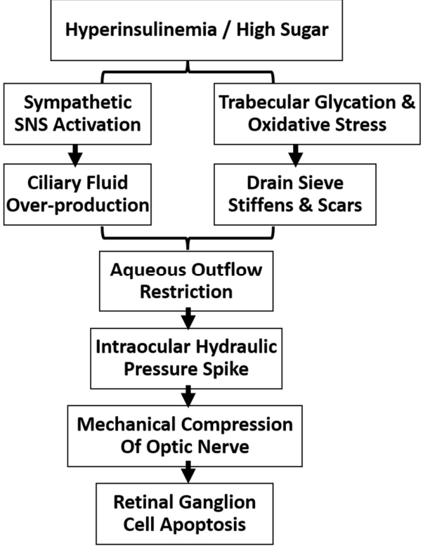
This elevated intraocular pressure can damage the optic nerve and compromise retinal ganglion cell survival which can cut off the microscopic blood vessels supplying our vision cells, starving them of oxygen and inducing rapid Retinal Ganglion Cell () apoptosis. Improving metabolic health may benefit overall vascular and ocular health, but established glaucoma requires appropriate ophthalmic evaluation and treatment to protect our senior vision from irreversible failure [23]. In fig.13 we show the summary of it.
14. Conclusion: Taking Control of Our Biological Machinery.
In classical engineering, when a highly complex mechanical system experiences a catastrophic operational failure, a skilled technician does not simply treat the surface symptoms. If an automated factory engine is overheating because a pressure valve is stuck open and the input fuel line is contaminated, adding external coolant is an inefficient, temporary patch. True resolution requires tracing the failure back to its source, repairing the primary control valve, and recalibrating the input parameters.
For us over 50, chronic health issues like hypertension, type 2 diabetes, non-alcoholic fatty liver disease, and visceral obesity are not random. Nor are mass accumulations like prostate hypertrophy, the structural wear of arthritic joint friction, erratic signal noise in our cardiac electrical grid, Alzheimer's neurodegenerative waste accumulation, or rising intraocular hydraulic pressure. They are systemic, predictable failures of an open thermodynamic machine subjected to prolonged input mismanagement.
Throughout this exploration, we have analysed the human body not through a traditional medical lens, but through the precise, universal laws of physics and systems mechanics to understand the system reversal path as shown below in the table:
| THE SYSTEM REVERSAL PATH | |
| THE OLD INPUT ENGINE | THE NEW OPTIMISED SYSTEM |
| • High Carbohydrate Loads | • Low-Carb, High-Protein/Fat Input |
| • Unrelenting Insulin Signalling | • Restored Signal Sensitivity |
| • Visceral Fat Accumulation | • Reduced Visceral Storage Tanks |
| • High Arterial Pipe Resistance | • Elastic Arteries & Clean Filters |
| • High Systemic Entropy (Decay) | • Autophagy & Low Entropy |
By understanding that the human digestive system is adapted to process a mixed diet containing nutrient-dense animal and plant foods (as discussed in section 2), we see why persistent excess energy intake and frequent consumption of rapidly absorbed refined carbohydrates can contribute to impaired metabolic regulation.
When the master Insulin Control Valve remains persistently activated (Section 3), cellular signalling undergoes downregulation, and the excess energy backs up into the liver and pancreas as toxic visceral fat. This metabolic backlog directly alters the fluid dynamics of our cardiovascular system (Section 4), raising osmotic pressure and stiffening the arterial pipes to cause chronic hypertension. Furthermore, this systemic signal failure acts as an uninhibited mitogenic growth accelerator may contribute to prostate growth-related signalling (Section 9), may adversely affect joint and connective-tissue metabolism and intervertebral discs to cause structural tensile breakdown (Section 10), introduces chaotic biological sparks that disrupt our cardiac electrophysiological grid (Section 11), diverts vital clearing enzymes to bury our neural CPU under high-entropy amyloid waste (Section 12), and blocks our ocular trabecular filtration sieve which may contribute to abnormal ocular pressure and vascular/metabolic stress (Section 13). In the appendix, we provide the Senior Operator's Daily Action Manual, including Table 2's General Metabolic Changes During Fasting [24], along with a separate note for the Vegetarian Operator on adapting this framework to a plant-based diet.
Hypothesis for reversing cascading failure: To reverse these cascading failures and restore the body to a state of low-entropy health, we as the operators must execute a systematic recalibration of habits, focusing on the type, amount, timing, and sequence of meal inputs together with physical activity and muscle preservation:
- Recalibrate the Input Quality (Macro-Shifting): Reduce excessive refined-carbohydrate intake while ensuring adequate protein and appropriate sources of dietary fat. Prioritizing protein intake overrides aging anabolic resistance to prevent the entropic decay of muscle tissue (Sarcopenia), while shifting the cellular power plants to clean-burning fat fuel (Beta-Oxidation and Ketones).
- Purge Industrial Lubricants: Reduce highly processed oils and emphasize an overall dietary pattern based on minimally processed foods and appropriate sources of dietary fat for the system to prevent lipid peroxidation and stop structurally driven insulin resistance within the cellular membrane bilayers.
- Enforce the Sequence of Phases: Utilize the physics of gastric emptying by consuming fibrous vegetables first, structural proteins and fats second, and any minimal carbohydrates dead last. This sequence may slow gastric emptying and reduce the post-meal glucose response, flattening the glucose absorption curve.
- Avoid carb-plus-fat combination diet: Repeatedly combining high-glycemic carbohydrates with fat in the same meal like bread with butter, pizza, cookies, sweetened desserts are particularly efficient at building visceral fat. Because, the insulin spike triggered by the carbohydrate simultaneously blocks fat oxidation and locks the co-ingested fat straight into storage. Like a fixed deposit that only ever receives contributions and is never withdrawn, this stored fat is rarely mobilized back out for fuel, making the carb-plus-fat combination one of the most avoidable drivers of visceral adiposity in a senior's daily diet [25]. This combination makes it easy to consume excess energy, particularly when refined carbohydrate and highly palatable foods are involved. Stored fat can later be mobilized when energy demand exceeds dietary energy availability
- Restrict Excess Fruit/Fructose Intake: Very high intake of added sugars and fructose-containing sweetened foods can increase hepatic fat production, particularly in the context of excess energy intake, which may convert excess fructose into fat (causing fatty liver). Rapid fructose metabolism can temporarily deplete cellular ATP and phosphate, increasing the breakdown of adenine nucleotides and thereby increasing uric acid production. When this uric acid spikes and settles into our joints as sharp, needle-like crystals, our immune system treats them as dangerous foreign invaders, unleashing an inflammatory attack that results in the excruciating pain and swelling of gout. Moderate portions of whole fruit can generally be incorporated into a healthy dietary pattern, limiting our intake of whole fruits daily prevents this overload because the natural fibre slows down sugar absorption, allowing the body to safely process the fructose. If you already suffer from joint pain or high uric acid, you should favour low-sugar options like berries, kiwis, cherries, and citrus fruits while restricting high-fructose varieties. Consuming fruits in low, controlled amounts delivers essential vitamins, antioxidants, and fibre that actually help our kidneys flush out uric acid and protect our cells, all without triggering metabolic stress or joint inflammation [26,27,28].
- Implement a Zero-Input Window (16:8 Fasting): Implement a daily 16-hour fasting window to lower insulin levels and increase reliance on stored energy. This allows the cellular transport mechanisms to reactivate and increase access to stored fat as an energy source and may help reduce visceral fat when it contributes to an overall reduction in energy intake.
- Apply Controlled Hormetic Stress: Introduce periodic physical stressors like brief fasting windows and intense muscle engagement. These forms of controlled metabolic stress can activate cellular maintenance and adaptive pathways, although the magnitude of these effects varies with the type, duration and intensity of the stress This triggers autophagy, the machine's built-in recycling mechanism sweeping away high-entropy biological debris, clearing "zombie" senescent cells, and forcing the creation of fresh, low-entropy mitochondrial power grids.
A Note on Individual Variation and Limitations: The framework and interventions described above are not universally appropriate. Extended fasting windows can cause dangerous hypoglycaemia in individuals taking insulin or sulfonylurea medications, and should only be attempted under medical supervision in these cases. Individuals with chronic kidney disease should consult a physician before increasing protein intake, as elevated protein loads may accelerate renal decline in compromised kidneys. Increasing saturated fat intake can meaningfully raise LDL cholesterol in a subset of individuals ("hyper-responders"), and lipid panels should be monitored during dietary transition. Finally, this article synthesizes mechanistic and observational evidence into a unifying framework; it does not report a randomized controlled trial, and the causal claims made here, while grounded in cited literature, should be read as a hypothesis-generating perspective rather than a proven clinical protocol. The framework therefore generates hypotheses and practical considerations rather than establishing causal treatment effects
Aging is an inevitable chronological process, but some aspects of metabolic deterioration may be modifiable. The human body is a self-healing, self-regulating biological machine of astonishing complexity. It possesses extensive internal mechanisms for repair, adaptation and cellular maintenance, although these mechanisms have limits and vary with age and disease.
For us as citizens over 50, taking control of our health is an act of deliberate system management. By shifting from passive consumption to active engineering, by managing the mechanics of our fuel and the timing of our inputs, we close the runaway feedback loops of disease. We don't just slow down the clock; we actively lower our systemic entropy, preserve our biological infrastructure, and support healthier aging and functional capacity. We cannot stop the clock, but we can potentially improve metabolic regulation, preserve biological function, and support healthier aging.
Data Availability Statement
This article does not report new experimental or clinical data. All claims and mechanisms discussed are drawn from previously published, peer-reviewed literature, cited throughout the text and listed in the References section.
Conflicts of Interest
The authors declare no conflicts of interest.
AI Disclosure: Artificial intelligence tools were used in the preparation of this manuscript. Google AI was used to search the literature and locate relevant published references. Claude was used to assist with editing, references reformatting, English and scientific validation. Finally, ChatGPT was used for reviewing and to make the article softer as per suggestion. Neither tool was used to generate the core scientific ideas, arguments, or conclusions presented in this article, which remain the intellectual work of the author. All AI-assisted edits and retrieved references were reviewed, verified, and approved by the author, who takes full responsibility for the accuracy and content of the final text.
Appendix: The Senior Operator’s Daily Action Manual
(A Quick-Reference System Calibration Guide for Reversing Metabolic Decay Past 50)
1. The Timing Parameters (Throughput Control)
- 16:8 Zero-Input Window: Restrict all caloric intake to an 8-hour daily window (e.g., 10:00 AM to 6:00 PM). Maintain absolute zero-input fasting for the remaining 16 hours to force baseline insulin depletion, deactivate Malonyl-CoA, and unlock the carnitine shuttle elevator to burn viscerally stored organ fat. A 16:8 eating pattern may be used by some individuals as one practical way of reducing eating frequency and total energy intake. It is not appropriate for everyone.
- Front-Load the Solar Cycle: Consume our largest meals during peak daylight hours when circadian insulin sensitivity is highest. Stop all thermodynamic inputs at least 3 hours before sleep to prevent glucose stagnation and midnight ROS accumulation.
2. The Phase Parameters (Sequence of Entry)
- Never Lead with Carbs: Consider beginning meals with vegetables, protein and other minimally processed foods before consuming refined carbohydrates. We must never let starches or simple sugars hit an empty stomach.
-
Execute the Mechanical Sequence: Consume our plate strictly in this order to slow gastric emptying and create a visco-elastic dampening mesh in our digestive tract:
- Phase 1: Fibrous non-starchy vegetables (Avocado, leafy greens, broccoli).
- Phase 2: Structural proteins and stable lipids (Meat, eggs, fish, ghee).
- Phase 3: High-glycemic carbohydrates/starches (Consume only at the very end of the meal, if at all).
3. The Quality Parameters (Macro & Micro Inputs)
- Aim for an Adequate Protein Dose: Have a meal containing roughly 35–40 g of high-quality protein in a single sitting which may be useful for some older adults, depending on body size and individual needs, to successfully flip the stubborn, anabolic-resistant mTOR switch for muscle repair (Sarcopenia protection)..
- Fuel via Clean Saturated/Monounsaturated Matrixes: Emphasize minimally processed sources of dietary fat, such as olive oil, nuts, seeds and other foods appropriate to the individual's overall dietary pattern. They are insulin-neutral and highly resistant to oxygen attack.
- Limit Highly Processed Foods and Industrially Processed Oils: We must restrict industrially processed oils as part of the overall dietary pattern such as canola, soybean, corn, sunflower, and cottonseed oils. They act as molecular shrapnel, causing lipid peroxidation and structurally ruining our cell membrane's insulin receptors.
4. The Self-Cleaning & Purging Protocols (System Flushing)
- Block Bile Reabsorption: Consume natural soluble fibre (avocados, berries, chia seeds) alongside meals. This acts as a physical sequestering agent, supporting normal bile-acid elimination carried by our liver's bile and forcing its exit via our stool.
- Trigger Mitophagy via Heavy Dynamic Loading: Engage in heavy resistance or interval exercises. The intentional dynamic stress may stimulate cellular pathways involved in mitochondrial maintenance and adaptation and build pristine, tightly sealed, spark-free mitochondrial power grids.
- Do Not Fear Matrix-Bound PUFAs: Continue eating whole nuts and seeds. Their natural structural matrix slowly releases nutrients over hours while providing built-in Vitamin E bodyguards to neutralize potential oxidative radical attacks right at the digestion door.
Table 2.
General Metabolic Changes During Fasting [24]:.
Table 2.
General Metabolic Changes During Fasting [24]:.
| Fasting duration | General metabolic changes |
|---|---|
| ~12 hours | Insulin generally declines; liver glycogen continues to supply glucose |
| ~16 hours | Greater reliance on stored fat begins; gluconeogenesis contributes to glucose maintenance |
| ~24 hours | Fat oxidation and ketone production generally increase |
| 24–48 hours | Ketone availability increases further and fuel use shifts progressively toward fat |
| Longer fasting | Ketone production and adaptation increase further, but responses vary substantially between individuals |
Core Biological Constants (What Never Changes):
- Red Blood Cells (RBCs): Because they completely lack mitochondria, RBCs demand a constant, fixed amount of glucose at hour 12, hour 72, and beyond. They fulfil this via baseline gluconeogenesis, converting the glycerol backbone of stored fats and recycled lactate back into glucose.
- The "Glucose Floor": Blood glucose will drop and stabilize, but it will never hit zero. If it falls below critical structural thresholds, the Na⁺/K⁺ ATPase pumps fail, instantly inducing CNS energy failure.
Note For the Vegetarian Operator: Adjusting Inputs.
The framework can also be adapted to a vegetarian dietary pattern. To overcome senior anabolic resistance and achieve the mandatory leucine trigger threshold (35–40 grams of high-quality protein per sitting), vegetarian operators must strategically select their structural macro-inputs. High-yield dairy products: such as paneer, ghee, butter, ultra-filtered milk, whey isolate, and fermented cheeses, serve as excellent possible protein/fat sources that do not spike our master insulin control valve. To fulfil remaining amino acid and fat matrix allocations without overloading the system with volatile carbohydrates, we should prioritize dense, plant-based whole foods like avocados, extra virgin olive oil, coconuts, and low-PUFA raw nuts and seeds. By front-loading these clean structural building blocks within our 8-hour window, the vegetarian senior can safely bypass the rusted mTOR accelerator pedal, clear out visceral organ backlogs, and achieve peak thermodynamic efficiency.
Key Takeaways:
- Shift the fuel mix, not just the calories. After 50, adequate protein and appropriate dietary fats can support metabolic and muscle health, while excessive refined carbohydrate intake may impair metabolic regulation.
- Sequence your plate. Eat fibre and protein/fat before carbohydrates in the same meal; this may reduce the post-meal glucose and insulin response.
- Avoid frequent combinations of refined sugar and energy-dense fats, particularly in highly processed foods. Combinations like pastries, pizza, or buttered toast are especially efficient at building visceral fat because the insulin spike from the carb locks the co-ingested fat straight into storage.
- Give your system a daily reset. A 16:8 eating pattern can lower insulin exposure and increase reliance on stored energy; fasting also influences cellular maintenance pathways. A 16:8 fasting window (eating within an 8-hour daytime span) lowers baseline insulin and triggers autophagy, the body's internal cellular cleanup process.
- Choose your fats carefully. Favor ghee, butter, and olive oil; avoid industrial seed oils, which are chemically unstable and drive inflammation at the cell-membrane level.
- Protect your muscle. Ensure adequate high-quality protein intake, with roughly 35–40g per meal being a possible target for some older adults to overcome age-related anabolic resistance and preserve muscle mass.
References
- von Bertalanffy, L. The theory of open systems in physics and biology. Science 1950, 111(2872), 23–29. [Google Scholar] [CrossRef] [PubMed]
- Aiello, L. C.; Wheeler, P. The expensive-tissue hypothesis: The brain and the digestive system in human and primate evolution. Curr. Anthropol. 1995, 36(2), 199–221. [Google Scholar] [CrossRef] [PubMed]
- Beasley, D. E.; et al. The evolution of stomach acidity and its relevance to the human microbiome. PLoS ONE 2015, 10(7), e0134116. [Google Scholar] [CrossRef] [PubMed]
- Poitout, V.; Robertson, R. P. Glucolipotoxicity: fuel excess and beta-cell dysfunction. Endocr. Rev. 2008, 29(3), 351–366. [Google Scholar] [CrossRef] [PubMed]
- Unger, R. H. Lipotoxicity in the pathogenesis of obesity-dependent NIDDM. Trends Endocrinol. Metab. 1995, 6(4), 118–122. [Google Scholar]
- Tiwari, S.; et al. Insulin activates epithelial sodium channels (ENaC) in renal collecting duct cells via a phosphatidylinositol 3-kinase-dependent pathway. J. Clin. Investig. 2007, 117(11), 3368–3378. [Google Scholar]
- Comminal, R.; et al. Kidney tubular ENaC activation in diabetes and salt-sensitive hypertension. Circ. Res. 2022, 131(4), 312–325. [Google Scholar] [CrossRef] [PubMed]
- Artunc, F.; et al. The epithelial sodium channel (ENaC) in salt-sensitive hypertension and its regulation by insulin. Nat. Rev. Nephrol. 2014, 10(9), 512–522. [Google Scholar]
- Wolfe, R. R. The underappreciated role of muscle in health and disease. Am. J. Clin. Nutr. 2006, 84(3), 475–482. [Google Scholar] [CrossRef] [PubMed]
- Kim, I. Y.; et al. Protein Consumption and the Elderly: What Is the Optimal Level of Intake? Nutrients 2016, 8(6), 359. [Google Scholar] [CrossRef] [PubMed]
- Moore, D. R.; et al. Protein ingestion to stimulate myofibrillar protein synthesis as a function of age and protein dose: a meta-analysis. J. Gerontol. Ser. A 2015, 70(1), 57–62. [Google Scholar]
- Westerterp, K. R. Diet induced thermogenesis. Nutr. Metab. 2004, 1(1), 5. [Google Scholar] [CrossRef] [PubMed]
- Esterbauer, H.; et al. Chemistry and biochemistry of 4-hydroxynonenal, malonaldehyde and related aldehydes. Free Radic. Biol. Med. 1991, 11(1), 81–128. [Google Scholar] [CrossRef] [PubMed]
- Ayala, A.; et al. Lipid peroxidation: production, metabolism, and signaling mechanisms of malondialdehyde and 4-hydroxy-2-nonenal. Oxidative Med. Cell. Longev. 2014, 360438. [Google Scholar] [CrossRef] [PubMed]
- Dayton, S.; et al. Composition of lipids in human serum and adipose tissue during prolonged feeding of a diet high in linoleic acid. J. Lipid Res. 1966, 7(1), 103–111. [Google Scholar] [CrossRef]
- Hammarsten, J.; Högstedt, B. Hyperinsulinaemia as a risk factor for developing benign prostatic hyperplasia. Eur. Urol. 2001, 39(2), 151–158. [Google Scholar] [CrossRef] [PubMed]
- Vikram, A.; et al. Insulin resistance and benign prostatic hyperplasia: the growth factor link. The Prostate 2010, 70(1), 75–87. [Google Scholar] [CrossRef] [PubMed]
- Verzijl, N.; et al. AGEs cross-link joint collagen and accelerate structural cartilage degradation. Arthritis Rheumatol. 2000, 43(8), 1740–1750. [Google Scholar]
- Erickson, J. R.; et al. Diabetic cardiomyopathy and arrhythmia are driven by CaMKII-mediated O-GlcNAcylation. Nature 2013, 502(7471), 372–376. [Google Scholar] [PubMed]
- Anderson, M. E.; et al. CaMKII signaling in heart failure and erratic electrical pulse dynamics. Circ. Res. 2011, 109(12), 1415–1426. [Google Scholar]
- Farris, W.; et al. Insulin-degrading enzyme regulates the levels of insulin, amyloid beta-protein, and the risk of Alzheimer's disease. Proc. Natl. Acad. Sci. (PNAS) 2003, 100(7), 4162–4167. [Google Scholar] [PubMed]
- Craft, S. Insulin resistance and Alzheimer's disease: Type 3 Diabetes mechanism. Nat. Rev. Neurosci. 2007, 8(12), 943–959. [Google Scholar]
- Tezel, G. Oxidative stress and advanced glycation end-products in the pathogenesis of glaucoma and retinal ganglion cell apoptosis. Prog. Retin. Eye Res. 2006, 25(5), 490–513. [Google Scholar] [PubMed]
- Cahill, G. F. Fuel metabolism in starvation. Annu. Rev. Nutr. 2006, 26, 1–22. [Google Scholar] [CrossRef] [PubMed]
- Ludwig, D. S.; Ebbeling, B. The Carbohydrate-Insulin Model of Obesity: Beyond "Calories In, Calories Out. JAMA Intern Med. 2018, 178(8), 1098–1103. [Google Scholar] [PubMed]
- Choi, J. W.; Ford, E. S.; Gao, X.; Choi, H. K. Fructose intake and risk of gout and hyperuricemia: a systematic review and meta-analysis. BMJ Open 2016, 6(10), e013191. [Google Scholar] [CrossRef] [PubMed]
- Cortez-Pinto, H.; Machado, M. V.; Alves, A.; Correia, L.; de Almeida, A. M.; de Moura, M. C.; Bedossa, P. Higher dietary fructose is associated with impaired hepatic ATP homeostasis in obese individuals with type 2 diabetes. Hepatology 2012, 55(2), 405–411. [Google Scholar]
- Johnson, R. J.; Perez-Pozo, S. E.; Sautin, Y. Y.; Manitius, J.; Sanchez-Lozada, L. G.; Feig, D. I.; Shafiu, M.; Segal, M.; Sanchez, R.; Lanaspa, M. A. Could excessive fructose intake and uric acid cause type 2 diabetes and a metabolic syndrome? Endocr. Rev. 2009, 30(1), 96–116. [Google Scholar] [CrossRef] [PubMed]
Figure 1.
Comparative Volumetric Layout of the Human Digestive System. Major organs involved in human digestion and nutrient absorption [Conceptual model].
Figure 1.
Comparative Volumetric Layout of the Human Digestive System. Major organs involved in human digestion and nutrient absorption [Conceptual model].
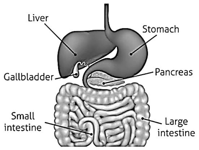
Figure 2.
The Carbohydrate Splitting Cascade. The classification of dietary carbohydrates, showing enzymatic breakdown of digestible saccharides into glucose, versus indigestible fibre, which passes through unbroken to feed gut microbiota and bind bile acids [Conceptual model].
Figure 2.
The Carbohydrate Splitting Cascade. The classification of dietary carbohydrates, showing enzymatic breakdown of digestible saccharides into glucose, versus indigestible fibre, which passes through unbroken to feed gut microbiota and bind bile acids [Conceptual model].
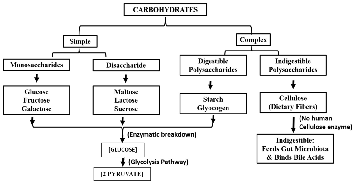
Figure 3.
Classification of Dietary Fatty Acids. Saturated fats have a maximum number of C–H bonds so from a thermodynamics standpoint they are highly stable [Conceptual model].
Figure 3.
Classification of Dietary Fatty Acids. Saturated fats have a maximum number of C–H bonds so from a thermodynamics standpoint they are highly stable [Conceptual model].
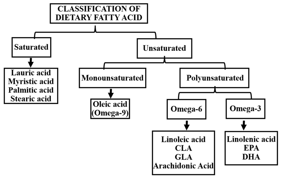
Figure 4.
Relative Fuel Use at Different Levels of Physical Activity. A structural systems mapping of fuel utilization based on workload velocity and kinetic output demands [Conceptual model].
Figure 4.
Relative Fuel Use at Different Levels of Physical Activity. A structural systems mapping of fuel utilization based on workload velocity and kinetic output demands [Conceptual model].
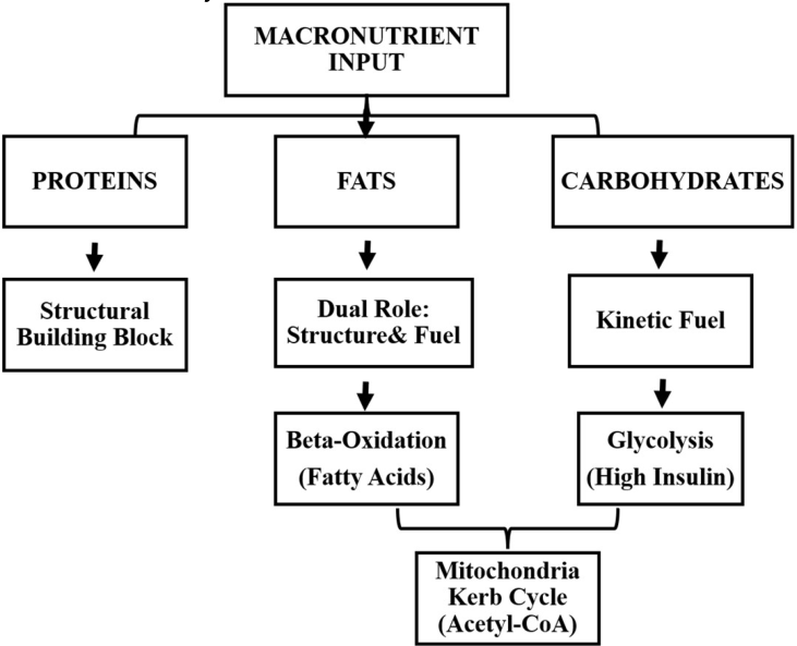
Figure 5.
Strategies for Supporting Muscle Preservation After 50. Overcoming senior cellular anabolic resistance by optimizing high-density leucine triggers and mechanical loading vectors [Conceptual model].
Figure 5.
Strategies for Supporting Muscle Preservation After 50. Overcoming senior cellular anabolic resistance by optimizing high-density leucine triggers and mechanical loading vectors [Conceptual model].
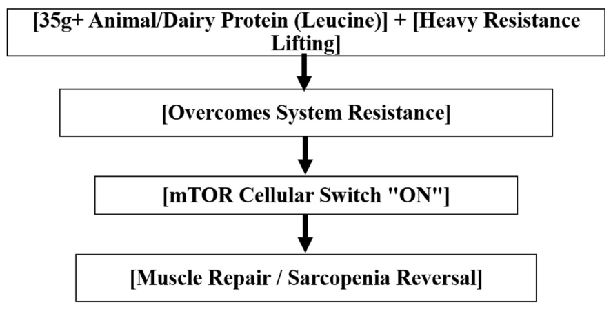
Figure 7.
Potential Effects of Lipid Oxidation on Membrane Signalling. A comparison showing how localized lipid peroxidation de-calibrates membrane-bound insulin receptors to induce resistance [Conceptual model].
Figure 7.
Potential Effects of Lipid Oxidation on Membrane Signalling. A comparison showing how localized lipid peroxidation de-calibrates membrane-bound insulin receptors to induce resistance [Conceptual model].
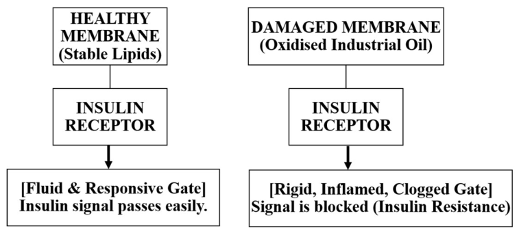
Figure 8.
Soluble Fibre and Bile Acid Recycling. Shows how Soluble fibre → altered bile-acid reabsorption → increased faecal bile-acid loss [Conceptual model].
Figure 8.
Soluble Fibre and Bile Acid Recycling. Shows how Soluble fibre → altered bile-acid reabsorption → increased faecal bile-acid loss [Conceptual model].
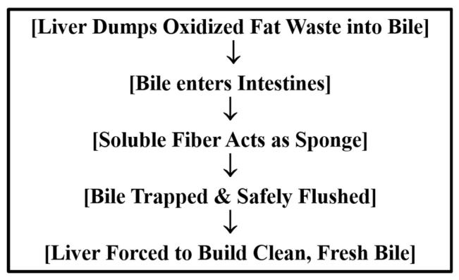
Figure 9.
Proliferative Feedback Mapping of the Prostate Matrix: Potential links between metabolic dysfunction, insulin signalling and prostate growth [Conceptual model].
Figure 9.
Proliferative Feedback Mapping of the Prostate Matrix: Potential links between metabolic dysfunction, insulin signalling and prostate growth [Conceptual model].
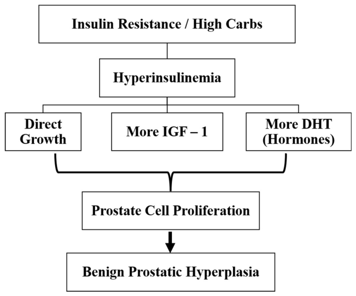
Figure 10.
The Musculoskeletal Tensile Failure Map: Potential links between metabolic dysfunction and musculoskeletal tissue changes [Conceptual model].
Figure 10.
The Musculoskeletal Tensile Failure Map: Potential links between metabolic dysfunction and musculoskeletal tissue changes [Conceptual model].
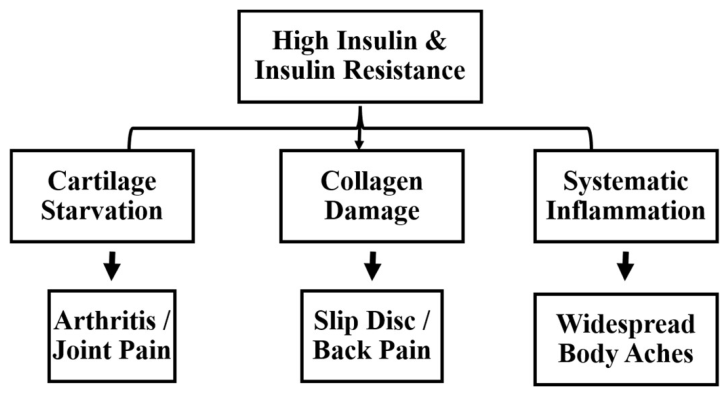
Figure 11.
Electrophysiological Phase Dispersion Path: Potential pathways linking metabolic dysfunction to cardiac electrical instability [Conceptual model].
Figure 11.
Electrophysiological Phase Dispersion Path: Potential pathways linking metabolic dysfunction to cardiac electrical instability [Conceptual model].
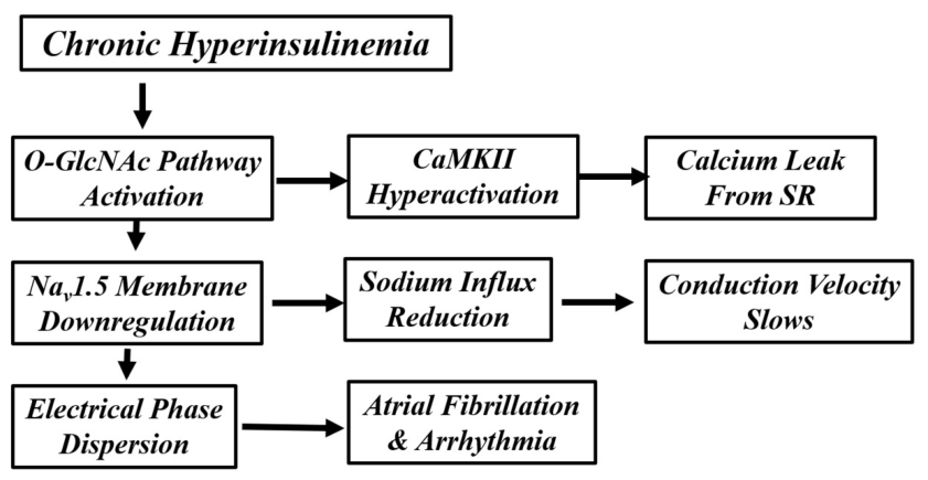
Figure 12.
Neurological Power Failure Sequencing. Potential links between brain insulin signalling, amyloid processing and cognitive decline [Conceptual model].
Figure 12.
Neurological Power Failure Sequencing. Potential links between brain insulin signalling, amyloid processing and cognitive decline [Conceptual model].
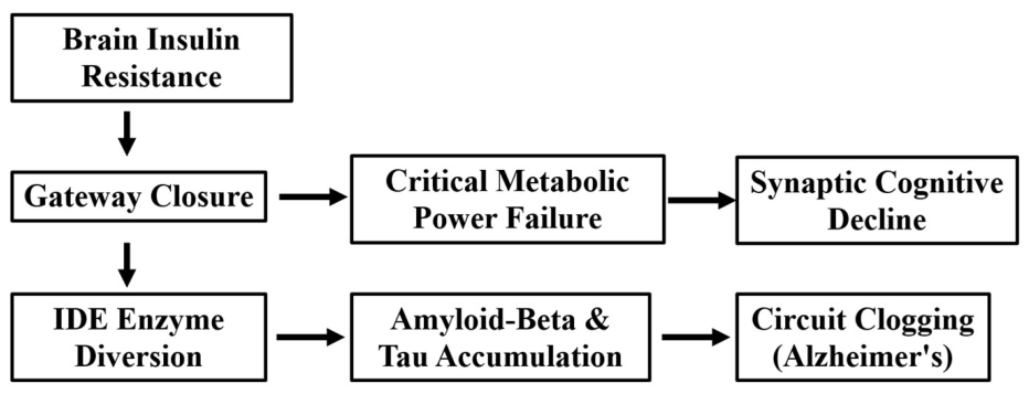
Disclaimer/Publisher’s Note: The statements, opinions and data contained in all publications are solely those of the individual author(s) and contributor(s) and not of MDPI and/or the editor(s). MDPI and/or the editor(s) disclaim responsibility for any injury to people or property resulting from any ideas, methods, instructions or products referred to in the content. |
© 2026 by the authors. Licensee MDPI, Basel, Switzerland. This article is an open access article distributed under the terms and conditions of the Creative Commons Attribution (CC BY) license (http://creativecommons.org/licenses/by/4.0/).
Copyright: This open access article is published under a Creative Commons CC BY 4.0 license, which permit the free download, distribution, and reuse, provided that the author and preprint are cited in any reuse.