

Submitted:
06 September 2026
Posted:
08 September 2026
You are already at the latest version
Abstract
Background/Objectives: BRAF-mutated (BRAFmut) colorectal cancer (CRC) is associated with a poor prognosis. In the metastatic setting, the role of metastasectomy in this subgroup remains controversial. However, treatment-induced molecular changes may help identify patients who are more likely to benefit from surgical resection of metastatic disease. Methods: A case-control study was conducted involving 14 patients with metastatic CRC, including 7 with BRAF-mutated tumors (case group) and 7 with tumors that were wild type for both RAS or BRAF (control group). Whole-exome sequencing (WES) was performed on FFPE tissue samples obtained before and after systemic therapy. All patients subsequently underwent surgical resection of metastatic lesions. Patients were matched according to primary tumor location (left- vs right-sided) and the site of metastasectomy (liver or peritoneal metastases). Results: Most patients underwent surgical resection of liver metastases (57%), followed by resection of peritoneal metastases (43%). All patients received fluoropyrimidine-based chemotherapy (capecitabine or 5-fluorouracil) in combination with oxaliplatin, and 36% also received irinotecan In addition, 64% received anti-angiogenic therapy (bevacizumab or aflibercept) and 14% received panitumumab. Distinct patterns of post-treatment mutational gain or loss were observed between the Case and Control groups. Furthermore, the profiles of altered genes differed according to metastatic site, primary tumor sidedness, and treatment regimen. No significant differences in overall survival or event-free survival were observed between the two groups. Conclusions: The mutational landscape of mCRC is dynamic rather than static, evolving in response to systemic therapy, the metastatic microenvironment, and the underlying molecular background. These findings support the potential clinical value of incorporating longitudinal genomic monitoring into therapeutic decision-making to identify treatment-induced evolutionary changes and optimize systemic treatment strategies.
Keywords:
colorectal cancer
; BRAF mutation
; whole-exome sequencing
; metastases surgery
; molecular changes
1. Introduction
Colorectal cancer (CRC) is one of the leading causes of morbidity and mortality worldwide. It is the third most commonly diagnosed cancer and the second leading cause of cancer-related death, after lung cancer. Its incidence and prevalence have increased in industrialized countries (1). Although many cases of CRC are diagnosed at an early stage and can be managed with surgery alone, approximately 25% of patients will develop metastatic disease during the course of their illness, while 20% present with metastatic disease at the time of diagnosis. The overall five-year survival rate for patients with metastatic CRC is approximately 13% (2).
Historically, the management of metastatic colorectal cancer (mCRC) has relied on combinations of cytotoxic agents such as irinotecan or oxaliplatin with fluoropyrimidines (5-fluorouracil plus leucovorin or capecitabine), resulting in a median overall survival (OS) of 18 months (3). Over the past decades, the introduction of targeted therapies, including anti-epidermal growth factor receptor (anti-EGFR) and anti-vascular endothelial growth factor (anti-VEGF) monoclonal antibodies, has transformed the treatment landscape for mCRC, extending median OS to 30 months in randomized clinical trials (4,5).
In selected patients, initially unresectable mCRC may become amenable to surgical resection following sufficient tumor shrinkage achieved with systemic therapies. These patients generally experience significantly improved survival outcomes compared with those whose disease remains unresectable (6,7).
Recent advances in molecular biology have demonstrated that mCRC is not a homogeneous disease but rather comprises distinct molecular subtypes characterized by specific genomic and pathological features. Mutations in the BRAF (B- Rapidly Accelerated Fibrosarcoma) gene represent one of the most clinically relevant oncogenic alterations and occur in approximately 10% of patients with CRC (8). These mutations are associated with female sex, right-sided primary tumors, advanced-stage disease at diagnosis, mucinous histology, and microsatellite instability (MSI). In addition, BRAF-mutated CRC is characterized by an unfavorable prognosis and reduced responsiveness to standard therapies, with a median OS of approximately 12 months (9).
The BRAF protein is a serine/threonine kinase that plays a central role in the mitogen-activated protein kinase (MAPK) signaling pathway, which regulates cellular proliferation, differentiation, migration, survival, and angiogenesis. Dysregulation of this pathway is a key driver of carcinogenesis. MAPK signaling is initiated by activation of the RAS (Rat Sarcoma) GTPase, which subsequently activates members of the RAF kinase family (ARAF, BRAF, and CRAF, also known as RAF1). Activated RAF proteins then phosphorylate and activate MEK1/2, which in turn phosphorylate and activate ERK1/2. Activated ERK kinases then phosphorylate a wide range of downstream substrates, including numerous transcription factors, thereby regulating critical cellular processes involved in tumor development and progression.
Approximately 96% of all BRAF mutations involve a c.1799T>A substitution in exon 15, resulting in a valine-to-glutamate substitution at codon 600 (BRAF V600E). This mutation increases BRAF kinase activity by approximately tenfold compared with the wild-type protein. Consequently, BRAF-mutated mCRC represents a distinct molecular subtype in CRC pathogenesis, characterized by marked clinical and molecular heterogeneity (10).
Given the poor prognosis associated with BRAF-mutated mCRC, comprehensive tumor molecular profiling using next-generation sequencing (NGS) is warranted to identify potential molecular alterations induced by prior chemotherapeutic regimens administered before metastatic resection.
2. Materials and Methods
We conducted a single-center retrospective observational study based on clinical data collected from medical records between 2010 and 2019. NGS of the whole exome was performed on formalin-fixed, paraffin-embedded tumor samples archived in the Department of Pathology at the Gregorio Marañón University General Hospital (HGUGM), Madrid.
Patients
We conducted a review of all mCRC patients seen in the Medical Oncology clinic at HGUGM between 2010 and 2019 to estimate the number of potential cases for analysis. Of the 554 patients who underwent clinical BRAF mutation profiling, 34 were identified as carriers of pathogenic BRAF mutations. To establish a control cohort, we selected patients with comparable clinical characteristics and wild-type (WT) BRAF, KRAS, and NRAS genes, based on electronic medical record data.
Eighteen patients (nine per group) were selected for whole-exome sequencing (WES) analysis. However, due to the poor quality of the sequencing data from various samples, only 14 samples (seven patients per group) were included in the final analysis.
Each cohort was defined by BRAF mutational status. Patients with pathogenic BRAF mutations were in the Case group, and those with WT BRAF, KRAS, and NRAS were in the Control group. Mutational profiling was performed as part of routine clinical care by the Department of Pathology at HGUGM.
Two samples per patient (n = 28) were analyzed to assess the impact of treatment exposure: (i) formalin-fixed, paraffin-embedded (FFPE) diagnostic specimens of metastatic colorectal cancer obtained before chemotherapy (pre-treatment, n = 14); and (ii) FFPE surgical specimens collected after chemotherapy (post-treatment, n = 14).
NGS
WES was performed on all FFPE samples (n = 28). To prepare the samples, 10-micron sections were obtained from FFPE blocks using a microtome. Each section was mounted on a glass slide. One section was stained with hematoxylin and eosin (H&E), to allow the pathologist to identify the infiltrating tumor area and assess its cellularity. Based on this assessment, the tumor area was macrodissected from the remaining sections. DNA was then extracted from these sections using a commercial QIAGEN kit that had been previously validated in the laboratory for DNA isolation.
The samples were sequenced at the National Center for Genomic Analysis (CNAG) using an Illumina platform and the Roche KAPA HyperExome + mt kit. The analytical pipeline included the Illumina Sequencing Analysis Viewer, Illumina run specifications, FastQC, and the INS-017 quality control alignment protocol.
Data Analysis
Sequence alignment was performed using BWA version 0.7.17, and variant calling was conducted with GATK Mutect2 version 4.1.9.0 and Strelka version 2.9.10. Variant annotation was carried out using SnpEff 5.0, SnpSift 5.1d, and Ensembl VEP version 109. The functional impact of variants was assessed using Ensembl impact scores and ClinVar annotations. The tumor mutational burden (TMB) was calculated with the use of the software program pyTMB version 1.3.0. The analysis of the variants that were either gained or lost was performed in R using the Maftools package version 2.24.0.
An initial analysis of the complete set of mutations was performed, followed by an evaluation focused solely on variants considered pathogenic and high-impact variants.
Secondary endpoints concentrated exclusively on pathogenic and high-impact mutations.
Primary and Secondary Endpoints
The primary objective of the study was to identify tumor mutational characteristics through WES, both before and after systemic treatment, and to evaluate differences between the two time points.
The secondary objectives were: 1) to describe and compare clinical and mutational characteristics identified in patients with BRAF mutations versus those without; 2) to characterize and correlate patients’ clinical features and treatments received with the genomic data obtained; and 3) to analyze survival outcomes based on the molecular alterations present in the samples studied.
3. Results
3.1. Demographics and Disease Characteristics of the Patients
A summary of the patients’ characteristics is presented in Table 1. The median age at the time of mCRC diagnosis was 67.47 years (range 58–72) for the Case group and 68.6 years (range 61–73) for the Control group.
The majority of patients presented with stage IV (57%) or III (36%) disease at the time of diagnosis, and primary tumors were predominantly located in the right colon (64%).
All patients received chemotherapy: 100% of patients received a combination of fluoropyrimidines and oxaliplatin, while 36% received irinotecan as well. With respect to biological therapy, 64% (n=9) of patients received antiangiogenic agents (8 bevacizumab and 1 aflibercept), 14% (n=2) received anti-EGFR therapy and 21% did not receive any biological treatment. Furthermore, two patients received targeted anti-BRAF therapy, with one patient undergoing treatment with encorafenib–cetuximab and the other with encorafenib–binimetinib–cetuximab.
The median follow-up period was 36.8 months. As illustrated in Figure 1, the chronogram for each patient is presented, with time designated as 0 representing the moment of diagnosis of metastatic disease.
The employment of high-throughput sequencing analyses facilitated the calculation of tumor mutational burden (TMB) for each sample (Figure 2). It was observed that Cases #3 and #7 exhibited notably higher values. These patients had previously reported microsatellite instability, which may explain the elevated TMB relative to the rest of the cohort. No consistent trend of increasing or decreasing TMB was observed in relation to patient group or chemotherapy exposure.
3.2. Differences in All Mutational Variants Between Cases and Controls After Treatment
When all categories of detected alterations were included in the analysis, several genes demonstrated statistically significant differences in the gain or loss of variants between the Case and Control groups after treatment, as determined by chi-square testing (Figure 3). The Case group demonstrated a substantial gain in variants in LMTK2, while the Control group exhibited a gain in TMC3. Conversely, a substantial loss of variants was observed exclusively in the Control group for NIPBL, HEATR5A, HTT, and TENM4.
3.3. Focused Analysis of Pathogenic and High-Impact Variants
An investigation into pathogenic and high-impact variants, as reported in the ClinVar database (11), revealed that the median number of somatic mutations identified as pathogenic was lower than the number of mutations categorized as high-impact (Figure 4). This discrepancy indicates that not all high-impact variants have been previously documented in clinical databases. High-impact variants encompass alterations in coding regions as well as canonical splice sites and exonic splicing enhancer (ESE) motifs. Variants of uncertain significance, benign variants, synonymous changes, and those predicted to be of moderate or low impact were excluded from this study due to the limited likelihood of biological relevance in the context of CRC (12).
A comparative analysis revealed that the median number of high-impact or pathogenic variants was lower in the Case group than in the Control group. Furthermore, a significant number of pathogenic mutations were gained by the Case group following treatment, while the Control group, exhibited a net loss of such mutations.
In light of the observed scenarios of mutational gains and losses between the Case and Control groups, it was imperative to characterize the specific genes implicated. The most frequently mutated genes following treatment (regardless of group), were as follows (Figure 5):
- Pathogenic mutations most commonly gained: CFAP47, CDH7, HDAC9, LNX1, MAP3K5, RHPN2, and ZZEF1;
- Pathogenic mutations most commonly lost: AGL, CDK13, CNGA3, DNAH7, DSP, OBSCN, SACS, SDAD1, SMARCA2, and TP53.
The set of genes exhibiting lost mutations versus those with gained mutations were found to be distinct. A lack of significant disparities was identified in the overall mutational profile between Case and Control groups both before and after treatment. However, several individual genes exhibited alterations that were group-specific, although these differences did not reach statistical significance in the chi-square analysis (Figure 6). The following observations merit emphasis: The gain of pathogenic mutations in LNX1 was exclusively observed in the Control group. The loss of mutations in CNGA3 and DNAH7 was similarly exclusive, occurring only in the Control group. The loss of mutations in OBSCN was confined to the Case group.
3.4. Correlation Between Clinical Characteristics and Treatments with Genomic Data
Differences in mutational profiles were also analyzed according to metastatic site (peritoneal versus hepatic metastases) and primary tumor location (left-sided versus right-sided). Restricting the analysis to pathogenic or high-impact mutations, the comparison between peritoneal and hepatic metastases revealed a notable gain of pathogenic mutations in CERT1, DCPS, LNX1, and PAX4, observed exclusively in Control patients with peritoneal metastases (Table 2). Among these, LNX1 once again emerged as a recurrently affected gene. In addition, frequent losses of mutations were observed in HERC1 among Cases with peritoneal metastases and in DNAH7 (previously identified in the primary analysis) among Controls with hepatic metastases (Table 2).
When analyzed by primary tumor sidedness, the highest frequencies of pathogenic mutation gains and losses were observed in left-sided colon cancer. Specifically, mutation gains were most frequently observed in ZZEF1, whereas mutation losses were identified in DSP, KDM5D, PRTG, and SACS (Table 3).
The interpretation of mutational changes under the selective pressure of anti-BRAF targeted therapy is limited by the small number of patients exposed to this treatment, with only 2 out of 14 patients receiving anti-BRAF agents. The genes most frequently exhibiting mutation gains and losses are summarized in Table 4.
3.5. Overall Survival
Overall survival (OS) and event-free survival (EFS) were assessed by sample group (Case vs Control) using Kaplan–Meier analysis. However, given the small sample size, the resulting survival curves were not considered representative.
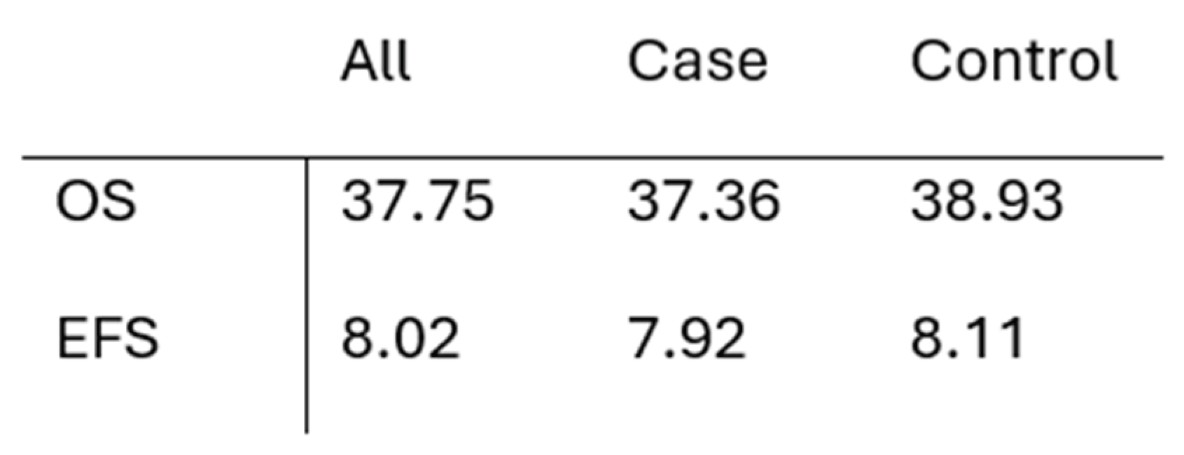
Median survival for each group is presented in Table 5. Overall survival was calculated from the date of metastatic disease diagnosis, whereas event-free survival was calculated from the date of metastasectomy. No statistically significant differences were observed between the groups in the Kaplan–Meier analysis.
4. Discussion
The present study thoroughly examined the mutational changes in mCRC patients following systemic treatment, with a particular emphasis on the distinctions between cases with BRAF mutations and matched controls. Utilizing NGS data and in silico pathogenicity predictions, we identified genes of interest that potentially correlated with treatment response/resistance and disease progression.
The distribution of subjects by sex reflects the availability of samples from patients who met the inclusion criteria for this study, taking into account the higher reported incidence of mCRC in men (13). All patients received fluoropyrimidine-based chemotherapy, agents known to induce DNA damage and genomic instability. Consequently, a substantial proportion of post-treatment variants likely represent chemotherapy-induced or chemotherapy-selected alterations. However, the distinct patterns observed between the study groups suggest that treatment pressure interacts with intrinsic biology of the tumors to drive different evolutionary trajectories. BRAF-mutated tumors consistently gained pathogenic mutations after treatment, whereas the wild-type control group predominantly exhibited mutation loss, indicating fundamentally different adaptive responses.
In BRAF-mutated tumors (Cases), the accumulation of pathogenic variants following therapy is indicative of adaptive clonal evolution, whereby selective pressure favors the expansion of resistant subclones rather than effective clonal depletion. The recurrent gain of mutations in genes such as LMTK2 is particularly noteworthy. LMTK2 encodes a serine-threonine kinase involved in vesicle trafficking, apoptosis regulation, and tumorigenesis. Mechanistically, LMTK2 appears to modulate the NF-κB signaling pathway, a central regulator of cellular growth, survival, and immune responses. Increased NF-κB pathway activity has been associated with enhanced cancer cell proliferation (14). In addition, LMTK2 participates in the phosphorylation of proteins within the MAPK signaling pathway, a key molecular pathway in patients with mCRC. This observation is consistent with findings from the BEACON CRC study, although that study reported the accumulation of mutations in other MAPK-related genes such as RAS, MAP2K1, and MET (15). Therefore, the increased mutational burden observed in LMTK2 in BRAF-mutated tumors may reflect either a compensatory mechanism that reinforces proliferative signaling downstream of BRAF or remodeling of stress-response pathways during treatment, providing an alternative route for MAPK activation as previously proposed (16; 17).
In contrast, tumors lacking baseline MAPK alterations (Controls) preferentially acquired mutations in TMC3 following chemotherapy. Although TMC3 has been poorly characterized in CRC, it belongs to a family of transmembrane ion channels with potential roles in signal modulation, suggesting a treatment-specific adaptive response in triple wild-type tumors. Growing evidence supports the involvement of ion channels and remodeling of the ionic microenvironment in cancer progression, therapeutic resistance, and metastatic dissemination (18; 19; 20).
When examining gene-specific mutation losses, alterations in NIPBL and TENM4 were observed exclusively in the Control group. NIPBL plays a key role in chromatin cohesion, and its disruption has been associated with chromosomal instability in gastrointestinal cancers (21; 22). TENM4 alterations, in turn, have been linked to poorer outcomes in patients with colorectal cancer treated with fluoropyrimidine-based chemotherapy (23). Although the specific variants identified in our cohort have not been previously reported in clinical databases, in silico prediction analyses suggest they are functionally relevant. Their disappearance following chemotherapy in the Control group may indicate that treatment preferentially targets pathways involved in chromatin organization, intracellular trafficking, and cell adhesion rather than classical driver oncogenes. Alternatively, it may reflect the selective elimination of chemotherapy-sensitive subclones (i.e., a chemotherapy-induced “genomic pruning” effect) rather than a biologically coordinated loss of these alterations.
When the analyses were restricted to pathogenic and high-impact variants, the contrasting evolutionary patterns became even more pronounced. In the Control group, pathogenic mutations were more frequently lost than gained after treatment, whereas the opposite trend was observed in BRAF-mutated tumors. Among the gene alterations gained in the Control group, LNX1 was particularly noteworthy. LNX1, a dual-function E3 ubiquitin ligase, regulates TP53 stability and modulates NOTCH signaling, both of which play critical roles in colorectal cancer biology (24; 25; 26; 27; 28; 29; 30). Increased mutational activity in LNX1 may therefore reflect a tumor-suppressive shift, modulating NOTCH-dependent survival pathways in response to cytotoxic stress. These findings suggest that BRAF-WT CRC may rely more heavily on signaling reprogramming to withstand treatment, rather than on widespread genomic instability.
Conversely, the loss of pathogenic mutations in CNGA3 and DNAH7 warrants a more nuanced biological interpretation (31). Both genes encode proteins associated with specialized cellular structures, including ion channels and cilia. CNGA3 encodes a cyclic nucleotide-activated cation channel that is essential for sensory transduction, including normal vision and olfactory signaling, by regulating the influx of Na+ and Ca2+ ions and, consequently, cellular excitability (32; 33). CNGA3 is also a component of the cAMP (cyclic Adenosine MonoPhosphate) signaling pathway (34), a central regulator of cellular metabolism, contractility, and signal transduction. Cells harboring alterations in ion channels function may be less able to compensate for the oxidative and mitochondrial damage induced by oxaliplatin, rendering them more susceptible to chemotherapy-induced calcium dysregulation. DNAH7 encodes a protein that is essential for ciliary and flagellar motility. Recent evidence indicates that tumors harboring DNAH7 mutations exhibit greater immune cell infiltration and a more immunologically active tumor microenvironment, features that have been associated with improved responses to immunotherapy (35). Such immunologically “visible” subclones may be also more efficiently eradicated by cytotoxic chemotherapy. Taken together, the loss of CNGA3 and DNAH7 mutations in the Control group following systemic therapy suggest that BRAF-WT tumors may preferentially eliminate subpopulations with altered ion-channel signaling or ciliary function because of their intrinsic vulnerability to fluoropyrimidine/oxaliplatin-based chemotherapy. In contrast, the greater genomic plasticity of BRAF-mutated tumors may enable compensation for these vulnerabilities. Although neither CNGA3 nor DNAH7 have previously been associated with mCRC, recent studies suggest that CNGA3 silencing may contribute to tumorigenesis in rectal adenocarcinoma (36).
The OBSCN gene is notable for its large size (~150 kb), which may predispose it to a higher mutational frequency. It encodes a giant cytoskeletal protein composed of 68 immunoglobulin-like domains, two fibronectin domains, a calcium/calmodulin-binding domain, a RhoGEF domain with an associated pH domain, and 2 serine/threonine kinase domains. OBSCN has been reported to be frequently mutated in both breast and colorectal cancers (37). In combination with TTN, it has also been proposed as a biomarker for identifying CRC patients who are most likely to benefit from immunotherapy (38). In our study, the loss of pathogenic OBSCN mutations was observed exclusively in the Case group and was associated with a poorer prognosis.
In the analysis of clinical factors associated with patterns of metastasis, stratified analyses by case-controls status and metastatic site (peritoneal vs hepatic) revealed distinct patterns of post-treatment mutational changes. In the Cases group, liver metastases were characterized by mutation gains in ATP2A2, CSMD1, F13B, GFAP, and HDAC9, genes involved in calcium-dependent signalling (ATP2A2) (39; 40), extracellular matrix remodelling (F13B) and signalling (CSMD1) (41; 42), cytoskeletal reorganization (GFAP) (43), and epigenetic reprogramming and therapeutic resistance (HDAC9) (44; 45). This pattern may reflect the selective survival and expansion of clones capable of modulating extracellular matrix interactions, cytoskeletal remodeling, and transcriptional plasticity, thereby facilitating adaptation to the oxidative and metabolically dynamic hepatic microenvironment. Conversely, the mutations lost in this group suggest the preferential elimination of subclones dependent on metastasis-suppressive pathways (BRMS1) (46), metabolic rigidity (ACAD8) (47), or vesicular trafficking (AP1M1) (48), allowing treatment-sensitive populations to be rapidly replaced by more adaptable clones. In contrast, the mutational profile of cases with peritoneal metastases was characterized predominantly by the loss of HERC1, an ubiquitin ligase involved in protein turnover and cellular stress-response signaling. This finding likely reflects the selective depletion of stress-vulnerable subclones within the hypoxic, poorly perfused peritoneal microenvironment. It is also consistent with the unique biological characteristics of the peritoneal compartment, where increased stromal density and limited drug penetration may create selective pressures that favor the elimination of specific subclonal populations while allowing others to persist.
Analysis of the mutational dynamics in the Control group revealed losses of DNAH7, CEP85, and DMD in liver metastases, suggesting that clones metastasizing to the liver may become less dependent on epithelial structural integrity and acquire greater mesenchymal plasticity (49; 50). In contrast, Control patients with peritoneal metastases gained mutations in genes such as CERT1, DCPS, LNX1, and PAX4, suggesting that BRAF-WT tumours may rely more heavily on signaling pathway reprogramming as a mechanism of chemotherapy resistance within the nutritionally heterogeneous peritoneal cavity. Notably, the recurrent gain of LNX1, a regulator of NOTCH signalling, further supports the hypothesis that adaptive signaling plasticity contributes to tumor survival in the peritoneal microenvironment.
Taken together, the distinct patterns of mutational gains and losses according to metastatic location in the cases and control groups reinforce the hypothesis that chemotherapy reshapes clonal composition based on the metabolic and physical constraints of each metastatic microenvironment.
Tumor sidedness also influenced mutational trajectories. Right-sided tumors, which are more frequently associated with BRAF mutations, exhibited a greater number of post-treatment mutational losses, suggesting more extensive remodeling of the clonal landscape. In contrast, left-sided tumors displayed a more balanced pattern, with a slight predominance of newly gained mutations, primarily involving genes associated with metabolic processes (ACN1) and membrane homeostasis (ACE2). This pattern is consistent with a more metabolically adaptable and invasive phenotype (51; 52).
Overall, the profile of affected genes differed between patients treated with chemotherapy plus anti-BRAF therapy and those treated without anti-BRAF agents. This observation is consistent with the concept that different treatment regimens exert selective pressure on distinct cellular pathways. The genes altered in patients receiving anti-BRAF therapy were primarily associated with metabolic processes, extracellular matrix remodeling, and the regulation of cell signaling (53), consistent with the compensatory mechanisms that are typically activated in response to BRAF inhibition (54; 55). Taken together, these findings suggest that the combination of chemotherapy and anti-BRAF therapy exerts adaptive selective pressure that is particularly reflected in metabolic reprogramming and tumor–microenvironment interactions. In contrast, among patients who did not receive anti-BRAF therapy (i.e., those treated with chemotherapy alone or chemotherapy combined with an anti-VEGF or anti-EGFR biologic agent; see Table 4), the altered genes were predominantly involved in cellular structure, cytoskeletal integrity, and homeostasis (DNAH7, DSP, SACS, and CNGA3). This pattern may reflect a broader and less specific adaptive response to treatment than that observed in patients receiving anti-BRAF therapy. However, the very small number of patients exposed to anti-BRAF agents in our cohort limits the ability to draw meaningful conclusions regarding the selective pressure exerted by targeted therapy in this setting.
Regarding survival outcomes, no significant differences in OS or EFS were observed between patients with BRAF-mutated and BRAF-WT tumors. However, the small sample size substantially limited the statistical power of these analyses.
A clinically important finding of this study is that patients with BRAF-mutated tumors who underwent surgical resection of metastases achieved OS comparable to that of patients with BRAF wild-type tumors, despite the adverse genomic features and poorer prognosis typically associated with BRAF mutations. Although this observation should be interpreted with caution because of the limited sample size, it suggests that metastasectomy may partially mitigate the intrinsically poor prognosis of BRAF-mutated mCRC by eliminating resistant subclones before they achieve irreversible clonal dominance occurs. This finding is consistent with previous surgical series demonstrating that, in carefully selected patients, aggressive multimodal treatment strategies can result in prolonged survival even in the presence of unfavorable molecular characteristics (56) (57).
5. Conclusions
Taken together, our findings demonstrate meaningful gene-specific gains and losses, indicating that the mutational landscape of mCRC is not static but evolves dynamically in response to systemic therapy, the metastatic microenvironment, and the underlying molecular background. In BRAF-mutated tumors, the post-treatment gain of pathogenic mutations may serve as a marker of adaptive resistance, converging on the reinforcement of MAPK and NF-κB signaling pathways. In contrast, in BRAF wild-type tumors, the preferential loss of pathogenic variants following chemotherapy may reflect more effective disruption of oncogenic pathways through the elimination of treatment-sensitive subclones, while more resistant populations persist by relaying on alternative mechanisms involving chromatin organization, ion transport, and cytoskeletal integrity. The recurrent alterations observed in genes such as LNX1, HDAC9, OBSCN, DSP and SACS further underscore the importance of structural remodeling, cellular stress response pathways and signaling plasticity in post-treatment tumor evolution. Moreover, the analysis of clinical-genomic correlations demonstrated that mutational remodeling varies according to both the primary tumor location and the site of metastasis, confirming that treatment-induced clonal selection is highly dependent on the biological context.
These findings, while exploratory, support the potential clinical utility of incorporating longitudinal genomic monitoring into therapeutic decision-making to capture treatment-induced evolutionary changes and guide the selection of systemic therapy.
Several limitations of this study should be acknowledged. First, the small sample size limits statistical power and precludes definitive conclusions regarding survival associations or treatment-specific effects, particularly those related to anti-BRAF therapy, which was administered to only a minority of patients. Second, the retrospective design introduces the potential for selection bias. In addition, functional validation of the identified variants was not performed, and variant pathogenicity was inferred using in silico prediction tools, which may not fully reflect their biological effects. Finally, whole-exome sequencing does not capture epigenetic alterations, transcriptomic changes, or clonal architecture, all of which may contribute to tumor adaptation and the development of treatment resistance.
Future prospective studies incorporating larger patient cohorts, serial tissue and liquid biopsy sampling, and functional validation of the identified variants will be essential to translate these findings into clinically actionable strategies for the management of patients with metastatic colorectal cancer.
Supplementary Materials
The following supporting information can be downloaded at the website of this paper posted on Preprints.org, Table S1.
Author Contributions
Conceptualization, JSA, CP, EA, MDMM, MM, PGA.; methodology, JSA, CP, EA, MDMM, NGA, MBB, MM, PGA; formal analysis, JSA, CP, EA, MDMM, PGA; investigation, JSA, CP, EA, MDMM, MM, PGA; data curation, JSA, CP, EA, MDMM, NGA, MBB, IP, MACM, CCL, RML, MPG, LOM, GTPS, PACF, TM, AJMM, IO, AA, FADLP, MM, PGA; writing—original draft preparation, JSA, CP, EA, MDMM, MM, PGA; writing—review and editing, all authors. All authors have read and agreed to the published version of the manuscript.
Funding
This research received funding from Pierre Fabre.
Institutional Review Board Statement
The study was conducted in accordance with the Declaration of Helsinki, and approved by the Ethics Committee of Gregorio Marañón Hospital (protocol code GOM-HGUGM-2022-06, approved on September 2022).
Informed Consent Statement
Informed consent was obtained from all subjects involved in the study.
Data Availability Statement
No new data were created.
Conflicts of Interest
JSA has played an advisory role for Takeda; received honoraria from Servier, LEO Pharma, MSD, BMS, Roche, Merck, Pfizer, Recordati, AstraZeneca, Nestlé, Abbott, Tahio, Techdow; received funding from Amgen and Pierre Fabre. NGA has received honoraria from MSD, Astrazeneca, Pfizer, BMS, Pierre Fabre, Merck. MBB has received honoraria from Pierre Fabre, AstraZeneca, MSD, Almirall, Novartis, Regeneron. MPG has received honoraria from Servier, Amgen, Abbott. AJMM has played an advisory role for Pfizer, BMS-Celgene, Sanofi, Astra-Zeneca, MSD, Servier, Roche, Taiho, Leo Pharma, Regeneron, Jazz Pharmaceuticals, Revolution Medicine, Novocure; received funding from LEO Pharma; received honoraria from Taiho, Rovi, Menarini, Stada, Medscape, Incyte, GSK; has a patent on risk assessment model in venous thromboembolism in cancer patients (genomic risk score) and liquid biopsy developed with Laser technology. FADLP has played an advisory role for Lilly, Pfizer, Roche, Seagen, Pfizer, Novartis; received honoraria from Lilly, Gilead, Pfizer, Novartis, Seagen, Adium, Roche. MM has played an advisory role for Astrazeneca, Lilly, Daiichi Sankyo, Menarinio-Stemline, Roche/Genentech; received honoraria from Astrazeneca, Lilly, Novartis, Pfizer, Roche/Genentech; received funding from Novartis, Puma, Roche. PGA has played an advisory role for Amgen, Takeda, MSD, BMS; received honoraria from Amgen, Servier, Takeda, BMS, Pierre Fabre.The rest of the authors declare no conflicts of interest.
References
- Bray, F. Global cancer statistics 2018: GLOBOCAN estimates of incidence and mortality worldwide for 36 cancers in 185 countries. S.l. CA Cancer J. Clin. 2018, 68, 394–424. [Google Scholar] [CrossRef] [PubMed]
- Figures, American Cancer Society; Cancer Facts & and American Cancer Society: Atlanta, GA, USA, 2016.
- Fuchs, C.S.; Marshall, J.; Mitchell, E. Randomized, controlled trial of irinotecan plus infusional, bolus, or oral fluoropyrimidines in first-line treatment of metastatic colorectal cancer: Results from the BICC-C Study. J. Clin. Oncol. 2007, Vols. 25, 4779–4786. [Google Scholar] [CrossRef] [PubMed]
- Venook, A.P.; Niedzwiecki, D.; Lenz, H.J. Phase III trial of irinotecan/5-FU/leucovorin (FOLFIRI) or oxaliplatin/5-FU/leucovorin (mFOLFOX6) with bevacizumab or cetuximab for patients with KRAS wild-type untreated metastatic adenocarcinoma of the colon or rectum. CALGB/SWOG 80405; J Clin Oncol. 2014; Vol. 32, (Suppl 18). [Google Scholar]
- Cervantes, A.; Adam, R.; Roselló, S. Metastatic colorectal cancer: ESMO Clinical Practice Guideline for diagnosis, treatment and follow-up. Ann. Oncol. 2023, 34(1). [Google Scholar] [CrossRef] [PubMed]
- Ma, R.; Li, T. Conversion therapy combined with individualized surgical treatment strategy improves survival in patients with colorectal cancer liver metastases. Int. J. Clin. Exp. Pathol. Vols. 2021, 14(3), 314–321. [Google Scholar] [PubMed] [PubMed Central]
- Granieri, S.; Cotsoglou, C.; Bonomi, A. Conversion Strategy in Left-Sided RAS/BRAF Wild-Type Metastatic Colorectal Cancer Patients with Unresectable Liver-Limited Disease: A Multicenter Cohort Study. Cancers 2022, 14(22), 5513. [Google Scholar] [CrossRef] [PubMed] [PubMed Central]
- Tjpar, S.; Bertagnolli, M.; Bosman, F. Prognostic and predictive biomarkers in resected colon cancer: Current status and future perspectives for integrating genomics into biomarker discovery. Oncologist 2010, 15, 390–404. [Google Scholar] [CrossRef] [PubMed]
- Clarke, C.; Kopetz, E. BRAF mutant colorectal cancer as a distinct subset of colorectal cancer: clinical characteristics, clinical behaviour, and response to targeted therapies. J. Gastrointest. Oncol. 2015, 6(6), 660–7. [Google Scholar] [CrossRef] [PubMed] [PubMed Central]
- Cantwell-Dorris, E.R.; O’Leary, J.J.; Sheils, O.M. BRAFV600E: Implications for carcinogenesis and molecular therapy. Mol. Cancer Ther. 2011, 10, 385–394. [Google Scholar] [CrossRef] [PubMed]
- https://www.ncbi.nlm.nih.gov/clinvar/ [Online].
- Cartegni, L.; Chew, S.; Krainer, A. Listening to silence and understanding nonsense: exonic mutations that affect splicing. Nat. Rev. Genet 2002, 3, 285–298. [Google Scholar] [CrossRef] [PubMed]
- Sung, H.; Ferlay, J.; Siegel, R.L. Global cancer statistics 2020: GLOBOCAN estimates of incidence and mortality worldwide for 36 cancers in 185 countries. CA Cancer J. Clin. [Internet] 2021, 71(3), 209–49. [Google Scholar] [CrossRef] [PubMed]
- Zhang, R.; Li, X.; Wei, L.; Qin, Y.; Fang, J. Lemur tyrosine kinase 2 acts as a positive regulator of NF-κB activation and colon cancer cell proliferation. Cancer Lett. 2019, 454, 70–77. [Google Scholar] [CrossRef] [PubMed]
- Kopetz, S.; Murphy, D.A.; Pu, J. Molecular profiling of BRAF-V600E-mutant metastatic colorectal cancer in the phase 3 BEACON CRC trial. Nat. Med. 2024, 30, 3261–3271. [Google Scholar] [CrossRef] [PubMed]
- Xie, X.; Liu, H.; Wang, Y. Nicotinamide N-methyltransferase enhances resistance to 5-fluorouracil in colorectal cancer cells through inhibition of the ASK1-p38 MAPK pathway. Oncotarget 2016, 7, 45837–45848. [Google Scholar] [CrossRef] [PubMed]
- Zhang, G.; Luo, X.; Wang, Z. TIMP-2 regulates 5-Fu resistance via the ERK/MAPK signaling pathway in colorectal cancer. Aging 2022, 14(1), 297–315. [Google Scholar] [CrossRef] [PubMed]
- Zhou, Y.; Cheng, X.; Zhang, F. Integrated multi-omics data analyses for exploring the co-occurring and mutually exclusive gene alteration events in colorectal cancer. Hum. Mutat. 2020, 41(9), 1588–1599. [Google Scholar] [CrossRef] [PubMed]
- Litan, A.; Langhans, S.A. Cancer as a channelopathy: ion channels and pumps in tumor development and progression. Front. Cell. Neurosci. 2015, 9, 86. [Google Scholar] [CrossRef] [PubMed]
- Gentile, R.; Feudi, D.; Sallicandro, L.; Biagini, A. Can the Tumor Microenvironment Alter Ion Channels? Unraveling Their Role in Cancer. Cancers 2025, 17(7), 1244. [Google Scholar] [CrossRef] [PubMed]
- Kim, M.S.; An, C.H.; Chung, Y.J. NIPBL, a cohesion loading factor, is somatically mutated in gastric and colorectal cancers with high microsatellite instability. Dig. Dis. Sci. 2013, 58(11), 3376–3378. [Google Scholar] [CrossRef] [PubMed]
- Leylek, T.R.; Jeusset, L.M.; Lichtensztejn, Z.; McManus, K.J. Reduced Expression of Genes Regulating Cohesion Induces Chromosome Instability that May Promote Cancer and Impact Patient Outcomes. Sci. Rep. 2020, 10(1), 592. [Google Scholar] [CrossRef] [PubMed]
- Heczko, L.; Liška, V.; Vyčítal, O. Targeted panel sequencing of pharmacogenes and oncodrivers in colorectal cancer patients reveals genes with prognostic significance. Hum. Genom. 2024, 18, 83. [Google Scholar] [CrossRef] [PubMed]
- Zhou, B.; Lin, W.; Long, Y. Notch signaling pathway: architecture, disease, and therapeutics. Sig Transduct. Target Ther. 2022, 7, 95. [Google Scholar] [CrossRef] [PubMed]
- Zenonos, K.; Kyprianou, K. RAS signaling pathways, mutations and their role in colorectal cancer. World J. Gastrointest. Oncol. 2013, 5(5), 97–101. [Google Scholar] [CrossRef] [PubMed] [PubMed Central]
- Michel, M.; Kaps, L.; Maderer, A.; Galle, P.R.; Moehler, M. The Role of p53 Dysfunction in Colorectal Cancer and Its Implication for Therapy. Cancers 2021, 13(10), 2296. [Google Scholar] [CrossRef] [PubMed] [PubMed Central]
- Ottaiano, A.; Santorsola, M.; Capuozzo, M. The prognostic role of p53 mutations in metastatic colorectal cancer: A systematic review and meta-analysis. Crit. Rev. Oncol. Hematol. 2023, 186, 104018. [Google Scholar] [CrossRef] [PubMed]
- Park, R.; Kim, H.; Jang, M. LNX1 contributes to tumor growth by down-regulating p53 stability. FASEB J. 2019, 33(12), 13216–13227. [Google Scholar] [CrossRef] [PubMed]
- Ma, L.; Wang, L.; Shan, Y. Suppression of cancer stemness by upregulating Ligand-of-Numb protein X1 in colorectal carcinoma. PLoS ONE 2017, 12(11), e0188665. [Google Scholar] [CrossRef] [PubMed] [PubMed Central]
- Baisiwala, S.; Hall, R.R., 3rd; Saathoff, M.R. LNX1 Modulates Notch1 Signaling to Promote Expansion of the Glioma Stem Cell Population during Temozolomide therapy in Glioblastoma. Cancers 2020, 12(12), 3505. [Google Scholar] [CrossRef] [PubMed] [PubMed Central]
- McFarland, C.D.; Mirny, L.A.; Korolev, K.S. Tug-of-war between driver and passenger mutations in cancer and other adaptive processes. Proc. Natl. Acad. Sci. U S A 2014, 111(42), 15138–43. [Google Scholar] [CrossRef] [PubMed] [PubMed Central]
- https://www.genecards.org/cgi-bin/carddisp.pl?gene=CNGA3. [Online].
- https://www.omim.org/entry/600053. [Online].
- https://www.kegg.jp/entry/hsa04024. [Online].
- Yang, W.; Shen, Z.; Yang, T.; Wu, M. DNAH7 mutations benefit colorectal cancer patients receiving immune checkpoint inhibitors. Ann. Transl. Med. 2022, 10(24), 1335. [Google Scholar] [CrossRef] [PubMed] [PubMed Central]
- Hua, Y.; Ma, X.; Liu, X. Abnormal expression of mRNA, microRNA alteration and aberrant DNA methylation patterns in rectal adenocarcinoma. PLoS ONE 2017, 12(3), e0174461. [Google Scholar] [CrossRef] [PubMed] [PubMed Central]
- Sjoblom, T.; Jones, S.; Wood, L.D. The consensus coding sequences of human breast and colorectal cancers. Science 2006, 314(5797), 268–74. [Google Scholar] [CrossRef] [PubMed]
- Liu, Z.; Wang, L.; Guo, C. TTN/OBSCN 'Double-Hit' predicts favourable prognosis, 'immune-hot' subtype and potentially better immunotherapeutic efficacy in colorectal cancer. J. Cell Mol. Med. 2021, 25(7), 3239–3251. [Google Scholar] [CrossRef] [PubMed] [PubMed Central]
- https://ckb.genomenon.com/gene/show?geneId=488. [Online].
- Chung, F.Y.; Lin, S.R.; Lu, C.Y. Sarco/endoplasmic reticulum calcium-ATPase 2 expression as a tumor marker in colorectal cancer. Am. J. Surg. Pathol. 2006, 30(8), 969–74. [Google Scholar] [CrossRef] [PubMed]
- Wei, D.; Xueming, X.; Fan, H.; Jiayun, Y. Extracellular matrix remodeling in the tumor immunity. Front. Immunol. 2024. [Google Scholar] [CrossRef]
- Gialeli, C.; Tuysuz, E.C.; Staaf, J. Complement inhibitor CSMD1 modulates epidermal growth factor receptor oncogenic signaling and sensitizes breast cancer cells to chemotherapy. J. Exp. Clin. Cancer Res. 2021, 40, 258. [Google Scholar] [CrossRef] [PubMed]
- Aseervatham. Cytoskeletal Remodeling in Cancer. J. Biology 2020, 9(11), 385. [Google Scholar] [CrossRef] [PubMed]
- Minisini, M.; Mascaro, M.; Brancolini, C. HDAC-driven mechanisms in anticancer resistance: epigenetics and beyond. Cancer Drug Resist 2024, 7, 46. [Google Scholar] [CrossRef] [PubMed]
- Giorgio, E.; Dalla, E.; Franforte, E. Different class IIa HDACs repressive complexes regulate specific epigenetic responses related to cell survival in leiomyosarcoma cells. Nucleic Acids Res. 2020, 48(2), 564–664. [Google Scholar] [CrossRef] [PubMed]
- Zhang, Y.; Guan, J.; Sun, Y. Effect of BRMS1 on Tumorigenicity and Metastasis of Human Rectal Cancer. Cell Biochem Biophys. 2014, 70, 505–509. [Google Scholar] [CrossRef] [PubMed]
- Zhuang, H.; Chen, Y.; Huang, S. Cuproptosis-related gene ACAD8 inhibits the metastatic ability of colorectal cancer by inducing cuproptosis. Front. Immunol. 2025, 16, 1560322. [Google Scholar] [CrossRef] [PubMed]
- Shin, J.; Nile, A.; Oh, J.-W. Role of adaptin protein complexes in intracellular trafficking and their impact on diseases. Bioengineered 2021, 12(1), 8259–8278. [Google Scholar] [CrossRef] [PubMed]
- Aditi; Khajuria, A.; Garima. Extracellular vesicles in cancer: biogenesis, oncogenic mechanisms, biomarker potential, and therapeutic applications. Med. Oncol. 2026, 43, 23. [Google Scholar] [CrossRef] [PubMed]
- Aseervatham. Cytoskeletal Remodeling in Cancer. J. Biology 2020, 9(11), 385. [Google Scholar] [CrossRef] [PubMed]
- Morris, M.T.; Jain, A.; Sun, B.; Ket. Multi-omic analysis reveals metabolic pathways that characterize right-sided colon cancer liver metastasis. Cancer Lett. 2023, 574, 216384. [Google Scholar] [CrossRef] [PubMed] [PubMed Central]
- Jain, A.; Morris, M.T.; Berardi, D. Charting the metabolic biogeography of the colorectum in cancer: challenging the right sided versus left sided classification. Mol. Cancer 2024, 23, 211. [Google Scholar] [CrossRef] [PubMed]
- https://www.proteinatlas.org. [Online].
- Ahronian, L.G.; Sennott, E.M.; Van Allen, Em. Clinical Acquired Resistance to RAF Inhibitor Combinations in BRAF-Mutant Colorectal Cancer through MAPK Pathway Alterations. Cancer Discov. 2015, 5(4), 358–67. [Google Scholar] [CrossRef] [PubMed] [PubMed Central]
- New therapeutic strategies for BRAF mutant colorectal cancers. RB., Corcoran. J. Gastrointest. Oncol. 2015, 6(6), 650–659. [CrossRef] [PubMed]
- Johnson, B.; Jin, Z.; Truty, M.J. Impact of Metastasectomy in the Multimodality Approach for BRAF V600E Metastatic Colorectal Cancer: The Mayo Clinic Experience. Oncologist 2018, 23(1), 128–134. [Google Scholar] [CrossRef] [PubMed] [PubMed Central]
- Petrelli, F.; Arru, M.; Colombo, S. BRAF mutations and survival with surgery for colorectal liver metastases: A systematic review and meta-analysis. Eur. J. Surg. Oncol. 2024, Vol. Jun;50(6), 108306. [Google Scholar] [CrossRef] [PubMed]
- https://www.ensembl.org/index.html
- Akalovich, S.; Portyanko, A.; Pundik, A. 5-FU resistant colorectal cancer cells possess improved invasiveness and βIII-tubulin expression. Exp. Oncol. 2021, 43(2), 111–117. [Google Scholar] [CrossRef] [PubMed]
- Rhodes, J.M.; McEwan, M.; Horsfield, J.A. Gene regulation by cohesin in cancer: is the ring an unexpected party to proliferation? Mol. Cancer Res. 2011, 9(12), 1587–607. [Google Scholar] [CrossRef] [PubMed]
- Estevez-Garcia, P.; Rivera, F.; Molina-Pinelo, S.; et al. Gene expression profile predictive of response to chemotherapy in metastatic colorectal cancer. Oncotarget 2015, 6(8), 6151–9. [Google Scholar] [CrossRef] [PubMed]
- Li, J.; Lan, Z.; Liao, W. Histone demethylase KDM5D upregulation drives sex differences in colon cancer. Nature 2023, 619(7970), 632–639. [Google Scholar] [CrossRef] [PubMed] [PubMed Central]
- Xiang, T.; Yuan, C.; Guo, X. The novel ZEB1-upregulated protein PRTG induced by Helicobacter pylori infection promotes gastric carcinogenesis through the cGMP/PKG signaling pathway. Cell Death Dis. 2021, 12(2), 150. [Google Scholar] [CrossRef] [PubMed] [PubMed Central]
Figure 1.
Timeline of events for the Case and Control Groups.
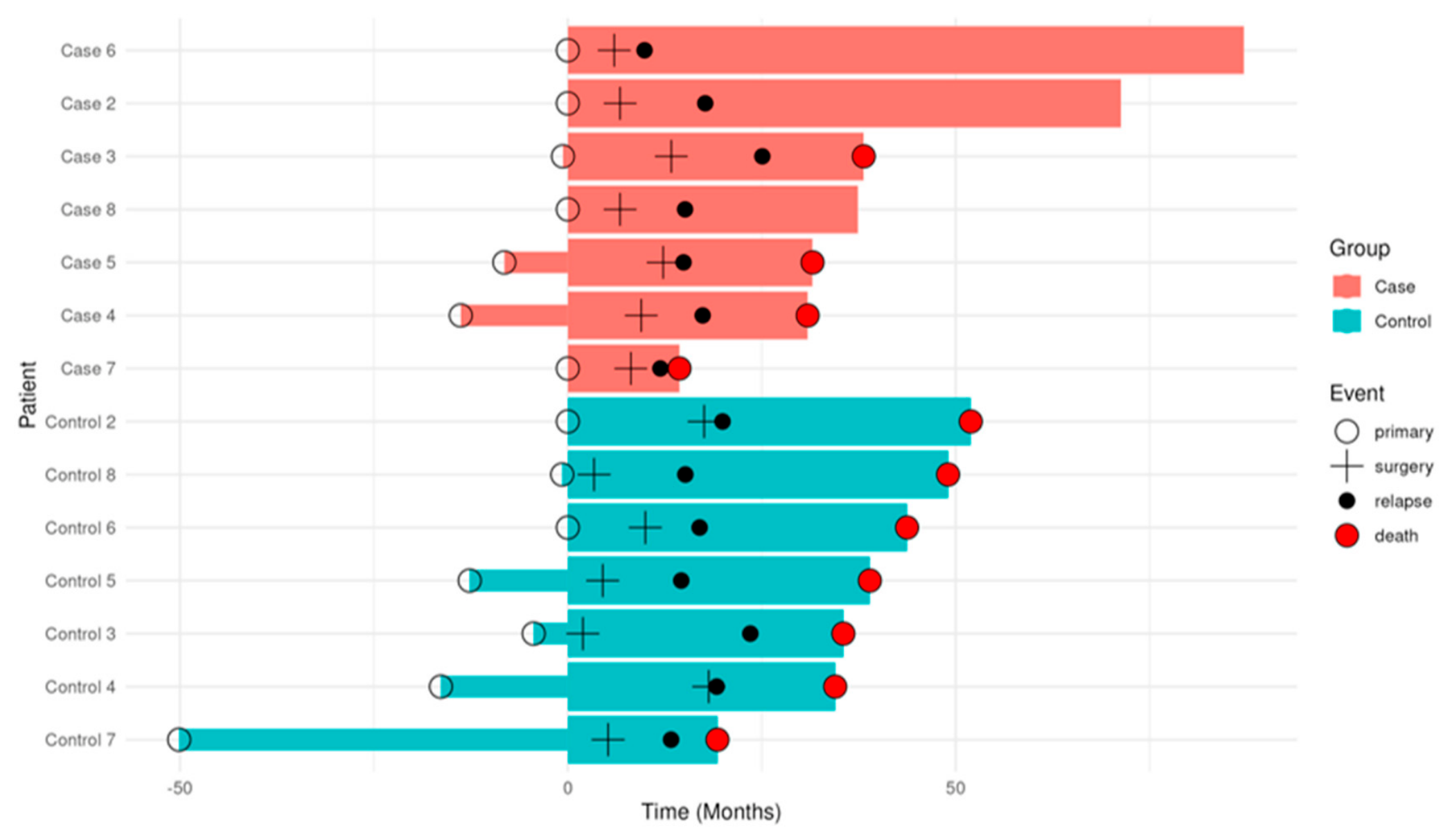
Figure 2.
TMB values per sample, with “Pre CTx” denoting the tissue specimen obtained prior to initiation of chemotherapy (mCRC diagnosis), and “Post CTx” denoting the surgical specimen collected following chemotherapy treatment.
Figure 2.
TMB values per sample, with “Pre CTx” denoting the tissue specimen obtained prior to initiation of chemotherapy (mCRC diagnosis), and “Post CTx” denoting the surgical specimen collected following chemotherapy treatment.
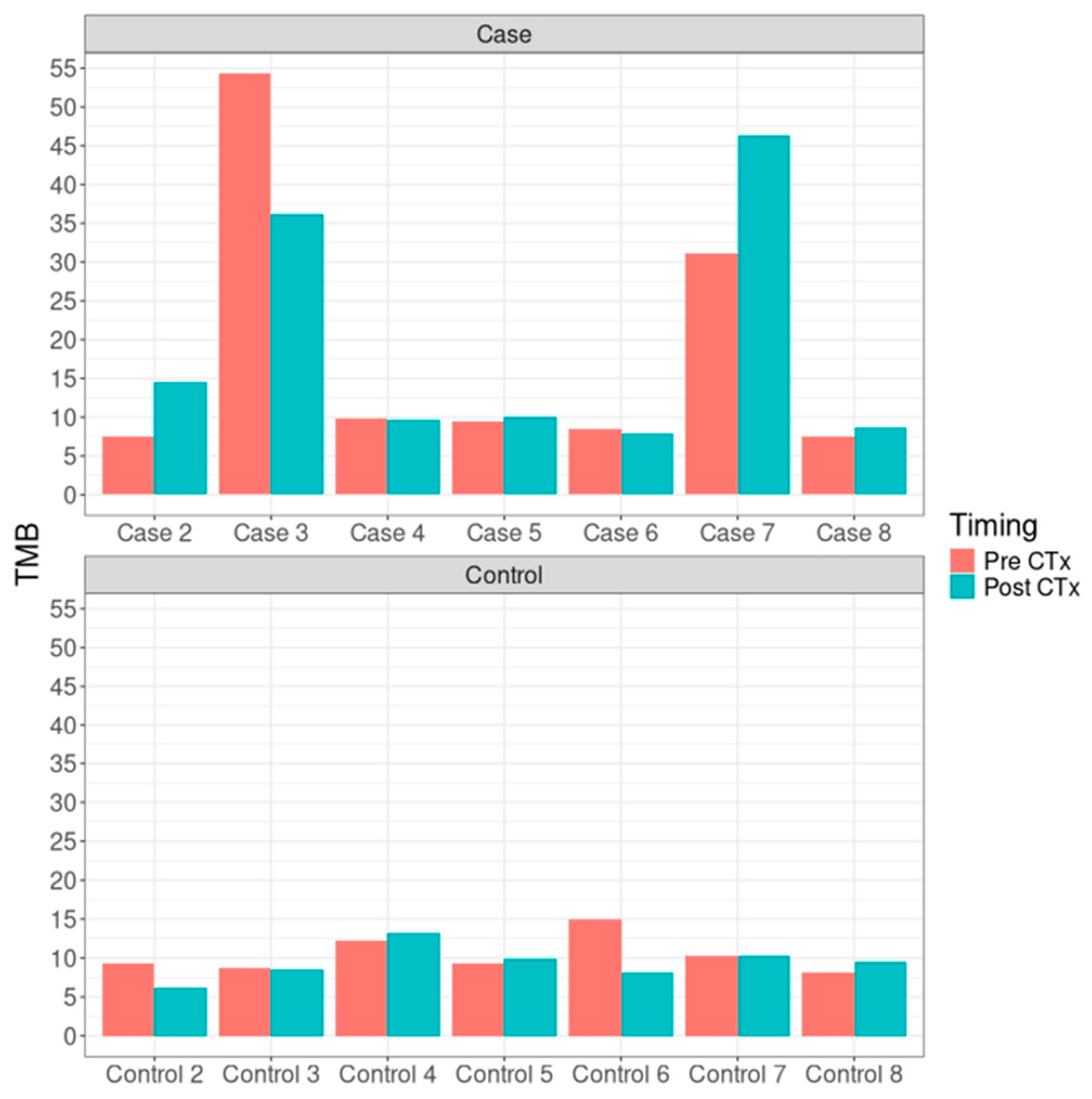
Figure 3.
Statistically significant gain and loss of mutations (chi-square analysis) observed differentially in the Case and Control groups. This analysis encompasses a comprehensive range of genetic alterations.
Figure 3.
Statistically significant gain and loss of mutations (chi-square analysis) observed differentially in the Case and Control groups. This analysis encompasses a comprehensive range of genetic alterations.
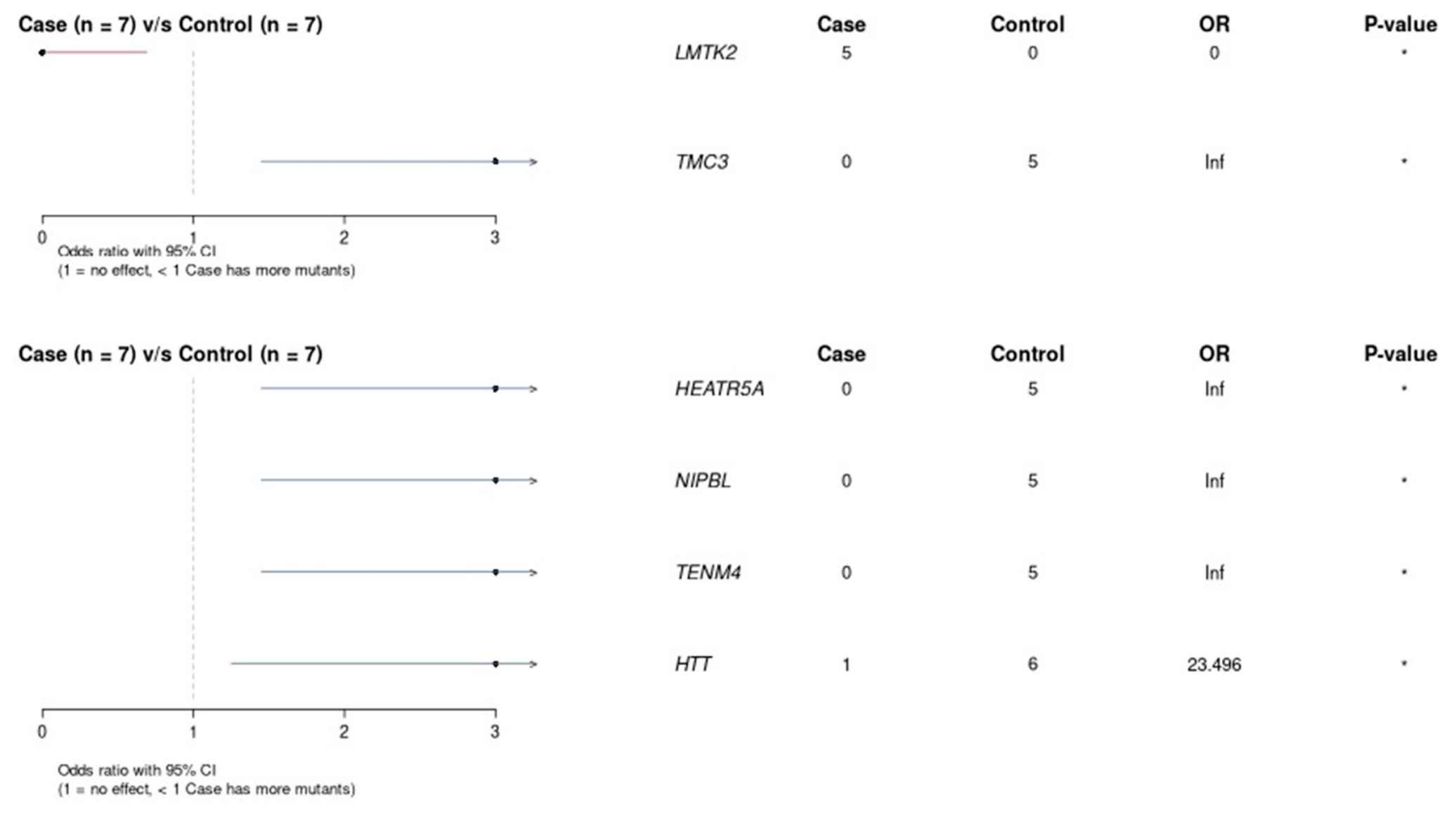
Figure 4.
Median number of pathogenic mutations, as well as those classified as high-impact by the VEP v.109 program, displayed across the groups on the X-axis.
Figure 4.
Median number of pathogenic mutations, as well as those classified as high-impact by the VEP v.109 program, displayed across the groups on the X-axis.
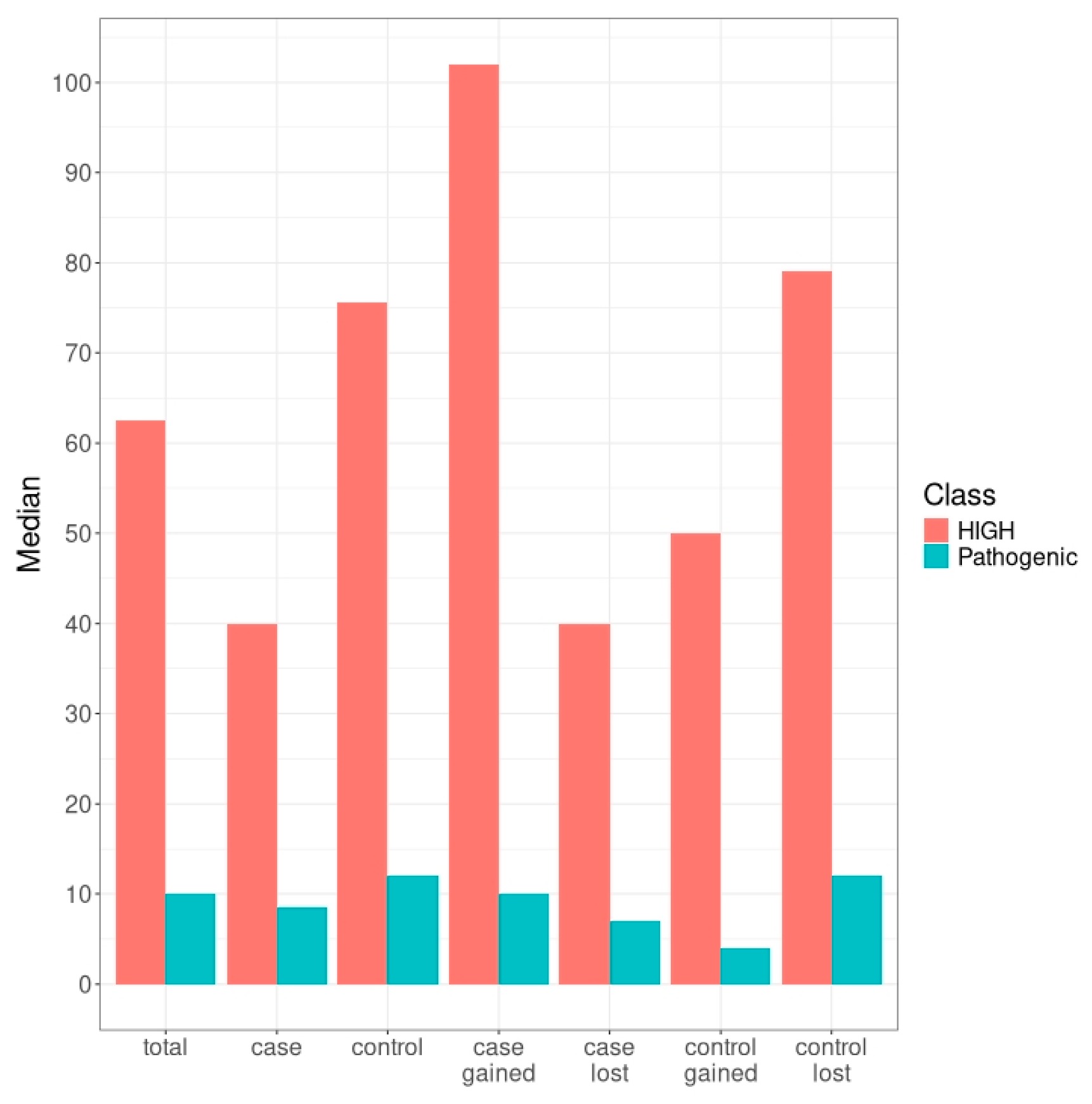
Figure 5.
Oncoplot of the most frequently mutated genes based on the gain of pathogenic mutations (A) and the loss of pathogenic mutations (B).
Figure 5.
Oncoplot of the most frequently mutated genes based on the gain of pathogenic mutations (A) and the loss of pathogenic mutations (B).
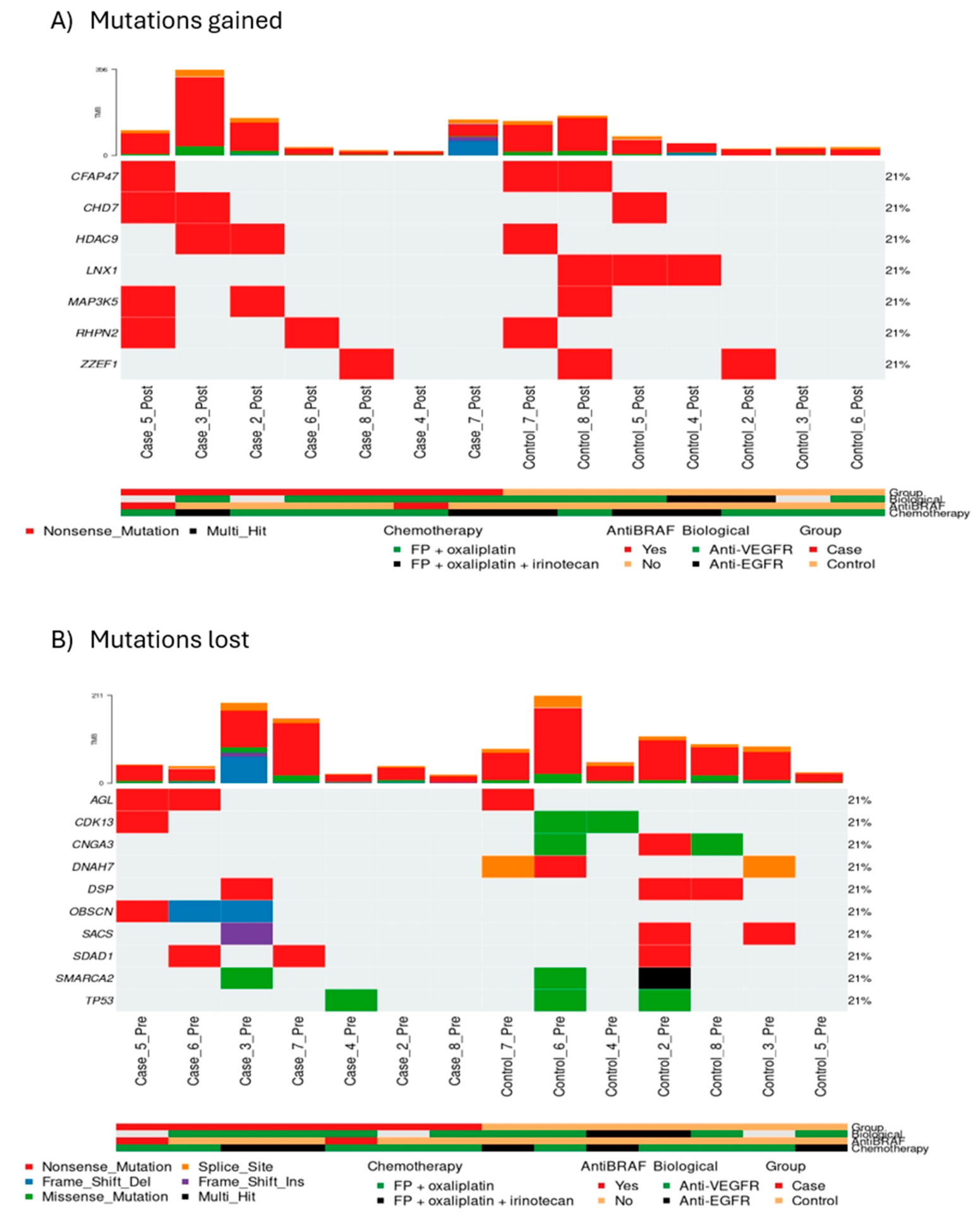
Figure 6.
Most frequently mutated genes in the control and case groups, as identified by differential mutation (gain or loss of pathogenic mutations following treatment).
Figure 6.
Most frequently mutated genes in the control and case groups, as identified by differential mutation (gain or loss of pathogenic mutations following treatment).
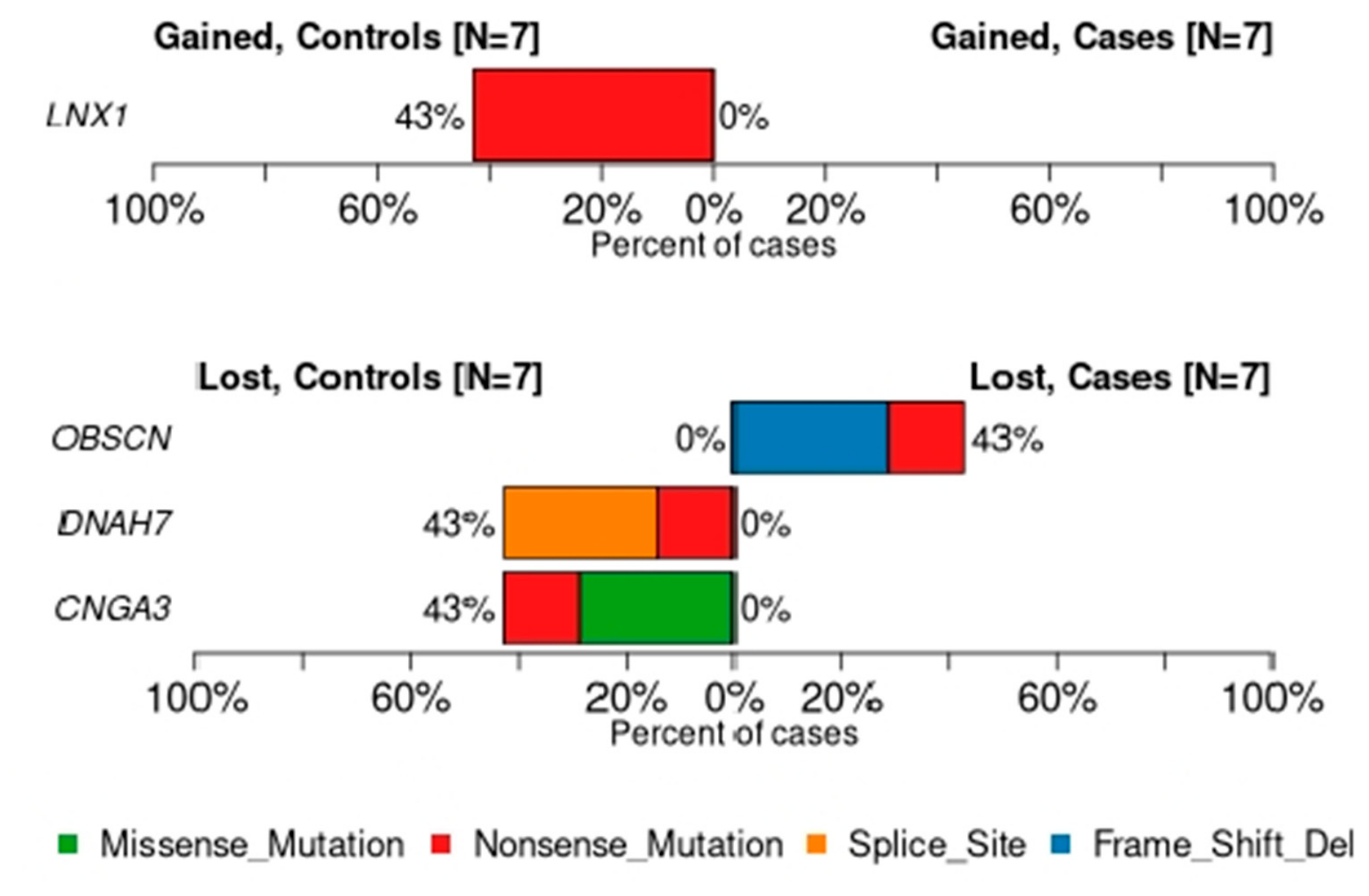

Table 1.
Clinical characteristics of the overall population, with the data stratified by Case and Control groups. The p-values for the variable associations were assessed by the chi-square test.
Table 1.
Clinical characteristics of the overall population, with the data stratified by Case and Control groups. The p-values for the variable associations were assessed by the chi-square test.
| Name | Detail | All | Case | Control | Χ2 |
| Group | Case | 7 (50)% | 7 (100)% | 0 (0)% | <0.01 |
| Control | 7 (50)% | 0 (0)% | 7 (100)% | ||
| Sex | Female | 8 (57)% | 4 (57)% | 4 (57)% | 1 |
| Male | 6 (43)% | 3 (43)% | 3 (43)% | ||
| Number of comorbidities | 0 | 3 (21)% | 1 (14)% | 2 (29)% | 0.86 |
| 1 | 2 (14)% | 1 (14)% | 1 (14)% | ||
| 2 | 6 (43)% | 3 (43)% | 3 (43)% | ||
| 3 | 2 (14)% | 1 (14)% | 1 (14)% | ||
| 4 | 1 (7)% | 1 (14)% | 0 (0)% | ||
| Histology | Adenocarcinoma | 8 (57)% | 3 (43)% | 5 (71)% | 0.61 |
| Mixed adenocarcinoma | 2 (14)% | 1 (14)% | 1 (14)% | ||
| Mucinous adenocarcinoma | 3 (21)% | 2 (29)% | 1 (14)% | ||
| Mucinous adenocarcinoma + signet ring cells | 1 (7)% | 1 (14)% | 0 (0)% | ||
| Histologic grade | Poorly differentiated | 4 (29)% | 2 (29)% | 2 (29)% | 0.79 |
| Moderately differentiated | 7 (50)% | 4 (57)% | 3 (43)% | ||
| Well differentiated | 3 (21)% | 1 (14)% | 2 (29)% | ||
| Tumor regression after treatment | Major tumor regression | 2 (14)% | 1 (14)% | 1 (14)% | 0.82 |
| Minor tumor regression | 3 (21)% | 3 (43)% | 0 (0)% | ||
| NA | 9 (64)% | 3 (43)% | 6 (86)% | ||
| BRAF mutation | Not mutated/Wild-type | 7 (50)% | 0 (0)% | 7 (100)% | <0.01 |
| V600E | 7 (50)% | 7 (100)% | 0 (0)% | ||
| HER2 overexpression | Negative | 2 (14)% | 1 (14)% | 1 (14)% | 1 |
| Positive (1+) | 1 (7)% | 1 (14)% | 0 (0)% | ||
| NA | 11 (79)% | 5 (71)% | 6 (86)% | ||
| Microsatellite instability | No | 9 (64)% | 4 (57)% | 5 (71)% | 0.52 |
| Yes | 2 (14)% | 2 (29)% | 0 (0)% | ||
| NA | 3 (21)% | 1 (14)% | 2 (29)% | ||
| RAS mutation | No | 13 (93)% | 6 (86)% | 7 (100)% | 1 |
| Yes | 1 (7)% | 1 (14)% | 0 (0)% | ||
| Stage at diagnosis | II | 1 (7)% | 0 (0)% | 1 (14)% | 0.43 |
| III | 5 (36)% | 2 (29)% | 3 (43)% | ||
| IV | 8 (57)% | 5 (71)% | 3 (43)% | ||
| Primary tumor side | Left | 5 (36)% | 2 (29)% | 3 (43)% | 1 |
| Right | 9 (64)% | 5 (71)% | 4 (57)% | ||
| Primary tumor surgery | No | 0 (0)% | 0 (0)% | 0 (0)% | - |
| Yes | 14 (100)% | 7 (100)% | 7 (100)% | ||
| Time at primary tumor surgery | Before metastases surgery | 9 (64)% | 4 (57)% | 5 (71)% | 0.57 |
| Same time of metastases surgery | 4 (29)% | 2 (29)% | 2 (29)% | ||
| After metastases surgery | 1 (7)% | 1 (14)% | 0 (0)% | ||
| Lung metastases | No | 14 (100)% | 7 (100)% | 7 (100)% | - |
| Yes | 0 (0)% | 0 (0)% | 0 (0)% | ||
| Liver metastases | No | 5 (36)% | 3 (43)% | 2 (29)% | 1 |
| Yes | 9 (64)% | 4 (57)% | 5 (71)% | ||
| Peritoneal metastases | No | 8 (57)% | 4 (57)% | 4 (57)% | 1 |
| Yes | 6 (43)% | 3 (43)% | 3 (43)% | ||
| Metastases surgery | No | 0 (0)% | 0 (0)% | 0 (0)% | - |
| Yes | 14 (100)% | 7 (100)% | 7 (100)% | ||
| Type of metastases surgery | Liver | 8 (57)% | 4 (57)% | 4 (57)% | 1 |
| Peritoneal | 6 (43)% | 3 (43)% | 3 (43)% | ||
| Resection | R0 | 12 (86)% | 6 (86)% | 6 (86)% | 0.37 |
| R1 | 1 (7)% | 1 (14)% | 0 (0)% | ||
| R2 | 1 (7)% | 0 (0)% | 1 (14)% | ||
| Type of chemotherapy | Fluoropyrimidine + oxaliplatin | 9 (64)% | 5 (71)% | 4 (57)% | 1 |
| Fluoropyrimidine + oxaliplatin + irinotecan | 5 (36)% | 2 (29)% | 3 (43)% | ||
| Biological treatment | Aflibercept | 1 (7)% | 0 (0)% | 1 (14)% | 0.18 |
| Anti-EGFR | 2 (14)% | 0 (0)% | 2 (29)% | ||
| Bevacizumab | 8 (57)% | 5 (71)% | 3 (43)% | ||
| NA | 3 (21)% | 2 (29)% | 1 (14)% | ||
| Anti BRAF treatment | No | 12 (86)% | 5 (71)% | 7 (100)% | 0.45 |
| Yes | 2 (14)% | 2 (29)% | 0 (0)% | ||
| Immunotherapy | No | 14 (100)% | 7 (100)% | 7 (100)% | - |
| Yes | 0 (0)% | 0 (0)% | 0 (0)% | ||
| ECOG | 0 | 7 (50)% | 4 (57)% | 3 (43)% | 1 |
| 1 | 7 (50)% | 3 (43)% | 4 (57)% | ||
| Site of relapse after metastases surgery | Liver | 3 (21)% | 1 (14)% | 2 (29)% | 0.6 |
| Liver and lung | 2 (14)% | 1 (14)% | 1 (14)% | ||
| Liver, spleen, lymph nodes | 1 (7)% | 0 (0)% | 1 (14)% | ||
| Lung | 3 (21)% | 2 (29)% | 1 (14)% | ||
| Lymph nodes | 1 (7)% | 0 (0)% | 1 (14)% | ||
| Peritoneal | 4 (29)% | 3 (43)% | 1 (14)% | ||
| Last contact status | Alive with active disease | 3 (21)% | 3 (43)% | 0 (0)% | 0.19 |
| Exitus | 11 (79)% | 4 (57)% | 7 (100)% | ||
| Cause of death | Disease progression | 13 (93)% | 7 (100)% | 6 (86)% | 1 |
| Infection | 1 (7)% | 0 (0)% | 1 (14)% |
Table 2.
Key differentially mutated genes based on sample group and metastatic site (hepatic vs. peritoneal).
Table 2.
Key differentially mutated genes based on sample group and metastatic site (hepatic vs. peritoneal).
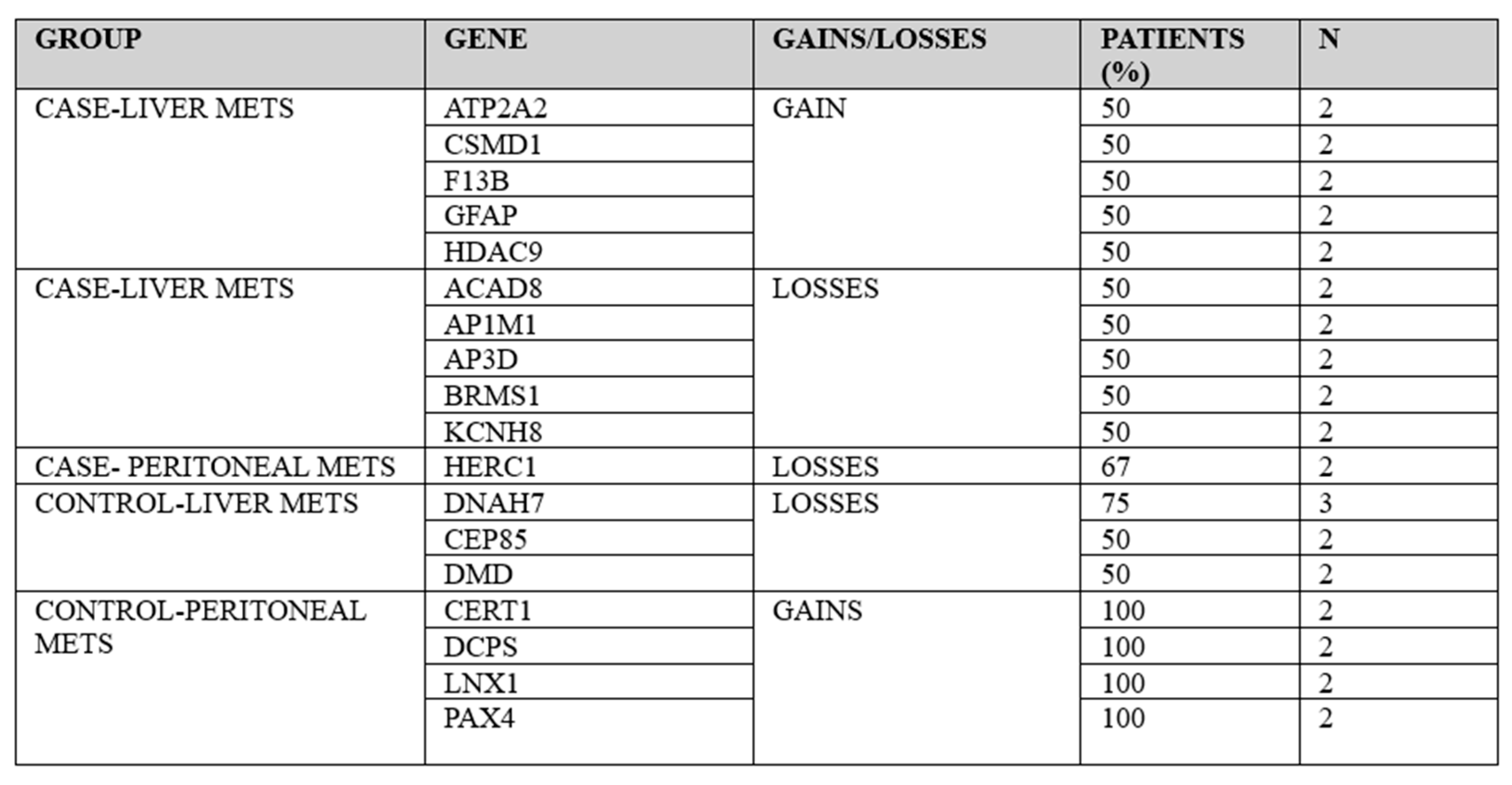 |
Table 3.
Key differentially mutated genes according to sample group and primary tumor sidedness (right vs. left).
Table 3.
Key differentially mutated genes according to sample group and primary tumor sidedness (right vs. left).
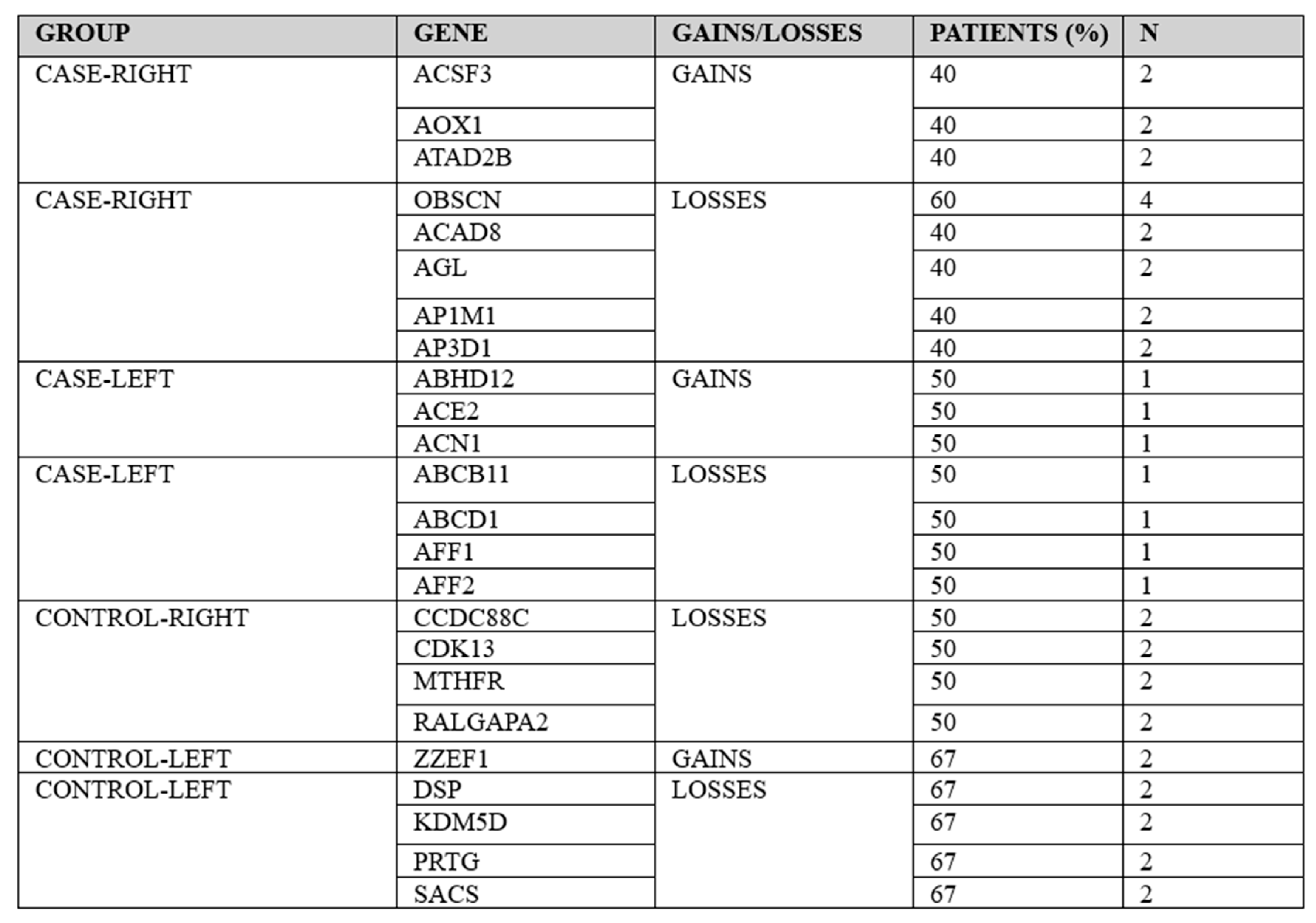 |
Table 4.
Key differentially mutated genes based on Anti-BRAF treatment status (treated, n = 2 vs. untreated, n = 12).
Table 4.
Key differentially mutated genes based on Anti-BRAF treatment status (treated, n = 2 vs. untreated, n = 12).
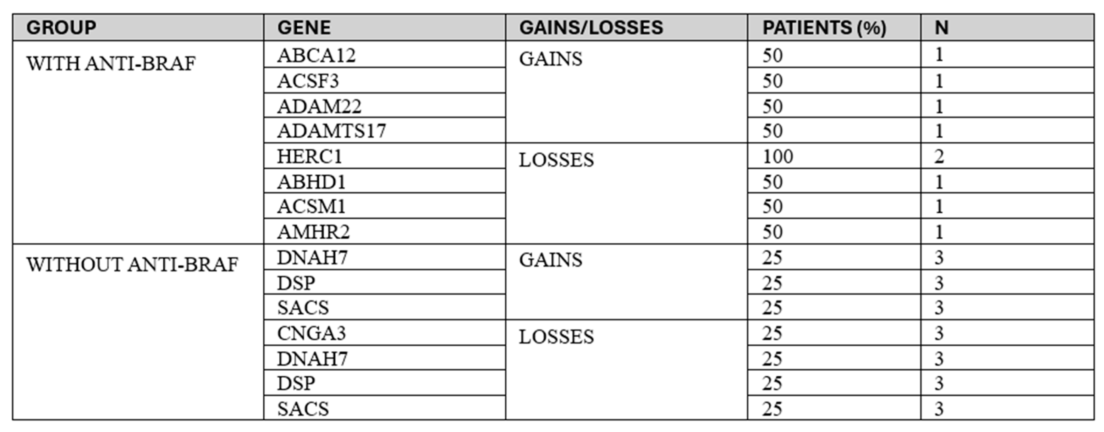 |
Table 5.
Median overall survival (OS) and event-free survival (EFS), measured in months, in the study population.
Table 5.
Median overall survival (OS) and event-free survival (EFS), measured in months, in the study population.
 |
Disclaimer/Publisher’s Note: The statements, opinions and data contained in all publications are solely those of the individual author(s) and contributor(s) and not of MDPI and/or the editor(s). MDPI and/or the editor(s) disclaim responsibility for any injury to people or property resulting from any ideas, methods, instructions or products referred to in the content. |
© 2026 by the authors. Licensee MDPI, Basel, Switzerland. This article is an open access article distributed under the terms and conditions of the Creative Commons Attribution (CC BY) license (http://creativecommons.org/licenses/by/4.0/).
Copyright: This open access article is published under a Creative Commons CC BY 4.0 license, which permit the free download, distribution, and reuse, provided that the author and preprint are cited in any reuse.