
Submitted:
07 September 2026
Posted:
08 September 2026
You are already at the latest version
Abstract
Background/Objectives: Cancer remains a leading cause of mortality worldwide, creating a persistent clinical need for accurate and minimally invasive approaches capable of detecting malignancy at earlier and more treatable stages. Circulating cell-free DNA (cfDNA) has emerged as a transformative liquid-biopsy analyte that provides information beyond somatic mutation detection. This review examines three complementary dimensions of cfDNA analysis-fragmentomics, methylomics, and CRISPR-enabled biosensing-and evaluates their potential roles in cancer detection, classification, and monitoring. Methods: This narrative review synthesizes current evidence on cfDNA fragmentomic features, including fragment size distribution, end motifs, jagged ends, nucleosome footprinting, orientation-aware fragmentation, and machine-learning-based classification. It also reviews bisulfite-based and bisulfite-free methylation profiling, multi-cancer early detection strategies, nanopore-based methylation analysis, and CRISPR-Cas systems adapted for sequence- and methylation-sensitive nucleic acid biosensing. Results: Fragmentomic approaches exploit structural characteristics of cfDNA that reflect chromatin organization, nuclease activity, and tissue of origin, while methylomic analyses provide highly tissue-specific epigenetic information relevant to early cancer detection and tumor localization. CRISPR-Cas12a-, Cas13a-, and related platforms provide programmable, highly specific detection of nucleic acid variants and selected epigenetic modifications with potential for rapid and instrument-minimal testing. Clinical applications across colorectal, lung, breast, hepatocellular, pancreatic, gastric, esophageal, and bladder cancers demonstrate complementary roles for these approaches in early detection, minimal residual disease monitoring, relapse detection, and treatment-response assessment. Conclusions: Fragmentomics, methylomics, and CRISPR-enabled biosensing represent complementary rather than interchangeable strategies for cfDNA analysis. Their broader clinical integration will depend on analytical standardization, prospective validation in diverse populations, improved sensitivity at low tumor fractions, and resolution of regulatory and implementation challenges.
Keywords:
cell‐free DNA (cfDNA)
; liquid biopsy
; fragmentomics
; DNA methylation
; CRISPR Cas biosensors
; minimal residual disease (MRD)
; early cancer detection
1. Introduction
Despite decades of progress in oncology, the majority of cancer related deaths continue to result from disease that is diagnosed at an advanced or metastatic stage, when curative treatment options are limited [1]. The development of tools capable of detecting malignant disease earlier, monitoring its evolution over time, and assessing residual disease burden after therapy has therefore become a central priority in translational cancer research [1]. Liquid biopsy, the analysis of tumor derived material circulating in accessible body fluids such as blood plasma, has emerged over the past two decades as a minimally invasive alternative and complement to conventional tissue biopsy [2].
Among the analytes accessible through liquid biopsy, circulating cfDNA has attracted particular attention because it captures both genetic and epigenetic information shed by tumor cells into the bloodstream [2]. CfDNA is not a homogeneous molecule but rather a heterogeneous population of fragments whose length, end sequence, chromatin context, and cytosine modification status are shaped by the biological processes that generated them [3]. Recent work has demonstrated that these structural and epigenetic features, collectively termed fragmentomics and methylomics, carry diagnostic information that extends well beyond the simple presence or absence of somatic mutations [3].
The clinical relevance of these signals has been demonstrated at scale through multi-cancer early detection programs, in which cancer-associated methylation patterns in plasma cell-free DNA are used to simultaneously screen for multiple cancer types and predict the most likely tissue of origin [4]. Complementary clinical validation studies have confirmed that targeted methylation based classifiers can detect and localize multiple cancer types from a single blood draw while maintaining high specificity, reinforcing the diagnostic value of the methylomic layer of circulating cfDNA [5].
While the interpretation of natural cfDNA signals has rapidly advanced, programmable nuclease systems derived from bacterial CRISPR Cas adaptive immunity have been repurposed as highly sensitive and specific biosensing platforms for nucleic acid detection [6]. Collateral cleavage activities exhibited by certain Cas enzymes upon target recognition have enabled the design of diagnostic assays capable of detecting single nucleotide variants (SNV) at low input concentrations with minimal instrumentation [6]. Related platforms exploiting the collateral single-stranded DNase activity of Cas12a enzymes have similarly been applied to nucleic acid diagnostics, broadening the toolkit of CRISPR-based biosensors now being adapted for circulating tumor DNA (ctDNA) and methylation analysis [7].
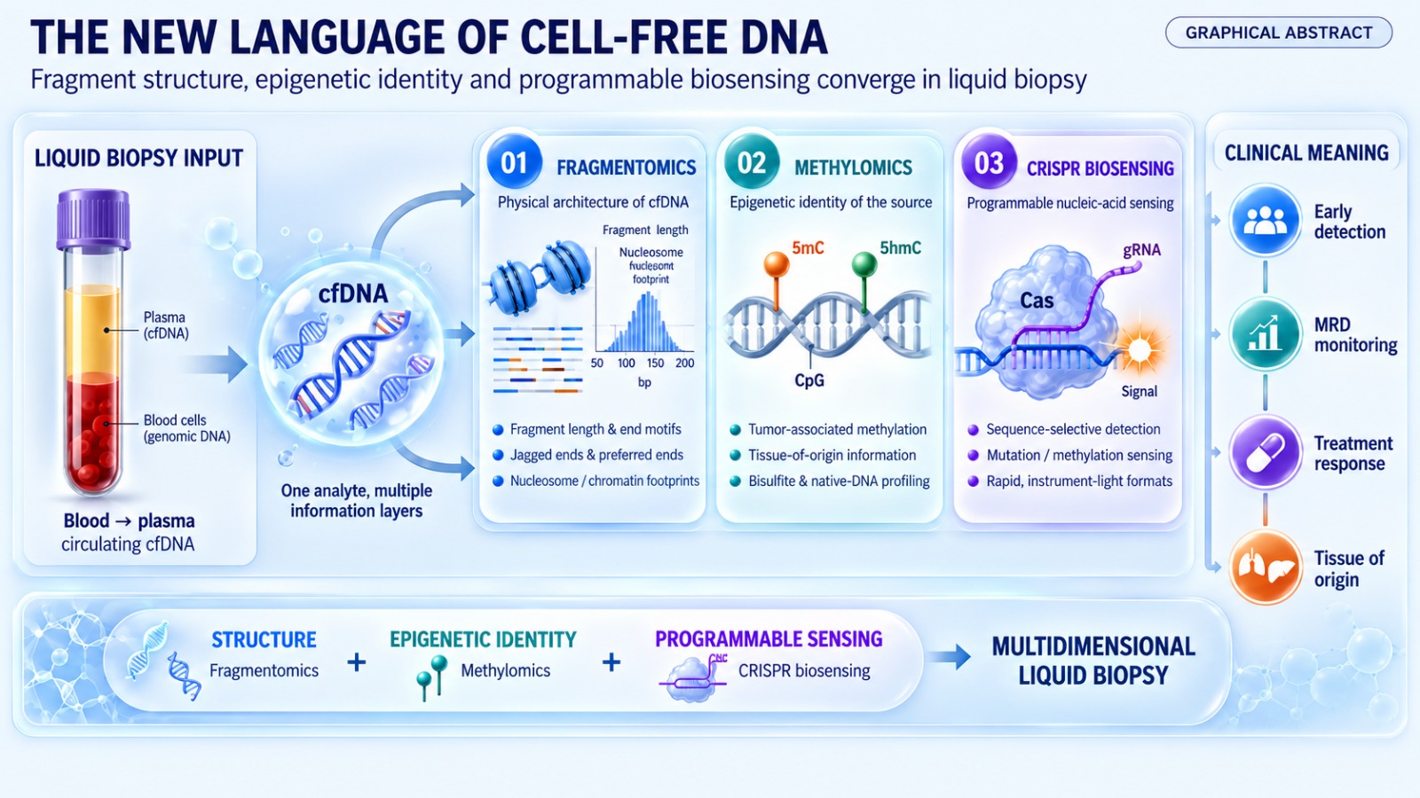
This review integrates these three converging dimensions of cfDNA analysis, namely fragmentomics, methylomics, and CRISPR-enabled biosensing, into a single framework for understanding their individual contributions and combined potential in cancer detection and monitoring. Following an overview of the biological origins of circulating cfDNA, the review examines structural fragmentomic signatures, epigenetic methylation based approaches, and emerging CRISPR-based detection platforms in turn, before considering their clinical application across major cancer types and the challenges that remain before widespread implementation in routine oncological practice.
2. The Biology and Origins of Circulating Cell-Free DNA
The presence of nucleic acids in human blood plasma was first reported in 1948, when free nucleic acids circulating outside of intact cells in human blood were described, laying the earliest foundation for what would later become the field of liquid biopsy [8].
Nearly three decades later, elevated concentrations of free DNA were observed in the serum of patients with cancer relative to healthy individuals, providing the first evidence that circulating nucleic acids might carry disease relevant information [9].
Following these early observations, researchersdemonstrated that DNA could be isolated and characterized from the plasma of patients with cancer, further establishing circulating DNA as a biologically measurable component of the cancer-associated bloodstream [10]. The mechanisms responsible for the release of DNA into the circulation were later clarified through studies showing that circulating cfDNA is predominantly generated through apoptosis, with a smaller contribution from necrosis, and that this apoptotic origin imprints a characteristic fragmentation pattern onto the released molecules [11].
In healthy individuals, the majority of circulating cell-free DNA is thought to originate from hematopoietic cell turnover, whereas in patients with cancer a variable fraction, referred to as ctDNA, is released from malignant cells predominantly through apoptosis and necrosis, with additional contributions from active secretion through extracellular vesicles and other nucleic acid-containing complexes [12]. A parallel and highly influential line of investigation began with the discovery that cell free fetal DNA circulates in maternal plasma during pregnancy, a finding that not only enabled the development of noninvasive prenatal testing but also established many of the analytical principles, including size based fractionation and targeted sequencing approaches, that would later be adapted for cancer detection [13].
The concentration of circulating cfDNA is normally low, but it rises substantially in patients with cancer, inflammatory conditions, trauma, and other states associated with increased cell death, a phenomenon partly attributable to greater release of high molecular weight genomic DNA from necrotic and incompletely processed cellular material, a feature captured by measures of plasma DNA integrity [14].
The clinical utility of detecting tumor derived alterations within this circulating pool was firmly established when broad genomic profiling demonstrated that ctDNA could be identified across the majority of common epithelial cancer types, with detection rates that increased with advancing disease stage while remaining measurable in a meaningful proportion of early stage and localized tumors [15].
A pivotal advance in understanding the structural organization of circulating cfDNA came with the recognition that its fragmentation is not random but instead reflects the nucleosomal packaging of chromatin within the cell of origin, such that mapping the genome wide pattern of fragment start and end positions in plasma can reveal footprints of nucleosome occupancy and transcription factor binding characteristic of the contributing tissue [16]. This observation, discussed further in Section 3, established the conceptual bridge between the biology of cfDNA release and the structural discipline now known as fragmentomics [16].
A chronological summary of these foundational discoveries, from the first description of circulating nucleic acids to the identification of the nucleosome footprint, is presented in Table 1. The principal cellular mechanisms responsible for the release of cfDNA into the circulation are summarized schematically in Figure 1.
3. Fragmentomics: The Structural Language of cfDNA
The term fragmentomics refers to the systematic study of the size, end sequence, and positional characteristics of cfDNA fragments as a source of biological and diagnostic information [3]. Because the enzymatic processes that generate circulating cfDNA are influenced by chromatin accessibility, nuclease activity, and cell type specific nucleosome positioning, the resulting fragmentation pattern encodes information about both the tissue of origin and the pathological state of the contributing cells [16].
3.1. Fragment Size and Length Distribution
Circulating cfDNA in healthy individuals is predominantly composed of fragments approximately one hundred and sixty six base pairs in length, corresponding to the length of DNA wrapped around a single nucleosome core particle together with its associated linker region, with a characteristic ten base pair periodicity reflecting the helical pitch of DNA as it winds around the histone octamer [16].
Fragments derived from tumor cells tend to be systematically shorter than those originating from nonmalignant hematopoietic turnover, a difference attributed to altered chromatin compaction and nuclease accessibility within malignant nuclei [17].
This size difference has been exploited analytically by selectively enriching for shorter fragments prior to sequencing, an approach shown to enhance the sensitivity of ctDNA detection across multiple cancer types without requiring an increase in sequencing depth [18].
Consistent with these findings, plasma DNA fragment size profiles have also been reported to become progressively shorter in patients with hepatocellular carcinoma as tumor burden increases, and to normalize following curative treatment, supporting the use of fragment length as a dynamic biomarker of disease activity [19].
3.2. DNA End Motifs and Jagged Ends
In addition to fragment length, the sequence context immediately flanking the cleavage sites at the ends of cfDNA fragments, referred to as end motifs, has been shown to differ systematically between healthy individuals and patients with cancer, reflecting differences in the nuclease activities responsible for fragment generation [20].
In addition to blunt double-stranded ends, a substantial proportion of plasma DNA molecules carry short single-stranded protrusions known as jagged ends, which arise from nuclease mediated trimming of nucleosome protected DNA and whose length and frequency vary with both physiological state and malignancy [21].
Preferred fragmentation end coordinates, meaning genomic positions at which cfDNA molecules are cleaved with higher than expected frequency, have similarly been shown to carry information about the tissue and disease state of origin, with a distinct set of preferred ends identified in the plasma of patients with hepatocellular carcinoma relative to healthy controls [22].
Analogous preferred end signatures have been described in the context of pregnancy, where fetal derived fragments display characteristic end coordinates distinguishable from maternal background DNA, illustrating that the fragmentomic principles informing prenatal diagnostics are directly transferable to oncology [23].
The abundance of jagged ends on circulating DNA molecules has also been linked to nuclease activity in autoimmune conditions such as systemic lupus erythematosus, demonstrating that end structure fragmentomic signals reflect nuclease biology more broadly and are not restricted to malignancy alone, a consideration relevant to the specificity of cancer focused fragmentomic assays [24].
End motif and jagged end analyses have since been extended beyond plasma to urinary cfDNA, where characterization of fragment end structures has demonstrated feasibility for the noninvasive detection of bladder cancer directly from voided urine samples [25].
Genome wide analysis of aberrant fragment end position and sequence has further shown that these signals can be integrated into composite scores capable of distinguishing patients with cancer from healthy individuals with performance comparable to more established genomic biomarkers [26].
Representative differences in fragment length, end motif frequency, and jagged end proportion between cfDNA of nonmalignant and tumor origin are illustrated in Figure 2.
3.3. Nucleosome Footprinting and Chromatin Accessibility
Because nucleosome occupancy protects the underlying DNA from nuclease digestion, the density of cfDNA fragment coverage across the genome reflects the positioning of nucleosomes within the chromatin of the cells that contributed the DNA, allowing genome wide nucleosome occupancy maps to be reconstructed directly from plasma sequencing data [16]. Regions of open chromatin corresponding to actively transcribed genes and transcription factor binding sites display reduced nucleosome protection and are therefore associated with shorter, more fragmented cfDNA, enabling inference of transcription factor occupancy and gene expression activity from plasma sequencing without the need for tissue biopsy [27].
Complementary approaches combining chromatin immunoprecipitation with plasma cfDNA sequencing have been used to directly identify histone modification patterns characteristic of specific cell types and gene expression programs, extending nucleosome footprinting toward direct epigenomic profiling of the cells of origin [28]. Building on these principles, computational frameworks have been developed to infer relative gene expression levels from the depth and shape of fragmentation coverage across gene bodies and regulatory regions, providing a noninvasive proxy for transcriptional activity within tumors that is not directly accessible through mutation based liquid biopsy assays [29].
3.4. Orientation Aware Fragmentation and Genome Wide Profiling
Orientation-aware analysis represents another major step forward, considering not only the position but also the orientation of cfDNA fragment ends relative to regulatory elements, an approach termed orientation aware cfDNA fragmentation analysis, which has been shown to more accurately infer the tissue of origin of circulating DNA within open chromatin regions than positional coverage alone [30].
Extending this concept genome wide, epigenetic fragmentomic profiling that integrates fragment size, end position, and coverage across the entire genome has been used to reconstruct tissue specific chromatin accessibility maps from plasma alone, achieving resolution sufficient to distinguish contributions from multiple normal and malignant tissue types simultaneously [31].
3.5. Machine Learning Enabled Fragmentomic Classifiers
The recognition that fragmentation patterns differ systematically between healthy individuals and patients with cancer motivated the development of genome wide fragmentation profiling combined with machine learning classification, an approach in which plasma samples from patients with various cancer types were shown to display altered genome wide fragmentation profiles relative to healthy controls with high overall classification accuracy [32].
This fragmentomic classification approach has since been adapted to specific cancer types, including a lung cancer detection model that combined genome wide fragmentation features with clinical risk factors to improve discrimination between patients with lung cancer and high risk individuals without malignancy [33].
A comparable fragmentome based classifier developed for hepatocellular carcinoma demonstrated that combining fragmentation features with conventional biomarkers improved detection sensitivity relative to either approach alone, particularly among patients with early stage or small volume tumors [34].
Multidimensional fragmentomic models incorporating fragment size, end motif, and copy number derived features have similarly been applied to colorectal cancer, where such models achieved high sensitivity for both early stage cancer and advanced precancerous lesions while maintaining specificity comparable to established screening modalities [35].
Deep learning architectures trained directly on end motif frequency distributions, without requiring extensive manual feature engineering, have further demonstrated that neural network based classifiers can achieve diagnostic performance comparable to conventional machine learning approaches while simplifying the analytical pipeline required for clinical implementation [36].
Beyond diagnosis, fragmentomic profiling has also shown promise for treatment monitoring, with longitudinal changes in genome wide fragmentation patterns shown to track with response to systemic therapy and, in some patients, to precede radiographic evidence of disease progression [37].
These findings indicate that the physical structure of cfDNA, spanning fragment length, end sequence, nucleosome footprint, and genome wide fragmentation pattern, constitutes an information rich signal that is complementary to, and in some contexts more sensitive than, mutation based ctDNA detection, particularly in early stage disease where tumor derived mutant allele fractions are frequently below the limit of detection of sequencing based assays [3].
A comparative summary of representative fragmentomic platforms discussed above, together with the fragmentomic feature exploited and the cancer types studied, is provided in Table 2.
The generalized analytical workflow underlying these machine learning based fragmentomic classifiers, from plasma collection through feature extraction to cancer probability output, is depicted in Figure 3.
4. Methylomics: The Epigenetic Language of cfDNA
In addition to its structural fragmentomic signature, circulating cfDNA carries the epigenetic imprint of the cells from which it originated, most notably in the pattern of cytosine methylation across the genome, a heritable and reversible chemical modification that regulates gene expression without altering the underlying DNA sequence [38].
Cancer cells characteristically display a combination of global genome wide hypomethylation and focal hypermethylation at the promoter regions of tumor suppressor genes, a pattern that differs substantially from that observed in normal somatic tissue and that is preserved, at least in part, in the cfDNA shed by tumor cells into the circulation [38].
4.1. Biological Basis and Detection Principles
Because methylation patterns are established early in development and are highly tissue specific, the methylome of circulating cfDNA can, in principle, be used both to detect the presence of a malignant clone and to infer the tissue in which that clone originated, a property that is more difficult to achieve using somatic mutation profiles alone, which are frequently shared across tissue types [39].
Tissue specific methylation signals in plasma were first exploited systematically through genome wide bisulfite sequencing approaches capable of mapping the tissue of origin of circulating DNA in the contexts of pregnancy, cancer, and solid organ transplantation, establishing methylation deconvolution as a broadly applicable framework for noninvasive tissue mapping [40].
4.2. Detection Technologies: Bisulfite-Based and Bisulfite Free Approaches
The most widely used method for interrogating cfDNA methylation remains sodium bisulfite conversion, in which unmethylated cytosines are chemically deaminated to uracil while methylated cytosines remain unchanged, allowing methylation status to be inferred from the resulting sequence following polymerase chain reaction (PCR) amplification and sequencing [41].
Because bisulfite conversion is associated with DNA degradation and loss of sequence complexity, methods that improve the efficiency of methylation marker recovery from low input, fragmented cfDNA, including strategies that simultaneously interrogate the sense and antisense strands generated during conversion, have been developed to increase the sensitivity of methylation based assays performed on limited plasma volumes [42].
Digital methylation profiling approaches that quantify methylation heterogeneity at the level of individual DNA molecules, rather than relying on bulk averaged methylation ratios, have further been shown to improve the sensitivity and cost effectiveness of cancer detection from low volume liquid biopsies [43].
An emerging alternative to bisulfite-based approaches involves the detection of five hydroxymethylcytosine (5hmC), an oxidized derivative of methylcytosine that is enriched in actively transcribed gene bodies and that can be profiled without the harsh chemical conversion required for conventional bisulfite sequencing, an approach that has been analytically validated for the early detection of pancreatic cancer [44].
4.3. Combined Mutation and Methylation Approaches
Because ctDNA mutant allele fractions can be extremely low in early stage disease, combining methylation analysis with conventional mutation detection on the same cfDNA molecules has been shown to improve overall sensitivity for hepatocellular carcinoma detection relative to either modality used alone [45].
4.4. Methylation Based Multi-Cancer Early Detection Platforms
The clinical maturity of methylation based liquid biopsy is most clearly illustrated by the development of multi-cancer early detection tests, which apply targeted methylation sequencing across thousands of genomic regions to simultaneously screen for the presence of dozens of cancer types from a single blood draw and to predict the tissue of origin of any detected signal [4].
In a large scale case control validation study, such a targeted methylation classifier achieved high specificity across cancer types, while sensitivity increased with both disease stage and the biological aggressiveness of the underlying tumor type [5].
Prospective evaluation of methylation based multi-cancer early detection testing within an intended use screening population has since supported the feasibility of implementing this approach at scale, while also highlighting the diagnostic workup burden associated with false positive and indeterminate results that must be addressed before broader population level adoption [4].
A comparative overview of the methylation based platforms discussed in this section, including their detection principle and reported clinical application, is provided in Table 3. The principal tumor associated methylation changes and the corresponding detection workflows are illustrated in Figure 4.
4.5. Nanopore and Single Molecule Methylation Profiling
Third generation long read nanopore sequencing platforms offer a further advantage for methylation analysis because they can detect base modifications directly from native DNA molecules without the need for bisulfite conversion, PCR amplification, or the associated loss of fragment length information [46].
Single molecule nanopore methylation profiling of cfDNA has been used to simultaneously capture methylation status and native fragment length from the same sequencing read, an integration that is difficult to achieve with short read bisulfite sequencing and that may enhance the combined diagnostic value of fragmentomic and methylomic signals [47].
Computational methods that infer both the cell of origin and cancer specific methylation features directly from nanopore sequencing data have further demonstrated that this technology can achieve classification performance approaching that of established short read bisulfite sequencing approaches while substantially reducing sequencing turnaround time [48].
Methylomic approaches to cfDNA analysis complement the structural information captured by fragmentomics, offering high tissue specificity and the capacity for multi-cancer screening from a single assay, and their continued technical refinement, including reduced input requirements and bisulfite free detection chemistries, is expected to further expand their role in early cancer detection and monitoring [3].
5. CRISPR-Enabled Biosensing for Cell-Free DNA Analysis
Distinct from sequencing based approaches, an entirely distinct detection paradigm has emerged from the repurposing of bacterial CRISPR Cas adaptive immune systems as programmable nucleic acid sensors, offering an alternative route to interrogating cfDNA that does not necessarily require next generation sequencing (NGS) infrastructure [6].
5.1. Foundational CRISPR Cas Diagnostic Platforms
Certain Cas enzymes, most notably Cas13a and Cas12a, exhibit a collateral, or trans, cleavage activity, in which recognition of a specific target nucleic acid sequence by the ribonucleoprotein complex triggers indiscriminate cleavage of nearby single-stranded reporter molecules, generating a fluorescent or colorimetric signal proportional to the abundance of the target sequence [6].
This principle was first translated into a diagnostic platform capable of detecting nucleic acids at attomolar concentrations through combination with isothermal preamplification, achieving single nucleotide specificity and multiplexed detection of several targets within a single reaction [6].
The collateral single-stranded DNase activity of the Cas12a enzyme was independently characterized and shown to enable a related diagnostic platform capable of detecting DNA targets, including human papillomavirus sequences, directly from clinical samples, establishing Cas12a based detection as a complementary approach to Cas13a based platforms for DNA target sensing [7].
Subsequent engineering efforts combined multiple Cas enzymes with distinct collateral cleavage preferences within a single multiplexed reaction, enabling simultaneous detection and quantification of several nucleic acid targets and expanding the practical utility of CRISPR-based diagnostics for panels of clinically relevant biomarkers [49].
The core ribonuclease activity underlying Cas13 mediated collateral cleavage was mechanistically characterized through structural and biochemical studies demonstrating that guide RNA processing and target dependent nonspecific RNA cleavage are performed by distinct catalytic domains of the enzyme, providing the biochemical foundation upon which subsequent diagnostic applications were built [50].
A schematic representation of the collateral cleavage mechanism underlying Cas12a and Cas13a based detection, and its adaptation for cfDNA biosensing, is provided in Figure 5.
5.2. CRISPR-Based Detection of CtDNA Mutations
Because CRISPR-based assays can be engineered for high sequence specificity, they are well suited to discriminating oncogenic single-nucleotide variants from abundant wild-type background, a property that has motivated their adaptation to ctDNA biosensing [51,52]. Integration of CRISPR Cas12a based target recognition with metal organic framework (MOF) enhanced electrochemical transduction platforms has been used to generate biosensors capable of detecting ctDNA sequences with improved sensitivity, while eliminating the requirement for fluorescence detection instrumentation, an advantage relevant to the development of low cost, point-of-care liquid biopsy devices [51].
Related impedimetric biosensing platforms employing catalytically inactive dead Cas9 (dCas9) for sequence specific target recognition, coupled with electrochemical signal transduction, have similarly demonstrated label free detection of ctDNA sequences without a collateral cleavage step, illustrating that multiple distinct Cas protein architectures can be adapted for electrochemical liquid biopsy biosensing [52].
5.3. CRISPR-Based Detection of DNA Methylation
More recently, CRISPR Cas12a based platforms have more recently been adapted to interrogate DNA methylation status directly, exploiting the sensitivity of Cas12a target recognition and subsequent trans cleavage activity to site specific cytosine modifications as a readout for methylation at defined genomic loci without requiring bisulfite conversion [53].
Complementary CRISPR Cas12a biosensor designs incorporating dual fluorescence reporter systems have enabled ratiometric, label free quantification of site specific DNA methylation from both blood and tissue samples, improving the robustness of methylation readout relative to single channel fluorescence approaches [54].
Further refinements combining CRISPR Cas12a with DNAzyme and split aptamer cascade architectures have been developed to enable label free detection of site specific DNA methylation with amplification free readout, reducing assay complexity relative to conventional bisulfite sequencing workflows while retaining locus specific resolution [55].
5.4. Integration with Point-of-Care and Multiplexed Formats
To translate CRISPR-based detection chemistry into formats suitable for resource-limited or point-of-care settings, microfluidic paper-based analytical devices integrating isothermal amplification with CRISPR-Cas12a have been developed for colorimetric visual detection of double-stranded DNA targets [56]. Beyond cfDNA itself, electrochemical CRISPR-Cas13a biosensors incorporating Ag⁺-mediated RNA probes on nanostructured gold electrodes have also been developed for amplification-free detection of circulating tumor-derived RNA, illustrating the broader applicability of CRISPR-based biosensing across liquid-biopsy nucleic acid analytes [57]. Ultimately, CRISPR-based biosensing platforms offer a complementary, and in several respects orthogonal, approach to sequencing based fragmentomic and methylomic analysis of cfDNA, combining high target specificity with the potential for rapid, low cost, and instrument minimal detection formats that may be particularly well suited to point-of-care screening, MRD monitoring, and resource limited clinical settings where access to NGS infrastructure remains constrained [6].
A comparative summary of the CRISPR-based biosensors discussed in this section, including the Cas enzyme employed, the target analyte, and the detection format, is provided in Table 4.
6. Clinical Applications Across Cancer Types
In parallel with the development of fragmentomic, methylomic, and CRISPR-based approaches, ultrasensitive sequencing technologies for ctDNA quantification have established the clinical value of plasma-based detection for MRD and longitudinal cancer monitoring [58].
6.1. CtDNA Quantification Technologies for MRD Detection
Hybrid-capture-based deep sequencing approaches targeting recurrently mutated genomic regions have been developed to quantify ctDNA with broad patient coverage across solid tumors [58]. Subsequent integrated digital error suppression strategies combined molecular barcoding with computational removal of stereotypical sequencing artifacts, substantially improving the sensitivity and specificity of CAPP-Seq for very-low-frequency ctDNA detection [59].
Molecular barcoding strategies that uniquely tag individual template molecules prior to amplification, allowing sequencing errors introduced during library preparation to be distinguished from true low frequency variants, provided an early foundation for the ultrasensitive mutation detection methods now used throughout ctDNA analysis [60].
Sequencing of phased genetic variants occurring on the same ctDNA molecule, rather than relying on individual SNV alone, has been shown to substantially improve the sensitivity of MRD detection by reducing the background error rate against which true tumor derived signal must be distinguished [61].
Complementary copy number based approaches that estimate tumor fraction directly from shallow whole genome or whole exome sequencing of cfDNA, without requiring prior knowledge of tumor specific mutations, have provided a scalable and cost effective alternative for tumor burden quantification across diverse cancer types [62].
A comparative summary of these ctDNA quantification technologies is provided in Table 5.
6.2. Colorectal Cancer
In colorectal cancer, postoperative detection of ctDNA using ultradeep sequencing of plasma obtained shortly after curative intent surgery has been shown to identify patients at markedly increased risk of disease recurrence, independent of conventional clinicopathological staging, supporting the use of ctDNA as a biomarker to help guide adjuvant treatment decisions [63].
Rather than simple binary detection of MRD, longitudinal ctDNA monitoring throughout adjuvant chemotherapy has been shown to reflect treatment efficacy, with clearance of detectable ctDNA during therapy associated with a substantially lower risk of subsequent recurrence than persistent detectability [64].
The multidimensional fragmentomic classifiers discussed in Section 3 have similarly been applied to colorectal cancer screening, where combined fragment size, end motif, and copy number features achieved high sensitivity for both invasive cancer and advanced precancerous adenomas, suggesting a potential role for fragmentomic assays in primary screening as well as postoperative monitoring [35].
6.3. Lung Cancer
In non-small cell lung cancer (NSCLC), prospective tracking of ctDNA throughout the course of curative intent treatment has demonstrated that detectable ctDNA following surgery, and its subsequent dynamics during adjuvant therapy, are associated with the risk and timing of metastatic relapse, in some patients preceding radiographic detection of recurrence by several months [65].
Phylogenetic analysis of ctDNA obtained longitudinally throughout early stage lung cancer treatment has further shown that this approach can capture the clonal evolution of the tumor over time, distinguishing truncal alterations present in all tumor subclones from subclonal alterations that emerge later in the disease course [66].
Fragmentome based classifiers combining genome wide fragmentation features with established clinical risk factors, discussed previously in Section 3, have similarly demonstrated an ability to improve discrimination between patients with lung cancer and high risk individuals without malignancy, supporting a potential role for fragmentomics in lung cancer screening programs [33].
6.4. Breast Cancer
In early stage breast cancer, detection of tumor specific mutations in plasma has been shown to identify patients harboring residual or recurrent disease, in some cases substantially earlier than clinical or radiographic evidence of relapse becomes apparent [67]. Quantification of ctDNA in the plasma of patients with early stage breast cancer has similarly been shown to correlate with established markers of disease burden, supporting the feasibility of ctDNA based monitoring across the breast cancer treatment continuum, from initial diagnosis through surveillance for recurrence [68].
6.5. Hepatocellular Carcinoma
In hepatocellular carcinoma, a cancer type for which early detection remains particularly challenging because it frequently arises against a background of chronic liver disease and cirrhosis, fragmentome based classifiers combining structural cfDNA features with conventional serum biomarkers have been shown to improve detection sensitivity relative to either approach used in isolation, as discussed in Section 3 [34].
Combined analysis of somatic mutations and methylation changes on the same circulating cfDNA molecules has likewise been shown to improve hepatocellular carcinoma detection sensitivity, particularly among patients with early stage, surgically resectable tumors for whom the clinical benefit of early diagnosis is greatest [45].
The preferred fragmentation end coordinates and characteristic patterns of fragment lengthening and shortening described in Section 3 in the context of hepatocellular carcinoma biology further illustrate how disease specific fragmentomic signatures identified through basic biological characterization can subsequently inform the design of clinically applicable detection assays [19,22].
6.6. Pancreatic, Gastric, Esophageal, and Bladder Cancers
In pancreatic cancer, another malignancy typically diagnosed at an advanced, unresectable stage, analytically validated assays profiling 5hmC signatures in circulating cfDNA have demonstrated feasibility for early detection, addressing an area of substantial unmet clinical need given the limited availability of established pancreatic cancer screening strategies [44]. Plasma cfDNA methylome profiling has similarly been applied to gastric cancer, where methylation based classifiers have demonstrated an ability to accurately distinguish patients with gastric cancer from healthy controls, supporting a potential future role in populations at elevated risk for this malignancy [39]. In esophageal squamous cell carcinoma, ensemble machine learning models integrating multiple fragmentomic features extracted from cfDNA sequencing have been developed for early detection, reflecting the broader trend toward combining several complementary fragmentomic signals within a single classifier rather than relying on any one feature in isolation [69]. In bladder cancer, characterization of fragment end structures, including jagged ends, in urinary rather than plasma derived cfDNA has demonstrated feasibility for noninvasive detection directly from voided urine samples, illustrating that fragmentomic principles established in blood based liquid biopsy can be extended to alternative, more accessible biofluids [25]. Complementary shallow depth genome wide bisulfite sequencing of urinary cfDNA, capturing both methylation and copy number information simultaneously, has similarly been applied to bladder cancer detection, further supporting urine as a viable alternative matrix for fragmentomic and methylomic liquid biopsy approaches in malignancies of the urinary tract [70]. Across these diverse cancer types, fragmentomic and methylomic approaches have demonstrated complementary diagnostic value, while CRISPR-based biosensors remain an emerging targeted modality. Integration of these orthogonal analytical strategies is conceptually promising, but its incremental clinical benefit over established single-modality assays will require direct prospective validation. A summary of the clinical applications, principal modality, and supporting references discussed across the cancer types reviewed in this section is presented in Table 6, and an integrated overview schematic is provided in Figure 6.
7. Discussion
Our review highlights how fragmentomics, methylomics, and CRISPR biosensing work synergistically rather than competitively in cfDNA analysis. Fragmentomic features capture information about chromatin structure and nuclease activity that is encoded in the physical architecture of cfDNA fragments, methylomic features capture the epigenetic identity and transformation state of the cells of origin, and CRISPR-based biosensors offer an orthogonal detection modality capable of interrogating both sequence variants and site specific methylation marks outside of a conventional sequencing workflow. Rather than representing interchangeable technologies, these approaches occupy complementary positions within the liquid-biopsy landscape. Multifeature sequencing classifiers increasingly integrate genomic, fragmentomic, and methylomic information, whereas CRISPR-based biosensors currently represent a largely distinct analytical strategy with particular potential for rapid, targeted, and instrument-minimal detection.
Despite this promise, several technical and analytical challenges remain before fragmentomic and methylomic liquid biopsy assays can be deployed uniformly across clinical laboratories. Pre analytical variables, including the type of blood collection tube used, the interval between blood draw and plasma separation, and the temperature and duration of sample storage, can all measurably alter the fragment size distribution and apparent methylation profile of extracted cfDNA, introducing a source of technical variability that is distinct from true biological signal. Standardization of pre analytical handling procedures across collecting sites is therefore a prerequisite for the reliable comparison of fragmentomic and methylomic results generated in different laboratories or at different time points within a longitudinal monitoring program.
Another critical barrier to widespread implementation is the fundamental sensitivity limit imposed by the low absolute quantity of tumor derived cfDNA present in the plasma of patients with early stage or MRD. Even with the most sensitive currently available sequencing based quantification technologies, reliable detection of ctDNA at very low mutant allele fractions remains technically demanding, and this limitation applies equally to fragmentomic and methylomic classifiers that rely on the same underlying pool of circulating tumor derived molecules. Increasing plasma input volume, deepening sequencing coverage, and combining multiple independent signal types within a single classifier each offer partial mitigation of this constraint, but none eliminates it entirely, and the theoretical floor imposed by the number of tumor derived molecules physically present in a standard blood draw remains an important consideration when interpreting negative results, particularly in the MRD setting.
The machine learning classifiers that underlie many fragmentomic and methylomic detection platforms introduce their own set of methodological considerations. Models trained on a limited number of cases from a single institution or a narrow demographic population risk overfitting to cohort specific technical or biological characteristics rather than learning generalizable disease associated signal, and such models may perform considerably less well when applied prospectively to an independent population that differs in ancestry, comorbidity profile, or sample processing pipeline. Rigorous external validation in prospectively collected, demographically diverse cohorts, together with transparent reporting of model architecture and training data composition, will be essential for establishing confidence in the clinical performance claims associated with these classifiers.
The application of these technologies within population level cancer screening programs raises additional considerations beyond analytical performance. Even a test with high specificity will generate a nontrivial absolute number of false positive results when applied to a large, mostly cancer free screening population, and each false positive result triggers a diagnostic workup, with associated cost, anxiety, and potential for procedural harm, that must be weighed against the benefit of the true positive cases correctly identified. Similarly, the possibility that highly sensitive fragmentomic or methylomic assays may detect indolent or clinically insignificant tumors that would never have caused harm during a patient’s lifetime, a phenomenon generally referred to as overdiagnosis, remains incompletely characterized for these newer detection modalities and warrants careful evaluation within long term prospective screening trials rather than assumption based on analytical sensitivity alone.
CRISPR-based biosensing platforms, while offering clear advantages in terms of instrumentation simplicity, turnaround time, and cost relative to sequencing based approaches, currently occupy an earlier stage of clinical development for cfDNA applications than fragmentomic and methylomic sequencing methods. Most CRISPR-based assays for ctDNA and methylation detection described in this review have been validated in proof of concept or small scale analytical studies rather than in large, prospectively enrolled clinical cohorts, and questions regarding their multiplexing capacity, quantitative precision across a wide dynamic range of target abundance, and performance directly on unprocessed or minimally processed plasma remain areas of active investigation. The matrix effects introduced by the complex protein and lipid composition of human plasma, which can inhibit or interfere with the collateral cleavage chemistry underlying many CRISPR-based assays, represent a further practical obstacle that must be addressed through assay engineering before these platforms can be reliably deployed outside of controlled laboratory settings.
Finally, the regulatory pathway for novel liquid biopsy assays, whether based on fragmentomics, methylomics, or CRISPR-based detection chemistry, requires demonstration not only of analytical validity but also of clinical validity and, increasingly, clinical utility, meaning evidence that use of the test in routine practice measurably improves patient outcomes rather than merely providing additional information. Generating this level of evidence typically requires large, resource intensive prospective trials with long follow up periods, and the pace of technological innovation in this field has, in several instances, outstripped the pace at which such outcome oriented evidence can realistically be generated, creating tension between the desire for early clinical access to promising new tests and the need for rigorous outcome based validation before their widespread adoption and reimbursement.
7.1. Limitations of This Review
This review is itself subject to several limitations. The literature summarized here spans a technically heterogeneous set of study designs, ranging from early proof of concept analytical studies to large prospective clinical validation cohorts, and the strength of evidence supporting the clinical applications described in Section 6 therefore varies considerably across the specific platforms and cancer types discussed. In addition, because this is a narrative rather than a systematic review, the literature search underlying it was not conducted according to a prespecified, reproducible search protocol, and it is possible that relevant studies, particularly very recent publications or those reported outside of the major journals emphasized here, were not captured. Finally, rapid ongoing progress in this field means that some of the platforms and performance figures described in this review may be superseded by newer data within a relatively short time following publication.
8. Conclusion
Circulating cfDNA has evolved from a simple carrier of tumor derived mutations into a rich, multidimensional source of biological information encompassing fragment structure, epigenetic state, and, through CRISPR-enabled biosensing, a versatile new detection modality. Fragmentomic analysis reveals how the physical architecture of circulating DNA fragments, from their length and end sequence to their genome wide nucleosome footprint, reflects both the tissue of origin and the pathological state of the cells that released them. Methylomic analysis captures the epigenetic identity of these cells with a level of tissue specificity that complements, and in several clinical contexts exceeds, that achievable through mutation detection alone. CRISPR-based biosensing platforms extend the reach of these analyses beyond laboratories equipped with NGS infrastructure, offering a pathway toward more rapid, lower cost, and potentially point-of-care liquid biopsy testing.
Across cancer types as varied as lung, breast, colorectal, hepatocellular, pancreatic, gastric, esophageal, and bladder carcinoma, fragmentomic and methylomic analyses have demonstrated complementary diagnostic value for early detection and disease monitoring, while CRISPR-based biosensing is emerging as a promising targeted detection strategy. Future integration of these orthogonal information layers may improve analytical sensitivity, tissue-of-origin resolution, and accessibility, but this potential will require direct prospective validation against established single-modality approaches. Realizing the full clinical potential of this new language of cfDNA will require continued methodological standardization, larger and more diverse prospective validation studies, and careful attention to the practical and regulatory challenges of population level implementation, but the trajectory of the field over the past decade suggests that fragmentomics, methylomics, and CRISPR-enabled biosensing are poised to become integral components of routine oncological practice.
Author Contributions
M.O.C. and M.O. contributed to the conception and design of the review, evaluation and interpretation of the literature, and critical revision of the manuscript. M.O.C. prepared the original draft and figures. Both authors reviewed and approved the final version of the manuscript and agree to be accountable for the integrity of the work.
Funding
This research received no external funding.
Institutional Review Board Statement
Not applicable. This review did not involve any new studies with human participants or animals performed by the authors.
Informed Consent Statement
Not applicable.
Data Availability Statement
No new data were generated or analyzed in this study. Data sharing is not applicable to this article.
Acknowledgments
During the preparation and design of Figures 1–6, the authors used OpenAI’s ChatGPT image-generation tool for initial visual conceptualization and drafting. All figures were subsequently reviewed, manually edited using BioRender, and verified for scientific accuracy by the authors. The authors take full responsibility for the final content and accuracy of all figures.
Conflicts of Interest
The authors declare no conflicts of interest.
Abbreviation
| cfDNA | Cell-Free DNA |
| ctDNA | Circulating Tumor DNA |
| CRISPR | Clustered Regularly Interspaced Short Palindromic Repeats |
| Cas | CRISPR-Associated Protein |
| MRD | Minimal Residual Disease |
| MCED | Multi-Cancer Early Detection |
| NGS | Next-Generation Sequencing |
| PCR | Polymerase Chain Reaction |
| 5mC | 5-Methylcytosine |
| 5hmC | 5-Hydroxymethylcytosine |
| DELFI | DNA Evaluation of Fragments for Early Interception |
| OCF | Orientation-Aware Cell-Free DNA Fragmentation |
| SHERLOCK | Specific High-Sensitivity Enzymatic Reporter UnLOCKing |
| DETECTR | DNA Endonuclease-Targeted CRISPR Trans Reporter |
| dCas9 | Catalytically Inactive Cas9 |
| MOF | Metal-Organic Framework |
| ssDNA | Single-Stranded DNA |
| dsDNA | Double-Stranded DNA |
| RNA | Ribonucleic Acid |
| DNA | Deoxyribonucleic Acid |
| CAPP-Seq | Cancer Personalized Profiling by Deep Sequencing |
| Safe-SeqS | Safe-Sequencing System |
| PhasED-Seq | Phased Variant Enrichment and Detection by Sequencing |
| ichorCNA | Inference of Copy Number Alterations from cfDNA |
| SNV | Single-Nucleotide Variant |
| POC | Point-of-Care |
| AI | Artificial Intelligence |
References
- Wan, J.C.M.; Massie, C.; Garcia-Corbacho, J.; Mouliere, F.; Brenton, J.D.; Caldas, C.; Pacey, S.; Baird, R.; Rosenfeld, N. Liquid biopsies come of age: Towards implementation of circulating tumour DNA. Nat. Rev. Cancer 2017, 17, 223–238. [Google Scholar] [CrossRef]
- Siravegna, G.; Marsoni, S.; Siena, S.; Bardelli, A. Integrating liquid biopsies into the management of cancer. Nat. Rev. Clin. Oncol. 2017, 14, 531–548. [Google Scholar] [CrossRef]
- Bruhm, D.C.; Vulpescu, N.A.; Foda, Z.H.; Phallen, J.; Scharpf, R.B.; Velculescu, V.E. Genomic and fragmentomic landscapes of cell-free DNA for early cancer detection. Nat. Rev. Cancer 2025, 25, 341–358. [Google Scholar] [CrossRef]
- Schrag, D.; Beer, T.M.; McDonnell, C.H., III; Nadauld, L.; Dilaveri, C.A.; Reid, R.; Marinac, C.R.; Chung, K.C.; Lopatin, M.; Fung, E.T.; et al. Blood-based tests for multicancer early detection (PATHFINDER): A prospective cohort study. Lancet 2023, 402, 1251–1260. [Google Scholar] [CrossRef]
- Klein, E.A.; Richards, D.; Cohn, A.; Tummala, M.; Lapham, R.; Cosgrove, D.; Chung, G.; Clement, J.; Gao, J.; Hunkapiller, N.; et al. Clinical validation of a targeted methylation-based multi-cancer early detection test using an independent validation set. Ann. Oncol. 2021, 32, 1167–1177. [Google Scholar] [CrossRef]
- Gootenberg, J.S.; Abudayyeh, O.O.; Lee, J.W.; Essletzbichler, P.; Dy, A.J.; Joung, J.; Verdine, V.; Donghia, N.; Daringer, N.M.; Freije, C.A.; et al. Nucleic acid detection with CRISPR-Cas13a/C2c2. Science 2017, 356, 438–442. [Google Scholar] [CrossRef]
- Chen, J.S.; Ma, E.; Harrington, L.B.; Da Costa, M.; Tian, X.; Palefsky, J.M.; Doudna, J.A. CRISPR-Cas12a target binding unleashes indiscriminate single-stranded DNase activity. Science 2018, 360, 436–439. [Google Scholar] [CrossRef]
- Mandel, P.; Metais, P. Les acides nucléiques du plasma sanguin chez l’homme. C. R. Seances Soc. Biol. Fil. 1948, 142, 241–243. [Google Scholar]
- Leon, S.A.; Shapiro, B.; Sklaroff, D.M.; Yaros, M.J. Free DNA in the serum of cancer patients and the effect of therapy. Cancer Res. 1977, 37, 646–650. [Google Scholar]
- Stroun, M.; Anker, P.; Lyautey, J.; Lederrey, C.; Maurice, P.A. Isolation and characterization of DNA from the plasma of cancer patients. Eur. J. Cancer Clin. Oncol. 1987, 23, 707–712. [Google Scholar] [CrossRef]
- Jahr, S.; Hentze, H.; Englisch, S.; Hardt, D.; Fackelmayer, F.O.; Hesch, R.D.; Knippers, R. DNA fragments in the blood plasma of cancer patients: Quantitations and evidence for their origin from apoptotic and necrotic cells. Cancer Res. 2001, 61, 1659–1665. [Google Scholar]
- Stejskal, P.; Goodarzi, H.; Srovnal, J.; Hajdúch, M.; van ‘t Veer, L.J.; Magbanua, M.J.M. Circulating tumor nucleic acids: Biology, release mechanisms, and clinical relevance. Mol. Cancer 2023, 22, 15. [Google Scholar] [CrossRef]
- Lo, Y.M.D.; Corbetta, N.; Chamberlain, P.F.; Rai, V.; Sargent, I.L.; Redman, C.W.; Wainscoat, J.S. Presence of fetal DNA in maternal plasma and serum. Lancet 1997, 350, 485–487. [Google Scholar] [CrossRef]
- Wang, B.G.; Huang, H.-Y.; Chen, Y.-C.; Bristow, R.E.; Kassauei, K.; Cheng, C.-C.; Roden, R.; Sokoll, L.J.; Chan, D.W.; Shih, I.-M. Increased plasma DNA integrity in cancer patients. Cancer Res. 2003, 63, 3966–3968. [Google Scholar]
- Bettegowda, C.; Sausen, M.; Leary, R.J.; Kinde, I.; Wang, Y.; Agrawal, N.; Bartlett, B.R.; Wang, H.; Luber, B.; Alani, R.M.; et al. Detection of circulating tumor DNA in early- and late-stage human malignancies. Sci. Transl. Med. 2014, 6, 224ra24. [Google Scholar] [CrossRef]
- Snyder, M.W.; Kircher, M.; Hill, A.J.; Daza, R.M.; Shendure, J. Cell-free DNA comprises an in vivo nucleosome footprint that informs its tissues-of-origin. Cell 2016, 164, 57–68. [Google Scholar] [CrossRef]
- Underhill, H.R.; Kitzman, J.O.; Hellwig, S.; Welker, N.C.; Daza, R.; Baker, D.N.; Gligorich, K.M.; Rostomily, R.C.; Bronner, M.P.; Shendure, J. Fragment length of circulating tumor DNA. PLoS Genet. 2016, 12, e1006162. [Google Scholar] [CrossRef]
- Mouliere, F.; Chandrananda, D.; Piskorz, A.M.; Moore, E.K.; Morris, J.; Ahlborn, L.B.; Mair, R.; Goranova, T.; Marass, F.; Heider, K.; et al. Enhanced detection of circulating tumor DNA by fragment size analysis. Sci. Transl. Med. 2018, 10, eaat4921. [Google Scholar] [CrossRef]
- Jiang, P.; Chan, C.W.M.; Chan, K.C.A.; Cheng, S.H.; Wong, J.; Wong, V.W.S.; Wong, G.L.H.; Chan, S.L.; Mok, T.S.K.; Chan, H.L.Y.; et al. Lengthening and shortening of plasma DNA in hepatocellular carcinoma patients. Proc. Natl. Acad. Sci. USA 2015, 112, E1317–E1325. [Google Scholar] [CrossRef]
- Jiang, P.; Sun, K.; Peng, W.; Cheng, S.H.; Ni, M.; Yeung, P.C.; Heung, M.M.S.; Xie, T.; Shang, H.; Zhou, Z.; et al. Plasma DNA end-motif profiling as a fragmentomic marker in cancer, pregnancy, and transplantation. Cancer Discov. 2020, 10, 664–673. [Google Scholar] [CrossRef]
- Jiang, P.; Xie, T.; Ding, S.C.; Zhou, Z.; Cheng, S.H.; Chan, R.W.Y.; Lee, W.-S.; Peng, W.; Wong, J.; Wong, V.W.S.; et al. Detection and characterization of jagged ends of double-stranded DNA in plasma. Genome Res. 2020, 30, 1144–1153. [Google Scholar] [CrossRef]
- Jiang, P.; Sun, K.; Tong, Y.K.; Cheng, S.H.; Cheng, T.H.T.; Heung, M.M.S.; Wong, J.; Wong, V.W.S.; Chan, H.L.Y.; Chan, K.C.A.; et al. Preferred end coordinates and somatic variants as signatures of circulating tumor DNA associated with hepatocellular carcinoma. Proc. Natl. Acad. Sci. USA 2018, 115, E10925–E10933. [Google Scholar] [CrossRef]
- Chan, K.C.A.; Jiang, P.; Sun, K.; Cheng, Y.K.Y.; Tong, Y.K.; Cheng, S.H.; Wong, A.I.C.; Hudecova, I.; Leung, T.Y.; Chiu, R.W.K.; et al. Second generation noninvasive fetal genome analysis reveals de novo mutations, single-base parental inheritance, and preferred DNA ends. Proc. Natl. Acad. Sci. USA 2016, 113, E8159–E8168. [Google Scholar] [CrossRef]
- Ding, S.C.; Chan, R.W.Y.; Peng, W.; Huang, L.; Zhou, Z.; Hu, X.; Volpi, S.; Hiraki, L.T.; Vaglio, A.; Fenaroli, P.; et al. Jagged ends on multinucleosomal cell-free DNA serve as a biomarker for nuclease activity and systemic lupus erythematosus. Clin. Chem. 2022, 68, 917–926. [Google Scholar] [CrossRef]
- Zhou, Z.; Cheng, S.H.; Ding, S.C.; Heung, M.M.S.; Xie, T.; Cheng, T.H.T.; Lam, W.K.J.; Peng, W.; Teoh, J.Y.C.; Chiu, P.K.F.; et al. Jagged ends of urinary cell-free DNA: Characterization and feasibility assessment in bladder cancer detection. Clin. Chem. 2021, 67, 621–630. [Google Scholar] [CrossRef]
- Budhraja, K.K.; McDonald, B.R.; Stephens, M.D.; Contente-Cuomo, T.; Markus, H.; Farooq, M.; Favaro, P.F.; Connor, S.; Byron, S.A.; Egan, J.B.; et al. Genome-wide analysis of aberrant position and sequence of plasma DNA fragment ends in patients with cancer. Sci. Transl. Med. 2023, 15, eabm6863. [Google Scholar] [CrossRef]
- Ulz, P.; Perakis, S.; Zhou, Q.; Moser, T.; Belic, J.; Lazzeri, I.; Wölfler, A.; Zebisch, A.; Gerger, A.; Pristauz, G.; et al. Inference of transcription factor binding from cell-free DNA enables tumor subtype prediction and early detection. Nat. Commun. 2019, 10, 4666. [Google Scholar] [CrossRef]
- Sadeh, R.; Sharkia, I.; Fialkoff, G.; Rahat, A.; Gutin, J.; Chappleboim, A.; Nitzan, M.; Fox-Fisher, I.; Neiman, D.; Meler, G.; et al. ChIP-seq of plasma cell-free nucleosomes identifies gene expression programs of the cells of origin. Nat. Biotechnol. 2021, 39, 586–598. [Google Scholar] [CrossRef]
- Esfahani, M.S.; Hamilton, E.G.; Mehrmohamadi, M.; Nabet, B.Y.; Alig, S.K.; King, D.A.; Steen, C.B.; Macaulay, C.W.; Schultz, A.; Nesselbush, M.C.; et al. Inferring gene expression from cell-free DNA fragmentation profiles. Nat. Biotechnol. 2022, 40, 585–597. [Google Scholar] [CrossRef]
- Sun, K.; Jiang, P.; Cheng, S.H.; Cheng, T.H.T.; Wong, J.; Wong, V.W.S.; Ng, S.S.M.; Ma, B.B.Y.; Leung, T.Y.; Chan, S.L.; et al. Orientation-aware plasma cell-free DNA fragmentation analysis in open chromatin regions informs tissue of origin. Genome Res. 2019, 29, 418–427. [Google Scholar] [CrossRef]
- Zhou, Q.; Kang, G.; Jiang, P.; Qiao, R.; Lam, W.K.J.; Yu, S.C.Y.; Ma, M.-J.L.; Ji, L.; Cheng, S.H.; Gai, W.; et al. Epigenetic analysis of cell-free DNA by fragmentomic profiling. Proc. Natl. Acad. Sci. USA 2022, 119, e2209852119. [Google Scholar] [CrossRef]
- Cristiano, S.; Leal, A.; Phallen, J.; Fiksel, J.; Adleff, V.; Bruhm, D.C.; Jensen, S.Ø.; Medina, J.E.; Hruban, C.; White, J.R.; et al. Genome-wide cell-free DNA fragmentation in patients with cancer. Nature 2019, 570, 385–389. [Google Scholar] [CrossRef]
- Mathios, D.; Johansen, J.S.; Cristiano, S.; Medina, J.E.; Phallen, J.; Larsen, K.R.; Bruhm, D.C.; Niknafs, N.; Ferreira, L.; Adleff, V.; et al. Detection and characterization of lung cancer using cell-free DNA fragmentomes. Nat. Commun. 2021, 12, 5060. [Google Scholar] [CrossRef]
- Foda, Z.H.; Annapragada, A.V.; Boyapati, K.; Bruhm, D.C.; Vulpescu, N.A.; Medina, J.E.; Mathios, D.; Cristiano, S.; Niknafs, N.; Luu, H.T.; et al. Detecting liver cancer using cell-free DNA fragmentomes. Cancer Discov. 2023, 13, 616–631. [Google Scholar] [CrossRef]
- Cao, Y.; Wang, N.; Wu, X.; Tang, W.; Bao, H.; Si, C.; Shao, P.; Li, D.; Zhou, X.; Zhu, D.; et al. Multidimensional fragmentomics enables early and accurate detection of colorectal cancer. Cancer Res. 2024, 84, 3286–3295. [Google Scholar] [CrossRef]
- Shen, H.; Yang, M.; Liu, J.; Chen, K.; Li, X. Development of a deep learning model for cancer diagnosis by inspecting cell-free DNA end-motifs. npj Precis. Oncol. 2024, 8, 160. [Google Scholar] [CrossRef]
- van ‘t Erve, I.; Alipanahi, B.; Lumbard, K.; Skidmore, Z.L.; Rinaldi, L.; Millberg, L.K.; Carey, J.; Chesnick, B.; Cristiano, S.; Portwood, C.; et al. Cancer treatment monitoring using cell-free DNA fragmentomes. Nat. Commun. 2024, 15, 8801. [Google Scholar] [CrossRef]
- Klutstein, M.; Nejman, D.; Greenfield, R.; Cedar, H. DNA methylation in cancer and aging. Cancer Res. 2016, 76, 3446–3450. [Google Scholar] [CrossRef]
- Qi, J.; Hong, B.; Wang, S.; Wang, J.; Fang, J.; Sun, R.; Nie, J.; Wang, H. Plasma cell-free DNA methylome-based liquid biopsy for accurate gastric cancer detection. Cancer Sci. 2024, 115, 3426–3438. [Google Scholar] [CrossRef]
- Sun, K.; Jiang, P.; Chan, K.C.A.; Wong, J.; Cheng, Y.K.Y.; Liang, R.H.S.; Chan, W.-K.; Ma, E.S.K.; Chan, S.L.; Cheng, S.H.; et al. Plasma DNA tissue mapping by genome-wide methylation sequencing for noninvasive prenatal, cancer, and transplantation assessments. Proc. Natl. Acad. Sci. USA 2015, 112, E5503–E5512. [Google Scholar] [CrossRef]
- Tan, W.Y.; Nagabhyrava, S.; Ang-Olson, O.; Das, P.; Ladel, L.; Sailo, B.; He, L.; Sharma, A.; Ahuja, N. Translation of epigenetics in cell-free DNA liquid biopsy technology and precision oncology. Curr. Issues Mol. Biol. 2024, 46, 6533–6565. [Google Scholar] [CrossRef]
- Jensen, S.Ø.; Øgaard, N.; Nielsen, H.J.; Bramsen, J.B.; Andersen, C.L. Enhanced performance of DNA methylation markers by simultaneous measurement of sense and antisense DNA strands after cytosine conversion. Clin. Chem. 2020, 66, 925–933. [Google Scholar] [CrossRef]
- Zhao, Y.; O’Keefe, C.M.; Hu, J.; Allan, C.M.; Cui, W.; Lei, H.; Chiu, A.; Hsieh, K.; Joyce, S.C.; Herman, J.G.; et al. Multiplex digital profiling of DNA methylation heterogeneity for sensitive and cost-effective cancer detection in low-volume liquid biopsies. Sci. Adv. 2024, 10, eadp1704. [Google Scholar] [CrossRef]
- Chowdhury, S.; Kesling, M.; Collins, M.; Lopez, V.; Xue, Y.; Oliveira, G.; Friedl, V.; Bergamaschi, A.; Haan, D.; Volkmuth, W.; et al. Analytical validation of an early detection pancreatic cancer test using 5-hydroxymethylation signatures. J. Mol. Diagn. 2024, 26, 888–896. [Google Scholar] [CrossRef]
- Wang, P.; Song, Q.; Ren, J.; Zhang, W.; Wang, Y.; Zhou, L.; Wang, D.; Chen, K.; Jiang, L.; Zhang, B.; et al. Simultaneous analysis of mutations and methylations in circulating cell-free DNA for hepatocellular carcinoma detection. Sci. Transl. Med. 2022, 14, eabp8704. [Google Scholar] [CrossRef]
- Si, H.-Q.; Wang, P.; Long, F.; Zhong, W.; Meng, Y.-D.; Rong, Y.; Meng, X.-Y.; Wang, F.-B. Cancer liquid biopsies by Oxford Nanopore Technologies sequencing of cell-free DNA: From basic research to clinical applications. Mol. Cancer 2024, 23, 265. [Google Scholar] [CrossRef]
- Lau, B.T.; Almeda, A.; Schauer, M.; McNamara, M.; Bai, X.; Meng, Q.; Partha, M.; Grimes, S.M.; Lee, H.; Heestand, G.M.; et al. Single-molecule methylation profiles of cell-free DNA in cancer with nanopore sequencing. Genome Med. 2023, 15, 33. [Google Scholar] [CrossRef]
- Katsman, E.; Orlanski, S.; Martignano, F.; Fox-Fisher, I.; Shemer, R.; Dor, Y.; Zick, A.; Eden, A.; Petrini, I.; Conticello, S.G.; et al. Detecting cell-of-origin and cancer-specific methylation features of cell-free DNA from Nanopore sequencing. Genome Biol. 2022, 23, 158. [Google Scholar] [CrossRef]
- Gootenberg, J.S.; Abudayyeh, O.O.; Kellner, M.J.; Joung, J.; Collins, J.J.; Zhang, F. Multiplexed and portable nucleic acid detection platform with Cas13, Cas12a, and Csm6. Science 2018, 360, 439–444. [Google Scholar] [CrossRef]
- East-Seletsky, A.; O’Connell, M.R.; Knight, S.C.; Burstein, D.; Cate, J.H.D.; Tjian, R.; Doudna, J.A. Two distinct RNase activities of CRISPR-C2c2 enable guide-RNA processing and RNA detection. Nature 2016, 538, 270–273. [Google Scholar] [CrossRef]
- Wu, S.; Liu, Y.; Zeng, T.; Zhou, T.; Sun, Y.; Deng, Y.; Zhang, J.; Li, G.; Yin, Y. Enhanced the trans-cleavage activity of CRISPR-Cas12a using metal-organic frameworks as stimulants for efficient electrochemical sensing of circulating tumor DNA. Adv. Sci. 2025, 12, 2417206. [Google Scholar] [CrossRef]
- Uygun, Z.O.; Yeniay, L.; Sağın, F.G. CRISPR-dCas9 powered impedimetric biosensor for label-free detection of circulating tumor DNAs. Anal. Chim. Acta 2020, 1121, 35–41. [Google Scholar] [CrossRef]
- Yang, F.; Xu, C.; Li, C.; Xiang, X.; Zhao, Y.; Hu, C.; Rong, H.; He, Y.; Li, J.; Wang, Y.; et al. Amplification-free cancer diagnosis based on inhibition of Cas12a activity by site-specific 5mC-modified cfDNA. Nucleic Acids Res. 2025, 53, gkaf1383. [Google Scholar] [CrossRef]
- Tian, W.; Yu, S.; Zhang, K.; Liu, T.; Ding, L.; Zhang, P. Engineered dual-fluorescence functional nucleic acid-based CRISPR/Cas12a biosensor for label-free ratiometric detection of site-specific DNA methylation. Synth. Syst. Biotechnol. 2026, 12, 52–58. [Google Scholar] [CrossRef]
- Tian, W.; Liu, T.; Niu, X.; Li, P.; Li, Z.; Yu, S.; Zhang, P. CRISPR/Cas12a-mediated DNAzyme/split-aptamer cascade for label-free detection of site-specific DNA methylation. Biosens. Bioelectron. 2025, 287, 117720. [Google Scholar] [CrossRef]
- Zhang, Z.; Fu, Q.; Wen, T.; Zheng, Y.; Ma, Y.; Liu, S.; Liu, G. Integrated colorimetric CRISPR/Cas12a detection of double-stranded DNA on microfluidic paper-based analytical devices. Biosensors 2026, 16, 32. [Google Scholar] [CrossRef]
- Lee, Y.-R.; Lee, M.-J.; Lee, S.-N.; Choi, J.-W. Amplification-free CRISPR/Cas13a-based electrochemical biosensor for the detection of circulating tumor RNA using RNA-3Ag⁺ probes on Au porous-lattice nanoelectrode. J. Biol. Eng. 2026, 20, 54. [Google Scholar] [CrossRef]
- Newman, A.M.; Bratman, S.V.; To, J.; Wynne, J.F.; Eclov, N.C.W.; Modlin, L.A.; Liu, C.L.; Neal, J.W.; Wakelee, H.A.; Merritt, R.E.; et al. An ultrasensitive method for quantitating circulating tumor DNA with broad patient coverage. Nat. Med. 2014, 20, 548–554. [Google Scholar] [CrossRef]
- Newman, A.M.; Lovejoy, A.F.; Klass, D.M.; Kurtz, D.M.; Chabon, J.J.; Scherer, F.; Stehr, H.; Liu, C.L.; Bratman, S.V.; Say, C.; et al. Integrated digital error suppression for improved detection of circulating tumor DNA. Nat. Biotechnol. 2016, 34, 547–555. [Google Scholar] [CrossRef]
- Kinde, I.; Wu, J.; Papadopoulos, N.; Kinzler, K.W.; Vogelstein, B. Detection and quantification of rare mutations with massively parallel sequencing. Proc. Natl. Acad. Sci. USA 2011, 108, 9530–9535. [Google Scholar] [CrossRef]
- Kurtz, D.M.; Soo, J.; Co Ting Keh, L.; Alig, S.; Chabon, J.J.; Sworder, B.J.; Schultz, A.; Jin, M.C.; Scherer, F.; Garofalo, A.; et al. Enhanced detection of minimal residual disease by targeted sequencing of phased variants in circulating tumor DNA. Nat. Biotechnol. 2021, 39, 1537–1547. [Google Scholar] [CrossRef]
- Adalsteinsson, V.A.; Ha, G.; Freeman, S.S.; Choudhury, A.D.; Stover, D.G.; Parsons, H.A.; Gydush, G.; Reed, S.C.; Rotem, D.; Rhoades, J.; et al. Scalable whole-exome sequencing of cell-free DNA reveals high concordance with metastatic tumors. Nat. Commun. 2017, 8, 1324. [Google Scholar] [CrossRef]
- Reinert, T.; Henriksen, T.V.; Christensen, E.; Sharma, S.; Salari, R.; Sethi, H.; Knudsen, M.; Nordentoft, I.; Wu, H.-T.; Tin, A.S.; et al. Analysis of plasma cell-free DNA by ultradeep sequencing in patients with stages I to III colorectal cancer. JAMA Oncol. 2019, 5, 1124–1131. [Google Scholar] [CrossRef]
- Henriksen, T.V.; Tarazona, N.; Frydendahl, A.; Reinert, T.; Gimeno-Valiente, F.; Carbonell-Asins, J.A.; Sharma, S.; Renner, D.; Hafez, D.; Roda, D.; et al. Circulating tumor DNA in stage III colorectal cancer, beyond minimal residual disease detection, toward assessment of adjuvant therapy efficacy and clinical behavior of recurrences. Clin. Cancer Res. 2022, 28, 507–517. [Google Scholar] [CrossRef]
- Abbosh, C.; Frankell, A.M.; Harrison, T.; Kisistok, J.; Garnett, A.; Johnson, L.; Veeriah, S.; Moreau, M.; Chesh, A.; Chaunzwa, T.L.; et al. Tracking early lung cancer metastatic dissemination in TRACERx using ctDNA. Nature 2023, 616, 553–562. [Google Scholar] [CrossRef]
- Abbosh, C.; Birkbak, N.J.; Wilson, G.A.; Jamal-Hanjani, M.; Constantin, T.; Salari, R.; Le Quesne, J.; Moore, D.A.; Veeriah, S.; Rosenthal, R.; et al. Phylogenetic ctDNA analysis depicts early-stage lung cancer evolution. Nature 2017, 545, 446–451. [Google Scholar] [CrossRef]
- Garcia-Murillas, I.; Schiavon, G.; Weigelt, B.; Ng, C.; Hrebien, S.; Cutts, R.J.; Cheang, M.; Osin, P.; Nerurkar, A.; Kozarewa, I.; et al. Mutation tracking in circulating tumor DNA predicts relapse in early breast cancer. Sci. Transl. Med. 2015, 7, 302ra133. [Google Scholar] [CrossRef]
- Beaver, J.A.; Jelovac, D.; Balukrishna, S.; Cochran, R.; Croessmann, S.; Zabransky, D.J.; Wong, H.Y.; Toro, P.V.; Cidado, J.; Blair, B.G.; et al. Detection of cancer DNA in plasma of patients with early-stage breast cancer. Clin. Cancer Res. 2014, 20, 2643–2650. [Google Scholar] [CrossRef]
- Jiao, Z.; Zhang, X.; Xuan, Y.; Shi, X.; Zhang, Z.; Yu, A.; Li, N.; Yang, S.; He, X.; Zhao, G.; et al. Leveraging cfDNA fragmentomic features in a stacked ensemble model for early detection of esophageal squamous cell carcinoma. Cell Rep. Med. 2024, 5, 101664. [Google Scholar] [CrossRef]
- Cheng, T.H.T.; Jiang, P.; Teoh, J.Y.C.; Heung, M.M.S.; Tam, J.C.W.; Sun, X.; Lee, W.-S.; Ni, M.; Chan, R.C.K.; Ng, C.-F.; et al. Noninvasive detection of bladder cancer by shallow-depth genome-wide bisulfite sequencing of urinary cell-free DNA for methylation and copy number profiling. Clin. Chem. 2019, 65, 927–936. [Google Scholar] [CrossRef]
Figure 1.
Schematic overview of the biological origins and release mechanisms of circulating cell-free DNA, including apoptosis, necrosis, active secretion, and the characteristic nucleosomal fragment-length distribution of nonmalignant and tumor-derived cfDNA. Created in BioRender. Begüm Kurt (2026). Available at https://BioRender.com (accessed on 30.08.2026).
Figure 1.
Schematic overview of the biological origins and release mechanisms of circulating cell-free DNA, including apoptosis, necrosis, active secretion, and the characteristic nucleosomal fragment-length distribution of nonmalignant and tumor-derived cfDNA. Created in BioRender. Begüm Kurt (2026). Available at https://BioRender.com (accessed on 30.08.2026).
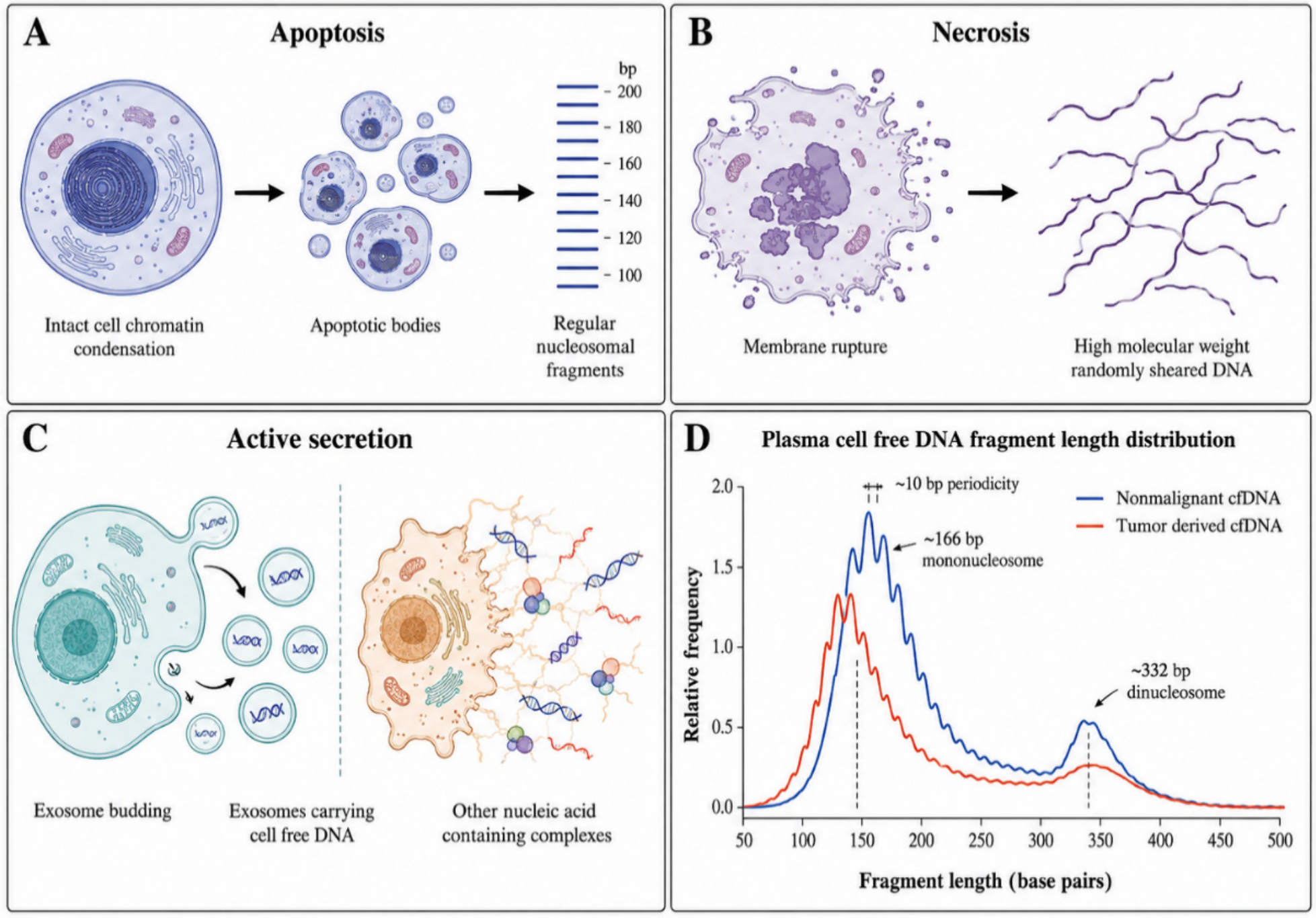
Figure 2.
Representative fragmentomic features distinguishing nonmalignant and tumor-derived cell-free DNA, including relative fragment shortening, altered four-base end-motif distributions, and differences in jagged-end abundance. The schematic illustrates qualitative patterns reported in the literature and does not represent data from a single experimental cohort. Created in BioRender. Begüm Kurt (2026). Available at https://BioRender.com (accessed on 30.08.2026).
Figure 2.
Representative fragmentomic features distinguishing nonmalignant and tumor-derived cell-free DNA, including relative fragment shortening, altered four-base end-motif distributions, and differences in jagged-end abundance. The schematic illustrates qualitative patterns reported in the literature and does not represent data from a single experimental cohort. Created in BioRender. Begüm Kurt (2026). Available at https://BioRender.com (accessed on 30.08.2026).
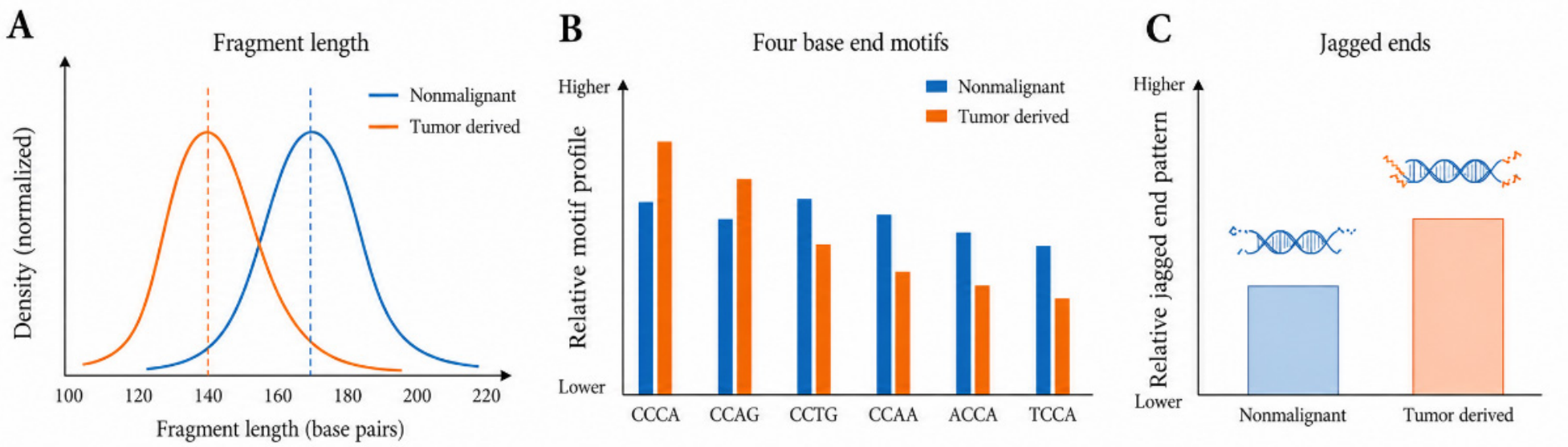
Figure 3.
Generalized analytical workflow for machine learning-based fragmentomic classification of plasma cell-free DNA, from blood collection and cfDNA sequencing to fragmentomic feature extraction, classifier analysis, cancer probability estimation, and tissue-of-origin prediction. Created in BioRender. Begüm Kurt (2026). Available at https://BioRender.com (accessed on 30.08.2026).
Figure 3.
Generalized analytical workflow for machine learning-based fragmentomic classification of plasma cell-free DNA, from blood collection and cfDNA sequencing to fragmentomic feature extraction, classifier analysis, cancer probability estimation, and tissue-of-origin prediction. Created in BioRender. Begüm Kurt (2026). Available at https://BioRender.com (accessed on 30.08.2026).
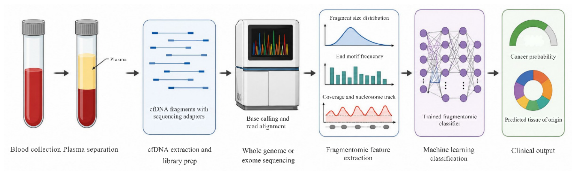
Figure 4.
Schematic comparison of tumor-associated and normal cell-free DNA methylation patterns and representative bisulfite-based and bisulfite-free workflows for cfDNA methylation profiling. Created in BioRender. Begüm Kurt (2026). Available at https://BioRender.com (accessed on 30.08.2026).
Figure 4.
Schematic comparison of tumor-associated and normal cell-free DNA methylation patterns and representative bisulfite-based and bisulfite-free workflows for cfDNA methylation profiling. Created in BioRender. Begüm Kurt (2026). Available at https://BioRender.com (accessed on 30.08.2026).
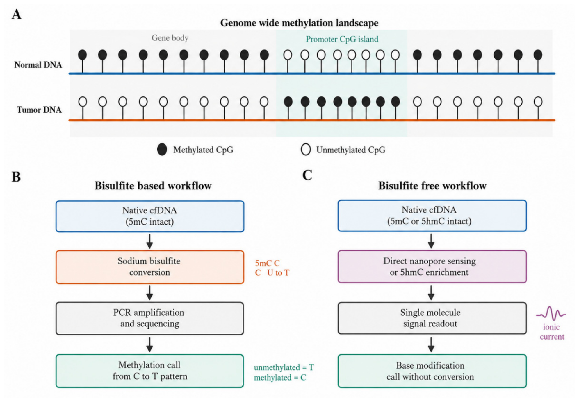
Figure 5.
Schematic overview of CRISPR-Cas12a- and Cas13a-based collateral cleavage mechanisms and their adaptation to liquid-biopsy biosensing. Cas12a-based systems primarily recognize DNA targets and cleave ssDNA reporters, whereas Cas13a-based systems recognize RNA targets and cleave ssRNA reporters. Selected Cas12a platforms can additionally exploit methylation-dependent target recognition for site-specific cfDNA methylation sensing. Created in BioRender. Begüm Kurt (2026). Available at https://BioRender.com (accessed on 30.08.2026).
Figure 5.
Schematic overview of CRISPR-Cas12a- and Cas13a-based collateral cleavage mechanisms and their adaptation to liquid-biopsy biosensing. Cas12a-based systems primarily recognize DNA targets and cleave ssDNA reporters, whereas Cas13a-based systems recognize RNA targets and cleave ssRNA reporters. Selected Cas12a platforms can additionally exploit methylation-dependent target recognition for site-specific cfDNA methylation sensing. Created in BioRender. Begüm Kurt (2026). Available at https://BioRender.com (accessed on 30.08.2026).
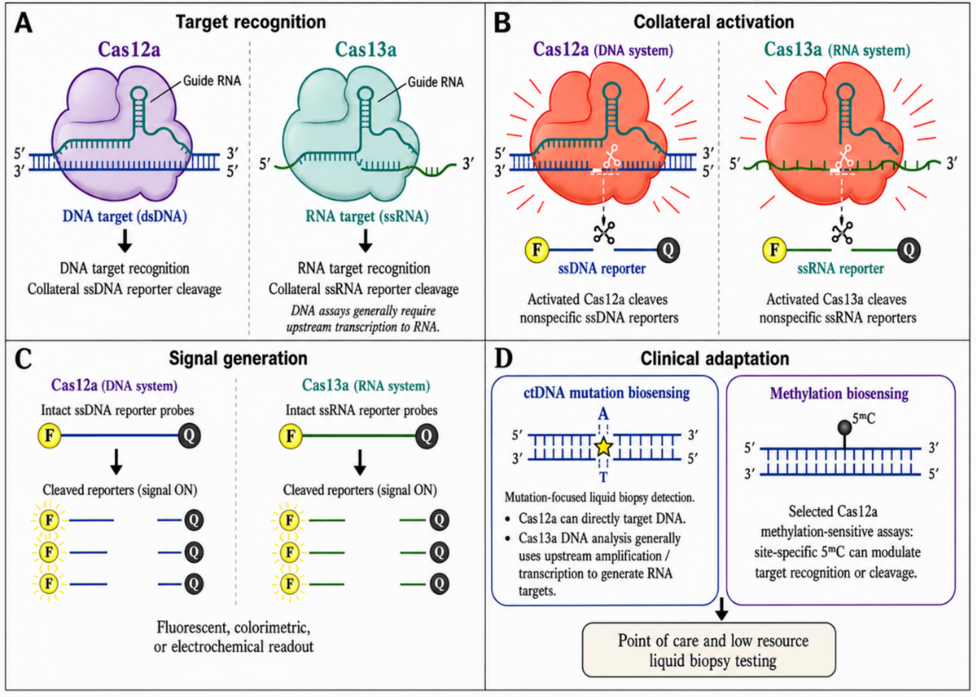
Figure 6.
Integrated overview of the principal liquid-biopsy modalities and clinical applications specifically discussed across the cancer types reviewed in Section 6. Colored markers indicate modalities directly supported by the studies summarized in Table 6. Created in BioRender. Begüm Kurt (2026). Available at https://BioRender.com (accessed on 30.08.2026).
Figure 6.
Integrated overview of the principal liquid-biopsy modalities and clinical applications specifically discussed across the cancer types reviewed in Section 6. Colored markers indicate modalities directly supported by the studies summarized in Table 6. Created in BioRender. Begüm Kurt (2026). Available at https://BioRender.com (accessed on 30.08.2026).
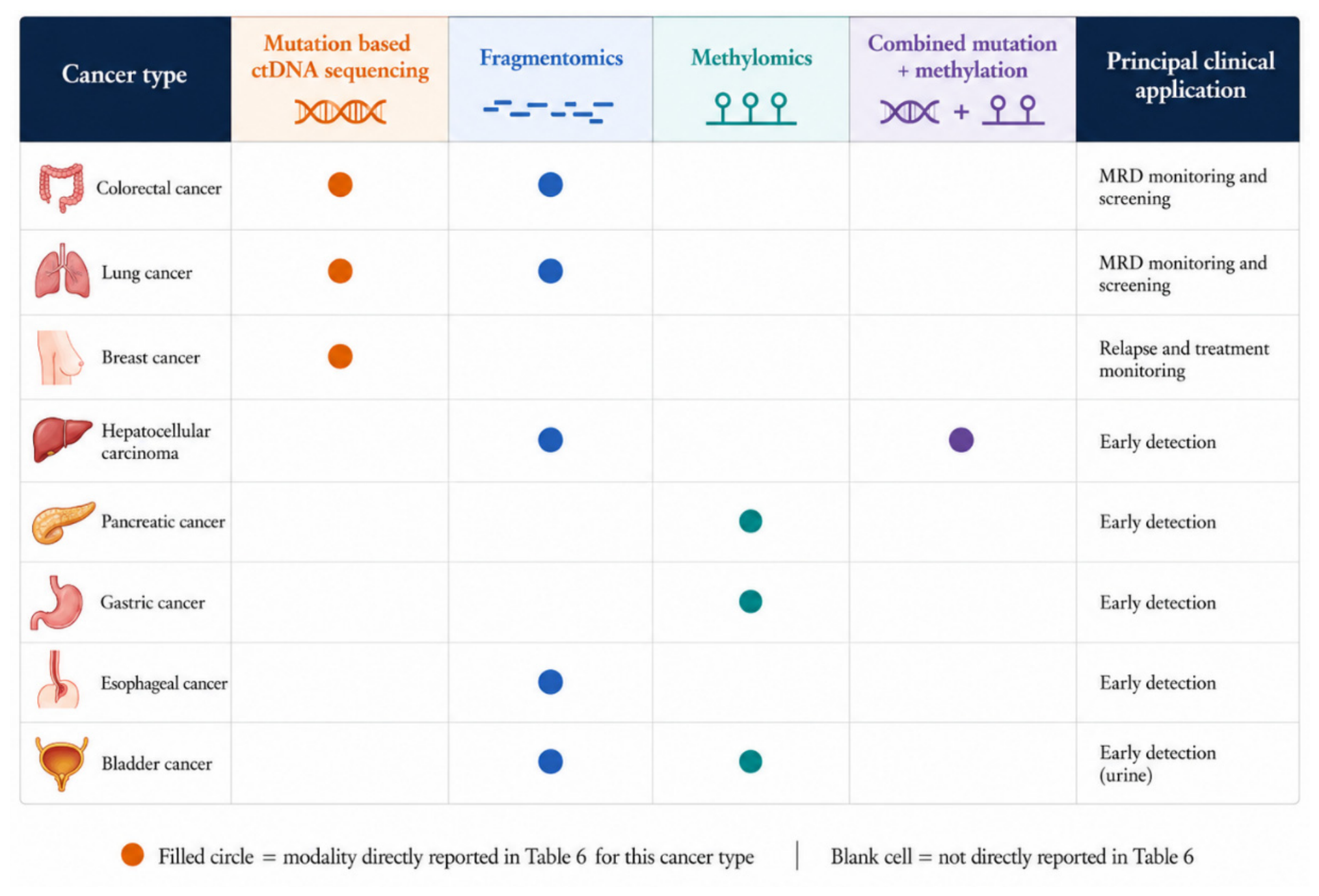

Table 1.
Key Milestones in the Discovery and Biological Characterization of Circulating Cell-free DNA (cfDNA).
Table 1.
Key Milestones in the Discovery and Biological Characterization of Circulating Cell-free DNA (cfDNA).
| Year | Milestone | Reference |
| 1948 | First description of circulating free nucleic acids in human plasma | [8] |
| 1977 | Elevated free DNA concentrations reported in the serum of cancer patients | [9] |
| 1987 | Isolation and characterization of circulating DNA from the plasma of cancer patients | [10] |
| 1997 | Discovery of cell free fetal DNA in maternal plasma, enabling noninvasive prenatal testing | [13] |
| 2001 | Apoptosis identified as the predominant mechanism generating circulating cfDNA fragments | [11] |
| 2003 | Plasma DNA integrity proposed as a quantitative marker associated with malignancy | [14] |
| 2014 | CtDNA detected across common epithelial cancer types using targeted and genome wide sequencing | [15] |
| 2016 | Genome wide nucleosome footprint identified within cfDNA fragmentation patterns | [16] |
Table 2.
Representative Fragmentomic Platforms and Their Reported Clinical Applications.
| Platform / Approach | Fragmentomic Feature | Cancer Type(s) Studied | Reference |
| Genome wide fragmentation classifier (DELFI type) | Genome wide fragmentation profile | Pan cancer, including lung, breast, colorectal, and ovarian | [32] |
| Lung cancer fragmentome model | Genome wide fragmentation plus clinical risk factors | Lung cancer | [33] |
| Liver cancer fragmentome model | Fragmentation features plus conventional biomarkers | Hepatocellular carcinoma | [34] |
| Multidimensional fragmentomics | Fragment size, end motif, and copy number | Colorectal cancer, including advanced adenoma | [35] |
| End motif deep learning classifier | End motif frequency distribution | Multiple cancer types | [36] |
| Orientation aware fragmentation (OCF) | Fragment end orientation in open chromatin | Tissue of origin inference | [30] |
| Jagged end and preferred end profiling | Single-stranded end structure and preferred cleavage sites | Hepatocellular carcinoma, bladder cancer | [21,22,25] |
Abbreviations: DELFI, DNA Evaluation of Fragments for Early Interception; OCF, orientation-aware cell-free DNA fragmentation.
Table 3.
Representative Methylation Based Liquid Biopsy Platforms and Their Reported Clinical Applications.
Table 3.
Representative Methylation Based Liquid Biopsy Platforms and Their Reported Clinical Applications.
| Platform / Approach | Detection Principle | Cancer Type(s) / Application | Reference |
| Targeted methylation multi-cancer classifier | Targeted bisulfite sequencing across thousands of genomic regions | Multi-cancer early detection and tissue of origin prediction | [4,5] |
| Genome wide bisulfite tissue mapping | Genome wide bisulfite sequencing | Tissue of origin deconvolution (prenatal, cancer, transplantation) | [40] |
| Digital single molecule methylation profiling | Molecule level methylation heterogeneity quantification | Low volume liquid biopsy cancer detection | [43] |
| 5hmC profiling | Enrichment of oxidized methylcytosine derivative | Pancreatic cancer early detection | [44] |
| Combined mutation and methylation sequencing | Simultaneous mutation and methylation analysis on the same molecules | Hepatocellular carcinoma detection | [45] |
| Nanopore single molecule methylation sequencing | Direct native base modification detection, no bisulfite conversion | Cell of origin and cancer classification | [46,47,48] |
Abbreviations: cfDNA, cell-free DNA; 5hmC, 5-hydroxymethylcytosine.
Table 4.
Representative CRISPR-Based Nucleic Acid Biosensors Relevant to Cancer Liquid Biopsy.
| Platform | Cas Enzyme | Target | Detection Format | Reference |
| SHERLOCK | Cas13a | RNA / DNA sequence variants | Fluorescent, isothermal preamplification | [6] |
| SHERLOCKv2 | Cas13a, Cas12a, Csm6 | Multiplexed nucleic acid targets | Fluorescent, multiplexed | [49] |
| DETECTR | Cas12a | DNA sequence variants (e.g., HPV) | Fluorescent, collateral ssDNA cleavage | [7] |
| Cas12a / MOF electrochemical sensor | Cas12a | CtDNA | Electrochemical, label free | [51] |
| dCas9 impedimetric biosensor | dCas9 | CtDNA | Electrochemical, label free | [52] |
| CRISPR methylation sensitive test | Cas12a | Site specific 5mC methylation | Amplification free, cleavage inhibition | [53] |
| Dual fluorescence Cas12a methylation sensor | Cas12a | Site specific DNA methylation | Ratiometric fluorescent | [54] |
| Paper based colorimetric Cas12a device | Cas12a | Double-stranded DNA targets | Colorimetric, microfluidic paper | [56] |
| Cas13a electrochemiluminescence sensor | Cas13a | Circulating tumor RNA | Electrochemiluminescence, amplification free | [57] |
Abbreviations: CRISPR, clustered regularly interspaced short palindromic repeats; Cas, CRISPR-associated protein; dCas9, catalytically inactive Cas9; MOF, metal-organic framework; 5mC, 5-methylcytosine; ssDNA, single-stranded DNA; dsDNA, double-stranded DNA; RNA, ribonucleic acid; DNA, deoxyribonucleic acid.
Table 5.
CtDNA Quantification Technologies for MRD Detection.
| Technology | Principle | Reference |
| CAPP Seq | Hybrid-capture deep sequencing of recurrently mutated genomic regions for broad patient coverage | [58] |
| Integrated digital error suppression | Molecular barcoding combined with in silico suppression of stereotypical sequencing artifacts | [59] |
| Safe SeqS | Unique molecular barcoding of template molecules prior to amplification | [60] |
| PhasED Seq | Detection of phased variants occurring on the same ctDNA molecule | [61] |
| ichorCNA | Tumor fraction estimation from shallow whole genome or whole exome copy number data | [62] |
Abbreviations: CAPP-Seq, Cancer Personalized Profiling by Deep Sequencing; Safe-SeqS, Safe-Sequencing System; PhasED-Seq, Phased Variant Enrichment and Detection by Sequencing; ichorCNA, inference of copy number alterations from cell-free DNA.
Table 6.
Summary of Clinical Applications by Cancer Type.
| Cancer Type | Clinical Application | Principal Modality | Reference |
| Colorectal cancer | Postoperative MRD detection, treatment response monitoring, screening | ctDNA ultradeep sequencing, fragmentomics | [35,63,64] |
| Lung cancer | Postoperative MRD detection, relapse detection, screening | Phylogenetic ctDNA analysis, fragmentomics | [33,65,66] |
| Breast cancer | Relapse detection, disease burden monitoring | Mutation tracking ctDNA | [67,68] |
| Hepatocellular carcinoma | Early detection in high risk (cirrhotic) populations | Fragmentomics, combined mutation and methylation analysis | [19,22,34,45] |
| Pancreatic cancer | Early detection | 5hmC methylation profiling | [44] |
| Gastric cancer | Early detection | Genome wide methylome profiling | [39] |
| Esophageal squamous cell carcinoma | Early detection | Ensemble fragmentomic model | [69] |
| Bladder cancer | Noninvasive detection from urine | Urinary jagged ends, bisulfite sequencing | [25,70] |
Abbreviations: ctDNA, circulating tumor DNA.
Disclaimer/Publisher’s Note: The statements, opinions and data contained in all publications are solely those of the individual author(s) and contributor(s) and not of MDPI and/or the editor(s). MDPI and/or the editor(s) disclaim responsibility for any injury to people or property resulting from any ideas, methods, instructions or products referred to in the content. |
© 2026 by the authors. Licensee MDPI, Basel, Switzerland. This article is an open access article distributed under the terms and conditions of the Creative Commons Attribution (CC BY) license.
Copyright: This open access article is published under a Creative Commons CC BY 4.0 license, which permit the free download, distribution, and reuse, provided that the author and preprint are cited in any reuse.