

Submitted:
07 September 2026
Posted:
08 September 2026
You are already at the latest version
Abstract
Ultraviolet B (UVB) radiation is a major cause of skin photodamage. This study investigated the protective effects of Diodon hystrix skin collagen (DHSC) against UVB-induced damage. In vitro, HaCaT cells were used to assess cell viability, reactive oxygen species (ROS), superoxide dismutase (SOD), malondialdehyde (MDA), and mRNA expression of IL-1β, TNF-α, and IL-6. In vivo, an acute UVB-induced photodamage model was established in Balb/c mice with topical DHSC application. Erythema scoring, histology (H&E, Masson’s trichrome), ELISA, Western blot, and qPCR were performed. DHSC prevented UVB-induced HaCaT cell death, reduced ROS production and inflammation, and showed no cytotoxicity. In vivo, DHSC alleviated skin erythema and thickening, enhanced SOD activity, reduced MDA, downregulated IL-1β, TNF-α, and IL-6, and suppressed MMP-1, -3, and -9 expression. Mechanistically, DHSC inhibited p38 and ERK phosphorylation, thereby suppressing NF-κB and AP-1 activation. These findings indicate that DHSC possesses potent antioxidative and anti-inflammatory properties and protects against skin photodamage via the MAPK/NF-κB/AP-1 signaling pathway.
Keywords:
Diodon hystrix
; collagen
; UVB
; acute skin photodamage
; MAPK signaling pathway
; oxidative stress
1. Introduction
Acute skin photodamage is a common pathological response to excessive ultraviolet (UV) irradiation, characterized by redness, inflammation, and potential long-term impairment of skin structure and function. With the increasing prevalence of outdoor activities and the progressive depletion of the stratospheric ozone layer, human skin is exposed to higher levels of UV radiation, particularly UVB, which is known to induce oxidative stress through the generation of reactive oxygen species (ROS). This oxidative burst triggers lipid peroxidation of cell membranes and causes oxidative damage to DNA and proteins, ultimately leading to collagen degradation and accelerated skin aging (Brar et al., 2025).
Despite the widespread use of sunscreens, concerns regarding their safety have emerged, as some UV filters may induce DNA photodamage, generate reactive oxygen species, and cause photogenotoxic effects. Moreover, current photosafety tests, such as the 3T3 NRU phototoxicity test, are limited because they only assess cell death while neglecting non-lethal but mutagenic damage (Paiva et al., 2020). These drawbacks highlight the need for alternative or complementary approaches. Consequently, there is growing interest in developing bioactive compounds that can mitigate UV-induced damage through multiple mechanisms, including antioxidant, anti-inflammatory, and matrix-protective effects.
To mitigate these effects, antioxidant-based therapeutics have been developed, with low-molecular-weight antioxidants being the most widely used agents. However, many lipophilic antioxidants suffer from poor aqueous solubility, low chemical stability, and limited bioavailability, restricting their application in food, nutraceutical, and pharmaceutical industries (Jug et al., 2025).
In parallel, collagen-based products have gained attention as potential preventive strategies against UV-induced skin damage, aiming to restore dermal integrity and counteract photoaging processes (Li et al., 2022). Collagen, a biopolymer abundant in the extracellular matrix, plays a crucial role in maintaining skin health and vitality. It is widely utilized in cosmetic skincare, food processing, immune regulation, and wound healing due to its biocompatibility, low immunogenicity, and regenerative properties (Park et al., 2021; Kang et al., 2021; Méndez-Flores et al., 2022; Rømer et al., 2021; Stahl et al., 2013). Traditional collagen sources, such as bovine and porcine collagen, have faced limitations due to concerns regarding disease transmission and religious beliefs. Consequently, there has been growing interest in exploring marine-derived collagen, which possesses unique features like the arginine-glycine-aspartic acid (RGD) domain, providing excellent cell adhesion properties and compatibility with human collagen (Niu et al., 2013). It has extremely low immunogenicity and high bioavailability. Therefore, it is an important issue to find a safe and effective fish-derived collagen as a UV protective agent.
Diodon hystrix, a unique fish species native to Hainan Province, boasts rich collagen content in its skin tissue. This collagen possesses low immunogenicity and high bioavailability, making it a promising candidate for developing safe and effective UV protective agents. The present study aims to investigate the protective effects of Diodonhystrix skin collagen (DHSC) against UVB-induced skin photodamage and explore its underlying mechanisms (Pan, 2023).
2. Materials and Methods
2.1. Cell Culture and Cell Viability Assay
HaCaT human keratinocytes were purchased from Procell Life Science and Technology Co., Ltd (Boukamp et al., 1988). Cells were cultured in Dulbecco’s modified Eagle’s medium (DMEM; Hyclone, Logan, UT, USA) supplemented with 10% fetal bovine serum (FBS; HyClone) and 1% penicillin-streptomycin (Gibco, CA, USA) at 37 °C in a humidified atmosphere with 5% CO2. For cell viability assays, an appropriate number of cells (1×10⁵ cells/mL) were seeded in 96-well plates and incubated for 24 hours. Cells were then treated with various concentrations of DHSC for an additional 24 hours. Cell viability was assessed using the 3-(4,5-dimethylthiazol-2-yl)-2,5-diphenyltetrazolium bromide (MTT) assay(Mosmann, 1983).
For UVB irradiation experiments, cells were treated with different concentrations of DHSC for 24 hours. The culture medium was replaced with 40 μL of PBS, and cells were exposed to UVB radiation using a Philips PL-S9W/01/2P lamp. After UVB exposure, fresh medium was added, and cell viability was measured using the MTT assay after an additional 24 hours of incubation.
2.2. Measurement of Oxidative Stress Indicators
After 6 hours of UVB irradiation and DHSC treatment, HaCaT cells were stained with 5 μmol/L 2’,7’-dichlorofluorescein diacetate (DCFH-DA; Nanjing Jiancheng Bioengineering Institute) for 20 minutes in a CO2 incubator. ROS production was analyzed using a fluorescence microscope (Leica Microsystems Germany Co., Ltd., 470797). Superoxide dismutase (SOD) activity and malondialdehyde (MDA) content were measured according to the instructions provided by the Nanjing Jiancheng Bioengineering Institute.
2.3. Animal Experiments
Balb/c male mice (18±2g) were obtained from Changzhou Cavince Laboratory Animal Co., Ltd. (JiangSu, China). Mice were housed in a specific pathogen-free (SPF) environment at Jilin University, China, maintained at 20-23 °C and 50-55% relative humidity with a 12-hour light/dark cycle. Animal care and experimental procedures were conducted in accordance with the guidelines of the National Institutes of Health (Institute of Laboratory Animal Resources, 1986) and approved by the Institutional Animal Care and Use Committee (IACUC permit number: 2026-KJCLL-050) of Hainan Tropical Ocean University. Efforts were made to minimize animal suffering and pain.
UV radiation dose was determined through preliminary experiments and set at 1000 mJ/cm² per day (Cao et al., 2021). Mice were randomly divided into different groups: control (CTL), model (MOD), DHSC treatment (H, M, L), matrix control (B), and positive control (Y) (Wu et al., 2020). Mice were anesthetized with chloral hydrate and applied with 0.3g of DHSC, matrix, or sunscreen 20 minutes before UVB irradiation. Mice in the MOD group were exposed to UVB irradiation without any treatment. UVB irradiation was performed for 6 days(Zeng et al., 2014) with a cumulative dose of 6000 mJ/cm2.
2.4. Histological Analysis
Dorsal skin samples were fixed in buffered formaldehyde solution and sectioned into 5 μm thick slides. Skin layers were stained with hematoxylin and eosin (H&E) to assess histopathological changes, and collagen fibers were analyzed using Masson’s trichrome staining, a well-established method for evaluating collagen distribution and morphology in skin tissue (de Medeiros et al., 2017; Inan et al., 2006).
2.5. RT-PCR
Total RNA was extracted from mouse skin tissues using Trizol reagent (Beyotime, R0016). Reverse transcription was performed using the PrimeScript TM reverse transcription reagent kit (Takara, Dalian, China, RR037A) according to the manufacturer’s instructions. Real-time PCR was performed using the SYBR Select Master Mix (Roche, 04913914001) and primer sequences listed in Tables 1 and 2. Data were analyzed using the 2−ΔΔCt method with β-actin as an internal control.
2.6. Western Blot
Proteins were extracted using RIPA lysis buffer containing PMSF (Beyotime, P0013B). Protein concentration was determined using the BCA Protein Assay Kit (Beyotime, P0012S). Proteins were separated using 12% SDS-PAGE and transferred to PVDF membranes (Millipore, Burlington, MA). Membranes were incubated with primary antibodies listed in Table 3 overnight at 4 °C followed by secondary antibody (Wanleibio, WLA037a) for 2 hours at room temperature. Chemiluminescence detection reagent (PE0010, Solarbio) was used for visualization, and analysis was performed using an Analytik Jena instrument (US).
2.7. Enzyme-Linked Immunosorbent Assay
Proteins were extracted using RIPA lysis buffer containing PMSF. Cytokine levels, including interleukin-1β (IL-1β), tumor necrosis factor-α (TNF-α), and interleukin-6 (IL-6), were measured using commercial ELISA kits (Biolegend) according to the manufacturer’s instructions.
2.8. Statistics Analysis
All experiments were performed in triplicate, and data are presented as mean ± SEM. Statistical differences among groups were analyzed using one-way analysis of variance (ANOVA) followed by Tukey’s post-hoc test. Statistical analyses were performed using GraphPad Prism 8.0.2, and significance was set at p < 0.05.
3. Results
3.1. Effect of DHSC on HaCaT Cell Viability
To evaluate the cytotoxicity of DHSC, HaCaT cells were treated with various concentrations and assessed using the MTT assay. As shown in Figure 1A, DHSC exhibited no significant cytotoxicity up to 5 mg/mL. Therefore, a concentration of 20 mg/mL was selected as the maximum safe dose for subsequent experiments.
3.2. Effect of UVB Radiation on HaCaT Cell Viability
HaCaT cells were exposed to increasing doses of UVB radiation, and cell viability was assessed. As depicted in Figure 1B, UVB radiation dose-dependently decreased cell viability, with the lowest survival rate observed at 248.4 mJ/cm2. This dose was chosen for subsequent experiments.
3.3. Effect of DHSC on UVB-Induced HaCaT Cell Death
To investigate the protective effect of DHSC against UVB-induced cell death, HaCaT cells were pretreated with DHSC followed by UVB irradiation. Figure 1C demonstrates that DHSC exerted a dose-dependent protective effect, with the highest concentration (20 mg/mL) restoring cell viability to levels comparable to the control group.
3.4. Effects of DHSC on UVB-Induced ROS Production in HaCaT Cells
UVB irradiation significantly increased intracellular ROS production, as evidenced by enhanced fluorescence in DCFH-DA-stained cells (Figure 2). In contrast, pretreatment with DHSC markedly reduced ROS levels, indicating its antioxidant potential. As shown in Figure 2, only weak fluorescence was observed in the normal control group, whereas the model group exhibited strong fluorescence, suggesting that UVB exposure induced substantial ROS generation. Notably, pretreatment with 20 mg/mL DHSC restored fluorescence to near-normal levels and significantly attenuated ROS production, consistent with the results of the MTT assay. These findings suggest that DHSC exerts its protective effect against UVB-induced photodamage in HaCaT cells, at least in part, by reducing ROS production.
3.5. Effects of DHSC on UVB-Induced SOD and MDA in HaCaT Cells
To further investigate the protective effect of DHSC against UVB-induced oxidative stress, the levels of superoxide dismutase (SOD) and malondialdehyde (MDA) were measured (Figure 3). UVB irradiation significantly decreased SOD activity and increased MDA content compared to the normal control group, indicating the induction of oxidative stress. However, pretreatment with DHSC effectively attenuated these changes by restoring SOD activity and reducing MDA levels, suggesting that DHSC alleviates UVB-induced oxidative damage in HaCaT cells.
3.6. Effects of DHSC on the Expression of Inflammatory Factors in HaCaT Cells Induced by UVB
To assess the anti-inflammatory effects of DHSC, the mRNA expression levels of the pro-inflammatory cytokines IL-1β, TNF-α, and IL-6 in HaCaT cells were measured by quantitative PCR (qPCR). As shown in Figure 4, UVB irradiation markedly upregulated the expression of these genes compared to the normal control group. However, this upregulation was significantly attenuated by pretreatment with DHSC, suggesting that DHSC mitigates UVB-induced cellular damage, at least in part, through the inhibition of inflammatory cytokine expression.
3.7. Effects of DHSC on Skin Appearance Morphology in Mice with Acute Photodamage
The dorsal skin changes and scores of the radiation area are shown in Figure 5, the back skin of mice in the CTL group is rosy and smooth (Figure 5A), and the back skin of mice in the MOD group produces photodamage such as roughness, scaling, sagging and wrinkles, indicating that the model is successfully established (Figure 5B); 20 min before irradiation, external DHSC has a notable protective effect on ultraviolet rays, and the high-dose group can improve the appearance of photodamage caused by ultraviolet rays and improve the degree of skin roughness. Erythema and wrinkles were alleviated (Figure 5E), and the difference between the erythema score and the model group was significant (Figure 6), while the effect was not obvious in the L and M groups (Figure 5C-D). In addition, the mice in group B had rough skin (Figure 5F), which was comparable to the MOD group, and there was no significant difference in the erythema score from the model group (Figure 6), which excluded the protective effect of the matrix against ultraviolet rays. Group Y significantly improved the skin condition, and there were no obvious wrinkles on the skin, accompanied only by peeling (Figure 5G).
3.8. Effect of DHSC on Skin Histopathology in Mice with Acute Photodamage
Skin thickening and inflammatory cell infiltration were observed in the skin tissues of each group of mice by H&E and the results were shown in Figure 7. Ultraviolet irradiation can lead to significant thickening of the skin epidermis of mice, damage to the stratum corneum (shown by the blue arrow of Figure 7B-F), irregular tissue, and diffuse inflammation (shown by the red circle of Figure 7B-F), compared with the MOD group, the thickening of the epidermis of mice in group H was significantly alleviated, which was similar to that of group Y, and inflammation was reduced; group L and M still had less inflammation, group B was accompanied by more inflammation, and group Y had almost no inflammatory cell infiltration.
3.9. Effect of DHSC on Collagen Content in Skin Tissue of Mice with Acute Photodamage
The back skin tissue of each group of mice was stained by Masson staining to observe the changes of collagen. The experimental results are shown in Figure 8, the skin of mice in the CTL group is rich in collagen (indicated by the red circle in Figure 8A), while the collagen content in the skin tissue of mice in the MOD group decreases significantly, and obvious skin fibrosis appears (shown by the blue arrow in Figure 8B). When the skin of mice was protected by different concentrations of DHSC before irradiation, the decrease of collagen content could be inhibited (indicated by the red circle of Figure 8C-E), and different groups could improve skin fibrosis to varying degrees (shown by the blue arrow of Figure 8C-D), among which the H group was the most obvious. Since the matrix Dabao has a certain moisturizing effect, group B can also reduce the degree of skin fibrosis to a certain extent, but the collagen content is not as good as that of L, M and H groups, which proves that the ultraviolet protection effect of DHSC is much greater than that of matrix, and only minimal fibrosis exists in the skin tissue of group Y (shown by the blue arrow in Figure 8G).
3.10. Effects of DHSC on Skin Oxidative Stress in Mice with Acute Photodamage
When the skin is exposed to ultraviolet radiation beyond its tolerance amount, tissue cells produce oxidative stress, and Nrf2 binds to the antioxidant response element, activating the transcription of antioxidant genes. The experimental results are shown in Figure 9, compared with the CTL group, a large amount of ROS is produced in the tissues of the MOD group (Figure 9A), and the activity of the antioxidant enzyme SOD is decreased (Figure 9B), the expression of Nrf2 protein is reduced (Figure 9D), and the content of the lipid peroxidation product MDA is significantly increased (Figure 9C), indicating that oxidative stress occurs in the tissue. Compared with the MOD group, the expression of nuclear protein Nrf2 in the skin of group H increased, the SOD activity was significantly increased, and the MDA content was significantly reduced. There were no significant differences in ROS content and nucleoprotein Nrf2 in the skin of group B and the MOD group, lower SOD activity than in H group, and higher MDA content than in H group, and there were statistical differences. The above results show that collagen can reduce the attack of oxygen free radicals by increasing the activity of antioxidant enzymes in the body, thereby reducing UVB damage to the skin.
3.11. Effects of DHSC on AP-1 Protein and MAPK Pathway in Skin of Mice with Acute Photodamage
UVB irradiation has been reported to activate the AP-1 protein and its upstream regulator, MAPK, thereby accelerating MMP transcription and skin inflammation. The experimental results are shown in Figure 10 and Figure 11, UVB induces phosphorylation and expression of AP-1 subunits c-fos and c-jun, and DHSC effectively inhibits this effect (Figure 10). In addition, UVB stimulates phosphorylation of ERK and p38 in the MAPK family, and DHSC inhibits their activation, but UVB has no effect on JNK signal activation (Figure 11), which is consistent with the findings of Kwon et al. (2019).
3.12. Effects of DHSC on Inflammation in Mice with Acute Photodamage
The expression of TNF-α and IL-6 in the skin tissue of mice in the MOD group was higher than that in the CTL group, while the expression of TNF-α and IL-6 in the skin tissue of the H group was lower than that in the MOD group (Figure 12B-C). Although the expression of IL-1β did not differ significantly among the groups, the MOD group increased compared with the CTL group, and the H group tended to inhibit its expression to some extent (Figure 12-A). When the body is stimulated by UVB, p65 is activated and transported to the nucleus, and the experimental results are shown in Figure 12-D, the MOD group has the highest content of p65 activated form, and DHSC effectively inhibits this effect.
3.13. Effects of DHSC on Skin Collagen Degradation and Expression in Mice with Acute Photodamage
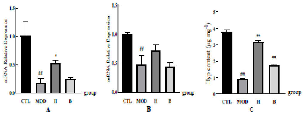
The increase in MMPs and the decrease in precollagen are the main causes of skin photodamage, and mRNA levels were measured. The mRNA levels of MMP-1, MMP-3 and MMP-9 in the skin of the MOD group treated with ultraviolet radiation were significantly increased, and the expression of MMPs genes in the H group with DHSC intervention was significantly inhibited, and there was no significant difference between the matrix group and the model group (Figure 13). The content of collagen type I and type III and hydroxyproline was higher than that of the MOD group (Figure 14).
4. Discussion
Repeated or prolonged exposure to ultraviolet (UV) radiation, particularly UVB, is a well-established risk factor for a range of skin pathologies, including inflammation, photoaging, and carcinogenesis (Bosch et al., 2015; Xu et al., 2010). The pathogenic effects of UVB are primarily initiated by the overproduction of reactive oxygen species (ROS), which triggers oxidative stress, DNA damage, and the activation of complex inflammatory signaling cascades. This oxidative insult subsequently stimulates the expression of matrix metalloproteinases (MMPs), such as collagenase (MMP-1), procollagen activator (MMP-3), and gelatinase (MMP-9), which are the principal mediators of collagen and extracellular matrix degradation, leading to the characteristic features of photoaged skin (Wen et al., 2012; Chiang et al., 2013; Han et al., 2019).
Our study demonstrates that Diodon hystrix skin collagen (DHSC) possesses potent protective effects against UVB-induced photodamage by intervening at multiple critical junctures of this cascade. Firstly, we confirmed the antioxidant properties of DHSC, showing it effectively reduces UVB-induced ROS production in both HaCaT cells and mouse skin, thereby mitigating the initial oxidative stress that drives subsequent damage. Consequently, this reduction in oxidative stress led to a significant inhibition of MMP-1, -3, and -9 expression, suggesting that DHSC helps preserve collagen integrity and dermal matrix structure. To elucidate the molecular mechanism underlying these effects, we investigated the upstream signaling pathways. UVB-induced ROS are known to activate key transcription factors, including activator protein-1 (AP-1) and nuclear factor-kappa B (NF-κB), which play pivotal roles in regulating MMP expression and the inflammatory response (Lu et al., 2016; Rittié and Fisher, 2002; Khan et al., 2006).. AP-1, a heterodimer of c-Fos and c-Jun, binds to promoter regions of MMP genes to activate their transcription, while NF-κB promotes the expression of pro-inflammatory cytokines (Rittié and Fisher, 2002; Khan et al., 2006). The activation of AP-1 and NF-κB is, in turn, largely governed by the mitogen-activated protein kinase (MAPK) signaling pathway, which includes the ERK, JNK, and p38 subgroups. Our results show that DHSC significantly inhibited the UVB-induced phosphorylation of both c-Fos and c-Jun, as well as the upstream activators ERK and p38. By suppressing the MAPK/AP-1 signaling axis, DHSC effectively curtails the expression of downstream MMPs and pro-inflammatory factors (such as TNF-α and IL-1β), thereby reducing both collagen degradation and inflammation. In summary, our findings suggest that DHSC exerts its photoprotective effects by acting as an antioxidant that quenches UVB-induced ROS. This, in turn, leads to the downregulation of the MAPK (ERK/p38) signaling pathway, inhibition of AP-1 and NF-κB activation, and subsequent suppression of MMPs and inflammatory cytokines. This multi-target mechanism positions DHSC as a promising candidate for preventing UVB-induced skin aging and damage.
While this study provides valuable insights, further research is warranted to fully elucidate the protective mechanisms of DHSC. Future investigations could explore its potential to promote DNA repair, inhibit apoptosis, and upregulate endogenous antioxidant enzymes. Additionally, long-term studies in more advanced animal models and subsequent clinical trials are essential to comprehensively evaluate the safety and efficacy of DHSC as a topical or systemic photoprotective agent.
5. Conclusions
Our findings demonstrate that DHSC possesses potent antioxidant and anti-inflammatory properties, effectively protecting against UVB-induced skin photodamage. This protection is achieved through the inhibition of ROS production, the suppression of MMPs, and the downregulation of the MAPK/AP-1/NF-κB signaling pathway. DHSC holds great potential as a safe and effective UV protective agent for the prevention and treatment of skin photodamage.
Author Contributions
Conceptualization, Y.S. (Yu Song) ; methodology, J.S., M.P., N.L. and H.H.; formal analysis, J.S., M.P., Y.N. and Y.S. (Ying Sun); investigation, H.Z., Z.L. and H.C.; data curation, J.S., M.P. and Y.S. (Ying Sun); writing—original draft preparation, J.S. and M.P.; writing—review and editing, Y.S. (Yu Song); visualization, N.L. and H.H.; supervision, Y.S. (Yu Song) ; project administration, Y.S. (Yu Song); funding acquisition, Y.S. (Yu Song). All authors have read and agreed to the published version of the manuscript.
Funding
This research was funded by the Education Reform Research Project of Hainan Tropical Ocean University (grant No. RHYjg2026-04) and the Fourth Batch of Nanhai Innovative Talents (Education Sector), Hainan Nanhai Yucai Project (grant No. 2025NHYC406).
Institutional Review Board Statement
The animal study protocol was approved by the Institutional Animal Care and Use Committee of Hainan Tropical Ocean University (approval No.2026-KJCLL-050) and was conducted in accordance with the National Institutes of Health Guide for the Care and Use of Laboratory Animals (8th edition, 2011). All efforts were made to minimize animal suffering and reduce the number of animals used.
Data Availability Statement
The data presented in this study are available on request from the corresponding authors (Yu Song, song_yu@hntou.edu.cn) The data are not publicly available due to privacy restrictions related to ongoing research projects.
Acknowledgments
The authors would like to thank the College of Fisheries and Life Sciences, Hainan Tropical Ocean University for providing laboratory facilities. We are also grateful to all members of the Hainan Key Laboratory for Conservation and Utilization of Tropical Marine Fishery Resources for their technical support and valuable discussions.
Conflicts of Interest
The authors declare no conflicts of interest. The funders had no role in the design of the study; in the collection, analyses, or interpretation of data; in the writing of the manuscript; or in the decision to publish the results.
References
- Brar G, Dhaliwal A, Brar AS, et al. A comprehensive review of the role of UV radiation in photoaging processes between different types of skin[J]. Cureus, 2025, 17(3): e81109. [CrossRef]
- Paiva JP, Diniz RR, Leitao AC, et al. Insights and controversies on sunscreen safety[J]. Critical Reviews in Toxicology, 2020, 50(8): 707-723. [CrossRef]
- Jug M, Radić K, Nižić Nodilo L, et al. Exploring Cyclodextrin Complexes of Lipophilic Antioxidants: Benefits and Challenges in Nutraceutical Development[J]. International Journal of Molecular Sciences, 2025, 26(23): 11682. [CrossRef]
- Li C, Fu Y, Dai H, et al. Recent progress in preventive effect of collagen peptides on photoaging skin and action mechanism[J]. Food Science and Human Wellness, 2022, 11(2): 218-229. [CrossRef]
- Park SJ, Kim D, Lee M, Yang JH, Yang JS, Lee J. GT Collagen Improves Skin Moisturization in UVB-Irradiated HaCaT Cells and SKH-I Hairless Mice[J]. Journal of Medicinal Food, 2021, 24(12): 1313-1322. [CrossRef]
- Kang MK, Kim DY, Oh H, et al. Dietary Collagen Hydrolysates Ameliorate Furrowed and Parched Skin Caused by Photoaging in Hairless Mice[J]. International Journal of Molecular Sciences, 2021, 22(11): 6137. [CrossRef]
- Méndez-Flores S, Priego-Ranero Á, Azamar-Llamas D, et al. Effect of polymerised type I collagen on hyperinflammation of adult outpatients with symptomatic COVID-19[J]. Clinical and Translational Medicine, 2022, 12(3): e763. [CrossRef]
- Rømer AMA, Thorseth ML, Madsen DH. Immune Modulatory Properties of Collagen in Cancer[J]. Frontiers in Immunology, 2021, 12: 791453. [CrossRef]
- Stahl M, Schupp J, Jäger B, et al. Lung collagens perpetuate pulmonary fibrosis via CD204 and M2 macrophage activation[J]. PLoS ONE, 2013, 8(11): e81382. [CrossRef]
- Niu LH, Zhou X, Yuan CQ, et al. Characterization of tilapia (Oreochromis niloticus) skin gelatin extracted with alkaline and different acid pretreatments[J]. Food Hydrocolloids, 2013, 33(2): 336-341. [CrossRef]
- Pan M. Protective effect of Diodon hystrix skin collagen on UVB-induced acute skin photodamage in mice [D]. Jilin University, 2023. (In Chinese).
- Boukamp P, Petrussevska RT, Breitkreutz D, et al. Normal keratinization in a spontaneously immortalized aneuploid human keratinocyte cell line[J]. The Journal of Cell Biology, 1988, 106(3): 761-771. [CrossRef]
- Mosmann T. Rapid colorimetric assay for cellular growth and survival: application to proliferation and cytotoxicity assays[J]. Journal of Immunological Methods, 1983, 65(1-2): 55-63. [CrossRef]
- Institute of Laboratory Animal Resources (US). Committee on Care and Use of Laboratory Animals. Guide for the care and use of laboratory animals[M]. Bethesda: US Department of Health and Human Services, Public Health Service, National Institutes of Health, 1986.
- Cao L, Yue X, Zhao Y, et al. Mechanisms of broad-band UVB irradiation‒induced itch in mice[J]. Journal of Investigative Dermatology, 2021, 141(10): 2499-2508.e3. [CrossRef]
- Wu H, Zhong Z, Lin S, et al. Coenzyme Q10 Sunscreen Prevents Progression of Ultraviolet-Induced Skin Damage in Mice[J]. BioMed Research International, 2020, 2020: 9039843. [CrossRef]
- Zeng J, Bi B, Chen L, et al. Repeated exposure of mouse dermal fibroblasts at a sub-cytotoxic dose of UVB leads to premature senescence: a robust model of cellular photoaging[J]. Journal of Dermatological Science, 2014, 73(1): 49-56. [CrossRef]
- de Medeiros V, de Biase MP, de Paiva AM, et al. Comparative measurement of collagen bundle orientation by Fourier analysis and semiquantitative evaluation: reliability and agreement in Masson’s trichrome, Picrosirius red and confocal microscopy techniques[J]. Journal of Microscopy, 2017, 267(2): 130-142. [CrossRef]
- Inan S, Oztukcan S, Vatansever S, et al. Histopathological and ultrastructural effects of glycolic acid on rat skin[J]. Acta Histochemica, 2006, 108(1): 37-47. [CrossRef]
- Kwon KR, Alam MB, Park JH, Kim TH, Lee SH. Attenuation of UVB-Induced Photo-Aging by Polyphenolic-Rich Spatholobus Suberectus Stem Extract Via Modulation of MAPK/AP-1/MMPs Signaling in Human Keratinocytes[J]. Nutrients, 2019, 11(6): 1341. [CrossRef]
- Bosch R, Philips N, Suárez-Pérez JA, et al. Mechanisms of Photoaging and Cutaneous Photocarcinogenesis, and Photoprotective Strategies with Phytochemicals[J]. Antioxidants, 2015, 4(2): 248-268. [CrossRef]
- Xu H, Yan Y, Li L, Peng S, Qu T, Wang B. Ultraviolet B-induced apoptosis of human skin fibroblasts involves activation of caspase-8 and -3 with increased expression of vimentin[J]. Photodermatology, Photoimmunology & Photomedicine, 2010, 26(4): 198-204. [CrossRef]
- Wen KC, Fan PC, Tsai SY, Shih IC, Chiang HM. Ixora parviflora Protects against UVB-Induced Photoaging by Inhibiting the Expression of MMPs, MAP Kinases, and COX-2 and by Promoting Type I Procollagen Synthesis[J]. Evidence-Based Complementary and Alternative Medicine, 2012, 2012: 417346. [CrossRef]
- Chiang HM, Chen HC, Chiu HH, Chen CW, Wang SM, Wen KC. Neonauclea reticulata (Havil.) Merr Stimulates Skin Regeneration after UVB Exposure via ROS Scavenging and Modulation of the MAPK/MMPs/Collagen Pathway[J]. Evidence-Based Complementary and Alternative Medicine, 2013, 2013: 324864. [CrossRef]
- Han HS, Shin JS, Myung DB, et al. Hydrangea serrata (Thunb.) Ser. Extract Attenuate UVB-Induced Photoaging through MAPK/AP-1 Inactivation in Human Skin Fibroblasts and Hairless Mice[J]. Nutrients, 2019, 11(3): 533. [CrossRef]
- Lu J, Guo JH, Tu XL, et al. Tiron Inhibits UVB-Induced AP-1 Binding Sites Transcriptional Activation on MMP-1 and MMP-3 Promoters by MAPK Signaling Pathway in Human Dermal Fibroblasts[J]. PLoS ONE, 2016, 11(8): e0159998. [CrossRef]
- Rittié L, Fisher GJ. UV-light-induced signal cascades and skin aging[J]. Ageing Research Reviews, 2002, 1(4): 705-720. [CrossRef]
- Khan MF, Kannan S, Wang J. Activation of transcription factor AP-1 and mitogen-activated protein kinases in aniline-induced splenic toxicity[J]. Toxicology and Applied Pharmacology, 2006, 210(1-2): 86-93. [CrossRef]
Figure 1.
uman immortalized keratinocytes (HaCaT). A. Relationship between DHSC and cell.viability. B. Relationship between UVB irradiation and cell viability. C. Relationship between DHSC and cell viability after UVB irradiation. #p <0.05 compared with the normal group, ∗ ∗p<0.05 compared with the model group.
Figure 1.
uman immortalized keratinocytes (HaCaT). A. Relationship between DHSC and cell.viability. B. Relationship between UVB irradiation and cell viability. C. Relationship between DHSC and cell viability after UVB irradiation. #p <0.05 compared with the normal group, ∗ ∗p<0.05 compared with the model group.
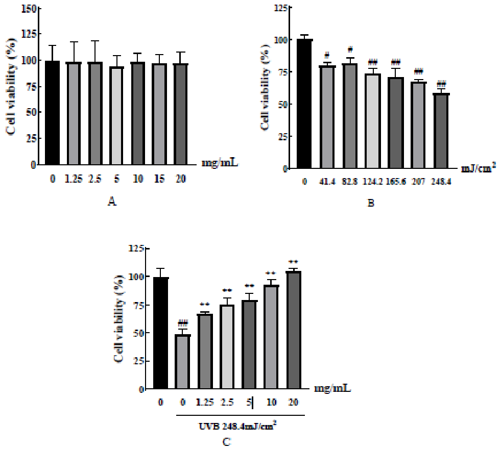
Figure 2.
The effects of DHSC on UVB-induced ROS production in HaCaT cells. A. Normal group. UV(-)+Vehicle. B. Model group. UV(+)+Vehicle. C. Protection group.UV(+)+DHSC(20mg/mL).
Figure 2.
The effects of DHSC on UVB-induced ROS production in HaCaT cells. A. Normal group. UV(-)+Vehicle. B. Model group. UV(+)+Vehicle. C. Protection group.UV(+)+DHSC(20mg/mL).
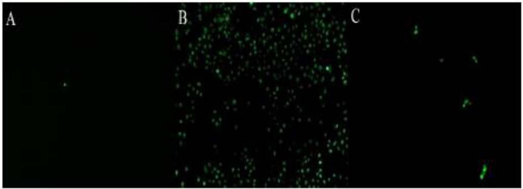
Figure 3.
The effects of DHSC on UVB-induced oxidative stress in HaCaT cells. A. SOD activity.B. MDA content. #p <0.05 compared with the normal group, ∗∗ p<0.05 compared with the model group.
Figure 3.
The effects of DHSC on UVB-induced oxidative stress in HaCaT cells. A. SOD activity.B. MDA content. #p <0.05 compared with the normal group, ∗∗ p<0.05 compared with the model group.
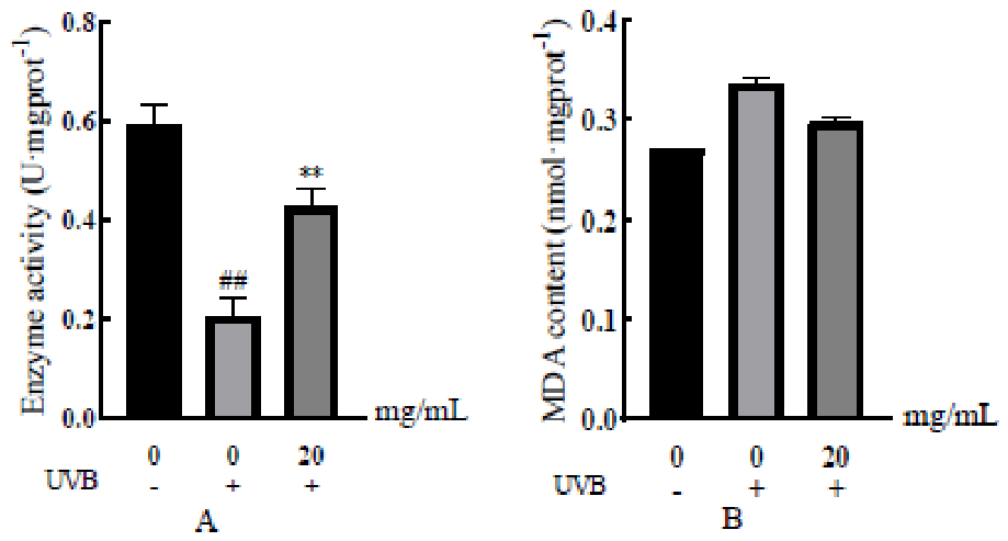
Figure 4.
The effects of DHSC on UVB-induced inflammation in HaCaT cells. A. IL-1β. B.TNF-α.C. IL-6. #p <0.05 compared with the normal group.
Figure 4.
The effects of DHSC on UVB-induced inflammation in HaCaT cells. A. IL-1β. B.TNF-α.C. IL-6. #p <0.05 compared with the normal group.
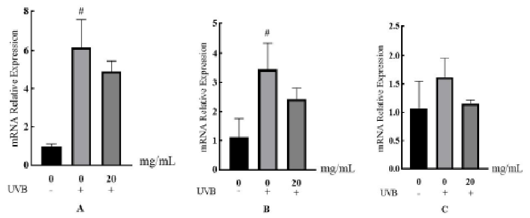
Figure 5.
Effect of DHSC on skin phenotype in mice with acute photodamage. A. The CTL group. UV(-)+Vehicle. B. The MOD group. UV(+)+Vehicle. C. L group. UV(+)+DHSC(80mg/mL). D.M group. UV(+)+DHSC(120mg/mL). E. H group. UV(+)+DHSC(160mg/mL). F. B group.UV(+)+matrix. G. Y group. UV(+)+commercially available sunscreen.
Figure 5.
Effect of DHSC on skin phenotype in mice with acute photodamage. A. The CTL group. UV(-)+Vehicle. B. The MOD group. UV(+)+Vehicle. C. L group. UV(+)+DHSC(80mg/mL). D.M group. UV(+)+DHSC(120mg/mL). E. H group. UV(+)+DHSC(160mg/mL). F. B group.UV(+)+matrix. G. Y group. UV(+)+commercially available sunscreen.
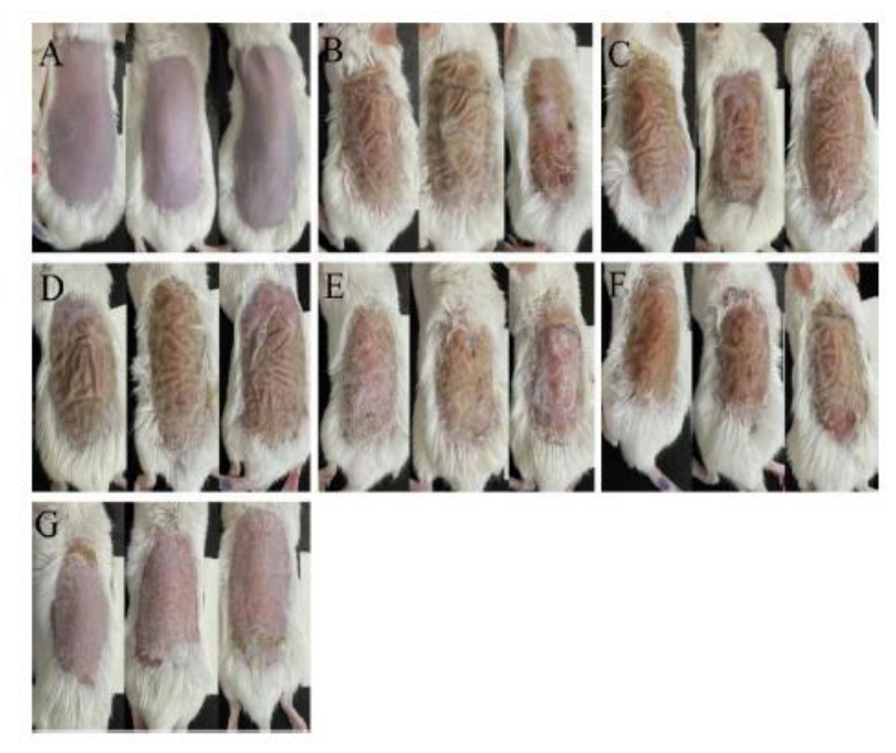
Figure 6.
Erythema scores of mice in each group.(N=9) #p <0.05 compared with the CTL group, ∗∗p<0.05 compared with the MOD group.
Figure 6.
Erythema scores of mice in each group.(N=9) #p <0.05 compared with the CTL group, ∗∗p<0.05 compared with the MOD group.
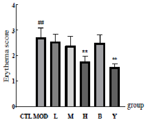
Figure 7.
Effect of DHSC on skin histopathology in mice with acute photodamage. A. The CTL group. UV(-)+Vehicle. B. The MOD group. UV(+)+Vehicle. C. L group. UV(+)+DHSC(80mg/mL). D. M group. UV(+)+DHSC(120mg/mL). E. H group. UV(+)+DHSC(160mg/mL). F. B group. UV(+)+matrix. G. Y group. UV(+)+commercially available sunscreen.
Figure 7.
Effect of DHSC on skin histopathology in mice with acute photodamage. A. The CTL group. UV(-)+Vehicle. B. The MOD group. UV(+)+Vehicle. C. L group. UV(+)+DHSC(80mg/mL). D. M group. UV(+)+DHSC(120mg/mL). E. H group. UV(+)+DHSC(160mg/mL). F. B group. UV(+)+matrix. G. Y group. UV(+)+commercially available sunscreen.
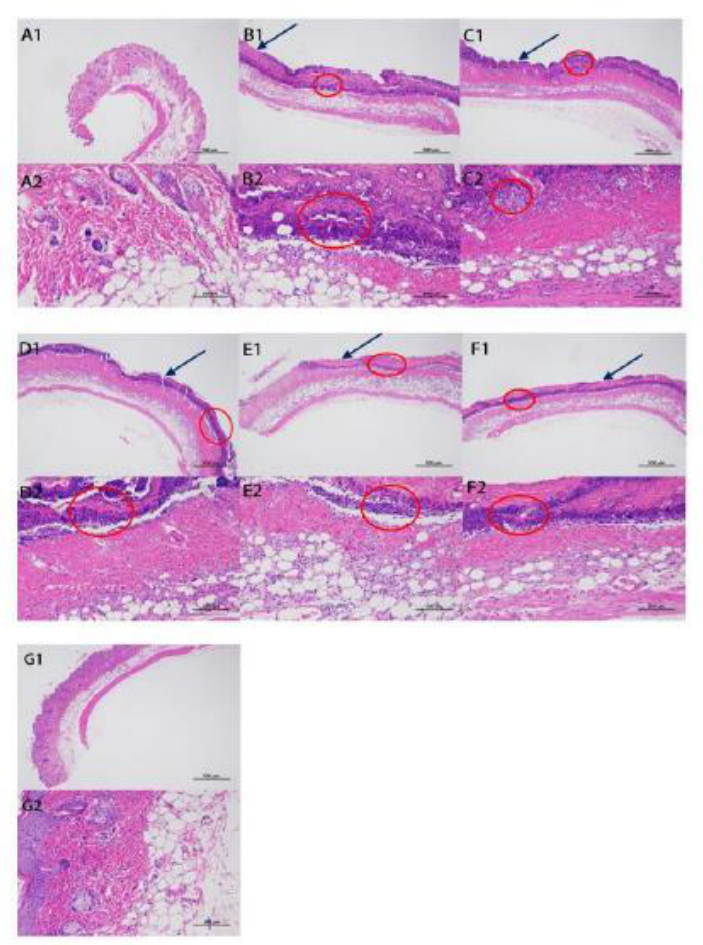
Figure 8.
Effects of DHSC on skin collagen structure in mice with acute photodamage. A. The CTL group. UV(-)+Vehicle. B. The MOD group. UV(+)+Vehicle. C. L group. UV(+)+DHSC(80mg/mL). D. M group. UV(+)+DHSC(120mg/mL). E. H group. UV(+)+DHSC(160mg/mL). F. B group. UV(+)+matrix. G. Y group. UV(+)+commercially available sunscreen.
Figure 8.
Effects of DHSC on skin collagen structure in mice with acute photodamage. A. The CTL group. UV(-)+Vehicle. B. The MOD group. UV(+)+Vehicle. C. L group. UV(+)+DHSC(80mg/mL). D. M group. UV(+)+DHSC(120mg/mL). E. H group. UV(+)+DHSC(160mg/mL). F. B group. UV(+)+matrix. G. Y group. UV(+)+commercially available sunscreen.
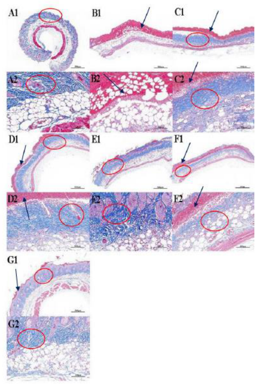
Figure 9.
Biochemical indexes of oxidative stress in each group of mice. A. ROS. B. SOD. C. MDA D. Nrf2. #p <0.05 compared with the CTL group, ∗∗p<0.05 compared with the MOD group.
Figure 9.
Biochemical indexes of oxidative stress in each group of mice. A. ROS. B. SOD. C. MDA D. Nrf2. #p <0.05 compared with the CTL group, ∗∗p<0.05 compared with the MOD group.
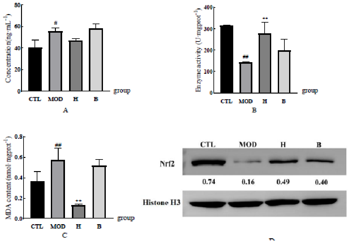
Figure 10.
Expression of AP-1 protein in mouse skin tissues in each group.
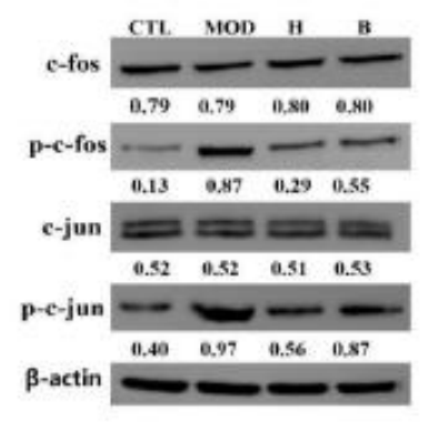
Figure 11.
Expression of MAPK pathway protein in mice skin tissues in each group.
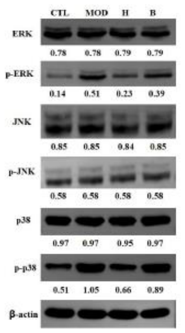
Figure 12.
Expression of inflammatory factors in skin tissue of mice in each group. A. IL-1β. B.TNF-α. C. IL-6. D. p-p65 #p <0.05 compared with the CTL group, ∗∗p<0.05 compared with the MOD group.
Figure 12.
Expression of inflammatory factors in skin tissue of mice in each group. A. IL-1β. B.TNF-α. C. IL-6. D. p-p65 #p <0.05 compared with the CTL group, ∗∗p<0.05 compared with the MOD group.
Figure 13.
Expression of MMPs in skin tissue of mice in each group. A. MMP-1. B. MMP-3.C.MMP-9. #p <0.05 compared with the CTL group, ∗∗p<0.05 compared with the MOD group.
Figure 13.
Expression of MMPs in skin tissue of mice in each group. A. MMP-1. B. MMP-3.C.MMP-9. #p <0.05 compared with the CTL group, ∗∗p<0.05 compared with the MOD group.
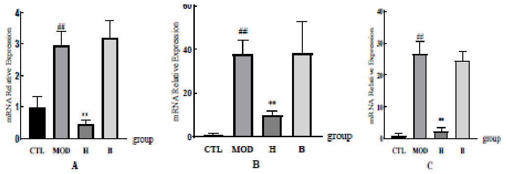
Figure 14.
Expression of collagen-related indexes in skin tissue of mice in each group. A.Type I collagen. B. Type III collagen. C. Hydroxyproline. #p <0.05 compared with the CTL group, ∗ ∗p<0.05 compared with the MOD group.
Figure 14.
Expression of collagen-related indexes in skin tissue of mice in each group. A.Type I collagen. B. Type III collagen. C. Hydroxyproline. #p <0.05 compared with the CTL group, ∗ ∗p<0.05 compared with the MOD group.
Disclaimer/Publisher’s Note: The statements, opinions and data contained in all publications are solely those of the individual author(s) and contributor(s) and not of MDPI and/or the editor(s). MDPI and/or the editor(s) disclaim responsibility for any injury to people or property resulting from any ideas, methods, instructions or products referred to in the content. |
© 2026 by the authors. Licensee MDPI, Basel, Switzerland. This article is an open access article distributed under the terms and conditions of the Creative Commons Attribution (CC BY) license.
Copyright: This open access article is published under a Creative Commons CC BY 4.0 license, which permit the free download, distribution, and reuse, provided that the author and preprint are cited in any reuse.