

Submitted:
03 September 2026
Posted:
04 September 2026
You are already at the latest version
Abstract
Polymeric microneedles have emerged as promising platforms for minimally invasive drug delivery, yet their clinical translation remains limited despite extensive proof-of-concept research. This review critically examines polymeric microneedles from a function-driven design perspective, linking classification, fabrication, and material selection to the principal determinants of performance and translational success. Particular attention is given to the interplay among microneedle geometry, polymer properties, skin biomechanics, and application conditions, which collectively define the mechanical window required for reliable insertion. Drug release is considered a programmable design function that should be aligned with dose and pharmacological objectives, while polymer dissolution, degradation, persistence, and potential accumulation are evaluated in relation to safety and regulatory acceptance. Stimuli-responsive and hybrid systems are also assessed by weighing their functional benefits against the additional complexity they introduce. Recurring mechanical, pharmacokinetic, manufacturing, and clinical design–performance mismatches are identified as major contributors to the gap between laboratory success and clinical implementation. Based on these findings, a function-driven framework is proposed that integrates therapeutic objectives, insertion performance, release kinetics, manufacturability, and safety from the earliest stages of development, thereby supporting the rational design of clinically viable next-generation microneedle systems.
Keywords:
microneedles
; transdermal drug delivery
; drug release programming
; insertion mechanics
; polymer fate
; translational barriers
; biomedical polymers
1. Introduction
The development of alternative routes for drug delivery remains a central objective in pharmaceutical research, driven by persistent limitations of conventional administration pathways. Oral delivery, while convenient, is frequently associated with reduced bioavailability due to gastrointestinal degradation and extensive first-pass metabolism, particularly for peptide- and protein-based therapeutics, stomach irritation and side effects [1,2]. Parenteral administration, although capable of achieving efficient systemic exposure, is often constrained by pain, the need for trained personnel, and reduced patient compliance, especially in the context of chronic therapy [1]. These limitations have stimulated growing interest in minimally invasive delivery strategies that aim to combine the efficacy of injections with the convenience of non-invasive approaches.
Transdermal and intradermal delivery systems have emerged as attractive alternatives, enabling controlled drug release, avoidance of first-pass metabolism, and improved patient adherence [3]. However, the effectiveness of conventional transdermal systems is inherently limited by the barrier function of the stratum corneum, which restricts permeation primarily to small, lipophilic molecules [4]. Consequently, expanding the range of therapeutics suitable for transdermal delivery remains a major challenge in the field.
Microneedle-based systems have been developed to overcome this limitation by creating micron-scale pathways across the stratum corneum, thereby enabling direct access to the viable epidermis and dermis while avoiding nerve-rich regions associated with pain [1,5]. Since their initial conceptualization, microneedles have evolved into a highly versatile platform encompassing multiple delivery strategies, including solid, coated, dissolving, hydrogel-forming, and hollow systems, each tailored to achieve specific pharmacokinetic and therapeutic objectives [5,6]. This versatility has established microneedles as a promising alternative route of administration capable of delivering a wide range of molecules, from small drugs to vaccines, peptides, and nucleic acids [6,7,8].
Among the various microneedle platforms, systems based on polymers have attracted particular attention due to their versatility and tunability.
Polymeric microneedles (MNs) are minimally invasive transdermal drug delivery systems consisting of micron-scale projections fabricated from biodegradable, biocompatible, or water-soluble polymers, which penetrate the stratum corneum to create transient pathways for drug transport into the skin. Their polymeric composition provides considerable flexibility in controlling mechanical strength, dissolution or degradation behavior, swelling, drug loading, and release kinetics profiles [9]. Polymers such as poly(vinylpyrrolidone) (PVP), poly(vinyl alcohol) (PVA), poly(lactic-co-glycolic acid) (PLGA), and polycaprolactone (PCL) have been widely investigated for microneedle fabrication, supporting applications ranging from rapid drug delivery to sustained and long-acting release systems [9,10]. Based on their behavior following skin insertion, dissolving and hydrogel-forming microneedles represent two important classes of polymeric MNs, offering distinct mechanisms for drug release and delivery. This versatility has driven substantial innovation in the field, enabling the development of polymeric microneedles tailored to diverse therapeutic requirements.
Despite the rapid expansion of research activity in this area, a significant gap persists between proof-of-concept studies and clinically translated microneedle systems. Although hundreds of studies report promising in vitro and preclinical results, only a limited number of microneedle-based products have progressed toward commercialization or clinical adoption [10,11]. This discrepancy reflects a fundamental limitation of the current literature: most studies emphasize feasibility rather than critically evaluating whether a given system is optimally designed for its intended therapeutic purpose.
A key limitation of existing reviews lies in their predominantly material- or technology-centered perspective, in which microneedle systems are classified according to polymer type, fabrication method, or structural design [5,10]. While such classifications provide valuable descriptive frameworks, they do not adequately explain why certain systems achieve clinically relevant performance whereas others remain confined to experimental validation. Increasing evidence indicates that translational success depends not solely on material properties, but on the alignment between microneedle design, drug release kinetics, and pharmacological requirements [12].
In this context, there is a clear need for a more integrative perspective that links material selection, fabrication strategy, and system architecture to functional performance and clinical relevance. Meeting this need requires moving beyond descriptive classification toward a function-driven framework, in which microneedle systems are evaluated according to their ability to satisfy clearly defined therapeutic objectives.
Accordingly, this review provides a comprehensive analysis of polymeric microneedles as an alternative route for drug delivery, integrating both classical and function-oriented perspectives. It is structured in two complementary parts. The first part outlines microneedle classification, fabrication methods, and material selection, thereby defining the fundamental design space of these systems. The second part adopts a function-driven perspective, focusing on the key determinants of microneedle performance, including mechanical integrity, release programming, stimuli-responsiveness, and polymer fate in the skin, as well as the factors limiting clinical translation. By identifying recurring design–performance relationships, this review aims to establish a rational framework for the development of next-generation microneedle systems with improved translational potential.
2. Classification of Microneedles
Microneedle systems can be broadly classified according to their mechanism of action and mode of drug delivery as solid, coated, hollow, dissolving, biodegradable, implantable and hydrogel-forming microneedles. This classification not only provides a structural overview of the field but also highlights fundamental differences in drug delivery efficiency, dosing control, and clinical applicability [5,11].
Below is a comparative table (Table 1) that reflects the general trends reported for microneedles in the literature [1,5,6,7,13,14,15,16,17,18,19,20,21,22,23,24,25,26,27,28,29,30]. Because performance depends on material composition, geometry, drug type, and application conditions, the rankings should be interpreted as relative qualitative estimates rather than absolute value. The qualitative ranking (very low to very high) reflects relative differences among the various microneedle types and does not represent a comparison with alternative drug delivery platforms.
The classification of microneedle systems based on their mechanism of action is schematically illustrated in Figure 1.
Although these categories are traditionally presented as discrete, many contemporary microneedle systems incorporate hybrid features that combine multiple mechanisms of action. Importantly, classification based solely on structure or material does not fully explain the clinical performance of microneedle systems. As will be discussed in subsequent sections, successful translation depends on the alignment between design parameters and therapeutic objectives rather than on categorical classification alone.
3. Fabrication Methods
Microneedle fabrication methods play a critical role in determining the mechanical properties, drug loading capacity, reproducibility, and scalability of polymeric microneedle systems. While a wide range of microfabrication techniques has been explored, the choice of fabrication method is not merely a technical consideration but a fundamental design parameter that directly influences the translational feasibility of the final system [39,40].
To provide a structured comparison of the principal fabrication techniques used in polymeric microneedle development, their key advantages and limitations are summarized in Table 2. As shown, fabrication method selection has direct implications not only for microneedle geometry and drug loading but also for scalability, reproducibility, and overall translational feasibility [39,41,42,43,44].
Hybrid fabrication strategies that combine multiple techniques have been developed to overcome the limitations of individual methods. For example, micromolding combined with layer-by-layer assembly or spray deposition enables the fabrication of multilayer microneedles with spatially controlled drug distribution [46]. Similarly, the integration of nanoparticles or responsive materials during fabrication allows the development of multifunctional systems with tailored release properties. While these approaches demonstrate significant innovation, they also introduce additional complexity in manufacturing, quality control, and regulatory validation. The increased number of process steps and material interfaces may reduce reproducibility and complicate scale-up, thereby reinforcing the need for careful alignment between fabrication strategy and intended clinical application.
Importantly, fabrication methods are closely linked to many of the translational challenges discussed in later sections. Variability in microneedle geometry, inconsistencies in drug distribution, and limitations in sterilization and scalability can often be traced back to the selected fabrication approach. Accordingly, fabrication should be considered an integral component of function-driven design rather than a downstream technical step.
As indicated in Table 2, no single fabrication method simultaneously satisfies all the requirements for ideal microneedle production, including mechanical robustness, precise drug loading, scalability, and cost-effectiveness. Instead, each technique involves inherent trade-offs that must be carefully balanced according to the intended application.
Micromolding remains the dominant approach due to its versatility and compatibility with drug-loaded systems; however, its sensitivity to processing conditions introduces variability that may hinder reproducibility at scale. In contrast, industrial techniques such as injection molding offer superior consistency and scalability but are limited by material constraints and incompatibility with thermolabile drugs. Emerging technologies such as 3D printing offer unprecedented design flexibility but currently face limitations in resolution and material availability, which restrict their widespread adoption.
Importantly, many of the translational challenges discussed in later sections, particularly those related to dose variability, mechanical failure, and manufacturing reproducibility, can be traced directly to limitations inherent in the selected fabrication method. This observation underscores that fabrication strategy should not be considered a purely technical choice, but rather an integral component of function-driven microneedle design.
In parallel with fabrication strategy, material selection represents a critical determinant of microneedle performance. The physicochemical properties of the polymers used not only influence manufacturability but also define mechanical behavior, drug loading capacity, and release characteristics.
4. Materials for Polymeric Microneedles
Microneedle performance is fundamentally governed by the physicochemical properties of the materials used for their fabrication. Polymer selection determines not only the mechanical strength required for effective skin penetration but also drug loading capacity, release kinetics, biocompatibility, and, ultimately, the translational potential of the system. Accordingly, materials should not be viewed as passive carriers but as active determinants of microneedle function and clinical applicability [6,12]. Based on their behavior following skin insertion, the polymers used for MN fabrication can be broadly categorized as water-soluble, biodegradable, or hydrogel-forming materials, each offering distinct advantages and limitations (Table 3).
Recent advances in polymer science have enabled the development of stimuli-responsive materials, including thermoresponsive polymers such as poly(N-isopropylacrylamide) (PNIPAm), pH-responsive polymers, and hybrid systems incorporating nanoparticles or bioactive components [10]. These materials offer the potential for on-demand drug release in response to physiological or external stimuli. However, their integration into microneedle systems introduces additional complexity in formulation, fabrication, and regulatory evaluation. In many cases, this added functionality must be carefully justified against the increased risk of variability, reduced reproducibility, and potential safety concerns associated with multi-component systems [10,49].
Importantly, material selection is intrinsically linked to fabrication methods and performance characteristics. Mechanical properties, degradation behavior, and drug–polymer interactions collectively determine whether a given microneedle system can achieve its intended therapeutic function. As will be discussed in later sections, successful translation depends not on the intrinsic novelty of the material but on its alignment with clearly defined pharmacological and clinical requirements.
Across all material classes, a recurring theme is that no polymer is intrinsically optimal for microneedle fabrication. Instead, material suitability is context-dependent and must be evaluated in relation to the intended therapeutic application, required release profile, and clinical use conditions. This observation reinforces the central premise of this review: material selection should be guided by functional requirements rather than by material availability or novelty. Integrating material properties with mechanical design, fabrication strategy, and pharmacological objectives represents a critical step toward the rational development of clinically viable microneedle systems.
5. Factors Limiting Drug Delivery Efficiency in Polymeric Microneedles and Design Strategies to Overcome Them
Polymeric microneedles have emerged as versatile platforms for transdermal drug delivery, offering minimally invasive administration and the potential for improved therapeutic efficacy and patient compliance [50]. Despite the substantial volume of research on polymeric microneedles over the past two decades, the number of systems that have progressed from proof-of-concept to clinical implementation remains strikingly limited. According to recent comprehensive analyses, fewer than 20 drug substances are currently delivered transdermally in any format, and no dissolving polymeric microneedle array has yet achieved successful commercialization. This disparity reflects a persistent translational gap that cannot be explained solely by technological immaturity [51,52,53].
The amount of drug incorporated into a microneedle array does not necessarily correspond to the amount successfully delivered into or across the skin. Drug delivery efficiency is governed by a series of interconnected processes, including successful skin penetration, polymer dissolution or swelling, drug release from the polymeric matrix, diffusion through the skin microenvironment, and subsequent absorption at the target site as presented in Figure 2. Failure or suboptimal performance at any of these stages may result in incomplete or highly variable drug delivery, thereby limiting therapeutic outcomes and hindering clinical translation [12,54].
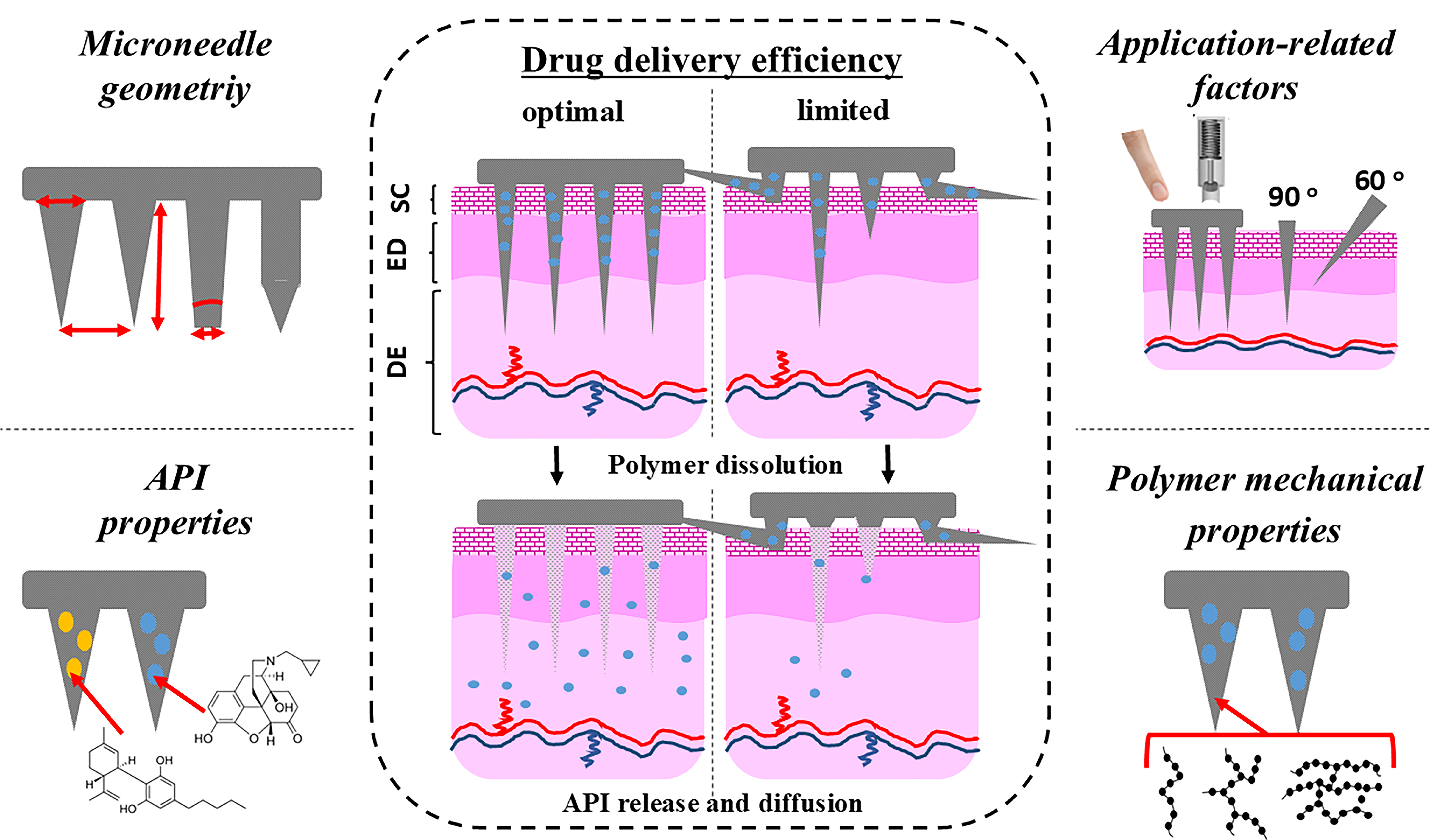
From a systems-level perspective, translational failure in polymeric microneedles can be broadly categorized into four interrelated domains: (i) mechanical–insertion mismatch, (ii) pharmacokinetic–dose mismatch, (iii) manufacturing and scalability limitations, and (iv) clinical–context misalignment. These domains are not independent; instead, they frequently interact, creating compounded barriers that prevent otherwise promising systems from advancing toward clinical implementation. Table 4 summarizes the principal subdomains within these four domains and illustrates their interconnected contributions to translational success or failure.
The factors influencing drug delivery efficiency are multifactorial and arise from their combined effects. Mechanical failure during insertion may prevent adequate skin penetration [5,59], while inappropriate polymer properties can impair dissolution, swelling, or drug diffusion [12]. In addition, drug-specific factors such as poor solubility, instability, or unfavorable interactions with the polymer matrix may reduce drug release, whereas variations in skin physiology and user-dependent application conditions further contribute to delivery variability [6]. Therefore, understanding the mechanisms responsible for ineffective drug delivery is essential for the rational design of next-generation polymeric microneedles with improved performance and greater potential for clinical implementation [7]. While manufacturing scalability and broader clinical implementation represent important considerations for translation, the present review focuses on the design-related determinants that directly govern drug delivery performance. Specifically, it examines how the physicochemical properties of the encapsulated drug, microneedle geometry, polymer-related factors, skin and application-related factors, release programming, and polymer fate interact to influence insertion efficiency, drug loading, release behavior, skin permeation, and overall therapeutic performance. By integrating these interconnected design parameters within a function-driven framework, this review highlights the critical mechanisms through which rational material and structural optimization can improve the translational potential of polymeric microneedle systems [11].
5.1. Mechanical–Insertion Performance
Mechanical performance represents the first and indispensable functional requirement for polymeric microneedles intended as an alternative route for drug delivery. Independent of polymer chemistry or drug loading strategy, microneedles must reliably penetrate the stratum corneum without fracturing, bending, or undergoing uncontrolled deformation. Mechanical–insertion performance represents the ability of polymeric microneedles to reliably penetrate the skin while maintaining their structural integrity and creating reproducible microchannels for effective drug delivery. It encompasses the interplay between microneedle geometry, polymer mechanical properties, skin characteristics, and application parameters, including insertion force, angle, method, and residence time, all of which collectively determine insertion efficiency and subsequent drug delivery performance. Failure to meet this requirement compromises drug delivery efficiency, reproducibility, and safety, ultimately limiting clinical translation [5,59,60].
5.1.1. Microneedle Geometry
Microneedle geometry plays a critical role in determining overall device performance, as it directly influences mechanical strength, skin insertion efficiency, and drug delivery outcomes. The most commonly investigated geometrical parameters include needle shape, base diameter, needle length, tip diameter, and inter-needle spacing, all of which must be carefully optimized to achieve effective transdermal penetration while maintaining structural integrity. In addition, the aspect ratio and apex angle are key design determinants that govern the balance between insertion capability and mechanical robustness. Collectively, these geometric features, presented in Figure 3, define the interaction of microneedles with the skin barrier and are therefore essential considerations in the rational design of polymeric microneedle systems.
The geometry of microneedle tips strongly influences insertion efficiency, mechanical strength, and pain perception [61,62]. Common shapes include conical, pyramidal, bevelled, and arrowhead structures as visualized in Figure 3 (A). Among these, conical and pyramidal geometries are most widely used for polymeric microneedles, as they provide a good balance between manufacturability and mechanical stability [63]. Sharp-tipped geometries are preferred because they reduce insertion force and improve skin penetration reliability. In general, sharper and more gradually tapered structures enhance insertion success while minimizing tissue deformation. The influence of microneedle shape on the mechanical performance, insertion behavior, and drug delivery characteristics of polymeric microneedle systems is summarized in Table 5.
Microneedle Base Diameter
The base diameter determines the mechanical robustness of the microneedle. Larger base diameters increase mechanical strength but may reduce insertion efficiency due to increased skin deformation and higher insertion force requirements. For polymeric microneedles, typical base diameters are in the range of 100–300 µm, with optimization aimed at ensuring sufficient mechanical strength while maintaining a small footprint to reduce the “bed-of-nails” effect in dense arrays [33,35,56,59,69,70,71].
Microneedle Length
Needle length is a critical parameter controlling the depth of skin penetration and thus drug delivery location (epidermis vs dermis). Most effective microneedles are designed to penetrate the stratum corneum and reach the viable epidermis or upper dermis without stimulating deeper nerves or blood vessels. Optimal lengths are typically 300–1000 µm for polymeric microneedles. Shorter needles failed to overcome skin deformation and may lead to incomplete barrier bypass, while excessively long needles increase pain risk and fracture probability [59,69].
Microneedle Tip Aspect Ratio
The aspect ratio (height/base diameter) governs both mechanical strength and insertion capability. High aspect ratios improve penetration ability but increase the risk of buckling and fracture, particularly in polymeric systems. For polymeric microneedles, a commonly reported optimal aspect ratio is 2:1 to 5:1. Within this range, microneedles can maintain structural integrity while still achieving effective skin penetration [33,35,56,59,69,70,71].
Microneedle Tip Diameter
The tip diameter is one of the most important determinants of insertion force. Sharper tips (smaller diameters) significantly reduce penetration resistance and improve reproducibility of insertion. For efficient skin piercing, tip diameters are generally designed to be below approximately 30 µm, with the smallest feasible tip diameter preferred to minimize insertion force while maintaining structural integrity [33,35,56,59,69,70,71]. MN tips less than 5 µm are easily prone to breaking and bending [35].
Apex (Tip) Angle
The apex angle defines the sharpness of the microneedle tip and strongly influences insertion force and mechanical stress distribution. A smaller apex angle results in sharper tips and lower insertion force but may reduce mechanical strength. Conversely, larger angles increase robustness but require higher force for insertion. Typical optimized apex angles are in the range of 20°–60°, with sharper angles generally preferred for dissolving and hydrogel-forming polymeric microneedles where insertion efficiency is critical [33,35,56,59,69,70,71].
MN Tip Spacing (Interspacing)
Inter-needle spacing is a critical design parameter that influences both insertion efficiency and drug delivery performance. Insufficient spacing may promote the bed-of-nails effect, leading to skin deformation and incomplete insertion, whereas excessive spacing reduces needle density and the total drug-loading capacity of the microneedle array. Typical spacing values range from 300–800 µm, depending on needle height and array density. Optimized spacing ensures adequate skin recovery between insertion points while maintaining sufficient delivery surface area [25,35,51,59,64,69].
The interplay between microneedle geometry and mechanical performance is summarized in Table 6 and visualized in Figure 4.
Overall, the geometric design of microneedles requires a careful balance between several interdependent performance criteria, including mechanical strength, insertion efficiency, and patient safety. Needle height, base diameter, tip radius, tip angle, and aspect ratio determine stress distribution during insertion and resistance to buckling or fracture [14,73] and must be optimized collectively rather than independently, as improving one feature often compromises another. For example, sharper and higher aspect ratio structures can significantly enhance skin penetration and reduce insertion force, but may also increase the risk of mechanical failure such as buckling or fracture, particularly in polymer-based systems during manual or self-administration [74]. Conversely, increasing base diameter can improve structural integrity but may reduce insertion efficiency due to greater skin deformation and higher insertion force requirements. Similarly, needle density must be optimized to maximize drug delivery area while minimizing the “bed-of-nails” effect. The last one can hinder effective penetration, influence force distribution across the patch, affect penetration uniformity and user-dependent variability [75]. Therefore, an optimal microneedle geometry represents a compromise between structural robustness, reliable skin insertion, and minimal tissue trauma, all of which are essential for achieving consistent drug delivery and supporting clinical translation of polymeric microneedle systems. These relationships highlight that mechanical performance must be evaluated as an integrated system property rather than an isolated material characteristic [14].
Several representative studies demonstrate how mechanical performance governs the success or failure of polymeric microneedle systems, largely independent of their chemical composition.
Kochhar et al. systematically evaluated the effect of needle height, base diameter, and tip angle on skin penetration and showed that increasing needle height beyond an optimal threshold does not necessarily improve penetration efficiency but instead increases the likelihood of buckling and fracture, particularly for polymeric microneedles with moderate elastic modulus. Their results emphasized that geometric optimization must be tailored to the mechanical properties of the selected polymer rather than maximized indiscriminately [51].
Practical limitations associated with blunt or short microneedles have been reported in investigations focusing on manual and self-administration scenarios. Gill and Prausnitz showed that microneedles with insufficient height or overly blunt tips require higher and less reproducible application forces, resulting in large inter-user variability and incomplete skin penetration [74]. These findings are particularly relevant for patch-based systems intended for home use, where controlled application forces cannot be guaranteed.
The role of tip diameter in microneedle insertion was investigated by Römgens et al., who demonstrated that tip diameter influences both the force required for skin penetration and the resulting penetration depth [76]. Beyond geometry, the magnitude of the applied force is also critical. Cheung et al. showed that insufficient insertion force may result in incomplete skin penetration and negligible insulin permeation, whereas increasing the applied force significantly enhanced insulin delivery across porcine skin [77]. Together, these findings demonstrate that reliable microneedle performance depends on the interaction between tip geometry and application force rather than on either parameter alone.
Early work by Park et al. showed that biodegradable PLA and PLGA microneedles with sufficient axial fracture force could reliably penetrate murine and human skin models, provided that needle geometry and tip sharpness were appropriately optimized [25]. However, the same study revealed that mechanically robust microneedles with suboptimal tip geometry exhibited incomplete insertion despite high fracture resistance, underscoring that material strength alone is insufficient to ensure functional penetration. This apparent contradiction reflects the fact that successful skin penetration is governed not by absolute mechanical strength, but by the balance between insertion force and fracture resistance within a narrow mechanical window (Figure 5). While PLA- and PLGA-based microneedles can achieve high axial fracture force, suboptimal tip geometry—particularly increased tip radius or a blunted apex—significantly increases the force required to breach the stratum corneum. As a result, even mechanically robust structures may fail to penetrate effectively, as the applied force is dissipated through skin deformation rather than concentrated at the needle tip. In contrast, microneedles with sharper tips and optimized aspect ratios reduce the insertion force threshold, enabling efficient penetration at forces well below the fracture limit. This interplay highlights that geometry-driven stress concentration at the needle–skin interface is a more critical determinant of functional insertion than bulk material strength alone.
Similarly, Davis et al. [14] systematically measured both parameters across microneedles with varying geometries and materials and showed that successful skin penetration occurs only within a narrow mechanical window, where the applied force exceeds the resistance of the stratum corneum but remains below the fracture threshold of the microneedle [14]. Their work revealed that increasing polymer stiffness or needle thickness beyond this window does not necessarily improve insertion efficiency and may instead promote brittle failure or uncontrolled fracture.
This behavior arises from the fundamentally different physical processes governing microneedle deformation and skin penetration. While axial fracture force reflects structural resistance to compressive failure, successful insertion depends on the generation of sufficiently high localized stress at the needle tip to overcome the barrier properties of the stratum corneum [14]. In systems with suboptimal tip geometry, the applied force is distributed over a larger contact area, reducing stress concentration and promoting skin deformation rather than penetration. Consequently, the skin behaves as a viscoelastic material that absorbs and redistributes the applied load, resulting in elastic indentation instead of barrier disruption [14].
Moreover, this mismatch is amplified by the time-dependent mechanical response of skin. Under typical application conditions, viscoelastic relaxation of the stratum corneum leads to partial dissipation of the applied force, thereby increasing the effective insertion threshold [14]. In such cases, even microneedles with high fracture resistance may fail to penetrate if the rate and mode of force application are not optimized. In contrast, microneedles with sharper tips and optimized aspect ratios promote stress localization and minimize energy dissipation, enabling efficient penetration at lower applied forces and within shorter timeframes [14].
Importantly, this highlights that insertion efficiency is not solely a function of material properties, but rather an emergent property arising from the interplay between geometry, polymer mechanics, and skin biomechanics.
Geometric effects have been further emphasized in studies examining array configuration and interspacing. Olatunji et al. demonstrated that increasing microneedle density within an array can significantly elevate the force required for successful penetration due to load sharing across needles, even when individual needles possess sufficient mechanical strength [64]. This phenomenon has important implications for patch design, as mechanically sound microneedles may fail collectively if array-level parameters are not properly optimized [64].
This behavior reflects the transition from single-needle mechanics to collective array behavior, in which force distribution and skin deformation become dominant factors. While individual microneedles may be designed to exceed the critical insertion threshold, increasing array density distributes the applied force across multiple needles rather than concentrating it at a single tip. As a result, the effective force per needle decreases, often falling below the threshold required to overcome the mechanical resistance of the stratum corneum [64].
In addition to load sharing, closely spaced microneedles induce cooperative deformation of the skin surface, leading to increased tissue indentation and lateral stress redistribution. Rather than acting as independent penetration units, densely packed arrays interact mechanically through the underlying skin, which behaves as a compliant, viscoelastic substrate. This interaction reduces stress localization at each needle tip and increases the total force required for successful insertion. Consequently, even arrays composed of mechanically robust microneedles may fail to penetrate uniformly if interspacing is insufficient [64].
Furthermore, this phenomenon is strongly influenced by application conditions. Under manual administration, variability in applied force and insertion angle can exacerbate uneven load distribution across the array, resulting in partial insertion or heterogeneous penetration depth. In contrast, optimized interspacing promotes more independent needle action, minimizing mechanical interference and improving insertion reliability [64].
These findings underscore that microneedle performance must be evaluated at the array level rather than solely at the level of individual needles. Failure to account for these collective mechanical effects represents a key source of discrepancy between in vitro mechanical characterization and in vivo performance.
Importantly, studies employing optical coherence tomography have demonstrated that actual microneedle penetration depth depends strongly on needle geometry and application force and may remain considerably lower than the nominal needle height. Donnelly et al. showed that increasing microneedle height and application force significantly increased penetration depth in neonatal porcine skin, highlighting the importance of evaluating insertion directly in skin rather than relying exclusively on nominal dimensions [78]. These observations illustrate a recurring design limitation in which mechanical performance is assessed in isolation, without accounting for the complex viscoelastic nature of skin [52,75,79].
Importantly, mechanical performance must be evaluated in relation to the intended drug delivery application. For intradermal vaccination, partial penetration may be sufficient provided antigen deposition occurs within immune-active skin layers [74,80,81]. In contrast, microneedle systems designed for systemic delivery or long-acting implantation require deeper and more consistent penetration to ensure reliable dosing and pharmacological effect [28,53]. Failure to align mechanical design with pharmacological intent has contributed to numerous systems demonstrating promising in vitro characteristics but limited in vivo efficacy [66].
5.1.2. Polymer Mechanical Properties
Polymer-related factors influencing drug delivery efficiency arise primarily from the physicochemical and mechanical characteristics of the polymeric matrix, which govern microneedle insertion, dissolution or swelling behavior, drug release kinetics, diffusion through the polymer network, and formulation reproducibility. Key determinants include polymer molecular weight, concentration, chemical structure, hydrophilicity, crosslink density, swelling capacity, dissolution rate, mechanical modulus, glass transition temperature, crystallinity, drug–polymer interactions, mesh size, degradation behavior, and residual moisture content.
Polymeric microneedles are usually fabricated from water-soluble, biodegradable and hydrogel polymers, either as single-component systems or polymer blends. Since these materials are generally mechanically weaker than silicon, metals, or ceramics, careful material selection is required. Achieving an optimal balance between strength, flexibility, and dissolution with a single material is often difficult; therefore, composite formulations are frequently used to improve both insertion performance and drug release [35]. Table 7 summarizes the principal polymer-related factors that influence the manufacturing quality, mechanical properties, drug release characteristics, and overall performance of polymeric microneedle systems.
Polymer-related parameters such as molecular weight, glass transition temperature (Tg), crystallinity, and elastic modulus directly influence microneedle stiffness and brittleness, thereby affecting insertion behavior [7,86]. Polymers with high Tg and crystallinity may provide sufficient mechanical strength but are also more prone to brittle fracture, whereas more ductile polymers may deform during insertion, reducing effective penetration depth [22]. These observations underscore that mechanical robustness alone does not guarantee functional skin insertion.
The influence of polymer-related parameters on microneedle insertion behavior has been clearly demonstrated across several commonly used synthetic systems. For instance, Park et al. reported that PLA- and PLGA-based microneedles with relatively high glass transition temperatures and semi-crystalline structures exhibited high fracture forces but were susceptible to brittle failure when fabricated with sharp tip geometries or applied under non-uniform loading conditions [25]. In these systems, mechanical strength alone did not ensure reliable skin penetration, particularly when minor geometric imperfections were present.
In contrast, dissolving microneedles fabricated from more ductile polymers such as PVP or PVA have been shown to penetrate skin effectively despite their lower elastic modulus, provided that molecular weight and needle geometry were carefully optimized. Lee et al. demonstrated that increasing the molecular weight of PVP improved microneedle stiffness sufficiently to enable insertion, while excessive ductility at lower molecular weights resulted in elastic deformation and reduced penetration depth [23]. These findings illustrate that an optimal balance between stiffness and deformability is required, rather than maximal mechanical strength.
Crystallinity has also been identified as a critical determinant of microneedle failure mode, as it governs not only stiffness but also fracture behavior at the microscale [7,9]. Highly crystalline polymers typically exhibit increased elastic modulus and strength; however, their limited chain mobility results in reduced capacity for plastic deformation, leading to brittle fracture once the critical stress threshold is exceeded. In the context of microneedles, this often manifests as abrupt tip failure during insertion, particularly under conditions where stress is highly localized at the needle apex [9]. Conversely, amorphous or semi-amorphous polymers possess greater chain mobility and energy dissipation capacity, enabling more gradual deformation through viscoelastic or plastic mechanisms. However, this apparent advantage is accompanied by a different type of functional limitation. Under compressive loading, amorphous systems may undergo progressive deformation, including tip blunting, axial shortening, or lateral bending, all of which reduce the effective stress concentration required for skin penetration. As a result, insertion may still occur but in a partial and non-uniform manner, leading to heterogeneous penetration depths across the microneedle array. This phenomenon is particularly problematic in drug delivery applications, where dose accuracy depends on consistent insertion of all needles within the array.
Importantly, the transition between brittle fracture and plastic deformation is not solely dictated by intrinsic polymer structure but is also influenced by environmental and formulation-dependent factors, including temperature, hydration, and plasticizer content [7]. For example, exposure to moisture can significantly reduce the glass transition temperature of amorphous polymers, shifting their behavior toward a more deformable state during application. Consequently, microneedles fabricated from nominally rigid materials may exhibit time- and condition-dependent mechanical responses in vivo [89].
These findings highlight that crystallinity-driven mechanical behavior must be carefully balanced to avoid both catastrophic fracture and excessive deformation. An optimal microneedle design requires a controlled combination of stiffness and toughness that allows sufficient resistance to compressive loading while maintaining the ability to dissipate stress without failure. Failure to achieve this balance results either in brittle tip fracture or in progressive deformation, both of which ultimately compromise penetration efficiency and drug delivery performance.
Collectively, these examples demonstrate that polymer molecular characteristics influence not only microneedle strength but also the dominant failure mechanism during skin insertion. Consequently, mechanical robustness assessed in isolation is insufficient to predict functional performance. Instead, polymer selection must be guided by how molecular weight, glass transition temperature, and crystallinity collectively shape the balance between stiffness and brittleness under realistic insertion conditions.
5.1.3. Skin- and Application-Related Factors
In addition to material and geometric factors, the therapeutic performance of polymeric microneedles is strongly influenced by application-related variables, including the mode of administration, applied force, residence time, anatomical site, skin condition, patch fixation, and user-dependent handling, all of which can affect insertion efficiency, drug release, absorption, and dose reproducibility. The following table summarizes the principal application-related factors, their main effects on insertion performance, drug release, absorption as well as the preferred conditions required for efficacy and reproducibility of polymeric microneedle delivery.

Table 8.
Skin and application-related factors affecting the performance and drug delivery efficiency of polymeric microneedles.
Table 8.
Skin and application-related factors affecting the performance and drug delivery efficiency of polymeric microneedles.
| Factor | Examples | Main influence on | General effect on microneedle performance and drug delivery |
Generally preferable condition |
|---|---|---|---|---|
| Application mode [35,90,91,92] |
Manual finger or applicator-assisted application; Dynamic impact insertion; Press-and-hold application. |
insertion depth; insertion reproducibility; drug delivery variability; patient usability. |
Determines the consistency of skin penetration and dose delivery. | Standardized applicator-assisted modes generally improve reproducibility. |
| Insertion force [35,93] |
Magnitude of force; Uniformity of force; Force distribution across the array. |
successful skin penetration; microchannel formation; incomplete insertion; delivered dose. |
Insufficient or non-uniform force can reduce penetration efficiency and increase variability in drug delivery. | Standardized force sufficient to exceed the insertion threshold and uniformly transmitted across the array |
| Application duration/ residence time [35,94,95] |
Short application; Intermediate application; Prolonged application. |
polymer dissolution; hydrogel swelling; drug release; delivered dose. |
Adequate residence time is required to achieve sufficient polymer dissolution or hydrogel swelling and reproducible drug delivery. | Formulation-specific, standardized residence time sufficient for the intended dissolution/swelling and drug delivery. |
| Anatomical application site [35,71,93,96,97,98] |
Upper arm; Forearm; Abdomen; Thigh; Back; Face. |
penetration depth; drug permeation; absorption; local/systemic exposure. |
Different skin sites produce different absorption profiles and bioavailability. | Abdomen or upper arm is the most common. |
| Skin condition before application [98] |
Hydrated vs dry skin; Healthy vs diseased skin; Hairy vs non-hairy skin; Intact vs damaged skin; Removal of excess surface lipids/moisture; Skin temperature. |
insertion efficiency; dissolution/swelling rate; drug permeation. irritation risk. |
Skin hydration and barrier integrity can substantially affect MN insertion, dissolution/swelling, and drug permeation. | Clean, intact, adequately hydrated skin with a preserved barrier and minimal hair interference. |
| Patch fixation and adhesion [99,100] |
Adhesive strength; Occlusion; Edge lifting; Movement during wear. |
skin contact; insertion; stability; sustained delivery, dose; reproducibility. |
Insufficient adhesion or patch displacement can reduce effective skin contact and compromise delivery consistency. | Uniform, sufficient adhesion throughout the intended application period. |
| Application angle [101] |
Perpendicular insertion (90°); Oblique insertion. |
penetration depth; tissue localization; needle fracture risk; insertion uniformity. |
Insertion angle strongly affects penetration depth and delivery localization; the optimal angle depends on the desired skin target and MN design. |
Perpendicular application generally provides more uniform insertion and lower fracture risk. |
| User-related factors [91,102] |
Training/experience; Hand strength; dexterity; Application technique; Compliance with instructions. |
self-administration success; dose variability; real-world performance. |
User-dependent variation in application technique and force can substantially affect insertion efficiency and dose reproducibility. | Trained user or standardized applicator. |
| Environmental conditions during application [98,100] |
Humidity; Ambient temperature; Skin moisture/Sweating. |
premature dissolution; adhesion; polymer stability. |
Excessive moisture and temperature extremes may alter polymer behavior and compromise patch adhesion or MN performance. | Controlled temperature and low-to-moderate skin moisture. |
| Post-application handling [91,103] |
Application duration; Patch movement; Premature removal; Physical activity. |
drug retention; microchannel closure; delivery duration. |
Premature removal or excessive movement during wear can reduce effective MN–skin contact and shorten the delivery period. | Maintain stable contact for the intended application period and minimize patch displacement. |
Overall, the efficiency and reproducibility of polymeric microneedle-mediated drug delivery are strongly influenced by application-related factors in addition to the intrinsic properties of the microneedle system. Standardized application using an appropriate applicator, controlled insertion force and angle, and a formulation-specific residence time can minimize user-dependent variability and promote consistent skin penetration and drug deposition. The condition and preparation of the skin, including its integrity, hydration, cleanliness, and surface characteristics, should also be carefully controlled, while adequate patch adhesion and minimal movement during wear are essential for maintaining effective skin–microneedle contact. Anatomical site selection should be tailored to the microneedle dimensions, target tissue, drug, and intended local or systemic delivery profile. Environmental conditions and post-application handling may further affect polymer behavior, adhesion, and delivery duration. Thus, rather than defining a single universally optimal application protocol, effective administration of polymeric microneedles requires a standardized, device- and formulation-specific approach that considers the interaction between application technique, skin characteristics, and intended therapeutic outcome. The practical influence of key application-related factors on microneedle insertion performance is summarized schematically in Figure 6.
Taken together, these findings emphasize that mechanical performance should be regarded as a functional design constraint rather than a material property alone. Rational development of polymeric microneedles requires integrated optimization of polymer characteristics, microneedle geometry, and application conditions to achieve reproducible skin penetration. Without such alignment, even advanced polymer formulations may fail to deliver therapeutically meaningful outcomes, undermining the potential of microneedles as a viable alternative route for drug delivery.
Once successful skin penetration has been achieved, the therapeutic effectiveness of microneedle systems depends on the subsequent release of the drug payload. The temporal profile of drug release must therefore be carefully aligned with the intended pharmacological objective, as discussed in the following section.
5.2. Pharmacokinetic–Dose Performance
Pharmacokinetic–dose performance represents the ability of a polymeric microneedle system to deliver a therapeutically effective drug exposure by achieving the appropriate dose, release profile, and absorption at the target site. It encompasses the interplay between the physicochemical properties of the encapsulated drug, drug loading capacity, release behavior, skin transport processes, and systemic pharmacokinetic characteristics, all of which collectively determine the amount, rate, and duration of drug delivery.
5.2.1. Physicochemical Properties of the Incorporated Drug
The physicochemical properties of the encapsulated drug are critical determinants of drug delivery efficiency from polymeric microneedle systems, as they influence drug incorporation within the polymeric matrix, its stability during manufacturing and storage, release from the microneedles, and subsequent partitioning and permeation through the skin. Parameters such as molecular size, aqueous solubility, lipophilicity, ionization state, solid-state properties, and drug–polymer interactions can substantially affect these processes and may ultimately determine the fraction of the loaded dose that reaches the intended tissue. Moreover, the required therapeutic dose and drug loading capacity can impose additional formulation constraints, particularly because the amount of drug that can be incorporated into microneedle arrays is often limited. Consequently, successful design of polymeric microneedles requires careful matching of the physicochemical characteristics of the encapsulated drug with the properties of the polymer matrix and the intended route and site of delivery. The major physicochemical properties of the encapsulated drug that may influence drug loading, release, skin permeation, and overall delivery efficiency of polymeric microneedles are summarized in Table 9.
5.2.2. Release Programming: Aligning Polymer Behavior with Pharmacological Intent
Drug release from polymeric microneedles should be regarded as a primary functional design parameter rather than a passive consequence of polymer selection. The pharmacological objective—whether rapid local anaesthesia, sustained systemic exposure, or controlled immunization—must dictate the release architecture, not the other way around. Failure to align release kinetics with therapeutic intent represents one of the most frequently overlooked translational bottlenecks in the field [25,53].
From a mechanistic perspective, drug release from polymeric microneedles is governed by three primary processes: polymer dissolution, polymer degradation, and diffusion through hydrated matrices. In dissolving systems, release is dominated by rapid polymer dissolution, whereas in biodegradable systems such as PLGA, drug liberation is controlled by a combination of diffusion and hydrolytic degradation. In hydrogel-forming systems, transport occurs primarily via diffusion through a swollen polymer network, often coupled with reservoir-based delivery. Recognizing these distinct mechanisms is essential for rational release design, as each imposes specific constraints on drug loading, stability, and achievable release kinetics.
The alignment between therapeutic objectives, release profiles, and polymer selection is summarized in Table 10, illustrating how different microneedle designs should be tailored to specific pharmacological requirements.
As shown in Table 10, no single release strategy is universally optimal; rather, the suitability of a given system depends on its ability to match the temporal requirements of the intended therapeutic application.
The principal release strategies employed in polymeric microneedle systems can be broadly categorized as immediate, biphasic, and sustained, each associated with distinct polymer choices, fabrication strategies, and pharmacological applications. Dissolving microneedles fabricated from rapidly water-soluble polymers such as poly(vinylpyrrolidone) (PVP) and poly(vinyl alcohol) (PVA) typically produce immediate or near-instant release profiles. Sullivan et al. demonstrated that PVP-based microneedles can deliver protein cargo within minutes of skin insertion, exploiting the high aqueous solubility and rapid dissolution of the polymer matrix [22]. This approach is well suited to vaccination, where rapid antigen deposition in immunologically active skin layers is desirable, but is inappropriate for indications requiring sustained systemic exposure [23].
In contrast, poly(lactic-co-glycolic acid) (PLGA)-based systems offer tunable sustained release through hydrolytic degradation, with release durations ranging from hours to months depending on monomer ratio, molecular weight, and encapsulation strategy. Park et al. demonstrated that PLGA microneedles can achieve controlled release either through direct drug incorporation into the polymer matrix or via encapsulation within PLGA microparticles, the latter introducing a two-stage release mechanism that prolongs delivery [25]. PLGA with an approximately 50:50 lactide-to-glycolide ratio generally undergoes faster degradation than copolymers with a higher lactide content, allowing polymer composition to be selected according to the intended release duration [9,108].
Biphasic release profiles, combining an initial rapid phase with prolonged delivery, have been achieved through multilayer microneedle architectures. For example, PLGA–PVP bilayer systems enable rapid dissolution of the outer PVP layer to deliver a bolus dose, followed by sustained release from the PLGA core [111]. While such systems are conceptually attractive for indications requiring both rapid onset and prolonged action, their increased fabrication complexity introduces reproducibility challenges and may limit scalability [53].
Beyond system architecture, the physicochemical properties of the drug payload impose additional constraints on release strategy selection. Small hydrophilic molecules diffuse readily through dissolving matrices, making burst-release systems suitable for many low-molecular-weight drugs. In contrast, biologics such as proteins, peptides, and nucleic acids exhibit restricted diffusion within dense polymer networks and are susceptible to degradation during fabrication, particularly in acidic microenvironments generated by PLGA hydrolysis. These factors necessitate careful polymer selection to preserve drug stability and functionality [64,73]. A critical but often underappreciated limitation in release characterization is the discrepancy between in vitro and in vivo conditions. Standard in vitro assays, typically conducted under sink conditions with constant agitation, do not replicate the complex environment of the skin, where limited interstitial fluid volume, heterogeneous tissue structure, and variable perfusion significantly influence drug transport. Consequently, release profiles that appear well controlled in vitro may not translate into predictable pharmacokinetic behavior in vivo. This discrepancy underscores the need for physiologically relevant testing models and robust in vitro–in vivo correlation strategies.
A recurring design misalignment in the literature concerns systems exhibiting well-defined in vitro release profiles that are poorly matched to clinical pharmacokinetic requirements. As reflected in Table 10, failure to align release kinetics with pharmacological needs remains a major barrier to translation. Koyani noted that many microneedle studies report favorable in vitro release without pharmacokinetic validation or therapeutic endpoint data, limiting their clinical relevance. Similarly, Prausnitz and Langer highlighted that only a limited number of drugs have been successfully translated into transdermal delivery systems, reflecting the stringent constraints imposed by the skin barrier [50].
Collectively, these observations demonstrate that successful release design in polymeric microneedles is not a material-driven outcome but a function-driven process requiring explicit alignment between drug properties, polymer behavior, and therapeutic objectives.
In addition to release behavior, an equally important consideration is the fate of the polymeric matrix following microneedle insertion. Whether the material dissolves, degrades, or remains within the skin has significant implications for safety, efficacy, and regulatory acceptance.
5.2.3. Polymer Fate in the Skin: Dissolution, Implantation, and Accumulation
The fate of polymers following microneedle insertion represents a critical yet systematically underreported aspect of microneedle design. While mechanical performance and release programming have received extensive attention, the post-delivery behavior of the polymeric matrix is rarely examined with the rigor required for safety evaluation, particularly in the context of repeated or chronic administration [52,53].
From a functional and safety perspective, polymer fate in microneedle systems can be categorized into three principal scenarios: (i) complete dissolution and clearance (e.g., PVP, PVA systems), (ii) temporary residence followed by degradation (e.g., PLGA-based systems), and (iii) structural retention with subsequent removal (e.g., hydrogel-forming systems). The three principal scenarios governing polymer fate in the skin are schematically illustrated in Figure 7. Each scenario is associated with a distinct safety profile and regulatory burden, and should therefore be considered a primary design parameter rather than a secondary consequence of material selection.
Dissolving microneedles fabricated from rapidly water-soluble polymers such as PVP and PVA undergo complete dissolution within seconds to minutes upon contact with interstitial fluid, leaving no residual material in the tissue. Koyani reported dissolution times ranging from 10 seconds to 15 minutes depending on composition, with PVP-based systems generally dissolving more rapidly due to their higher hygroscopicity [53]. For single-use applications such as vaccination, this rapid clearance is both pharmacologically and toxicologically acceptable, given the low polymer dose and limited tissue exposure.
The situation changes substantially for systems designed for sustained drug delivery, in which the polymer must persist within the skin to fulfil its release function. PLGA-based microneedles have been developed for intradermal implantation, with drug-loaded tips designed to remain in the tissue for weeks to months [112,113]. While this property enables sustained release, it also results in the local accumulation of polymer and its degradation products—lactic and glycolic acids—throughout the release period. Although PLGA is an FDA-approved biomaterial with an established safety record in implants, systematic dermal safety data under conditions of repeated microneedle administration remain limited, representing an important regulatory gap [52,73].
In addition to polymer accumulation, the local microenvironment generated during degradation plays a critical role in determining tissue response. The acidification associated with PLGA hydrolysis may alter enzymatic activity, protein stability, and cellular behavior, particularly under repeated dosing conditions where local buffering capacity may be exceeded. While these effects are well characterized in bulk implant systems, they remain insufficiently explored in the context of distributed intradermal microneedle depots.
An additional dimension of polymer fate relates to the microchannels created during microneedle insertion. Microneedle-created microchannels generally reseal within several hours, although the closure time varies considerably depending on microneedle geometry, skin condition, and whether the application site is occluded [10]. This resealing kinetics defines the window of increased susceptibility to infection and has direct implications for repeated-use safety. For systems intended for chronic application, the cumulative effect of repeated microchannel formation at the same anatomical site warrants systematic evaluation.
Hydrogel-forming microneedles represent a distinct delivery strategy in which the crosslinked polymer matrix absorbs interstitial fluid and swells without dissolving in the skin, thereby enabling drug diffusion from an attached reservoir. The swollen microneedle array is intended to remain mechanically intact and to be removed after use [11,57,105]. Consequently, its development requires evaluation of local biocompatibility, irritation and sensitization potential, mechanical integrity after swelling, and the possibility of polymer fragment retention, particularly under repeated-use conditions. Such evaluations should follow the applicable parts of the ISO 10993 series, including ISO 10993-23 for irritation and ISO 10993-10 for skin sensitization. A further, often overlooked factor is the spatial distribution of polymer material within the skin. Unlike conventional implants, microneedles generate a distributed pattern of microdeposits across the dermis. This dispersion may influence degradation kinetics, clearance pathways, and local immune response, as heterogeneous polymer distribution can lead to localized concentration gradients and variable tissue exposure.
The risk of polymer accumulation becomes particularly relevant in repeated-use scenarios. Koyani emphasized that the long-term consequences of cumulative polymer deposition following repeated microneedle application remain poorly characterized, and that adverse outcomes such as granuloma formation or chronic inflammation cannot be excluded based on current evidence [53,114]. Similarly, Waghule et al. identified retained microneedle fragments as a potential safety concern in cases of mechanical failure during insertion, particularly for slowly degrading synthetic polymers . Bonfante et al. further showed that material shrinkage during fabrication may introduce internal stress, increasing the likelihood of tip fracture—a phenomenon that, although reported for polysaccharides, is also relevant for synthetic systems undergoing volumetric changes during processing [86]. Interindividual variability in skin properties represents an additional layer of complexity in predicting polymer fate. Factors such as age, hydration, skin thickness, and vascularization may influence polymer degradation, clearance, and local tissue response, potentially contributing to variability in both drug delivery and safety outcomes. These variables are rarely incorporated into preclinical studies but may become critical determinants of clinical performance.
From a regulatory perspective, the fate of the polymeric component must be evaluated alongside the pharmacological payload within the overall safety dossier. Accordingly, approval requires robust evidence that microneedle-induced skin disruption does not result in persistent tissue damage or an unacceptable infection risk [6,7]. For synthetic polymer systems, this requirement extends to demonstrating the biocompatibility of both the intact material and its degradation products under realistic conditions of use—a requirement that remains insufficiently addressed in the current literature and represents a significant barrier to clinical translation.
The interplay between material behavior, mechanical performance, and release characteristics ultimately determines whether a microneedle system can achieve clinically relevant outcomes. However, despite promising experimental data, many systems fail to progress toward clinical application, as discussed in the following section. These considerations highlight that polymer fate should be regarded as a critical determinant of long-term safety rather than a secondary outcome of material choice.
While conventional microneedle systems rely on predefined release profiles, increasing attention has been directed toward systems capable of dynamic, stimuli-responsive drug delivery, in which release is triggered by physiological or external cues.
6. Responsive and Smart Microneedle Systems: Added Value or Added Complexity?
The emergence of stimuli-responsive microneedle systems represents one of the most actively pursued directions in the field, driven by the appeal of on-demand drug release triggered by physiological or external stimuli. pH-, temperature-, near-infrared (NIR)-, glucose-, and magnetically responsive systems have all been reported for synthetic polymeric microneedles. However, despite their conceptual appeal, a critical appraisal indicates that responsiveness does not invariably translate into clinical added value and often introduces fabrication complexity, reproducibility challenges, and safety concerns that may hinder rather than facilitate translation [51,53].
From a functional standpoint, stimuli-responsive systems can be broadly categorized into internally triggered systems (e.g., pH-, glucose-, or enzyme-responsive) and externally triggered systems (e.g., NIR irradiation or magnetic fields), as visualizaed in Figure 8. This distinction has direct implications for clinical applicability: internally triggered systems are inherently more compatible with autonomous drug delivery, whereas externally triggered systems require user intervention and device integration, potentially limiting their practicality in routine clinical use.
Among thermoresponsive systems, poly(N-isopropylacrylamide) (PNIPAm)-based microneedles have attracted particular attention due to their lower critical solution temperature near physiological conditions. Li et al. developed rapidly separable thermosensitive microneedles based on gelatin-g-PNIPAm, which undergo a temperature-induced gel-to-sol transition at physiological temperature, enabling rapid deposition of the insulin-loaded component into the skin followed by controlled drug release [109]. In diabetic animal models, this system produced slower and more prolonged glucose reduction compared to non-modified gelatin microneedles. However, its fabrication required multi-step synthesis, crosslinking optimization, and a complex rapidly separating architecture, raising questions about whether such complexity provides clinically meaningful advantages over simpler sustained-release systems based on PLGA or PVP [28,53]. Externally triggered systems, including NIR- and magnetically responsive microneedles, have also been explored for on-demand drug delivery. For example, PCL-based microneedles incorporating photothermal nanostructures (like carbon nanoparticles) enable drug release upon laser irradiation, while Fe₃O₄-loaded systems allow modulation via alternating magnetic fields [53]. Although these approaches demonstrate proof-of-concept responsiveness, their clinical applicability is constrained by the requirement for specialized external devices, which limits patient self-administration and compatibility with standard clinical settings. In addition, the long-term safety of retained inorganic nanomaterials within the skin remains insufficiently characterized.
Glucose-responsive microneedle systems for closed-loop insulin delivery represent one of the most clinically relevant subclasses of smart systems. Luo et al. developed a microneedle-array patch incorporating pH-sensitive insulin-loaded nanoparticles together with glucose oxidase- and catalase-loaded nanoparticles. Under hyperglycaemic conditions, glucose oxidation induces a local decrease in pH, triggering nanoparticle dissociation and insulin release. The system maintained blood glucose levels within the normal range for a prolonged period in a type 1 diabetic mouse model [115]. While the pharmacological rationale is compelling, translation remains limited by challenges including enzyme stability, sensor calibration drift, and the risk of hypoglycaemia due to unintended activation. To date, glucose-responsive microneedle systems remain at the preclinical stage, and their translation is limited by challenges related to trigger reliability, dose accuracy, enzyme stability, and hypoglycaemia risk [110]. A comprehensive analysis of both glucose oxidase-containing and phenylboronic acid-containing microneedle systems concluded that, despite their mechanistic elegance, significant challenges — including enzyme stability, dose accuracy, and hypoglycaemia risk — must be resolved before clinical translation can be considered feasible [110].
A critical determinant of the clinical viability of responsive systems is the reliability and specificity of the triggering mechanism under physiological conditions. Many systems demonstrate clear responsiveness in vitro; however, physiological stimuli in vivo—such as local pH or temperature variations—are often modest and may be insufficient to consistently activate release. This raises the risk of under-dosing due to incomplete activation or unintended release caused by non-specific environmental fluctuations.
A key distinction must therefore be made between responsive systems that address a genuine clinical need—such as glucose-triggered insulin delivery or infection-responsive antibiotic release—and those that introduce responsiveness primarily as a conceptual or technological novelty. While complexity is not inherently undesirable, it must be justified by a clear pharmacological advantage. For example, biphasic release architectures are appropriate when both rapid onset and sustained exposure are required, as in hormonal contraception or post-operative pain management [25]. However, in the absence of such justification, additional system complexity may reduce robustness, reproducibility, and translational feasibility.
From a function-driven design perspective, over-engineered microneedle systems that are insufficiently aligned with biological and clinical requirements may introduce additional manufacturing, safety, and regulatory uncertainties without providing a corresponding therapeutic benefit [35]. From a function-driven design perspective, stimuli-responsiveness should therefore be implemented only when it directly addresses a clearly defined therapeutic requirement. Otherwise, it risks transforming potentially viable delivery systems into unnecessarily complex and clinically impractical constructs.
Collectively, these observations demonstrate that translational failure in microneedle systems arises not from isolated technical limitations, but from systemic misalignment between design, application, and clinical reality. Overcoming this gap requires a shift from technology-driven innovation toward rigorously validated, function-oriented system design.
The recurring design–performance mismatches identified above highlight the need for a more systematic approach to microneedle development. These observations form the basis for the function-driven design principles outlined in the following section.
7. Function-Driven Design Principles for Next-Generation Polymeric Microneedles
The evidence reviewed in the preceding sections converges on a central conclusion: the translational success of polymeric microneedle systems depends not on the sophistication of polymer chemistry or the novelty of fabrication techniques, but on the extent to which each design decision is guided by a clearly defined functional requirement [6,9,14,22,28,51,52,53,64,73,74]. The following principles are proposed as a practical framework for rational microneedle development, derived from the recurring design–performance patterns identified throughout this review.
These principles can be organized into four interconnected design axes: (i) therapeutic target definition, (ii) mechanical–insertion optimization, (iii) release–kinetic alignment, and (iv) safety and regulatory integration. Together, these axes form a function-driven framework that supports systematic development from early-stage design to clinical translation.
The most consequential design decision in microneedle development is not polymer selection, but therapeutic target definition. Parameters such as required release profile, target tissue depth, administration frequency, and delivered dose must be established prior to material selection [53,73]. Mechanical performance must satisfy the functional insertion threshold—defined by the balance between insertion force and fracture resistance—without exceeding it unnecessarily [14,86]. Importantly, mechanical design must account for real-world use conditions. Systems optimized under controlled laboratory conditions may fail during patient self-administration, where applied force, insertion angle, and dwell time vary significantly. Incorporating user-dependent variability into design criteria is therefore essential for achieving clinically robust performance.
Once the therapeutic objective is defined, the temporal requirements of drug delivery determine the acceptable degradation or dissolution window of the polymer matrix [53,73].
The incorporation of stimuli-responsive elements, nanoparticle carriers, multilayer architectures, or hybrid polymer systems should be justified only when these features address a clearly defined functional limitation [53,86]. In many cases, simpler systems with well-characterized behavior may offer greater translational potential than highly engineered platforms whose added complexity introduces uncertainty in performance, safety, and manufacturability.
Biocompatibility assessment must extend beyond the immediate insertion phase to encompass the full residence time of the polymer, the nature of its degradation products, and cumulative tissue exposure during repeated administration. Integrating biocompatibility assessment, including skin sensitization and irritation testing in accordance with ISO 10993-10 and ISO 10993-23, respectively, into early-stage design is essential for regulatory readiness [6,14].
Finally, it must be recognized that polymeric microneedles are not universally applicable. For indications requiring high drug loading, immediate systemic exposure, or precise dose titration, alternative delivery strategies—such as hollow microneedles or conventional parenteral routes—may be more appropriate. Acknowledging these limitations is not a weakness of the field, but a prerequisite for its rational advancement [50].
8. Conclusions and Outlook
Polymeric microneedles represent one of the most promising strategies for overcoming the limitations of conventional drug delivery routes, offering the potential to combine the efficacy of parenteral administration with the convenience of minimally invasive systems. However, as demonstrated throughout this review, the rapid expansion of research activity in this field has not been matched by a proportional increase in clinically translated systems. This discrepancy reflects a fundamental challenge: the majority of microneedle designs are optimized for technical feasibility rather than for functional and clinical performance.
A central conclusion emerging from this analysis is that microneedle systems cannot be effectively developed through material- or technology-driven approaches alone. Instead, successful translation requires a function-driven design paradigm in which therapeutic objectives, mechanical performance, release kinetics, and material behavior are integrated from the earliest stages of development. Mechanical integrity must be understood as a system-level property arising from the interplay between polymer characteristics, microneedle geometry, array configuration, and application conditions. Similarly, drug release should be regarded as a programmable function aligned with pharmacological intent, rather than a passive consequence of polymer selection.
The analysis of fabrication strategies, material classes, and mechanical behavior further highlights that no single polymer or manufacturing method is universally optimal. Each system involves inherent trade-offs between mechanical strength, drug loading capacity, reproducibility, scalability, and safety. These trade-offs become particularly critical in the context of clinical translation, where factors such as user variability, polymer fate in the skin, and regulatory requirements impose constraints that are often underrepresented in early-stage research.
Importantly, the recurring failure modes identified in this review—mechanical–insertion mismatch, pharmacokinetic–dose limitations, manufacturing constraints, and clinical misalignment—demonstrate that translational barriers are rarely due to isolated technical limitations. Rather, they arise from cumulative inconsistencies between system design and real-world application conditions. Addressing these challenges requires a shift toward integrated, systems-level optimization supported by physiologically relevant testing and rigorous in vitro–in vivo correlation.
Future research should therefore prioritize the development of standardized evaluation frameworks that couple mechanical characterization with validated skin insertion models, as well as the establishment of robust pharmacokinetic and pharmacodynamic endpoints. In parallel, greater attention must be given to long-term safety, particularly with respect to polymer degradation, accumulation, and repeated-use scenarios. Advances in manufacturing technologies and scalable fabrication processes will also be essential for bridging the gap between laboratory-scale prototypes and clinically viable products.
Finally, it is important to recognize that microneedle systems are not universally applicable to all drug delivery challenges. Their successful implementation depends on careful selection of therapeutics that are inherently compatible with transdermal or intradermal delivery. Within these boundaries, however, function-driven design provides a powerful framework for transforming microneedle technologies from experimental constructs into clinically relevant drug delivery platforms.
Ultimately, the future of polymeric microneedles will depend not on how advanced they are, but on how precisely they are designed to meet real clinical needs.
Author Contributions
Conceptualization, T.P., C.V.; Methodology, T.P.; Software, C.V.; Literature investigation, T.P., I.G., C.V.; Writing—original draft preparation, I.G., T.P., C.V.; Writing—review and editing, T.P., I.G., C.V.; Visualization, T.P., C.V.; Supervision, T.P., C.V.; Project administration, C.V.; All authors have read and agreed to the published version of the manuscript.
Funding
This research was funded by the Medical Science Council at the Medical University of Sofia under the Grant-2026 program with contract number Д-146/2026.
Acknowledgments
The authors acknowledge the use of artificial intelligence tools for the purpose of language editing, grammar correction, and improving readability. These tools did not contribute to the scientific content, study design, data analysis, or interpretation. The authors take full responsibility for the content of the manuscript.
Institutional Review Board Statement
Not applicable.
Informed Consent Statement
Not applicable.
Data Availability Statement
No new data were created or analyzed in this study. Data sharing is not applicable to this article.
Conflicts of Interest
The authors declare no conflicts of interest.
Abbreviations
The following abbreviations are used in this manuscript:
| APIs | Active pharmaceutical ingredients |
| CMC | Carboxymethylcellulose |
| DLP | Digital light processing |
| FDA | United States Food and Drug Administration |
| GMP | Good manufacturing practice |
| HA | Hyaluronic acid |
| ISO | International Organization for Standardization |
| MN | Microneedle |
| MNs | Microneedles |
| NIR | Near-infrared |
| PAA | Poly(acrylic acid) |
| PCL | Polycaprolactone |
| PEG | Polyethylene glycol |
| PLA | Poly(lactic acid) |
| PLGA | Poly(lactic-co-glycolic acid) |
| PMVE/MA | Poly(methyl vinyl ether-co-maleic anhydride) |
| PNIPAm | Poly(N-isopropylacrylamide) |
| PVA | Poly(vinyl alcohol) |
| PVP | Poly(vinylpyrrolidone) |
| SLA | Stereolithography |
| Tg | Glass transition temperature |
References
- Prausnitz, M.R. Microneedles for Transdermal Drug Delivery. Adv. Drug Deliv. Rev. 2004, 56, 581–587. [Google Scholar] [CrossRef]
- Pastore, M.N.; Kalia, Y.N.; Horstmann, M.; Roberts, M.S. Transdermal Patches: History, Development and Pharmacology. Br. J. Pharmacol. 2015, 172, 2179–2209. [Google Scholar] [CrossRef]
- Brown, M.B.; Martin, G.P.; Jones, S.A.; Akomeah, F.K. Dermal and Transdermal Drug Delivery Systems: Current and Future Prospects. Drug Deliv. 2006, 13, 175–187. [Google Scholar] [CrossRef]
- Prausnitz, M.R.; Mitragotri, S.; Langer, R. Current Status and Future Potential of Transdermal Drug Delivery. Nat. Rev. Drug Discov. 2004, 3, 115–124. [Google Scholar] [CrossRef]
- Kim, Y.-C.; Park, J.-H.; Prausnitz, M.R. Microneedles for Drug and Vaccine Delivery. Adv. Drug Deliv. Rev. 2012, 64, 1547–1568. [Google Scholar] [CrossRef]
- Donnelly, R.F.; Raj Singh, T.R.; Woolfson, A.D. Microneedle-Based Drug Delivery Systems: Microfabrication, Drug Delivery, and Safety. Drug Deliv. 2010, 17, 187–207. [Google Scholar] [CrossRef]
- Larrañeta, E.; Lutton, R.E.M.; Woolfson, A.D.; Donnelly, R.F. Microneedle Arrays as Transdermal and Intradermal Drug Delivery Systems: Materials Science, Manufacture and Commercial Development. Mater. Sci. Eng. R Rep. 2016, 104, 1–32. [Google Scholar] [CrossRef]
- Hamdan, I. Microneedle and Drug Delivery across the Skin: An Overview. Pharmacia 2024, 71, 1–12. [Google Scholar] [CrossRef]
- Makadia, H.K.; Siegel, S.J. Poly Lactic-Co-Glycolic Acid (PLGA) as Biodegradable Controlled Drug Delivery Carrier. Polymers 2011, 3, 1377–1397. [Google Scholar] [CrossRef]
- Waghule, T.; Singhvi, G.; Dubey, S.K.; Pandey, M.M.; Gupta, G.; Singh, M.; Dua, K. Microneedles: A Smart Approach and Increasing Potential for Transdermal Drug Delivery System. BioMed Pharmacother. 2019, 109, 1249–1258. [Google Scholar] [CrossRef]
- Donnelly, R.F.; Singh, T.R.R.; Garland, M.J.; Migalska, K.; Majithiya, R.; McCrudden, C.M.; Kole, P.L.; Mahmood, T.M.T.; McCarthy, H.O.; Woolfson, A.D. Hydrogel-Forming Microneedle Arrays for Enhanced Transdermal Drug Delivery. Adv. Funct. Mater. 2012, 22, 4879–4890. [Google Scholar] [CrossRef]
- Ita, K. Transdermal Delivery of Drugs with Microneedles-Potential and Challenges. Pharmaceutics 2015, 7, 90–105. [Google Scholar] [CrossRef]
- Verbaan, F.J.; Bal, S.M.; van den Berg, D.J.; Dijksman, J.A.; van Hecke, M.; Verpoorten, H.; van den Berg, A.; Luttge, R.; Bouwstra, J.A. Improved Piercing of Microneedle Arrays in Dermatomed Human Skin by an Impact Insertion Method. J. Control Release 2008, 128, 80–88. [Google Scholar] [CrossRef]
- Davis, S.P.; Landis, B.J.; Adams, Z.H.; Allen, M.G.; Prausnitz, M.R. Insertion of Microneedles into Skin: Measurement and Prediction of Insertion Force and Needle Fracture Force. J. Biomech. 2004, 37, 1155–1163. [Google Scholar] [CrossRef]
- Henry, S.; McAllister, D.V.; Allen, M.G.; Prausnitz, M.R. Microfabricated Microneedles: A Novel Approach to Transdermal Drug Delivery. J. Pharm. Sci. 1999, 88, 948. [Google Scholar] [CrossRef]
- Mdanda, S.; Ubanako, P.; Kondiah, P.P.D.; Kumar, P.; Choonara, Y.E. Recent Advances in Microneedle Platforms for Transdermal Drug Delivery Technologies. Polymers 2021, 13, 2405. [Google Scholar] [CrossRef]
- Rzhevskiy, A.S.; Singh, T.R.R.; Donnelly, R.F.; Anissimov, Y.G. Microneedles as the Technique of Drug Delivery Enhancement in Diverse Organs and Tissues. J. Control Release 2018, 270, 184–202. [Google Scholar] [CrossRef]
- Gill, H.S.; Prausnitz, M.R. Coated Microneedles for Transdermal Delivery. J. Control Release 2007, 117, 227–237. [Google Scholar] [CrossRef]
- Matriano, J.A.; Cormier, M.; Johnson, J.; Young, W.A.; Buttery, M.; Nyam, K.; Daddona, P.E. Macroflux Microprojection Array Patch Technology: A New and Efficient Approach for Intracutaneous Immunization. Pharm. Res. 2002, 19, 63–70. [Google Scholar] [CrossRef]
- Guillot, A.J.; Cordeiro, A.S.; Donnelly, R.F.; Montesinos, M.C.; Garrigues, T.M.; Melero, A. Microneedle-Based Delivery: An Overview of Current Applications and Trends. Pharmaceutics 2020, 12, 569. [Google Scholar] [CrossRef]
- Ito, Y.; Hagiwara, E.; Saeki, A.; Sugioka, N.; Takada, K. Feasibility of Microneedles for Percutaneous Absorption of Insulin. Eur. J. Pharm. Sci. 2006, 29, 82–88. [Google Scholar] [CrossRef]
- Sullivan, S.P.; Murthy, N.; Prausnitz, M.R. Minimally Invasive Protein Delivery with Rapidly Dissolving Polymer Microneedles. Adv. Mater. 2008, 20, 933–938. [Google Scholar] [CrossRef]
- Lee, J.W.; Park, J.-H.; Prausnitz, M.R. Dissolving Microneedles for Transdermal Drug Delivery. Biomaterials 2008, 29, 2113–2124. [Google Scholar] [CrossRef]
- Ganguly, P.; Saha, P.; Khawas, S.; Chatterjee, S.; Patel, A.K.; Pati, P.P. An Overview on Microneedle Technology: Superior Pharmaceutical Transdermal Delivery Forms. Int. J. Pharm. Investig. 2025, 16, 77–88. [Google Scholar] [CrossRef]
- Park, J.-H.; Allen, M.G.; Prausnitz, M.R. Biodegradable Polymer Microneedles: Fabrication, Mechanics and Transdermal Drug Delivery. J. Control Release 2005, 104, 51–66. [Google Scholar] [CrossRef]
- Li, W.; Terry, R.N.; Tang, J.; Feng, M.R.; Schwendeman, S.P.; Prausnitz, M.R. Rapidly Separable Microneedle Patch for the Sustained Release of a Contraceptive. Nat. BioMed Eng. 2019, 3, 220–229. [Google Scholar] [CrossRef]
- Kim, Y.-C.; Quan, F.-S.; Compans, R.W.; Kang, S.-M.; Prausnitz, M.R. Formulation and Coating of Microneedles with Inactivated Influenza Virus to Improve Vaccine Stability and Immunogenicity. J. Control Release 2010, 142, 187–195. [Google Scholar] [CrossRef]
- Caffarel-Salvador, E.; Brady, A.J.; Eltayib, E.; Meng, T.; Alonso-Vicente, A.; Gonzalez-Vazquez, P.; Torrisi, B.M.; Vicente-Perez, E.M.; Mooney, K.; Jones, D.S.; et al. Hydrogel-Forming Microneedle Arrays Allow Detection of Drugs and Glucose In Vivo: Potential for Use in Diagnosis and Therapeutic Drug Monitoring. PLoS ONE 2015, 10, e0145644. [Google Scholar] [CrossRef]
- Hou, X.; Li, J.; Hong, Y.; Ruan, H.; Long, M.; Feng, N.; Zhang, Y. Advances and Prospects for Hydrogel-Forming Microneedles in Transdermal Drug Delivery. Biomedicines 2023, 11, 2119. [Google Scholar] [CrossRef]
- Davis, S.P.; Martanto, W.; Allen, M.G.; Prausnitz, M.R. Hollow Metal Microneedles for Insulin Delivery to Diabetic Rats. IEEE Trans. BioMed Eng. 2005, 52, 909–915. [Google Scholar] [CrossRef]
- Nagarkar, R.; Singh, M.; Nguyen, H.X.; Jonnalagadda, S. A Review of Recent Advances in Microneedle Technology for Transdermal Drug Delivery. J. Drug Deliv. Sci. Technol. 2020, 59, 101923. [Google Scholar] [CrossRef]
- Ita, K. Dissolving Microneedles for Transdermal Drug Delivery: Advances and Challenges. Biomed. Pharmacother. 2017, 93, 1116–1127. [Google Scholar] [CrossRef]
- Jia, B.; Xia, T.; Wang, X.; Xu, Y.; Guo, Z. Morphology Design of Polymer Microneedle Arrays: Key Factors from the Application Perspective. J. Drug Deliv. Sci. Technol. 2023, 88, 104883. [Google Scholar] [CrossRef]
- Jung, J.H.; Jin, S.G. Microneedle for Transdermal Drug Delivery: Current Trends and Fabrication. J. Pharm. Investig. 2021, 51, 503–517. [Google Scholar] [CrossRef]
- Moawad, F.; Pouliot, R.; Brambilla, D. Dissolving Microneedles in Transdermal Drug Delivery: A Critical Analysis of Limitations and Translation Challenges. J. Control. Release 2025, 383, 113794. [Google Scholar] [CrossRef]
- McAlister, E.; Kirkby, M.; Domínguez-Robles, J.; Paredes, A.J.; Anjani, Q.K.; Moffatt, K.; Vora, L.K.; Hutton, A.R.J.; McKenna, P.E.; Larrañeta, E.; et al. The Role of Microneedle Arrays in Drug Delivery and Patient Monitoring to Prevent Diabetes Induced Fibrosis. Adv. Drug Deliv. Rev. 2021, 175, 113825. [Google Scholar] [CrossRef]
- Shriky, B.; Babenko, M.; Whiteside, B.R. Dissolving and Swelling Hydrogel-Based Microneedles: An Overview of Their Materials, Fabrication, Characterization Methods, and Challenges. Gels 2023, 9, 806. [Google Scholar] [CrossRef]
- van der Maaden, K.; Jiskoot, W.; Bouwstra, J. Microneedle Technologies for (Trans)Dermal Drug and Vaccine Delivery. J. Control. Release 2012, 161, 645–655. [Google Scholar] [CrossRef]
- Indermun, S.; Luttge, R.; Choonara, Y.E.; Kumar, P.; du Toit, L.C.; Modi, G.; Pillay, V. Current Advances in the Fabrication of Microneedles for Transdermal Delivery. J. Control Release 2014, 185, 130–138. [Google Scholar] [CrossRef]
- Aldawood, F.K.; Andar, A.; Desai, S. A Comprehensive Review of Microneedles: Types, Materials, Processes, Characterizations and Applications. Polymers 2021, 13, 2815. [Google Scholar] [CrossRef]
- Park, J.-H.; Yoon, Y.-K.; Choi, S.-O.; Prausnitz, M.R.; Allen, M.G. Tapered Conical Polymer Microneedles Fabricated Using an Integrated Lens Technique for Transdermal Drug Delivery. IEEE Trans. BioMed Eng. 2007, 54, 903–913. [Google Scholar] [CrossRef]
- Kim, J.D.; Kim, M.; Yang, H.; Lee, K.; Jung, H. Droplet-Born Air Blowing: Novel Dissolving Microneedle Fabrication. J. Control. Release 2013, 170, 430–436. [Google Scholar] [CrossRef]
- Economidou, S.N.; Lamprou, D.A.; Douroumis, D. 3D Printing Applications for Transdermal Drug Delivery. Int. J. Pharm. 2018, 544, 415–424. [Google Scholar] [CrossRef]
- Nguyen, H.X.; Nguyen, C.N. Microneedle-Mediated Transdermal Delivery of Biopharmaceuticals. Pharmaceutics 2023, 15, 277. [Google Scholar] [CrossRef]
- Sartawi, Z.; Blackshields, C.; Faisal, W. Dissolving Microneedles: Applications and Growing Therapeutic Potential. J. Control. Release 2022, 348, 186–205. [Google Scholar] [CrossRef]
- Chen, M.-C.; Ling, M.-H.; Lai, K.-Y.; Pramudityo, E. Chitosan Microneedle Patches for Sustained Transdermal Delivery of Macromolecules. Biomacromolecules 2012, 13, 4022–4031. [Google Scholar] [CrossRef]
- Dharadhar, S.; Majumdar, A.; Dhoble, S.; Patravale, V. Microneedles for Transdermal Drug Delivery: A Systematic Review. Drug Dev. Ind. Pharm. 2019, 45, 188–201. [Google Scholar] [CrossRef]
- Danhier, F.; Ansorena, E.; Silva, J.M.; Coco, R.; Le Breton, A.; Préat, V. PLGA-Based Nanoparticles: An Overview of Biomedical Applications. J. Control Release 2012, 161, 505–522. [Google Scholar] [CrossRef]
- Raza, A.; Hayat, U.; Rasheed, T.; Bilal, M.; Iqbal, H.M.N. “Smart” Materials-Based near-Infrared Light-Responsive Drug Delivery Systems for Cancer Treatment: A Review. J. Mater. Res. Technol. 2019, 8, 1497–1509. [Google Scholar] [CrossRef]
- Prausnitz, M.R.; Langer, R. Transdermal Drug Delivery. Nat. Biotechnol. 2008, 26, 1261–1268. [Google Scholar] [CrossRef]
- Kochhar, J.S.; Quek, T.C.; Soon, W.J.; Choi, J.; Zou, S.; Kang, L. Effect of Microneedle Geometry and Supporting Substrate on Microneedle Array Penetration into Skin. J. Pharm. Sci. 2013, 102, 4100–4108. [Google Scholar] [CrossRef]
- McAllister, D.V.; Wang, P.M.; Davis, S.P.; Park, J.-H.; Canatella, P.J.; Allen, M.G.; Prausnitz, M.R. Microfabricated Needles for Transdermal Delivery of Macromolecules and Nanoparticles: Fabrication Methods and Transport Studies. Proc. Natl. Acad. Sci. U S A 2003, 100, 13755–13760. [Google Scholar] [CrossRef]
- Koyani, R.D. Synthetic Polymers for Microneedle Synthesis: From Then to Now. J. Drug Deliv. Sci. Technol. 2020, 60, 102071. [Google Scholar] [CrossRef]
- Nguyen, H.X.; Banga, A.K. Advanced Transdermal Drug Delivery System: A Comprehensive Review of Microneedle Technologies, Novel Designs, Diverse Applications, and Critical Challenges. Int. J. Pharm. 2025, 670, 125118. [Google Scholar] [CrossRef]
- Donnelly, R.F.; Singh, T.R.R.; Morrow, D.I.J.; Woolfson, A.D. Microneedle-Mediated Transdermal and Intradermal Drug Delivery; John Wiley & Sons, 2012; ISBN 978-1-119-95966-3. [Google Scholar]
- Avcil, M.; Çelik, A. Microneedles in Drug Delivery: Progress and Challenges. Micromachines 2021, 12, 1321. [Google Scholar] [CrossRef]
- Turner, J.G.; White, L.R.; Estrela, P.; Leese, H.S. Hydrogel-Forming Microneedles: Current Advancements and Future Trends. Macromol. Biosci. 2021, 21, e2000307. [Google Scholar] [CrossRef]
- Tucak, A.; Sirbubalo, M.; Hindija, L.; Rahić, O.; Hadžiabdić, J.; Muhamedagić, K.; Čekić, A.; Vranić, E. Microneedles: Characteristics, Materials, Production Methods and Commercial Development. Micromachines 2020, 11, 961. [Google Scholar] [CrossRef]
- Xiu, X.; Gao, G.; Liu, Y.; Ma, F. Drug Delivery with Dissolving Microneedles: Skin Puncture, Its Influencing Factors and Improvement Strategies. J. Drug Deliv. Sci. Technol. 2022, 76, 103653. [Google Scholar] [CrossRef]
- Dave, R.; Shinde, S.; Kalayil, N.; Budar, A. Engineering Microscopic Delivery Systems: A Review of Dissolving Microneedle Design, Fabrication, and Function. Micro Nano Syst. Lett. 2024, 12, 14. [Google Scholar] [CrossRef]
- De Martino, S.; Battisti, M.; Napolitano, F.; Palladino, A.; Serpico, L.; Amendola, E.; Martone, A.; De Girolamo, P.; Squillace, A.; Dardano, P.; et al. Effect of Microneedles Shape on Skin Penetration and Transdermal Drug Administration. Biomater. Adv. 2022, 142, 213169. [Google Scholar] [CrossRef]
- Min, H.S.; Kim, Y.; Nam, J.; Ahn, H.; Kim, M.; Kang, G.; Jang, M.; Yang, H.; Jung, H. Shape of Dissolving Microneedles Determines Skin Penetration Ability and Efficacy of Drug Delivery. Biomater. Adv. 2023, 145, 213248. [Google Scholar] [CrossRef]
- Seetharam, A.; Choudhry, H.; Bakhrebah, M.; Abdulaal, W.; Gupta, S.; Rizvi, S.; Alam; Siddaramaiah; Gowda, D.; Moin, A. Microneedles Drug Delivery Systems for Treatment of Cancer: A Recent Update. Pharmaceutics 2020, 12. [Google Scholar] [CrossRef]
- Olatunji, O.; Das, D.B.; Garland, M.J.; Belaid, L.; Donnelly, R.F. Influence of Array Interspacing on the Force Required for Successful Microneedle Skin Penetration: Theoretical and Practical Approaches. J. Pharm. Sci. 2013, 102, 1209–1221. [Google Scholar] [CrossRef]
- Chang, K.-T.; Shen, Y.-K.; Fan, F.-Y.; Lin, Y.; Kang, S.-C. Optimal Design and Fabrication of a Microneedle Arrays Patch. J. Manuf. Process. 2020, 54, 274–285. [Google Scholar] [CrossRef]
- Larrañeta, E.; McCrudden, M.T.C.; Courtenay, A.J.; Donnelly, R.F. Microneedles: A New Frontier in Nanomedicine Delivery. Pharm. Res. 2016, 33, 1055–1073. [Google Scholar] [CrossRef]
- Olatunji, O.; Das, D.B.; Nassehi, V. Modelling Transdermal Drug Delivery Using Microneedles: Effect of Geometry on Drug Transport Behaviour. J. Pharm. Sci. 2012, 101, 164–175. [Google Scholar] [CrossRef]
- McCrudden, M.T.C.; McAlister, E.; Courtenay, A.J.; González-Vázquez, P.; Raj Singh, T.R.; Donnelly, R.F. Microneedle Applications in Improving Skin Appearance. Exp. Dermatol. 2015, 24, 561–566. [Google Scholar] [CrossRef]
- Visscher, M.; Frijlink, H.W.; Hinrichs, W.L.J. What Is the Optimal Geometry of Dissolving Microneedle Arrays? A Literature Review. Pharmaceutics 2025, 17, 124. [Google Scholar] [CrossRef]
- Le, Z.; Yu, J.; Quek, Y.J.; Bai, B.; Li, X.; Shou, Y.; Myint, B.; Xu, C.; Tay, A. Design Principles of Microneedles for Drug Delivery and Sampling Applications. Mater. Today 2023, 63, 137–169. [Google Scholar] [CrossRef]
- Mohite, P.; Puri, A.; Munde, S.; Ade, N.; Kumar, A.; Jantrawut, P.; Singh, S.; Chittasupho, C. Hydrogel-Forming Microneedles in the Management of Dermal Disorders Through a Non-Invasive Process: A Review. Gels 2024, 10, 719. [Google Scholar] [CrossRef]
- Chu, L.Y.; Choi, S.-O.; Prausnitz, M.R. Fabrication of Dissolving Polymer Microneedles for Controlled Drug Encapsulation and Delivery: Bubble and Pedestal Microneedle Designs. J. Pharm. Sci. 2010, 99, 4228–4238. [Google Scholar] [CrossRef]
- Costa, P.; Sousa Lobo, J.M. Modeling and Comparison of Dissolution Profiles. Eur. J. Pharm. Sci. 2001, 13, 123–133. [Google Scholar] [CrossRef]
- Gill, H.S.; Denson, D.D.; Burris, B.A.; Prausnitz, M.R. Effect of Microneedle Design on Pain in Human Volunteers. Clin. J. Pain 2008, 24, 585–594. [Google Scholar] [CrossRef]
- Quinn, H.L.; Kearney, M.-C.; Courtenay, A.J.; McCrudden, M.T.C.; Donnelly, R.F. The Role of Microneedles for Drug and Vaccine Delivery. Expert Opin. Drug Deliv. 2014, 11, 1769–1780. [Google Scholar] [CrossRef]
- Römgens, A.M.; Bader, D.L.; Bouwstra, J.A.; Baaijens, F.P.T.; Oomens, C.W.J. Monitoring the Penetration Process of Single Microneedles with Varying Tip Diameters. J. Mech. Behav. BioMed Mater. 2014, 40, 397–405. [Google Scholar] [CrossRef]
- Cheung, K.; Han, T.; Das, D.B. Effect of Force of Microneedle Insertion on the Permeability of Insulin in Skin. J. Diabetes Sci. Technol. 2014, 8, 444–452. [Google Scholar] [CrossRef]
- Donnelly, R.F.; Garland, M.J.; Morrow, D.I.J.; Migalska, K.; Singh, T.R.R.; Majithiya, R.; Woolfson, A.D. Optical Coherence Tomography Is a Valuable Tool in the Study of the Effects of Microneedle Geometry on Skin Penetration Characteristics and In-Skin Dissolution. J. Control Release 2010, 147, 333–341. [Google Scholar] [CrossRef]
- Alrimawi, B.H.; Lee, J.Y.; Ng, K.W.; Goh, C.F. In Vitro Evaluation of Microneedle Strength: A Comparison of Test Configurations and Experimental Insights. RSC Pharm. 2024, 1, 227–233. [Google Scholar] [CrossRef]
- Prausnitz, M.R.; Mikszta, J.A.; Cormier, M.; Andrianov, A.K. Microneedle-Based Vaccines. Curr. Top. Microbiol. Immunol. 2009, 333, 369–393. [Google Scholar] [CrossRef]
- Alkilani, A.Z.; McCrudden, M.T.C.; Donnelly, R.F. Transdermal Drug Delivery: Innovative Pharmaceutical Developments Based on Disruption of the Barrier Properties of the Stratum Corneum. Pharmaceutics 2015, 7, 438–470. [Google Scholar] [CrossRef]
- Chi, Y.; Huang, Y.; Kang, Y.; Dai, G.; Liu, Z.; Xu, K.; Zhong, W. The Effects of Molecular Weight of Hyaluronic Acid on Transdermal Delivery Efficiencies of Dissolving Microneedles. Eur. J. Pharm. Sci. 2022, 168, 106075. [Google Scholar] [CrossRef]
- Babapour, F.; Faraji Rad, Z.; Ganji, F. Mechanics of Dissolving Microneedles Insertion into the Skin: Finite Element and Experimental Analyses. J. Appl. Polym. Sci. 2024, 141. [Google Scholar] [CrossRef]
- McGrath, M.G.; Vucen, S.; Vrdoljak, A.; Kelly, A.; O’Mahony, C.; Crean, A.M.; Moore, A. Production of Dissolvable Microneedles Using an Atomised Spray Process: Effect of Microneedle Composition on Skin Penetration. Eur. J. Pharm. Biopharm. 2014, 86, 200–211. [Google Scholar] [CrossRef]
- Lee, J.; van der Maaden, K.; Gooris, G.; O’Mahony, C.; Jiskoot, W.; Bouwstra, J. Engineering of an Automated Nano-Droplet Dispensing System for Fabrication of Antigen-Loaded Dissolving Microneedle Arrays. Int. J. Pharm. 2021, 600, 120473. [Google Scholar] [CrossRef]
- Bonfante, G.; Lee, H.; Bao, L.; Park, J.; Takama, N.; Kim, B. Comparison of Polymers to Enhance Mechanical Properties of Microneedles for Bio-Medical Applications. Micro Nano Syst. Lett. 2020, 8, 13. [Google Scholar] [CrossRef]
- Azarikhah, P.; Mushtaq, A.; Saifullah, K.M.; Prewett, P.D.; Davies, G.J.; Faraji Rad, Z. A Critical Review of Advances and Challenges in Microinjection Moulding of Polymeric Microneedles. J. Drug Deliv. Sci. Technol. 2025, 114, 107435. [Google Scholar] [CrossRef]
- Moon, S.-S.; Richter-Roche, M.; Resch, T.K.; Wang, Y.; Foytich, K.R.; Wang, H.; Mainou, B.A.; Pewin, W.; Lee, J.; Henry, S.; et al. Microneedle Patch as a New Platform to Effectively Deliver Inactivated Polio Vaccine and Inactivated Rotavirus Vaccine. npj Vaccines 2022, 7. [Google Scholar] [CrossRef]
- Hancock, B.C.; Zografi, G. Characteristics and Significance of the Amorphous State in Pharmaceutical Systems. J. Pharm. Sci. 1997, 86, 1–12. [Google Scholar] [CrossRef]
- Leone, M.; Van Oorschot, B.H.; Nejadnik, M.R.; Bocchino, A.; Rosato, M.; Kersten, G.; O’Mahony, C.; Bouwstra, J.; Van der Maaden, K. Universal Applicator for Digitally-Controlled Pressing Force and Impact Velocity Insertion of Microneedles into Skin. Pharmaceutics 2018, 10, 211. [Google Scholar] [CrossRef]
- van der Maaden, K.; Sekerdag, E.; Jiskoot, W.; Bouwstra, J. Impact-Insertion Applicator Improves Reliability of Skin Penetration by Solid Microneedle Arrays. AAPS J. 2014, 16, 681–684. [Google Scholar] [CrossRef]
- Li, J.; Zhou, Y.; Yang, J.; Ye, R.; Gao, J.; Ren, L.; Liu, B.; Liang, L.; Jiang, L. Fabrication of Gradient Porous Microneedle Array by Modified Hot Embossing for Transdermal Drug Delivery. Mater. Sci. Eng. C 2019, 96, 576–582. [Google Scholar] [CrossRef]
- Kim, J.; Park, S.; Nam, G.; Choi, Y.; Woo, S.; Yoon, S.-H. Bioinspired Microneedle Insertion for Deep and Precise Skin Penetration with Low Force: Why the Application of Mechanophysical Stimuli Should Be Considered. J. Mech. Behav. Biomed. Mater. 2018, 78, 480–490. [Google Scholar] [CrossRef]
- Korkmaz, E.; Friedrich, E.E.; Ramadan, M.H.; Erdos, G.; Mathers, A.R.; Burak Ozdoganlar, O.; Washburn, N.R.; Falo, L.D. Therapeutic Intradermal Delivery of Tumor Necrosis Factor-Alpha Antibodies Using Tip-Loaded Dissolvable Microneedle Arrays. Acta Biomater. 2015, 24, 96–105. [Google Scholar] [CrossRef]
- He, M.; Yang, G.; Zhang, S.; Zhao, X.; Gao, Y. Dissolving Microneedles Loaded With Etonogestrel Microcrystal Particles for Intradermal Sustained Delivery. J. Pharm. Sci. 2018, 107, 1037–1045. [Google Scholar] [CrossRef]
- Joodaki, H.; Panzer, M.B. Skin Mechanical Properties and Modeling: A Review. Proc. Inst. Mech. Eng. H. 2018, 232, 323–343. [Google Scholar] [CrossRef]
- Sabri, A.H.; Cater, Z.; Ogilvie, J.; Scurr, D.J.; Marlow, M.; Segal, J. Characterisation of Mechanical Insertion of Commercial Microneedles. J. Drug Deliv. Sci. Technol. 2020, 58, 101766. [Google Scholar] [CrossRef]
- Limcharoen, B.; Wanichwecharungruang, S.; Kröger, M.; Sansureerungsikul, T.; Schleusener, J.; Lena Klein, A.; Banlunara, W.; Meinke, M.C.; Darvin, M.E. Dissolvable Microneedles in the Skin: Determination the Impact of Barrier Disruption and Dry Skin on Dissolution. Eur. J. Pharm. Biopharm. 2024, 199, 114303. [Google Scholar] [CrossRef]
- Freundlich, E.; Shimony, N.; Gross, A.; Mizrahi, B. Bioadhesive Microneedle Patches for Tissue Sealing. Bioeng. Transl. Med. 2024, 9, e10578. [Google Scholar] [CrossRef]
- Liu, Z.; Yuan, Z.; Mao, C.; Wang, C.; Liu, H.; Liu, X.; Li, Z.; Zhu, S.; Jiang, H.; Cui, Z.; et al. Bioinspired Ultra-Adhesive Microneedle Patch for Long-Lasting Deep Drug Delivery. Cell Biomater. 2025, 1, 100100. [Google Scholar] [CrossRef]
- Murty, R.; Sankaranarayanan, A.; Bowland, I.I.; Mena-Lapaix, J.; Prausnitz, M.R. Angled Insertion of Microneedles for Targeted Antigen Delivery to the Epidermis. Pharmaceutics 2022, 14, 347. [Google Scholar] [CrossRef]
- Larrañeta, E.; Moore, J.; Vicente-Pérez, E.M.; González-Vázquez, P.; Lutton, R.; Woolfson, A.D.; Donnelly, R.F. A Proposed Model Membrane and Test Method for Microneedle Insertion Studies. Int. J. Pharm. 2014, 472, 65–73. [Google Scholar] [CrossRef]
- Wang, H.; Pastorin, G.; Lee, C. Toward Self-Powered Wearable Adhesive Skin Patch with Bendable Microneedle Array for Transdermal Drug Delivery. Adv. Sci. (Weinh) 2016, 3, 1500441. [Google Scholar] [CrossRef]
- Hadgraft, J.; Lane, M.E. Skin Permeation: The Years of Enlightenment. Int. J. Pharm. 2005, 305, 2–12. [Google Scholar] [CrossRef]
- Donnelly, R.F.; McCrudden, M.T.C.; Zaid Alkilani, A.; Larrañeta, E.; McAlister, E.; Courtenay, A.J.; Kearney, M.-C.; Singh, T.R.R.; McCarthy, H.O.; Kett, V.L.; et al. Hydrogel-Forming Microneedles Prepared from “Super Swelling” Polymers Combined with Lyophilised Wafers for Transdermal Drug Delivery. PLoS ONE 2014, 9, e111547. [Google Scholar] [CrossRef]
- Yu, L. Amorphous Pharmaceutical Solids: Preparation, Characterization and Stabilization. Adv. Drug Deliv. Rev. 2001, 48, 27–42. [Google Scholar] [CrossRef]
- Sullivan, S.P.; Koutsonanos, D.G.; del Pilar Martin, M.; Lee, J.W.; Zarnitsyn, V.; Choi, S.-O.; Murthy, N.; Compans, R.W.; Skountzou, I.; Prausnitz, M.R. Dissolving Polymer Microneedle Patches for Influenza Vaccination. Nat. Med. 2010, 16, 915–920. [Google Scholar] [CrossRef]
- Nayak, A.; Das, D.B. Potential of Biodegradable Microneedles as a Transdermal Delivery Vehicle for Lidocaine. Biotechnol. Lett. 2013, 35, 1351–1363. [Google Scholar] [CrossRef]
- Li, J.Y.; Feng, Y.H.; He, Y.T.; Hu, L.F.; Liang, L.; Zhao, Z.Q.; Chen, B.Z.; Guo, X.D. Thermosensitive Hydrogel Microneedles for Controlled Transdermal Drug Delivery. Acta Biomater. 2022, 153, 308–319. [Google Scholar] [CrossRef]
- Martínez-Navarrete, M.; Pérez-López, A.; Guillot, A.J.; Cordeiro, A.S.; Melero, A.; Aparicio-Blanco, J. Latest Advances in Glucose-Responsive Microneedle-Based Systems for Transdermal Insulin Delivery. Int. J. Biol. Macromol. 2024, 263, 130301. [Google Scholar] [CrossRef]
- Park, S.C.; Kim, M.J.; Baek, S.-K.; Park, J.-H.; Choi, S.-O. Spray-Formed Layered Polymer Microneedles for Controlled Biphasic Drug Delivery. Polymers 2019, 11, 369. [Google Scholar] [CrossRef]
- Zhao, L.; Sabri, A.; McGuckin, M.; Li, L.; Wang, Z.; Larrañeta, E.; Donnelly, R. Implantable Microarray Patch: Engineering at the Nano and Macro Scale for Sustained Therapeutic Release via Synthetic Biodegradable Polymers. Adv. Mater. Technol. 2024, 10. [Google Scholar] [CrossRef]
- Li, W.; Tang, J.; Terry, R.N.; Li, S.; Brunie, A.; Callahan, R.L.; Noel, R.K.; Rodríguez, C.A.; Schwendeman, S.P.; Prausnitz, M.R. Long-Acting Reversible Contraception by Effervescent Microneedle Patch. Sci. Adv. 2019, 5, eaaw8145. [Google Scholar] [CrossRef]
- Arya, J.; Prausnitz, M.R. Microneedle Patches for Vaccination in Developing Countries. J. Control Release 2016, 240, 135–141. [Google Scholar] [CrossRef]
- Luo, F.-Q.; Chen, G.; Xu, W.; Zhou, D.; Li, J.-X.; Huang, Y.-C.; Lin, R.; Gu, Z.; Du, J.-Z. Microneedle-Array Patch with pH-Sensitive Formulation for Glucose-Responsive Insulin Delivery. Nano Res. 2021, 14, 2689–2696. [Google Scholar] [CrossRef]
Figure 1.
Mechanism-based classification of microneedle systems. The schematic highlights distinct drug delivery strategies, including microchannel formation, surface coating, polymer dissolution, depot formation, diffusion through swollen matrices, and direct liquid infusion.
Figure 1.
Mechanism-based classification of microneedle systems. The schematic highlights distinct drug delivery strategies, including microchannel formation, surface coating, polymer dissolution, depot formation, diffusion through swollen matrices, and direct liquid infusion.
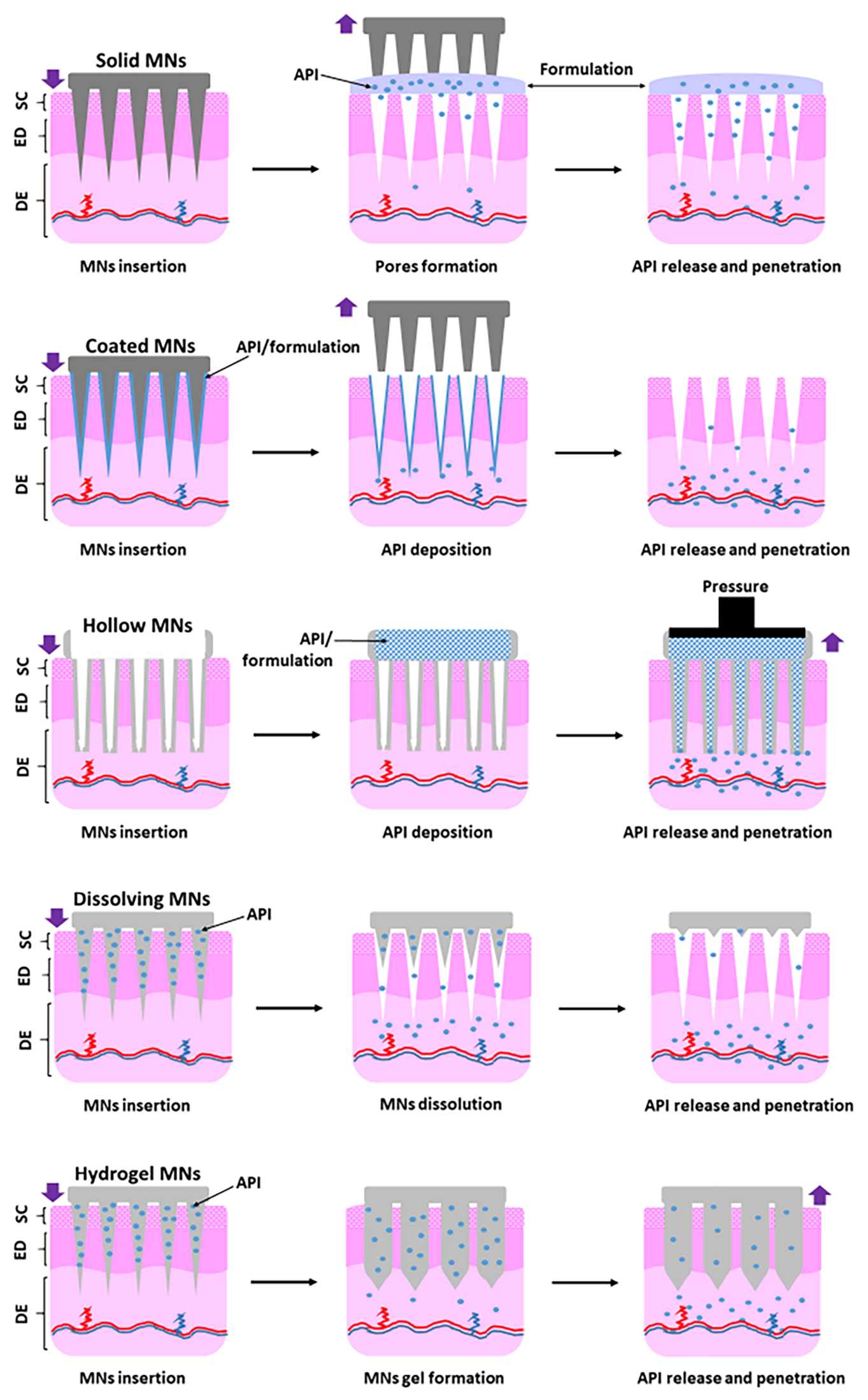
Figure 2.
Schematic representation of the drug delivery process from polymeric microneedles and the three principal polymer-behavior mechanisms following skin insertion. Drug delivery involves sequential stages of skin penetration, polymer dissolution, biodegradation, or hydrogel swelling, followed by drug release from the polymeric matrix, diffusion through the skin microenvironment, and subsequent absorption at the target site.
Figure 2.
Schematic representation of the drug delivery process from polymeric microneedles and the three principal polymer-behavior mechanisms following skin insertion. Drug delivery involves sequential stages of skin penetration, polymer dissolution, biodegradation, or hydrogel swelling, followed by drug release from the polymeric matrix, diffusion through the skin microenvironment, and subsequent absorption at the target site.
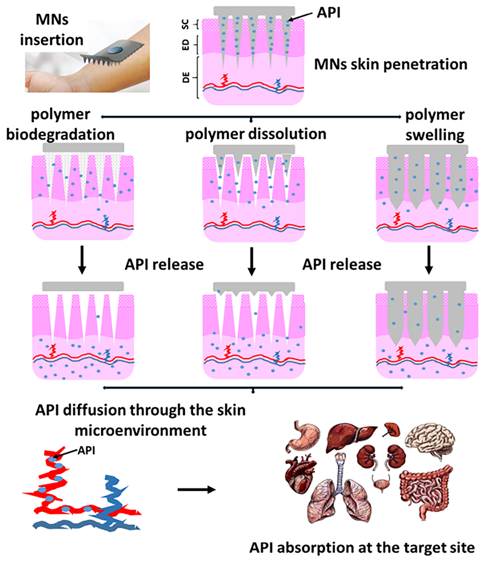
Figure 3.
The most common geometrical shapes for microneedles (A) and a schematic side view of polymeric microneedles (B).
Figure 3.
The most common geometrical shapes for microneedles (A) and a schematic side view of polymeric microneedles (B).
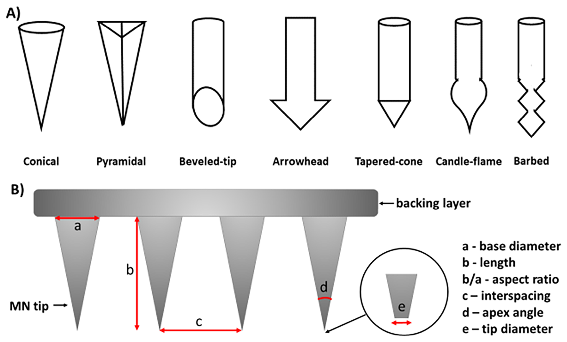
Figure 4.
Influence of microneedle geometry on mechanical performance, skin penetration, and drug delivery efficiency. Schematic representation of the effects of MN base diameter (A), MN length (B), aspect ratio (C), MN tip diameter (D), and MN tip-to-tip spacing (E) on insertion behavior, mechanical stability, and drug delivery performance. The reported geometric ranges are indicative rather than universally applicable, as optimal MN dimensions depend on the material and geometry of the microneedles, anatomical site, application method and force, and the intended depth of skin penetration.
Figure 4.
Influence of microneedle geometry on mechanical performance, skin penetration, and drug delivery efficiency. Schematic representation of the effects of MN base diameter (A), MN length (B), aspect ratio (C), MN tip diameter (D), and MN tip-to-tip spacing (E) on insertion behavior, mechanical stability, and drug delivery performance. The reported geometric ranges are indicative rather than universally applicable, as optimal MN dimensions depend on the material and geometry of the microneedles, anatomical site, application method and force, and the intended depth of skin penetration.
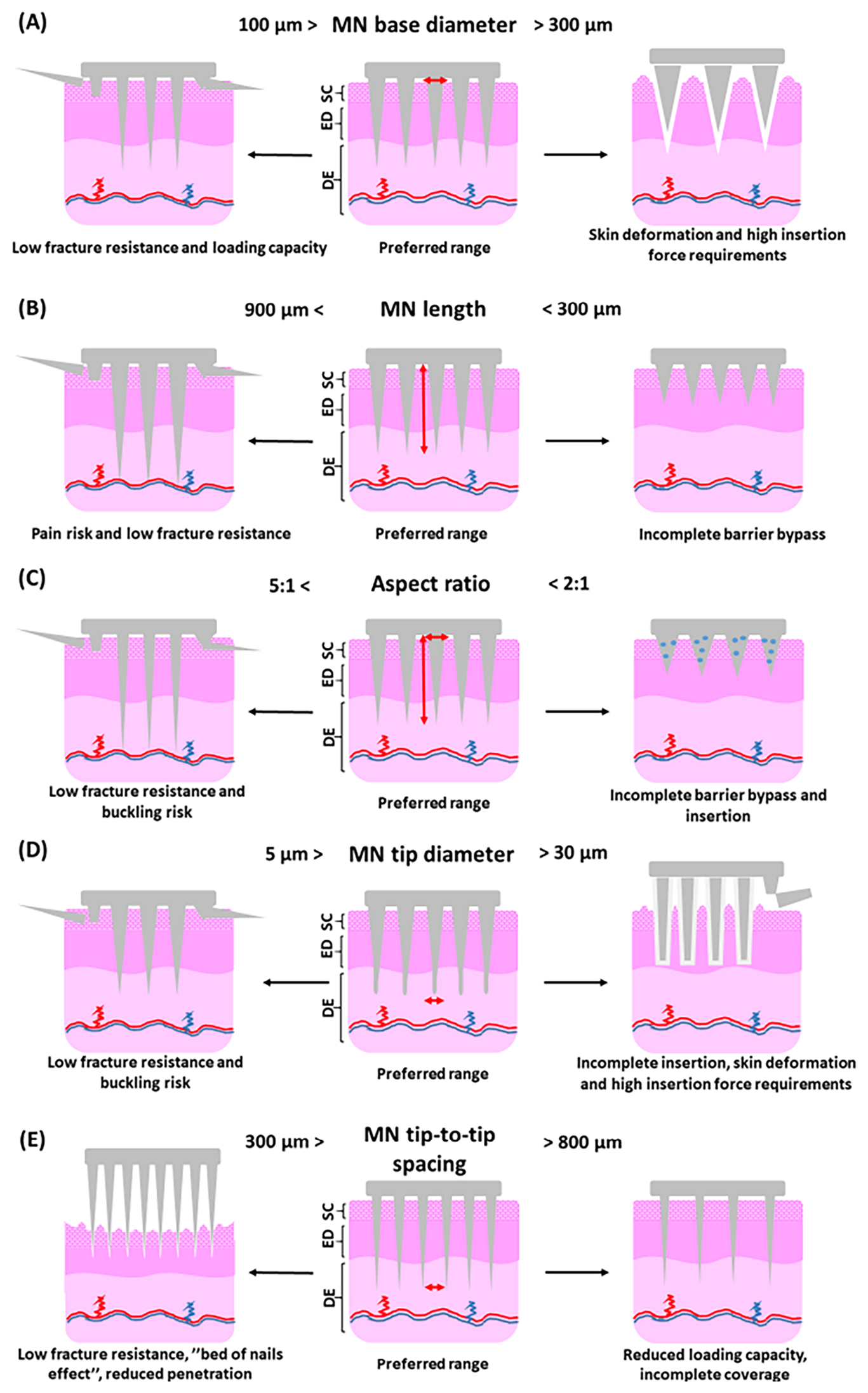
Figure 5.
Mechanical constraints governing microneedle insertion and failure. The schematic highlights the narrow functional window required for effective microneedle performance. Insufficient stiffness results in failed insertion, whereas excessive stiffness or applied force leads to fracture. This balance defines a critical design constraint for reliable drug delivery.
Figure 5.
Mechanical constraints governing microneedle insertion and failure. The schematic highlights the narrow functional window required for effective microneedle performance. Insufficient stiffness results in failed insertion, whereas excessive stiffness or applied force leads to fracture. This balance defines a critical design constraint for reliable drug delivery.
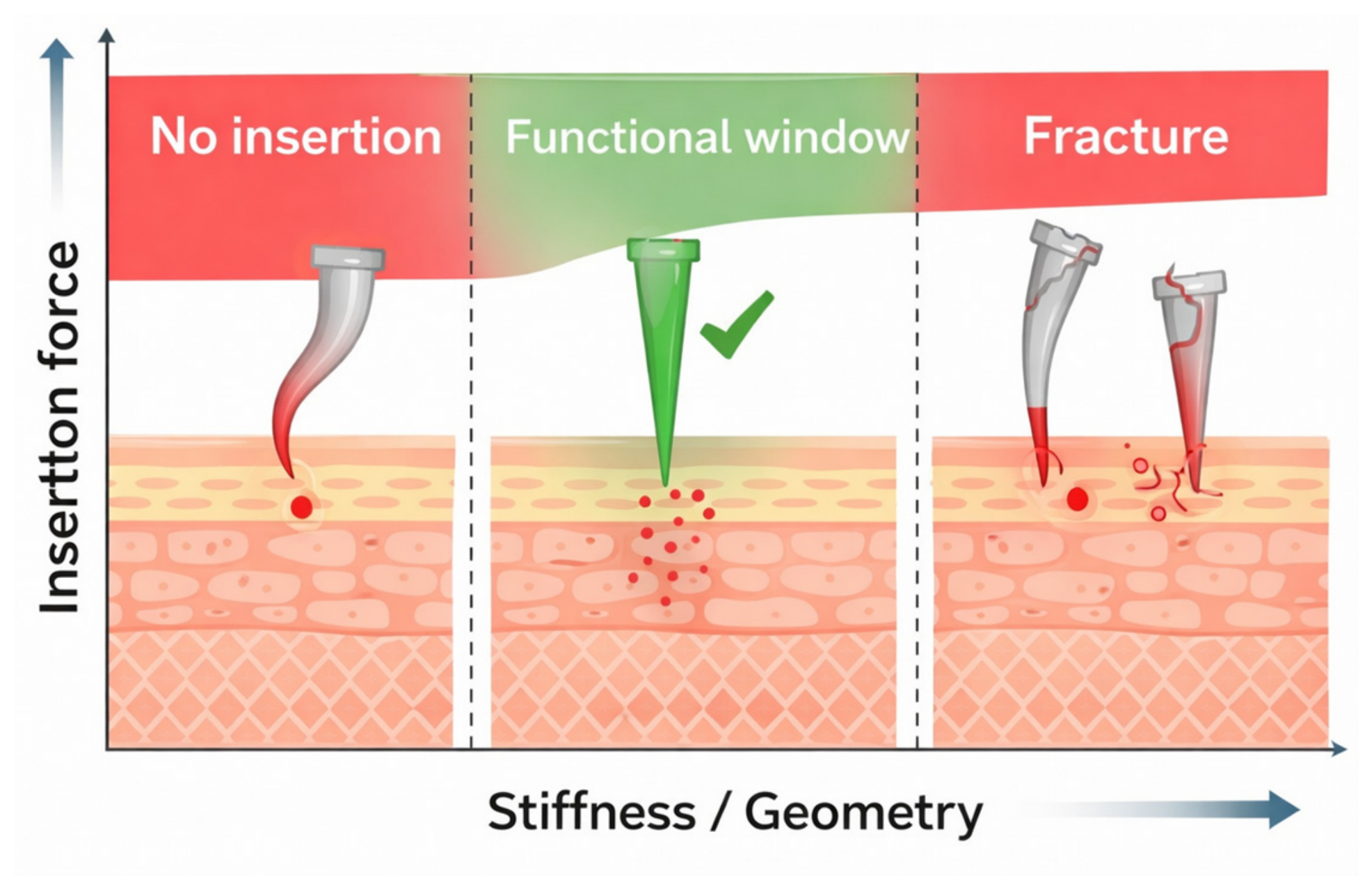
Figure 6.
Representative examples of application-related factors influencing microneedle insertion performance. Schematic comparison of suboptimal and optimized application conditions, including: application mode - manual finger application vs. applicator-assisted application (A); patch fixation and adhesion during wear - excessive skin deformation vs. stable fixation (B); application angle – oblique vs. perpendicular insertion (C); skin condition before application - hairy vs non-hairy skin (D). The illustrated scenarios are conceptual representations based on reported application-related mechanisms in the literature.
Figure 6.
Representative examples of application-related factors influencing microneedle insertion performance. Schematic comparison of suboptimal and optimized application conditions, including: application mode - manual finger application vs. applicator-assisted application (A); patch fixation and adhesion during wear - excessive skin deformation vs. stable fixation (B); application angle – oblique vs. perpendicular insertion (C); skin condition before application - hairy vs non-hairy skin (D). The illustrated scenarios are conceptual representations based on reported application-related mechanisms in the literature.
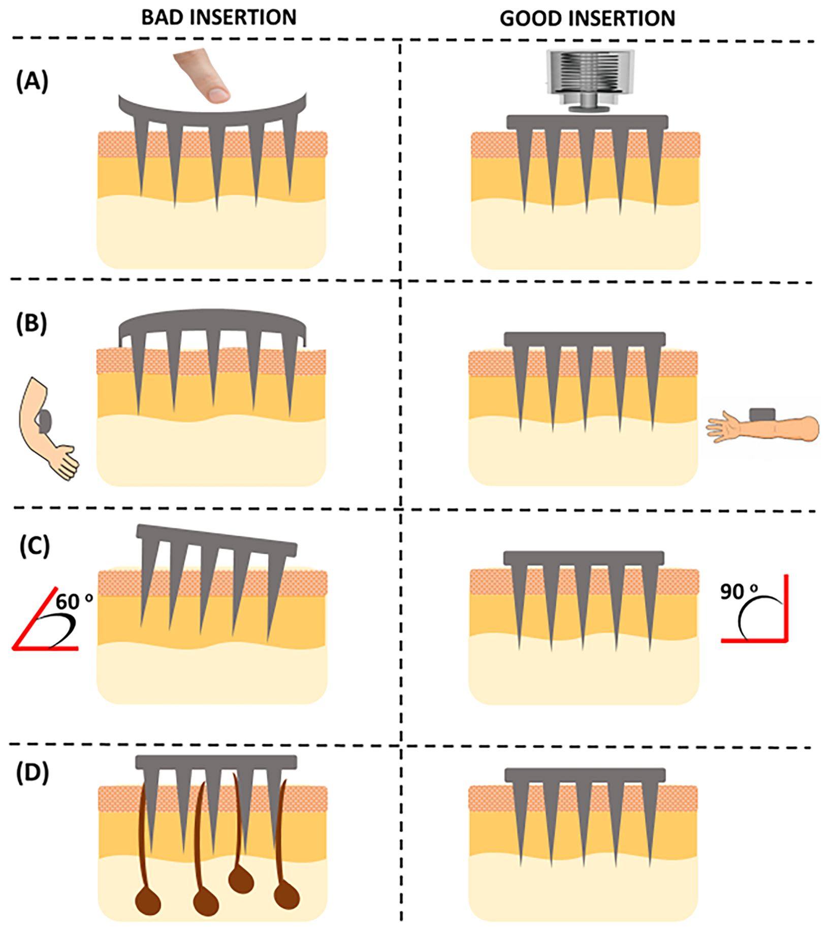
Figure 7.
Polymer fate in the skin. Illustration of polymer dissolution, intradermal degradation, and hydrogel removal following microneedle insertion.
Figure 7.
Polymer fate in the skin. Illustration of polymer dissolution, intradermal degradation, and hydrogel removal following microneedle insertion.
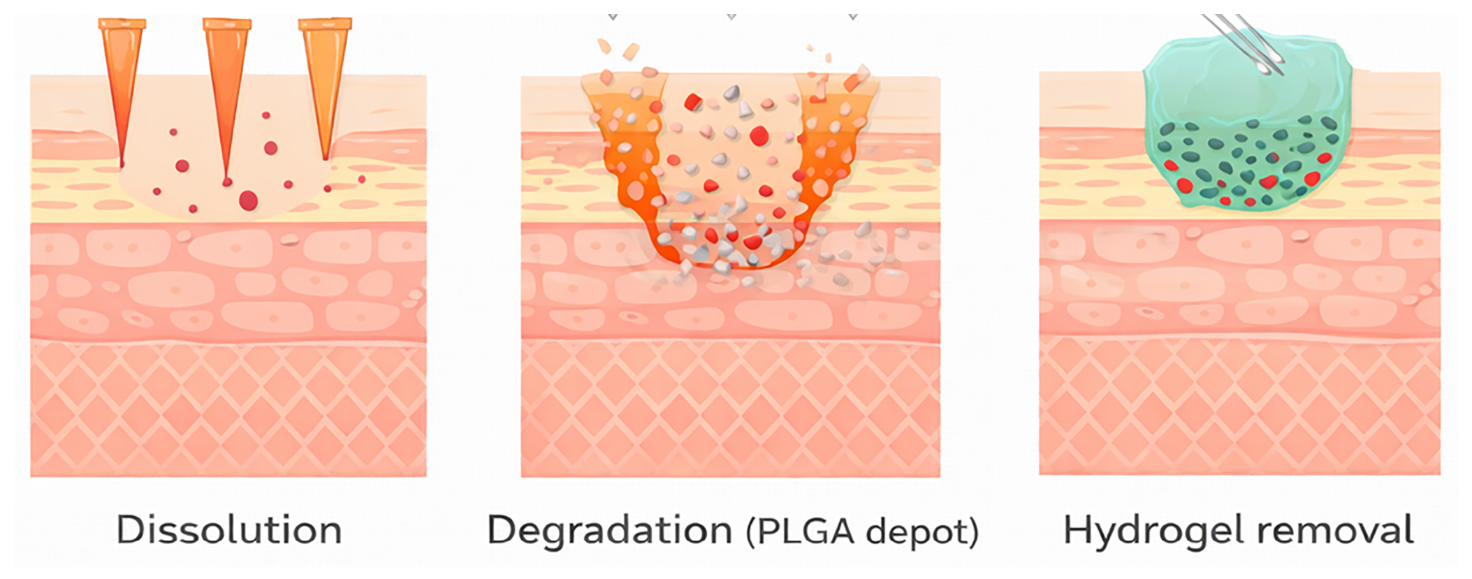
Figure 8.
Stimuli-responsive polymeric microneedles based on activation mechanism. Schematic representation of internally triggered systems, which respond to physiological cues such as pH, temperature, and enzymes (A), and externally triggered systems, which are activated by external stimuli such as near-infrared (NIR) irradiation and magnetic fields (B).
Figure 8.
Stimuli-responsive polymeric microneedles based on activation mechanism. Schematic representation of internally triggered systems, which respond to physiological cues such as pH, temperature, and enzymes (A), and externally triggered systems, which are activated by external stimuli such as near-infrared (NIR) irradiation and magnetic fields (B).
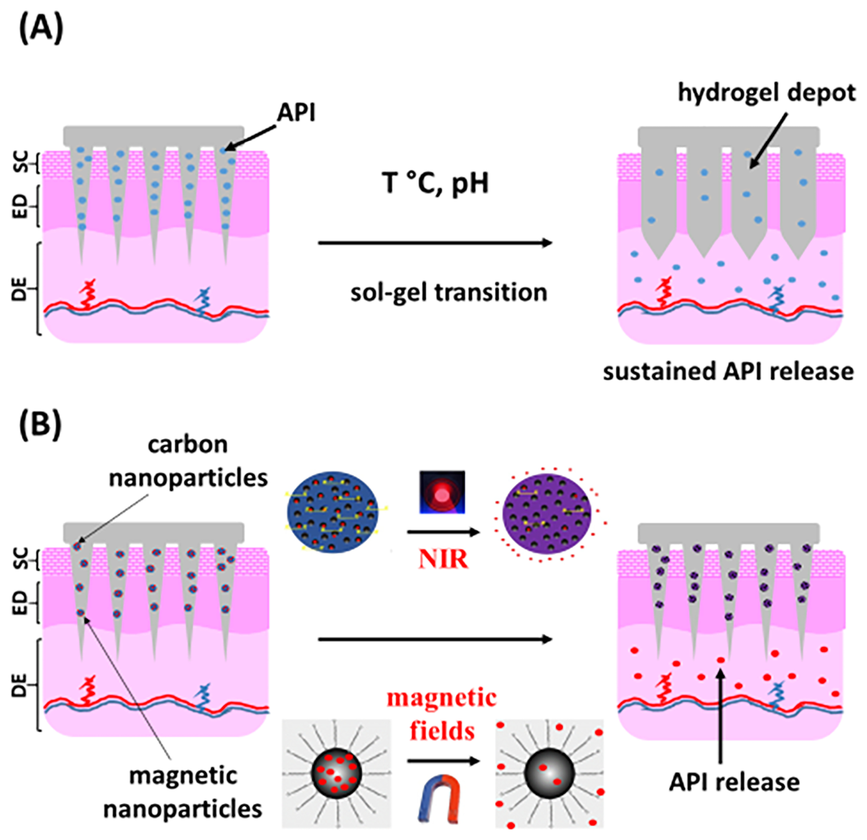
Table 1.
Qualitative comparison of major microneedle types with respect to key performance and translational parameters.
Table 1.
Qualitative comparison of major microneedle types with respect to key performance and translational parameters.
| Type | Solid MNs | Coated MNs | Hollow MNs | Dissolving, biodegradable and implantable MNs | Hydrogel-forming MNs |
|---|---|---|---|---|---|
| Mechanism [7,14,18,21,27] |
“poke-and-patch” | “coat-and-poke” | “poke and flow” | “poke-and-release”, “poke-and-implant” | “poke-and-permeate” |
| Insertion and mechanics parameters | |||||
| Insertion efficiency [5,7,20,31] |
Very high | High/very high | Moderate | Low/moderate | Low/moderate |
| Mechanical strength [5,7,31] |
Very high | High/very high | Moderate | Low | Low/moderate |
| Microchannel persistence after removal [7,12,32] |
Very low | Very low | Moderate | High | Very high |
| Drug delivery performance parameters | |||||
| Drug loading capacity [7,20,31,33,34] |
Very low | Low | Very high | Moderate | Moderate/High |
| Drug retention in the skin [11,35,36] |
Low | Moderate/high | Moderate | High | Very High |
| Controlled drug release ability [11,16,28,37] |
Very low | Low | Moderate | Moderate/high | Very high |
| Bioavailability [5,10,24,37,38] |
Low/moderate | Moderate/high | Very high | High | High |
| Precise dosing ability [6,33,38] |
Very low | Moderate | Very high | High | High |
| Onset of action [5,10,20,34,38] |
Slow | Fast/Very fast | Very fast | Fast | Moderate |
| Delivery period [12,31,33,34] |
Very short | Short | Short/moderate | Moderate/long | Long/Very long |
| Translation and usability parameters | |||||
| Risk of pain [5,10,12,16] |
High | High | Moderate | Very low | Very low |
| Risk of hazardous waste [5,6,10,31,34] |
Very high | Very high | Very high/high | Very low | Low |
| Fabrication complexity [6,7,24,31] |
Low | Moderate | Very high | Moderate | High |
| Drug delivery variability [6,11,37,38] |
High | Moderate | Low/moderate | Moderate | Low |
| Self-administration ability [5,10,31,37] |
Low/moderate | Moderate | Low | Very high | High |
| Patient compliance [5,11,31] |
Low | Moderate | Moderate | Very high | High/very high |
Table 2.
Fabrication methods for polymeric microneedles: advantages and limitations.
| Fabrication method | Principle | Advantages | Limitations |
|---|---|---|---|
| Micromolding [39,40] |
Casting polymer solution/melt into microstructured molds followed by drying or curing | Simple, versatile; compatible with wide range of polymers; allows drug incorporation; low cost | Sensitive to processing parameters; batch variability; limited scalability; potential defects (air bubbles, incomplete filling) |
| Drawing-based methods [42,45] |
Stretching polymer droplets into microneedle structures using controlled airflow or mechanical force | Rapid fabrication; no mold required; sharp tip formation; suitable for prototyping | Poor control over uniformity; limited reproducibility; difficult scale-up; high process variability |
| 3D printing (SLA/DLP) [35,43] |
Layer-by-layer photopolymerization to build microneedle structures | High design flexibility; customizable geometries; rapid prototyping; potential for personalized devices | Limited material selection; insufficient resolution for sharp tips (in some systems); post-processing required |
| Hot embossing / Injection molding [41] |
Thermoplastic polymer shaping under heat and pressure in microstructured molds | High reproducibility; industrial scalability; precise geometry control | High temperature may degrade drugs; limited to thermoplastic polymers; complex equipment |
| Layer-by-layer / hybrid fabrication [46] |
Sequential deposition or combination of multiple fabrication techniques | Enables multilayer structures; spatial drug distribution; advanced functionalities | Increased complexity; reproducibility challenges; difficult regulatory validation |
Table 3.
Major polymer classes used for the fabrication of polymeric microneedles: mechanisms, advantages, limitations, and representative examples.
Table 3.
Major polymer classes used for the fabrication of polymeric microneedles: mechanisms, advantages, limitations, and representative examples.
| Water-soluble polymers | Biodegradable polymers | Hydrogel polymers |
|---|---|---|
| Mechanism/function | ||
| Form dissolving MN matrices that rapidly hydrate and dissolve upon contact with interstitial fluid, releasing the incorporated drug into the skin | Form solid or matrix-type MNs that gradually degrade in the skin, allowing prolonged and controlled drug release; degradation occurs through hydrolysis and/or enzymatic processes depending on polymer chemistry | The MN matrix itself generally does not dissolve during application; instead, it forms crosslinked hydrophilic MN networks that absorb interstitial fluid, swell and form hydrated pathways through which drug from a reservoir can diffuse. |
| Examples | ||
| Poly(vinylpyrrolidone) (PVP); poly(vinyl alcohol) (PVA); hyaluronic acid (HA); carboxymethylcellulose (CMC); maltose; trehalose | Poly(lactic-co-glycolic acid) (PLGA); polylactic acid (PLA); polycaprolactone (PCL); poly(lactic acid) (PLA)-based copolymers | Poly(acrylic acid) (PAA); poly(methyl vinyl ether-co-maleic anhydride) (PMVE/MA); polyethylene glycol (PEG)-based networks; poly(vinyl alcohol) (PVA) hydrogels; hyaluronic acid-based crosslinked systems |
| Advantages | ||
| Rapid drug release; no residual sharp waste; rapid polymer clearance; generally good biocompatibility; relatively simple processing; suitable for single-step application | Sustained and long-acting drug delivery; tunable degradation and release kinetics; potential for high structural integrity; suitable for poorly soluble drugs and depot-type delivery; degradation products can be eliminated through normal biological pathways | Sustained and controlled drug delivery; high water uptake; MNs remain structurally intact and can be removed after application; minimal polymer deposition in skin; swelling can facilitate prolonged drug transport |
| Disadvantages | ||
| Relatively limited mechanical strength; risk of deformation or fracture during insertion; dissolution rate can be difficult to control; drug loading may be limited; sensitive to moisture | Slow degradation may result in prolonged polymer residence; acidic degradation products from polyesters may affect drug stability or cause local irritation; release kinetics can be difficult to predict; residual polymer may remain in tissue for extended periods; more complex manufacturing | Drug release is primarily diffusion-controlled; relatively slow onset compared with dissolving MNs; requires adequate patch–skin contact; crosslink density must balance mechanical strength and swelling; high swelling may compromise structural integrity or adhesion |
| References | ||
| [10,47]. | [9,48] | [10,11] |
Table 4.
Systems-level framework of the principal domains and subdomains contributing to translational failure in polymeric microneedles.
Table 4.
Systems-level framework of the principal domains and subdomains contributing to translational failure in polymeric microneedles.
|
Mechanical–Insertion Mismatch Failure to achieve reliable skin penetration while maintaining structural integrity. [7,35,40,55,56] | |||||||
| Microneedle geometry: Shape Length Base diameter Tip diameter, etc. |
Polymer mechanical properties: Elastic modulus Fracture strength Flexibility Molecular weight |
Skin-related factors: Skin thickness Hydration Elasticity Anatomical site |
Application-related factors: Insertion force Application angle Application method Residence time |
||||
|
Pharmacokinetic–Dose Mismatch Failure to achieve the desired drug exposure despite successful insertion. [40,57,58] | |||||||
| Drug physicochemical properties: Molecular weight Solubility Lipophilicity Ionization (pKa) Stability |
Drug loading constraints: Matrix loading capacity Therapeutic dose requirements Drug distribution within the MN |
Release behavior: Dissolution rate Hydrogel swelling Polymer degradation |
Skin transport limitations: Diffusion through microchannels Skin partitioning Microchannel closure |
Systemic pharmacokinetics: Absorption rate Sustained exposure Bioavailability |
|||
|
Manufacturing and Scalability Limitations Failure to reproduce laboratory performance under industrial production. [40,56,58] | |||||||
| Material processing: Polymer viscosity Mold filling Drying conditions Crosslinking |
Manufacturing consistency: Dimensional uniformity Needle integrity Drug content uniformity |
Scale-up challenges: Batch-to-batch reproducibility Automation compatibility Production throughput |
Product stability: Moisture sensitivity Storage stability Packaging requirements |
Quality and regulatory considerations: Mechanical quality control Sterility GMP |
|||
|
Clinical–Context Misalignment Failure of otherwise functional devices to meet real-world clinical and patient requirements. [40,56,58] | |||||||
| Patient usability: Self-administration Dexterity Training Compliance |
Patch performance during wear: Adhesion Movement Wear time |
Clinical application factors: Anatomical site selection Skin preparation Environmental conditions |
Safety considerations: Skin irritation Residual polymer Infection risk |
Clinical implementation: Target population Therapeutic indication Healthcare workflow Cost-effectiveness |
|||
Table 5.
Qualitative comparison of the effects of conical, pyramidal, beveled, and arrowhead microneedle geometries on key performance characteristics relevant to transdermal drug delivery, including skin penetration, insertion force, mechanical strength, tissue interaction, drug retention, and fabrication complexity. The qualitative rankings represent general trends reported in the literature and may vary depending on material composition, dimensions, and fabrication method.
Table 5.
Qualitative comparison of the effects of conical, pyramidal, beveled, and arrowhead microneedle geometries on key performance characteristics relevant to transdermal drug delivery, including skin penetration, insertion force, mechanical strength, tissue interaction, drug retention, and fabrication complexity. The qualitative rankings represent general trends reported in the literature and may vary depending on material composition, dimensions, and fabrication method.
| Performance characteristics |
Conical | Pyramidal | Beveled tip | Arrowhead |
|---|---|---|---|---|
| Initial skin puncture [5,14,61] | Moderate/High | High/Very high | High/Very high | Very high |
| Insertion force [14,59,64] | Low/Moderate | Moderate/High | Low | High |
| Mechanical strength [65,66] | High | Very high | Low/Moderate | Low/Moderate |
| Risk of incomplete insertion [62,67] | Low/Moderate | Moderate | Moderate | Low |
| Bending/buckling risk [62,66] | Moderate/High | Low | High | Low/Moderate |
| Risk of pain or tissue damage [5,32] | low | Low/Moderate | Moderate/High | Very high |
| Risk of accidental removal [59] | High | Moderate | Moderate/High | Low |
| Drug retention in the skin [64] | Low | Moderate | Moderate | Very high |
| Fabrication complexity [68] | Low | Moderate | High | Very high |
| Suitability for polymeric microneedles [11] | Very high | Very high | High | Low |
Table 6.
Influence of microneedle geometry on mechanical performance, supported by representative studies. The qualitative rankings represent general trends reported in the literature and may vary depending on material composition and fabrication method.
Table 6.
Influence of microneedle geometry on mechanical performance, supported by representative studies. The qualitative rankings represent general trends reported in the literature and may vary depending on material composition and fabrication method.
| ↑length | ↑base diameter | ↑aspect ratio | ↑tip diameter /apex angle | ↑interspacing | |
|---|---|---|---|---|---|
| Drug loading capacity | ↑ | ↑ | ↓ | ||
| Skin deformation | ↑ | ↑ | ↓ | ||
| Penetration depth | ↑ | ↓ | Opt. | ↓ | |
| Risk of pain | ↑ | ↑ | ↑ | ||
| Fabrication complexity | ↑ | ↑ | ↑ | ↑ | |
| Bending/bucking resistance/forces | ↓ | ↑ | ↓ | ↑ | |
| Mechanical strength | ↓ | ↑ | ↓ | ↑ | |
| Required insertion force | ↑ | ↑ | ↑ | ↓ | |
| Bed-of-nail effect | ↑ | ↓ | |||
| Insertion reliability/ratio | ↑ | ↓ | ↑/opt. | ↓ | ↑ |
| References | [25,33,35,51,56,59,64,69,70,71] | ||||
Table 7.
Influence of polymer physicochemical, mechanical, rheological, surface, and processing properties on the manufacturing quality and drug delivery performance of polymeric microneedle systems.
Table 7.
Influence of polymer physicochemical, mechanical, rheological, surface, and processing properties on the manufacturing quality and drug delivery performance of polymeric microneedle systems.
| Factor | Main influence on | General effect on microneedle performance and drug delivery |
|---|---|---|
| Molecular weight [7,23,31,35,54,82] |
strength; viscosity of casting solution; mold filling ability; dissolution rate; swelling ability; drug diffusion through the matrix. |
Higher molecular weight generally increases mechanical strength and viscosity, but slows dissolution and drug diffusion. |
| Polymer concentration [11,35,55,83] |
needle formation; structural integrity (brittleness or flexibility); drug loading capacity; dissolution/swelling kinetics; diffusion through the matrix. |
Higher concentration usually improves structural integrity and drug loading, but may reduce dissolution and increase brittleness. |
| Polymer type [7,35] |
mechanical performance; hydrophilicity/hydrophilicity; biocompatibility; biodegradability; drug–polymer interactions; release behavior. |
Determines mechanical performance, water uptake, biodegradability, drug interactions, and release behavior. |
| Hydrophilicity / hydrophobicity [37,68] |
swelling; dissolution; drug transport. |
More hydrophilic polymers swell or dissolve faster and usually enhance drug release. |
| Crosslink density [29,37,68] |
Mechanical strength; swelling, drug diffusion; release duration. |
Higher crosslinking increases mechanical strength but decreases swelling and drug diffusion. |
| Swelling capacity [29,37,68] |
drug diffusion; release duration. |
Greater swelling facilitates diffusion and may prolong drug release. |
| Dissolution rate [11,54,55] |
drug release; delivery efficiency. |
Faster dissolution accelerates drug release but may shorten delivery duration. |
| Glass transition temperature (Tg) [7,11,55] |
rigidity at room and body temperature; storage stability; needle deformation during insertion; moisture sensitivity. |
Higher Tg generally improves rigidity and resistance to deformation during storage and insertion. |
| Crystallinity [7,54] |
strength; dissolution; moisture uptake; drug stability; processability. |
Higher crystallinity increases rigidity and stability but slows dissolution and reduces water uptake. |
| Residual moisture [7,55,84,85] |
mechanical strength; stability, insertion; brittleness; tip sharpness; storage stability; premature swelling or dissolution. |
Higher moisture content softens the matrix, reduces insertion efficiency, and may compromise stability. |
| Mechanical modulus [7,14,35,78,86] |
buckling; fracture; insertion; elastic modulus; tensile strength; fracture toughness; flexibility. |
Higher modulus improves penetration and resistance to bending or fracture. |
| Rheology [7,35,55,87,88] |
mold filling; reproducibility; morphology; air bubble entrapment; tip replication; layer uniformity. |
Critical for micromolding and multilayer fabrication. |
| Film-forming and drying properties [45,54,55] |
needle morphology; surface smoothness; crack formation; shrinkage; dose uniformity. |
Rapid or uneven drying may cause defects and drug migration. |
| Drug–polymer interactions [54,55] |
loading; stability; release. |
Strong interactions may improve drug stability but can retard drug release. |
| Polymer blend composition [7,55] |
strength; dissolution; swelling; release kinetics; processability. |
Blending polymers can balance mechanical performance and release characteristics. |
| Mesh size / pore size [37,68] |
diffusion; sustained release. |
Larger mesh size enhances drug diffusion, whereas smaller mesh size prolongs release. |
| Degradation / erosion behavior [5,68] |
release duration; biocompatibility; stability during storage; In vivo persistence. |
Faster degradation accelerates release, while slower degradation supports prolonged delivery. Important for biodegradable and long-acting systems. |
| Surface properties of the polymer [5,7,40] |
wettability; adhesion to skin; friction during insertion; coating efficiency; protein adsorption. |
Influences insertion and biological interactions. |
| Thermal and processing stability [7,45,55] |
sterilization compatibility; drying temperature tolerance; compatibility with APIs; shelf-life. |
Essential for industrial manufacturing. |
Table 9.
Drug-related physicochemical factors affecting the performance and drug delivery efficiency of polymeric microneedles.
Table 9.
Drug-related physicochemical factors affecting the performance and drug delivery efficiency of polymeric microneedles.
| Major factor | Main influence on delivery efficiency |
General effect on microneedle performance and drug delivery |
|---|---|---|
| Molecular size [32,35] |
Diffusion through polymer and skin; permeation rate | Smaller molecules generally diffuse more readily through the polymer matrix and skin |
| Lipophilicity [32,35,104] |
Partitioning into skin; permeation | Moderate lipophilicity is generally favorable for transdermal permeation; very hydrophilic or highly lipophilic compounds may exhibit limited permeation. |
| Aqueous solubility [32,105] |
Drug dissolution, release, and thermodynamic activity | Poorly soluble drugs may show incomplete dissolution and limited delivery. |
| Ionization (pKa, degree of ionization, pH) [4,104] |
Solubility, skin partitioning, and permeation | The non-ionized fraction generally exhibits greater partitioning into the lipid-rich stratum corneum. |
| Solid-state properties (polymorphism, amorphous/crystalline state, melting point) [104,106] |
Drug dissolution, stability, and release | Amorphous drug may provide increased apparent solubility but can present greater physical instability. |
| Chemical and physical stability [6,32] |
Drug integrity during manufacture, storage, and application | Unstable drugs may undergo degradation during formulation, drying, storage, or application, reducing the effective delivered dose and potentially generating degradation products. |
| Drug–polymer interactions [6,35] |
Drug loading, retention, dissolution, and release | Strong interactions may improve drug retention and loading but can slow dissolution/release or cause incomplete drug recovery. Weak or poorly controlled interactions may result in drug migration or crystallization. |
| Drug–skin interactions (Partition coefficient, binding to skin components) [104,107] |
Skin retention and transdermal permeation | Excessive skin binding may increase local retention and reduce systemic availability. |
| Dose/loading requirement [66,107] |
Feasibility of loading and achieving therapeutic delivery | High-dose drugs may exceed the loading capacity of MN arrays and can compromise mechanical strength, needle formation, dissolution, or patch dimensions. |
| Diffusion coefficient [32,105] |
Release driving force and permeation rate | Higher diffusivity generally facilitates faster drug release, whereas strong matrix interactions can reduce diffusivity. |
| Thermodynamic activity (drug activity; degree of saturation/supersaturation) [104] |
Release driving force and permeation rate | Increasing thermodynamic activity can enhance transdermal flux |
Table 10.
Alignment between release strategy, polymer selection, and therapeutic objective.
| Therapeutic goal | Release profile | Polymers | Ref |
|---|---|---|---|
| Vaccination | Immediate | PVP, PVA | [22] |
| Local anesthesia | Rapid | PVP, PVA | [108] |
| Chronic therapy | Sustained | PLGA | [25] |
| Long-term delivery | Extended | PCL, PLGA | [53,108] |
| Biosensing | Diffusion-controlled | Hydrogels | [28] |
| Smart delivery | Triggered | PNIPAm, hybrids | [109,110] |
Disclaimer/Publisher’s Note: The statements, opinions and data contained in all publications are solely those of the individual author(s) and contributor(s) and not of MDPI and/or the editor(s). MDPI and/or the editor(s) disclaim responsibility for any injury to people or property resulting from any ideas, methods, instructions or products referred to in the content. |
© 2026 by the authors. Licensee MDPI, Basel, Switzerland. This article is an open access article distributed under the terms and conditions of the Creative Commons Attribution (CC BY) license.
Copyright: This open access article is published under a Creative Commons CC BY 4.0 license, which permit the free download, distribution, and reuse, provided that the author and preprint are cited in any reuse.