
Submitted:
02 September 2026
Posted:
03 September 2026
You are already at the latest version
Abstract
Novel iodinated cobalt bis(dicarbollide) conjugates with acridine were synthesized by the direct iodination reaction of cobalt bis(dicarbollide) conjugates and acridine with crystalline iodine. The cytotoxicity of new compounds against different human cell lines was evaluated. One of these compounds was found to be non-toxic for all cell types and was combined with polyvinylpyrrolidone to form a non-covalent complex to increase its water solubility. The BNCT experiment showed that pre-incubation with a new boron-containing complex followed by neutron irradiation reduces the survival of T98G glioblastoma cells to 25% and B16 melanoma cells to 17%. The biodistribution of the boron-containing compound ACR-PVP was investigated in a B16F10 melanoma murine model. Maximum uptake in tumor and healthy tissues was observed 15 minutes following single intravenous administration. ACR-PVP exhibited a considerably higher tumor-to-muscle ratio (5.4) relative to BPA (3.0), thereby indicating its potential as a promising candidate for boron neutron capture therapy. This study creates prerequisites for further research in the development of methods for synthesizing BNCT agents based on acridine.
Keywords:
iodinated cobalt bis(dicarbollide)
; acridine
; polyhedral boron hydrides
; poly(vinylpyrrolidone)
; in vivo study
; boron neutron capture therapy
1. Introduction
Cancer is a widespread disease that requires scientists to develop new, effective treatment methods [1]. Therefore, the development of drugs that can be used for cancer treatment is becoming increasingly important. One effective cancer treatment method is boron neutron capture therapy, the high potential of which has been demonstrated in both preclinical and clinical trials. After administration of the drug, selective accumulation of non-radioactive nucleus 10B boron atoms occurs in cancer cells, and subsequent thermal neutron irradiation stimulates a nuclear reaction 10B + 10n → [11B]* → α + 7Li + 2.31 MeV, that produces an excited [11B]* nucleus, which then generates high linear energy transfer fission 4He (α particles) and 7Li [2,3,4]. The energy released by this reaction leads to the destruction of cancer cells [5,6]. However, the development of this method is hampered by the lack of effective drugs that can selectively penetrate and accumulate within cancer cells. This poses the challenge for scientists to develop boron-containing drugs with a number of specific characteristics.
The realization of a therapeutic effect in boron neutron capture therapy (BNCT) is contingent upon several critical prerequisites for potential pharmaceutical agents. These include the capacity for transmembrane penetration, selective targeting, and accumulation within neoplastic cells, and attainment of the requisite therapeutic boron concentration, conventionally defined as ≥20 μg 10B/g of tumor [7,8,9,10,11,12]. Furthermore, it has been established that the radiobiological efficacy of the 10B(n,α)7Li capture reaction is substantially enhanced when the event occurs within the cell nucleus, as opposed to the cytoplasm or plasma membrane [13]. This intracellular localization is of particular significance, since nuclear confinement of BNCT drugs leads to more pronounced cellular destruction upon neutron irradiation than would be achieved with an equivalent boron load distributed homogenously throughout the cell. Accordingly, targeted nuclear accumulation may reduce the dose of administered boron, thereby reducing systemic toxicity [14]. These considerations have stimulated growing research interest in boron-functionalized DNA intercalators, including derivatives of phenanthridinium [15,16], naphthalimide [17,18,19,20,21,22,23], and related structural scaffolds [24].
An analysis of the literature revealed that acridine-based DNA intercalators are of great interest for medical applications [25,26]. Acridines and their derivatives are known to have a planar structure consisting of three conjugated six-membered rings. These structural features of acridine determine its intercalating properties and ability to bind to DNA and RNA molecules. The acridine structure inserts between nitrogenous base pairs, altering the double helix structure. These structural distortions manifest as partial unwinding of the double helix and an increase in the vertical separation between neighboring ones at the intercalation site.
Consequently, acridine derivatives functionalized with polyhedral boron hydrides have attracted considerable interest as prospective BNCT agents. The synthetic chemistry of boron-containing acridines has been explored in several reported studies, wherein the boron moiety is typically represented by an ortho- or para-carborane framework [27,28,29]. More recently, a series of acridine conjugates bearing ortho- and meta-carborane clusters have been synthesized and evaluated with respect to their DNA-binding affinity and cytotoxic profiles [28]. In a complementary development, a conjugate of cobalt bis(dicarbollide) with acridine has been prepared; this compound exhibits low toxicity and holds promise as a candidate for BNCT applications [30]. Among the diverse family of polyhedral boron hydrides, the cobalt bis(dicarbollide) anion, [3,3’-Co(1,2-C2B9H11)2]− [31,32,33,34,35], has emerged as a particularly attractive boron delivery vehicle for BNCT, owing to its remarkable chemical robustness and favorable toxicological profile [36,37,38,39]. Notably, this metallacarborane possesses an amphiphilic character [40], which facilitates its passive permeation across lipid bilayer membranes and subsequent intracellular accumulation [10,41,42,43].
Furthermore, it is known that the introduction of halogens into the cobalt bis(dicarbollide) molecule improves compound penetration and achieves the required pharmacokinetic parameters [44,45]. In particular, the introduction of an iodine atom hinders ligands rotation through the formation of intramolecular C-H···I hydrogen bonds between the C-H groups of one dicarbollide ligand and the halogen substituent on the other [46,47]. Moreover, the presence of iodine facilitates transbilayer diffusion while concurrently reducing the propensity for nonspecific interactions with cellular components [44,45,46,47,48]. The present study was undertaken with the aim of synthesizing the iodine-containing derivatives of cobalt bis(dicarbollide) with acridine. To achieve the stated goal, the presented work uses iodination of previously obtained click-reaction conjugates of cobalt bis(dicarbollide) with acridine with a triazole ring in the spacer.
2. Materials and Methods
2.1. Instruments, General Information and Synthetic Procedures
Conjugates 1-4 were prepared according to the literature procedures [49]. Iodine, CН3OH, acetonitrile, CH2Cl2, Na2S2O3×5H2O, and Na2SO4 were purchased from commercial vendors (Carl Roth GmbH, Abcr GmbH, Komponent-Reactiv) and used without further purification. The reaction progress was monitored by thin-layer chromatography (Merck F254 silica gel on aluminum plates) and visualized using 0.5% PdCl2 in 1% HCl in aq. MeOH (1:10). Macherey-Nagel Silica 60 (0.063-0.200 mm) was used for column chromatography. The NMR spectra at 400.0 MHz (1H), 128.3 MHz (11B) and 100.6 MHz (13C) were recorded with the Varian Inova 400 Instrument with Qone QOT 400 X/HF-05 probe. The residual signal of the NMR solvent relative to TMS was taken as the internal reference for 1H and 13C{1H} NMR spectra. 11B and 11B{1H} NMR spectra were referenced against BF3×Et2O. The 11B{1H} spectra were used to determine the chemical shifts and signal intensities of the B-H and B-I groups, 11B spectra — to determine B-H coupling constants. Infrared spectra were recorded on an FSM2201 instrument (OKB SPECTRUM, Saint-Petersburg, Russia). High-resolution mass spectra (HRMS) were measured on a mictOTOF II (Bruker Daltonic, Bremen, Germany) instrument using electrospray ionization (ESI). The measurements were performed in a negative ion mode (interface capillary voltage 3000 V), mass range from m/z 50 to m/z 3000, external or internal calibration was carried out with ESI Tuning Mix, Agilent. A syringe injection was used for solutions in acetonitrile (flow rate 3 µL/min). Nitrogen was applied as a drying gas; the interface temperature was set at 180 °C. Elemental analyses were performed at the Laboratory of Microanalysis of the A.N. Nesmeyanov Institute of Organoelement Compounds.
General Method of Synthesis of Iodinated Cobalt bis(dicarbollide) Conjugates with Acridine (5-8). Crystalline iodine (3 equiv.) was added to CH3OH and stirred for one hour until complete dissolution. Then, the solution was added to the conjugate of cobalt bis(dicarbollide) with acridine 1-4, and the mixture was refluxed for three hours. The reaction mixture was cooled, and the solvent was evaporated. The residue was extracted with ethyl acetate, washed with an aqueous solution of Na2S2O3×5H2O (2×50 mL) and H2O (2×50 mL). The organic fraction was dried over anhydrous Na2SO4. The inorganic precipitate was filtered off, and the solvent was evaporated.
Synthesis of [p-{8-NHCH2CH2-N3CHC-CH2O(CH2CH2O)2-8′-I-3,3′-Co(1,2-C2B9H10)
(1′,2′-C2B9H10)}-(C6H4)2[b,e]pyridinate]Cs (5). Obtained from 120 mg (0.14 mmol) of compound 1, 100 mg (0.42 mmol) of сrystalline I2, and 70 ml of CH3OH. The yield of product 5 was 100 mg (88%), an orange solid. NMR 1H (400 MHz, acetone-d6) δ, ppm: 8.54 (d, 2H, J = 9.0 Hz, CHAr), 8.03 (t, 2H, J = 7.8 Hz, CHAr), 7.96 (s, 1H, CHCN3), 7.92 (d, 2H, J = 8.7 Hz, CHAr), 7.64 (t, 2H, J = 8.0 Hz, CHAr), 5.10 (t, 2H, J = 5.1 Hz, СН2N), 4.91 (t, 2H, J = 5.2 Hz, СН2N), 4.51 (s, 2H, OСН2CCHN3), 4.41 (br.s, 2H, СНCarb), 4.13 (br.s, 2H, СНCarb), 3.55 (m, 4H, 2×CH2O), 3.46 (m, 4H, 2×CH2O). 11B NMR (128 MHz, acetone-d6) δ, ppm: 21.5 (s, 1B, B-O), –0.4 (d, 2В, J = 119 Hz, B-H), –5.9 (m, 9B, J = 119 Hz, B-H+B-I), –17.9 (d, 2В, J = 162 Hz, B-H), –19.9 (d, 2В, J = 151 Hz, B-H), –23.1 (d, 1В, J = 142 Hz, B-H), –27.0 (d, 1В, J = 142 Hz, B-H). 13C{1H} NMR (101 MHz, acetone-d6) δ, ppm: 159.7 (CAr), 146.0 (CAr), 140.6 (CCHN3), 136.6 (CHAr), 126.1 (CHAr), 125.3 (CHAr,), 125.1 (CCHN3), 119.8 (CHAr), 113.8 (CAr), 72.7 (OCH2), 71.1 (OCH2), 70.4 (OCH2), 69.1 (OCH2), 64.9 (OСН2CCHN3), 57.3 (CHCarb), 55.4 (CHCarb), 50.1 (CH2N), 49.9 (CH2N). IR (film, cm–1): 2566 (vB-H), 1582 (vtriaz). Found: C 36.71, H 5.23, B 22.82, N 8.39; calcd: C 36.52, H 5.19, B 22.76, N 8.19. HRMS (ESI) of C26H44B18CoIN5O3, m/z: calcd for [M+H]+ 855.3625, found: 855.3624.
Synthesis of [p-{8-NHCH2CH2-N3CHC-CH2CH2O(CH2CH2O)2-8′-I-3,3′-Co(1,2-C2B9H10)
(1′,2′-C2B9H10)}-(C6H4)2[b,e]pyridinate]Cs (6). Obtained from 230 mg (0.26 mmol) of compound 2, 200 mg (0.78 mmol) of сrystalline I2, and 90 ml of CH3OH. The yield of product 6 was 220 mg (84%), an orange solid. NMR 1H (400 MHz, acetone-d6) δ, ppm: 8.54 (d, 2H, J = 8.8 Hz, CHAr), 8.05 (t, 2H, J = 7.8 Hz, CHAr), 7.95 (d, 2H, J = 8.0 Hz, CHAr), 7.87 (s, 1H, CHCN3), 7.65 (m, 2H, J = 7.8 Hz, CHAr), 5.07 (t, 2H, J = 5.3 Hz, СН2N), 4.89 (t, 2H, J = 5.1 Hz, СН2N), 4.45 (br.s, 2H, СНCarb), 4.16 (br.s, 2H, СНCarb), 3.59 (t, 2H, J = 6.3 Hz, OСН2CH2CCHN3), 3.50 (m, 6H, 3×OCH2), 3.44 (m, 2H, OCH2), 2.84 (t, 2H, J = 6.5 Hz, СН2CCHN3). 11B NMR (128 MHz, acetone-d6) δ, ppm: 21.5 (s, 1B, B-O), –0.7 (d, 2В, J = 125 Hz, B-H), ––5.8 (m, 9B, B-H+B-I), –17.9 (d, 2В, J = 148 Hz, B-H), –20.0 (d, 2В, J = 167 Hz, B-H), –23.1 (d, 1В, J = 147 Hz, B-H), –27.3 (d, 1В, J = 176 Hz, B-H). 13C{1H} NMR (101 MHz, acetone-d6) δ, ppm: 159.5 (CAr), 147.4 (CAr), 142.0 (CCHN3), 136.6 (CHAr), 126.1 (CHAr), 125.3 (CHAr), 124.2 (CCHN3), 119.7 (CHAr), 113.7 (CAr), 72.6 (OCH2), 71.0 (OCH2), 70.7 (OCH2), 70.4 (OСН2CH2CCHN3), 69.2 (OCH2), 57.3 (CHCarb), 55.4 (CHCarb), 50.1 (CH2N), 49.6 (CH2N), 27.1 (CH2CCHN3). IR (film, cm–1): 2564 (vB-H), 1583 (vtriaz). Found: C 32.74, H 4.79, B 19.64, N 7.12; calcd: C 32.36, H 4.63, B 19.42, N 6.99. HRMS (ESI) of C27H46IB18CoN5O3, m/z: calcd for [M+H]+ 869.3782, found: 869.3781.
Synthesis of [p-{8-NHCH2CH2-N3CHC-CH2O(CH2)5O-8′-I-3,3′-Co(1,2-C2B9H10)
(1′,2′-C2B9H10)}-(C6H4)2[b,e]pyridinate]Cs (7). Obtained from 270 mg (0.31 mmol) of compound 3, 240 mg (0.93 mmol) of сrystalline I2, and 90 ml of CH3OH. The yield of product 6 was 270 mg (85%), an orange solid. NMR 1H (400 MHz, acetone-d6) δ, ppm: 8.56 (d, 2H, J = 9.1 Hz, CHAr), 8.02 (m, 3H, CHAr+CHCN3), 7.94 (d, 2H, CHAr, J = 9.0 Hz), 7.63 (m, 2H, CHAr), 5.14 (m, 2H, СН2N), 4.94 (m, 2H, СН2N), 4.47 (s, 2H, OСН2CCHN3), 4.32 (br.s, 2H, СНCarb), 4.18 (br.s, 2H, СНCarb), 3.38 (t, 2H, J = 5.9 Hz, OCH2), 3.33 (t, 2H, J = 5.0 Hz, OCH2), 1.47 (m, 2H, CH2), 1.40 (m, 2H, CH2), 1.28 (m, 2H, CH2). 11B NMR (128 MHz, acetone-d6) δ, ppm: 21.7 (s, 1B, B-O), –0.6 (d, 2В, J = 136 Hz, B-H), –6.5 (m, 9B, B-H+B-I), –18.0 (d, 2В, J = 157 Hz, B-H), –19.9 (d, 2В, J = 163 Hz, B-H), –23.4 (d, 1В, J = 159 Hz, B-H), –27.4 (d, 1В, J = 158 Hz, B-H). 13C{1H} NMR (101 MHz, acetone-d6) δ, ppm: 159.5 (CAr), 146.4 (CAr), 140.7 (CCHN3), 136.6 (CHAr), 126.2 (CHAr), 125.3 (CHAr), 124.9 (CCHN3), 119.7 (CHAr), 113.7 (CAr), 70.9 (OCH2), 69.4 (OCH2), 64.7 (OСН2CCHN3), 57.3 (CHCarb), 55.2 (CHCarb), 50.0 (CH2N), 49.7 (CH2N), 32.3 (CH2), 30.1 (CH2), 23.5 (CH2). IR (film, cm–1): 2555 (vB-H), 1583 (vtriaz). Found: C 33.13, H 4.91, B 19.85, N 7.27; calcd: C 32.89, H 4.70, B 19.73, N 7.10. HRMS (ESI) of C27H46IB18CoN5O2, m/z: calcd for [M+H]+ 853.3833, found: 853.3831.
Synthesis of [p-{8-NHCH2CH2-N3CHC-CH2CH2O(CH2)5O-8′-I-3,3′-Co(1,2-C2B9H10)
(1′,2′-C2B9H10)}-(C6H4)2[b,e]pyridinate]Cs (8). Obtained from 100 mg (0.11 mmol) of compound 4, 80 mg (0.33 mmol) of сrystalline I2, and 40 ml of CH3OH. The yield of product 8 was 90 mg (81%), an orange solid. NMR 1H (400 MHz, acetone-d6) δ, ppm: 8.55 (d, 2H, J = 9.0 Hz, CHAr), 8.06 (t, 2H, J = 7.7 Hz, CHAr), 7.95 (d, 2H, J = 9.1, CHAr), 7.84 (s, 1H, CHCN3), 7.66 (t, 2H, J = 7.7 Hz, CHAr), 5.09 (m, 2H, СН2N), 4.90 (m, 2H, СН2N), 4.33 (br.s, 2H, СНCarb), 4.19 (br.s, 2H, СНCarb), 3.51 (t, 2H, J = 6.2 Hz, OСН2CH2CCHN3), 3.31 (t, 2H, J = 6.2 Hz, 2×ОCH2), 2.83 (m, 2H, СН2CCHN3), 1.40 (m, 4H, 2×CH2), 1.28 (m, 4H, CH2). 11B NMR (128 MHz, acetone-d6) δ, ppm: 21.8 (s, 1B, B-O), –0.6 (d, 2В, J = 141 Hz, B-H), –5.9 (m, 9B, B-H+B-I), –18.0 (d, 2В, J = 162 Hz, B-H), –19.9 (d, 2В, J = 146 Hz, B-H), –23.5 (d, 1В, J = 158 Hz, B-H), –27.5 (d, 1В, J = 154 Hz, B-H). 13C{1H} NMR (101 MHz, acetone-d6) δ, ppm: 159.6 (CAr), 146.3 (CAr), 140.7 (CCHN3), 136.7 (CHAr), 126.2 (CHAr), 125.3 (CHAr), 124.1 (CCHN3), 119.7 (CHAr), 113.7 (CAr), 71.3 (OCH2), 70.1 (OСН2CH2CCHN3), 69.5 (OCH2), 57.3 (CHCarb), 55.2 (CHCarb), 50.2 (CH2N), 49.6 (CH2N), 32.3 (CH2), 30.2 (CH2), 27.2 (CH2CCHN3), 23.6 (CH2). IR (film, cm–1): 2553 (vB-H), 1583 (vtriaz). Found: C 33.63, H 4.84, B 19.46, N 7.00; calcd: C 33.89, H 5.02, B 19.89, N 7.19. HRMS (ESI) of C27H46IB18CoN5O2, m/z: calcd for [M+H]+ 867.3990, found: 867.3991.
Preparation of 5-PVP non-covalent complexes
160 mg of compound 5 was suspended in 8 mL of EtOH and mixed over 4 hours at 56 °C until the complete dissolution. The resultant solution was further mixed with 70 mL of aqueous PLASDONE® K-17 (ISP Technologies Inc., USA) solution ([PVP] = 10 mg/mL), placed in a 100 mm Petri dish, frozen at –28 °C for 24 hours, and then lyophilized using a FreeZone freeze-dryer (Labconco, USA). The content of compound 5 in the sample was measured by spectrophotometry using a UV-Vis spectrophotometer (OKB SPECTRUM, Saint-Petersburg, Russia) at a wavelength of 264 nm.
2.2. Cells and MTT Assay
The human HCT116 colorectal carcinoma, MCF7 breast adenocarcinoma, A549 non-small cell lung carcinoma, and WI38 nonmalignant lung fibroblast cell lines were obtained from the European collection of authenticated cell cultures (ECACC; Salisbury, UK). All cells were grown in DMEM medium (Gibco™, Ireland) supplemented with 10% fetal bovine serum (Gibco™, Brazil). The cells were cultured in an incubator at 37 °C in a humidified 5% CO2 atmosphere and subcultured 2 times a week. The MTT (3-(4,5-dimethylthiazol-2-yl)-2,5-diphenyltetrazolium bromide) assay was performed as described earlier [50]. In brief, the cells were seeded in 96-well plates («TPP», Switzerland) at a 1×10⁴ cells/well in 100 µL. After 24 h incubation at 37 °C, the cells were incubated with the tested compounds in concentrations from 0 to 200 µM.
2.3. Boron Neutron Capture Therapy In Vitro
There were 3 cell lines were used in BNCT studies: B16, Mouse skin melanoma, obtained at the Institute of Cytology and Genetics SB RAS (Novosibirsk), and two human cell lines T98G (glioblastoma) and DF1 (dermal fibroblasts), obtained from the Russian Collection of Vertebrate Cell Cultures at the Institute of Cytology RAS (St. Petersburg). The cells were cultured in DMEM/F12 medium (Biolot, Russia) supplemented with 10% fetal bovine serum (DiaM, Russia) and penicillin-streptomycin antibiotics (Biolot, Russia) at 37 °C in a humidified 5% CO2 atmosphere. The cells were subcultured 3 times a week.
Since preliminary experiments on the cytotoxicity of the iodinated cobalt bis(dicarbollide) conjugates of with acridine 5-8 against a number of cell lines did not reveal antiproliferative activity (IC50) on all cell lines studied for compound 5 only, it was decided to investigate the effect of compound 5-PVP on the viability of the B16 line over a wide range of boron-10 concentrations. The MTT test was performed as described in articles [51,52].
To assess boron accumulation, B16 cells were seeded into culture flasks, with 3 replicates for each experimental point, and incubated for 24 hours. The studied boron-containing compound was added at a concentration of 40 μg/mL, and the cells were incubated with it for 24 hours, after which the cells were counted and centrifuged. Sample preparation of the cell pellet was carried out using concentrated nitric acid and heating to 90 °C for 2 hours; the samples were then left for 24 hours at room temperature. The analysis was performed by ICP AES using an ICPE-9820 spectrometer (Shimadzu, Japan).
Before BNCT experiment the cells B16, T98G, and DF1 were seeded into culture flasks; the boron delivery agent 5-PVP with a boron-10 concentration of 40 μg/ml was added to the “BNCT” and “5-PVP “ groups. The “control” and “irradiation” groups were incubated without boron-containing compounds. Before irradiation, the cells were washed and placed in cryotubes (Biologix, China). The cryotubes with cells from the “irradiation” and “BNCT” groups were placed in a phantom made of polymethyl methacrylate, designed for in vitro experiments. The irradiation was carried out for 30 minutes with a proton energy of 2.0 MeV and a current integral of 1 mA×h at accelerator based neutron source VITA at the Budker Institute of Nuclear Physics, Novosibirsk [53].
For the MTT assay, 5×10³ cells per well were seeded into 96-well plates and incubated for 4 days. The test was performed according to the procedure described above.
To assess cell migration after irradiation, culture-inserts (IBIDI) were placed into a 24-well plate, and 5×10³ cells in 100 μL of medium were seeded into each well of the insert. After 24 hours, the inserts were removed, the wells were washed, and complete culture medium was added. Microphotographs were taken the day after BNCT and 3 days after BNCT (magnification X100).
For the clonogenic assay, 200 cells per well were seeded into a 6-well plate. Colonies were stained using 6% glutaraldehyde (Neofroxx, Germany) and a 0.5% aqueous solution of crystal violet (DiaM, Russia). Colony counting was performed using an inverted light microscope (Olympus, Japan); a colony was defined as a clone containing more than 50 cells.
Data were statistically processed using STATISTICA 10.0 software, employing the nonparametric Mann-Whitney U-test. Results are presented as mean ± standard deviation (M ± SD). Differences were considered statistically significant at p < 0.05.
2.4. Ex Vivo Biodistribution Study and Tumor Uptake
All procedures involving animals were performed in accordance with local ethical regulations and approved by the institutional ethical committee (protocol No. 2026-5.1, issued on 15.05.2026). Female C57Bl/6 mice weighing 20-22 g aged 6-8 weeks (obtained from the Animal Breeding Facility of the N.N. Blokhin National Medical Research Center of Oncology) were used in this study. Melanoma B16F10 cells (obtained from cryobank of the N.N. Blokhin National Medical Research Center of Oncology) were transplanted subcutaneously into the right flank of the animals. Inoculation was performed using 0.2 mL of ex tempore prepared 14% (w/v) tumor cell suspension in Hanks’ solution.
At a tumor volume of 0.2-0.4 cm3, the animals were randomly divided into 5 groups of 5 animals each. All mice received a single intravenous injection of 0.2 mL of the 5-PVP compound at a concentration of 214 µg B/mL via the tail vein. At 15, 30, 60, 90, and 120 min post-injection, the respective groups were euthanized by isoflurane overdose. Immediately after euthanasia, the following organs and tissues were collected: blood, tumor, muscle, skin, liver, kidneys, heart, lungs, spleen, and brain. All tissue samples except blood were rinsed in distilled water, dried, and weighed. After collection, the samples were stored at –20 °C until further analysis.
For elemental analysis, tissue samples (0.2-1.0 g) were mineralized by microwave-assisted digestion in an Ethos Easy system (MILESTONE, Sorisole, Italy) using 5 mL of concentrated HNO3 and 1 mL of H2O2. The temperature was gradually raised to 190 °C over 30 min and maintained at 190 °C for 10 min. After cooling to 50 °C, the digestate was transferred to 50 mL boron-free plastic Falcon tubes, supplemented with 50 μL of Triton X-100, and dilute.
Boron concentrations were determined by inductively coupled plasma optical emission spectrometry (ICP-OES) using a PlasmaQuant 9100 Series spectrometer (Analytik Jena, Jena, Germany). Measurements were performed in triplicate at two emission wavelengths: 249.773 nm and 249.678 nm. Calibration was performed using a multi-element standard solution IV (Supelco). The calibration curve was linear in the range of 0.02-12 µg B/mL with R2 > 0.99. Final concentrations were calculated based on the dilution factors applied during sample preparation to 10 mL with deionized water.
3. Results and Discussion
3.1. Chemistry
Modification of polyhedral boron hydrides with acridine allows for the production of derivatives that combine the unique properties of boron clusters with the exceptional characteristics of acridine. In this context, it is noteworthy that N9-substituted acridine derivatives, by virtue of their unique chemical architecture, display a broad spectrum of biological activities [54]. In particular, 9-aminoacridines have been shown to accumulate selectively in cell nuclei and in other DNA- and RNA-containing subcellular compartments. Currently, the clinically approved antitumor agent amsacrine, an N9-substituted acridine derivative, remains in active use for the treatment of various leukemias [55], and its structural analogues continue to be the subject of intensive investigation [56]. Structural studies of N9-substituted acridines further indicate that the introduction of substituents at the 9-amino group attenuates the intrinsic toxicity of these compounds [56]. These observations provide a compelling rationale for the design of novel potential BNCT agents wherein a boron-rich fragment, containing up to 18 boron atoms per molecule, is attached to the acridine scaffold via the N9 position of the aromatic system. This strategy has previously been exemplified by the synthesis of cobalt bis(dicarbollide)-acridine conjugates employing a copper(I)-catalyzed azide-alkyne cycloaddition (“click”-reaction) with azido derivative of acridine and cobalt bis(dicarbollide) terminal alkynes [49]. In this article, we describe the modification of acridine with iodinated cobalt bis(dicarbollide) at the N9 position of the aromatic system (Figure 1).
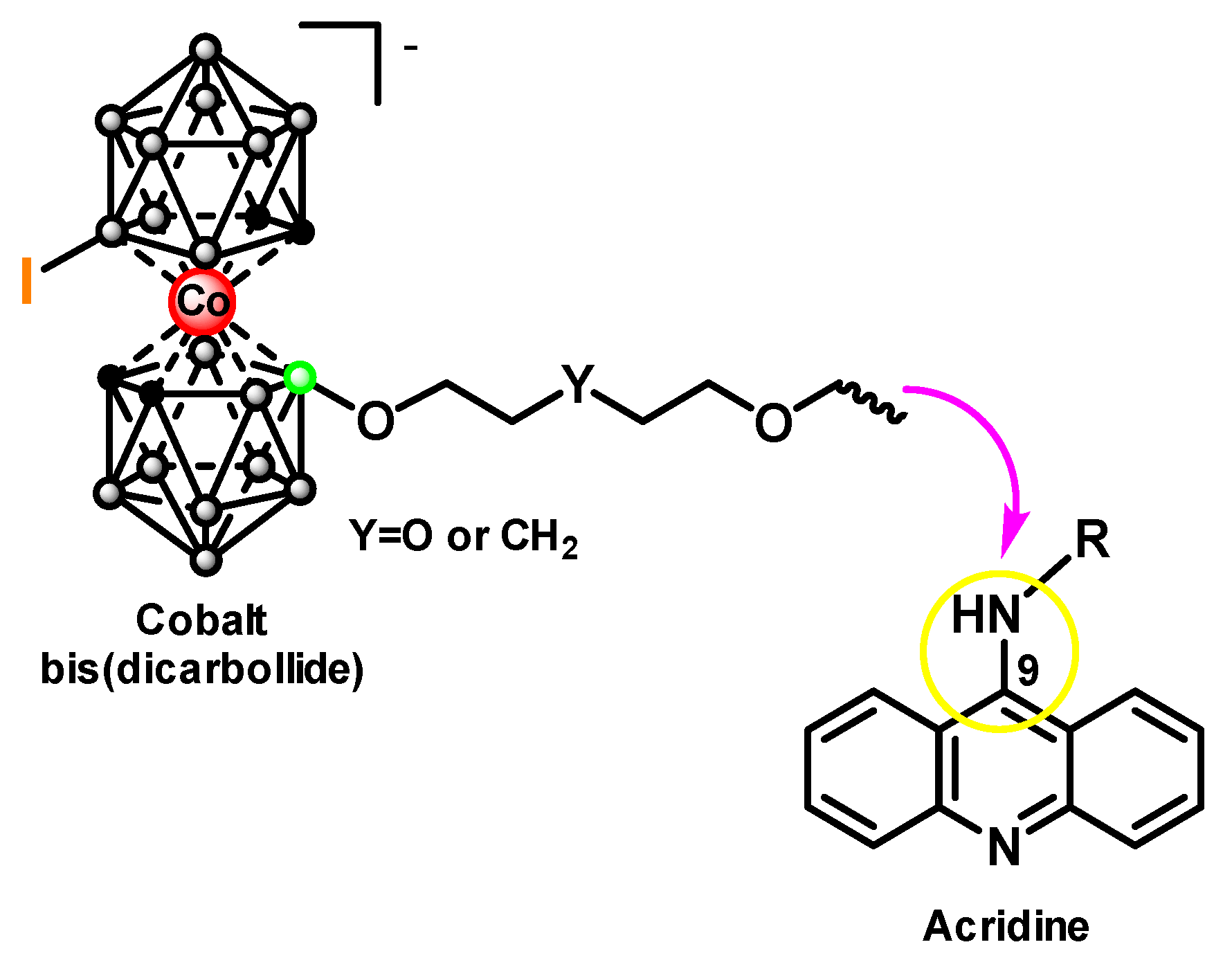
To obtain iodine-containing conjugates of cobalt bis(dicarbollide) with acridine, iodination of 1,2,3-triazoles 1-4 with excess of crystalline I2 was carried out by boiling in MeOH for one hour, which led to the formation of new derivatives 5-8 with yields over 81% (Scheme 1), which can potentially be used as effective BNCT agents. The structures of conjugates 5-8 were confirmed by 1H, 11B{1H}, 11B, and 13C{1H} NMR spectroscopy, IR-spectroscopy, and high-resolution mass spectrometry (see Supplementary Materials, Figures S1–S31).
The 1H NMR spectra of conjugates 5-8 contain the characteristic signals of triazole CH group at 7.84-8.01 ppm, signals of acridine CH groups at 7.63-8.56 ppm, and signals of carborane CH groups at 4.13-4.45 ppm. In the 13С{1Н} NMR spectra characteristic signals from triazole CH and C groups at 124.1-125.1 and 140.6-142.0 ppm, signals from acridine CH, C, and C-N groups at 119.7-136.7, 146.0-159.7, and 113.7-113.8 ppm, and signals from carborane CH groups at 55.2-53.3 ppm, respectively, are observed. In the NMR 11B{1H} spectra of compounds 5-8, the B-O signals are present approx. at 21.6 ppm and the B-I signals appear as hindered behind the group of BH signals at the area at –6.0 ppm, as expected for such bis(dicarbollide) cobalt derivatives.
The IR spectra of compounds 5-8 demonstrate a strong band of the BH stretching at ~2565 cm–1.
3.2. Biological Properties
3.2.1. Cytotoxic Activity of Conjugates of Iodinated Cobalt Bis(dicarbollide) with Acridine 5-8
The cytotoxic effects of compounds 5-8 against human cancer cell lines HCT116, MCF7, and A549, alongside nonmalignant WI38 lung fibroblasts, were assessed using a standard MTT colorimetric assay after a 72 hour incubation period (Table 1). Compound 5 was non-toxic (IC50 > 100 μM) across all tested cell lines. All four compounds were found to be non-toxic against the lung cancer A549 cell line and WI38 normal fibroblast line. In contrast, compounds 6, 7, and 8 exhibited mid-micromolar cytotoxic activity against the HCT116 and MCF7 lines, which restricts their suitability as potential agents for BNCT.
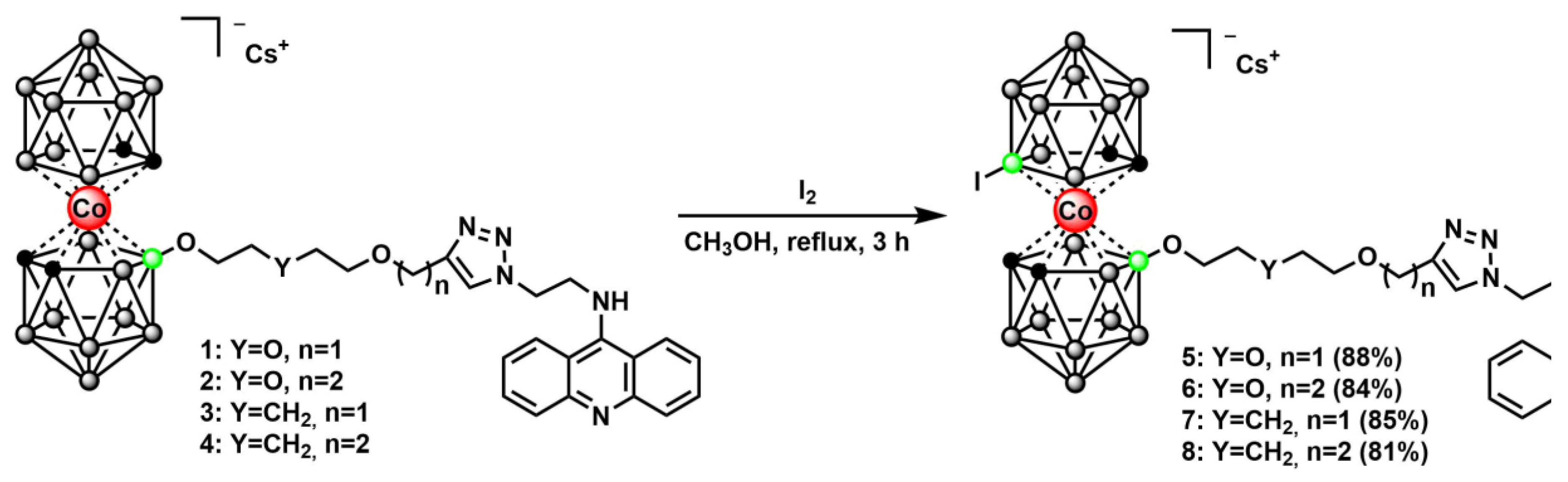
Based on the data obtained, it was decided to select conjugate 5 for further biological studies. Earlier it has been shown that the solubility in water of cobalt bis(dicarbollide) can be increased by using of suitable biocompatible excipients carriers of pharmaceutically active compounds, including poly(vinylpyrrolidone) (PVP) [57,58]. Specifically, to increase the water solubility of compound 5, the latter one was transformed to the non-covalent complex with PVP (Scheme 2). This resulted in a water-soluble form of compound 5-PVP, which was used to determine cytotoxicity, intracellular accumulation and in the BNCT experiment in vitro.
The fabrication of 5-PVP was performed in accordance with the patented procedure [59,60]. For the case of 5-PVP the sequence of steps included the dissolution of compound 5 in ethanol, and PVP (MW 10 kDa) — in water, mixing these solutions, followed by freezing the resultant liquid system and then its freeze-drying (see Expermental). The conjugate content in the 5-PVP sample by weight was 19.7%, and the water solubility reached 250 mg/ml. The solid matter thus produced was shown to possess at least 100-fold higher solubility in aqueous media with the physiological pH level as compared to the water solubility of compound 5 itself.
3.2.2. Cytotoxicity, Intracellular Accumulation and BNCT Experiment In Vitro of 5-PVP
The results of determining the cytotoxicity of the 5-PVP for B16 cell line at various boron-10 concentrations are presented in Figure 2.
During the first 24 hours of incubation of the 5-PVP with B16 cell line, no decrease in viability was observed; a slight, statistically insignificant increase in viability during incubation with high concentrations may be associated with the interaction of the PVP and MTT. Upon longer period of incubation, a significant decrease in viability occurs even at the lowest concentration used, which indicates the safety of incubation with the investigated compound 5-PVP for no more than 24 hours at boron-10 concentrations up to 125 μg/mL.
The boron content in the B16 cells was 0.98 ± 0.08 μg/106 cells. When converted to the boron-10 isotope, the natural content of which is 20%, the boron-10 content is 0.20 μg/106 cells.
The results of cell viability are shown in Figure 3; no pronounced effect of irradiation in the presence of the new boron-containing compound 5-PVP on B16 and DF1 cell lines was observed compared to the other groups. A statistically significant reduction in cell viability of T98G was observed, with a survival rate of 71%.
According to the migration test data, the cells do not lose their migration ability, but their morphology in the “BNCT” groups changes after 3 days (Figure 4).
Figure 5 presents the results of the clonogenic assay. It was found that preincubation with the new boron-containing 5-PVP followed by neutron irradiation reduces the survival of T98G glioblastoma cells to 25% and B16 melanoma cells to 17%. The effect of BNCT on DF1 fibroblasts was less pronounced, with a cloning efficiency of 39%.
3.2.3. Ex Vivo Biodistribution and Tumor Uptake Study of 5-PVP
An ex vivo biodistribution and tumor uptake study of 5-PVP was performed in B16F10 melanoma-bearing mice using ICP-OES. Boron concentrations in organs and tissues were measured after a single intravenous injection at a dose of 2.01 ± 0.08 mg B/kg. The resulting time-dependent concentration profiles are presented in Figure 6 and Figure 7.
As expected for intravenous administration, the highest boron concentration in blood was observed at the earliest time point (15 min), with a value of 4.3 ± 1.4 µg B/g. Thereafter, the blood boron level declined rapidly by 30 min, followed by a slower decrease up to 90 min. Between 90 and 120 min, the concentration remained relatively stable, fluctuating around 1.02 ± 0.14 µg B/g.
The highest boron concentration in the tumor was also detected at 15 min post-injection, corresponding to 1.4 ± 0.2 µg B/g, and gradually decreased to 0.4 ± 0.1 µg B/g by 120 min. A slight increase at 60 min was not statistically significant (p = 0.216, ANOVA). Overall, the boron concentration profile in the tumor was similar in shape to that in the blood.
The concentration profiles in skin and muscle were also comparable to those in blood and tumor, but with lower absolute values. These findings suggest a relative specificity of 5-PVP accumulation in the tumor compared to the surrounding normal tissues.
Figure 7.
Boron concentration profiles in the heart, lungs, spleen, brain, liver, and kidneys after a single intravenous injection of 5-PVP. Data are presented as mean ± SD, n = 5 per time point.
Figure 7.
Boron concentration profiles in the heart, lungs, spleen, brain, liver, and kidneys after a single intravenous injection of 5-PVP. Data are presented as mean ± SD, n = 5 per time point.
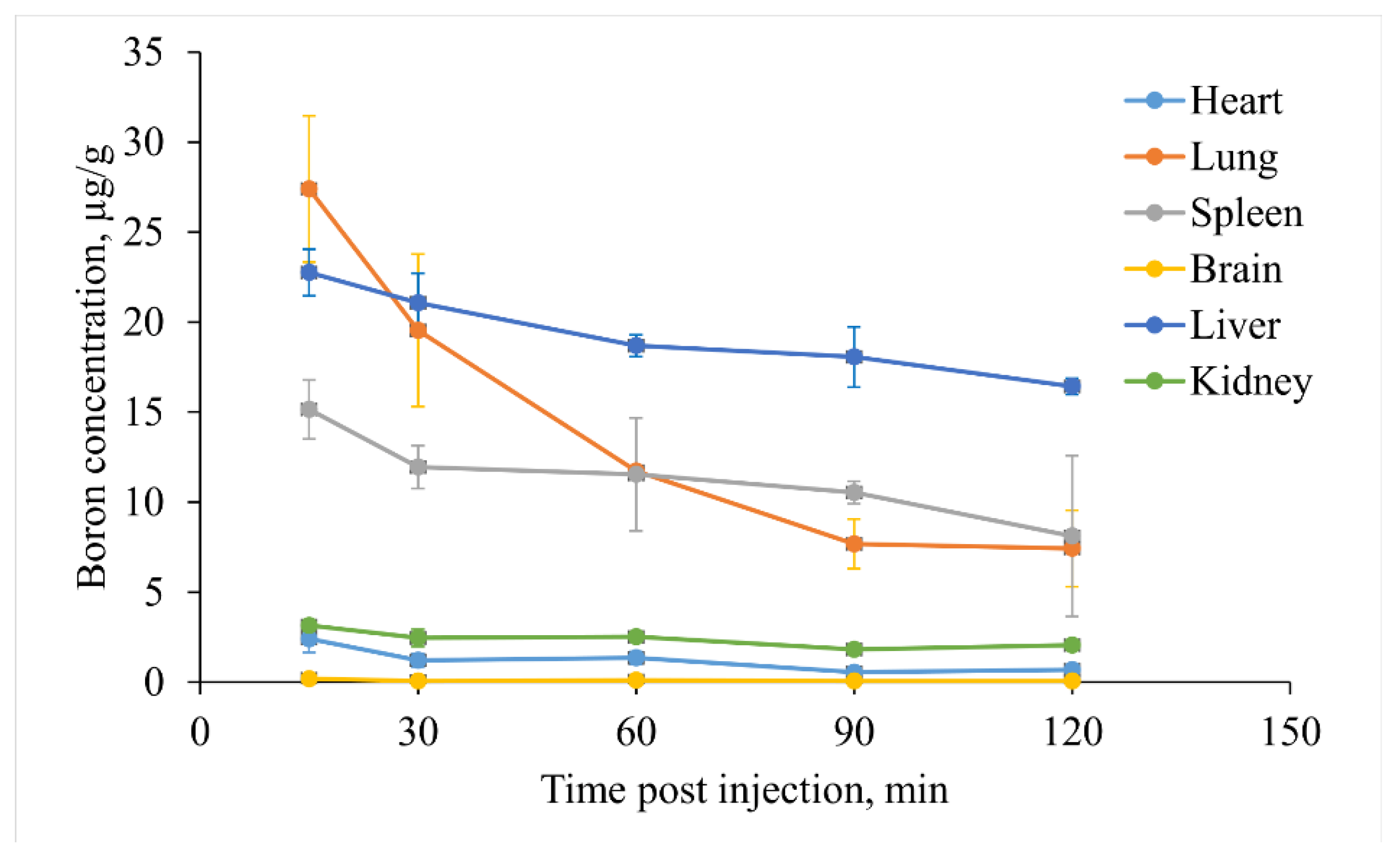
All other healthy organs shown in Figure 7 also exhibited the highest boron concentration at 15 min after administration. The most intense boron accumulation was observed in the lungs, liver, and spleen. In these organs, absolute boron concentrations ranged from 15-27 µg B/g at 15 min to 7-16 µg B/g by 120 min post injection. In the brain, boron levels remained very low throughout the observation period, ranging from 0.20 to 0.06 µg B/g over 120 min. These results imply that 5-PVP does not appear to cross the blood-brain barrier, and that the boron detected in the brain is most likely attributable to the presence of boron in the cerebral blood vessels.
To evaluate the potential of 5-PVP for boron neutron capture therapy (BNCT), the tumor to normal tissue ratio was plotted as a function of time and is presented in Figure 8.
The tumor/muscle and tumor/skin ratios reached values of 5.4 and 3.4, respectively, at 30 min post-injection, and gradually decreased to 2.1 and 1.8 by 120 min. These values indicate a high selectivity of tumor accumulation, with tumor-to-normal tissue ratios sustained over a time frame sufficient for a BNCT session. Consequently, the absorbed dose delivered to healthy tissues upon neutron irradiation would be substantially lower than that delivered to the tumor.
In our previous study [61], the accumulation and retention of boronophenylalanine (BPA), the conventional BNCT agent, were evaluated in the same B16F10 melanoma model. The tumor/muscle ratio reached a maximum of 3 at 30 min post-injection, gradually declined to 1.5 at 90 min, and then remained stable up to 120 min. By contrast, 5-PVP achieved a tumor/muscle ratio of 5.4 at 30 min, indicating a potential advantage over BPA and warranting further investigation.
At the same time, the maximum tumor/blood ratio reached with 5-PVP was 0.8, which suggests a moderate level of bioavailability. In this context, careful determination of the therapeutic window will be required in subsequent translational studies, as a relatively high dose may be necessary to deliver sufficient boron to the tumor tissue. Optimization of the compound’s bioavailability may therefore represent a promising direction for future research.
4. Conclusions
A series of novel iodinated cobalt bis(dicarbollide)-acridine conjugates was successfully synthesized via direct electrophilic iodination of the parent cobalt bis(dicarbollide)-acridine adducts using molecular iodine as the halogenating agent. The cytotoxic profiles of the newly synthesized compounds were assessed against a panel of human cell lines of diverse origin. Notably, one of them alone exhibited no antiproliferative activity against both cancerous and non-malignant cells.
To enhance its aqueous solubility, this iodinated cobalt bis(dicarbollide) conjugate with acridine was subsequently formulated as a non-covalent inclusion complex with polyvinylpyrrolidone. It was demonstrated that it successfully penetrates cells and accumulates at the boron concentration required for successful BNCT. It was found that preincubation with the new boron-containing drug followed by neutron irradiation reduces the survival of T98G glioblastoma cells to 25% and B16 melanoma cells to 17%.
The present study established that this complex exhibits pronounced selectivity in tumor uptake, as evidenced by a tumor-to-muscle concentration ratio of 5.4, which exceeds that of the conventional boron delivery agent BPA. Further optimization of the compound may include the improvement of pharmacokinetic properties, particularly regarding bioavailability.
In this way, the conjugation of acridine with iodinated cobalt bis(dicarbollide) is a promising strategy for therapeutic applications in boron neutron capture therapy.
Supplementary Materials
The following supporting information can be downloaded at the website of this paper posted on Preprints.org, Figure S1–S31.
Author Contributions
Nadezhda V. Dudarova: Formal analysis, Investigation. Egor V. Sidorskii: Data curation, Investigation. Anna I. Kasatova: Formal analysis, Investigation, Writing – original draft. Timofey A. Bykov: Formal analysis, Investigation. Anastasia A. Antonets: Formal analysis, Investigation. Alexey A. Nazarov: Formal analysis, Investigation. Vsevolod A. Skribitsky: Data curation, Investigation, Writing – original draft. Kristina E. Shpakova: Data curation, Investigation. Anton A. Kasianov: Data curation, Investigation. Yulia A. Finogenova: Formal analysis, Writing – original draft. Vladimir I. Lozinsky: Conceptualization, Supervision, Writing – review & editing. Sergey Yu. Taskaev: Conceptualization, Supervision. Alexey A. Lipengolts: Conceptualization, Supervision. Sergey A. Anufriev: Formal analysis, Investigation. Anna A. Druzina: Conceptualization, Formal analysis, Writing – original draft. Igor B. Sivaev: Writing – review & editing, Supervision. Vladimir I. Bregadze: Conceptualization, Supervision.
Funding
Synthesis and characterization of new compounds described in this study were supported by the Ministry of Science and Higher Education of the Russian Federation as part of the State Assignment of the A.N. Nesmeyanov Institute of Organoelement Compounds of the Russian Academy of Sciences (075-03-2026-024). The NMR spectra were obtained using equipment of the Center for Molecular Structure Research at Nesmeyanov Institute of Organoelement Compounds operating with support of the Ministry of Science and Higher Education of the Russian Federation.
Institutional Review Board Statement
All experiments involving laboratory animals were carried out in accordance with the ethical standards for the treatment of animals adopted by the European Convention for the Protection of Vertebrate Animals used for Research and other Scientific Purposes and was approved by the Local Biomedical Ethics Committee (No. 2026-5.1, issued on 15.05.2026).
Data Availability Statement
Data is contained within the article.
Acknowledgments
The authors acknowledge support from the M.V. Lomonosov Moscow State University Program of Development («Feyond-A400» microplate reader (Allsheng, Hangzhou, China), automated pipetting system «EzMate» (Blue-Ray Biotech, Taipei, Taiwan).
Conflicts of Interest
The authors declare no conflicts of interest.
References
- Bray, F.; Laversanne, M.; Sung, H.; Ferlay, J.; Siegel, R.L.; Soerjomataram, I.; Jemal, A. Global cancer statistics 2022: GLOBOCAN estimates of incidence and mortality worldwide for 36 cancers in 185 countries. CA Cancer J. Clin. 2024, 74, 229-263. [CrossRef]
- Sauerwein, W.; Wittig, A.; Moss, R.; Nakagawa, Y., Eds. Neutron Capture Therapy: Principles and Applications; Springer: Berlin/Heidelberg, Germany, 2012; pp 554. [CrossRef]
- Sivaev, I.B.; Bregadze, V.I. Polyhedral boranes for medical applications: Current status and perspectives. Eur. J. Inorg. Chem. 2009, 2009, 1433-1450. [CrossRef]
- Zhu, Y.; Hosmane, N.S. Applications and perspectives of boron-enriched nanocomposites in cancer therapy. Future Med. Chem. 2013, 5, 705-714. [CrossRef]
- Uspenskii, S.A.; Khaptakhanova, P.A. Boron nanoparticles in chemotherapy and radiotherapy: the synthesis, state-of-the-art, and prospects. Russ. Chem. Bull. 2022, 71, 2533-2560. [CrossRef]
- Barth, R.F.; Zhang, Z.; Liu, T. A realistic appraisal of boron neutron capture therapy as a cancer treatment modality. Cancer Commun. 2018, 38, 36. [CrossRef]
- Lipengolts, A.A.; Finogenova, Y.A.; Skribitsky, V.A.; Grigorieva, E.Y. Binary technologies of malignant tumors radiotherapy. J. Phys. Conf. Ser. 2021, 2058, 012039. [CrossRef]
- Tsygankova, A.R.; Kanygin, V.V.; Kasatova, A.I.; Zavyalov, E.L.; Guselnikova, T.Ya.; Kichigin, A.I.; Mukhamadiyarov, R.A. Determination of boron by inductively coupled plasma atomic emission spectroscopy. Biodistribution of 10B in tumor-bearing mice. Russ. Chem. Bull. 2020, 69, 601-607. [CrossRef]
- Okada, S.; Nishimura, K.; Ainaya, Q.; Shiraishi, K.; Anufriev, S.A.; Sivaev, I.B.; Sakurai, Y.; Suzuki, M.; Yokoyama, M.; Nakamura, H. Development of a Gadolinium-Boron-Conjugated Albumin for MRI-Guided Neutron Capture Therapy. Mol. Pharm. 2023, 20, 6311-6318. [CrossRef]
- Hawthorne, M.F. The role of chemistry in the development of boron neutron capture therapy of cancer. Angew. Chem. Int. Ed. 1993, 32, 950-984. [CrossRef]
- Soloway, A.H.; Tjarks, W.; Barnum, B.A.; Rong, F.G.; Barth, R.F.; Codogni, I.M.; Wilson, J.G. The Chemistry of Neutron Capture Therapy. Chem. Rev. 1998, 98, 1515-1562. [CrossRef]
- Kasatova, A.I.; Kanygin, V.V.; Zavjalov, E.L.; Razumov, I.A.; Kichigin, A.I.; Tsygankova, A.R.; Solovieva, O.I.; Kasatov, D.A.; Chernov, V.I. Accumulation and toxic effects of boron-containing drugs in SW-620 models. Vestn. Tomsk. gos. univ., Biol. 2021, 55, 97-113. [CrossRef]
- Hartman, T.; Carlsson, J. Radiation dose heterogeneity in receptor and antigen mediated boron neutron capture therapy. Radiother. Oncol. 1994, 31, 61-75. [CrossRef]
- Gabel, D.; Foster, S.; Fairchild, R.G. The Monte Carlo simulation of the biological effect of the 10B(n, α)7Li reaction in cells and tissue and its implication for boron neutron capture therapy. Radiat. Res. 1987, 111, 14-25. [CrossRef]
- Tjarks, W.; Ghaneolhosseini, H.; Henssen, C.L.; Malmquist, J.; Sjöberg, S. Synthesis of para-and nido-carboranyl phenanthridinium compounds for neutron capture therapy. Tetrahedron Lett. 1996, 37, 6905-6908. [CrossRef]
- Ghaneolhosseini, H.; Tjarks, W.; Sjöberg, S. Synthesis of boronated phenanthridinium derivatives for potential use in boron neutron capture therapy (BNCT). Tetrahedron 1997, 53, 17519-17526. [CrossRef]
- Ghaneolhosseini, H.; Sjöberg, S. Synthesis of a boronated naphthalimide for potential use in boron neutron capture therapy (BNCT). Acta Chem. Scand. 1999, 53, 298-300. [CrossRef]
- Nekvinda, J.; Różycka, D.; Rykowski, S.; Wyszko, E.; Fedoruk-Wyszomirska, A.; Gurda, D.; Orlicka-Płocka, M.; Giel-Pietraszuk, M.; Kiliszek, A.; Rypniewski, W.; Bachorz, R.; Wojcieszak, J.; Grüner, B.; Olejniczak, A.B. Synthesis of naphthalimide-carborane and metallacarborane conjugates: Anticancer activity, DNA binding ability. Bioorg. Chem. 2020, 94, 103432. [CrossRef]
- Rykowski, S.; Gurda-Woźna, D.; Orlicka-Płocka, M.; Fedoruk-Wyszomirska, A.; Giel-Pietraszuk, M.; Wyszko, E.; Kowalczyk, A.; Stączek, P.; Bak, A.; Kiliszek, A.; Rypniewski, W.; Olejniczak, A.B. Design, synthesis, and evaluation of novel 3-carboranyl-1,8-naphthalimide derivatives as potential anticancer agents. Int. J. Mol. Sci. 2021, 22, 2772. [CrossRef]
- Laskova, J.; Kosenko, I.; Serdyukov, A.; Sivaev, I.; Bregadze, V.I. Synthesis of naphthalimide derivatives of closo-dodecaborate and nido-carborane. J. Organomet. Chem. 2022, 959, 122186. [CrossRef]
- Bogucka-Kocka, A.; Kołodziej, P.; Makuch-Kocka, A.; Różycka, D.; Rykowski, S.; Nekvinda, J.; Grüner, B.; Olejniczak, A.B. Nematicidal activity of naphthalimide-boron cluster conjugates. Chem. Commun. 2022, 58, 2528-2531. [CrossRef]
- Rykowski, S.; Gurda-Woźna, D.; Orlicka-Płocka, M.; Fedoruk-Wyszomirska, A.; Giel-Pietraszuk, M.; Wyszko, E.; Kowalczyk, A.; Stączek, P.; Biniek-Antosiak, K.; Rypniewski, W.; Olejniczak, A.B. Design of DNA intercalators based on 4-carboranyl-1,8-naphthalimides: Investigation of their DNA-binding ability and anticancer activity. Int. J. Mol. Sci. 2022, 23, 4598. [CrossRef]
- Rykowski, S.; Gurda-Woźna, D.; Fedoruk-Wyszomirska, A.; Orlicka-Płocka, M.; Kowalczyk, A.; Stączek, P.; Denel-Bobrowska, M.; Biniek-Antosiak, K.; Rypniewski, W.; Wyszko, E.; Olejniczak, A.B. Carboranyl-1,8-naphthalimide intercalators induce lysosomal membrane permeabilization and ferroptosis in cancer cell lines. J. Enzyme Inhib. Med. Chem. 2023, 38, 2171028. [CrossRef]
- Crossley, E.L.; Ziolkowski, E.J.; Coderre, J.A.; Rendina, L.M. Boronated DNA-binding compounds as potential agents for boron neutron capture therapy. Mini-Rev. Med. Chem. 2007, 7, 303-313. [CrossRef]
- Singh, R.; Singh, J.; Kaur, A. Recent Advances in QSAR, SAR, and Therapeutic Potentials of Acridine Derivatives: A Review. Future Med. Chem. 2026, 1347, 143226. [CrossRef]
- Gallala, M.; Tka, N.; Aroui, S. Recent advances in the development of acridine-based hybrids with antiproliferative activity. RSC Med. Chem. 2026, 17, 2768–2849. [CrossRef]
- Ghaneolhosseini, H.; Tjarks, W.; Sjöberg, S. Synthesis of novel boronated acridines and spermidines as possible agents for BNCT. Tetrahedron 1998, 54, 3877-3884. [CrossRef]
- Różycka, D.; Kowalczyk, A.; Denel-Bobrowska, M.; Kuźmycz, O.; Gapińska, M.; Stączek, P.; Olejniczak, A.B. Acridine/Acridone-Carborane Conjugates as Strong DNA-Binding Agents with Anticancer Potential. ChemMedChem 2023, 18, e202200666. [CrossRef]
- Soloway, A.H.; Hatanaka, H.; Davis, M.A. Penetration of brain and brain tumor. VII. Tumor-binding sulfhydryl boron compounds. J. Med. Chem. 1967, 10, 714-717. [CrossRef]
- Swietnicki, W.; Goldeman, W.; Psurski, M.; Nasulewicz-Goldeman, A.; Boguszewska-Czubara, A.; Drab, M.; Sycz, J.; Goszczyński, T.M. Metallacarborane derivatives effective against Pseudomonas aeruginosa and Yersinia enterocolitica. Int. J. Mol. Sci. 2021, 22, 6762. [CrossRef]
- Sivaev, I.B.; Bregadze, V.I. Chemistry of cobalt bis(dicarbollides). A review. Collect. Czech. Chem. Commun. 1999, 64, 783-805. [CrossRef]
- Dash, B.P.; Satapathy, R.; Swain, B.R.; Mahanta, C.S.; Jena, B.B.; Hosmane, N.S. Cobalt bis(dicarbollide) anion and its derivatives. J. Organomet. Chem. 2017, 849, 170-194. [CrossRef]
- Dudarova, N.V.; Kosenko, I.D.; Maltseva, V.E.; Ananyev, I.V.; Druzina, A.A.; Bregadze, V.I.; Sivaev, I.B. On the Issue of Biological Activity of Cobalt bis(dicarbollide) Derivatives: Conformation of the [8-EtO-8′-I-3,3′-Co(1,2-C2B9H10)2]– Anion. J. Clust. Sci. 2025, 36, 161. [CrossRef]
- Nuez-Martinez, M.; Pinto, C.I.G.; Guerreiro, J.F.; Mendes, F.; Marques, F.; Muñoz-Juan, A.; Xavier, J.A.M.; Laromaine, A.; Bitonto, V.; Protti, N.; Geninatti Crich, S.; Teixidor, F.; Viñas, C. Cobaltabis(dicarbollide) ([o-COSAN]–) as multifunctional chemotherapeutics: A prospective application in boron neutron capture therapy (BNCT) for glioblastoma. Cancers 2021, 13, 6367. [CrossRef]
- Gos, M.; Cebula, J.; Goszczyński, T.M. Metallacarboranes in medicinal chemistry: Current advances and future perspectives. J. Med. Chem. 2024, 67, 8481-8501. [CrossRef]
- Bennour, I.; Ramos, M.N.; Nuez-Martínez, M.; Xavier, J.A.M.; Buades, A.B.; Sillanpää, R.; Teixidor, F.; Choquesillo-Lazarte, D.; Romero, I.; Martinez-Medina, M.; Viñas, C. Water soluble organometallic small molecules as promising antibacterial agents: synthesis, physical-chemical properties and biological evaluation to tackle bacterial infections. Dalton Trans. 2022, 51, 7188-7209. [CrossRef]
- Fuentes, I.; García-Mendiola, T.; Sato, S.; Pita, M.; Nakamura, H.; Lorenzo, E.; Teixidor, F.; Marques, F.; Viñas, C. Metallacarboranes on the Road to Anticancer Therapies: Cellular Uptake, DNA Interaction, and Biological Evaluation of Cobaltabisdicarbollide [COSAN]–. Chem. Eur. J. 2018, 24, 17239-17254. [CrossRef]
- Tarrés, M.; Canetta, E.; Paul, E.; Forbes, J.; Azzouni, K.; Viñas, C.; Teixidor, F.; Harwood, A.J. Biological interaction of living cells with COSAN-based synthetic vesicles. Sci. Rep. 2015, 5, 7804. [CrossRef]
- Spryshkova, R.A.; Karaseva, L.I.; Brattsev, V.A.; Serebriakov, N.G. Toxicity of the functional derivatives of polyhedral carboranes. Med. Radiol. 1981, 26, 62-64. PMID: 7266291.
- Bauduin, P.; Prevost, S.; Farràs, P.; Teixidor, F.; Diat, O.; Zemb, T. A theta-shaped amphiphilic cobaltabisdicarbollide anion: Transition from monolayer vesicles to micelles. Angew. Chem. Int. Ed. 2011, 50, 5298-5300. [CrossRef]
- Teixidor, F.; Pedrajas, J.; Rojo, I.; Viñas, C.; Sillanpää, R.; Sivaev, I.; Bregadze, V.; Sjöberg, S. Chameleonic Capacity of [3,3′-Co(1,2-C2B9H11)2]– in Coordination. Generation of the Highly Uncommon S(thioether)-Na Bond. Organometallics 2003, 22, 3414-3423. [CrossRef]
- Llop, J.; Masalles, C.; Viñas, C.; Teixidor, F.; Sillanpää, R.; Kivekäs, R. The [3,3′-Co(1,2-C2B9H11)2]– anion as a platform for new materials: Synthesis of its functionalized monosubstituted derivatives incorporating synthons for conducting organic polymers. Dalton Trans. 2003, 32, 556-561. [CrossRef]
- Rokitskaya, T.I.; Kosenko, I.D.; Sivaev, I.B.; Antonenko, Y.N.; Bregadze, V.I. Fast flip-flop of halogenated cobalt bis(dicarbollide) anion in a lipid bilayer membrane. Phys. Chem. Chem. Phys. 2017, 19, 25122-25128. [CrossRef]
- Assaf, K.I.; Begaj, B.; Frank, A.; Nilam, M.; Mougharbel, A.S.; Kortz, U.; Nekvinda, J.; Grüner, B.; Gabel, D.; Nau, W.M. High-Affinity Binding of Metallacarborane Cobalt Bis(dicarbollide) Anions to Cyclodextrins and Application to Membrane Translocation. J. Org. Chem. 2019, 84, 11790-11798. [CrossRef]
- Zakret-Drozdowska, K.; Szermer-Olearnik, B.; Goldeman, W.; Gos M.; Drozdowski, D.; Gągor, A.; Goszczyński, T.M.; Unraveling the correlation between biological effects and halogen substituents in cobalt bis(dicarbollide). Inorg. Chem. Front. 2025; 12, 191-204. [CrossRef]
- Sivaev, I.B.; Kosenko, I.D. Rotational conformation of 8,8′-dihalogenated derivatives of cobalt bis(dicarbollide) in solution. Russ. Chem. Bull. 2021, 70, 753-756. [CrossRef]
- Sivaev, I.B. Bis(Dicarbollide) Complexes of Transition Metals: How Substituents in Dicarbollide Ligands Affect the Geometry and Properties of the Complexes. Molecules 2024, 29, 3510. [CrossRef]
- Verdiá-Báguena, C.; Alcaraz, A.; Aguilella, V.M.; Cioran, A.M.; Tachikawa, S.; Nakamura, H.; Teixidor, F.; Viñas, C. Amphiphilic COSAN and I2-COSAN crossing synthetic lipid membranes: Planar bilayers and liposomes. Chem. Commun. 2014, 50, 6700-6703. [CrossRef]
- Druzina, A.A.; Dudarova, N.V.; Ananyev, I.V.; Antonets, A.A.; Kaluzhny, D.N.; Nazarov, A.A.; Sivaev, I.B.; Bregadze, V.I. New Boron Containing Acridines: Synthesis and Preliminary Biological Study. Molecules 2023, 28, 6636. [CrossRef]
- Laskova, J.; Serdyukov, A.; Kosenko, I.; Ananyev, I.; Titova, E.; Druzina, A.; Sivaev, I.; Antonets, A.A.; Nazarov, A.A.; Bregadze, V.I. New azido coumarins as potential agents for fluorescent labeling and their “click” chemistry reactions for the conjugation with closo-dodecaborate anion. Molecules 2022, 27, 8575. [CrossRef]
- Kumar, P.; Nagarajan, A.; Uchil, P.D. Analysis of Cell Viability by the MTT Assay. Cold Spring Harb. Protoc. 2018, pdb.prot095505. [CrossRef]
- Ghasemi, M.; Turnbull, T.; Sebastian, S.; Kempson, I. The MTT Assay: Utility, Limitations, Pitfalls, and Interpretation in Bulk and Single-Cell Analysis. Int. J. Mol. Sci. 2021, 22, 12827. [CrossRef]
- Taskaev, S.; Berendeev, E.; Bikchurina, M.; Bykov, T.; Chesnokova, Yu.; Deeb, R.; Ibrahim, I.; Kasatova, A.; Kasatov, D.; Kolesnikov, Ya.; Koshkarev, A.; Kuzmina, K.; Maltseva, V.; Ostreinov, G.; Savinov, S.; Shchudlo, I.; Shchukin, S.; Shein, T.; Shuklina, A.; Singatulina, N.; Sokolova, E.; Sorokin, I.; Taskaeva, Iu.; Verkhovod, G. VITA accelerator neutron sources: status and research results. Cancer. 2026, 18, 1886. [CrossRef]
- Chilin, A.; Marzaro, G.; Marzano, C.; Dalla Via, L.; Ferlin, M.G.; Pastorini, G.; Guiotto, A. Synthesis and antitumor activity of novel amsacrine analogs: The critical role of the acridine moiety in determining their biological activity. Bioorg. Med. Chem. 2009, 17, 523-529. [CrossRef]
- Lang, X.; Li, L.; Chen, Y.; Sun, Q.; Wu, Q.; Liu, F.; Tan, C.; Liu, H.; Gao, C.; Jiang, Y. Novel synthetic acridine derivatives as potent DNA-binding and apoptosis-inducing antitumor agents. Bioorg. Med. Chem. 2013, 21, 4170-4177. [CrossRef]
- Tonelli, M.; Vettoretti, G.; Tasso, B.; Novelli, F.; Boido, V.; Sparatore, F.; Busonera, B.; Ouhtit, A.; Farci, P.; Blois, S.; Giliberti, G.; La Colla, P. Acridine derivatives as anti-BVDV agents. Antiviral Res. 2011, 91, 133-141. [CrossRef]
- Rak, J.; Kaplánek, R.; Král, V. Solubilization and deaggregation of cobalt bis(dicarbollide) derivatives in water by biocompatible excipients. Bioorg. Med. Chem. Lett. 2010, 20, 1045-1048. [CrossRef]
- Rak, J.; Jakubek, M.; Kaplánek, R., Matejícek, P.; Král, V. Cobalt bis(dicarbollide) derivatives: Solubilization and self-assembly suppression. Eur. J. Med. Chem. 2011, 46, 1140-1146. [CrossRef]
- Lozinsky, V.I.; Ivanov, R.V.; Kravchenko, D.V.; Ivaschenko, A.A. Composition for making dosage form with prolonged effect and method of making said form. Russian Federation Patent No. 2363451, 2007.
- Ivanov, R.V.; Lozinsky, V.I.; Balakin, K.V.; Bachurin, S.O. Pharmaceutical composition based on hydrogenated pyrido(4,3-b)indole for treatment of neurodegenerative diseases, method for preparation of the composition and pharmacological dosage form on the basis of this composition. Russian Federation Patent No. 2428185, 2009.
- Ryabchikova, M.N.; Nelyubin, A.V.; Klyukin, I.N.; Selivanov, N.A.; Bykov, A.Y.; Kubasov, A.S.; Skribitsky, V.A.; Finogenova, Y.A.; Shpakova, K.E.; Kasianov, A.A.; Lipengolts, A.A.; Zhdanov, A.P.; Grigoreva, E.Y.; Zhizhin, K.Y.; Kuznetsov, N.T. Synthesis of New Promising BNCT Agents Based on Conjugates of closo-Dodecaborate Anion and Aliphatic Diamino Acids. Int. J. Mol. Sci. 2025, 26, 68. [CrossRef]
Figure 1.
The structure of N9-substituted acridine.
Scheme 1.
The synthesis of iodinated cobalt bis(dicarbollide) conjugates with acridine 5-8.
Scheme 2.
The non-covalent complex of poly(vinylpyrrolidone) with iodinated cobalt bis(dicarbollide) conjugate with acridine 5-PVP.
Scheme 2.
The non-covalent complex of poly(vinylpyrrolidone) with iodinated cobalt bis(dicarbollide) conjugate with acridine 5-PVP.
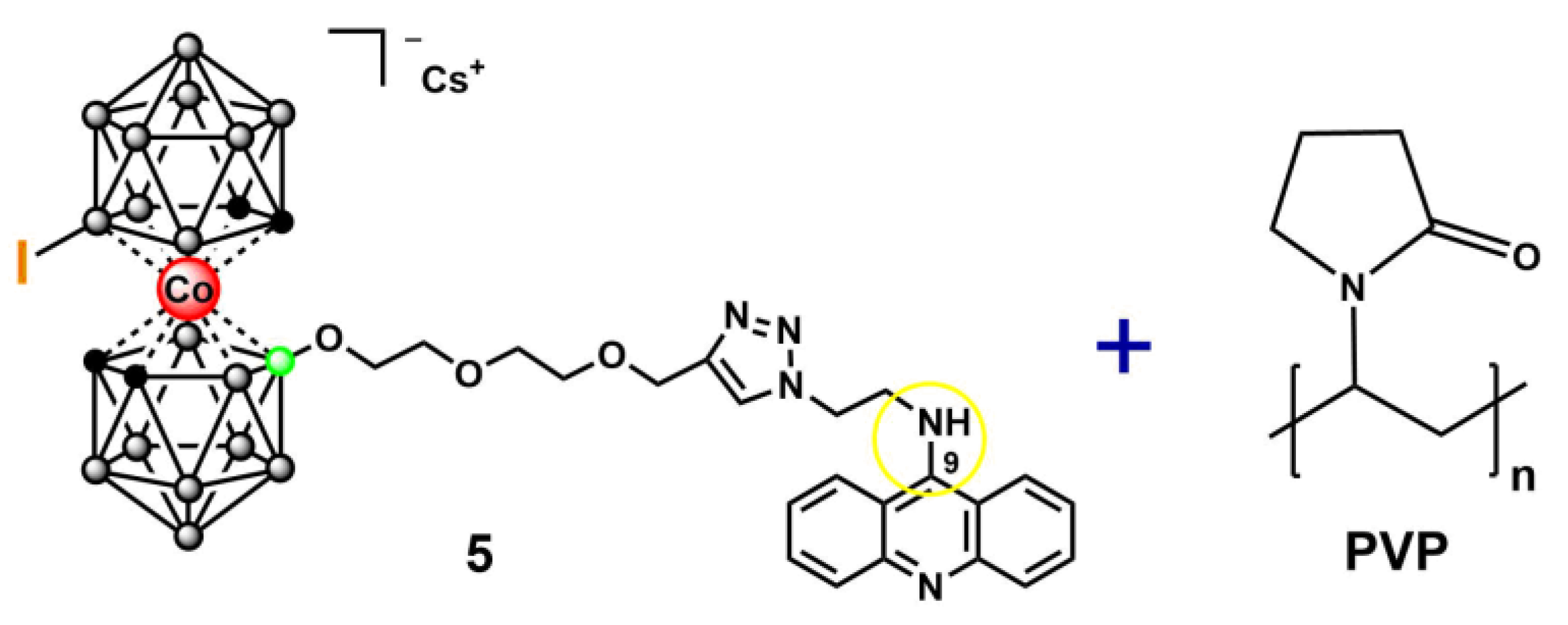
Figure 2.
The cytotoxicity of the 5-PVP for B16 cell line.
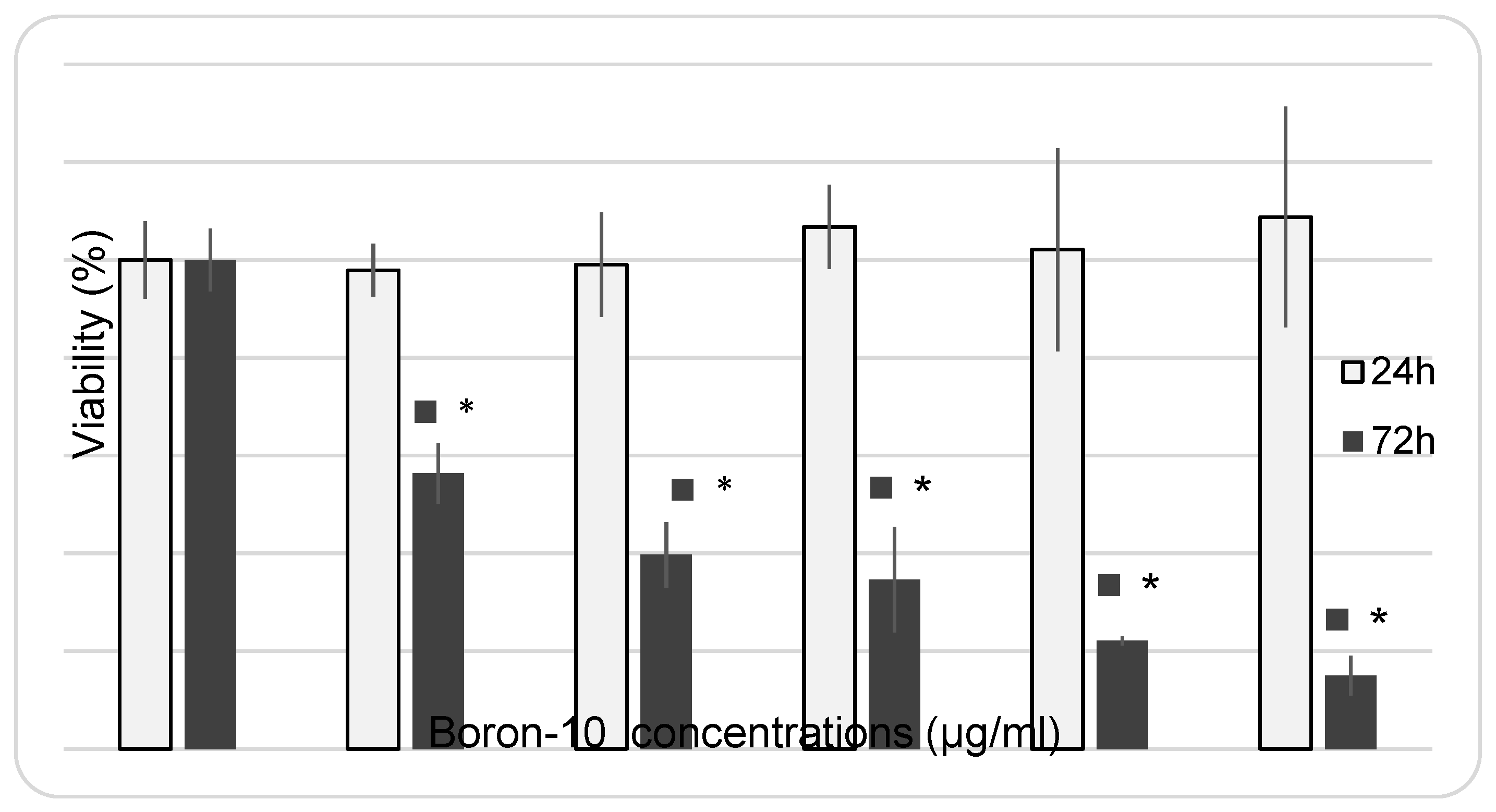
Figure 3.
Cytotoxicity indicators of different types of exposure: neutron irradiation, the drug alone, and BNCT. МТТ assay 96 hours after BNCT with the 5-PVP, n=5.
Figure 3.
Cytotoxicity indicators of different types of exposure: neutron irradiation, the drug alone, and BNCT. МТТ assay 96 hours after BNCT with the 5-PVP, n=5.
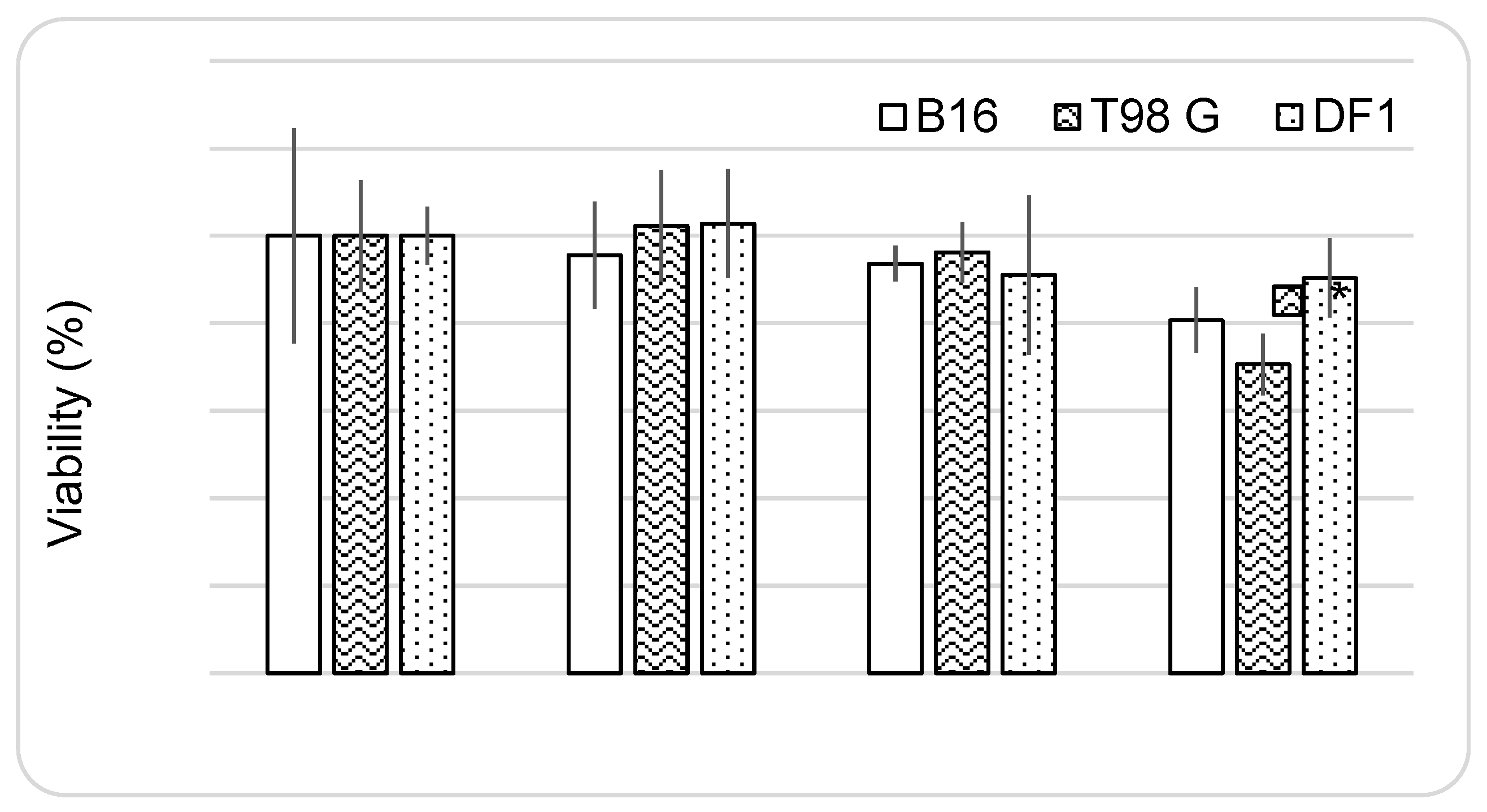
Figure 4.
Cell migration assay. Magnification: ×100.
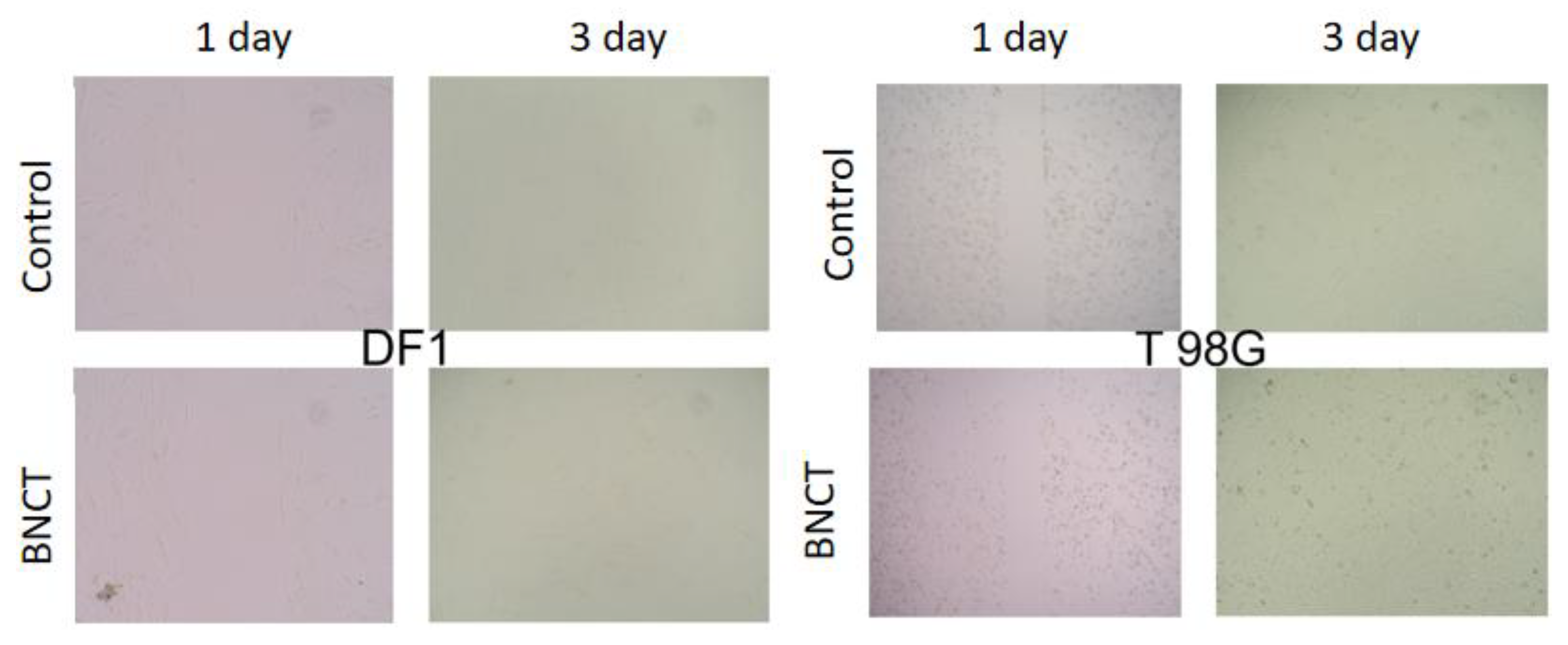
Figure 5.
Assessment of the plating efficacy following various treatments: clonogenic assay, n=3.
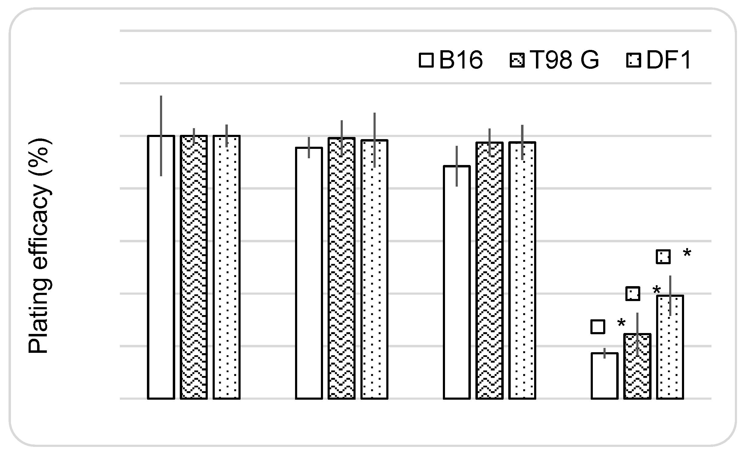
Figure 6.
Boron concentration profiles in blood, tumor (B16F10 melanoma), muscle, and skin after a single intravenous injection of 5-PVP. Data are presented as mean ± SD, n = 5 per time point.
Figure 6.
Boron concentration profiles in blood, tumor (B16F10 melanoma), muscle, and skin after a single intravenous injection of 5-PVP. Data are presented as mean ± SD, n = 5 per time point.
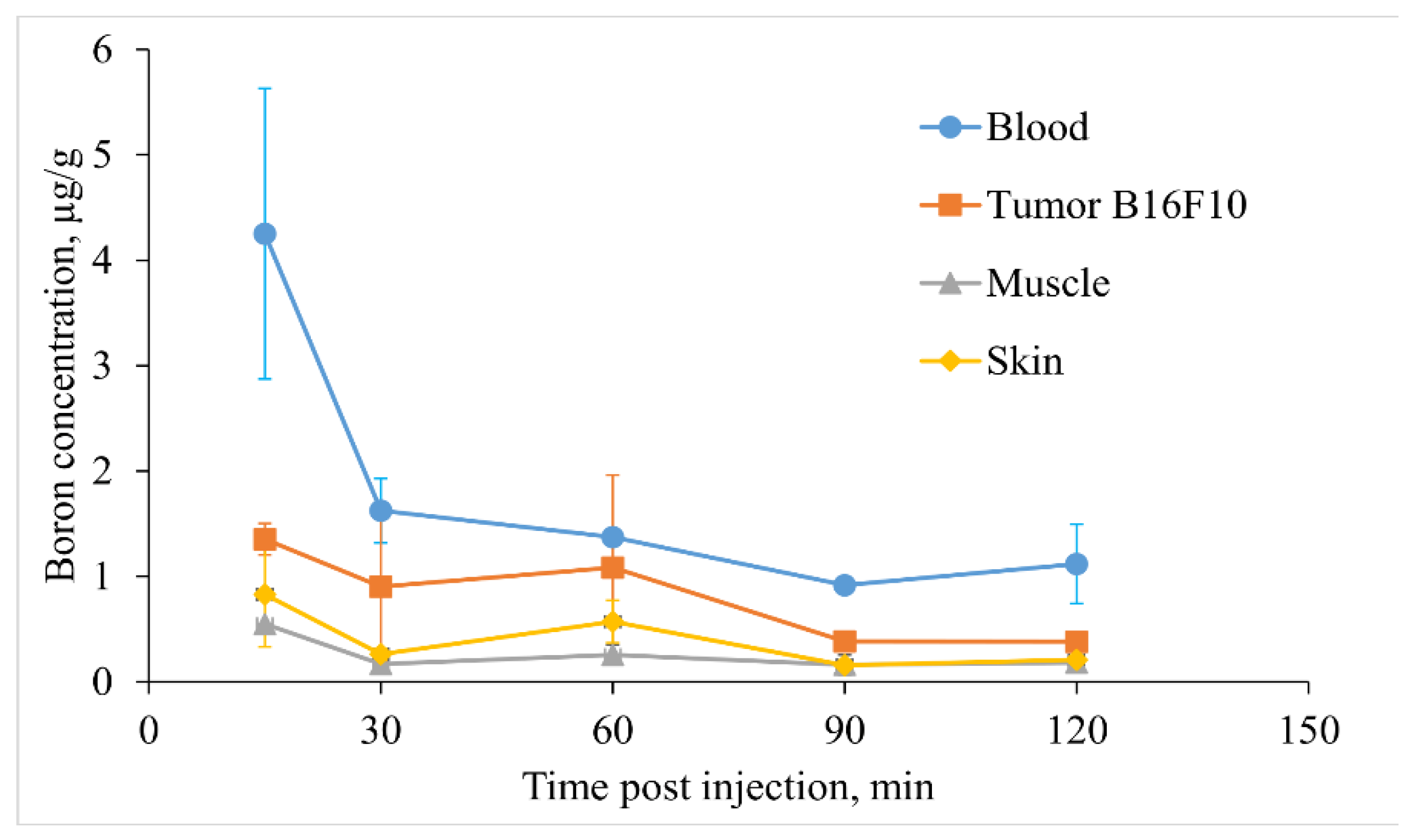
Figure 8.
Time course of tumor-to-normal tissue concentration ratios (tumor/blood, tumor/skin, and tumor/muscle) in B16F10 melanoma-bearing mice after a single intravenous injection of 5-PVP. Data are presented as mean ± SD, n = 5 per time point.
Figure 8.
Time course of tumor-to-normal tissue concentration ratios (tumor/blood, tumor/skin, and tumor/muscle) in B16F10 melanoma-bearing mice after a single intravenous injection of 5-PVP. Data are presented as mean ± SD, n = 5 per time point.
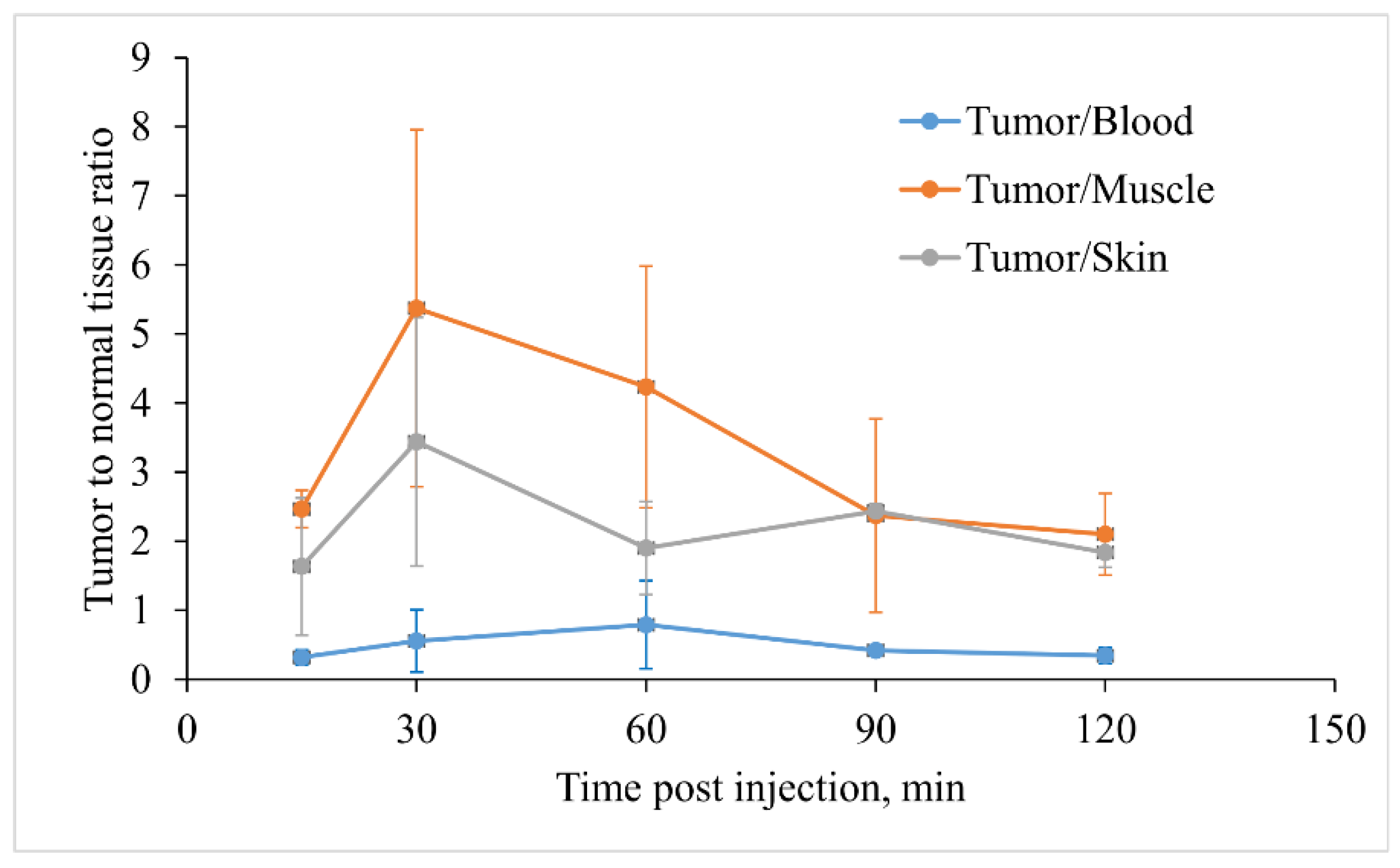

Table 1.
Cytotoxic activity of conjugates of cobalt bis(dicarbollide) with acridine 5-8 against A549, HCT116, MCF-7, and WI-38 cells after a 72 hour cell exposure and cisplatin served as an internal standard. Shown are mean ± SD, n=3.
Table 1.
Cytotoxic activity of conjugates of cobalt bis(dicarbollide) with acridine 5-8 against A549, HCT116, MCF-7, and WI-38 cells after a 72 hour cell exposure and cisplatin served as an internal standard. Shown are mean ± SD, n=3.
| Cell line | Cytotoxisity IC50, µM | ||||
|---|---|---|---|---|---|
| 5 | 6 | 7 | 8 | Cisplatin [46] | |
| WI38 | > 100 | > 100 | > 100 | > 100 | 8 ± 3 |
| A549 | > 100 | > 100 | > 100 | > 100 | 13 ± 3 |
| HCT116 | > 100 | 21 ± 9 | 63 ± 6 | 53 ± 4 | 13 ± 4 |
| MCF7 | > 100 | 44 ± 8 | 45 ± 4 | 22 ± 4 | 30 ± 9 |
Disclaimer/Publisher’s Note: The statements, opinions and data contained in all publications are solely those of the individual author(s) and contributor(s) and not of MDPI and/or the editor(s). MDPI and/or the editor(s) disclaim responsibility for any injury to people or property resulting from any ideas, methods, instructions or products referred to in the content. |
© 2026 by the authors. Licensee MDPI, Basel, Switzerland. This article is an open access article distributed under the terms and conditions of the Creative Commons Attribution (CC BY) license (http://creativecommons.org/licenses/by/4.0/).
Copyright: This open access article is published under a Creative Commons CC BY 4.0 license, which permit the free download, distribution, and reuse, provided that the author and preprint are cited in any reuse.