

Submitted:
02 September 2026
Posted:
03 September 2026
You are already at the latest version
Abstract
Musculoskeletal ultrasonography (MSKUS) is increasingly used to assess masticatory-muscle morphology and mechanical properties, but its clinical interpretability in temporomandibular disorders (TMDs) and bruxism remains uncertain. We systematically reviewed quantitative B-mode, elastographic, functional, diagnostic, and measurement-property evidence. The final pre-submission search combined PubMed/MEDLINE, Scopus, Embase, and Web of Science Core Collection, with supplementary discovery in Europe PMC and Lens, through 20 August 2026 (PROSPERO CRD420261481058). Random-effects meta-analyses used restricted maximum likelihood (REML) with Hartung-Knapp inference. Resting masseter thickness showed no consistent case-control difference (k=14; MD -0.19 mm, 95% CI -0.78 to 0.40; I²=88.9%), nor did contracted masseter thickness (k=11; MD -0.06 mm, 95% CI -1.16 to 1.03; I²=95.9%). Anterior temporalis thickness remained inconclusive at rest (k=5; MD 0.33 mm, 95% CI -0.59 to 1.25; I²=83.4%) and during contraction (k=5; MD -0.05 mm, 95% CI -1.59 to 1.49; I²=94.5%). Resting masseter stiffness measured by shear-wave elastography remained higher in clinical/bruxism groups (k=4; Hedges' g 1.80, 95% CI 0.15 to 3.44; I²=83.5%). Certainty was very low for all pooled outcomes. MSKUS is currently better supported for adjunctive phenotyping and longitudinal monitoring than as a stand-alone diagnostic test.
Keywords:
temporomandibular disorders
; bruxism
; ultrasonography
; masseter
; temporalis
; shear-wave elastography
; muscle thickness
; stiffness
; meta-analysis
1. Introduction
Temporomandibular disorders (TMDs) comprise a heterogeneous group of musculoskeletal conditions affecting the temporomandibular joints, masticatory muscles, and associated structures. Pain-related TMDs, particularly myalgia and arthralgia, can impair mandibular function and contribute to persistent orofacial pain, headache, altered muscle recruitment, and reduced quality of life. Standardized clinical frameworks such as the Diagnostic Criteria for Temporomandibular Disorders (DC/TMD) have improved diagnostic consistency; however, objective characterization of masticatory-muscle morphology and mechanical behavior remains challenging.
Imaging of the temporomandibular region has traditionally emphasized articular morphology and internal derangement. Magnetic resonance imaging and computed or cone-beam computed tomography remain important when soft-tissue or osseous joint pathology is suspected, but they are less suited to repeated, dynamic, point-of-care assessment of superficial masticatory muscles. Musculoskeletal ultrasonography (MSKUS) is non-ionizing, repeatable, relatively accessible, and capable of real-time examination during rest and contraction, making it attractive for both clinical assessment and longitudinal monitoring.
B-mode ultrasonography has been widely used to quantify masseter and temporalis thickness and, less frequently, cross-sectional area, volume, architecture, and internal echotexture. Several studies have explored whether TMD, myofascial pain, or bruxism is associated with muscle hypertrophy, atrophy, asymmetry, or altered contraction-related thickening. Yet the literature has produced inconsistent findings, raising the question of whether absolute muscle thickness is sufficiently stable to function as a clinically useful biomarker.
Elastographic techniques extend ultrasonographic assessment beyond morphology. Shear-wave elastography (SWE) estimates tissue stiffness using shear-wave speed or derived elastic modulus, whereas strain elastography estimates relative tissue deformation. These techniques may capture mechanical alterations not visible on conventional B-mode imaging. Reports of increased masseter stiffness in myofascial pain and bruxism have therefore generated interest in a mechanical phenotype of masticatory dysfunction. However, device/vendor effects, jaw state, scan plane, tissue preload, region-of-interest definition, and reported units complicate direct comparison across studies.
Ultrasound-derived echogenicity, grayscale intensity, fractal dimension, radiomic features, trigger-point characteristics, diagnostic thresholds, and treatment-related changes have also been investigated. Before these measures can be translated into routine practice, their reliability, measurement error, diagnostic performance, responsiveness, and external validity must be considered together. High within-study reliability alone does not establish interchangeability across devices, operators, or acquisition protocols.
This distinction is especially important because ultrasonography is operator- and protocol-dependent. Patient position, head and neck support, mandibular state, anatomical landmark, transducer orientation and pressure, acquisition frequency, number of repetitions, bilateral handling, ROI placement, and examiner blinding may all influence results. For SWE, additional sources of variation include scan orientation relative to muscle fibers, ROI depth and size, machine-specific processing, and conversion between m/s and kPa. Consequently, methodological heterogeneity may contribute substantially to the statistical heterogeneity reported across the literature.
Accordingly, this systematic review and meta-analysis aimed to critically synthesize quantitative MSKUS evidence in the masticatory system across morphometric, mechanical, functional, diagnostic, and measurement-property domains. Where clinical and methodological comparability permitted, random-effects meta-analyses were performed for masseter and anterior temporalis thickness and resting masseter stiffness. Secondary objectives were to synthesize dynamic functional behavior, architecture and echotexture, diagnostic performance, reliability and reproducibility, treatment responsiveness, and prognostic applications, while explicitly evaluating protocol variability as a source of between-study heterogeneity.
2. Materials and Methods
2.1. Protocol and Reporting Standard
This systematic review and meta-analysis was conducted in accordance with a prespecified protocol and is reported in line with the PRISMA 2020 statement. The protocol was prospectively registered in the International Prospective Register of Systematic Reviews (PROSPERO; CRD420261481058).
2.2. Eligibility Criteria
Eligible studies investigated quantitative ultrasonography of the masticatory system in human participants, including B-mode morphometry, cross-sectional area or volume, architecture or echotexture, SWE or strain elastography, dynamic rest-to-contraction behavior, reliability or measurement properties, diagnostic performance, treatment responsiveness, or prognostic/predictive applications. Clinical populations included TMD, myofascial pain, bruxism, combined TMD-bruxism phenotypes, and related masticatory-muscle conditions. Healthy cohorts were eligible when they provided normative, comparator, reliability, or methodological data relevant to the review. Studies using ultrasound only as a therapeutic intervention or procedural-guidance tool, without a quantitative masticatory-muscle imaging outcome, were excluded from the primary imaging synthesis. Reviews, editorials, non-human studies, and reports without extractable primary quantitative data were excluded.
2.3. Information Sources and Search Strategy
The review used a combined historical and pre-submission search strategy. The original PubMed/MEDLINE screening universe contained 769 records. Before submission, an updated electronic search was conducted across PubMed/MEDLINE (n=680; 18 August 2026), Scopus (n=970; 18 August 2026), Embase (n=1,223; 19 August 2026), and Web of Science Core Collection (n=673; 20 August 2026). Supplementary discovery and recovery searches were performed in Europe PMC (n=4,022) and Lens (n=6,101) on 18 August 2026, together with backward and forward citation checking of eligible studies and relevant reviews. Across the updated sources, 13,669 raw records were retrieved. The historical and updated searches were reconciled by identifier and normalized title to a combined universe of 7,672 unique records. Search concepts covered the masseter, temporalis and other masticatory muscles; ultrasonography and musculoskeletal ultrasound; B-mode imaging; shear-wave and strain elastography; muscle thickness, architecture, echogenicity, stiffness, reliability, diagnostic performance, and treatment responsiveness. Exact PubMed and Embase strategies, source counts, deduplication rules, the Web of Science RIS reconciliation, and the final search audit are provided in the Supplementary Materials. The Web of Science RIS export retained title, abstract, keywords, DOI, and accession-number fields; the exact platform query-history string was not embedded in the RIS file and is therefore reported transparently as unavailable from the export.
2.4. Study Selection
Records were consolidated and screened sequentially by title, abstract, and full text by two reviewers (A.M. and E.A.P.) working independently, with disagreements resolved by discussion and consensus. During the pre-submission update, AI-assisted and rule-based prioritization was used only to organize and flag records for adjudication; it was not used as a substitute for final reviewer eligibility decisions. Screening quality control included targeted false-negative review of records containing ultrasonography, sonography, elastography, masseter, temporalis, or masticatory terminology. Publication families and repeated reports from the same cohort were linked before quantitative synthesis to reduce analytic double counting. Reasons for full-text exclusion and update-stage adjudication were retained in the Supplementary Materials. Both reviewers approved the final reconciled eligibility freeze, cohort-overlap decisions, and updated quantitative pools before submission.
2.5. Data Extraction and Unit-of-Analysis Rules
Data were independently extracted and/or cross-checked by two reviewers (A.M. and E.A.P.), with discrepancies resolved by consensus. Extraction was performed at participant or cohort level whenever possible. Extracted fields included study design, population, diagnostic criteria, sample size, muscle, device and transducer characteristics, body and jaw position, anatomical landmark, probe orientation and pressure, measurement side, number of repeated acquisitions, outcome units, group means and variances, diagnostic metrics, reliability statistics, and longitudinal time points. For studies reporting bilateral measurements separately without a participant-level bilateral mean and variance or an estimable within-person correlation, the right side was prespecified for the primary analysis and the left side was used in sensitivity analysis. Shared controls were counted once; compatible clinical subgroups were combined before pooling when necessary. Standard errors were converted to standard deviations using SD=SE×√n, and reconstructed variances were retained only when derivation was transparent and sensitivity-tested.
2.6. Protocol Standardization Assessment
Because ultrasonographic measurements are sensitive to acquisition technique, protocol characteristics were systematically mapped across studies. Domains included transducer type and frequency, patient and head position, anatomical landmark, probe orientation, probe pressure, jaw state, side handling, number of repetitions, SWE metric and acquisition settings, ROI geometry and depth, echo-intensity settings, and examiner blinding. These factors were used to determine poolability and to interpret residual heterogeneity.
2.7. Risk of Bias
Risk of bias was assessed independently by two reviewers (A.M. and E.A.P.), with disagreements resolved by consensus, using design-specific frameworks. Observational analytical cross-sectional/case-control studies contributing to the main quantitative syntheses were appraised using the revised JBI analytical cross-sectional framework, with judgments focused on selection and representativeness, phenotype ascertainment, ultrasonographic outcome measurement, confounding, and statistical analysis. Diagnostic studies were routed to QUADAS-3, randomized trials to RoB 2, non-randomized comparative interventions to ROBINS-I, and reliability/measurement-property studies to COSMIN-informed appraisal. Risk-of-bias judgments were not used as automatic exclusion criteria; methodological sensitivities were used where a specific issue could materially affect an effect estimate.
2.8. Quantitative Synthesis
For continuous morphometric outcomes reported in the same unit, pooled mean differences (MDs) were calculated. Because SWE studies reported stiffness in different units and device-dependent scales, standardized mean differences using Hedges’ g were used. Random-effects models were fitted using restricted maximum likelihood (REML), with Hartung-Knapp inference for pooled confidence intervals. Statistical heterogeneity was summarized using I2 and τ2. Outcomes were not pooled when constructs, activation states, measurement units, reporting formats, or study designs were insufficiently comparable; these were synthesized narratively.
2.9. Sensitivity Analyses
Sensitivity analyses addressed material analytic uncertainties, including participant-level reweighting of Aksu et al., exclusion of Kılınç et al. where reconstructed variance estimates were used, and left-side substitution for Palinkas et al. under the prespecified bilateral-handling rule. The purpose was to determine whether these decisions materially changed pooled interpretation.
2.10. Small-Study Effects and Publication Bias
Small-study effects were explored only when the number of contributing studies permitted meaningful interpretation. Funnel-plot asymmetry testing was therefore limited to resting masseter thickness (k=11). Formal asymmetry tests were not interpreted for outcomes with fewer than 10 studies.
2.11. Certainty of Evidence
Certainty of evidence for the five principal quantitative outcomes was assessed using GRADE, considering risk of bias, inconsistency, indirectness, imprecision, and publication bias. Because the main quantitative evidence was observational, certainty started at low and was downgraded where serious concerns were present. No upgrading was applied to the large apparent SWE effect because heterogeneity, small-study effects, and residual confounding remained plausible.
The complete reproducible PubMed/MEDLINE and Embase search strategies, final search dates, source-specific record counts, deduplication rules, and supplementary discovery procedures are provided in the Supplementary Materials. Result-level exports were retained for Scopus, Europe PMC, and Lens; where platform query-history strings were not preserved verbatim, the platform-adapted search concepts and audit trail are reported transparently rather than retrospectively reconstructed.
3. Results
3.1. Study Selection and Characteristics
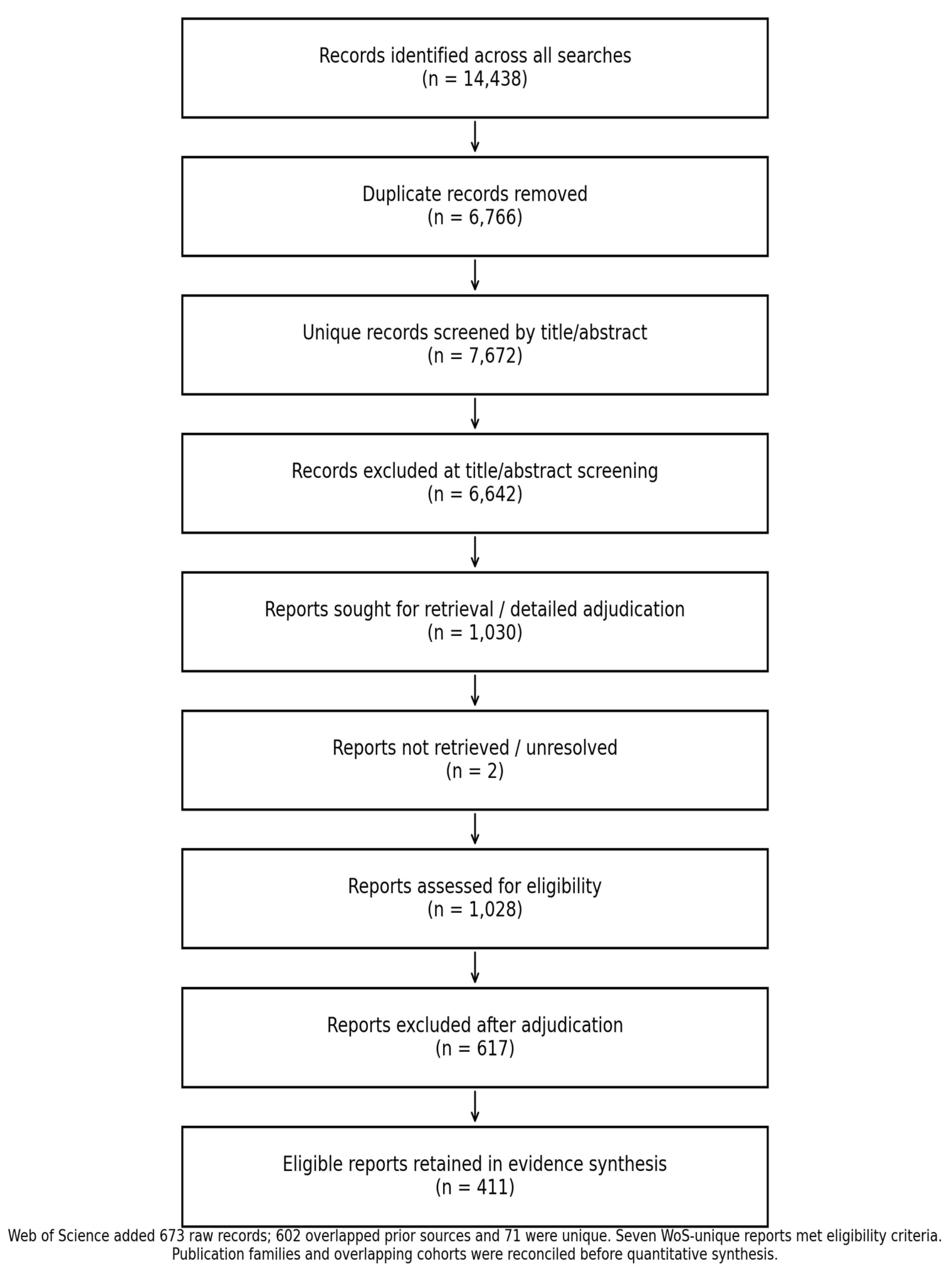
Across the original and updated searches, 14,438 raw records were identified. After removal of 6,766 duplicate records, 7,672 unique records entered title/abstract screening. Of these, 6,642 were excluded at title/abstract level and 1,030 reports entered detailed retrieval or eligibility adjudication. Two historical reports remained unretrieved or unresolved, leaving 1,028 reports for completed assessment; 617 were excluded after detailed adjudication and 411 eligible reports were retained in the evidence synthesis (Figure 1). The final Web of Science export contributed 673 raw records: 602 overlapped records already captured by the other sources and 71 were unique after identifier/title reconciliation. Of these 71 WoS-unique records, seven met the review eligibility criteria and 64 were excluded at title/abstract adjudication. Publication families and potentially overlapping cohorts were reconciled before quantitative pooling so that duplicate reports from the same cohort were not treated as independent datasets.
The included literature encompassed B-mode morphometry, SWE and strain elastography, architecture and quantitative texture analysis, dynamic rest-to-contraction assessment, reliability and measurement-property studies, diagnostic-accuracy investigations, and longitudinal treatment-response cohorts. The masseter was the most frequently examined muscle, followed by the anterior temporalis. Considerable methodological diversity was present in diagnostic criteria, age and sex distribution, jaw state, body position, transducer location, ultrasonographic device, elastography output, and handling of bilateral measurements.

Table 1.
Principal quantitative meta-analytic findings.
| Outcome | k | Effect | 95% CI | I2 | GRADE |
| Masseter thickness - rest | 14 | MD -0.19 mm | -0.78 to 0.40 | 88.9% | Very low |
| Masseter thickness - contraction | 11 | MD -0.06 mm | -1.16 to 1.03 | 95.9% | Very low |
| Anterior temporalis - rest | 5 | MD 0.33 mm | -0.59 to 1.25 | 83.4% | Very low |
| Anterior temporalis - contraction | 5 | MD -0.05 mm | -1.59 to 1.49 | 94.5% | Very low |
| Resting masseter stiffness (SWE) | 4 | Hedges’ g 1.80 | 0.15 to 3.44 | 83.5% | Very low |
3.2. Masseter Thickness
Fourteen adult datasets contributed to the updated meta-analysis of resting masseter thickness. Three newly recovered datasets provided compatible case-control data: Ünal et al. (2026), Jafari et al. (2017), and Lecaros et al. (2017). For Lecaros et al., published standard errors were converted to standard deviations using SD=SE×√n and the right side was used under the prespecified bilateral-handling rule. The random-effects pooled MD was -0.19 mm (95% CI -0.78 to 0.40), with substantial heterogeneity (I2=88.9%). Eleven adult datasets contributed to contraction-state masseter thickness after addition of Ünal et al. and Lecaros et al., yielding MD -0.06 mm (95% CI -1.16 to 1.03; I2=95.9%). These updates did not materially alter the near-null interpretation of masseter morphometry.
3.3. Anterior Temporalis Thickness
Five adult datasets contributed to resting anterior temporalis thickness, and the pooled estimate remained MD 0.33 mm (95% CI -0.59 to 1.25; I2=83.4%). Lecaros et al. reported a markedly lower resting temporalis thickness in TMD, but the clinical-group standard error was rounded to 0.00 and a defensible variance could not be reconstructed; this measurement was therefore retained narratively rather than pooled. Five datasets contributed to anterior temporalis thickness during contraction after inclusion of the right-side Lecaros dataset, producing MD -0.05 mm (95% CI -1.59 to 1.49; I2=94.5%). The updated analysis therefore remained highly heterogeneous and inconclusive.
3.4. Sensitivity Analyses
Sensitivity analyses did not materially alter the morphometric conclusions. Participant-level reweighting of Aydin Aksu et al. [6], exclusion of Kılınç et al. [24] when reconstructed variance estimates were used, and substitution of the left rather than right side from Palinkas et al. [9] produced materially similar interpretations. The near-null morphometric findings were therefore not attributable to a single prespecified analytic decision.
3.5. Resting Masseter Stiffness
Four adult case-control datasets with primary-source verified mean and variance data contributed to the quantitative synthesis of resting masseter stiffness [4,5,6,7]. All four showed higher stiffness in the clinical or bruxism group. Random-effects REML meta-analysis with Hartung-Knapp inference yielded Hedges’ g 1.80 (95% CI 0.15 to 3.44), with substantial heterogeneity (I2=83.5%; τ2=0.887). Reweighting Aydin Aksu et al. [6] at participant level produced a similar estimate (g 1.72, 95% CI 0.18 to 3.26; I2=73.3%).
3.6. Contraction-State SWE and Strain Elastography
Contraction-state stiffness was not meta-analyzed because of the small number of compatible studies, different activation protocols, and non-parametric reporting in some cohorts. Across the structured evidence, clinical groups generally showed higher masseter stiffness during contraction, although effect magnitude varied substantially. Strain-elastography evidence was likewise synthesized without meta-analysis because strain metrics, reference materials, compression approaches, and study designs were not harmonized. Symptomatic-side and treatment-responsive changes were repeatedly observed, supporting biological sensitivity but not metric interchangeability.
3.7. Architecture, Echo Intensity, and Texture
Architecture and echotexture outcomes were heterogeneous and were not meta-analyzed. Earlier studies used visual or ordinal internal-pattern classifications of the masseter, including the three-type echogenic-band framework described in women with TMD-related myofascial pain [27] and later phenotype studies [8,10]. More recent work introduced quantitative grayscale echo intensity, fractal dimension, and radiomics. These constructs were kept separate because they represent different image properties and rely on different acquisition and preprocessing pipelines. Quantitative grayscale values were considered particularly sensitive to gain, depth, dynamic range, transducer characteristics, ROI definition, and post-processing. The evidence suggests phenotype-sensitive information beyond thickness, but no common architecture or texture metric is currently established.
3.8. Dynamic Functional Ultrasonography
Dynamic B-mode ultrasonography consistently demonstrated muscle thickening during contraction, but between-group differences in the magnitude of this response were not uniform. Some cohorts reported reduced relative masseter thickening in myofascial TMD, whereas others found nearly identical contraction/rest ratios between clinical and control groups. A pooled paired-change analysis was not performed because most studies did not report the SD of change or the rest-contraction correlation required for valid reconstruction.
3.9. Reliability and Measurement Properties
Reliability was generally moderate to excellent but varied by muscle, anatomical site, imaging construct, and acquisition protocol [13,14,15,16,17]. Contemporary multi-muscle studies reported intra-rater ICCs of approximately 0.897-0.976 for thickness and 0.862-0.893 for stiffness, while inter-rater estimates were lower and more variable (approximately 0.724-0.957 for thickness and 0.745-0.805 for stiffness) [13]. A standardized masseter protocol reported intra-session ICCs of 0.85-0.98, inter-rater ICCs of 0.87-0.92, coefficients of variation below 3%, and mean ultrasound-CT error below 6%, with correlations r≥0.93 [15]. Reliability findings were not pooled because ICC model, observer structure, muscle, state, device, and repetition scheme differed materially across studies.
3.10. Diagnostic Performance
Diagnostic-performance evidence remained limited and heterogeneous. Study-specific masseter SWE thresholds showed promising discrimination for bruxism-related phenotypes, including an AUC of 0.894 in the adult study by Güven et al. [7], but thresholds differed materially across age groups, jaw states, units, and devices. Separate work evaluating the temporomandibular disc and masseter with SWE also illustrates that anatomically distinct targets should not be combined into a single diagnostic threshold [12]. A 2024 masseter myofascial-trigger-point study in the audit dataset reported sensitivity of 94.6% and specificity of 90.0% at the site level (116 sites nested within 29 patients); because the unit was the examined site rather than the patient, these estimates were not pooled with participant-level diagnostic studies. No formal diagnostic-accuracy meta-analysis was therefore performed and no universal cutoff was supported.
3.11. Treatment Responsiveness
Longitudinal studies demonstrated that ultrasonographic measures can respond to treatment-related change. Exercise-based management in bruxism altered masseter thickness and elasticity [11], while stabilization-splint treatment in painful myofascial TMD produced significant reductions in masseter dimensions after three months [28]. Botulinum toxin A studies also demonstrated time-dependent reductions in masseter thickness on ultrasonography [29,30]. Across conservative, splint, manual/massage, and botulinum-toxin interventions, however, protocols and outcome metrics were too heterogeneous for a defensible pooled treatment effect. Responsiveness should not be equated with surrogate validity; imaging change did not consistently correlate with the magnitude of symptom change.
3.12. Prognostic and Predictive Evidence
Evidence for prognostic or predictive use of masticatory-muscle ultrasonography was preliminary. The audit dataset identified small responder/predictor cohorts in which early imaging changes during massage-based interventions differed between subsequent responders and non-responders, together with emerging radiomic or multiparametric classification models. These studies were retained as structured narrative evidence because they lacked external validation, stable calibration, and demonstrated incremental value over established clinical variables. Current evidence therefore supports hypothesis generation rather than validated prognostic use.
3.13. Risk of Bias
Among the 16 unique studies contributing to the five principal quantitative pools after the search update, two were judged at high risk of bias in the conservative freeze-candidate assessment and 14 had some concerns; none was classified as uniformly low risk. Recurrent concerns involved selection or representativeness, phenotype ascertainment, residual confounding, small sample sizes, heterogeneous diagnostic definitions, and unit-of-analysis or reporting limitations. Ultrasonographic outcome measurement was generally the strongest domain because outcomes were directly acquired, although protocol-specific variability remained.
3.14. Certainty of Evidence
Certainty was rated very low for all five principal quantitative outcomes. For morphometric outcomes, downgrading was driven mainly by risk of bias and substantial-to-extreme inconsistency, with additional imprecision for smaller evidence bases. Resting masseter stiffness showed a large pooled standardized effect, but certainty remained very low because only four studies contributed, heterogeneity was substantial, the Hartung-Knapp interval was wide, and small-study or publication-bias concerns could not be excluded.
3.15. Small-Study Effects
For resting masseter thickness (k=14), exploratory Egger regression indicated funnel-plot asymmetry (intercept 2.75, p=0.025). This finding should be interpreted cautiously because between-study heterogeneity remained substantial and the evidence base combined clinically heterogeneous TMD and bruxism phenotypes. Formal asymmetry testing was not emphasized for the other outcomes; anterior temporalis and resting SWE syntheses remained below the conventional 10-study threshold, and extreme heterogeneity limited interpretation of contraction-state masseter asymmetry.
Figure 2.
Random-effects meta-analysis of resting masseter thickness.
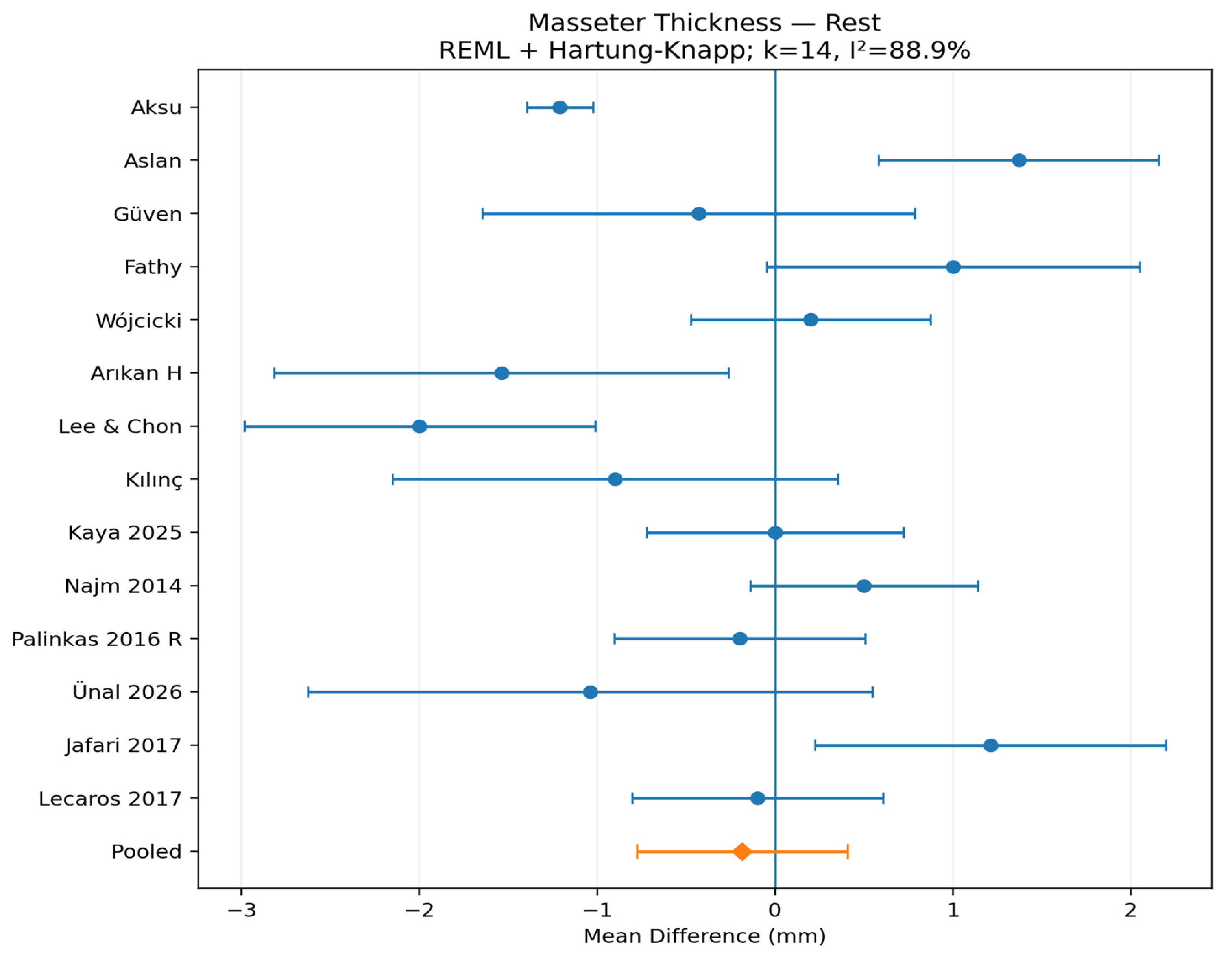
Figure 3.
Random-effects meta-analysis of masseter thickness during contraction.
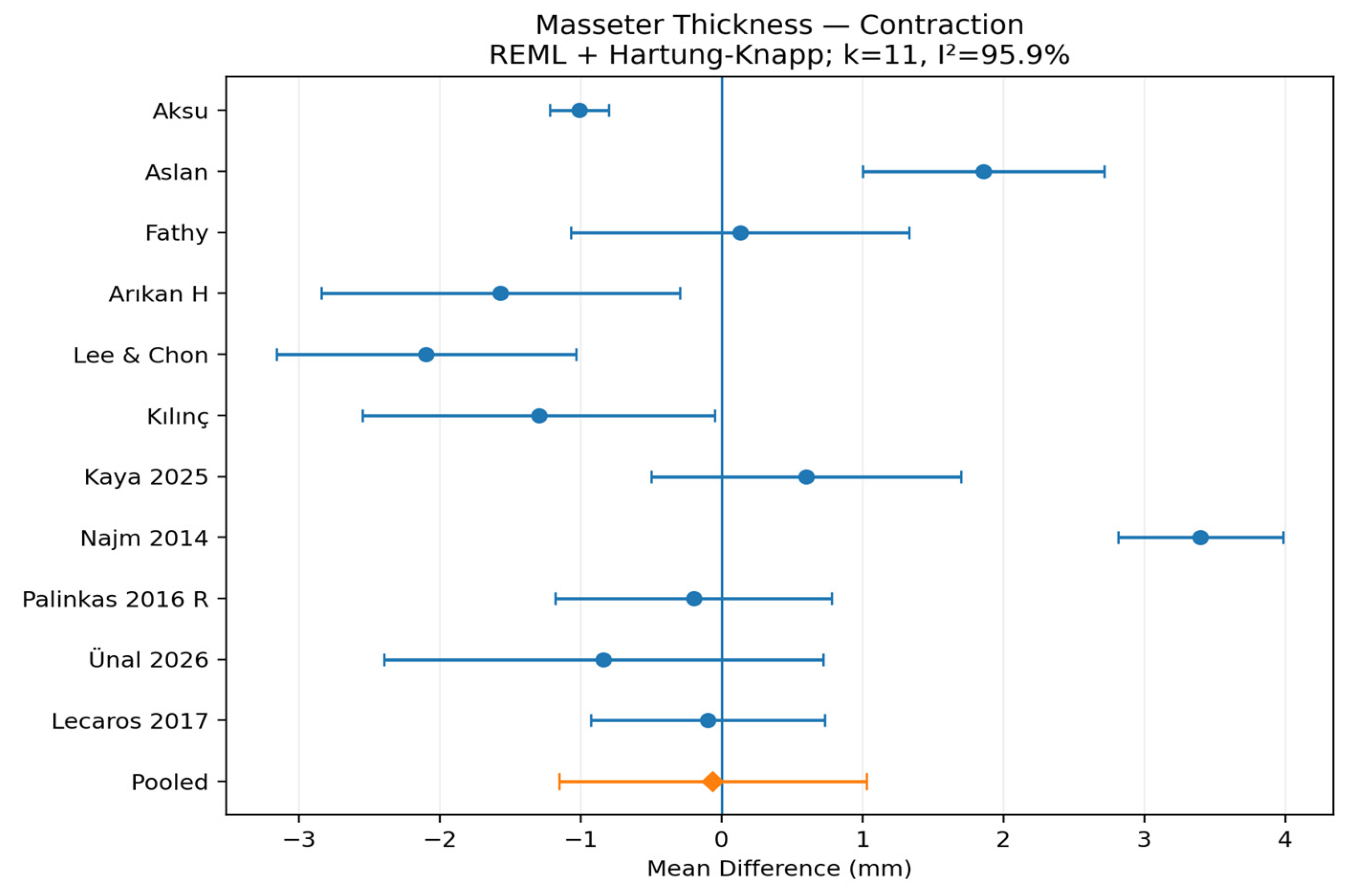
Figure 4.
Random-effects meta-analysis of resting anterior temporalis thickness.
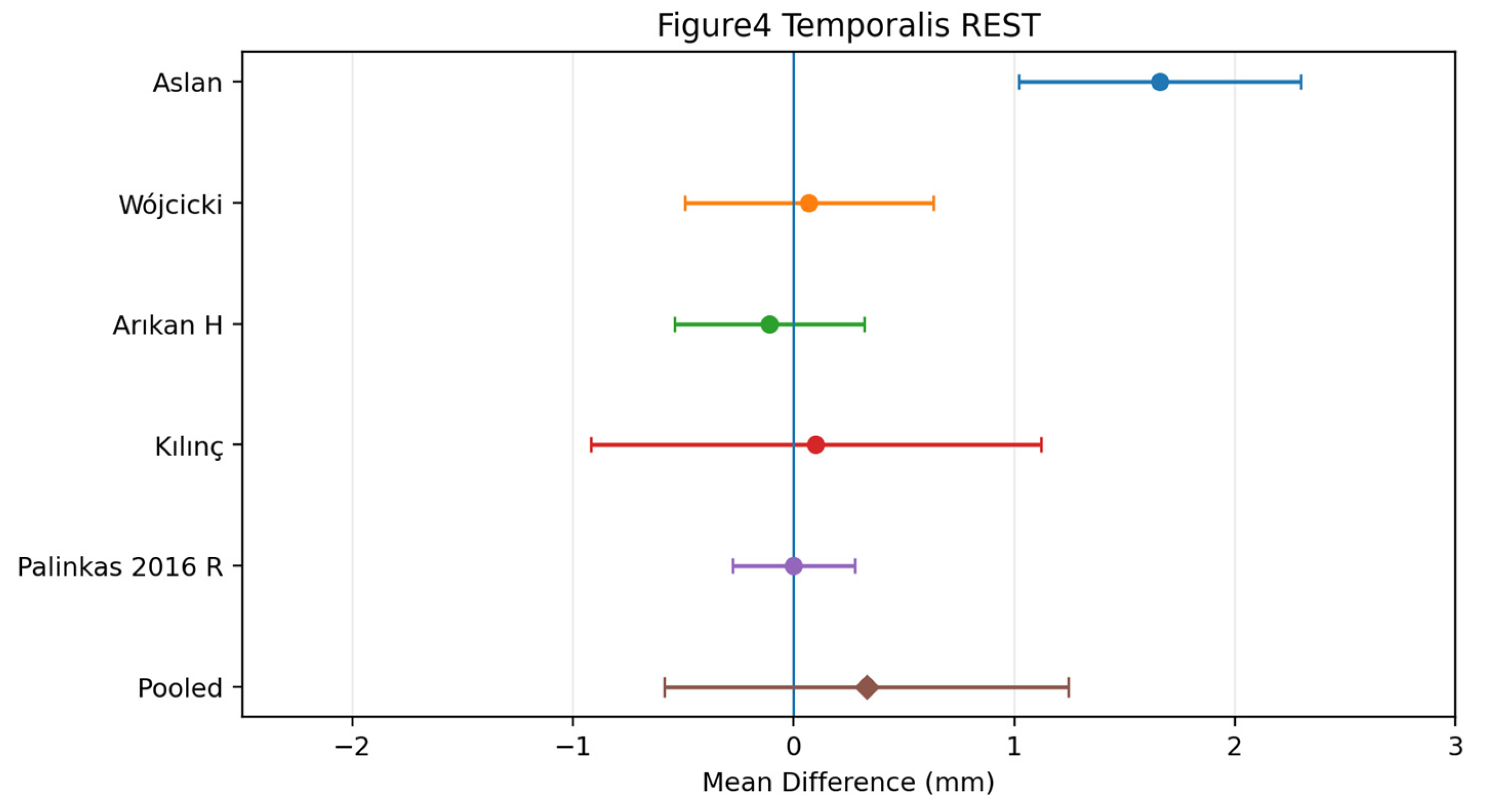
Figure 5.
Random-effects meta-analysis of anterior temporalis thickness during contraction.
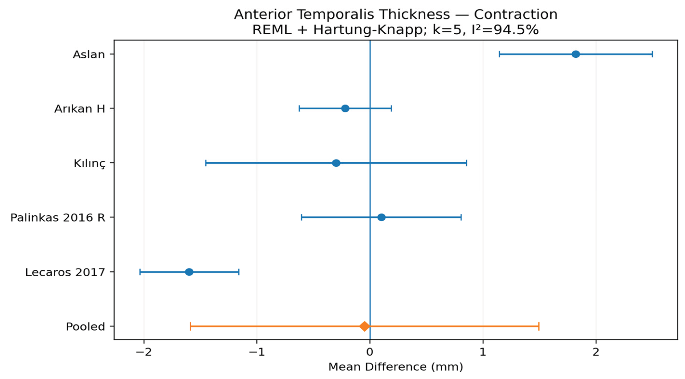
Figure 6.
Random-effects meta-analysis of resting masseter stiffness assessed with shear-wave elastography.
Figure 6.
Random-effects meta-analysis of resting masseter stiffness assessed with shear-wave elastography.
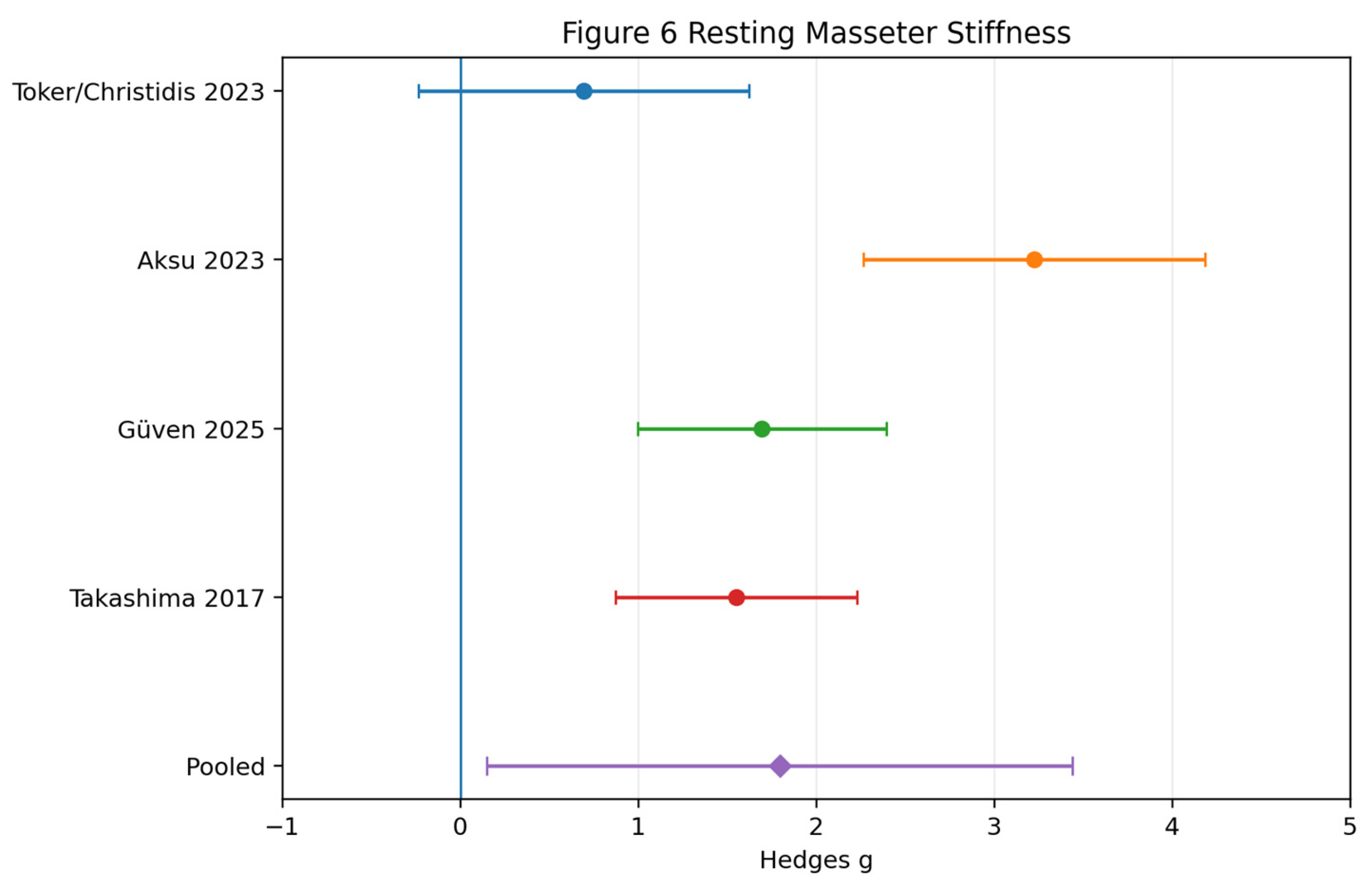
Table 2.
Key protocol-standardization domains affecting cross-study comparability.
| Domain | Observed variation | Potential impact | Recommended harmonization |
| Transducer/frequency | Linear probes approximately 2-15 MHz; device/vendor variation | Resolution and apparent fascial borders; SWE device dependence | Report manufacturer/model, probe and active frequency; avoid cross-device equivalence assumptions |
| Patient position | Supine, seated/upright, variable head support | Changes geometry and resting tone | Pool only comparable postures; report head/neck support |
| Masseter landmark | Thickest belly, occlusal plane, ramus midpoint, fixed coordinates | Thickness varies spatially | Use reproducible anatomical coordinates |
| Probe pressure | Minimal/no pressure often stated but rarely quantified | Compression biases resting thickness and ratios | Standardize pressure and flag unreported compression control |
| Jaw state | Variable rest definitions, clench/MVC instructions and duration | Directly alters thickness and stiffness | Separate rest and contraction; report activation protocol |
| Side handling | Bilateral, symptomatic, dominant, more painful, or averaged sides | Can introduce asymmetry/phenotype bias | Do not treat bilateral sides as independent observations |
| SWE metric/ROI | m/s vs kPa; ROI depth and geometry vary | Vendor algorithms and ROI placement alter stiffness | Report unit, ROI size/count/depth and quality criteria |
| Echo intensity | Ordinal patterns vs 0-255 grayscale/ImageJ | Different constructs; grayscale depends on settings | Do not pool categorical and continuous constructs; standardize acquisition settings |
| Blinding | Operator/reader blinding inconsistently reported | Potential observer bias | Report blinding and calibration explicitly |
4. Discussion
4.1. Principal Findings
This updated review indicates that quantitative ultrasonography of the masticatory system yields a multidimensional rather than a single structural signal. Incorporating newly recovered studies increased the masseter morphometry evidence base but did not materially change the pooled conclusion: case-control thickness effects remained close to zero at rest and during contraction. Anterior-temporalis estimates also remained inconclusive, whereas resting masseter stiffness continued to show a directionally consistent positive effect across eligible adult case-control datasets. The most clinically useful interpretation is therefore not that ultrasonography is negative in TMD or bruxism, but that gross morphometry and mechanical phenotype appear to convey different information.
4.2. Why Morphometry Is Inconsistent
The marked heterogeneity in thickness estimates is biologically and technically plausible. TMD and bruxism are heterogeneous phenotypes, and absolute muscle thickness is influenced by sex, body size, craniofacial morphology, habitual loading, dentition, age, pain duration, and recruitment strategy. The protocol audit additionally showed substantial variation in body position, jaw state, anatomical landmark, transducer orientation, pressure, and bilateral handling. Because masseter thickness varies spatially and is compression-sensitive, even technically reliable measurements may not be directly exchangeable across laboratories. This helps explain why individual studies may report meaningful local differences while the pooled case-control effect remains near null.
4.3. Mechanical Phenotype Versus Structural Phenotype
The large positive pooled resting-stiffness effect contrasts with the near-null morphometric findings and supports a distinction between muscle size and mechanical behavior. A muscle can retain normal gross thickness while exhibiting altered resting tone, stiffness, pain-related guarding, microstructural organization, or motor control. SWE may therefore be more sensitive to some pain- or bruxism-related states than simple thickness. However, this result should not be overinterpreted: only four studies contributed, heterogeneity was high, the Hartung-Knapp interval was wide, and certainty was very low. The appropriate conclusion is comparative rather than diagnostic: mechanical stiffness is currently more directionally consistent than gross thickness, but it is not yet a validated universal biomarker.
4.4. Dynamic Assessment
Dynamic ultrasonography may bridge static morphology and function, but the current evidence remains difficult to synthesize. Some cohorts reported reduced relative thickening during contraction, whereas others observed similar contraction/rest ratios between clinical and control groups. Reporting limitations are central. Without the variance of paired change or rest-contraction correlations, change-score meta-analysis cannot be reconstructed reliably. Future work should report participant-level change scores and their dispersion directly and should standardize contraction intensity and duration.
4.5. Architecture and Texture
B-mode images contain information beyond muscle thickness. Ordinal echogenic patterns, grayscale intensity, fractal dimension, and radiomics may reflect muscle composition, fibrosis, edema, intramuscular organization, or other tissue properties. These constructs, however, are not interchangeable. Quantitative echo intensity is strongly affected by gain, depth, dynamic range, transducer characteristics, and ROI selection, while radiomic features are additionally sensitive to preprocessing and segmentation. Clinical translation will therefore depend more on harmonized acquisition and external validation than on accumulation of isolated feature sets.
4.6. Reliability and Standardization
Reliability evidence is encouraging but protocol-dependent. Standardized acquisition can yield high intra- and inter-rater reliability for B-mode thickness and good repeatability for SWE and texture metrics, particularly in the masseter. Reliability should therefore be understood as a property of a specific protocol applied to a specific muscle and state rather than of ultrasonography in the abstract. Studies should report the ICC model and confidence interval together with absolute error metrics such as SEM, MDC, CV, or limits of agreement.
4.7. Diagnostic Implications
Current evidence does not support a universal ultrasonographic diagnostic threshold. SWE cutoffs differed across age groups, jaw states, devices, and study populations, while trigger-point accuracy studies were limited by reference standards and clustering of measurement sites within participants. High within-study AUCs should not be interpreted as transportable clinical thresholds. Ultrasonography is better supported as an adjunctive quantitative marker integrated with validated clinical diagnosis than as a stand-alone test.
4.8. Treatment Responsiveness and Monitoring
The longitudinal literature suggests a potentially stronger role for ultrasonography in monitoring than in diagnosis. Thickness, stiffness, and strain-derived measures changed after conservative care, splint therapy, manual/massage interventions, and particularly botulinum toxin A. Repeated MSKUS may therefore be useful for documenting tissue-level change over time. Nevertheless, responsiveness is not synonymous with clinical importance or surrogate validity; an imaging measure can change without closely tracking pain, disability, or patient-reported recovery.
4.9. Protocol Heterogeneity as a Source of Statistical Heterogeneity
The protocol-standardization matrix provides a coherent explanation for the high I2 values observed in the principal meta-analyses. Variation in anatomical landmark, posture, probe pressure and orientation, jaw activation, repeated-measure averaging, side handling, and SWE ROI/device conventions can shift absolute estimates even when within-study reliability is good. SMD can harmonize scale differences but cannot remove differences in biological construct or measurement protocol. Future quantitative syntheses may become more informative if sufficiently large evidence bases permit subgroup or meta-regression analyses by these technical factors.
4.10. Risk of Bias and Certainty
Risk-of-bias findings reinforce cautious interpretation of both the near-null morphometric effects and the positive stiffness signal. The enlarged quantitative evidence base still contained no study judged uniformly low risk under the conservative framework, and all five principal outcomes remained very low certainty. Newly recovered case-control datasets increased precision only modestly because between-study heterogeneity remained substantial. The near-null thickness estimates should therefore not be interpreted as definitive evidence that structural differences never occur in specific phenotypes. Conversely, the large stiffness effect should not be interpreted as a validated diagnostic biomarker.
4.11. Small-Study Effects
Reporting bias remains a concern. After addition of newly recovered datasets, exploratory Egger regression for resting masseter thickness showed statistically detectable funnel-plot asymmetry (intercept 2.75, p=0.025). This signal is not proof of publication bias because clinical and technical heterogeneity was substantial, but it strengthens the rationale for cautious interpretation and very-low certainty. For SWE and temporalis outcomes, the evidence bases remained too small for reliable formal asymmetry testing.
4.12. Clinical Implications
For clinical practice, the present evidence supports masticatory-muscle ultrasonography as an adjunctive phenotyping and monitoring tool. Thickness alone should not be used as a universal case-control discriminator. Mechanical stiffness may provide complementary information in selected pain or bruxism phenotypes, while dynamic and texture measures may add functional or compositional context. Interpretation should remain muscle-specific, state-specific, device-aware, and protocol-specific and should be integrated with validated clinical examination rather than replacing it.
4.13. Strengths and Limitations
This review integrates morphometric, mechanical, architectural, dynamic, diagnostic, reliability, treatment, and predictive ultrasonographic constructs while preserving their methodological distinctions. It explicitly audited cohort overlap, bilateral measurements, shared controls, reconstructed variance, participant-versus-muscle denominators, risk of bias, GRADE certainty, and protocol standardization. A major strength of the final update is the broadened and auditable search across PubMed/MEDLINE, Scopus, Embase, and Web of Science Core Collection, supplemented by Europe PMC and Lens. The final Web of Science reconciliation identified seven additional eligible reports, including reference morphometry, JIA/TMJ phenotype, longitudinal orthodontic, prognostic, and treatment-responsive SWE evidence, but none supplied a compatible additional clinical-versus-control effect for the five prespecified quantitative pools; pooled estimates therefore remained unchanged after the WoS update. Important limitations remain. The exact Web of Science platform query-history string was not embedded in the supplied RIS export, although the complete 673-record RIS file and record-level identifiers were retained and audited. Search-platform syntax was likewise not recoverable verbatim for every supplementary source. Clinical and technical heterogeneity remained substantial, several reports provided incomplete variance data, many evidence domains were unsuitable for pooling, and two historical reports remained unretrieved or unresolved. These limitations reduce certainty and generalizability but are explicitly represented in the search and adjudication audit.
4.14. Research Priorities
Future work should prioritize harmonization rather than proliferation of isolated protocols. Studies should prespecify muscle region, transducer location, participant and head position, jaw state, contraction intensity, device and software, ROI size and depth, side-handling strategy, and repeated-acquisition procedures. Dynamic studies should report paired variance; reliability studies should report ICCs and absolute error; diagnostic studies should externally validate prespecified thresholds; and prognostic studies should demonstrate calibration and incremental predictive value. Multicenter studies using shared acquisition protocols are particularly needed to establish clinically transferable reference values.
5. Conclusions
Quantitative ultrasonography provides reproducible and clinically responsive information about the masticatory musculature, but simple muscle thickness is not a stable universal biomarker across TMD and bruxism phenotypes. The expanded pre-submission search increased the masseter evidence base without materially changing the central conclusion: pooled masseter thickness differences remained close to null at rest and during contraction, and anterior-temporalis estimates remained highly heterogeneous and inconclusive. Resting masseter stiffness continued to show a large positive pooled effect, although certainty was very low. MSKUS is therefore best regarded as an adjunctive tool for phenotyping and longitudinal monitoring, with mechanical measures potentially more informative than gross morphometry in selected clinical contexts. Standardized acquisition, transparent reliability reporting, harmonized dynamic metrics, and external validation of diagnostic and prognostic models are required before clinically transferable thresholds can be established.
Supplementary Materials
The following supporting information can be downloaded at: Preprints.org, An accompanying workbook contains S1, final information-source and search documentation; S2, eligibility rules and reviewer process; S3, verified study characteristics and report-to-cohort linkage, including Web of Science-unique eligible reports; S4, updated effect-size data and protocol standardization matrix; S5, sensitivity analyses; S6, risk-of-bias assessments; S7, GRADE certainty assessments; S8, updated small-study effects/publication-bias assessment; S9, historical full-text exclusions and unresolved reports; S10, duplicate-publication/cohort-overlap audit; S11, final PRISMA 2020/PRISMA-S audit; S12, combined search-update reconciliation; S13, new meta-analysis extraction and variance reconstruction; S14, reviewer sign-off; and S15, record-level Web of Science reconciliation audit.
Author Contributions
Conceptualization, A.M.; methodology, A.M., G.S.I., D.G.B. and E.A.P.; validation, G.S.I., M.I., D.G.B., A.P. and E.A.P.; formal analysis, A.M. and E.A.P.; investigation, A.M., M.I., D.G.B., A.P. and E.A.P.; data curation, A.M. and E.A.P.; writing—original draft preparation, A.M.; writing—review and editing, G.S.I., M.I., D.G.B., A.P. and E.A.P.; visualization, A.M.; supervision, G.S.I. and E.A.P.; project administration, A.M. A.M. and E.A.P. independently performed study selection, data extraction/cross-checking, and risk-of-bias assessment, resolving discrepancies by consensus. All six authors contributed equally to the overall work. All authors have read and agreed to the published version of the manuscript.
Funding
This research received no external funding.
Institutional Review Board Statement
Not applicable.
Informed Consent Statement
Not applicable.
Data Availability Statement
The data supporting the findings of this systematic review are derived from published studies. The study-level extraction framework, analysis datasets, sensitivity analyses, and supplementary audit materials can be made available with the article as Supplementary Materials.
Conflicts of Interest
The authors declare no conflicts of interest.
References
- Schiffman, E.; Ohrbach, R.; Truelove, E.; et al. Diagnostic Criteria for Temporomandibular Disorders (DC/TMD) for Clinical and Research Applications. J. Oral Facial Pain Headache 2014, 28(1), 6–27. [Google Scholar] [CrossRef]
- Reis Durão, A.P.; Morosolli, A.; Brown, J.; Jacobs, R. Masseter muscle measurement performed by ultrasound: a systematic review. Dentomaxillofac Radiol. 2017, 46(6), 20170052. [Google Scholar] [CrossRef]
- Blicharz, G.; Rymarczyk, M.; Rogulski, M.; Linek, P. Methods of Masseter and Temporal Muscle Thickness and Elasticity Measurements by Ultrasound Imaging: A Literature Review. Curr. Med. Imaging 2021, 17(6), 707–713. [Google Scholar] [CrossRef]
- Takashima, M.; Arai, Y.; Kawamura, A.; Hayashi, T.; Takagi, R. Quantitative evaluation of masseter muscle stiffness in patients with temporomandibular disorders using shear wave elastography. J. Prosthodont Res. 2017, 61(4), 432–438. [Google Scholar] [CrossRef]
- Toker, C.; Marquetand, J.; Symmank, J.; et al. Shear Wave Elastography in Bruxism-Not Yet Ready for Clinical Routine. Diagnostics 2023, 13(2), 276. [Google Scholar] [CrossRef]
- Aydin Aksu, S.; Kursoglu, P.; Turker, I.; et al. Dynamic Quantitative Imaging of the Masseter Muscles in Bruxism Patients with Myofascial Pain: Could It Be an Objective Biomarker? J. Pers. Med. 2023, 13(10), 1467. [Google Scholar] [CrossRef]
- Güven, F.; Akkuş, A.T.; Dinçkal Yanikoğlu, N.; Akdoğan, C.; Kaşali, K. Shear wave elastography as a diagnostic method for bruxism. Medicine 2025, 104(48), e46183. [Google Scholar] [CrossRef]
- Fathy, S.; Abdelrhman, I.G.; Aggour, R.L. Ultrasonographic evaluation of the masseter muscle in temporomandibular disorders patients with myofascial pain: A case-control observational study. Cranio 2025, 43(1), 78–84. [Google Scholar] [CrossRef]
- Palinkas, M.; Bataglion, C.; de Luca Canto, G.; et al. Impact of sleep bruxism on masseter and temporalis muscles and bite force. Cranio 2016, 34(5), 309–315. [Google Scholar] [CrossRef]
- Arslan, Z.B.; Yaşar, F. Evaluation of the thickness and internal structure of the masseter muscle with ultrasonography in female bruxism patients. Oral Radiol. 2023, 39(4), 708–714. [Google Scholar] [CrossRef]
- Uçar, İ.; Kararti, C.; Dadali, Y.; Özüdoğru, A.; Okçu, M. Masseter Muscle Thickness And Elasticity in Bruxism After Exercise Treatment: A Comparison Trial. J. Manip. Physiol. Ther. 2022, 45(4), 282–289. [Google Scholar] [CrossRef]
- Habibi, H.A.; Ozturk, M.; Caliskan, E.; Turan, M. Quantitative assessment of temporomandibular disc and masseter muscle with shear wave elastography. Oral Radiol. 2022, 38(1), 49–56. [Google Scholar] [CrossRef]
- Chen, Y.J.; Lin, H.Y.; Chu, C.A.; et al. Assessing thickness and stiffness of superficial/deep masticatory muscles in orofacial pain: an ultrasound and shear wave elastography study. Ann. Med. 2023, 55(2), 2261116. [Google Scholar] [CrossRef]
- Akkoca, F.; Ozyurek, S.; Ilhan, G.; Koyuncu, E.; Ozdede, M. Role of the masseter, anterior temporalis, and sternocleidomastoid muscles in myofascial temporomandibular disorder pain: evaluation of thickness and stiffness by ultrasonography. Oral Radiol. 2025, 41(3), 363–371. [Google Scholar] [CrossRef]
- Rogulski, M.; Pałac, M.; Wolny, T.; Linek, P. Assessment of Reliability, Agreement, and Accuracy of Masseter Muscle Ultrasound Thickness Measurement Using a New Standardized Protocol. Diagnostics 2024, 14(16), 1771. [Google Scholar] [CrossRef]
- Chang, P.H.; Chen, Y.J.; Chang, K.V.; Wu, W.T.; Özçakar, L. Ultrasound measurements of superficial and deep masticatory muscles in various postures: reliability and influencers. Sci. Rep. 2020, 10(1), 14357. [Google Scholar] [CrossRef]
- Emshoff, R.; Emshoff, I.; Rudisch, A.; Bertram, S. Reliability and temporal variation of masseter muscle thickness measurements utilizing ultrasonography. J. Oral Rehabil. 2003, 30(12), 1168–1172. [Google Scholar] [CrossRef]
- Page, M.J.; McKenzie, J.E.; Bossuyt, P.M.; et al. The PRISMA 2020 statement: an updated guideline for reporting systematic reviews. BMJ 2021, 372, n71. [Google Scholar] [CrossRef]
- Guyatt, G.H.; Oxman, A.D.; Vist, G.E.; et al. GRADE: an emerging consensus on rating quality of evidence and strength of recommendations. BMJ 2008, 336, 924–926. [Google Scholar] [CrossRef]
- Aslan, E.M.; Artaş, A. Ultrasonographic Assessment of Masseter and Anterior Temporal Muscle Thickness and Internal Structure in Young Adult Patients With Bruxism. J. Clin. Ultrasound 2025, 53(2), 286–293. [Google Scholar] [CrossRef]
- Wójcicki, M.; Litko-Rola, M.; Szkutnik, J.; Ginszt, M.; Gawda, P.; Różyło-Kalinowska, I. Association Between Psychological Factors and Masticatory Muscle Thickness Assessed by Ultrasonography in Female Dental Students with TMD-Related Myalgia: A Cross-Sectional Study. J. Clin. Med. 2026, 15(9), 3183. [Google Scholar] [CrossRef]
- Arıkan, H.; Çıtaker, S.; Batıbay, S.C.; Üçok, C.; Günendi, Z. Investigation of the masseter and temporalis muscles thicknesses in individuals with and without temporomandibular disorders by ultrasonography: A randomized controlled study. Adıyaman Üniversitesi Sağlık Bilim. Derg. 2024, 10(1), 35–44. [Google Scholar] [CrossRef]
- Lee, K.; Chon, S. Assessments of Muscle Thickness and Tonicity of the Masseter and Sternocleidomastoid Muscles and Maximum Mouth Opening in Patients with Temporomandibular Disorder. Healthcare 2021, 9(12), 1640. [Google Scholar] [CrossRef]
- Kilinc, H.E.; Onan, D.; Ulger, O. Investigation of masticatory muscle thickness and mechanosensitivity of cervical and masticatory muscles in myofascial temporomandibular disorder patients with bruxism: A cross-sectional study. Musculoskelet. Sci. Pract. 2024, 70, 102919. [Google Scholar] [CrossRef]
- Kaya, S.; Koç, A.; Huyut, Z. Bruxism and Stress: Ultrasonographic Masseter Muscle Assessment and Salivary Melatonin-Cortisol Dynamics. J. Oral Rehabil. 2025, 52(9), 1489–1495. [Google Scholar] [CrossRef]
- Najm, A.A. Sonographic Evaluation of Masseter Muscle Thickness in Bruxist and Non-Bruxist Subjects. J. Baghdad Coll. Dent. 2014, 26(3), 49–52. [Google Scholar] [CrossRef]
- Ariji, Y.; Sakuma, S.; Izumi, M.; et al. Ultrasonographic features of the masseter muscle in female patients with temporomandibular disorder associated with myofascial pain. Oral Surg. Oral Med. Oral Pathol. Oral Radiol. Endod. 2004, 98(3), 337–341. [Google Scholar] [CrossRef]
- Aldemir, K.; Üstüner, E.; Erdem, E.; Demiralp, A.S.; Oztuna, D. Ultrasound evaluation of masseter muscle changes in stabilization splint treatment of myofascial type painful temporomandibular diseases. Oral Surg. Oral Med. Oral Pathol. Oral Radiol. 2013, 116(3), 377–383. [Google Scholar] [CrossRef]
- Park, G.; Choi, Y.C.; Bae, J.H.; Kim, S.T. Does Botulinum Toxin Injection into Masseter Muscles Affect Subcutaneous Thickness? Aesthet. Surg. J. 2018, 38(2), 192–198. [Google Scholar] [CrossRef]
- Erdil, D.; Bagis, N.; Eren, H.; Camgoz, M.; Orhan, K. The Evaluation of the Relationship between Changes in Masseter Muscle Thickness and Tooth Clenching Habits of Bruxism Patients Treated with Botulinum Toxin A. J. Med. Ultrasound 2023, 31(1), 22–28. [Google Scholar] [CrossRef]
- Ünal, S.Y.; Budak, A.O.; Keser, G.; Pekiner, F.N. Ultrasonographic Evaluation of Masseter Muscle in Temporomandibular Joint Dysfunction: Preliminary Study. Clin. Exp. Health Sci. 2026, 16, 154–161. [Google Scholar] [CrossRef]
- Jafari, M.; Ghasemi, M.; Dehghan Manshdi, F.; Akbarzadeh Baghban, A. A Comparison of Ultrasonic Thickness of Masseter Muscle between Patients with Bruxism and Healthy People. J. Babol Univ. Med. Sci. 2017, 19(8), 28–32. [Google Scholar] [CrossRef]
- Lecaros, A.M.C.; Palinkas, M.; Nassar, M.S.P.; et al. Effects of Muscular Temporomandibular Disorder on Masticatory Muscles’ Thickness. Annu. Res. Rev. Biol. 2017, 15(2), 1–7. [Google Scholar] [CrossRef]
- Hernández Caldera, A.; Frugone Zambra, R.; Valenzuela, H.; Retamal, V. Masseter Muscle Deep Measured by Ultrasound per Facial Index Related to Sex. Int. J. Morphol. 2012, 30(3), 964–969. [Google Scholar] [CrossRef]
- Çelik, H.; Alpöz, E.; Aysel, A.; et al. Evaluating the Changes in Soft Tissue Profile and Masseter Muscle Because of Early Treatment of Anterior Crossbite in Children. Meandros Med. Dent. J. 2024, 25(1), 70–77. [Google Scholar] [CrossRef]
- Melchiorre, D.; Giani, T.; Vierucci, S.; Simonini, G.; de Martino, M.; Falcini, F. Asymmetrical masseter muscle thickness in oligoarticular JIA patients with monolateral TMJ involvement: sonographic evaluation. Ann. Rheum. Dis. 2006, 65, 443–444. [Google Scholar]
- Şahin-Sağlam, A.M.; Aksoy-Doğan, A.; Çetin, M. Masseter Muscle Thickness and Maxillofacial Morphology - A Pilot Study. Turk. J. Orthod. 2009, 22(2), 134–146. [Google Scholar] [CrossRef]
- Melchiorre, D.; Giani, T.; Vierucci, S.; Simonini, G.; de Martino, M.; Falcini, F.; Cerinic, M.M. Asymmetrical masseter muscle thickness in oligoarticular juvenile idiopathic arthritis patients with monolateral temporomandibular joint involvement: sonographic evaluation. Arthritis Rheum. 2006, 54((9) Suppl, S682. [Google Scholar]
- Feland, J.B.; Blotter, J.; Smallwood, C.; DiPeri, J.; Cowan, B.; Vanderpool, K.; Johnson, A.W. Acute Effect of Localized Vibration on Reducing Masseter Stiffness as Measured by Elastography. Med. Sci. Sports Exerc. 2018, 50((5) Suppl, 556. [Google Scholar] [CrossRef]
- Kiliardis, S.; Jonasson, G.; Ghislanzoni, L.T.H. Functions factors associated with continuous eruption of maxillary incisors in adulthood: a 10-year prospective cohort study. Int. J. Oral Implantol. 2019, 12(3), 329–335. [Google Scholar]
Figure 1.
PRISMA 2020 study-selection flow based on the auditable search and adjudication record.
Disclaimer/Publisher’s Note: The statements, opinions and data contained in all publications are solely those of the individual author(s) and contributor(s) and not of MDPI and/or the editor(s). MDPI and/or the editor(s) disclaim responsibility for any injury to people or property resulting from any ideas, methods, instructions or products referred to in the content. |
© 2026 by the authors. Licensee MDPI, Basel, Switzerland. This article is an open access article distributed under the terms and conditions of the Creative Commons Attribution (CC BY) license (http://creativecommons.org/licenses/by/4.0/).
Copyright: This open access article is published under a Creative Commons CC BY 4.0 license, which permit the free download, distribution, and reuse, provided that the author and preprint are cited in any reuse.