
Submitted:
02 September 2026
Posted:
02 September 2026
You are already at the latest version
Abstract
Type 2 diabetes (T2D) has long been viewed as a disease driven primarily by progressive loss of pancreatic β-cells resulting from chronic metabolic stress and apoptosis. Increasing evidence, however, indicates that impaired insulin secretion frequently arises from disruption of β-cell identity rather than extensive cellular depletion. Under diabetic conditions, mature β-cells may relinquish their differentiated phenotype through dedifferentiation, acquiring progenitor-like characteristics, or undergo transdifferentiation toward alternative endocrine cell fates, particularly α-cell-like phenotypes. These observations have shifted the current understanding of T2D toward a disorder characterized by β-cell plasticity and altered cell-state dynamics. In this review, we examine evidence from human pancreatic tissues, lineage-tracing studies, single-cell transcriptomic analyses, and experimental models to summarize the molecular mechanisms responsible for β-cell dedifferentiation and transdifferentiation. Particular emphasis is placed on the contribution of glucotoxicity, lipotoxicity, oxidative stress, mitochondrial dysfunction, endoplasmic reticulum stress, inflammatory signaling, and epigenetic regulation in destabilizing β-cell identity. To complement the published literature, we performed an integrated systems-level bioinformatics analysis of ten established regulators of β-cell identity, including PDX1, MAFA, NKX6.1, FOXO1, ALDH1A3, NEUROG3, BACH2, TCF7L2, UHRF1, and SMOC1. Functional enrichment and interaction network analyses revealed extensive coordination among pathways involved in pancreatic development, endocrine differentiation, glucose homeostasis, chromatin regulation, and diabetes-associated signaling, highlighting the interconnected molecular framework that maintains β-cell fate. We further discuss emerging concepts of endocrine plasticity, including β-to-α-cell conversion, hybrid endocrine cell states, and the growing evidence that β-cell dedifferentiation may be reversible. Finally, we evaluate current and emerging therapeutic strategies aimed at restoring β-cell identity through metabolic interventions, incretin-based therapies, epigenetic modulation, and regenerative approaches, emphasizing the potential of preserving or re-establishing endogenous β-cell function rather than simply enhancing insulin secretion.
Keywords:
type 2 diabetes
; β-cell dedifferentiation
; β-cell transdifferentiation
; β-cell plasticity
; FOXO1
; ALDH1A3
; BACH2
; TCF7L2
; endocrine identity
; pancreatic islets
1. Introduction
Type 2 diabetes (T2D) is among the most prevalent metabolic disorders worldwide and remains a major cause of cardiovascular disease, chronic kidney disease, blindness, and premature mortality. The disease develops when pancreatic β-cells can no longer produce sufficient insulin to compensate for increasing insulin resistance in peripheral tissues. For many years, this progressive decline in insulin secretion was attributed primarily to irreversible loss of β-cell mass caused by chronic metabolic stress and apoptosis. According to this conventional model, worsening hyperglycemia reflected the gradual depletion of insulin-producing cells. Although β-cell apoptosis undoubtedly contributes to disease progression, accumulating evidence suggests that it does not fully explain the profound functional impairment observed in patients with T2D. Histological analyses of pancreatic tissue have consistently shown that a considerable proportion of endocrine cells persists even in advanced stages of the disease, despite marked defects in insulin secretion. The relatively modest reduction in β-cell mass therefore appears insufficient to account for the severity of endocrine dysfunction, prompting investigators to reconsider the mechanisms responsible for β-cell failure.
Advances in lineage-tracing approaches, single-cell transcriptomics, epigenomic profiling, and studies of human pancreatic tissue have fundamentally reshaped this view. These studies indicate that many β-cells survive prolonged metabolic stress but no longer retain the molecular characteristics required for normal insulin secretion. Instead of being eliminated, they progressively lose features of mature β-cells while adopting alternative cellular states. This process, referred to as β-cell dedifferentiation, involves suppression of the transcriptional programs that maintain β-cell identity together with reactivation of developmental or progenitor-associated gene networks (Table 1). As a consequence, affected cells exhibit reduced insulin biosynthesis, impaired glucose responsiveness, and diminished secretory capacity while continuing to retain their endocrine lineage. Unlike apoptosis, however, dedifferentiation represents a dynamic cellular state that may be reversible under appropriate physiological or therapeutic conditions.
The spectrum of β-cell plasticity extends beyond dedifferentiation alone. Under persistent glucolipotoxic and inflammatory stress, β-cells may also undergo transdifferentiation, acquiring molecular and functional characteristics of other endocrine cell types, particularly glucagon-producing α-cells. Recent single-cell sequencing studies have identified intermediate endocrine populations displaying mixed β- and α-cell transcriptional signatures, suggesting that endocrine cell identity is considerably more flexible than previously appreciated. These observations challenge the long-standing concept that differentiated pancreatic endocrine cells are permanently committed to a single lineage and instead support a model in which cell identity remains responsive to changes in the metabolic environment. The molecular events driving these phenotypic transitions are highly interconnected. Chronic glucotoxicity, lipotoxicity, oxidative damage, mitochondrial dysfunction, endoplasmic reticulum stress, inflammatory signaling, and epigenetic alterations converge to disrupt the transcriptional circuitry responsible for maintaining β-cell identity. Central regulators including PDX1, MAFA, NKX6.1, FOXO1, TCF7L2, BACH2, and ALDH1A3 coordinate these responses and collectively determine whether β-cells preserve their differentiated phenotype or transition toward dysfunctional cellular states (Table 2) [2,3,6,10,11,15,16,17]. Consequently, β-cell failure is increasingly viewed as the breakdown of an integrated regulatory network rather than the consequence of defects in isolated signaling pathways.
This evolving understanding also carries important therapeutic implications. If a substantial fraction of dysfunctional β-cells remains viable, strategies that restore cellular identity may prove more effective than approaches aimed solely at replacing lost cells or stimulating insulin secretion. Experimental evidence demonstrating redifferentiation of dedifferentiated β-cells and lineage reprogramming among pancreatic endocrine cells supports the concept that endocrine plasticity may represent not only a mechanism of disease progression but also an opportunity for regeneration [33]. In this review, we summarize current knowledge of β-cell dedifferentiation and transdifferentiation in T2D, integrating evidence from human studies, experimental models, lineage-tracing experiments, and single-cell analyses. We also present an integrated systems-level bioinformatics analysis of key regulators of β-cell identity to illustrate the molecular networks governing endocrine stability. Finally, we discuss how these emerging concepts are reshaping therapeutic strategies aimed at preserving, restoring, or re-establishing functional β-cell identity as a means of improving long-term glycemic control.
2. β-Cell Failure Revisited: Beyond Cell Death
Across multiple studies, the reduction in β-cell mass observed in individuals with T2D is often considerably smaller than expected when compared with the profound impairment in insulin secretion. Even in patients with longstanding disease, substantial numbers of endocrine cells remain within the islets, suggesting that many β-cells survive despite marked functional deterioration [1]. These observations have prompted an important question: if a large fraction of β-cells remains present, what accounts for their inability to maintain glucose homeostasis? Lineage-tracing experiments, together with analyses of human islets and single-cell transcriptomic datasets, demonstrate that β-cells frequently persist but undergo extensive changes in cellular identity rather than disappearing altogether. Instead of maintaining the molecular program required for efficient insulin secretion, stressed β-cells progressively abandon features associated with mature endocrine function and adopt alternative cellular states. This shift has fundamentally changed how β-cell failure is interpreted, placing cellular plasticity alongside apoptosis as a major contributor to disease progression [7]. Dedifferentiation represents one of the best-characterized manifestations of this plasticity. During this process, mature β-cells suppress genes responsible for insulin biosynthesis, glucose sensing, and stimulus-secretion coupling while reactivating developmental pathways that are normally confined to endocrine progenitor cells. Consequently, insulin production declines, glucose responsiveness becomes impaired, and secretory capacity is progressively lost. Importantly, these cells generally retain their endocrine lineage, distinguishing dedifferentiation from irreversible cell death. Experimental models have further shown that restoration of a favorable metabolic environment can promote redifferentiation, indicating that this altered cellular state may be reversible rather than terminal. Evidence supporting this concept has also emerged from studies of human pancreatic tissue. Diabetic islets contain increasing numbers of endocrine cells that express general neuroendocrine markers such as chromogranin A or synaptophysin while lacking detectable expression of insulin or other canonical islet hormones [34]. The abundance of these hormone-negative endocrine cells tends to increase with disease severity and correlates with declining β-cell function. Such findings are difficult to reconcile with a model based solely on apoptosis and instead support the view that extensive remodeling of endocrine cell identity occurs during diabetes progression [32]. Cellular plasticity in diabetic islets is not limited to dedifferentiation. Increasing evidence indicates that some β-cells acquire characteristics of neighboring endocrine populations, particularly glucagon-producing α-cells. Hyperglycemia, lipotoxicity, inflammatory signaling, oxidative damage, and mitochondrial dysfunction collectively destabilize the differentiated β-cell phenotype, reducing insulin secretory capacity while leaving many cells structurally intact (Table 3). Single-cell sequencing studies have identified transitional cell populations exhibiting mixed β- and α-cell transcriptional profiles, suggesting that endocrine identity can shift along a continuum rather than through discrete binary transitions. These hybrid states highlight the remarkable flexibility of adult pancreatic endocrine cells and indicate that transdifferentiation may contribute to both adaptive and pathological responses under chronic metabolic stress [6,7].
3. The Biology of β-Cell Identity: What Makes a β-Cell a β-Cell?
Maintenance of β-cell identity relies on an interconnected transcriptional circuitry rather than on any single master regulator. Among the best-characterized components of this network are pancreatic and duodenal homeobox 1 (PDX1), MAFA, NKX6.1, NeuroD1, PAX6, and forkhead box O1 (FOXO1), each of which contributes distinct yet complementary functions in preserving endocrine differentiation. Epigenetic regulation like DNA methylation, histone modifications, chromatin accessibility, and non-coding RNAs cooperate to preserve β-cell-specific transcriptional programs while restricting inappropriate activation of developmental genes [37]. PDX1 occupies a central position by directing pancreatic development during embryogenesis and sustaining insulin gene transcription, glucose sensing, and β-cell survival in adult islets. MAFA primarily supports glucose-stimulated insulin secretion and metabolic maturation, whereas NKX6.1 reinforces β-cell identity by coordinating genes involved in insulin biosynthesis, mitochondrial metabolism, and stimulus-secretion coupling. FOXO1 assumes particular importance during metabolic stress, functioning as a molecular safeguard that protects differentiated β-cells from losing their mature phenotype. Establishing β-cell identity requires not only activation of lineage-specific genes but also persistent repression of genes that are incompatible with mature β-cell physiology. This concept is exemplified by the so-called disallowed genes, whose expression is selectively suppressed in differentiated β-cells. Enzymes such as LDHA and HK1, together with transporters including MCT1, are normally excluded because their activity would uncouple glucose metabolism from insulin secretion and diminish the exquisite glucose sensitivity characteristic of mature β-cells. Chronic exposure to glucotoxicity, lipotoxicity, oxidative stress, and inflammatory signaling progressively remodels the chromatin landscape, allowing developmental regulators such as NEUROG3, SOX9, and ALDH1A3 to re-emerge while mature β-cell markers decline. [2,4,6]. Although these regulators are frequently studied individually, their collective organization within broader biological networks has received comparatively less attention. To address this question, we performed a systems-level interaction analysis using ten representative regulators of β-cell identity and plasticity, including PDX1, MAFA, NKX6.1, FOXO1, ALDH1A3, NEUROG3, BACH2, TCF7L2, UHRF1, and SMOC1. Functional interaction mapping analyzed with GeneMANIA showed that these molecules do not operate as isolated signaling nodes but form a densely interconnected network with several other genes (Table 4) [38]. To investigate the biological processes associated with these interconnected networks of genes we analyzed them using Gene Ontology. The analysis revealed various biological processes like pancreatic development, endocrine differentiation, glucose homeostasis, hormone secretion, chromatin organization, and cellular responses to metabolic stimuli (Figure 1A). Complementary KEGG pathway analysis identified strong representation of pathways intimately associated with β-cell biology, including maturity-onset diabetes of the young (MODY), FoxO signaling, Wnt signaling, glucagon signaling, and glucose metabolic pathways (Figure 1B). Further, several genes overlapped in both GO and KEGG analysis related to common developmental programs and intracellular signaling (Figure 1C). These observations emphasize that preservation of β-cell identity depends on the coordinated behavior of complex regulatory networks that is frequently perturbed in T2D.
Recent advances in single-cell transcriptomics have added another dimension to our understanding of β-cell biology by revealing that adult β-cells do not constitute a uniform population. Instead, healthy islets contain multiple β-cell subpopulations that differ in metabolic activity, insulin secretory capacity, stress responsiveness, and proliferative potential [39,40]. Single-cell analyses consistently demonstrate expansion of β-cell populations characterized by reduced expression of maturity markers, impaired mitochondrial function, activation of stress-response pathways, and partial acquisition of progenitor-like or α-cell-associated transcriptional signatures. This emerging view raises an important biological question. Is β-cell plasticity simply a pathological consequence of chronic metabolic stress, or does it represent an adaptive mechanism that becomes maladaptive when stress persists? Current evidence favors the latter interpretation. Temporary relaxation of the differentiated program may allow β-cells to survive otherwise lethal metabolic insults, thereby preserving cellular viability at the expense of specialized function. Persistent stress, however, appears to stabilize these altered cellular states, preventing efficient recovery of mature identity and ultimately contributing to sustained insulin deficiency. From this perspective, β-cell plasticity represents both a vulnerability and an opportunity: the same mechanisms that permit cells to relinquish their differentiated phenotype may also enable restoration of β-cell function once the adverse metabolic environment is corrected. This conceptual framework has important therapeutic implications (Table 5). If many dysfunctional β-cells remain viable but occupy reversible intermediate states, restoring endogenous insulin secretion may depend less on replacing lost cells than on re-establishing the molecular networks that define mature β-cell identity.
4. From Identity Maintenance to Identity Collapse: The Progressive Destabilization of β-Cell Fate
Among the various forms of endocrine plasticity, β-to-α cell conversion has received the greatest attention. Experimental studies in mice, together with analyses of human diabetic islets, have demonstrated that β-cells exposed to prolonged metabolic stress may progressively suppress insulin expression while acquiring molecular characteristics typical of glucagon-producing α-cells [2,3]. This transition is not an abrupt lineage switch but occurs through intermediate cellular states in which β- and α-cell markers coexist. Single-cell transcriptomic analyses have identified populations simultaneously expressing INS, GCG, and lineage-associated transcription factors, supporting the concept that endocrine identity exists along a continuum rather than as discrete cellular categories [39,40]. The molecular basis of this transition lies in disruption of the transcriptional circuitry responsible for maintaining β-cell identity. Reduced expression of PDX1, MAFA, NKX6.1, and FOXO1 weakens repression of α-cell-specific gene programs, allowing transcription factors such as ARX to become increasingly dominant. Conversely, maintenance of β-cell identity depends on continued activity of factors including PAX6, which suppresses alternative endocrine lineages and preserves differentiated β-cell function [18]. These reciprocal regulatory relationships illustrate that endocrine identity is maintained through active repression of competing developmental programs rather than by irreversible lineage commitment.
Importantly, transdifferentiation is not restricted to β-cells. Other endocrine populations also display considerable plasticity under appropriate physiological or experimental conditions. Classical lineage-tracing studies demonstrated that ectopic expression of PAX4 in α-cells can promote their conversion into insulin-producing cells, whereas misexpression of ARX favors acquisition of α-cell characteristics at the expense of β-cell identity [21]. Likewise, δ-cells have been shown to generate insulin-producing cells following severe β-cell depletion, particularly in younger animals, highlighting an unexpected regenerative capacity within the endocrine pancreas [16]. Together, these findings indicate that multiple endocrine cell populations retain the ability to remodel their identity when exposed to developmental cues or tissue injury. Whether transdifferentiation represents a pathological consequence of chronic metabolic stress or an adaptive mechanism remains an area of active investigation. One emerging hypothesis proposes that temporary relaxation of lineage commitment enables endocrine cells to survive conditions that would otherwise trigger apoptosis. By reducing the energetic demands associated with sustained insulin synthesis, partial conversion toward alternative endocrine phenotypes may preserve cellular viability during prolonged metabolic overload. However, persistence of these altered states ultimately compromises insulin secretion and contributes to progressive deterioration of glucose homeostasis. Thus, mechanisms that initially enhance cellular survival may become maladaptive when metabolic stress remains unresolved. Recent single-cell studies have further refined this concept by revealing the presence of hybrid endocrine cells that simultaneously express features of multiple mature lineages without undergoing complete lineage conversion. These intermediate states suggest that endocrine plasticity encompasses a spectrum of transcriptional configurations rather than a series of irreversible binary transitions (Figure 2). Such observations are consistent with the broader concept of cellular state transitions increasingly recognized across developmental biology, tissue regeneration, and cancer biology, where differentiated cells can reversibly occupy intermediate phenotypic states before either recovering their original identity or progressing toward alternative fates [49,50].
5. Molecular Drivers of β-Cell Dedifferentiation: Converging Stress Pathways That Destabilize β-Cell Identity
Once β-cell identity begins to erode, multiple stress-responsive pathways interact to reinforce this transition. Rather than acting independently, chronic hyperglycemia, excess lipid exposure, mitochondrial dysfunction, oxidative injury, endoplasmic reticulum (ER) stress, inflammatory signaling, and epigenetic remodeling establish a self-perpetuating network that progressively weakens the molecular framework responsible for maintaining β-cell differentiation. These pathways differ in their initiating stimuli, yet they converge on a common biological outcome: suppression of mature β-cell gene programs accompanied by activation of stress-associated and developmental transcriptional networks. Appreciating this convergence is essential for understanding why β-cell dysfunction in type 2 diabetes reflects a systems-level failure rather than disruption of a single signaling cascade.
5.1. Glucotoxicity
Persistent hyperglycemia imposes a sustained metabolic burden on β-cells by increasing the demand for insulin synthesis while simultaneously disrupting the transcriptional programs required for normal endocrine function. During the early stages of insulin resistance, enhanced insulin production represents an adaptive response that preserves glucose homeostasis. Prolonged exposure to elevated glucose concentrations, however, transforms this adaptive state into one of chronic cellular stress. As glucose toxicity persists, expression of critical β-cell transcription factors including PDX1, MAFA, and NKX6.1 declines, leading to impaired insulin biosynthesis, defective glucose-stimulated insulin secretion, and progressive deterioration of β-cell identity (3,6). Hyperglycemia also alters intracellular signaling through excessive nutrient flux, activation of stress-responsive kinases, and accumulation of advanced glycation end products. These changes modify transcriptional activity, chromatin organization, and mitochondrial metabolism, further compromising the differentiated phenotype. Importantly, experimental models demonstrate that normalization of glycemia can partially restore expression of β-cell identity genes, indicating that glucotoxicity-induced dedifferentiation is not necessarily irreversible (7). This reversibility provides one of the strongest arguments that many dysfunctional β-cells remain viable and retain the capacity for functional recovery.
5.2. Lipotoxicity and Glucolipotoxicity
Although hyperglycemia is a major contributor to β-cell dysfunction, elevated circulating free fatty acids substantially amplify its detrimental effects. Chronic exposure to saturated fatty acids promotes intracellular lipid accumulation, perturbs membrane composition, disrupts mitochondrial function, and impairs insulin secretory machinery. Under physiological conditions, fatty acids provide an important metabolic substrate for β-cells. Excessive lipid exposure, however, overwhelms adaptive metabolic pathways and initiates cellular stress responses that progressively destabilize endocrine identity. The combined effects of hyperglycemia and lipotoxicity commonly referred to as glucolipotoxicity are considerably more damaging than either insult alone. Simultaneous exposure to excess glucose and lipids intensifies oxidative stress, ER stress, inflammatory signaling, and mitochondrial dysfunction, producing synergistic impairment of β-cell function. These metabolic insults suppress genes associated with mature β-cell differentiation while promoting expression of progenitor-associated markers such as ALDH1A3, a hallmark of failing β-cells in both experimental models and human diabetes (10,11). Rather than representing independent pathogenic mechanisms, glucotoxicity and lipotoxicity therefore function cooperatively to accelerate progressive loss of β-cell identity.
5.3. Mitochondrial Dysfunction and Oxidative Stress
Mitochondria occupy a central position in β-cell physiology by coupling glucose metabolism to insulin secretion through ATP generation. Efficient oxidative phosphorylation enables closure of ATP-sensitive potassium channels, membrane depolarization, calcium influx, and regulated insulin exocytosis. Consequently, disruption of mitochondrial function compromises not only cellular energy metabolism but also the fundamental mechanism underlying glucose-stimulated insulin secretion (13). Chronic metabolic stress progressively impairs mitochondrial integrity through defective oxidative phosphorylation, altered mitochondrial dynamics, and excessive production of reactive oxygen species (ROS). Unlike many other cell types, β-cells express relatively low levels of antioxidant enzymes, rendering them particularly susceptible to oxidative damage. Elevated ROS modifies proteins, lipids, mitochondrial DNA, and transcription factors that maintain β-cell differentiation, thereby linking metabolic stress directly to identity loss. Oxidative injury also activates stress-responsive signaling pathways capable of suppressing insulin gene expression while promoting transcriptional programs associated with dedifferentiation. These observations suggest that oxidative stress should be regarded not simply as a consequence of diabetes but as an active participant in the progressive destabilization of β-cell fate.
5.4. Endoplasmic Reticulum Stress and the Unfolded Protein Response
The extraordinary secretory activity of β-cells places continuous demands on the endoplasmic reticulum for proper folding and processing of proinsulin. Under physiological conditions, activation of the unfolded protein response (UPR) preserves protein homeostasis by reducing translational load, increasing chaperone expression, and facilitating degradation of misfolded proteins. These adaptive mechanisms are essential for maintaining endocrine function during periods of increased insulin demand. Persistent metabolic stress, however, converts this protective response into a maladaptive process. Sustained ER stress activates signaling pathways involving PERK, IRE1α, and ATF6, leading to prolonged translational inhibition, inflammatory activation, and eventually apoptosis if homeostasis cannot be restored (14). Importantly, chronic UPR activation also suppresses expression of β-cell maturity genes while permitting reactivation of developmental transcriptional programs, thereby contributing directly to dedifferentiation. Rather than functioning solely as a trigger of cell death, prolonged ER stress appears to reshape the transcriptional landscape of β-cells, linking secretory overload with progressive erosion of endocrine identity.
5.5. Epigenetic Regulation
Maintenance of β-cell identity depends not only on transcription factor activity but also on epigenetic mechanisms that preserve lineage-specific patterns of gene expression. Among the emerging regulators of this process is UHRF1, an epigenetic maintenance factor that coordinates DNA methylation during cell division and contributes to preservation of β-cell differentiation. Recent studies demonstrate that reduced UHRF1 expression decreases FOXO1 activity, thereby accelerating dedifferentiation and impairing insulin secretion in experimental models of T2D [26]. These findings illustrate how disruption of epigenetic maintenance can destabilize transcriptional networks essential for β-cell identity. Growing evidence also implicates non-coding RNAs in shaping the epigenetic landscape of β-cells. Several microRNAs regulate transcription factors responsible for endocrine maturation, mitochondrial homeostasis, and insulin secretion. For example, miR-195 promotes β-cell dedifferentiation by targeting MFN2, thereby disrupting mitochondrial dynamics and attenuating PI3K/Akt signaling [28]. Importantly, the dynamic nature of epigenetic regulation also provides a compelling therapeutic opportunity. Because epigenetic modifications are potentially reversible, pharmacological interventions targeting chromatin remodeling, histone-modifying enzymes, or DNA methylation may restore differentiated gene expression without requiring replacement of lost β-cells. Experimental studies demonstrating redifferentiation following manipulation of TGF-β, Wnt, and BACH2 signaling further support the concept that restoration of epigenetic stability may recover endogenous β-cell function rather than merely slowing disease progression [17,23,33].
6. Conclusion
Rather than failing abruptly, pancreatic β-cells typically undergo a gradual erosion of their differentiated state in response to persistent metabolic stress. This transition is best viewed as a continuum rather than a binary event. Healthy β-cells maintain a remarkably stable transcriptional and epigenetic landscape that supports efficient glucose sensing and insulin secretion. During the early stages of metabolic overload, however, this stability is challenged by sustained exposure to hyperglycemia, elevated circulating lipids, inflammatory mediators, and oxidative stress. Initially, many of these responses are adaptive, allowing β-cells to reduce biosynthetic demand, activate stress-response pathways, and preserve cellular viability. As the duration and intensity of metabolic stress increase, these protective mechanisms become progressively insufficient, and the molecular circuitry responsible for maintaining β-cell identity begins to deteriorate.
The earliest alterations often involve reduced expression or functional impairment of transcription factors that define mature β-cell identity, including PDX1, MAFA, NKX6.1, and FOXO1. Because these regulators function within an interconnected network, disruption of one component rarely occurs in isolation. Instead, cumulative metabolic insults weaken multiple regulatory nodes simultaneously, resulting in coordinated suppression of insulin biosynthesis, impaired glucose responsiveness, mitochondrial dysfunction, and activation of developmental gene programs. The systems-level interaction network presented in our GeneMANIA analysis illustrates this principle, highlighting the extensive functional connectivity among regulators that collectively preserve endocrine identity. Consequently, β-cell failure should be viewed as the destabilization of an integrated regulatory system rather than the consequence of defects in individual genes. Although these pathways are often described separately, they operate simultaneously within diabetic islets and reinforce one another through extensive molecular crosstalk. Their combined effect is the progressive destabilization of the transcriptional, metabolic, and epigenetic networks that define β-cell identity. Understanding this sequence of events provides a conceptual framework linking environmental stressors to cellular plasticity and ultimately to impaired insulin secretion in type 2 diabetes.
7. Materials and Methods
Ten genes representing key regulators of pancreatic β-cell identity, differentiation, metabolism, epigenetic regulation, and cellular plasticity (PDX1, MAFA, NKX6-1, FOXO1, ALDH1A3, NEUROG3, BACH2, TCF7L2, UHRF1, and SMOC1) were selected based on published evidence. Functional interaction networks were generated for each gene using GeneMANIA, and the resulting interacting genes were merged into a non-redundant gene set for downstream analyses. Gene symbols were standardized using the HGNChelper package and converted to Entrez Gene identifiers with the org.Hs.eg.db Bioconductor annotation database.
Functional enrichment analyses of Gene Ontology (GO) Biological Processes and Kyoto Encyclopedia of Genes and Genomes (KEGG) pathways were performed using the clusterProfiler package in R. Statistical significance was determined using the hypergeometric test with Benjamini–Hochberg correction, and terms with a false discovery rate (FDR)-adjusted P value <0.05 were considered significant. Redundant GO terms were reduced by semantic similarity analysis to improve biological interpretation. Enrichment results were visualized using dot plots based on gene count and adjusted P values.
To integrate functional annotations, significantly enriched GO biological processes and KEGG pathways related to β-cell biology, glucose metabolism, endocrine function, and diabetes were connected through shared genes and visualized as GO-Gene-KEGG interaction networks using the circlize package. All analyses were performed in R (version 4.5.3) using Bioconductor packages including clusterProfiler, org.Hs.eg.db, HGNChelper, enrichplot, and circlize.
Author Contributions
D.P. performed the experiments, conducted data analysis, and contributed to manuscript writing. A.B. conceived and supervised the study, review writing, acquired funding, contributed to data analysis. All authors reviewed and approved the final manuscript.
Funding
This research received no external funding. No specific grant from any funding agency in the public, commercial, or not-for-profit sectors was received for this study.
Acknowledgments
The authors gratefully acknowledge financial support from the West Bengal Department of Science and Technology and Biotechnology [Memo No. 804(Sanc.)/STBT-13015/20/2025-WBSCST SEC dated 17.10.2025, PI: A.B] for providing fellowship support to D.P.
Competing Interests
The authors declare no competing interests.
References
- Son, J.; Accili, D. Reversing pancreatic β-cell dedifferentiation in the treatment of type 2 diabetes. Exp. Mol. Med. 2023, 55(8), 1652–1658. [Google Scholar] [CrossRef] [PubMed]
- Spijker, H.S.; et al. Conversion of mature human β-cells into glucagon-producing α-cells. Diabetes 2013, 62(7), 2471–80. [Google Scholar] [CrossRef] [PubMed]
- Gao, T.; et al. Pdx1 maintains β cell identity and function by repressing an α cell program. Cell Metab. 2014, 19(2), 259–71. [Google Scholar] [CrossRef] [PubMed]
- Bensellam, M.; Jonas, J.C.; Laybutt, D.R. Mechanisms of β-cell dedifferentiation in diabetes: recent findings and future research directions. J. Endocrinol. 2018, 236(2), R109–r143. [Google Scholar] [CrossRef] [PubMed]
- Khin, P.P.; Lee, J.H.; Jun, H.S. A Brief Review of the Mechanisms of β-Cell Dedifferentiation in Type 2 Diabetes. Nutrients 2021, 13(5). [Google Scholar] [CrossRef] [PubMed]
- Talchai, C.; et al. Pancreatic β cell dedifferentiation as a mechanism of diabetic β cell failure. Cell 2012, 150(6), 1223–34. [Google Scholar] [CrossRef] [PubMed]
- Wang, Z.; et al. Pancreatic β cell dedifferentiation in diabetes and redifferentiation following insulin therapy. Cell Metab. 2014, 19(5), 872–82. [Google Scholar] [CrossRef] [PubMed]
- Puri, S.; Akiyama, H.; Hebrok, M. VHL-mediated disruption of Sox9 activity compromises β-cell identity and results in diabetes mellitus. Genes Dev. 2013, 27(23), 2563–75. [Google Scholar] [CrossRef] [PubMed]
- Sun, T.; Han, X. Death versus dedifferentiation: The molecular bases of beta cell mass reduction in type 2 diabetes. Semin Cell Dev. Biol. 2020, 103, 76–82. [Google Scholar] [CrossRef] [PubMed]
- Son, J.; et al. Genetic and pharmacologic inhibition of ALDH1A3 as a treatment of β-cell failure. Nat. Commun. 2023, 14(1), 558. [Google Scholar] [CrossRef] [PubMed]
- Kim-Muller, J.Y.; et al. Aldehyde dehydrogenase 1a3 defines a subset of failing pancreatic β cells in diabetic mice. Nat. Commun. 2016, 7, 12631. [Google Scholar] [CrossRef] [PubMed]
- Cinti, F.; et al. Evidence of β-Cell Dedifferentiation in Human Type 2 Diabetes. J. Clin. Endocrinol. Metab. 2016, 101(3), 1044–54. [Google Scholar] [CrossRef] [PubMed]
- Maechler, P.; Wollheim, C.B. Mitochondrial function in normal and diabetic beta-cells. Nature 2001, 414(6865), 807–12. [Google Scholar] [CrossRef] [PubMed]
- Sharma, R.B.; Landa-Galván, H.V.; Alonso, L.C. Living Dangerously: Protective and Harmful ER Stress Responses in Pancreatic β-Cells. Diabetes 2021, 70(11), 2431–2443. [Google Scholar] [CrossRef] [PubMed]
- Wu, H.H.; et al. Inhibitory Effect of TCF7L2 on Pancreatic β-Cell Dedifferentiation via ERK/MAPK Signaling Pathway in Diabetes. Clin. Med. Insights Endocrinol. Diabetes 2024, 17, 11795514241295620. [Google Scholar] [CrossRef] [PubMed]
- Chera, S.; et al. Diabetes recovery by age-dependent conversion of pancreatic δ-cells into insulin producers. Nature 2014, 514(7523), 503–7. [Google Scholar] [CrossRef] [PubMed]
- Son, J.; et al. BACH2 inhibition reverses β cell failure in type 2 diabetes models. J. Clin. Invest 2021, 131(24). [Google Scholar] [CrossRef] [PubMed]
- Swisa, A.; et al. PAX6 maintains β cell identity by repressing genes of alternative islet cell types. J. Clin. Invest 2017, 127(1), 230–243. [Google Scholar] [CrossRef] [PubMed]
- Zhou, Q.; et al. In vivo reprogramming of adult pancreatic exocrine cells to beta-cells. Nature 2008, 455(7213), 627–32. [Google Scholar] [CrossRef] [PubMed]
- Kang, R.B.; et al. Human pancreatic α-cell heterogeneity and trajectory inference analyses reveal SMOC1 as a β-cell dedifferentiation gene. Nat. Commun. 2025, 16(1), 8434. [Google Scholar] [CrossRef] [PubMed]
- Collombat, P.; et al. Embryonic endocrine pancreas and mature beta cells acquire alpha and PP cell phenotypes upon Arx misexpression. J. Clin. Invest 2007, 117(4), 961–70. [Google Scholar] [CrossRef] [PubMed]
- Sheng, C.; et al. Reversibility of β-Cell-Specific Transcript Factors Expression by Long-Term Caloric Restriction in db/db Mouse. J. Diabetes Res. 2016, 2016, 6035046. [Google Scholar] [CrossRef] [PubMed]
- Blum, B.; et al. Reversal of β cell de-differentiation by a small molecule inhibitor of the TGFβ pathway. Elife 2014, 3, e02809. [Google Scholar] [CrossRef] [PubMed]
- Son, J.; Accili, D. Dedifferentiation as a Stealthy Disruptor of β-Cell Function in Diabetes. J. Clin. Endocrinol. Metab. 2025, 110(6), e2096–e2097. [Google Scholar] [CrossRef] [PubMed]
- Tong, X.; et al. Lipid Droplet Accumulation in Human Pancreatic Islets Is Dependent On Both Donor Age and Health. Diabetes 2020, 69(3), 342–354. [Google Scholar] [CrossRef] [PubMed]
- Fu, L.; et al. Uhrf1 downregulation promotes β-cell dedifferentiation by decreasing Foxo1 expression in type 2 diabetes. J. Diabetes Investig. 2025, 16(8), 1371–1381. [Google Scholar] [CrossRef] [PubMed]
- Chen, Y.; et al. Novel Loss-of-Function Variant in HNF1a Induces β-Cell Dysfunction through Endoplasmic Reticulum Stress. Int. J. Mol. Sci. 2022, 23(21). [Google Scholar] [CrossRef] [PubMed]
- Xu, Y.; et al. MiR-195 promotes pancreatic β-cell dedifferentiation by targeting Mfn2 and impairing Pi3k/Akt signaling in type 2 diabetes. Obesity 2022, 30(2), 447–459. [Google Scholar] [CrossRef] [PubMed]
- Shao, S.; et al. Protective action of liraglutide in beta cells under lipotoxic stress via PI3K/Akt/FoxO1 pathway. J. Cell Biochem 2014, 115(6), 1166–75. [Google Scholar] [CrossRef] [PubMed]
- Stamateris, R.E.; et al. Noncanonical CDK4 signaling rescues diabetes in a mouse model by promoting β cell differentiation. J. Clin. Invest 2023, 133(18). [Google Scholar] [CrossRef] [PubMed]
- Wang, W.; Zhang, C. Targeting β-cell dedifferentiation and transdifferentiation: opportunities and challenges. Endocr. Connect 2021, 10(8), R213–r228. [Google Scholar] [CrossRef] [PubMed]
- Collombat, P.; et al. The ectopic expression of Pax4 in the mouse pancreas converts progenitor cells into alpha and subsequently beta cells. Cell 2009, 138(3), 449–62. [Google Scholar] [CrossRef] [PubMed]
- Lenz, A.; Toren-Haritan, G.; Efrat, S. Redifferentiation of adult human β cells expanded in vitro by inhibition of the WNT pathway. PLoS ONE 2014, 9(11), e112914. [Google Scholar] [CrossRef] [PubMed]
- Amo-Shiinoki, K.; et al. Islet cell dedifferentiation is a pathologic mechanism of long-standing progression of type 2 diabetes. JCI Insight 2021, 6(1). [Google Scholar] [CrossRef] [PubMed]
- Gutiérrez, G.D.; et al. Pancreatic β cell identity requires continual repression of non-β cell programs. J. Clin. Invest 2017, 127(1), 244–259. [Google Scholar] [CrossRef] [PubMed]
- Iftikhar, A.; et al. Effect of Caesalpinia bonduc Polyphenol Extract on Alloxan-Induced Diabetic Rats in Attenuating Hyperglycemia by Upregulating Insulin Secretion and Inhibiting JNK Signaling Pathway. Oxid. Med. Cell Longev. 2020, 2020, 9020219. [Google Scholar] [CrossRef] [PubMed]
- Sun, X.; et al. Epigenetic Regulation of β Cell Identity and Dysfunction. Front Endocrinol. 2021, 12, 725131. [Google Scholar] [CrossRef] [PubMed]
- Warde-Farley, D.; et al. The GeneMANIA prediction server: biological network integration for gene prioritization and predicting gene function. Nucleic Acids Res. (Web Server issue). 2010, 38, W214–20. [Google Scholar] [CrossRef] [PubMed]
- Segerstolpe, Å.; et al. Single-Cell Transcriptome Profiling of Human Pancreatic Islets in Health and Type 2 Diabetes. Cell Metab. 2016, 24(4), 593–607. [Google Scholar] [CrossRef] [PubMed]
- Xin, Y.; et al. RNA Sequencing of Single Human Islet Cells Reveals Type 2 Diabetes Genes. Cell Metab. 2016, 24(4), 608–615. [Google Scholar] [CrossRef] [PubMed]
- Taylor, R.; Al-Mrabeh, A.; Sattar, N. Understanding the mechanisms of reversal of type 2 diabetes. Lancet Diabetes Endocrinol. 2019, 7(9), 726–736. [Google Scholar] [CrossRef] [PubMed]
- Liu, T.; et al. Bariatric Surgery: Targeting pancreatic β cells to treat type II diabetes. Front Endocrinol. 2023, 14, 1031610. [Google Scholar] [CrossRef] [PubMed]
- Ackeifi, C.; et al. GLP-1 receptor agonists synergize with DYRK1A inhibitors to potentiate functional human β cell regeneration. Sci. Transl. Med. 2020, 12(530). [Google Scholar] [CrossRef] [PubMed]
- Heise, T.; et al. Effects of subcutaneous tirzepatide versus placebo or semaglutide on pancreatic islet function and insulin sensitivity in adults with type 2 diabetes: a multicentre, randomised, double-blind, parallel-arm, phase 1 clinical trial. Lancet Diabetes Endocrinol. 2022, 10(6), 418–429. [Google Scholar] [CrossRef] [PubMed]
- Zinman, B.; et al. Empagliflozin, Cardiovascular Outcomes, and Mortality in Type 2 Diabetes. N Engl. J. Med. 2015, 373(22), 2117–28. [Google Scholar] [CrossRef] [PubMed]
- Marso, S.P.; et al. Liraglutide and Cardiovascular Outcomes in Type 2 Diabetes. N Engl. J. Med. 2016, 375(4), 311–22. [Google Scholar] [CrossRef] [PubMed]
- Wiviott, S.D.; et al. Dapagliflozin and Cardiovascular Outcomes in Type 2 Diabetes. N Engl. J. Med. 2019, 380(4), 347–357. [Google Scholar] [CrossRef] [PubMed]
- Hanefeld, M.; et al. Metabolic syndrome and its single traits as risk factors for diabetes in people with impaired glucose tolerance: the STOP-NIDDM trial. Diab Vasc. Dis. Res. 2009, 6(1), 32–7. [Google Scholar] [CrossRef] [PubMed]
- Banerjee, A.; et al. Molecular Landscape and Therapeutic Strategies of Lung Cancer Lineage Plasticity. J. Thorac. Oncol. 2025, 20(11), 1582–1593. [Google Scholar] [CrossRef] [PubMed]
- Vathiotis, I.; et al. Histologic Transformation in Cancer: The Path for Clinical Translation. Cancer Discov. 2025, 15(9), 1783–1793. [Google Scholar] [CrossRef] [PubMed]
Figure 1.
Functional enrichment analysis of key regulators of pancreatic β-cell identity. (A) Gene Ontology (GO) Biological Process enrichment analysis showing the major biological processes associated with the interaction-derived gene set. (B) Similar analysis using Kyoto Encyclopedia of Genes and Genomes (KEGG) pathway enrichment. (C) Integrated GO-Gene-KEGG chord diagram illustrating shared genes linking diabetes-associated GO biological processes and KEGG pathway.
Figure 1.
Functional enrichment analysis of key regulators of pancreatic β-cell identity. (A) Gene Ontology (GO) Biological Process enrichment analysis showing the major biological processes associated with the interaction-derived gene set. (B) Similar analysis using Kyoto Encyclopedia of Genes and Genomes (KEGG) pathway enrichment. (C) Integrated GO-Gene-KEGG chord diagram illustrating shared genes linking diabetes-associated GO biological processes and KEGG pathway.
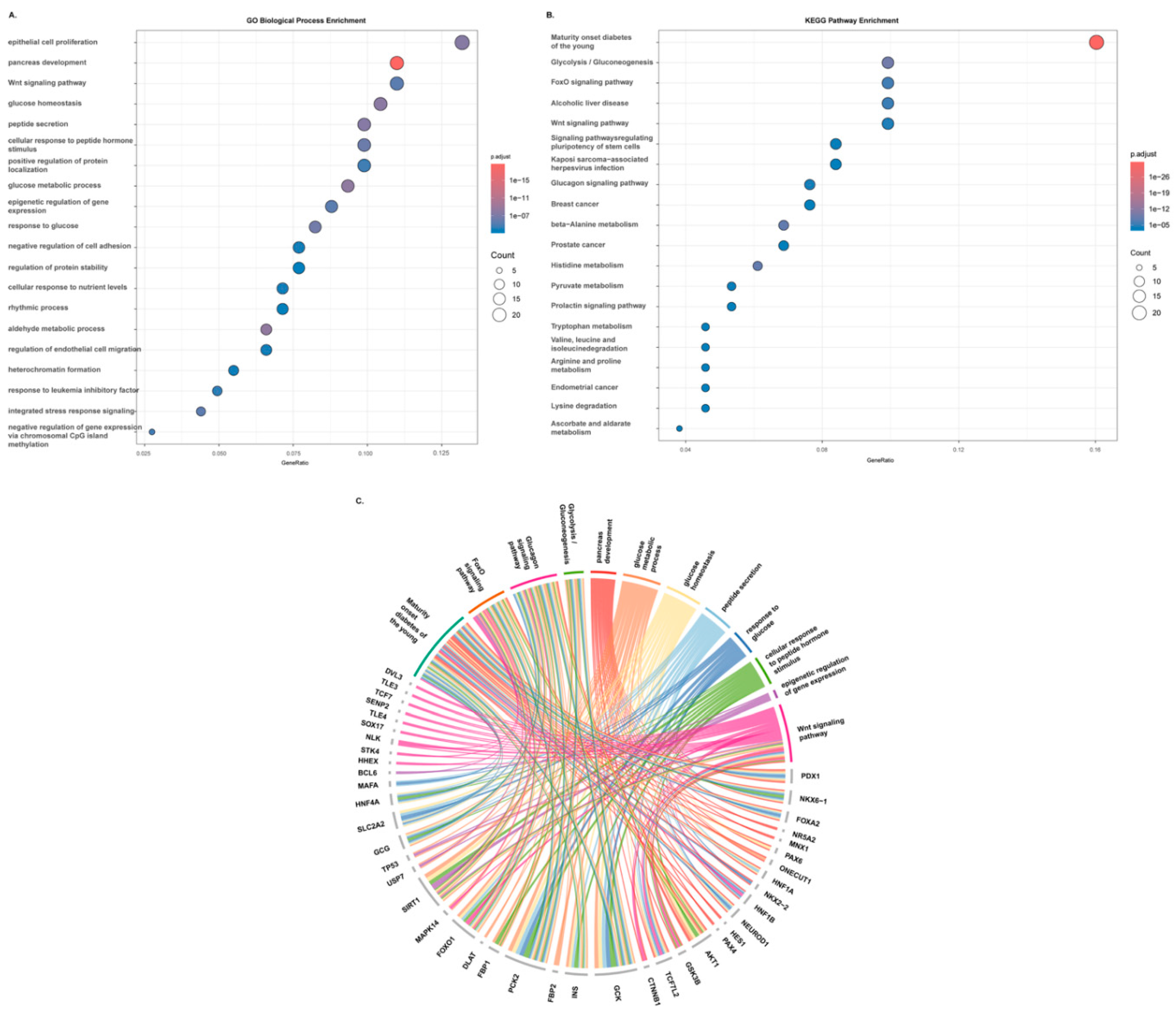
Figure 2.
Steps involed in the β-cell plasticity.
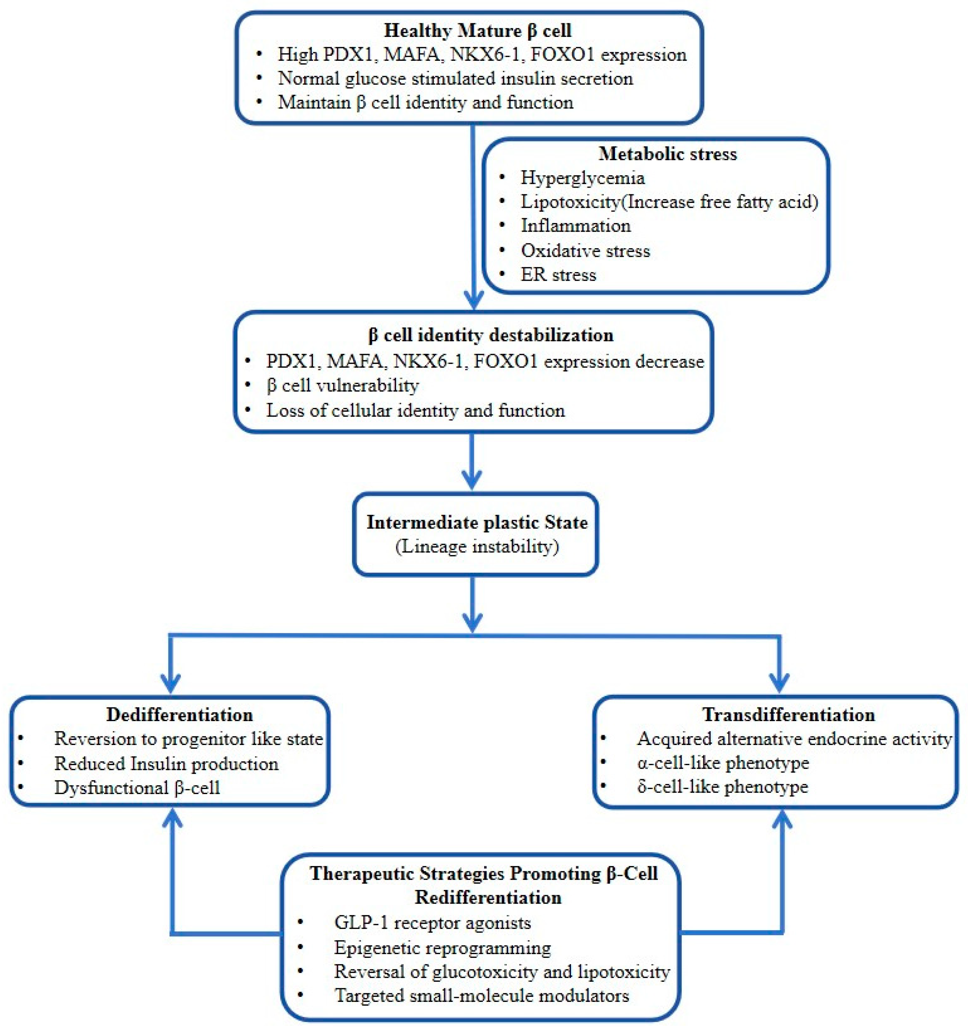

Table 1.
β-Cell dedifferentiation in T2D.
| Feature | Mature β-cell | Dedifferentiated β-cell | Functional consequence | Reference |
| Insulin production | High INS expression | Insulin resistance | Impaired glucose control | [1] |
| β-cell transcription factors | High PDX1, MAFA, NKX6.1, NeuroD1 | Reduced lineage factors | Loss of β-cell identity | [2,3,4] |
| Cellular phenotype | Fully differentiated endocrine state | Progenitor-like features | Functional instability | [5] |
| Progenitor markers | Absent/low | NGN3, SOX9, OCT4, NANOG ↑ | Identity regression | [6,7,8,9] |
| ALDH1A3 | Low/absent | High expression | Marker of dysfunctional β-cells | [10,11,12] |
| Mitochondrial function | Efficient ATP generation | Oxidative stress, ATP decline | Secretory dysfunction | [13] |
| ER homeostasis | Adaptive unfolded protein response | Chronic ER stress | Proteostasis imbalance | [14] |
Table 2.
Major Molecular Drivers of β-Cell Dedifferentiation in T2D.
| Category | Key genes/proteins | Major function in β-cell biology | Role in T2D dedifferentiation/ transdifferentiation | Reference |
| β-cell identity regulators | PDX1, MAFA, NeuroD1, PAX6 | Maintain mature β-cell identity, insulin secretion, glucose sensing | Downregulated during β-cell dedifferentiation | [2,3,4,18] |
| Master stress/identity regulator |
FOXO1 | Preserves β-cell maturity during metabolic stress | Loss promotes identity collapse and progenitor-like state | [6,16] |
| Dedifferentiation markers | ALDH1A3, NGN3 (NEUROG3), SOX9, OCT4 (POU5F1), NANOG | Progenitor-like/endocrine precursor programs | Re-expressed during β-cell dedifferentiation | [6,8,10,11,12,19] |
| Transdifferentiation/α-cell lineage regulators | ARX, GCG, SMOC1 | Promote α-cell identity and glucagon program | Increased during β→α-like transition | [12,20,21] |
| β-cell functional markers |
INS, GLUT2 | Insulin synthesis, glucose sensing, mature β-cell function | Reduced in dysfunctional β-cells | [22,23,24,25] |
| Epigenetic regulators | UHRF1, BACH2 | Chromatin remodeling, transcriptional repression | Regulate β-cell identity stability | [17,26] |
| Genetic susceptibility genes | TCF7L2, HNF1A, HNF4A | β-cell stress resilience and insulin secretion | Risk-associated dysfunction and identity loss | [15,27] |
| miRNA regulators | miR-195, miR-204, miR-7 | Post-transcriptional regulation of β-cell genes | Promote β-cell dysfunction | [28] |
| Insulin signaling pathway | PI3K, AKT1 | β-cell survival and insulin signaling | Suppressed in glucolipotoxicity | [29] |
| Cell-cycle and regeneration regulators |
CDK4, Cyclin D2 (CCND2), REG family genes | β-cell proliferation and regeneration | Linked to redifferentiation and β-cell recovery | [30] |
| Disallowed genes | Hk1, Dlk1, Pdgfra, Oat, and Mylk) | Normally repressed in mature β-cells | Derepression disrupts glucose sensing | [31] |
| Reprogramming factors (experimental) | PAX4, PDX1, MAFA, NGN3, NeuroD1 | Endocrine lineage conversion | Used for α→β or ductal/acinar reprogramming | [3,4,32] |
Table 3.
Evidence for β-Cell Transdifferentiation in T2D.
| Cell fate transition | Trigger | Key molecular regulators | Biological significance | Reference |
| β → α-like cell | Metabolic stress | PDX1↓, NKX2.2↓ | Hyperglucagonemia | [3,20,35] |
| β → progenitor-like state | Glucolipotoxicity | NGN3, SOX9 reactivation | Functional insulin loss | [8,36] |
| α → β-cell conversion | β-cell depletion | PAX4, ARX suppression | Endogenous regeneration | [21,32] |
| Acinar → β-like cell | Reprogramming | PDX1, NeuroD1 | Experimental regeneration | [4,31] |
Table 4.
Functional Gene Network.
| Hub Gene | Functionally Related Genes | Hub Gene | Functionally Related Genes |
| PDX1 | NKX61, FOXA2, MAFA, FOXO1, NR5A2, MNX1, PAX6, HHEX, ONECUT1, PCIF1, SPOP, SLC2A2, PSMD9, HNF1A, FGF10, NKX2-2, IAPP, HNF1B, GCK, SP1 |
MAFA | PDX1, NKX2-8, NKX2-2, INS, PAX6, PSME3, MAPK14, FOXA2, NRL, MAFB, MAF, FOXO1, NFE2L3, MAFG, MAFK, NFE2L1, MAFF, NFE2L2, NFE2, NEUROD1 |
| NKX6-1 | PDX1, NKX2-2, ATF1, FBXL13, NKX28, PCDHGC4, PTGER1, IL26, S100A2, PLA2G2F, RAI1, NAB2, SDR9C7, SCNN1A, LMNTD1, TP53AIP1, TGM4, C4orf36, CXXC5, PODXL |
FOXO1 | SFN, PDX1, YWHAG, YWHAZ, FHL2, DACH1, FBP2, AKT1, WDFY2, CCNB1, YWHAH, FOXG1, A2M, LRPPRC, CREBBP, SIRT1, STK4, ANGPT2, PCK2, FBP1 |
| ALDH1A3 | GSK3B, HIF1A, TP53, CDA, ALDH1A2, CFAP46, ALDH3B2, ALDH8A1, ALDH4A1, ALDH1B1, ALDH5A1, ALDH3B1, ALDH9A1, ALDH3A1, ALDH2, ALDH1A1, ALDH3A2, STS, ALDH6A1, ALDH7A1 | NEUROG3 | INSM1, NEUROD1, NKX2-2, ONECUT1, HES6, HES1, PAX4, MYOD1, NTN1, CCDC71L, SOX1, ZNF575, ZFPM1, CDH24, NKX2-8, AVP, HES7, FOXC2, HAGHL, FBXL17 |
| BACH2 | MAFK, PATZ1, BACH1, MAFG, BCL6B, MAFF, KLF12, MAFB, PLCG2, BCL6, PRDM1, AICDA, EGR2, TNFAIP2, CD22, BATF3, MDM4, OGG1, FOSL2, NFE2L2 |
TCF7L2 | NLK, CTNNB1, TNIK, GCG, SOX17, HIC1, IGF2BP1, RUNX3, TLE4, DLAT, ZCCHC12, SENP2, KLF4, TCF7, HOXB13, HNF4A, TLE3, DVL3, NUMA1, TJP1 |
| UHRF1 | DNMT1, UHRF1BP1, CEBPA, H2BC14, H2BC13, ZPR1, EME1, H2AX, H2AC7, MUS81, H2AC6, STAT5A, UHRF2, DNMT3A, USP7, WIPI1, DPPA3, H3C13, WDHD1, EZH2 | SMOC1 | DNAJB11, CORO1C, CCDC93, ADORA3, RACGAP1, SMOC2, CLMN, BMPR1B, TNIP1, SPOCK3, SPOCK2, SPOCK1, PCSK6, CISD3, TACSTD2, ELOVL2, EPCAM, CD74, HOXA1, SPARC |
Table 5.
Emerging Therapeutic Strategies to Restore β-Cell Identity.
| Clinical strategy | Representative trial/intervention | Mechanistic relevance to β-cell dedifferentiation | Key findings | Relevance to review | Reference |
| Metabolic unloading / remission | DiRECT Trial (Diabetes Remission Clinical Trial) | Reduces glucotoxicity and lipotoxicity, allowing β-cell recovery | Major weight loss induced diabetes remission and recovery of β-cell function in a subset of patients | Strong evidence for reversibility of dysfunctional β-cell states | [41] |
| Caloric restriction | Counterpoint / Counterbalance studies | Reduces pancreatic fat and β-cell stress | Rapid normalization of fasting glucose and improved first-phase insulin secretion | Supports redifferentiation after metabolic normalization | [41] |
| Bariatric surgery | STAMPEDE Trial | Profound metabolic unloading and incretin remodeling | Durable diabetes remission and improved endogenous insulin secretion | Suggests β-cells can recover after stress reduction | [42] |
| GLP-1 receptor agonists | LEADER (NCT01179048) (liraglutide), SUSTAIN (NCT01131676) (semaglutide), SURPASS (NCT03951753) (tirzepatide) | β-cell rest, reduced ER stress, anti-inflammatory effects | Improved β-cell function markers and glycemic control | Mechanistically linked to preservation of β-cell identity | [43,44,45,46] |
| SGLT2 inhibitors | EMPA-REG, DECLARE-TIMI (NCT01730534) | Relieves glucotoxicity and lowers β-cell workload | Improved insulin secretion indices in some studies | May indirectly favor β-cell redifferentiation | [47] |
| Intensive insulin therapy | Early insulinization trials in newly diagnosed T2D (NCT00629213) | Functional β-cell rest | Temporary diabetes remission in subsets | Supports concept of recoverable β-cell dysfunction | [48] |
| Cell-state restoration (emerging) | ALDH1A3 inhibitor programs (preclinical → translational stage) | Direct inhibition of dedifferentiation pathways | Reversal of β-cell dysfunction in animal models | Strong mechanistic future direction, but no established human trial yet | [11,12] |
| β-cell regeneration | DYRK1A inhibitor (harmine-derived compounds) | Promotes β-cell proliferation while preserving identity | Human β-cell expansion in preclinical studies | Relevant to restoration of β-cell mass + function | [43] |
| Precision/genetic risk modulation | Studies involving TCF7L2-associated diabetes subgroups | β-cell identity preservation | Emerging stratified medicine concept | Relevant for personalized β-cell therapies | [15] |
Disclaimer/Publisher’s Note: The statements, opinions and data contained in all publications are solely those of the individual author(s) and contributor(s) and not of MDPI and/or the editor(s). MDPI and/or the editor(s) disclaim responsibility for any injury to people or property resulting from any ideas, methods, instructions or products referred to in the content. |
© 2026 by the authors. Licensee MDPI, Basel, Switzerland. This article is an open access article distributed under the terms and conditions of the Creative Commons Attribution (CC BY) license (http://creativecommons.org/licenses/by/4.0/).
Copyright: This open access article is published under a Creative Commons CC BY 4.0 license, which permit the free download, distribution, and reuse, provided that the author and preprint are cited in any reuse.