

Submitted:
02 September 2026
Posted:
02 September 2026
You are already at the latest version
Abstract
HLA class I antigen presentation depends on coordinated peptide processing, transport and loading; yet inflammatory signalling may modify the consequences of defects in this machinery. We examined wild-type HAP1 cells and isogenic ERAP1-, TAP1- and TAP2-knockout cells before and after interferon-γ (IFNγ) treatment using flow cytometry, proteomics, and immunopeptidomics. IFNγ strongly induced residual antigen-processing machinery in all HAP1-genotypes, increased HLA-I surface expression and expanded peptide recovery, including peptides derived from cancer-associated antigens. However, repertoire restoration was incomplete and defect specific. ERAP1-deficient cells retained enrichment of longer, terminally extended peptides, whereas TAP2-deficient cells retained alterations in peptide length, processing of peptide flanking regions and source-protein location of the peptide. TAP1-deficient cells showed the greatest convergence towards wild-type immunopeptidome features upon IFNγ stimulation. Components of the pep-tide-loading and ER quality-control machinery remained associated with immature HLA-I heavy chains in TAP-deficient cells and were enhanced by exposure to IFNγ. These findings demonstrate that inflammatory signalling can quantitatively and partially qualitatively compensate for defects in HLA-I antigen processing by amplifying intact pathway components and alternative peptide supply, without recreating a fully wild-type immunopeptidome.
Keywords:
antigen processing
; antigen presentation
; mass spectrometry
; HLA
; immunopeptidomics
; cytokine signaling
; immune evasion
1. Introduction
Human leukocyte antigen (HLA) class I molecules (HLA-I) enable most nucleated cells to continuously “display” fragments of intracellular proteins at the cell surface for inspection by CD8⁺ T cells. In the classical HLA-I pathway, normal turnover of proteins, as well as defective ribosomal products and pathogen- or tumour-derived proteins generate peptide precursors predominantly through the actions of the proteasome. These peptide precursors can be further processed by cytosolic peptidases, prior to their transport into the endoplasmic reticulum (ER) by the heterodimeric transporter associated with antigen processing (TAP1/2) (1). In the ER, nascent HLA-I heavy chain associates with β2-microglobulin and engages a peptide-loading “assembly line” that includes calnexin, calreticulin, ERp57 and the formation of a peptide-loading complex (PLC) centered around the specialised chaperone tapasin. The formation of the PLC links HLA-I to TAP and promotes the acquisition and editing of high-stability peptide ligands. For many HLA allotypes, ER aminopeptidases (ERAP1 and ERAP2) trim N-terminally extended peptide precursors to optimal 8–12mer ligands, increasing the abundance of stable HLA-I–peptide complexes that can traffic through the secretory pathway to the plasma membrane (2). The result is a dynamic “immunopeptidome”, a collection of HLA class I bound peptides that reflects the cellular proteome, inflammatory state (including interferon-driven remodeling of antigen processing), and the binding specificities of an individual’s HLA allotypes (3).
This antigen presentation system underpins immune surveillance by providing a molecular readout of intracellular health. CD8⁺ T cells scan HLA-I–bound peptides via their clonally distributed T-cell receptors and can eliminate cells presenting non-self or aberrant self-peptides arising from infection, oncogenic transformation, or cellular stress. In parallel, natural killer (NK) cells integrate signals from HLA-I and other ligands to detect “missing-self” states, providing a complementary safeguard when HLA-I expression is reduced. The breadth and sensitivity of surveillance are further extended by cross-presentation, in which professional antigen-presenting cells can acquire exogenous material and present it on HLA-I to prime CD8⁺ T-cell responses. Together these mechanisms help maintain tissue homeostasis and constrain the emergence of infected or malignant cells.
In cancer, immune surveillance and tumour evolution are often framed as immunoediting, comprising elimination, equilibrium, and escape (4). Tumours can be eliminated when antigen presentation and effector responses are intact, but persistent immune pressure can select for cancer cell variants that evade recognition, which can commonly occur through quantitative or qualitative disruption of components of the HLA-I pathway (e.g., loss of β2-microglobulin, downregulation of TAP/tapasin/ERAP, defects in interferon signaling, or loss of HLA alleles) (5). Such aberrations reduce tumour immunogenicity, reshape the immunopeptidome, and contribute to resistance to T-cell based immunotherapies. Conversely, therapies that reinvigorate T-cell function (e.g., checkpoint blockade) highlight the centrality of effective antigen presentation for durable anti-tumour control, motivating efforts to understand and therapeutically modulate the antigen processing and presentation machinery (6).
Thus, HLA-I antigen presentation is orchestrated by integrated and synergistic antigen processing and presentation machinery (APM) that generates proteasome-derived peptides, transports them into the endoplasmic reticulum (ER), and loads high-affinity ligands onto nascent HLA-I molecules. Disruption of individual APM components can profoundly alter cell surface HLA-I abundance and reshape the presented immunopeptidome, with direct consequences for anti-viral and anti-tumour T-cell recognition and for mechanisms of immune escape in cancer. To enable controlled, component-by-component dissection of this pathway, Spaapen and colleagues generated PAKC (Panel of APM Knockout Cells): a set of isogenic human HAP1 cell lines created by CRISPR/Cas9, each lacking a single, key HLA-I pathway gene (7). The original PAKC collection includes knockouts spanning the pathway from HLA-I expression (HLA-A/B/C and B2M) through ER peptide supply and editing (TAP1, TAP2, tapasin, ERAP1) and ER chaperoning/quality control molecules (calnexin/CANX, calreticulin, ERp57, glucosidase IIα). These cell lines were validated at the sequence and protein levels and provide a shared genetic background in which changes in HLA-I surface expression, peptide repertoire, and T-cell recognition can be attributed to the loss of a specific APM node rather than confounded by cell-line differences. The PAKC panel has become a useful community resource for mechanistic studies of peptide loading and antigen presentation, benchmarking of immunopeptidomics workflows, and functional interrogation of immune-evasion phenotypes relevant to immunotherapy. Building on the original panel, recent work has applied integrated immunopeptidomics and proteomics across HAP1 APM perturbations to quantify how specific gene losses induce distinct, pathway-consistent shifts in peptide length distributions, peptide ligand sequence motif usage, and antigen abundance (7, 8). Together, these isogenic knockout systems provide a tractable platform for linking APM genotype to immunopeptidome phenotype and, ultimately, to T-cell–mediated immunity.
Building on this earlier work we go on further to explore how IFNγ stimulation can compensate for some of these defects and to varying extents repair deficits in antigen presentation and reinstate wildtype-like immunopeptidomes.
2. Materials and Methods
2.1. Cell lines
HAP1 is a near-haploid human cell line derived from KBM-7, a chronic myelogenous leukaemia (7). Unlike KBM-7, HAP1 cells contain a disomy of chromosome 15, while only containing a single copy of other chromosomes (9, 10). This cell line is commonly used in genetic screening assays due to its haploid nature, and this haploidy has been used advantageously to study the phenotypic characteristics of different genes (9, 11, 12). This study uses a Panel of HLA class I antigen presentation machinery HAP1-Knockout Cells (PAKC), which were provided by Dr Robbert Spaapen, Sanquin Research, Amsterdam (7). The variants used in this study include HAP1 wildtype (WT) and knockouts of TAP1, TAP2 and ERAP1. All cell lines have three common HLA-I allotypes—HLA-A*02:01, HLA-B*40:01, and HLA-C*03:04. HAP1 cell lines were grown Iscove’s Modified Dulbecco’s Medium (IMDM) supplemented with 10% fetal bovine serum (FBS) (IMDM-10) media and incubated at 37 °C, 5% CO2.
T2 (174×CEM.T2) is a human T–B lymphoblastoid hybrid cell line derived from fusion of the B-lymphoblastoid line 721.174 with the T-cell line CEMR.3. T2 is a variant of the T1 hybrid that has a deletion of the MHC class II region encompassing the antigen-processing machinery (13). It lacks functional TAP1/TAP2-mediated peptide transport, resulting in inefficient peptide loading and markedly reduced HLA class I surface expression (14). T2 cells were cultured in RPMI 1640 medium supplemented with 10% FBS (RPMI-10) media and incubated at 37 °C, 5% CO2.
2.2. Quantification of surface HLA-I expression by flow cytometry
Flow cytometry was performed regularly on all cell lines to ensure uniform levels of surface HLA-I expression. Cells were washed twice with 200 µL of ice-cold PBS and incubated (30 minutes, 4 °C) with 100 µL of primary antibody supernatant (W6/32 (pan anti-HLA-I) (15), BB7.2 (anti-HLA A2) (16), anti-Bw6 (17) and DT9 (anti-HLA C) (18) all produced in house from B cell hybridomas). Cells were washed twice with ice-cold PBS and incubated in 50 µL of Goat anti-mouse IgG conjugated to phycoerythrin (PE) (Southern Biotechnology Associates, Birmingham, UK (Cat#1032-09)) and 50 µL of LIVE/DEADTM Fixable Aqua (ThermoFischer Scientific) for 30 minutes, 4 °C, in dark. Cells were washed twice with ice-cold PBS and fixed with 1% paraformaldehyde (PFA; ProSciTech). Cells were washed and resuspended in 300 µL of ice-cold 1X PBS, following which flow cytometry was performed using a LSRFortessa™ X-20 Flow Cytometer (BD Biosciences). HLA-I surface expression was analysed using FlowJo® software v10.8.1 (BD Biosciences). Geometric mean fluorescence intensity (gMFI) was used to quantitate the HLA-I levels in cell lines and data was visualised using GraphPad Prism® v10.1.1 (Insight Partners).
2.3. Optimisation of IFNγ treatment—cell viability and surface HLA-I expression
HAP1 cell lines were grown in IMDM-10 for 24 hours until cells reached a 60% confluence. Cell lines were then treated with varying concentrations of IFNγ (20, 50, 100, 200, and 1000 International Units (IU)/mL) for 24-72 hours. The IFNγ incubation time and concentration were optimised for highest cell surface HLA-I expression and cell viability as determined by flow cytometry (Figure S1).
2.4. Proteomics of cell lines
2.4.1. Suspension trapping (S-Trap) digestion and tryptic peptide isolation
HAP1 wildtype and KO cells were grown in the absence and presence of IFNγ, in triplicate, to generate a cell pellet of 2 x 10⁶ cells, following which they were flash frozen in liquid nitrogen. For extracting the global proteome, cell pellets were lysed using 2X lysis buffer (10% SDS with 200 mM TEAB, pH 7.55), following which the lysate was sonicated to using a probe sonicator to shear DNA (amplitude 10, 10 seconds bursts x 3). The lysate was clarified by centrifugation at 4000xg for 5 minutes, and supernatant was transferred to a new LoBind Eppendorf tube. Protein concentration was measured using a Direct Detect® Spectrometer (Merck), and 100 μg of protein was reduced by 20 mM TCEP solution (ThermoFisher Scientific) and alkylated using 20mM Iodoacetamide (IAA; Merck). Proteins were processed using S-Trap™ micro spin columns (ProtiFi) according to the manufacturer’s protocol and digested with trypsin as previously described (19). Peptides were recovered in three wash steps. 80 μL of digestion buffer followed by 80 μL of 0.2% formic acid (FA; ThermoFisher Scientific) and finally 80 µL of 0.2% FA in 50% acetonitrile (ACN; Fisher Scientific). Peptides were then frozen, and vacuum concentrated. The dried samples were then reconstituted in 100 µL of 0.1% Trifluoroacetic Acid (TFA; ThermoFisher Scientific). For clean-up and concentration of tryptic peptides, 100 µL OMIX C18 tips (Agilent, USA) were conditioned three times in 100 µL of buffer B (0.1% FA and 50% ACN, in optima water). Tips were equilibrated with three washes of 100 µL of buffer A (0.1% FA, in optima water). Peptides were then loaded onto the tips, followed by washes by 100 µL of buffer A (x3). Peptides were eluted from the tip with 150 µL of buffer B, frozen, and vacuum concentrated and finally reconstituted in 20 µL of MS loading buffer (0.1% FA + 2% ACN). Peptide concentration was determined using a ThermoFisher Scientific NanoDrop and a total of 75 ng of peptides spiked with 1 µL of 0.33 pmol/μL of Indexed retention time (iRT; GL Biochem) peptides and transferred to MS vials.
2.4.2. LC-MS/MS of tryptic peptides
Samples were analysed using a hybrid trapped ion mobility-quadropole time of flight mass spectrometer (Bruker timsTOF Pro2) coupled to a nanoElute UHPLC liquid chromatography system operated using a DIA-PASEF method. Tryptic peptides were loaded onto a Trap PepMap Neo (C185mm x 300um 5um) trap column (ThermoFisher Scientific), eluted, and separated on an Aurora (25 cm x 75 μm internal diameter x1.7µm particle size) (IonOpticks) analytical column using a linear stepwise gradient of Buffer A (Optima water, 2% ACN, 0.1% FA) to Buffer B (ACN, 0.1% FA). The gradient used started from 0–17% buffer B over 60 min, then to 25% over the next 30 min, 37% over the next 10 min followed by a rapid rise to 95% Buffer B over a subsequent 10 min period with flow rate set to 300 nL/min in dia-PASEF mode. Data dependent acquisition was performed with following settings: m/z range: 100-1700 mz, capillary voltage:1600 V, Target intensity of 30000, TIMS ramp of 0.60 to 1.60 Vs/cm2 with collision energy ramped linearly as a function of the mobility from 59 eV at 1/k0 = 1.6 Vs cm-2 to 20 eV at 1/k0 = 0.6 Vs cm-2. For DIA, two windows were used in each 100 ms dia-PASEF scan with 16 of these scans (32 steps) covering the doubly and triply charged peptides in the m/z range of 400-1200 m/z and for singly charged peptides in the m/z range of 800-1200 narrow with a 26 Da width isolation window.
2.4.3. Analysis of proteome data
DIA-NN software v1.8.1 (20) was used to analyse LC-DIA-MS proteomics data using the following settings—Library free search mode; precursor FDR 1%; mass accuracy at MS1 and MS2 set to 20ppm; scan window set to 0; isotopologues turned on; MBR turned on; heuristic protein inference turned on; no shared spectra turned on; protein inference at protein names (from FASTA); neural network classifier double-pass mode; quantification strategy any LC (high precision); cross-run normalisation global; library generation smart profiling. The database used was UniProt Human proteome (20,372 entries; version February 2022; iRT and cRAP proteins included in list). Both FASTA digest for library-free search/library generation and Deep learning-based spectra RTs and IMs prediction were selected. Trypsin/P with 2 missed cleavages; Cysteine Carbamidomethylation (+57.02146) was used as the fixed modification, protein N-terminal M-excision, oxidation of methionine (+15.9949), and protein N-terminus acetylation (+42.0106) were used as variable modifications. Peptide length range from 7 to 30; precursor charge range from 1 to 4; precursor m/z range from 300 to 1800; fragment ion m/z range 200 to 1800.
The output of the DIA-NN search (report.pg_matrix.tsv, see Table S1) was imported into DIA-Analyst for differential protein abundance analysis and visualization (Monash Proteomic Analyst Suites, Monash University), based on the framework described by Shah et al. (21). Data was pre-filtered to remove potential contaminant sequences and reverse sequences. Following pre-filtering, LFQ intensities were Log2 transformed, rows were filtered to remove proteins with a high proportion of missing values (proteins that have ≥50% valid values in at least one group), following which missing values for protein intensities were imputed using Perseus-type missing value imputation. False discovery rate (FDR) correction option selected was Benjamini Hochberg method. Output of DIA-Analyst was exported, the clusters were then exported in a .txt file format, and gene set enrichment analysis (GSEA) was performed using EnrichR (22, 23).
2.4.4. Analysis of HC10 immunoprecipitates and peptide loading complex inference
T2, HAP wildtype and KO cells were harvested, washed with ice-cold PBS and lysed at 4 °C in a mild detergent-based lysis buffer containing 1% IGEPAL CA-630 and protease inhibitors to preserve HLA-I peptide-loading and ER-associated protein complexes as described (24). Insoluble material was removed by centrifugation and clarified lysates were incubated with HC10 antibody coupled to protein A Sepharose beads with rotation at 4 °C. Beads were washed extensively using lysis buffer containing a reduced concentration of IGEPAL CA-630 (0.1%) to remove non-specifically associated proteins while maintaining protein–protein interactions. HC10-associated proteins were subsequently reduced, alkylated and digested with trypsin as described above. Peptides were analysed by LC–DIA-MS and proteins identified using DIANN software. Relative recovery of HLA-I-associated proteins across experimental conditions was assessed using peptide-spectrum matches/spectral counts as a semi-quantitative measure of protein abundance.
2.5. HLA-I immunopeptidomics
Immunopeptidomics was performed on HAP1 cell lines that were cultured in the presence and absence of IFNγ. Cells were grown to high density followed by regular cell pelleting until cell pellet size of 5x10⁸ cells was achieved. Following harvesting, cell pellets were washed twice with ice-cold 1X PBS, flash frozen, and stored in stored at -80ºC until their time of use.
2.5.1. Cross-linking of purified antibody to protein A resin
1 mL of Protein A Sepharose (PAS; GE Healthcare, UK) was transferred into a tube containing purified antibody and rolled end-over-end for an hour at 4ºC prior to being transferred into a 10 mL Bio-Rad Poly-prep chromatography column and washing with 20 column volumes of 0.05M borate buffer pH8.0 and the antibody cross-linked to the protein A on column using Dimethyl pimelimidate dihydrochloride (DMP; Sigma, USA). Cross-linking was quenched by the flow of 20 column volumes of ice-cold 0.2M Tris-HCl (pH 8.0) followed by 20 column volumes of 0.1M citrate buffer (pH 3.0) to strip off any unbound antibody. Crosslinked antibody-PAS was then washed with 20 mL of borate buffer and stored at 4ºC until their time of use.
2.5.2. Isolation of HLA-I bound peptides
HAP1 cell pellets (5x10⁸ cells) were lysed using a combination of mechanical and chemical techniques. Pellets were initially pulverised into a fine powder using a cryogenic mill (Mixer Mill MM 400, Retsch, Germany). Once pulverised, cells were lysed in 10mL of ice-cold 2X lysis buffer (1% IGEPAL CA-630 (Merck, USA); 100mM Tris HCl (pH 8.0); 300mM NaCl; supplemented with cOmpleteTM Protease Inhibitor Cocktail (Sigma Aldrich)), incubated for 45 minutes at 4ºC, and rotated end-over-end. The cell lysate was then clarified by centrifugation (3166xg, 10 minutes, 4 °C), and the supernatant was transferred to a precooled ultracentrifuge tube. The clarified supernatant was then ultracentrifuged (117,524xg, 4ºC, 45 minutes; Ti70 rotor– radius: 6.57cm;) to remove cellular debris and nuclear material. Following ultracentrifugation, any lipid layer was removed from the lysate, and the clarified lysate was passed over a poly-prep column containing 1 mL of PAS to remove any non-specific binders. The flow through was then passed over the W6/32- PAS column with subsequent washing steps as described elsewhere (25). To elute HLA-peptide complexes from PAS, 4.5 mL of 10% acetic acid was added to the column which also resulted in dissociation the peptides from the heavy chain and β2m. The eluate mixture was further fractionated using reversed-phase high-performance liquid chromatography (RP-HPLC) to separate the peptides from the HC and β2m. RP-HPLC was performed using a 4.6 mm internal diameter x 100 mm long monolithic C18 reverse phase column (Chromolith® SpeedROD, Merck) using an ÄKTAmicroTM HPLC system (GE Healthcare) and UNICORNTM software (GE Healthcare). The mobile phase buffers used in the system were buffer A (0.1% TFA (v/v) in optima water) and buffer B (80% ACN (v/v), 0.1% TFA (v/v) in optima water). Column was equilibrated at 2 mL/minute for 10 minutes using 2% of buffer B. Following equilibration, eluate was injected onto the column at a flow rate of 2 mL/minute and peptides were separated using an increasing gradient of buffer B. Peptides, β2m, and HC, were separated into fractions by using an increasing linear gradient of buffer B and 500 µL fractions collected, frozen and vacuum dried.
Following drying, samples were reconstituted in 12 µL of MS loading buffer (0.1% FA + 2% ACN) and 1 µL of 0.33pmol/μL of iRT peptides, sonicated, and loaded onto Evotips (EvoSep; Denmark). Evotips were first washed with 20 µL of solvent B (80% ACN + 0.1% FA) and centrifuged (800xg, 60 seconds, RT), following which tips were conditioned by soaking in 100 µL of 2-propanol. The soaked tips were then equilibrated with 20 µL of solvent A (2% ACN + 0.1% FA) and centrifuged (800xg, 60 seconds, RT). Following equilibration, sample was loaded twice onto equilibrated Evotips and the tip was centrifuged (800xg, 60 seconds, RT). Tip was then washed with 20 µL of solvent A, centrifuged (800xg, 60 seconds, RT), followed by loading with 100 µL of solvent A and centrifugation (800xg, 10 seconds, RT).
2.5.3. Analysis of HLA-I bound peptides by mass spectrometry
For HAP1 cell lines, data dependent acquisition (DDA) LC-MS was performed using a Bruker timsTOF Pro2 (Bruker Daltonics) coupled to an EvoSep One system (EvoSep). Peptides were loaded on a Evotip PureTM disposable trap column, and peptides were eluted and separated with a C18 Aurora Elite CSI 15 cm nanoflow UHPLC column (75 μm internal diameter, 1.7µm particle size) (IonOpticks) kept at 50 °C using the Whisper 20 SPD method as described (26). The mobile phases consisted of buffer A (2% ACN, 0.1% FA, in optima water) and buffer B (100% ACN, 0.1% FA).
2.5.4. Peptide identification and immunopeptidome analysis
Bruker timsTOF .d MS files were exported to PEAKS® Online 11 software (Bioinformatics Solutions Inc) (27) and searched against the human proteome (UniProt, version: 9 February 2022; 20,372 entries) with contaminant database containing iRT peptides and cRAP proteins. The search was performed using the following parameters: for data refinement, enzyme was set to none, activation mode set to CID, instrument set to timsTOF, acquisition to DDA. Once the .d files were refined, de novo search was performed using precursor and fragment mass error tolerance set to 15 ppm and 0.05 Da respectively, with enzyme set to none, two variable PTMs (M [+15.99] and NQ [+0.98]), and a maximum of three PTMs for each peptide. Following de novo search, PEAKS DB (in-depth de novo assisted search) was performed. The following settings were used: mass correction and associate features with chimera spectra were ticked on, precursor mass error tolerance set to 15 ppm, fragment mass error tolerance to 0.05 Da, digest mode to unspecific, two variable PTMs (M [+15.99] and NQ [+0.98]), a maximum of three PTMs for each peptide, with peptide length between 7 and 30 amino acids long, and deep learning boost turned on.
3. Results
3.1. Knockout of APM components differentially impacts surface HLA-I expression
Cell surface HLA-I expression is dependent on the optimal function of various APM components. Previous studies however have shown that this expression is modulated following the knockout of APM components (7, 28). To determine the impact of knocking out ERAP1, TAP1 or TAP2 on the presentation of the HLA-I alleles, selected PAKC KO cell lines (which express three HLA-I allotypes (HLA-A*02:01, HLA-B*40:01, and HLA-C*03:04)) were treated in the absence or presence of 20 IU/mL IFNγ for 24h. Specific HLA allotype cell surface expression was quantified by flow cytometry using four primary antibodies: W6/32 (pan HLA-I) (Figure 1A), BB7.2 (HLA-A2 specific) (Figure 1B), anti-Bw6 (HLA-B40 specific in this context) (Figure 1C), and DT9 (HLA-C specific) (Figure 1D). For all cell lines, flow cytometry revealed that, as expected, IFNγ treatment led to a significant increase in HLA-I cell surface expression. HAP1WT cells expressed high levels of surface HLA-I in both untreated and IFNγ treated conditions (Figure 1A,B and Figure S1), whilst APM knockout modulated cell surface HLA-I expression in an allele dependent manner. ERAP1KO led to a significant decrease in HLA-A2 surface expression in both untreated and IFNγ treated cells (Figure 1A,B). However, no significant decrease was observed in HLA-B40 or HLA-C3 expression (Figure 1A,B). Knockout of TAP1 and TAP2 on the other hand led to a significant decrease in surface expression of all three allotypes that was partially restored upon IFN-g treatment (Figure 1A,B).
3.2. APM Components Dominate the IFNγ-Induced Proteomic Response despite Loss of ERAP1, TAP1, or TAP2
To determine whether the partial restoration of HLA-I surface expression reflected broader remodelling of the antigen-presentation pathway, we next compared the proteomes of untreated and IFNγ-treated cells within each genotype. In HAP1WT cells, IFNγ induced a prominent, coordinated increase in multiple APM proteins, including HLA-A, HLA-B, HLA-C, β2-microglobulin (B2M), TAP1, TAP2, ERAP1, and the immunoproteasome component PSMB9 (Figure 2A). These proteins were among the most strongly induced species in the IFNγ-responsive proteome, consistent with broad activation of the HLA-I antigen-presentation axis (Table S1). A similarly coordinated response was retained in each knockout line despite the absence of the targeted APM component, with the knock out confirmed by an absence of proteomic data. ERAP1KO cells strongly increased HLA-I heavy chains, B2M, TAP1, TAP2, and PSMB9 (Figure 2B). TAP1KO cells upregulated HLA-A, HLA-B, HLA-C, B2M, TAP2, ERAP1, and PSMB9 (Figure 2C), whereas TAP2KO cells upregulated TAP1, ERAP1, HLA-I heavy chains, and the immunoproteasome subunits PSMB9 and PSMB10 (Figure 2D). The deleted protein was not detected, but the remaining pathway components retained strong IFNγ responsiveness. These data indicate that the IFNγ signalling response remains functionally intact in the APM knockout cells and can substantially amplify the residual antigen-processing capacity, providing a molecular basis for the partial restoration of HLA-I expression observed by flow cytometry.
3.3. IFNγ Expands and Partially Repairs APM-Deficient Immunopeptidomes
We next asked whether the IFNγ induction of the remaining APM components translated into increased peptide antigen presentation. IFNγ markedly increased the number of HLA-I-bound peptides recovered from every HAP1 genotype (Figure 3A, Table S2). The increase was evident in HAP1WT and ERAP1KO cells and was especially pronounced in the TAP-deficient lines, which had very low peptide yields under basal conditions. Following IFNγ treatment, the TAP1KO immunopeptidome expanded to a depth approaching that of HAP1WT cells, whereas TAP2KO cells showed a substantial but incomplete recovery. ERAP1KO cells generated the largest peptide repertoire after IFNγ treatment, consistent with preserved peptide transport combined with altered ER trimming.
IFNγ altered not only the depth but also the composition of the peptide repertoire. Treatment-overlap analysis further showed that the expansion was driven largely by peptides detected only after IFNγ exposure, particularly in TAP1KO and TAP2KO cells (Figure 3B). The peptide length distribution remained dominated by canonical 9-mers, although IFNγ modestly reduced the relative 9-mer contribution while increasing several longer peptide classes (Figure 3C). ERAP1KO cells showed the expected broader length distribution, with fewer 9-mers and proportionally more 10- to 13-mers. TAP1KO cells became more strongly 9-mer dominated after IFNγ treatment, whereas TAP2KO cells retained a broader distribution with increased representation of 8-mers and peptides longer than 10 residues. Thus, IFNγ did not simply increase the abundance of an existing repertoire; it remodeled the immunopeptidome.
We then examined whether the expanded repertoires retained sequence features compatible with the three endogenous HLA-I allotypes. Across the complete immunopeptidomes, IFNγ reduced the proportion of peptides not predicted to bind HLA-A*02:01, HLA-B*40:01, or HLA-C*03:04 in HAP1WT and, more prominently, in the TAP-deficient cell lines (Figure 3D). The change was accompanied by increased assignment to HLA-B*40:01 in TAP1KO cells and to HLA-C*03:04 in TAP2KO cells. Separation of peptides according to whether they were untreated-specific, shared, or IFNγ-specific revealed that the newly induced peptide pools in TAP1KO and TAP2KO cells still contained a substantial fraction of peptides not predicted to bind any of the three HLA allotypes (Figure 3D). Therefore, IFNγ increased recovery of canonical HLA-associated ligands in TAP-deficient cells while simultaneously exposing a sizeable non-canonical or poorly predicted component of the rescued peptide repertoire.
The effect of IFNγ on repertoire identity was next assessed across genotypes using intersection analysis (Figure 4). Untreated cells contained large genotype-restricted peptide populations, demonstrating that loss of ERAP1, TAP1, or TAP2 produces qualitatively distinct HLA-I repertoires rather than simply reducing repertoire size. IFNγ increased the total number of peptides in all four cell lines and generated additional shared intersections, consistent with partial convergence on an interferon-responsive antigen-presentation program. Nevertheless, large genotype-specific peptide sets remained after treatment. Accordingly, restoration of peptide yield and surface HLA-I expression did not equate to restoration of a wild-type immunopeptidome; the identity of the deleted APM component continued to strongly determine which peptides were presented.
3.5. ERAP1 and TAP Deficiency Alter Peptide Terminal Processing, Hydrophobicity, and Source-Protein Position
To further define the processing features of the knockout-specific ligands, we examined terminal extensions relative to an eight-residue core. In untreated cells, ERAP1KO and TAP2KO immunopeptidomes showed the strongest enrichment of both N- and C-terminally extended peptides, whereas HAP1WT contained comparatively few extended species (Figure 5A,C). TAP1KO cells displayed an intermediate extension phenotype. Following IFNγ treatment, terminal extension frequencies in TAP1KO cells were reduced toward HAP1WT levels, consistent with the broader evidence for partial immunopeptidome repair in this line. In contrast, extended peptides remained prominent in ERAP1KO cells and were especially evident in TAP2KO cells (Figure 5B,D). Thus, increased peptide presentation after IFNγ does not bypass the characteristic processing defects associated with loss of ERAP1 or TAP2.
APM disruption also changed the physicochemical properties of the presented ligands. Across several peptide-length classes, peptides unique to the knockout lines showed shifts toward higher grand average of hydropathy (GRAVY) scores relative to HAP1WT, with the most prominent differences observed among 8- to 11-mers (Figure S2). These differences persisted after IFNγ treatment and remained significant for multiple length classes (Figure 5B), indicating that cytokine-driven increases in HLA-I expression and peptide yield do not eliminate the altered hydrophobicity profile created by APM disruption. The data therefore support a role for the APM in shaping not only peptide abundance and length but also the physicochemical space sampled by the HLA-I immunopeptidome.
Finally, we mapped presented peptides to their relative position within the source protein. Peptides from HAP1WT and ERAP1KO cells were distributed broadly across parental proteins, with relatively little positional bias (Figure 6A). By contrast, untreated TAP1KO and TAP2KO cells were strongly enriched for peptides derived from the N-terminal portion of source proteins. IFNγ largely flattened this positional bias in TAP1KO cells, producing a distribution more similar to HAP1WT, whereas TAP2KO cells retained a pronounced enrichment at the N terminus and additional terminal skewing (Figure 6B). This differential response again distinguishes TAP1 and TAP2 loss and suggests that IFNγ can restore access to a broader source-protein landscape in TAP1-deficient cells more efficiently than in TAP2-deficient cells.
3.6. Peptide-loading complex components remain associated with immature HLA-I heavy chains in TAP-deficient cells and are enhanced by IFNγ.
We next assessed which components of the peptide-loading complex (PLC) remained associated with HLA-I heavy chains under conditions of disrupted antigen processing. For this, we used the HC10 antibody, which preferentially recognises peptide-receptive or incompletely assembled MHC-I heavy chains, to immunoprecipitate HLA-I and examine associated PLC components. Notably, HC10 immunoprecipitation continued to recover components of the HLA-I peptide-loading machinery following loss of an individual TAP subunit (Figure 7). HC-10 immunoprecipitation was performed from HAP1WT, TAP1KO, -TAP2KO, and -ERAP1KO cells cultured in the absence or presence of IFNγ, together with TAP1/TAP2-deficient T2 cells. Co-immunoprecipitated proteins were identified by LC–MS/MS and spectral counts were used as a semi-quantitative measure of protein recovery. The heatmap shows key peptide-loading complex (PLC) and ER-associated proteins, including TAP1, TAP2, tapasin, calreticulin, calnexin and immunoglobulin binding protein (BiP). Values represent the mean spectral count from two independent samples and are visualised as log2(mean spectral count + 1). Retention of tapasin, calreticulin, calnexin and BiP in HC-10 immunoprecipitates from TAP-deficient cells indicates that immature HLA-I heavy chains remain associated with components of the ER peptide-loading and quality-control machinery despite disruption of the canonical TAP transporter. IFNγ treatment increased the recovery of several PLC-associated proteins, most notably tapasin, in the knockout backgrounds.
HC10 preferentially recognises immature or peptide-receptive HLA-I heavy chains, thus these findings suggest that disruption of the canonical TAP1–TAP2 transporter does not abolish engagement of nascent HLA-I with ER-resident loading and quality-control factors. Consistent with this interpretation, HLA-I–tapasin association has previously been demonstrated in TAP2-deficient T2 cells expressing TAP1 alone (29), and partially folded HC10-reactive HLA-I species can associate with tapasin–ERp57 complexes (30, 31). Thus, residual HLA-I–PLC interactions may persist in TAP-deficient cells even though canonical TAP-dependent peptide transport is lost.
3.7. IFNγ Broadens Presentation of Leukemia-Associated Antigen-Derived Peptides in APM-Deficient Cells
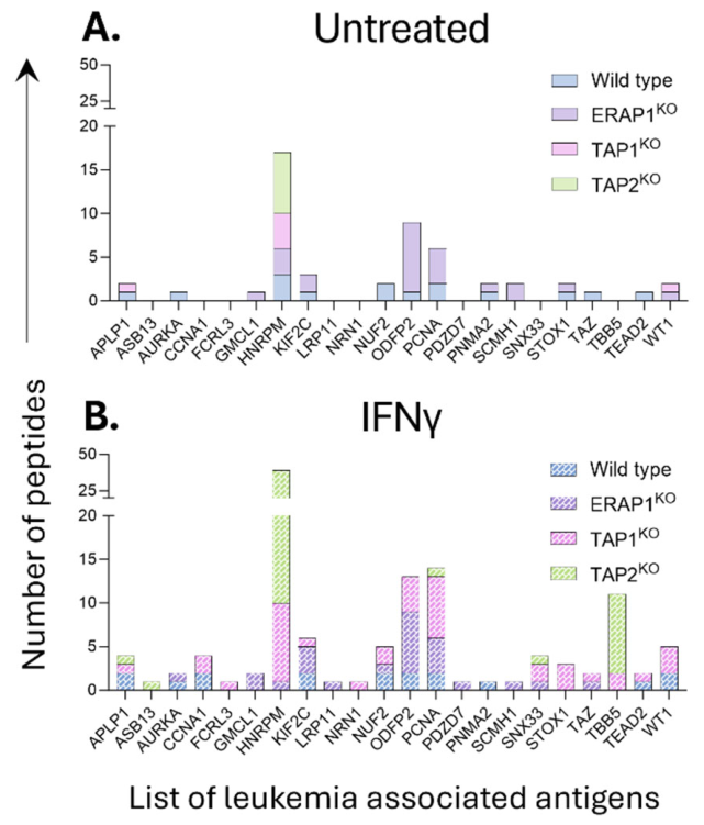
To assess whether the observed changes extended to potentially disease-relevant antigens, we interrogated the immunopeptidomes for peptides derived from a curated set of leukemia-associated antigens (LAAs). In untreated cells, LAA-derived peptide presentation was sparse and strongly genotype dependent, with loss of TAP1 or TAP2 altering both the number and source of detectable LAA ligands (Figure 8A). IFNγ substantially increased the breadth and number of LAA-derived peptides across the panel (Figure 8B). Importantly, peptides from multiple LAAs that were absent or poorly represented under basal conditions became detectable in the TAP-deficient cells following treatment. The rescue was not uniform across antigens or knockout genotypes, indicating that cytokine stimulation expands clinically relevant antigen presentation without erasing the underlying APM-dependent selectivity.
Collectively, these data show that IFNγ drives a powerful compensatory antigen-presentation program in HAP1 cells. Upregulation of the remaining APM increases HLA-I surface expression, expands peptide yield, increases presentation of predicted HLA binders, and broadens LAA-derived antigen display. However, the extent of repair is component specific. TAP1KO cells show substantial recovery toward wild-type-like peptide length and source-position characteristics, whereas TAP2KO and ERAP1KO cells retain pronounced processing signatures. Thus, IFNγ produces a quantitatively expanded but not fully wild-type-equivalent immunopeptidome in cells with defined defects in HLA-I antigen processing.
4. Discussion
The present study uses the isogenic HAP1 Panel of Antigen Processing Machinery Knockout Cells to examine how an inflammatory stimulus interacts with defined defects in HLA class I antigen processing. The principal finding is that IFNγ can compensate substantially for loss of individual antigen-processing components at the level of HLA-I abundance and peptide yield, but this compensation does not recreate a fully wild-type antigen-processing state. Rather, the extent and character of “repair” are strongly component dependent. TAP1-deficient cells showed the greatest convergence toward the wild-type immunopeptidome after IFNγ treatment, whereas TAP2- and ERAP1-deficient cells retained prominent signatures of altered antigen processing. These data extend the original description of the PAKC resource and the recent systematic analysis of its basal immunopeptidomes (7, 8) by demonstrating that inflammatory signaling acts on, but does not erase, the molecular consequences of individual APM lesions.
The proteomic and flow-cytometric data provide a mechanistic framework for this partial compensation. IFNγ induced a coordinated increase in HLA-A, HLA-B, HLA-C, B2M and multiple peptide-processing components, including TAP1/TAP2 where genetically intact, ERAP1, and immunoproteasome subunits such as PSMB9 and PSMB10. This is consistent with the well-established role of IFNγ in transcriptionally coordinating the HLA-I pathway and shifting proteasomal processing toward an immunoproteasome-dominated state; indeed, ERAP1 was originally characterised as an IFNγ-inducible ER aminopeptidase (32). Importantly, the targeted protein remained absent in each knockout, indicating that the response represents amplification of the residual pathway rather than reversal of the genetic lesion. This distinction is particularly clear in the surface-expression data. ERAP1 loss preferentially reduced HLA-A*02:01 while comparatively preserving HLA-B*40:01 and HLA-C*03:04, whereas loss of either TAP subunit broadly reduced all three allotypes. Such allele-selective effects are consistent with previous analysis of the PAKC panel, which showed that perturbation of individual APM components can produce HLA-allotype-specific changes rather than uniform effects across the entire class I repertoire (8).
IFNγ treatment also produced extensive qualitative remodeling of the immunopeptidome. In each HAP1 genotype investigated, cytokine exposure increased the number and diversity of recovered HLA-I ligands, with a large proportion of the expanded repertoire comprised of peptides detected only after the IFNγ treatment. Thus, IFNγ did not simply increase the abundance of peptides already presented under basal conditions. This behaviour is consistent with immunopeptidomic studies in tumour cells showing that IFNγ modifies antigen presentation through several superimposed mechanisms, including changes in source-protein abundance, induction of the immunoproteasome, increased HLA-I expression and allotype-specific changes in peptide loading (33, 34). IFNγ-driven inflammation can also generate qualitatively distinct or aberrant peptide products that are poorly represented in untreated cells (35-37). The current data extend these observations to cells carrying defined APM defects: even when peptide supply or ER trimming is genetically compromised, IFNγ can markedly broaden the detectable peptide repertoire. However, the continued presence of large genotype-specific peptide sets and the substantial fraction of IFNγ-induced peptides not assigned by prediction to HLA-A*02:01, HLA-B*40:01 or HLA-C*03:04 emphasise that increased peptide recovery is not synonymous with restoration of a canonical wild-type repertoire. The latter group may include atypical ligands, peptides poorly modelled by current prediction algorithms, or co-purifying species and should therefore be interpreted cautiously.
The ERAP1KO phenotype illustrates clearly the distinction between quantitative rescue and restoration of processing fidelity. ERAP1 normally removes N-terminal residues from peptide precursors and can either generate or destroy individual HLA-I ligands, with a strong influence on the abundance of optimally sized peptides (28, 32, 38). Consistent with this role, ERAP1-deficient cells retained a broad length distribution enriched for 10–13mers and terminally extended ligands even after IFNγ treatment. IFNγ therefore increased the amount of material entering the antigen-presentation pathway but could not substitute for the missing trimming step. The persistence of the ERAP1-dependent signature is also consistent with pharmacological studies in which ERAP1 inhibition substantially remodels the immunopeptidome rather than simply reducing its size (39, 40). The selective reduction in HLA-A*02:01 surface expression in ERAP1KO cells further suggests that the requirement for optimal ER trimming differs between the endogenous HAP1 HLA allotypes. This finding reinforces the view that ERAP1 perturbation should be considered in an HLA-specific context, particularly when ERAP1 inhibition is contemplated as a strategy to generate novel tumour-associated peptide repertoires.
The allele-selective effects observed following ERAP1 deletion further emphasise that the consequences of altered ER peptide trimming depend on the HLA molecules available for loading. Loss of ERAAP in mice has previously been associated with reduced MHC-I surface expression (41, 42), whereas reduced ERAP1 expression in human monocytic cells increased the accumulation of aberrant HLA-B27 species (43) without producing equivalent changes in HLA-B18 or HLA-B51 (44). Together with the selective reduction of HLA-A*02:01 observed here, these findings indicate that dependence on ERAP1-mediated trimming varies substantially between HLA allotypes. Thus, the effect of ERAP1 deficiency should not be viewed simply as a global reduction in peptide maturation, but as an allotype-dependent redistribution of the peptide repertoire in which individual HLA molecules differ in their capacity to acquire stable ligands in the absence of optimal ER trimming.
The HLA allotype binding-prediction analysis provides further evidence that disruption of peptide processing alters the contribution of individual HLA allotypes. In the untreated TAP-deficient immunopeptidomes, assignment of peptides to HLA-B*40:01 was markedly reduced, while a considerable fraction of the TAP2KO repertoire was not predicted to bind any of the three endogenous HLA-I molecules using NetMHCpan 4.1 (45). This poor representation of predicted HLA-B40 ligands demonstrates a strong dependence of this allotype on conventional TAP-mediated peptide supply, consistent with previous evidence that HLA-B allotypes differ substantially in their capacity to maintain antigen presentation during TAP deficiency (46). The increased recovery of conventionally predicted ligands following IFNγ treatment therefore represents qualitative as well as quantitative rescue, although the persistence of substantial genotype-specific and poorly predicted peptide populations indicates that canonical allotype usage is not completely restored.
The TAP-deficient lines revealed a different form of plasticity. Both TAP1KO and TAP2KO cells displayed markedly reduced HLA-I surface abundance and peptide yield under basal conditions, as expected for disruption of the heterodimeric transporter that normally supplies the ER with cytosol-derived peptides. Nevertheless, IFNγ substantially increased HLA-I expression and immunopeptidome depth in both lines despite the fact that the deleted TAP subunit was not be restored. This differs from tumour models in which IFNγ rescues antigen presentation principally by re-inducing low or suppressed TAP expression (47). In the genetically TAP-deficient setting studied here, increased surface HLA-I must instead reflect a combination of increased HLA heavy-chain and B2M availability, altered peptide generation and greater use of TAP-independent peptide sources. TAP-independent HLA-I presentation is well established and can involve peptides generated from proteins entering the secretory pathway, including signal-peptide and intramembrane-processing routes (48). The enrichment of peptides from the N-terminal regions of source proteins in TAP1KO and TAP2KO cells, together with shifts toward more hydrophobic peptide populations, is compatible with increased contribution from such ER-associated or membrane-proximal processing pathways, although the present data do not establish the origin of individual ligands.
An intriguing feature of the data is the different response of TAP1KO and TAP2KO cells to IFNγ. Following stimulation, TAP1KO cells approached wild-type-like peptide length and source-protein positional distributions more closely than TAP2KO cells, whereas TAP2KO retained a broader length profile, terminal extensions and stronger source-protein positional bias. Because canonical TAP function requires both TAP1 and TAP2, these differences should not be interpreted as evidence that one subunit can independently restore normal peptide transport. Rather, they suggest that loss of the two subunits generates distinct downstream states, potentially through differential compensatory changes in the remaining APM or altered access to non-canonical peptide sources. This interpretation is consistent with the broader PAKC analysis of Shapiro et al., in which individual APM knockouts generated distinct, pathway-consistent immunopeptidomic phenotypes even when the disrupted proteins participated in the same functional module (8).
The changes in peptide hydrophobicity and source-protein position further demonstrate that APM defects alter peptide quality as well as quantity. The persistence of higher hydrophobicity among subsets of knockout-specific ligands after IFNγ treatment indicates that inflammatory stimulation does not eliminate the physicochemical bias imposed by defective processing. Some of this effect may reflect altered access to membrane- or signal-sequence-derived peptides in the TAP-deficient lines, but changes in the relative contribution of HLA-A*02:01, HLA-B*40:01 and HLA-C*03:04 will also influence amino-acid composition because each allotype imposes distinct anchor requirements. Thus, peptide hydrophobicity should be interpreted as an emergent property of both processing and HLA selection rather than as a direct readout of a single enzymatic step. The same principle applies to the altered length distributions: peptide length reflects the combined effects of proteolysis, transport, ER trimming, peptide editing and the structural permissiveness of the individual HLA molecule.
The increased presentation of peptides derived from leukemia-associated antigens (LAAs) after IFNγ treatment provides a clinically relevant illustration of this pathway plasticity. IFNγ broadened LAA-derived antigen presentation not only in HAP1WT cells but also in cells lacking ERAP1, TAP1 or TAP2, indicating that inflammatory signaling can expose potentially targetable antigens even in the setting of defined antigen-processing defects. This observation is important for immunotherapy because the antigenic landscape encountered by T cells in an inflamed tumour may differ substantially from that measured under basal culture conditions. Previous work has similarly shown that IFNγ can diversify tumour immunopeptidomes and alter the relative presentation of candidate therapeutic targets (33-35).
Loss or dysfunction of HLA class I antigen-processing machinery is common across human cancers and provides an important route of tumour immune escape. The prevalence depends strongly on whether genomic alterations or loss of protein expression is considered. At the genomic level, somatic loss of heterozygosity (LOH) of the HLA-I locus occurs in approximately 17% of solid tumours, ranging from ~2–42% between tumour types and reaching ~30% in squamous carcinomas; in NSCLC, HLA-I LOH has been reported in approximately 40% of tumours in some cohorts (49, 50). Somatic HLA-I mutations are less frequent, occurring in approximately 3.3% of 7,930 TCGA tumours, but are enriched in head and neck, lung squamous, gastric and colorectal cancers (51). Alterations affecting other APM genes are similarly heterogeneous: analysis of head and neck cancers identified genomic alterations involving HLA-I and APM genes in approximately 20% of cases, with TAP1 and TAP2 altered in ~3–4% of individuals (52). Somatic B2M mutations are relatively uncommon in unselected tumours, occurring in approximately 1.8% of cancers in a pan-cancer TCGA analysis and in ~1–5% of several unselected tumour cohorts, but are markedly enriched in hypermutated and immunogenic settings. For example, B2M mutations were detected in 24.2% of MSI-H colorectal cancers compared with only 0.9% of microsatellite-stable colorectal cancers (53, 54). Importantly, defective protein expression is substantially more frequent than structural mutation, reflecting transcriptional, epigenetic and post-transcriptional regulation. Across historical tumour series, reduced or absent TAP1/TAP2 expression has been reported in ≥40% of many cancer types, although frequencies are lower in breast and ovarian cancers (<30%), while abnormalities in HLA-I heavy-chain expression range from ~36% in renal cancer to nearly 88% in thyroid cancer (55). Thus, complete genetic disruption of individual APM components is relatively uncommon, whereas partial loss, downregulation or allele-specific disruption of the antigen-presentation pathway is widespread, providing a strong biological rationale for understanding how inflammatory signals such as IFNγ compensate for individual APM lesions and reshape the residual immunopeptidome.
5. Conclusions
This study demonstrates that inflammatory signaling can substantially compensate for defined defects in the HLA-I antigen-processing machinery, but that increased HLA-I surface expression and peptide yield should not be equated with restoration of a wild-type immunopeptidome. IFNγ strongly induced the residual APM in HAP1 cells, expanded HLA-I ligand recovery and increased presentation of LAA-derived peptides across ERAP1-, TAP1- and TAP2-deficient backgrounds. Nevertheless, each genetic lesion retained a characteristic processing signature. ERAP1 deficiency preserved an extended-peptide phenotype despite IFNγ, while TAP2 deficiency remained associated with pronounced alterations in peptide length, terminal processing and source-protein position. TAP1-deficient cells showed a greater degree of convergence toward wild-type features, highlighting unexpected heterogeneity in the capacity of inflammatory signaling to compensate for defects within the same antigen-transport module.
Collectively, the findings support a model in which IFNγ provides quantitative and partial qualitative “repair” of antigen presentation by amplifying intact pathway components and increasing access to alternative peptide sources, rather than by bypassing the molecular requirement for the deleted APM component. This distinction has direct implications for tumour immune escape and antigen-targeted therapy: APM-deficient cancer cells may remain visible to T cells under inflammatory conditions, but the peptides they present can differ substantially from those displayed by antigen-processing-competent cells. Defining these defect- and inflammation-specific immunopeptidomes may therefore reveal both mechanisms of immune evasion and antigenic vulnerabilities that are not apparent from analysis of the untreated cellular state.
Supplementary Materials
The following supporting information can be downloaded at the website of this paper posted on Preprints.org, Figure S1: optimization of IFNg treatment of HAP1 cells; Figure S2 Knockout of APM components increases the presentation of hydrophobic peptides; Table S1: Proteomics data; Table S2: Immunopeptidome data.
Author Contributions
Conceptualization, R. Arahna, N.P.C., A.W.P; methodology, D.D., K.P., M.D; validation, R. Aranha, D.D; formal analysis, R. Arahna, M.D.; investigation, R. Arahna, K.P., M.D., D.D., R. Ayala; resources, A.W.P.; data curation, R. Arahna; writing—original draft preparation, R. Arahna, A.W.P.; writing—review and editing, all; supervision, N.P.C., A.W.P.; project administration, A.W.P.; funding acquisition, A.W.P. All authors have read and agreed to the published version of the manuscript.
Funding
This research was funded by the National Health and Medical Research Council (NHMRC) Australia, grant number 2016596 to A.W.P. and the APC was funded by Monash University.
Data Availability Statement
The mass spectrometry proteomics and immunopeptidomics data generated in this study will be deposited with the ProteomeXchange Consortium via the PRIDE partner repository (56) and made publicly available upon publication. The corresponding ProteomeXchange/PRIDE dataset accession number(s) will be provided upon completion of data deposition.
Acknowledgments
Computational resources were supported by the R@CMon/Monash Node of the Nectar Research Cloud, an initiative of the Australian Government’s Super Science Scheme and the Education Investment Fund.
Conflicts of Interest
The authors declare the following financial interests/personal relationships which may be considered as potential competing interests: Anthony Purcell is a scientific advisor for Bioinformatics Solutions Inc (Canada), a shareholder and scientific advisor for Evaxion Biotech (Denmark), and a co-founder of Resseptor Therapeutics (Australia). These entities had no role in the design of the study; in the collection, analyses, or interpretation of data; in the writing of the manuscript; or in the decision to publish the results.
Abbreviations
The following abbreviations are used in this manuscript:
| ACN | Acetonitrile |
| APM | Antigen Presentation and Processing Machinery |
| CML | Chronic Myelogenous Leukaemia |
| cRAP | Common Repository of Adventitious Proteins |
| DDA | Data Dependent Acquisition |
| DIA | Data Independent Acquisition |
| DMSO | Dimethyl-Sulphoxide |
| ER | Endoplasmic Reticulum |
| ERAP | Endoplasmic Reticulum Aminopeptidase |
| FA | Formic Acid |
| FDR | False Discovery Rate |
| gMFI | Geometric Mean Fluorescence Intensity |
| GSEA | Gene Set Enrichment Analysis |
| HC | Heavy Chain |
| HLA | Human Leukocyte Antigen |
| HLA-I | HLA Class I |
| IFNγ | Interferon γ |
| iRT | Indexed Retention Time |
| LC-MS/MS | Liquid Chromatography-Tandem Mass Spectrometry |
| PAKC | Panel Of HLA Class I Antigen Presentation Machinery Knockout Cells |
| PBS | Phosphate Buffered Saline |
| PFA | Paraformaldehyde |
| PLC | Peptide Loading Complex |
| PTM | Post-Translational Modification |
| PΩ | C-Terminal Anchor Residue |
| RP-HPLC | Reversed-Phase High-Performance Liquid Chromatography |
| RT | Room Temperature |
| TAA | Tumour Associated Antigen |
| TAP | Transporter Associated with Antigen Processing |
| TFA | Trifluoroacetic Acid |
| β2m | Beta-2-Microglobulin |
References
- K. L. Rock, E. Reits, J. Neefjes, Present Yourself! By MHC Class I and MHC Class II Molecules. Trends Immunol 37, 724-737 (2016). [CrossRef]
- J. Neefjes, M. L. Jongsma, P. Paul, O. Bakke, Towards a systems understanding of MHC class I and MHC class II antigen presentation. Nat Rev Immunol 11, 823-836 (2011). [CrossRef]
- J. W. Yewdell, E. Reits, J. Neefjes, Making sense of mass destruction: quantitating MHC class I antigen presentation. Nat Rev Immunol 3, 952-961 (2003). [CrossRef]
- G. P. Dunn, L. J. Old, R. D. Schreiber, The Immunobiology of Cancer Immunosurveillance and Immunoediting. Immunity 21, 137-148 (2004). [CrossRef]
- R. Kim, M. Emi, K. Tanabe, Cancer immunoediting from immune surveillance to immune escape. Immunology 121, 1-14 (2007). [CrossRef]
- Z. Zhang, Y. Zhang, J. Lieberman, Innate immunity in tumour immunoediting and immunosurveillance. The EMBO Journal 45, 7-29 (2026). [CrossRef]
- A. de Waard et al., PAKC: A novel panel of HLA class I antigen presentation machinery knockout cells from the same genetic origin. Eur J Immunol 51, 734-737 (2021). [CrossRef]
- E. Shapiro et al., Deleterious KOs in the HLA Class I Antigen Processing and Presentation Machinery Induce Distinct Changes in the Immunopeptidome. Mol Cell Proteomics 24, 100951 (2025). [CrossRef]
- T. B. Beigl, I. Kjosås, E. Seljeseth, N. Glomnes, H. Aksnes, Efficient and crucial quality control of HAP1 cell ploidy status. Biol Open 9 (2020). [CrossRef]
- M. Kotecki, P. S. Reddy, B. H. Cochran, Isolation and characterization of a near-haploid human cell line. Experimental cell research 252, 273-280 (1999). [CrossRef]
- M. Cavanaugh, S. L. Jaspersen, Big lessons from little yeast: budding and fission yeast centrosome structure, duplication, and function. Annual review of genetics 51, 361-383 (2017). [CrossRef]
- M. J. Stark (2001) The awesome power of yeast genetics-practical approaches and recipes for success: Methods in Yeast Genetics: A Cold Spring Harbor Laboratory Course Manual, 2000 Edition. (Company of Biologists).
- R. D. Salter, D. N. Howell, P. Cresswell, Genes regulating HLA class I antigen expression in T-B lymphoblast hybrids. Immunogenetics 21, 235-246 (1985). [CrossRef]
- R. D. Salter, P. Cresswell, Impaired assembly and transport of HLA-A and -B antigens in a mutant TxB cell hybrid. Embo j 5, 943-949 (1986). [CrossRef]
- P. Parham, C. J. Barnstable, W. F. Bodmer, Use of a monoclonal antibody (W6/32) in structural studies of HLA-A,B,C, antigens. J Immunol 123, 342-349 (1979). [CrossRef]
- P. Parham, F. M. Brodsky, Partial purification and some properties of BB7.2. A cytotoxic monoclonal antibody with specificity for HLA-A2 and a variant of HLA-A28. Hum Immunol 3, 277-299 (1981). [CrossRef]
- S. F. Radka, D. D. Kostyu, D. B. Amos, A monoclonal antibody directed against the HLA-Bw6 epitope. J Immunol 128, 2804-2806 (1982). [CrossRef]
- V. M. Braud, D. S. Allan, D. Wilson, A. J. McMichael, TAP- and tapasin-dependent HLA-E surface expression correlates with the binding of an MHC class I leader peptide. Curr Biol 8, 1-10 (1998). [CrossRef]
- M. HaileMariam et al., S-Trap, an Ultrafast Sample-Preparation Approach for Shotgun Proteomics. J Proteome Res 17, 2917-2924 (2018). [CrossRef]
- V. Demichev, C. B. Messner, S. I. Vernardis, K. S. Lilley, M. Ralser, DIA-NN: neural networks and interference correction enable deep proteome coverage in high throughput. Nat Methods 17, 41-44 (2020). [CrossRef]
- D. Shah, R. J. A. Goode, C. Huang, D. R. Powell, R. B. Schittenhelm, LFQ-Analyst: An Easy-To-Use Interactive Web Platform To Analyze and Visualize Label-Free Proteomics Data Preprocessed with MaxQuant. J Proteome Res 19, 204-211 (2020). [CrossRef]
- E. Y. Chen et al., Enrichr: interactive and collaborative HTML5 gene list enrichment analysis tool. BMC Bioinformatics 14, 128 (2013). [CrossRef]
- M. V. Kuleshov et al., Enrichr: a comprehensive gene set enrichment analysis web server 2016 update. Nucleic Acids Research 44, W90-W97 (2016). [CrossRef]
- W. Wynne et al., Characterization of the Antigen Processing Machinery and Endogenous Peptide Presentation of a Bat MHC Class I Molecule. J Immunol 196, 4468-4476 (2016). [CrossRef]
- W. Purcell, S. H. Ramarathinam, N. Ternette, Mass spectrometry-based identification of MHC-bound peptides for immunopeptidomics. Nat Protoc 14, 1687-1707 (2019). [CrossRef]
- Pandey et al., Multiple Classes of Antigen Contribute to the Antigenic Landscape of Mesothelioma. Mol Cell Proteomics 24, 100925 (2025). [CrossRef]
- Xin et al., A streamlined platform for analyzing tera-scale DDA and DIA mass spectrometry data enables highly sensitive immunopeptidomics. Nature Communications 13, 3108 (2022). [CrossRef]
- A. York et al., The ER aminopeptidase ERAP1 enhances or limits antigen presentation by trimming epitopes to 8–9 residues. Nature immunology 3, 1177-1184 (2002). [CrossRef]
- N. Antoniou, S. Ford, E. S. Pilley, N. Blake, S. J. Powis, Interactions formed by individually expressed TAP1 and TAP2 polypeptide subunits. Immunology 106, 182-189 (2002). [CrossRef]
- S. G. Santos et al., Major Histocompatibility Complex Class I-ERp57-Tapasin Interactions within the Peptide-loading Complex *. Journal of Biological Chemistry 282, 17587-17593 (2007). [CrossRef]
- M. Paulsson et al., Assembly of tapasin-associated MHC class I in the absence of the transporter associated with antigen processing (TAP). Int Immunol 13, 23-29 (2001). [CrossRef]
- T. Saric et al., An IFN-gamma-induced aminopeptidase in the ER, ERAP1, trims precursors to MHC class I-presented peptides. Nature Immunology 3, 1169-1176 (2002). [CrossRef]
- G. Goncalves et al., IFNγ Modulates the Immunopeptidome of Triple Negative Breast Cancer Cells by Enhancing and Diversifying Antigen Processing and Presentation. Frontiers in Immunology 12, 645770 (2021). [CrossRef]
- Newey et al., Multifactorial Remodeling of the Cancer Immunopeptidome by IFNγ. Cancer Research Communications 3, 2345-2357 (2023). [CrossRef]
- Bartok et al., Anti-tumour immunity induces aberrant peptide presentation in melanoma. Nature 590, 332-337 (2021). [CrossRef]
- M. Fahmy et al., STING dampens the unfolded protein response to enable the presentation of self-antigens on MHC-I during inflammation. Cell Rep 45, 117497 (2026). [CrossRef]
- Carré et al., Interferon-α promotes HLA-B-restricted presentation of conventional and alternative antigens in human pancreatic β-cells. Nat Commun 16, 765 (2025). [CrossRef]
- Leib et al., ERAP1-dependent extreme antigen processing efficacy can govern MHC class I expression hierarchy. J Immunol 214, 1147-1159 (2025). [CrossRef]
- D. Koumantou et al., Editing the immunopeptidome of melanoma cells using a potent inhibitor of endoplasmic reticulum aminopeptidase 1 (ERAP1). Cancer Immunology, Immunotherapy 68, 1245-1261 (2019). [CrossRef]
- P. Tempora et al., Combining ERAP1 silencing and entinostat therapy to overcome resistance to cancer immunotherapy in neuroblastoma. J Exp Clin Cancer Res 43, 292 (2024). [CrossRef]
- T. Kanaseki, N. Blanchard, G. E. Hammer, F. Gonzalez, N. Shastri, ERAAP synergizes with MHC class I molecules to make the final cut in the antigenic peptide precursors in the endoplasmic reticulum. Immunity 25, 795-806 (2006). [CrossRef]
- G. E. Hammer, F. Gonzalez, M. Champsaur, D. Cado, N. Shastri, The aminopeptidase ERAAP shapes the peptide repertoire displayed by major histocompatibility complex class I molecules. Nat Immunol 7, 103-112 (2006). [CrossRef]
- Chen et al., Critical role of endoplasmic reticulum aminopeptidase 1 in determining the length and sequence of peptides bound and presented by HLA-B27. Arthritis Rheumatol 66, 284-294 (2014). [CrossRef]
- T. M. Tran, S. Hong, J. H. Edwan, R. A. Colbert, ERAP1 reduces accumulation of aberrant and disulfide-linked forms of HLA-B27 on the cell surface. Molecular Immunology 74, 10-17 (2016). [CrossRef]
- Reynisson, B. Alvarez, S. Paul, B. Peters, M. Nielsen, NetMHCpan-4.1 and NetMHCIIpan-4.0: improved predictions of MHC antigen presentation by concurrent motif deconvolution and integration of MS MHC eluted ligand data. Nucleic Acids Research 48, W449-W454 (2020). [CrossRef]
- J. Geng, A. J. Zaitouna, M. Raghavan, Selected HLA-B allotypes are resistant to inhibition or deficiency of the transporter associated with antigen processing (TAP). PLOS Pathogens 14, e1007171 (2018). [CrossRef]
- K. Stifter et al., IFN-γ treatment protocol for MHC-Ilo/PD-L1+ pancreatic tumor cells selectively restores their TAP-mediated presentation competence and CD8 T-cell priming potential. Journal for ImmunoTherapy of Cancer 8, e000692 (2020). [CrossRef]
- C. Oliveira et al., New role of signal peptide peptidase to liberate C-terminal peptides for MHC class I presentation. The Journal of Immunology 191, 4020-4028 (2013). [CrossRef]
- Montesion et al., Somatic HLA Class I Loss Is a Widespread Mechanism of Immune Evasion Which Refines the Use of Tumor Mutational Burden as a Biomarker of Checkpoint Inhibitor Response. Cancer Discov 11, 282-292 (2021). [CrossRef]
- McGranahan et al., Allele-Specific HLA Loss and Immune Escape in Lung Cancer Evolution. Cell 171, 1259-1271.e1211 (2017). [CrossRef]
- S. A. Shukla et al., Comprehensive analysis of cancer-associated somatic mutations in class I HLA genes. Nat Biotechnol 33, 1152-1158 (2015). [CrossRef]
- F. Concha-Benavente, R. Srivastava, S. Ferrone, R. L. Ferris, Immunological and clinical significance of HLA class I antigen processing machinery component defects in malignant cells. Oral Oncol 58, 52-58 (2016). [CrossRef]
- J. Budczies et al., Mutation patterns in genes encoding interferon signaling and antigen presentation: A pan-cancer survey with implications for the use of immune checkpoint inhibitors. Genes Chromosomes Cancer 56, 651-659 (2017). [CrossRef]
- S. Middha et al., Majority of B2M-Mutant and -Deficient Colorectal Carcinomas Achieve Clinical Benefit From Immune Checkpoint Inhibitor Therapy and Are Microsatellite Instability-High. JCO Precis Oncol 3 (2019). [CrossRef]
- Cai et al., Defective HLA class I antigen processing machinery in cancer. Cancer Immunol Immunother 67, 999-1009 (2018). [CrossRef]
- Y. Perez-Riverol et al., The PRIDE database at 20 years: 2025 update. Nucleic Acids Res 53, D543-d553 (2025). [CrossRef]
Figure 1.
Knockout of APM components significantly impacts surface HLA-I expression. To examine the effect of IFNγ on cell surface HLA-I expression in HAP1 cell lines with KO of various components of the APM, flow cytometry was performed using four primary antibodies. using anti pan HLA-I (W6/32), anti HLA-A2 (BB7.2), anti HLA-B40 (Bw6), and anti HLA-C03 (DT9) antibodies respectively. The graphs represent gMFI HLA-I expression of HAP1WT, HAP1-ERAP1KO, HAP1-TAP1KO, and HAP1-TAP2KO in (A) Untreated cells and (B) IFNγ treated cells. All bar graphs represent (mean ± SD), n=3.
Figure 1.
Knockout of APM components significantly impacts surface HLA-I expression. To examine the effect of IFNγ on cell surface HLA-I expression in HAP1 cell lines with KO of various components of the APM, flow cytometry was performed using four primary antibodies. using anti pan HLA-I (W6/32), anti HLA-A2 (BB7.2), anti HLA-B40 (Bw6), and anti HLA-C03 (DT9) antibodies respectively. The graphs represent gMFI HLA-I expression of HAP1WT, HAP1-ERAP1KO, HAP1-TAP1KO, and HAP1-TAP2KO in (A) Untreated cells and (B) IFNγ treated cells. All bar graphs represent (mean ± SD), n=3.
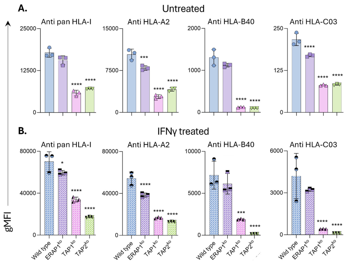
Figure 2.
IFNγ treatment significantly modulates the expression of APM proteins. Volcano plots of protein expression showing significantly up/downregulated proteins in the absence and presence of IFNγ, in (A) HAP1-WT cells, (B) -ERAP1KO cells, (C) -TAP1KO cells, and (D) -TAP2KO cells. Significance was determined following a two-sided student’s T-test (n = 3; adjusted -log10 p-value cutoff = 0.05; log2 fold change = 1).
Figure 2.
IFNγ treatment significantly modulates the expression of APM proteins. Volcano plots of protein expression showing significantly up/downregulated proteins in the absence and presence of IFNγ, in (A) HAP1-WT cells, (B) -ERAP1KO cells, (C) -TAP1KO cells, and (D) -TAP2KO cells. Significance was determined following a two-sided student’s T-test (n = 3; adjusted -log10 p-value cutoff = 0.05; log2 fold change = 1).
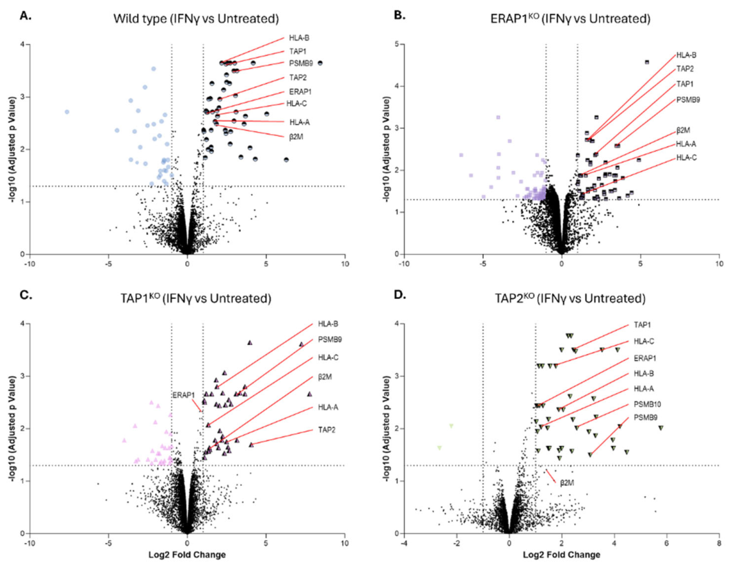
Figure 3.
IFNγ expands and remodels the immunopeptidome of HAP1 cells. HAP1 cell lines were treated without or with IFNγ (20 IU/mL, 24 h), followed by W6/32 immunoaffinity purification, RP-HPLC, LC-MS/MS, and peptide identification. (A) Total detected peptides. (B) Percentage of peptides detected only in untreated cells, common to both conditions, or detected only after IFNγ treatment. (C) Distribution of 8- to 14-mer HLA-bound peptides. Bars show mean ± SD, n = 3. (D) Peptides were grouped as detected only in untreated cells, common to untreated and IFNγ-treated cells, or detected only after IFNγ treatment. Stacked bars show the proportions of the peptides identified that are predicted to bind HLA-A*02:01, HLA-B*40:01, or HLA-C*03:04; NB indicates peptides not predicted to bind any of the three HLA-I allotypes.
Figure 3.
IFNγ expands and remodels the immunopeptidome of HAP1 cells. HAP1 cell lines were treated without or with IFNγ (20 IU/mL, 24 h), followed by W6/32 immunoaffinity purification, RP-HPLC, LC-MS/MS, and peptide identification. (A) Total detected peptides. (B) Percentage of peptides detected only in untreated cells, common to both conditions, or detected only after IFNγ treatment. (C) Distribution of 8- to 14-mer HLA-bound peptides. Bars show mean ± SD, n = 3. (D) Peptides were grouped as detected only in untreated cells, common to untreated and IFNγ-treated cells, or detected only after IFNγ treatment. Stacked bars show the proportions of the peptides identified that are predicted to bind HLA-A*02:01, HLA-B*40:01, or HLA-C*03:04; NB indicates peptides not predicted to bind any of the three HLA-I allotypes.
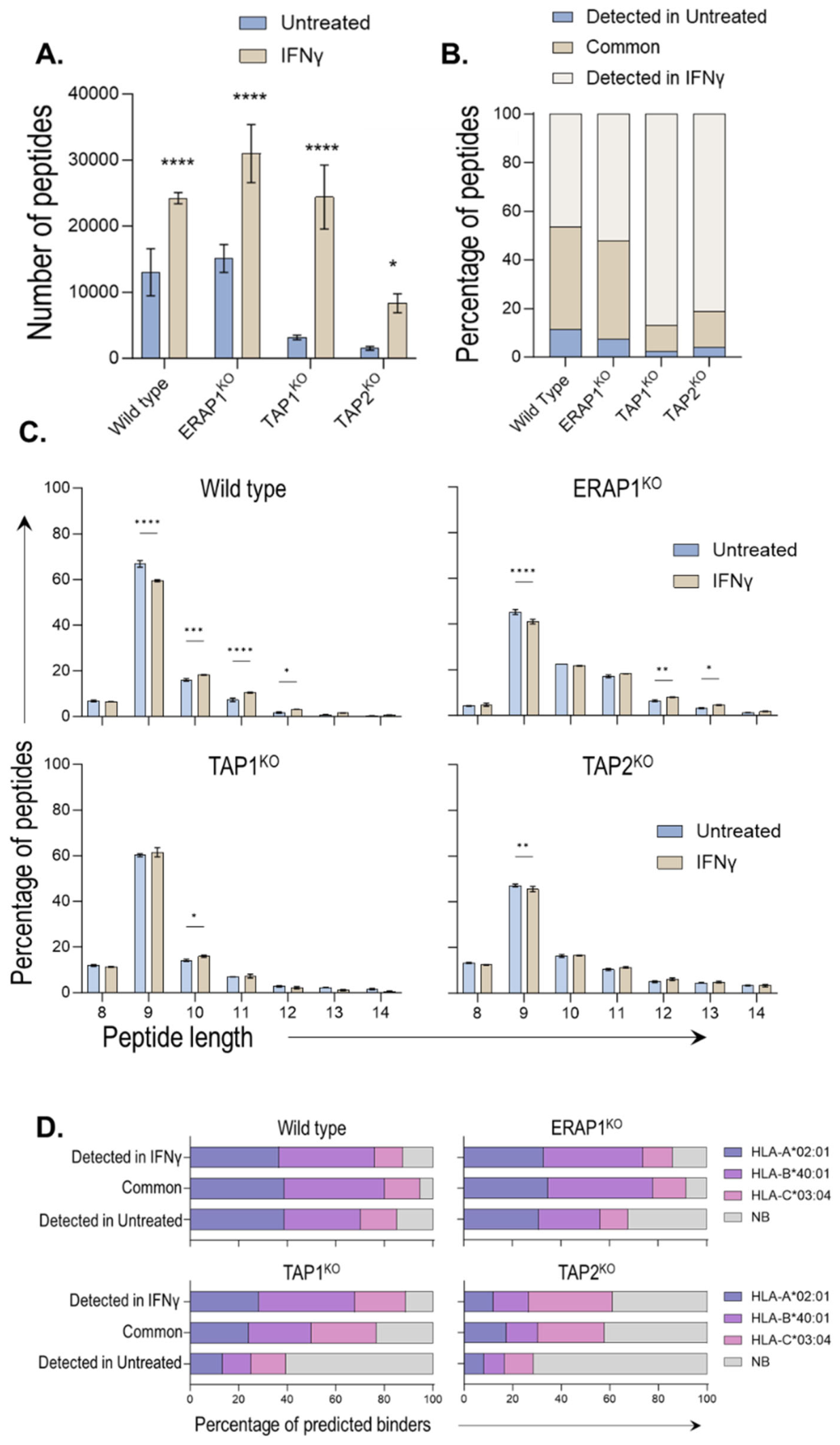
Figure 4.
APM knockout cells retain distinct peptide repertoires after IFNγ treatment. UpSet plots show peptide-set sizes and intersections among HAP1WT, ERAP1KO, TAP1KO, and TAP2KO cells under untreated and IFNγ-treated conditions. Rows indicate the cell-line peptide sets and columns indicate their intersections.
Figure 4.
APM knockout cells retain distinct peptide repertoires after IFNγ treatment. UpSet plots show peptide-set sizes and intersections among HAP1WT, ERAP1KO, TAP1KO, and TAP2KO cells under untreated and IFNγ-treated conditions. Rows indicate the cell-line peptide sets and columns indicate their intersections.
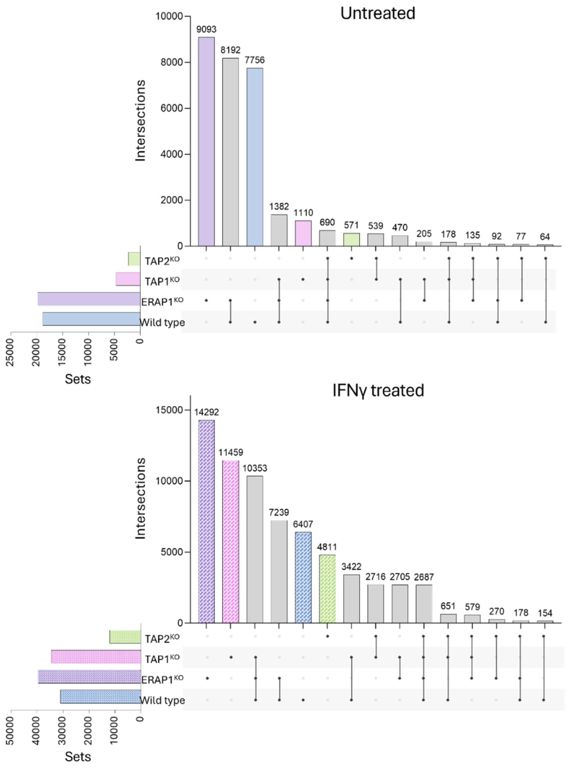
Figure 5.
ERAP1KO and TAP2KO cells preferentially present N- and C-terminally extended peptides. Peptide extensions were assessed relative to an eight-amino-acid core. (A, B) Frequencies of N- and C-terminal extensions in untreated and IFNγ-treated cells. (C, D) Normalised distributions of terminal extensions from −6 to −1 residues at the N terminus and +1 to +6 residues at the C terminus.
Figure 5.
ERAP1KO and TAP2KO cells preferentially present N- and C-terminally extended peptides. Peptide extensions were assessed relative to an eight-amino-acid core. (A, B) Frequencies of N- and C-terminal extensions in untreated and IFNγ-treated cells. (C, D) Normalised distributions of terminal extensions from −6 to −1 residues at the N terminus and +1 to +6 residues at the C terminus.
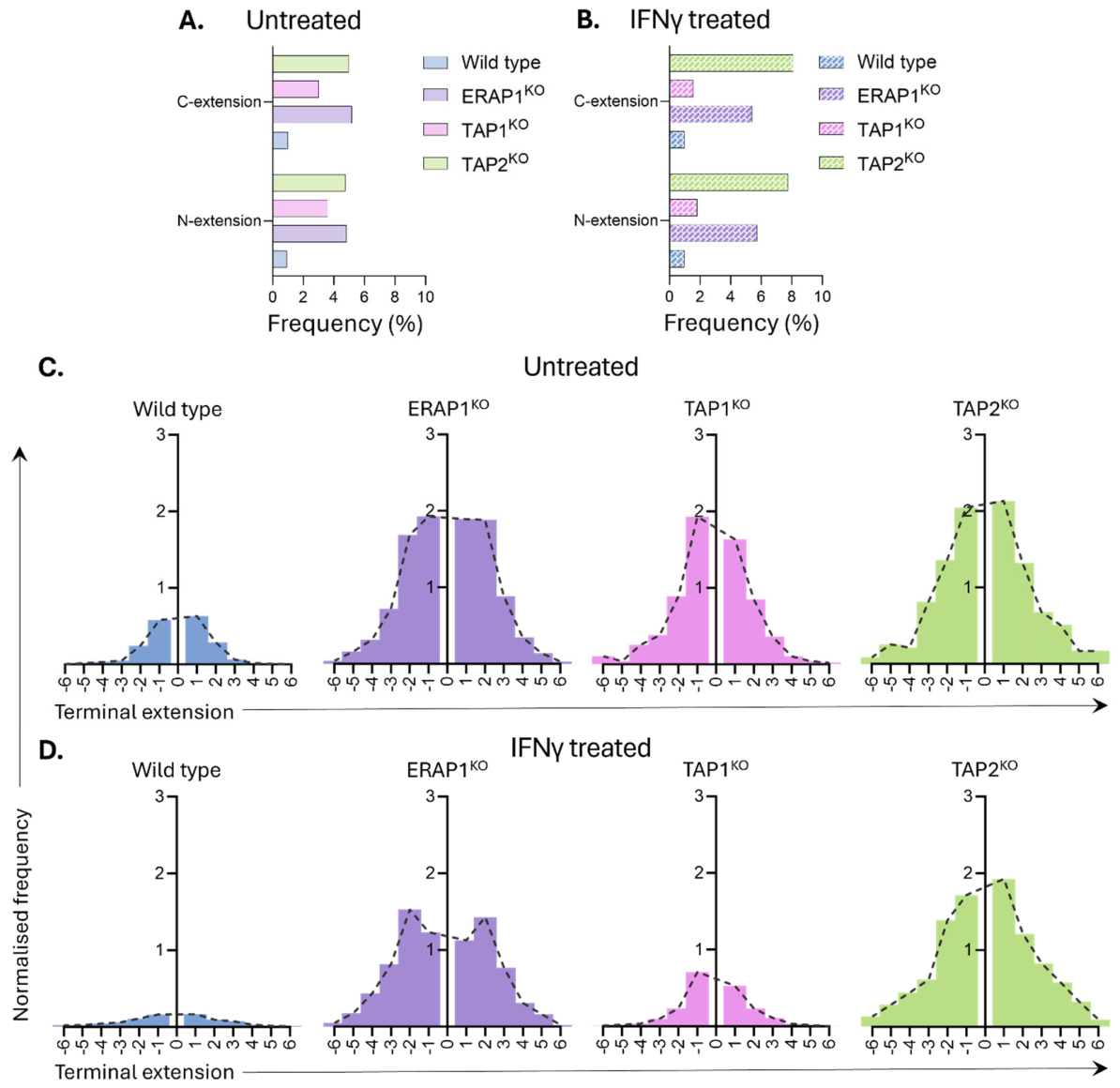
Figure 6.
TAP1KO and TAP2KO cells show altered source-protein positional bias. The position of each presented peptide within its parental protein was normalised and distributed across 20 equal bins (vigintiles). Histograms show relative peptide frequency across the source-protein sequence in (A) untreated and (B) IFNγ-treated HAP1WT, ERAP1KO, TAP1KO, and TAP2KO cells.
Figure 6.
TAP1KO and TAP2KO cells show altered source-protein positional bias. The position of each presented peptide within its parental protein was normalised and distributed across 20 equal bins (vigintiles). Histograms show relative peptide frequency across the source-protein sequence in (A) untreated and (B) IFNγ-treated HAP1WT, ERAP1KO, TAP1KO, and TAP2KO cells.
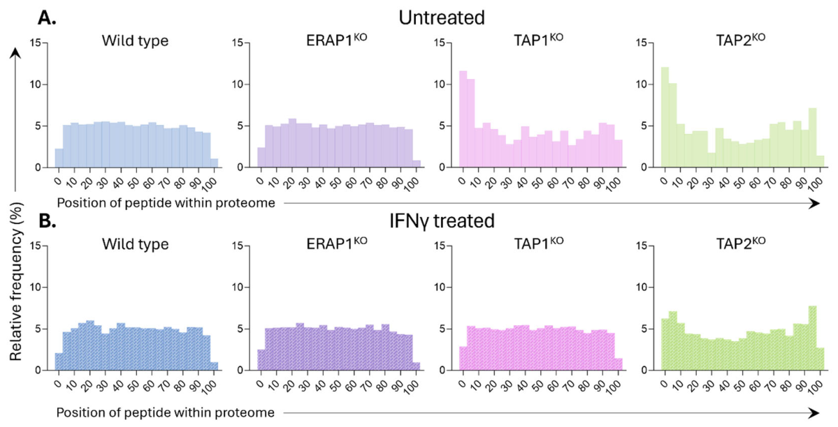
Figure 7.
Peptide-loading complex components remain associated with HC-10-reactive HLA class I heavy chains in cells deficient in antigen-processing machinery.
Figure 7.
Peptide-loading complex components remain associated with HC-10-reactive HLA class I heavy chains in cells deficient in antigen-processing machinery.
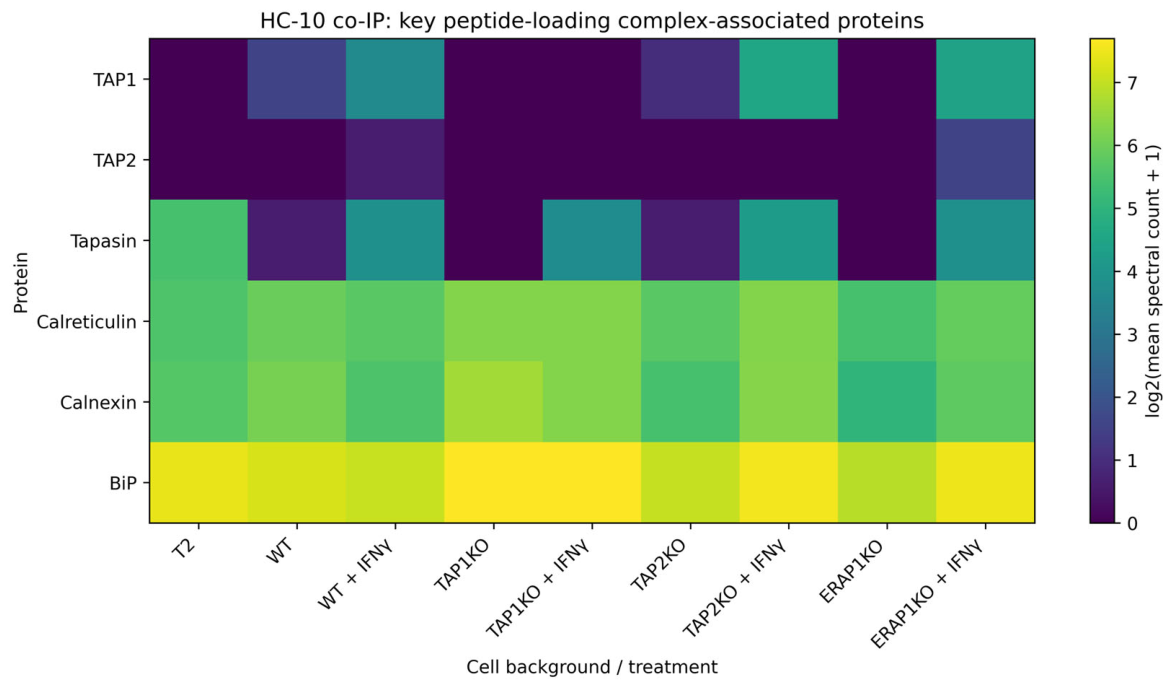
Figure 8.
IFNγ increases presentation of leukemia-associated antigen-derived peptides in APM-deficient cells. Numbers of peptides derived from the indicated leukemia-associated antigens are shown for (A) untreated and (B) IFNγ-treated HAP1WT, ERAP1KO, TAP1KO, and TAP2KO cells.
Figure 8.
IFNγ increases presentation of leukemia-associated antigen-derived peptides in APM-deficient cells. Numbers of peptides derived from the indicated leukemia-associated antigens are shown for (A) untreated and (B) IFNγ-treated HAP1WT, ERAP1KO, TAP1KO, and TAP2KO cells.
Disclaimer/Publisher’s Note: The statements, opinions and data contained in all publications are solely those of the individual author(s) and contributor(s) and not of MDPI and/or the editor(s). MDPI and/or the editor(s) disclaim responsibility for any injury to people or property resulting from any ideas, methods, instructions or products referred to in the content. |
© 2026 by the authors. Licensee MDPI, Basel, Switzerland. This article is an open access article distributed under the terms and conditions of the Creative Commons Attribution (CC BY) license.
Copyright: This open access article is published under a Creative Commons CC BY 4.0 license, which permit the free download, distribution, and reuse, provided that the author and preprint are cited in any reuse.