

Submitted:
02 September 2026
Posted:
02 September 2026
You are already at the latest version
Abstract
Synaptobrevin (Syb/VAMP) is the vesicular SNARE that drives synaptic vesicle fusion. Before fusion, Syb is located in the vesicle membrane. After fusion, Syb is translocated to the presynaptic plasma membrane and exposed to a different lipid environment. How its membrane-proximal juxtamembrane and transmembrane domains (JMD–TMDs) organ-ize these different surrounding membrane environments remains incompletely under-stood. Here, we used MARTINI coarse-grained molecular dynamics simulations to in-vestigate Syb1/2 membrane organization and collective behavior. Syb1 and Syb2 induced similar and robust local lipid disorder, whereas the spatial organization of lipid disorder and membrane mismatch depended more strongly on membrane lipid composition. Higher order Syb clustering was promoted by the presence of PIP₂ in the membrane, which is decreased in its absence and insensitive to PS depletion. Consistently, Syb cluster formation was associated with pronounced PIP₂ redistribution and enrichment, while PS showed substantially weaker enrichment and decreased association with Syb in the presence of PIP₂. Clustering spatially overlapped otherwise localized lipid-disordered regions without proportionally amplifying their magnitude. Together, these results dis-tinguish robust local membrane disorder from composition-dependent geometric re-modeling and identify PIP₂ as a prominent correlate of higher-order Syb organization that may contribute to post-fusion Syb reorganization before endocytic retrieval.
Keywords:
synaptobrevin
; PIP₂
; lipid disorder
; hydrophobic mismatch
; protein clustering
; coarse-grained molecular dynamics
1. Introduction
Chemical neurotransmission depends on the regulated release of neurotransmitters from synaptic vesicles (SVs) at presynaptic terminals [1]. Fusion of the SV membrane with the presynaptic plasma membrane is driven by assembly of the neuronal SNARE complex, consisting of the vesicular SNARE synaptobrevin (Syb/VAMP) and the plasma-membrane SNAREs Syntaxin-1 (Stx1) and SNAP-25 [2,3,4]. Dysfunction of this tightly regulated membrane-fusion machinery can impair neurotransmission and has been implicated in a broad spectrum of neurodevelopmental, neuromuscular, and neurodegenerative disorders [5].
Among the neuronal SNARE components, Syb1/VAMP1 and Syb2/VAMP2 are two closely related vesicular SNARE isoforms involved in Ca²⁺-dependent synaptic vesicle exocytosis [6,7]. Syb1/VAMP1 and Syb2/VAMP2 exhibit distinct but partially overlapping neuronal expression patterns. Syb2 is broadly expressed and represents a major vesicular SNARE at central synapses in the brain, whereas Syb1 is particularly enriched in the spinal cord, motor neurons, and neuromuscular terminals [8]. Pathogenic variants in both Syb1 and Syb2 demonstrate the physiological importance of these vesicular SNAREs. Recessive loss-of-function variants in VAMP1 have been associated with severe congenital myasthenic syndromes characterized by impaired neuromuscular transmission [9,10], whereas de novo variants in VAMP2 cause neurodevelopmental disorders associated with hypotonia, intellectual disability, autistic features, movement abnormalities, and impaired synaptic vesicle fusion [11]. Despite their high sequence similarity and shared role as vesicular SNAREs, the molecular mechanisms by which their membrane-proximal regions organize the SV membrane remain incompletely understood.
Increasing evidence indicates that the juxtamembrane domain (JMD) and transmembrane domain (TMD) of SNARE proteins participate actively in fusion and membrane organization rather than serving solely as passive membrane anchors [12,13,14,15,16,17,18]. In our previous study, Stx1 JMD–TMD fragments were found to induce robust local lipid disorder in plasma-membrane models, while the coupling between lipid disorder and hydrophobic mismatch depended on the local membrane environment [15]. Both, solitary Stx1 molecules and Stx1/SNAP-25 t-SNARE complexes also underwent concentration-dependent clustering that was strongly associated with PIP₂ redistribution [15,19], indicating a close relationship between collective Stx organization and its surrounding anionic lipid environment.
The membrane environment encountered by Syb during the synaptic vesicle cycle is dynamic. Before exocytosis, Syb resides in the SV membrane, which is compositionally distinct from the PIP₂-enriched presynaptic plasma membrane [20]. Following membrane fusion, vesicular membrane proteins, including Syb, become incorporated into the plasma membrane before being retrieved through endocytosis and recycled into newly formed synaptic vesicles [21,22,23]. Conversely, phosphoinositides undergo tightly regulated redistribution and interconversion during exocytosis and endocytosis: PIP₂ is enriched at the plasma membrane [24] , whereas changes in phosphoinositide identity and phosphorylation state accompany membrane internalization and vesicle recycling [25,26,27]. Thus, Syb can encounter distinct anionic lipid environments during the synaptic vesicle cycle, providing a physiological rationale for examining how its membrane organization responds to changes in PS and PIP₂ composition.
Compared with the polybasic Stx1 JMD, a prominent feature of the membrane-proximal Syb1/2 sequence is the pair of conserved interfacial tryptophans. Previous experimental studies demonstrated that substitution of these tryptophans alters vesicle priming and spontaneous neurotransmitter release, implicating the Syb membrane interface in regulation of release competence [13]. However, how these conserved residues organize the surrounding SV lipids, whether Syb-induced membrane remodeling depends on membrane lipid composition, and how these local membrane effects relate to collective Syb clustering remain unknown.
Here, we used coarse-grained molecular dynamics (CGMD) simulations to characterize the membrane organization and collective behavior of Syb1/2 JMD–TMD fragments across plasma membrane and SV-like membrane environments. We examined local lipid disorder, hydrophobic mismatch, anionic lipid redistribution, and collective Syb clustering, and investigated their sensitivity to conserved interfacial tryptophans mutations, and PIP₂ and PS content in the membrane lipid composition, under preserved initial local protein configurations. Particular attention was given to how PIP₂ modulates higher-order Syb organization and anionic lipid redistribution in the plasma membrane environment as encountered by Syb following vesicle fusion. By relating these responses to those previously identified for Stx1, we sought to distinguish robust local membrane-remodeling properties of Syb from lipid-dependent regulation of its collective organization.
2. Materials and Methods
2.1. Syb-Based Models
MARTINI CGMD simulations were performed using 1 or 10 copies of the rat Syb1 (residues 87–118) or Syb2 (residues 85–116) JMD–TMD regions embedded in five planar membrane models with the lipid composition described in the next section.
The atomistic reference structure of Syb2 was obtained from our previous study [28], whereas the initial Syb1 structure was generated from an AlphaFold-predicted model.
To evaluate the contribution of the conserved interfacial tryptophans (W89/W90 in Syb2 and W91/W92 in Syb1), single-copy simulations were performed using double tryptophan-to-alanine (WWAA) and double tryptophan-to-lysine (WWKK) mutants. These mutations were designed to distinguish the contributions of aromatic interfacial anchoring and positively charged interfacial residues to local membrane remodeling. All other simulation parameters were identical to those used for the corresponding wild-type systems.
2.2. CG Model Conversion and Membrane Construction
All atomistic Syb1/2 models were converted into MARTINI coarse-grained representations using the MARTINI 2.1 force field and a modified version of martinize_CYP.py (v2.5), as described previously [15]. Corresponding MARTINI topology (.itp) files were generated for all constructs. For systems containing multiple Syb molecules, individual proteins were duplicated and rotationally arranged around the membrane normal at angular intervals of , where N denotes the number of Syb molecules in the system.
Five membrane lipid compositions were examined in this study.
First, to enable direct comparison with the membrane organization previously observed for Syntaxin-1 [15], Syb1/2 systems were constructed in PIP₂-containing plasma membranes using the same plasma membrane composition employed in the previous Syntaxin-1 simulations. These systems had dimensions of approximately 20 × 20 × 15 nm³ and contained ~1,500 lipids. Detailed lipid compositions are provided in Table S1.
Second, to determine the contribution of PIP₂ to Syb-induced membrane remodeling and collective organization, corresponding PIP₂-depleted plasma membrane systems containing one or ten copies of Syb1/2 were generated and self-assembled using membrane compositions described previously [15,28,29]. These systems also had dimensions of approximately 20 × 20 × 15 nm³ and contained ~1,500 lipids. Detailed lipid compositions are provided in Table S2.
Third, simplified SV-like membrane systems were constructed following previously described self-assembly and production protocols [17,29]. The resulting simulation boxes had dimensions of approximately 20 × 20 × 15 nm³ and contained ~1800 lipids. The simplified SV membrane lipid composition is summarized in Table S3.
Fourth, to examine the contribution of PS in the PIP₂-depleted plasma membrane background, all PS molecules were replaced with phosphatidylcholine (PC), thereby removing the monovalent anionic lipid component while preserving the corresponding acyl-chain composition. The resulting membrane was depleted of both PIP₂ and PS, and its detailed composition is provided in Table S4.
Fifth, PS-depleted SV-like membrane systems were generated using the same PS-to-PC replacement procedure while otherwise preserving the simplified SV membrane composition. These systems were constructed to examine the contribution of PS to Syb-induced membrane remodeling and collective clustering. Detailed lipid compositions are provided in Table S5.
2.3. MD Simulation Parameters
All simulations were performed following the simulation protocol established in our previous study [15], using GROMACS version 2024 [30] on the Bridges-2 high-performance computing system [31].
During the 1 ns equilibration stage, the systems were coupled to a Berendsen thermostat (310 K, τ = 1.0 ps) and Berendsen barostat (1 bar, τ = 1.0 ps) using a timestep of 10 fs. Production simulations were carried out using a velocity-rescale thermostat (310 K, τ = 1.0 ps) and a semi-isotropic Parrinello–Rahman barostat (1 bar, τ = 12.0 ps) with a timestep of 20 fs.
Neighbor lists were updated every 20 integration steps using a Verlet buffer tolerance of 0.005. For each initial condition, n = 10 independent 2-μs simulations were performed using different initial velocity distributions. Trajectory coordinates were saved every 1 ns for subsequent analysis.
Additional simulation parameters, input files, and analysis scripts are available through the Data Availability Statement.
2.4. Analysis
Trajectory preprocessing, lipid selection, lipid disorder analysis, local membrane mismatch analysis, protein clustering analysis, and statistical analyses were performed using the membrane-analysis pipeline established in our previous study [15], unless otherwise specified below.
2.4.1. Trajectory Preprocessing
Production trajectories were reconstructed across periodic boundaries and stripped of water and ions using GROMACS before analysis.
2.4.2. Lipid Disorder and Membrane Mismatch
Lipids within a cutoff distance = 0.8 nm from any Syb molecule were identified using the same distance-based procedure described previously [15].
Lipid disorder parameters (S2), mean lipid order, and rim-wise lipid order were calculated using the previously described protocol [15]. At each frame, mean lipid order was defined as the mean across all membrane lipids in the corresponding system, providing an instantaneous global membrane-average reference rather than a reference derived from the outermost radial region. Radial lipid-order perturbations were quantified relative to this global reference.
For single-copy systems, radial analyses were performed up to 8 nm to characterize recovery toward the global membrane-average reference while maintaining adequate lipid sampling. For ten-copy Syb1/2 JMD–TMD systems, each lipid was assigned according to its minimum distance from the nearest Syb JMD–TMD region. Thus, each lipid contributed only once to the radial profile, even when it was spatially proximal to multiple Syb molecules, preventing double counting in regions where Syb-associated membrane environments overlapped. Radial analyses for ten-copy systems were restricted to 5 nm because more distant regions were increasingly affected by heterogeneous and limited sampling.
Local membrane mismatch was quantified for single-copy systems using the annular interleaflet-distance method described previously [15]. The reference interleaflet distance was defined as the average interleaflet distance within the outermost annular region (7.2–8 nm). A positive indicates a decrease in the local interleaflet distance relative to the reference value and therefore local membrane thinning, whereas a negative indicates an increase in interleaflet distance and therefore local membrane thickening.
2.4.3. PIP₂ and PS Redistribution and Protein Clustering
PIP₂ redistribution was quantified by identifying unique PIP₂ molecules located within 0.8 nm of any Syb JMD–TMD region. The fraction of intracellular-leaflet PIP₂ associated with Syb , was calculated as the number of unique Syb-associated PIP₂ molecules divided by the total PIP₂ population in the intracellular/cytosolic (IC) leaflet. Local PIP₂ enrichment , was calculated as the fraction of PIP₂ within the Syb-associated lipid population relative to the corresponding bulk PIP₂ fraction in the IC leaflet. For all PS-containing membrane systems, the fraction of Syb-associated PS , and local PS enrichment , were quantified using the same procedure.
The mean number of associated PIP₂ or PS molecules per Syb ( or ) was calculated as the number of unique PIP₂ or PS molecules located within 0.8 nm of any Syb JMD–TMD region divided by the number of Syb molecules in the system. The overbar denotes normalization of the number of unique Syb-associated lipid molecules by the number of Syb molecules. A PIP₂ or PS molecule simultaneously associated with multiple Syb molecules was counted only once.
Protein clustering was analyzed using the pairwise-distance algorithm established previously [15]. Two Syb molecules were considered to be in contact when the minimum pairwise distance between their backbone beads was ≤1.0 nm, and mutually connected Syb molecules were assigned to the same cluster. Collective clustering was characterized by the maximum cluster size and the mean cluster size . Monomers were treated as clusters of size one when calculating the maximum and mean cluster size. Time-dependent ensemble averages of these quantities were calculated across independent simulation replicates, whereas final-state statistics were calculated from values averaged over the final 100 ns of each trajectory.
2.4.4. Statistics
All quantities are reported as mean ± SEM (n = 10 independent simulations). Averaging was performed over the final 100 ns of each trajectory unless otherwise specified. Statistical comparisons between two groups were performed using the Mann–Whitney U test implemented in SciPy, and the corresponding p values are reported in the figures. Multiple-group comparisons were performed using the Kruskal–Wallis test.
3. Results
3.1. Syb2 and Syb1 Exhibit Similar Proximal Lipid Disorder but Membrane-Composition-Dependent Radial Remodeling
We first quantified lipid orientational order (), radial membrane properties, and local membrane mismatch () using the membrane-analysis framework established previously [15]. The definitions of lipid order and local membrane mismatch correspond to Equations (1) and (5), respectively, in Ref. [15]. Briefly, lipids were grouped according to their minimum distance from the nearest Syb JMD–TMD region, and lipid was calculated using the previously defined coarse-grained bead vectors connecting the second and third hydrophobic beads for phospholipids and the R5 and ROH beads for cholesterol.
Local lipid disorder was quantified for lipids located within 0.8 nm of the Syb1/2 JMD–TMD region. Despite ten residue differences between the Syb2 and Syb1 JMD–TMD sequences (Figure 1a), their local lipid , mean lipid , and local membrane mismatch trajectories showed broadly similar behavior and approached relatively stable levels within approximately 250 ns in the PIP₂-containing plasma membrane (Figure 1b). Consistently, no significant differences between Syb2 and Syb1 were detected in the corresponding values averaged over the final 100 ns of the simulations (Figure 1c; Table S6).
Radial analysis further showed that Syb2 and Syb1 produced nearly identical proximal lipid-disordering responses, but differed in their longer-range recovery toward the global membrane-average reference (Figure 1d; Table S7 and Table S8). Syb2 exhibited a more extended negative lipid-order perturbation, requiring approximately 5 nm to recover to within 5% of the mean reference, whereas Syb1 lipid order recovered more rapidly and approached the mean lipid reference within a shorter distance. Thus, the two isoforms generated similar proximal disorder magnitudes but differed modestly in the spatial propagation of this perturbation. In contrast, radial interleaflet-distance profiles remained broadly comparable between Syb2 and Syb1 and showed no consistent isoform-dependent membrane-mismatch response (Figure 1e; Table S7 and Table S8).
To determine whether these membrane responses were preserved in PIP₂-free environments, we performed the same analyses in PIP₂-depleted plasma membrane and SV-like membrane models (Figure S1). Absolute local and mean lipid differed substantially between the two membrane compositions, whereas Syb2 and Syb1 remained closely similar within each background (Figure S1a, b). Despite these differences in absolute membrane order, both isoforms retained pronounced proximal lipid-disordering profiles relative to their corresponding membrane-average references (Figure S1c). The substantially different absolute lipid order between the plasma-membrane and SV-like membrane likely reflects their distinct membrane compositions, including differences in lipid acyl-chain chemistry (Table S1–S3). The radial membrane-mismatch response was more membrane-composition dependent. Neither isoform exhibited a pronounced mismatch response in the PIP₂-depleted plasma membrane, whereas the SV-like membrane showed a stronger proximal geometric response (Figure S1b,d). In the SV-like membrane, Syb2 exhibited a significantly greater mean local mismatch than Syb1 (0.059 ± 0.026 vs. −0.020 ± 0.017 nm; Mann–Whitney U test, ), although neither isoform individually differed significantly from its corresponding bulk reference (Wilcoxon signed-rank test, and , respectively). These results indicate that proximal Syb-induced lipid disorder is preserved across substantially different membrane backgrounds, whereas its longer-range spatial organization and membrane mismatch are more sensitive to membrane composition and isoform context.
Because Syb2 and Syb1 exhibited closely similar local membrane-mismatch response under matched conditions, subsequent main-text analyses focused on Syb2 as the representative isoform. Corresponding Syb1 analyses across additional membrane environments and perturbations are provided in the Supporting Information and Supporting Tables.
3.2. Anionic Lipids Weakly Modulate the Spatial Organization of Syb-Induced Membrane Remodeling
The JMDs of Syb2 and Syb1 contain multiple basic residues, whereas PIP₂ and PS represent the major anionic lipid components of the intracellular/cytosolic leaflet in the plasma membrane models and can potentially interact with the polybasic Syb JMDs (Table S1). PIP₂-absent membrane backgrounds, including the PIP₂-depleted plasma membrane and SV-like membrane, retain PS as their major anionic lipid component (Table S2 and Table S3). We therefore examined whether changes in the surrounding anionic lipid environment modulate Syb-induced membrane organization.
To examine the contributions of PIP₂ and PS in the plasma membrane, PIP₂ was first removed from the membrane composition, followed by replacement of PS with PC to generate a membrane depleted of both PIP₂ and PS while preserving the corresponding acyl-chain composition. Specifically, POP2, PAP2, and PUP2 were replaced by POPS, PAPS, and PUPS, respectively, for PIP₂ depletion. For PS depletion, POPS, PAPS, and PUPS were replaced by POPC, PAPC, and PUPC, respectively. Neither PIP₂ nor subsequent PS depletion significantly altered local lipid order , or local membrane mismatch , for Syb2 (all ; Figure 2a). Radial analysis likewise showed that the proximal lipid-disordering response was preserved across all three plasma-membrane compositions, although modest differences emerged in the longer-range recovery toward the global membrane-average reference under PIP₂ depletion only (Figure 2b). The corresponding membrane-mismatch profiles showed similar modest spatial variation without significant changes in mean local mismatch. Syb1 exhibited comparable responses to PIP₂ and PS depletion (Figure S2a,b). Thus, neither PIP₂ nor PS was required for the generation of the proximal Syb-induced lipid-disordering response in the plasma-membrane background.
In the SV-like membrane, PS depletion likewise produced no significant changes in local lipid order or local membrane mismatch for either Syb2 or Syb1, and the corresponding radial profiles remained broadly preserved (Figure S2c, d). Moreover, the substantial difference in absolute lipid order between the PIP₂-depleted plasma-membrane and SV-like membrane compositions persisted following PS depletion (Figure S2c), indicating that this membrane-background-dependent difference cannot be attributed primarily to PS.
Together, these results indicate that PIP₂ and PS have limited effects on the magnitude of proximal Syb-induced lipid disorder and mean local membrane mismatch, while producing modest changes in their longer-range spatial organization. The proximal lipid-disordering response therefore remains comparatively robust to changes in anionic lipid headgroup composition.
3.3. Conserved Interfacial Tryptophans Regulate Syb-Induced Local Membrane Remodeling
Sequence analysis of the Syb2 and Syb1 JMDs revealed two highly conserved interfacial tryptophan residues (Figure 1a). According to the Wimley–White hydrophobicity scale, tryptophan exhibits a strong preference for the membrane–water interface [32], suggesting that these conserved aromatic residues may contribute to Syb-induced membrane remodeling. We therefore hypothesized that the interfacial tryptophans regulate local lipid packing and the accompanying membrane geometric response.
To test this hypothesis, tryptophan-to-alanine (WWAA) and tryptophan-to-lysine (WWKK) double-substitution mutants were generated for both Syb isoforms (Figure 3a). WWAA reduces the contribution of aromatic interfacial anchoring, whereas WWKK replaces the aromatic residues with positively charged side chains, shifting the local interfacial chemistry toward electrostatic interactions.
In the PIP₂-containing plasma membrane, WW substitutions differentially modulated proximal lipid packing and membrane mismatch. For Syb2, WWAA did not significantly alter local lipid order relative to WT, whereas WWKK produced a modest but significant increase in , corresponding to reduced proximal lipid disorder (Figure 3b; Table S6). In contrast, local membrane mismatch was significantly shifted by both WWAA and WWKK relative to WT, whereas the two mutants exhibited comparable mean mismatch responses. Radial analysis further showed that the extended Syb2 lipid-disordering profile was retained following both substitutions, whereas the corresponding membrane-mismatch profiles were more strongly reorganized, particularly within the proximal region (Figure 3c). Syb1 exhibited a similar separation between lipid disorder and membrane mismatch: neither WWAA nor WWKK significantly altered local , whereas both substitutions significantly shifted local membrane mismatch relative to WT (Figure S3a,b). Thus, perturbation of the conserved interfacial tryptophans affected the local geometric response more consistently than the magnitude or spatial persistence of Syb-induced lipid disorder.
The effects of WW substitution nevertheless depended on membrane lipid composition and Syb isoform. In the PIP₂-depleted plasma membrane and SV-like membrane, all WWAA and WWKK constructs retained the characteristic proximal lipid-disordering response, while mutation-dependent changes in its magnitude and in the accompanying membrane-mismatch profiles varied among isoforms and membrane environments (Figure S3c,d). Despite this variability, none of the substitutions abolished the proximal disorder field. These observations indicate that the conserved interfacial tryptophans are not required for Syb-induced lipid disorder but modulate how this robust packing perturbation is accompanied by local changes in membrane geometry.
Together, these results distinguish the generation of Syb-induced lipid disorder from its geometric manifestation. The proximal disorder response remains comparatively robust to WW substitution, whereas the magnitude and spatial organization of the accompanying membrane-mismatch response are more sensitive to interfacial residue chemistry and membrane composition. Thus, the conserved interfacial tryptophans modulate the geometric manifestation of Syb-induced membrane remodeling in a context-dependent manner.
3.4. PIP₂ Promotes Higher-Order Syb Assembly Whereas PS Has Limited Effects
We next examined the collective clustering dynamics of Syb1/2 in PIP₂-containing and PIP₂-depleted plasma membrane environments and in SV-like membranes (Figure 4 and Figure S4). Representative configurations showed progressive association of initially separated Syb JMD–TMD fragments into higher-order assemblies during the 2-μs simulations (Figure 4a; Videos S1–S3). Maximum cluster size, , and mean cluster size, , increased rapidly during the first ~250 ns, followed by a slower phase of higher-order assembly and cluster rearrangement (Figure 4b,Figure S4a,c,e). Syb2 and Syb1 exhibited similar clustering dynamics and final clustering states across the membrane environments examined, with no consistent isoform-dependent differences in either metric (Figure S4a; Table S9). We therefore focused subsequent main-text analyses of collective organization on Syb2 as the representative isoform.
We next examined whether the major anionic lipids PIP₂ and PS differentially influenced collective Syb organization. In all three plasma-membrane compositions, rapid initial oligomerization was preserved, whereas the subsequent growth of higher-order assemblies depended more strongly on PIP₂ availability (Figure 4b). Maximum and mean cluster sizes continued to increase throughout the simulations in the PIP₂-containing membrane but approached lower values following PIP₂ depletion. Final-state analysis showed a significant reduction in mean cluster size upon PIP₂ depletion (; Figure 4c, Table S9), while the corresponding reduction in maximum cluster size showed a similar tendency but did not reach statistical significance (; Figure 4c, Table S9).
Syb1 exhibited a comparable response as Syb2. PIP₂ depletion reduced higher-order cluster growth, whereas subsequent PS depletion produced little additional effect (Figure S4c; Table S9). The limited effect of PS depletion was also observed in the SV-like membrane, where removal of PS produced no consistent change in maximum or mean cluster size for either Syb isoform (Figure S4e; Table S9). Thus, PIP₂ availability preferentially promoted the continued growth of higher-order Syb assemblies, whereas PS exerted comparatively limited effects on collective organization.
3.5. PIP₂-Dependent Higher-Order Syb2 Assembly Is Preserved Following WW Substitution
We previously showed that WWAA and WWKK substitutions modulated Syb-induced local lipid disorder and membrane mismatch (Figure 3 and Figure S3). We next examined whether perturbation of the conserved interfacial tryptophans affected collective Syb organization and, importantly, whether the PIP₂-dependent enhancement of higher-order assembly identified for WT Syb was retained following WW substitution. Because WT Syb2 and Syb1 exhibited closely similar proximal membrane-remodeling and collective clustering behaviors under matched conditions (Figure 1, Figure 2, Figure 3 and Figure 4 and Figure S1–S4), Syb2 was used as the representative isoform for the ten-copy WWAA and WWKK simulations.
Both WWAA and WWKK retained the characteristic PIP₂-dependent divergence in clustering dynamics. Maximum and mean cluster sizes continued to increase in the PIP₂-containing plasma membrane but approached substantially lower values following PIP₂ depletion (Figure 5a). Final-state comparisons confirmed significant reductions in both clustering metrics upon PIP₂ depletion for WWAA and WWKK, whereas no significant differences were detected between the two mutants within either membrane background (Figure 5b; Table S9). Thus, substitution of the conserved interfacial tryptophans did not abolish the PIP₂-dependent promotion of higher-order Syb2 assembly.
In contrast, WW substitution itself produced no consistent clustering phenotype across the membrane backgrounds examined (Figure S5). Neither WWAA nor WWKK significantly altered final maximum or mean cluster size in the PIP₂-containing plasma membrane, and the effects observed in PIP₂-depleted plasma membrane and SV-like membrane backgrounds were modest and context dependent, without a consistent response across clustering metrics or membrane compositions (Figure S5a; Table S9). Together, these results indicate that the conserved interfacial tryptophans exert comparatively limited effects on collective Syb2 clustering, whereas the PIP₂-dependent enhancement of higher-order assembly is preserved following both WWAA and WWKK substitution.
3.6. Local Syb-Induced Lipid Disorder Is Preserved During Collective Clustering
We next examined how collective Syb clustering reorganizes the spatial distribution of Syb-induced local lipid disorder. Representative spatial maps showed that, when individual Syb JMD–TMD fragments were initially separated, regions of elevated lipid disorder were predominantly localized around individual proteins. As Syb molecules assembled into higher-order clusters, these initially separated disordered regions increasingly overlapped, producing more continuous regions of altered lipid packing around collective Syb assemblies in PIP₂-containing plasma membrane, PIP₂-depleted plasma membrane, and SV-like membrane backgrounds (Figure 6a and Figure S6a, b).
Radial analyses further showed that the characteristic lipid-disorder signature within 0.8 nm of the nearest Syb JMD-TMDs identified in single-copy systems was retained during collective clustering. Ten-copy Syb systems exhibited pronounced reductions in lipid within 0.8 nm of the nearest JMD–TMD across Syb isoforms, membrane lipid compositions, PS perturbation, and the Syb2 WWAA and WWKK substitutions (Figure 6b and Figure S6c). Although the radial profiles were reorganized beyond the immediate protein environment, collective assembly did not produce a simple proportional amplification of the single-copy disorder response.
Notably, PIP₂ promoted the continued growth of higher-order Syb assemblies (Figure 4 and Figure 5), yet this enhanced collective organization was not accompanied by a proportional increase in proximal lipid disorder (Figure 6b and Figure S6c). Thus, collective Syb assembly primarily reorganizes the spatial distribution and overlap of locally induced disorder fields while preserving the proximal membrane-disordering response associated with individual Syb JMD–TMD regions.
Together, these results indicate that collective Syb assembly preserves the magnitude of the proximal membrane-disordering response while reorganizing its longer-range spatial distribution. Rather than behaving as independent additive perturbations, clustered Syb molecules generate overlapping and increasingly shared membrane environments.
3.7. Collective Syb Assembly Is Associated with Distinct PIP₂ and PS Redistribution
Finally, we examined how the major anionic lipids PIP₂ and PS redistribute during collective Syb organization and how these responses depend on protein copy number, membrane lipid composition, and the WWAA and WWKK substitutions (Figure 7 and ). In the PIP₂-containing plasma membrane, representative spatial maps showed progressive localization of PIP₂ around clustered Syb JMD–TMD fragments, whereas PS remained more broadly distributed and did not exhibit comparable accumulation within Syb assemblies (Figure 7a, b).
The distinction between PIP₂ and PS was also evident from their temporal redistribution (Figure 7c). In single-copy systems, the fraction of intracellular-leaflet PIP₂ associated with Syb, , approached a relatively stable level within approximately 250 ns. In ten-copy systems, however, PIP₂ association continued to increase throughout the simulations, reaching approximately 70–80% of the intracellular-leaflet PIP₂ pool near the end of the trajectories. In the same PIP₂-containing membrane, the fraction of PS associated with Syb progressively decreased during the ten-copy simulations. These contrasting temporal responses were observed for both WT Syb isoforms and were retained following WWAA and WWKK substitution (Figure 7c and Figure S7).
Final-state analysis further distinguished the collective organization of PIP₂ and PS (Figure 7d,e; Table S10,S11). Increasing Syb copy number strongly increased the fraction of the total PIP₂ pool associated with Syb, while PIP₂ remained locally enriched by approximately 7–8-fold relative to its intracellular-leaflet abundance. In contrast, although the fraction of the global PS pool associated with Syb was also greater in ten-copy than in single-copy systems, PS showed substantially weaker local enrichment. Thus, increasing protein copy number increased global association with both anionic lipid species, but only PIP₂ remained strongly enriched within the collective Syb-associated membrane environment.
Normalization by Syb copy number further revealed the non-additive nature of this collective lipid organization. The mean number of unique associated PIP₂ or PS molecules per Syb, , was generally lower in ten-copy than in corresponding single-copy systems (Figure 7d,e; Table S10 and Table S11). Because lipid molecules associated with multiple Syb molecules were counted only once, this reduction is consistent with increasing overlap and sharing of the Syb-associated lipid environment rather than independent additive lipid shells surrounding individual proteins. Importantly, PIP₂ association nevertheless increased over time within the ten-copy systems (Figure 7c), indicating that the lower final per-Syb value does not reflect loss of PIP₂ association during collective assembly. The strong copy-number-dependent PIP₂ redistribution and comparatively weak PS enrichment were also retained following WWAA and WWKK substitution (Figure S7a-c).
In PIP₂-free membrane environments, PS exhibited a distinct collective redistribution pattern (Figure S8 and Figure S9). Increasing Syb copy number substantially increased the fraction of the global PS pool associated with Syb in both PIP₂-depleted plasma membrane and SV-like membrane backgrounds, whereas local PS enrichment remained comparatively modest (Figure S8 and Figure S9). The number of unique associated PS molecules normalized per Syb was generally lower in ten-copy systems (Figure S9). These patterns were broadly preserved across Syb isoforms and WW substitutions, indicating that collective assembly increases association with the global PS pool without generating the pronounced local enrichment observed for PIP₂.
Together, these results reveal distinct modes of anionic-lipid organization during collective Syb assembly. Increasing Syb copy number expands association with the global pools of both PIP₂ and PS, but PIP₂ undergoes substantially stronger local enrichment and becomes preferentially represented within the collective Syb-associated membrane environment when both anionic lipid species are present. Combined with the enhanced higher-order cluster growth observed in PIP₂-containing membranes (Figure 4 and Figure 5), these findings reveal a strong association between preferential PIP₂ redistribution and higher-order Syb organization.
4. Discussion
4.1. Syb Clustering Is Robust but Selectively Modulated by PIP₂ and Interfacial Tryptophans
Our simulations indicate that collective Syb organization is comparatively robust to several perturbations examined here. Syb1 and Syb2 exhibited closely similar clustering dynamics and final higher-order assembly states across the membrane environments examined (Figure S4). Depletion of PS likewise produced no consistent clustering phenotype across Syb isoforms or PIP₂-free membrane backgrounds (Figure 4 and Figure S4). PIP₂ represented the most prominent compositional effect. In PIP₂-containing plasma membranes, maximum and mean cluster sizes continued to increase after the initial phase of oligomerization, whereas PIP₂ depletion preferentially suppressed this continued higher-order growth (Figure 4). Thus, PIP₂ does not appear to be required for the initial association of Syb molecules but is associated with the subsequent growth and/or stabilization of larger Syb assemblies.
The distinct clustering responses to PS and PIP₂ parallel their different collective redistribution behaviors. Increasing Syb copy number increased the fraction of the global pools of both anionic lipid species associated with Syb-containing regions. However, only PIP₂ remained strongly enriched within the collective Syb-associated membrane environment, whereas PS enrichment was comparatively modest (Figure 7, Figure S8 and Figure S9). Moreover, the number of unique associated lipid molecules normalized per Syb was generally lower in ten-copy than in single-copy systems, consistent with clustered Syb molecules increasingly sharing overlapping membrane environments rather than generating independent additive lipid shells. When PIP₂ and PS were simultaneously present, PIP₂ association progressively increased during collective assembly while PS association decreased (Figure 7). These observations indicate that higher-order Syb organization is accompanied by preferential representation of PIP₂ rather than a nonspecific increase in association with anionic lipids.
This distinction is particularly interesting in comparison with the pronounced association between Syntaxin-1 clustering and PIP₂ redistribution reported previously [15]. Both vesicular Syb and plasma-membrane Syntaxin contain membrane-proximal basic residues capable of interacting with anionic lipids, yet monovalent PS had comparatively limited effects on Syb clustering whereas polyvalent PIP₂ was strongly associated with higher-order organization. Thus, in addition to JMD sequence and charge distribution, anionic-lipid valency may contribute to lipid-dependent organization of membrane-proximal SNARE regions. Directly separating lipid valency from headgroup chemistry, abundance, and other membrane-compositional differences will require more controlled membrane systems.
In contrast to the pronounced PIP₂-dependent collective phenotype, substitution of the conserved membrane-proximal tryptophans produced no consistent effect on higher-order Syb2 clustering across the membrane backgrounds examined (Figure S5). Importantly, the PIP₂-dependent enhancement of higher-order assembly was retained following both WWAA and WWKK substitution (Figure 5), and the pronounced copy-number-dependent PIP₂ redistribution was likewise preserved in the mutant systems (Figure S7). This contrasts with the substantially stronger effects of WW substitution on local lipid packing and membrane mismatch (Figure 3 and Figure S3), supporting a partial separation between regulation of the local Syb–membrane interface and collective Syb organization.
This separation between local and collective phenotypes is relevant to the functional effects of the membrane-proximal tryptophans reported by Borisovska et al. [12]. Substitution of W89/W90 impaired the exocytotic burst and reduced releasable vesicle pools in chromaffin cells while leaving vesicle docking and the kinetics of release from the remaining primed vesicles comparatively preserved. These observations were interpreted as evidence that the membrane-proximal tryptophans contribute to stabilization of the primed vesicle state. In our simulations, WW substitutions produced substantially stronger effects on local membrane remodeling than on higher-order Syb clustering. The experimentally observed priming phenotype therefore does not necessarily require major disruption of collective Syb organization and is consistent with an important contribution from regulation of the local Syb–membrane interface.
Collectively, these results support distinct but interacting levels of Syb membrane organization. Interfacial tryptophan chemistry preferentially modulates local membrane remodeling while exerting comparatively limited effects on higher-order assembly, whereas PIP₂ undergoes pronounced collective redistribution and is associated with continued higher-order cluster growth across WT and WW-mutant constructs. PIP₂ therefore emerges as the dominant anionic-lipid correlate of higher-order Syb organization among the membrane compositions examined here.
4.2. Syb-Induced Lipid Disorder Is Robust Whereas Its Spatial and Geometric Manifestation Is Context Dependent
Local lipid disorder was among the most robust membrane responses to Syb1/2 JMD–TMD insertion. Syb2 and Syb1 produced closely similar proximal disorder within matched membrane environments, and this response persisted following PIP₂ and PS depletion, WW substitution, and collective clustering (Figure 1, Figure 2 and Figure 3, 6, and S1–S3). Nevertheless, the longer-range radial organization of the disorder field was more variable. In the PIP₂-containing plasma membrane, Syb2 exhibited a more extended lipid-order perturbation than Syb1 despite their similar proximal disorder magnitudes (Figure 1). Changes in anionic-lipid composition likewise preserved the proximal disorder response while modestly reorganizing its longer-range recovery toward the global membrane-average reference (Figure 2 and Figure S2). During collective assembly, initially localized disorder fields increasingly overlapped, while the proximal disorder response remained preserved and the longer-range radial profile was reorganized (Figure 6). These observations suggest that proximal Syb-induced lipid disorder represents a comparatively robust membrane response, whereas its spatial propagation is more sensitive to Syb isoform, membrane composition, and collective organization.
The accompanying membrane-mismatch response was more context dependent. Neither Syb isoform exhibited a pronounced mismatch response in the PIP₂-containing or PIP₂-depleted plasma membrane, whereas the SV-like membrane showed a stronger proximal geometric response (Figure 1 and Figure S1). WWAA and WWKK substitutions produced more pronounced changes in membrane mismatch than in proximal lipid disorder, particularly in the PIP₂-containing plasma membrane (Figure 3 and Figure S3). Thus, perturbations that leave the local disorder field largely intact can nevertheless reorganize the accompanying membrane-geometric response. This distinction indicates that the generation of proximal lipid disorder and its manifestation as local membrane-thickness remodeling are partially separable features of the Syb–membrane interaction.
This distinction may provide a membrane-physical context for the functional importance of the conserved membrane-proximal tryptophans. Borisovska et al. reported that W89/W90 substitution impaired vesicle priming while largely preserving release kinetics from the remaining primed vesicles and proposed that these residues influence the positioning of neighboring basic residues at the membrane–water interface [12]. Fang et al. further reported that WWAA increased spontaneous fusion and accelerated release, consistent with an additional fusion-restraining function [13]. In our simulations, WW substitution altered local membrane mismatch more consistently than it altered proximal lipid disorder or higher-order clustering. The experimentally observed WW-dependent release phenotypes may therefore involve regulation of the local Syb–membrane interface rather than requiring loss of Syb-induced disorder or major disruption of collective Syb organization. The present simulations, however, do not establish a causal relationship between these membrane-physical responses and exocytotic behavior.
Syb-induced membrane remodeling also differs from that previously observed for Syntaxin-1 [15]. Although both SNARE JMD–TMD regions generate local lipid-packing perturbations, their spatial organization and membrane-mismatch responses differ across their respective membrane environments. These differences raise the possibility that vesicular and plasma-membrane SNAREs impose distinct local physical perturbations on the two membranes brought together during exocytosis. Whether these responses are functionally complementary or directly influence fusion remains unresolved. Moreover, the present simulations employed planar membranes and therefore do not reproduce the strong pre-existing curvature or full lipid complexity of native synaptic vesicles.
4.3. Post-Fusion Syb Organization in the Changing Phosphoinositide Environment
Our results reveal a notable similarity between the collective organization of Syb1/2 and Stx1: PIP₂ is associated with enhanced higher-order clustering of both SNARE proteins and undergoes pronounced redistribution toward their JMD–TMD assemblies. In the present Syb simulations, approximately 70–80% of the intracellular-leaflet PIP₂ pool became associated with collective Syb assemblies, consistent with the pronounced PIP₂ redistribution previously observed during Stx1 clustering [15]. This similarity is notable because the distributions of basic residues differ substantially between the two JMDs, with cationic residues concentrated toward the N-terminal portion of the Syb1/2 JMD but toward the membrane-proximal C-terminal portion of the Stx1 JMD. Thus, distinct membrane-proximal SNARE sequences may nevertheless share a strong collective response to PIP₂.
Following exocytosis, Syb2 enters the plasma membrane before subsequent retrieval, and Syb2 itself has been implicated in regulating single-vesicle endocytosis [33,34]. Our simulation observations raise the broader question of how SNARE JMD organization responds to the changing phosphoinositide environment encountered during membrane trafficking. Syb2 can rapidly diffuse laterally away from the fusion site within the plasma membrane [21,22,23,35], whereas its subsequent retrieval involves the organization of vesicular proteins into endocytic intermediates. Recent work has identified preformed Ω-profiles associated with Syb [36] as precursors for synaptic vesicle endocytosis and proposed that readily retrievable vesicular proteins may preferentially associate with these structures before internalization [26,36,37]. In this context, it is tempting to speculate that the PIP₂-associated reclustering of post-fusion Syb observed in our simulations could contribute to its organization before subsequent endocytic retrieval. Because PIP₂ is subsequently remodeled during membrane retrieval, including through synaptojanin-dependent conversion toward PI4P [26], the pronounced PIP₂-dependent Syb clustering and redistribution observed here further raise the possibility that phosphoinositide remodeling contributes to transitions between post-fusion Syb organization and subsequent endocytic retrieval. Whether Syb clustering directly participates in pre-Ω formation or its phosphoinositide-dependent internalization remains to be established.
4.4. Limitations and Future Directions
Several limitations of the present study should be acknowledged. First, although the membrane models capture important compositional differences between SV-like and plasma-membrane environments, the simplified SV-like membrane does not reproduce the full lipid complexity of native synaptic vesicles [20]. In particular, it does not represent the full diversity and abundance of polyunsaturated fatty acid (PUFA)-containing lipids reported for neuronal and synaptic vesicle membranes [20,38,39]. Because lipid orientational order and membrane mechanics depend strongly on acyl-chain chemistry, differences in PUFA content could influence baseline membrane packing as well as the magnitude and spatial organization of Syb-induced membrane remodeling. Future simulations systematically varying acyl-chain saturation and PUFA content will be required to distinguish lipid-tail effects from those arising from headgroup composition.
Second, planar membrane geometries were deliberately employed to isolate protein–lipid and protein–protein interactions from pre-existing curvature and to enable controlled comparisons among membrane compositions, protein copy numbers, and mutations. Native synaptic vesicles, however, are only several tens of nanometers in diameter and exhibit substantial intrinsic curvature [3]. Simulations of Syb1/2 in ~20–40 nm vesicles will therefore be required to determine whether Syb-induced lipid disorder and membrane-mismatch responses are preserved or reorganized by membrane curvature. Incorporating additional vesicular proteins, particularly synaptophysin, could further establish how Syb is spatially organized in more physiologically representative vesicle environments [40,41].
Finally, the MARTINI 2 coarse-grained representation reduces molecular resolution and simplifies side-chain packing, lipid-tail conformational dynamics, and specific electrostatic interactions [42,43]. Coarse-graining also accelerates effective dynamics relative to atomistic representations; therefore, the simulated timescales should not be interpreted as directly equivalent to experimental kinetics. The present simulations are instead most appropriate for identifying mesoscale trends in protein organization and membrane remodeling [44]. Targeted atomistic simulations could validate key molecular interactions involving the conserved interfacial tryptophans, anionic lipids, proximal lipid disorder, and local hydrophobic mismatch.
Together, these extensions would connect the present planar coarse-grained framework to the compositional complexity, curvature, and molecular resolution of native synaptic vesicle membranes.
5. Conclusions
In this study, we used CGMD simulations to characterize how Syb1/2 JMD–TMD regions reorganize their surrounding membranes and undergo collective assembly. Syb1 and Syb2 consistently induced pronounced proximal lipid disorder, whereas the longer-range spatial organization of this disorder and the accompanying membrane-mismatch response were more dependent on Syb isoform, membrane composition, and interfacial residue chemistry. The conserved membrane-proximal tryptophans modulated local membrane remodeling, with WWAA and WWKK substitutions producing more pronounced changes in membrane mismatch than in the robust proximal lipid-disordering response.
Collective Syb organization was comparatively robust to Syb isoform, PS depletion, and WW substitution. In contrast, PIP₂-containing plasma membranes promoted continued higher-order cluster growth, and this PIP₂-dependent phenotype was retained following WWAA and WWKK substitution. Collective assembly was accompanied by pronounced PIP₂ redistribution and strong local enrichment. Although increasing Syb copy number also increased the fraction of the global PS pool associated with Syb, PS exhibited substantially weaker local enrichment than PIP₂ when both anionic lipid species were present.
Together, these findings distinguish three partially separable features of Syb membrane organization: robust proximal lipid disorder, context-dependent spatial and geometric membrane remodeling, and PIP₂-associated higher-order collective organization. This framework highlights how local Syb–membrane remodeling and collective Syb assembly can be regulated at different physical levels and suggests that the changing lipid environment encountered across the synaptic vesicle cycle may contribute to reorganizing Syb before and after membrane fusion.
Supplementary Materials
The following supporting information can be downloaded at website of this paper posted on Preprints.org, Supporting Information file and Video S1-S3.
Author Contributions
Conceptualization, D.A. and M.L.; Methodology, D.A. and M.L.; Software, D.A.; Formal analysis, D.A.; Investigation, D.A.; Data curation, D.A.; Writing—original draft preparation, D.A.; Writing—review and editing, D.A. and M.L.; Visualization, D.A.; Funding acquisition, M.L. All authors have read and agreed to the published version of the manuscript.
Funding
This research was funded by NIGMS, R35GM139608, provided to ML, and by the University of Miami. All computation resources were funded by Bridges2 supercomputer from NSF ACCESS Allocation, BIO250149, to DA.
Institutional Review Board Statement
Not applicable.
Informed Consent Statement
Not applicable.
Data Availability Statement
All files required to reproduce the simulations and analyses described in this study, including starting structures, topology files, molecular dynamics parameter files, processed datasets, and analysis scripts, will be made publicly available upon publication at https://zenodo.org/records/22150503 (accessed on 28 August 2026). The trajectory (.xtc) files are too large to be deposited in the public repository but can be obtained from the corresponding author upon reasonable request.
Acknowledgments
During the preparation of this work, the first author used ChatGPT 5.5 to assist with the development and refinement of analysis code and assist with the drafting and refinement of the manuscript text. The authors reviewed and edited all AI-assisted content and take full responsibility for the final manuscript.
Conflicts of Interest
The authors declare no conflicts of interest.
Abbreviations
The following abbreviations are used in this manuscript:
| CGMD | Coarse-grained molecular dynamics |
| Syb | Synaptobrevin |
| SV | Synaptic vesicle |
| PM | Plasma membrane |
| JMD | Juxtamembrane domains |
| TMD | Transmembrane domains |
| PIP₂ | Phosphatidylinositol 4,5-bisphosphate |
| PS | Phosphatidylserine |
| PC | Phosphatidylcholine |
| IC | Intracellular/cytosolic leaflet |
| noP2 | PIP₂-depleted |
| noPS | PS-depleted |
| noP2PS | PIP₂/PS double-depleted |
References
- Wei, Z.; Wei, M.; Yang, X.; Xu, Y.; Gao, S.; Ren, K. Synaptic Secretion and Beyond: Targeting Synapse and Neurotransmitters to Treat Neurodegenerative Diseases. Oxid. Med. Cell Longev. 2022, 2022, 9176923. [Google Scholar] [CrossRef]
- Südhof, T. C. Neurotransmitter release: the last millisecond in the life of a synaptic vesicle. Neuron 2013, 80((3)), 675–90. [Google Scholar] [CrossRef]
- Südhof, T. C.; Rizo, J. Synaptic vesicle exocytosis. Cold Spring Harb. Perspect. Biol. 2011, 3((12)), a005637. [Google Scholar] [CrossRef]
- Rizo, J. Molecular Mechanisms Underlying Neurotransmitter Release. Annu Rev. Biophys. 2022, 51((1)), 377–408. [Google Scholar] [CrossRef]
- Melland, H.; Arvell, E. H.; Gordon, S. L. Disorders of synaptic vesicle fusion machinery. J. Neurochem 2021, 157((2)), 130–164. [Google Scholar] [CrossRef]
- Archer, B., 3rd; Ozcelik, T.; Jahn, R.; Francke, U.; Südhof, T. Structures and chromosomal localizations of two human genes encoding synaptobrevins 1 and 2. J. Biol. Chem. 1990, 265((28)), 17267–17273. [Google Scholar] [CrossRef]
- Raptis, A.; Torrejon-Escribano, B.; Gomez de Aranda, I.; Blasi, J. Distribution of synaptobrevin/VAMP 1 and 2 in rat brain. J. Chem. Neuroanat. 2005, 30((4)), 201–11. [Google Scholar] [CrossRef]
- Liu, Y.; Sugiura, Y.; Lin, W. The role of synaptobrevin1/VAMP1 in Ca2+-triggered neurotransmitter release at the mouse neuromuscular junction. J. Physiol. 2011, 589((7)), 1603–1618. [Google Scholar] [CrossRef]
- Israr, S.; Ikrama, M.; Usama, M.; Humayon, M. The Impact of VAMP1 Mutations on Synaptic Vesicle Fusion Dynamics in Familial Spastic Disorders. J. Child Neurol. 2026, 41((1)), 109–117. [Google Scholar] [CrossRef]
- Shen, X. M.; Scola, R. H.; Lorenzoni, P. J.; Kay, C. S.; Werneck, L. C.; Brengman, J.; Selcen, D.; Engel, A. G. Novel synaptobrevin-1 mutation causes fatal congenital myasthenic syndrome. Ann. Clin. Transl. Neurol. 2017, 4((2)), 130–138. [Google Scholar] [CrossRef]
- Salpietro, V.; Malintan, N. T.; Llano-Rivas, I.; Spaeth, C. G.; Efthymiou, S.; Striano, P.; Vandrovcova, J.; Cutrupi, M. C.; Chimenz, R.; David, E.; Di Rosa, G.; Marce-Grau, A.; Raspall-Chaure, M.; Martin-Hernandez, E.; Zara, F.; Minetti, C.; Deciphering Developmental Disorders, S.; Group, S. S.; Bello, O. D.; De Zorzi, R.; Fortuna, S.; Dauber, A.; Alkhawaja, M.; Sultan, T.; Mankad, K.; Vitobello, A.; Thomas, Q.; Mau-Them, F. T.; Faivre, L.; Martinez-Azorin, F.; Prada, C. E.; Macaya, A.; Kullmann, D. M.; Rothman, J. E.; Krishnakumar, S. S.; Houlden, H. Mutations in the Neuronal Vesicular SNARE VAMP2 Affect Synaptic Membrane Fusion and Impair Human Neurodevelopment. Am. J. Hum. Genet 2019, 104((4)), 721–730. [Google Scholar] [CrossRef]
- Borisovska, M.; Schwarz, Y. N.; Dhara, M.; Yarzagaray, A.; Hugo, S.; Narzi, D.; Siu, S. W.; Kesavan, J.; Mohrmann, R.; Bockmann, R. A.; Bruns, D. Membrane-proximal tryptophans of synaptobrevin II stabilize priming of secretory vesicles. J. Neurosci. 2012, 32((45)), 15983–97. [Google Scholar] [CrossRef]
- Fang, Q.; Zhao, Y.; Lindau, M. Juxtamembrane tryptophans of synaptobrevin 2 control the process of membrane fusion. Febs Lett. 2013, 587((1)), 67–72. [Google Scholar] [CrossRef]
- Dhara, M.; Yarzagaray, A.; Makke, M.; Schindeldecker, B.; Schwarz, Y.; Shaaban, A.; Sharma, S.; Bockmann, R. A.; Lindau, M.; Mohrmann, R.; Bruns, D. v-SNARE transmembrane domains function as catalysts for vesicle fusion. Elife 2016, 5. [Google Scholar] [CrossRef]
- An, D.; Lindau, M. Lipid Disorder and PIP2-Regulated Clustering of Syntaxin-1 JMD–TMD Regions Govern Membrane Fusion Competence. Int. J. Mol. Sci. 2026, 27((15)), 6673. [Google Scholar] [CrossRef]
- Vardar, G.; Salazar-Lazaro, A.; Zobel, S.; Trimbuch, T.; Rosenmund, C. Syntaxin-1A modulates vesicle fusion in mammalian neurons via juxtamembrane domain dependent palmitoylation of its transmembrane domain. Elife 2022, 11. [Google Scholar] [CrossRef]
- An, D.; Sharma, S.; Lindau, M. Syntaxin 1A transmembrane domain palmitoylation induces a fusogenic conformation. Biophys. J. 2025, 124((22)), 3948–3965. [Google Scholar] [CrossRef]
- Chang, C.-W.; Chiang, C.-W.; Gaffaney, J. D.; Chapman, E. R.; Jackson, M. B. Lipid-anchored synaptobrevin provides little or no support for exocytosis or liposome fusion. J. Biol. Chem. 2016, 291((6)), 2848–2857. [Google Scholar] [CrossRef]
- Sharma, S.; Lindau, M. t-SNARE Transmembrane Domain Clustering Modulates Lipid Organization and Membrane Curvature. J. Am. Chem. Soc. 2017, 139((51)), 18440–18443. [Google Scholar] [CrossRef]
- Binotti, B.; Jahn, R.; Perez-Lara, A. An overview of the synaptic vesicle lipid composition. Arch. Biochem Biophys. 2021, 709, 108966. [Google Scholar] [CrossRef]
- Dittman, J. S.; Kaplan, J. M. Factors regulating the abundance and localization of synaptobrevin in the plasma membrane. Proc. Natl. Acad. Sci. 2006, 103((30)), 11399–11404. [Google Scholar] [CrossRef]
- Wienisch, M.; Klingauf, J. Vesicular proteins exocytosed and subsequently retrieved by compensatory endocytosis are nonidentical. Nat. Neurosci. 2006, 9((8)), 1019–1027. [Google Scholar] [CrossRef]
- Hua, Y.; Sinha, R.; Thiel, C. S.; Schmidt, R.; Hüve, J.; Martens, H.; Hell, S. W.; Egner, A.; Klingauf, J. A readily retrievable pool of synaptic vesicles. Nat. Neurosci. 2011, 14((7)), 833–839. [Google Scholar] [CrossRef]
- Martin, T. F. PI (4, 5) P2-binding effector proteins for vesicle exocytosis. Biochim. Et. Biophys. Acta (BBA) -Mol. Cell Biol. Lipids 2015, 1851((6)), 785–793. [Google Scholar]
- Zhao, W.-D.; Hamid, E.; Shin, W.; Wen, P. J.; Krystofiak, E. S.; Villarreal, S. A.; Chiang, H.-C.; Kachar, B.; Wu, L.-G. Hemi-fused structure mediates and controls fusion and fission in live cells. Nature 2016, 534((7608)), 548–552. [Google Scholar] [CrossRef]
- Guo, X.; Wei, L.; Kundu, N.; Shin, W.; Chan, C. Y.; Han, S.; Mohseni, A.; Molakarimi, M.; Sun, M.; Wu, X.-S.; Jin, Y.; Hinshaw, J. E.; Wu, L.-G. A phosphoinositide switch from PI (4, 5) P2 to PI4P triggers endocytosis by inducing dynamin-mediated fission in secretory cells. Sci. Adv. 2025, 11((42)), eady8065. [Google Scholar] [CrossRef]
- Logothetis, D. E.; Petrou, V. I.; Zhang, M.; Mahajan, R.; Meng, X. Y.; Adney, S. K.; Cui, M.; Baki, L. Phosphoinositide control of membrane protein function: a frontier led by studies on ion channels. Annu Rev. Physiol. 2015, 77, 81–104. [Google Scholar] [CrossRef]
- Sharma, S.; Lindau, M. Molecular mechanism of fusion pore formation driven by the neuronal SNARE complex. Proc. Natl. Acad. Sci. U S A 2018, 115((50)), 12751–12756. [Google Scholar] [CrossRef]
- Sharma, S.; Kim, B. N.; Stansfeld, P. J.; Sansom, M. S.; Lindau, M. A Coarse Grained Model for a Lipid Membrane with Physiological Composition and Leaflet Asymmetry. PLoS ONE 2015, 10((12)), e0144814. [Google Scholar] [CrossRef]
- Lemkul, J. A. Introductory Tutorials for Simulating Protein Dynamics with GROMACS. J. Phys. Chem. B 2024, 128((39)), 9418–9435. [Google Scholar] [CrossRef]
- Brown, S. T.; Buitrago, P.; Hanna, E.; Sanielevici, S.; Scibek, R.; Nystrom, N. A. Bridges-2: A Platform for Rapidly-Evolving and Data Intensive Research. In Practice and Experience in Advanced Research Computing 2021; Pearc 2021 2021. [Google Scholar] [CrossRef]
- White, S. H.; Wimley, W. C. Membrane protein folding and stability: physical principles. Annu. Rev. Biophys. Biomol. Struct. 1999, 28((1)), 319–365. [Google Scholar] [CrossRef]
- Chanaday, N. L.; Kavalali, E. T. Synaptobrevin-2 dependent regulation of single synaptic vesicle endocytosis. Mol. Biol. Cell 2021, 32((19)), 1818–1823. [Google Scholar] [CrossRef]
- Deák, F.; Schoch, S.; Liu, X.; Südhof, T. C.; Kavalali, E. T. Synaptobrevin is essential for fast synaptic-vesicle endocytosis. Nat. Cell Biol. 2004, 6((11)), 1102–1108. [Google Scholar] [CrossRef]
- Allersma, M. W.; Wang, L.; Axelrod, D.; Holz, R. W. Visualization of regulated exocytosis with a granule-membrane probe using total internal reflection microscopy. Mol. Biol. Cell 2004, 15((10)), 4658–4668. [Google Scholar] [CrossRef]
- Guo, X.; Chan, C. Y.; Wu, L.-G. Membrane Merger Dynamics in Exocytosis and Endocytosis. In Cell Fusion: New Insights into the Mechanism of Cell Fusion and Its Biological Consequences; Springer, 2026; pp. 51–86. [Google Scholar]
- Shin, W.; Wei, L.; Arpino, G.; Ge, L.; Guo, X.; Chan, C. Y.; Hamid, E.; Shupliakov, O.; Bleck, C. K.; Wu, L.-G. Preformed Ω-profile closure and kiss-and-run mediate endocytosis and diverse endocytic modes in neuroendocrine chromaffin cells. Neuron 2021, 109((19)), 3119–3134. e5. [Google Scholar] [CrossRef]
- Breckenreidge, W. C.; Gombos, G.; Morgan, I. G. The docosahexaenoic acid of the phospholipids of synaptic membranes, vesicles and mitochondria. Brain Res. 1971, 33((2)), 581–3. [Google Scholar] [CrossRef]
- Tsai, M. C.; Fleuriot, L.; Janel, S.; Gonzalez-Rodriguez, D.; Morel, C.; Mettouchi, A.; Debayle, D.; Dallongeville, S.; Olivo-Marin, J. C.; Antonny, B.; Lafont, F.; Lemichez, E.; Barelli, H. DHA-containing phospholipids control membrane fusion and transcellular tunnel dynamics. J. Cell Sci. 2022, 135((5)). [Google Scholar] [CrossRef]
- Rothman, J. E.; Grushin, K.; Bera, M.; Pincet, F. Turbocharging synaptic transmission. Febs Lett. 2023, 597((18)), 2233–2249. [Google Scholar] [CrossRef]
- Bera, M.; Radhakrishnan, A.; Coleman, J.; Sundaram, R. V. K.; Ramakrishnan, S.; Pincet, F.; Rothman, J. E. Synaptophysin chaperones the assembly of 12 SNAREpins under each ready-release vesicle. P Natl. Acad. Sci. USA 2023, 120((45)). [Google Scholar] [CrossRef]
- Marrink, S. J.; de Vries, A. H.; Mark, A. E. Coarse grained model for semiquantitative lipid simulations. J. Phys. Chem. B 2004, 108((2)), 750–760. [Google Scholar] [CrossRef]
- Marrink, S. J.; Risselada, H. J.; Yefimov, S.; Tieleman, D. P.; de Vries, A. H. The MARTINI force field: coarse grained model for biomolecular simulations. J. Phys. Chem. B 2007, 111((27)), 7812–24. [Google Scholar] [CrossRef]
- An, D.; Sharma, S.; Lindau, M. Using multiscale molecular dynamics simulations to explore the fusion machinery underlying neurotransmitter release. Q. Rev. Biophys. 2025, 58, e14. [Google Scholar] [CrossRef]
Figure 1.
Syb2 and Syb1 induce similar proximal lipid disorder but distinct longer-range radial responses in the PIP₂-containing plasma membrane. (a) Sequence alignment of the rat Syb2 and Syb1 juxtamembrane and transmembrane (JMD–TMD) regions used in the simulations. The JMD and TMD regions are indicated, with the conserved interfacial tryptophan residues highlighted in orange and isoform-specific substitutions highlighted in red. (b) Time evolution of the local lipid order parameter (), mean lipid order parameter, and local membrane mismatch () for single-copy Syb2 and Syb1 systems during the 2-μs simulations. Solid lines indicate ensemble means across ten independent simulations, with shaded regions representing the corresponding variability. (c) Mean local lipid , mean lipid , and calculated over the final 100 ns of each trajectory. Individual circles represent independent simulations, and error bars indicate SEM. (d) Radial lipid-order profiles as a function of the minimum distance from the nearest Syb JMD–TMD region (left) and the corresponding perturbations relative to the mean membrane values (right). Black solid line: mean membrane reference. Black dashed line: 5% negative perturbation relative to the mean membrane value. (e) Radial local membrane mismatch as a function of the minimum distance from the nearest Syb JMD–TMD region (left) and the corresponding relative interleaflet-distance changes with respect to the bulk membrane (right). Detailed numerical values and statistical comparisons for (c–e) across all Syb–membrane constructs are provided in Table S6–S8. Black solid line: . Pairwise comparisons were performed using the Mann–Whitney U test unless otherwise indicated.
Figure 1.
Syb2 and Syb1 induce similar proximal lipid disorder but distinct longer-range radial responses in the PIP₂-containing plasma membrane. (a) Sequence alignment of the rat Syb2 and Syb1 juxtamembrane and transmembrane (JMD–TMD) regions used in the simulations. The JMD and TMD regions are indicated, with the conserved interfacial tryptophan residues highlighted in orange and isoform-specific substitutions highlighted in red. (b) Time evolution of the local lipid order parameter (), mean lipid order parameter, and local membrane mismatch () for single-copy Syb2 and Syb1 systems during the 2-μs simulations. Solid lines indicate ensemble means across ten independent simulations, with shaded regions representing the corresponding variability. (c) Mean local lipid , mean lipid , and calculated over the final 100 ns of each trajectory. Individual circles represent independent simulations, and error bars indicate SEM. (d) Radial lipid-order profiles as a function of the minimum distance from the nearest Syb JMD–TMD region (left) and the corresponding perturbations relative to the mean membrane values (right). Black solid line: mean membrane reference. Black dashed line: 5% negative perturbation relative to the mean membrane value. (e) Radial local membrane mismatch as a function of the minimum distance from the nearest Syb JMD–TMD region (left) and the corresponding relative interleaflet-distance changes with respect to the bulk membrane (right). Detailed numerical values and statistical comparisons for (c–e) across all Syb–membrane constructs are provided in Table S6–S8. Black solid line: . Pairwise comparisons were performed using the Mann–Whitney U test unless otherwise indicated.
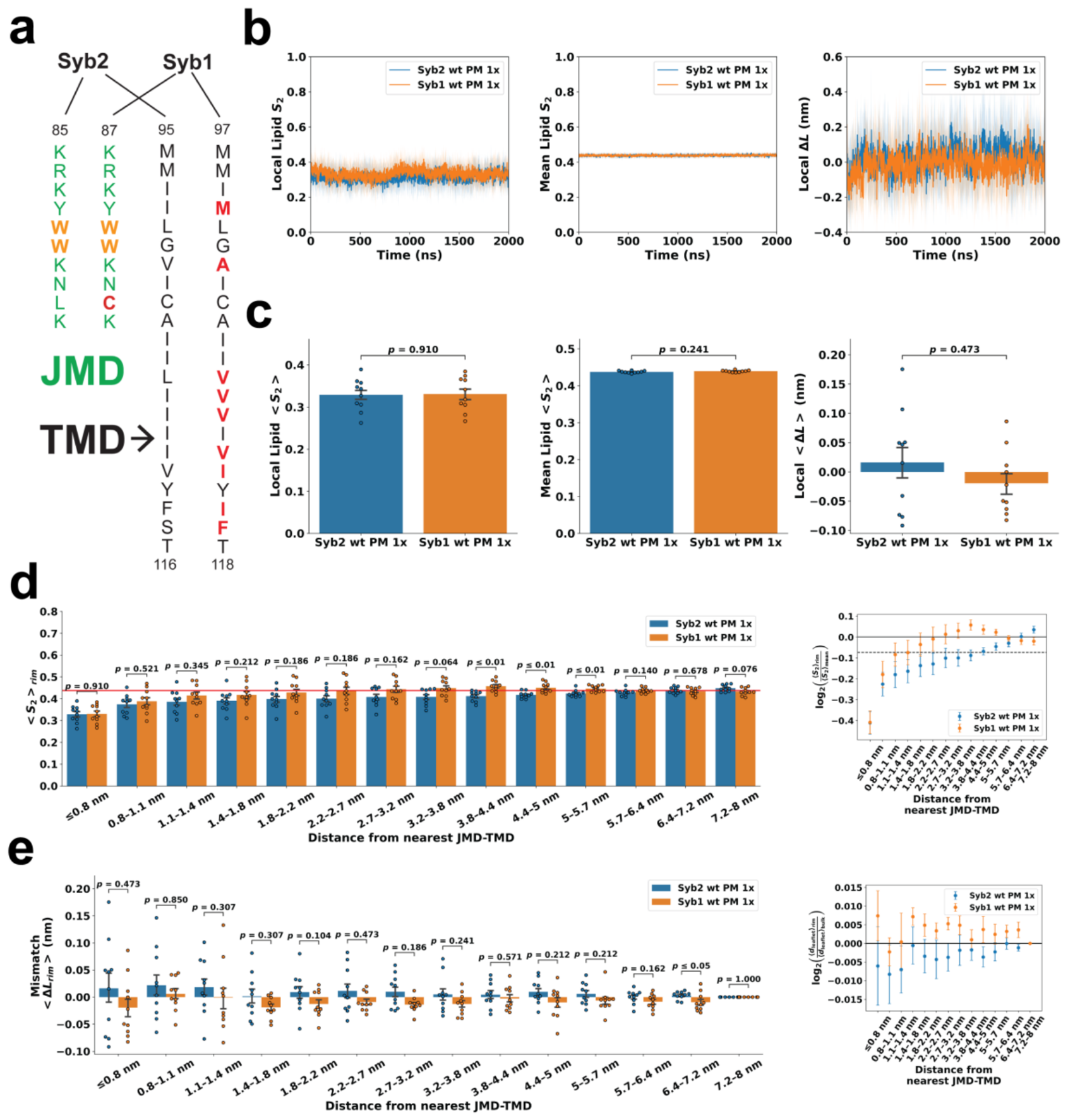
Figure 2.
PIP₂ and PS depletion have only minor effects on Syb2-induced proximal lipid disorder and membrane mismatch. (a) Local lipid order, and local membrane mismatch, for single-copy Syb2 in PIP₂-containing plasma membrane (PM), PIP₂-depleted plasma membrane (noP2 PM), and plasma membrane depleted of both PIP₂ and PS (noP2PS PM). Values were averaged over the final 100 ns of each trajectory. Individual circles represent independent simulations; bars and error bars indicate the mean and SEM, respectively. Pairwise comparisons were performed using the Mann–Whitney U test. (b) Corresponding radial lipid-order perturbations relative to the global membrane-average reference (left) and radial membrane-mismatch profiles (right) for the three membrane environments. Black solid lines indicate the mean membrane reference for lipid order and the bulk reference for interleaflet distance, respectively. Black dashed line indicates a 5% negative lipid-order perturbation relative to the mean membrane value. Detailed numerical values and statistical comparisons are provided in Table S6–S8.
Figure 2.
PIP₂ and PS depletion have only minor effects on Syb2-induced proximal lipid disorder and membrane mismatch. (a) Local lipid order, and local membrane mismatch, for single-copy Syb2 in PIP₂-containing plasma membrane (PM), PIP₂-depleted plasma membrane (noP2 PM), and plasma membrane depleted of both PIP₂ and PS (noP2PS PM). Values were averaged over the final 100 ns of each trajectory. Individual circles represent independent simulations; bars and error bars indicate the mean and SEM, respectively. Pairwise comparisons were performed using the Mann–Whitney U test. (b) Corresponding radial lipid-order perturbations relative to the global membrane-average reference (left) and radial membrane-mismatch profiles (right) for the three membrane environments. Black solid lines indicate the mean membrane reference for lipid order and the bulk reference for interleaflet distance, respectively. Black dashed line indicates a 5% negative lipid-order perturbation relative to the mean membrane value. Detailed numerical values and statistical comparisons are provided in Table S6–S8.
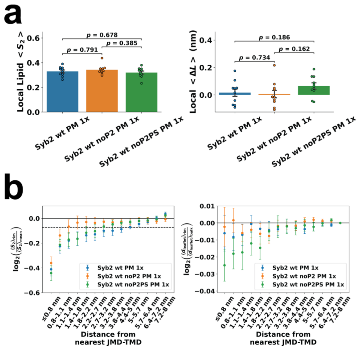
Figure 3.
Interfacial tryptophan substitutions modulate Syb-induced local membrane remodeling in PIP₂-containing plasma membrane. (a) JMD sequences of Syb1 and Syb2 wild-type (WT), double tryptophan-to-alanine (WWAA), and double tryptophan-to-lysine (WWKK) constructs. Conserved interfacial tryptophans are shown in bold, and substituted residues are highlighted in red. (b) Local lipid order parameters and local membrane mismatch for single-copy WT, WWAA, and WWKK Syb2 in the PIP₂-containing plasma membrane. Values were averaged over the final 100 ns of each trajectory. Individual circles represent independent simulations; bars and error bars indicate the mean and SEM, respectively. (c) Corresponding radial lipid-order perturbations relative to the mean membrane (left) and radial membrane-mismatch profiles (right). Black solid lines indicate the mean membrane reference for lipid order and the bulk reference for interleaflet distance, respectively. Black dashed line indicates a 5% negative lipid-order perturbation relative to the mean membrane value. Detailed numerical values and statistical comparisons in (b,c) are provided in Table S6–S8. Pairwise comparisons were performed using the Mann–Whitney U test unless otherwise indicated.
Figure 3.
Interfacial tryptophan substitutions modulate Syb-induced local membrane remodeling in PIP₂-containing plasma membrane. (a) JMD sequences of Syb1 and Syb2 wild-type (WT), double tryptophan-to-alanine (WWAA), and double tryptophan-to-lysine (WWKK) constructs. Conserved interfacial tryptophans are shown in bold, and substituted residues are highlighted in red. (b) Local lipid order parameters and local membrane mismatch for single-copy WT, WWAA, and WWKK Syb2 in the PIP₂-containing plasma membrane. Values were averaged over the final 100 ns of each trajectory. Individual circles represent independent simulations; bars and error bars indicate the mean and SEM, respectively. (c) Corresponding radial lipid-order perturbations relative to the mean membrane (left) and radial membrane-mismatch profiles (right). Black solid lines indicate the mean membrane reference for lipid order and the bulk reference for interleaflet distance, respectively. Black dashed line indicates a 5% negative lipid-order perturbation relative to the mean membrane value. Detailed numerical values and statistical comparisons in (b,c) are provided in Table S6–S8. Pairwise comparisons were performed using the Mann–Whitney U test unless otherwise indicated.
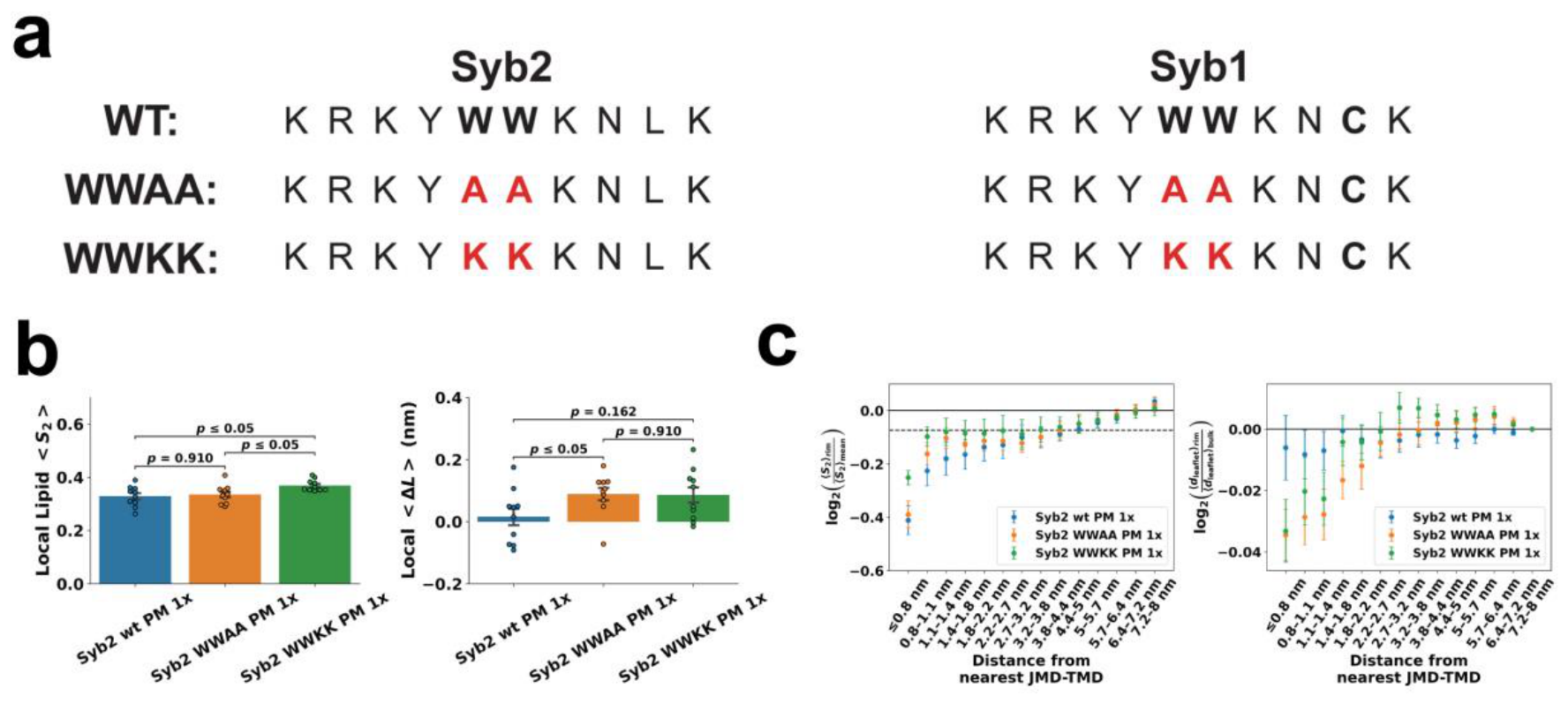
Figure 4.
PIP₂ promotes higher-order Syb2 cluster growth whereas PS has limited effects on collective organization. (a) Representative top and side views of a ten-copy Syb2 system in PIP₂-containing plasma membrane at 0 and 1900 ns. Syb2 JMD–TMD fragments are shown in blue, and lipid headgroups are shown as spheres. The red ellipse indicates the higher-order Syb assembly observed at 1900 ns, containing all ten Syb2 molecules in the representative trajectory. The corresponding trajectory is provided as Video S1. (b) Time evolution of maximum cluster size , and mean cluster size for ten-copy Syb2 systems in PIP₂-containing plasma membrane (PM), PIP₂-depleted plasma membrane (noP2 PM), and plasma membrane depleted of both PIP₂ and PS (noP2PS PM). Solid lines indicate ensemble means across independent simulations, with shaded regions representing the corresponding variability. (c) Corresponding maximum cluster size, and mean cluster size averaged over the final 100 ns of each trajectory. Individual points represent independent simulations; bars and error bars indicate the mean and SEM, respectively. Pairwise comparisons were performed using the Mann–Whitney U test, with p values indicated above the corresponding comparisons. Detailed numerical values and statistical comparisons are provided in Table S9.
Figure 4.
PIP₂ promotes higher-order Syb2 cluster growth whereas PS has limited effects on collective organization. (a) Representative top and side views of a ten-copy Syb2 system in PIP₂-containing plasma membrane at 0 and 1900 ns. Syb2 JMD–TMD fragments are shown in blue, and lipid headgroups are shown as spheres. The red ellipse indicates the higher-order Syb assembly observed at 1900 ns, containing all ten Syb2 molecules in the representative trajectory. The corresponding trajectory is provided as Video S1. (b) Time evolution of maximum cluster size , and mean cluster size for ten-copy Syb2 systems in PIP₂-containing plasma membrane (PM), PIP₂-depleted plasma membrane (noP2 PM), and plasma membrane depleted of both PIP₂ and PS (noP2PS PM). Solid lines indicate ensemble means across independent simulations, with shaded regions representing the corresponding variability. (c) Corresponding maximum cluster size, and mean cluster size averaged over the final 100 ns of each trajectory. Individual points represent independent simulations; bars and error bars indicate the mean and SEM, respectively. Pairwise comparisons were performed using the Mann–Whitney U test, with p values indicated above the corresponding comparisons. Detailed numerical values and statistical comparisons are provided in Table S9.
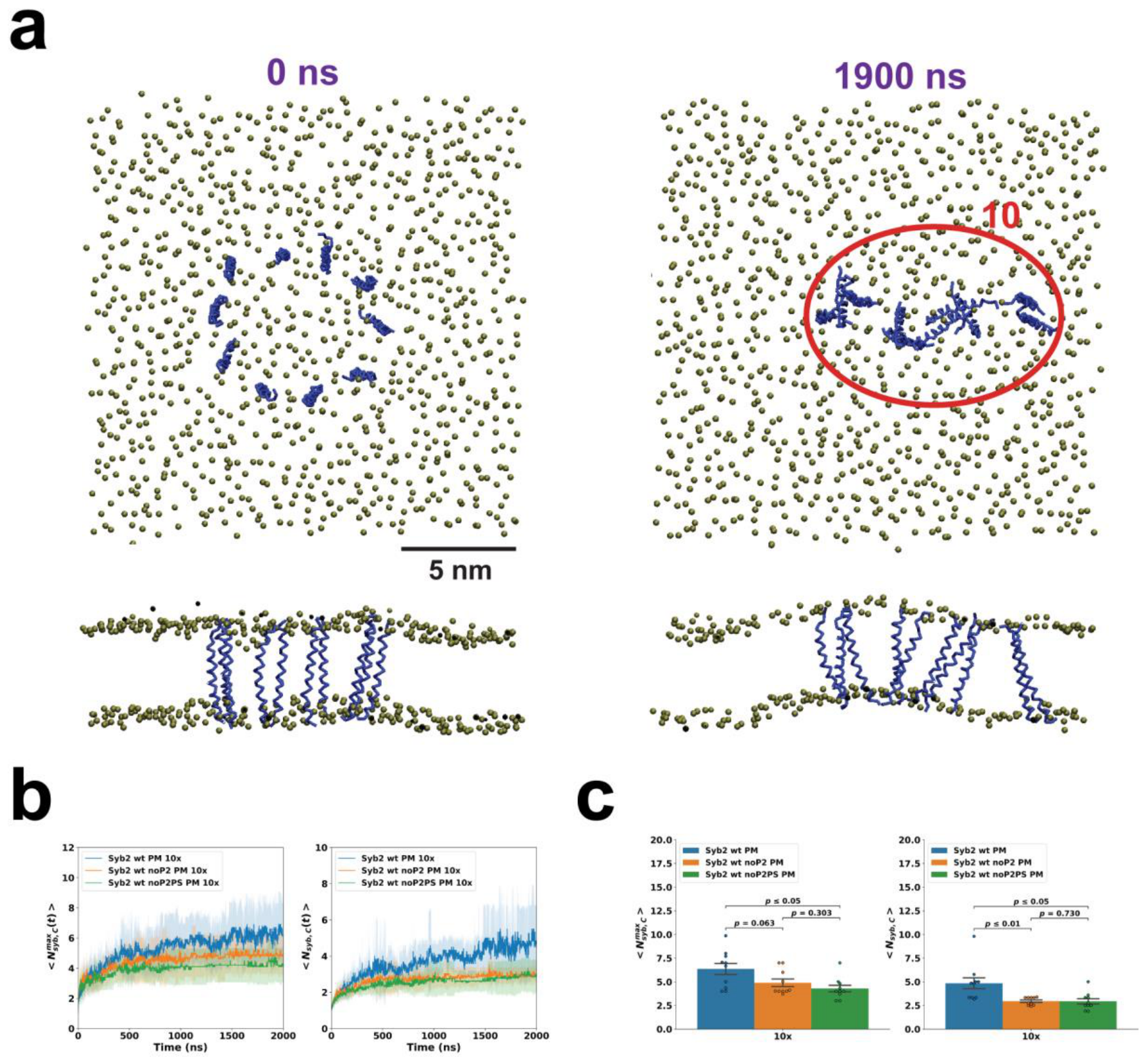
Figure 5.
PIP₂ promotes higher-order Syb2 assembly despite substitution of the conserved interfacial tryptophans. (a) Time evolution of the maximum cluster size and mean cluster size for ten-copy Syb2 WWAA and WWKK systems in PIP₂-containing and PIP₂-depleted plasma membranes. Solid lines indicate ensemble means across independent simulations, with shaded regions representing the corresponding variability. (b) Corresponding maximum and mean cluster sizes averaged over the final 100 ns of each trajectory. Individual points represent independent simulations; bars and error bars indicate the mean and SEM, respectively. Pairwise comparisons were performed using the Mann–Whitney U test, with values indicated above the corresponding comparisons. Detailed numerical values and statistical comparisons are provided in Table S9.
Figure 5.
PIP₂ promotes higher-order Syb2 assembly despite substitution of the conserved interfacial tryptophans. (a) Time evolution of the maximum cluster size and mean cluster size for ten-copy Syb2 WWAA and WWKK systems in PIP₂-containing and PIP₂-depleted plasma membranes. Solid lines indicate ensemble means across independent simulations, with shaded regions representing the corresponding variability. (b) Corresponding maximum and mean cluster sizes averaged over the final 100 ns of each trajectory. Individual points represent independent simulations; bars and error bars indicate the mean and SEM, respectively. Pairwise comparisons were performed using the Mann–Whitney U test, with values indicated above the corresponding comparisons. Detailed numerical values and statistical comparisons are provided in Table S9.
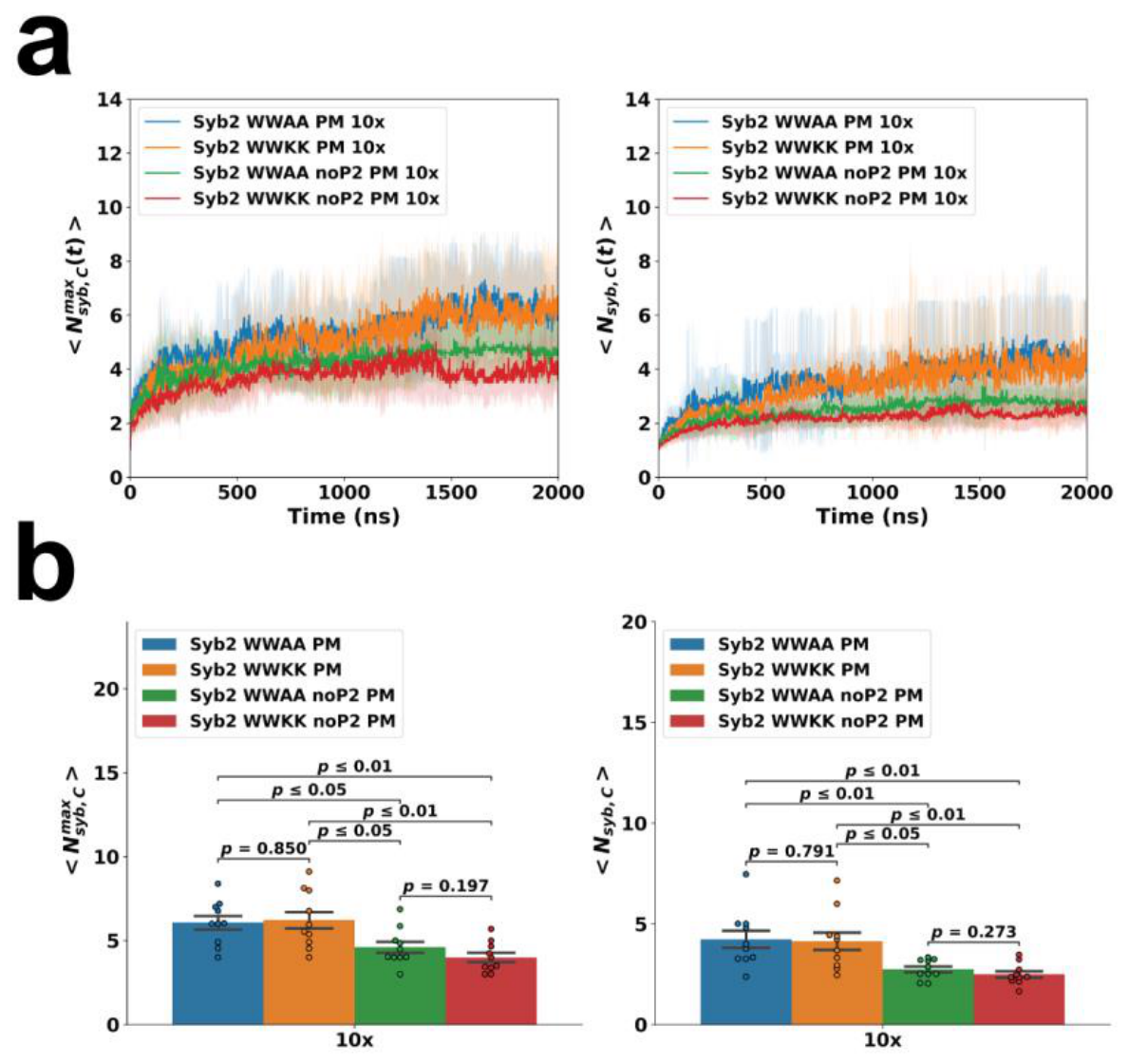
Figure 6.
Local Syb-induced lipid disorder is preserved during collective clustering. (a) Representative spatial maps of lipid disorder around ten Syb2 JMD–TMD fragments in the PIP₂-containing plasma membrane at 10 (left), 800 (middle), and 1900 ns (right). Black crosses indicate the centers of mass of individual Syb2 JMD–TMD fragments. Dashed ellipses highlight regions occupied by higher-order Syb assemblies at later simulation times. Lipid disorder is represented as the rolling-mean local (), with larger values indicating greater lipid disorder. (b) Radial lipid-order perturbations relative to the corresponding global membrane-average references for single-copy and ten-copy WT (left), WWAA (middle), and WWKK (right) Syb2 systems in PIP₂-containing (top) and PIP₂-depleted (bottom) plasma membranes. For ten-copy systems, each lipid was assigned exclusively to the radial bin corresponding to its minimum distance from the nearest Syb JMD–TMD region and was therefore counted only once. Values are plotted as , as a function of the minimum distance from the nearest Syb JMD–TMD region. Black solid line: mean membrane reference. Black dashed line: 5% negative perturbation relative to the mean membrane value. For direct comparison with the ten-copy systems, single-copy profiles are displayed only up to 5 nm, although the corresponding single-copy radial analyses were performed up to 8 nm. Error bars indicate SEM across independent simulations. Detailed numerical values are provided in Table S8.
Figure 6.
Local Syb-induced lipid disorder is preserved during collective clustering. (a) Representative spatial maps of lipid disorder around ten Syb2 JMD–TMD fragments in the PIP₂-containing plasma membrane at 10 (left), 800 (middle), and 1900 ns (right). Black crosses indicate the centers of mass of individual Syb2 JMD–TMD fragments. Dashed ellipses highlight regions occupied by higher-order Syb assemblies at later simulation times. Lipid disorder is represented as the rolling-mean local (), with larger values indicating greater lipid disorder. (b) Radial lipid-order perturbations relative to the corresponding global membrane-average references for single-copy and ten-copy WT (left), WWAA (middle), and WWKK (right) Syb2 systems in PIP₂-containing (top) and PIP₂-depleted (bottom) plasma membranes. For ten-copy systems, each lipid was assigned exclusively to the radial bin corresponding to its minimum distance from the nearest Syb JMD–TMD region and was therefore counted only once. Values are plotted as , as a function of the minimum distance from the nearest Syb JMD–TMD region. Black solid line: mean membrane reference. Black dashed line: 5% negative perturbation relative to the mean membrane value. For direct comparison with the ten-copy systems, single-copy profiles are displayed only up to 5 nm, although the corresponding single-copy radial analyses were performed up to 8 nm. Error bars indicate SEM across independent simulations. Detailed numerical values are provided in Table S8.
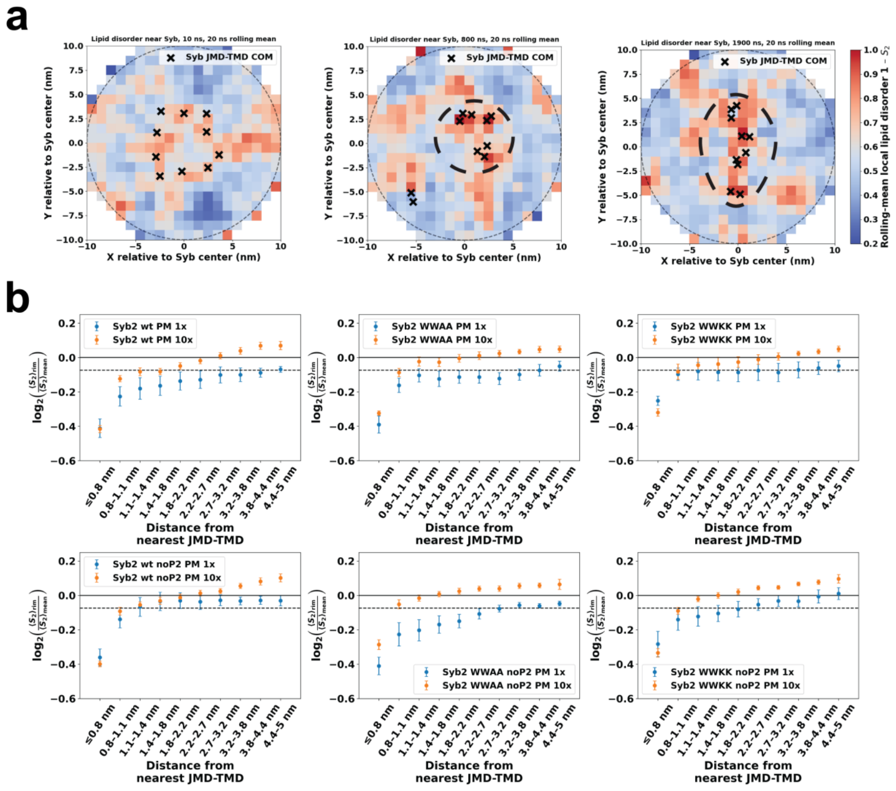
Figure 7.
Collective Syb assembly is associated with distinct PIP₂ and PS redistribution. (a, b) Representative spatial density maps of PIP₂ (a) and PS (b) around ten Syb2 JMD–TMD fragments in the PIP₂-containing plasma membrane at 10, 800, and 1900 ns. Black crosses indicate the centers of mass of individual Syb2 JMD–TMD fragments, and dashed red ellipses highlight regions occupied by higher-order Syb assemblies at later simulation times. (c) Time evolution of PIP₂ (left) and PS (right) redistribution for single-copy and ten-copy WT Syb2 and Syb1 systems in the PIP₂-containing plasma membrane. For each lipid species, the fraction of the intracellular-leaflet lipid pool associated with Syb , and the mean number of unique associated lipid molecules per Syb , are shown. Solid lines indicate ensemble means across independent simulations, with shaded regions representing the corresponding variability. (d) Final-state PIP₂ redistribution quantified by the fraction of intracellular-leaflet PIP₂ associated with Syb, (left); local PIP₂ enrichment relative to its intracellular-leaflet abundance, (middle); and mean number of unique associated PIP₂ molecules per Syb, (right), for single-copy and ten-copy WT Syb2 and Syb1 systems. (e) Corresponding final-state PS redistribution quantified by the fraction of intracellular-leaflet PS associated with Syb, (left); local PS enrichment relative to its intracellular-leaflet abundance, (middle); and mean number of unique associated PS molecules per Syb, (right). Final-state values in (d,e) were averaged over the final 100 ns of each trajectory. Individual points represent independent simulations; bars and error bars indicate the mean and SEM, respectively. A lipid molecule simultaneously associated with multiple Syb molecules was counted only once when calculating . Pairwise comparisons were performed using the Mann–Whitney U test, with values indicated above the corresponding comparisons. Detailed numerical values and statistical comparisons are provided in Table S10.
Figure 7.
Collective Syb assembly is associated with distinct PIP₂ and PS redistribution. (a, b) Representative spatial density maps of PIP₂ (a) and PS (b) around ten Syb2 JMD–TMD fragments in the PIP₂-containing plasma membrane at 10, 800, and 1900 ns. Black crosses indicate the centers of mass of individual Syb2 JMD–TMD fragments, and dashed red ellipses highlight regions occupied by higher-order Syb assemblies at later simulation times. (c) Time evolution of PIP₂ (left) and PS (right) redistribution for single-copy and ten-copy WT Syb2 and Syb1 systems in the PIP₂-containing plasma membrane. For each lipid species, the fraction of the intracellular-leaflet lipid pool associated with Syb , and the mean number of unique associated lipid molecules per Syb , are shown. Solid lines indicate ensemble means across independent simulations, with shaded regions representing the corresponding variability. (d) Final-state PIP₂ redistribution quantified by the fraction of intracellular-leaflet PIP₂ associated with Syb, (left); local PIP₂ enrichment relative to its intracellular-leaflet abundance, (middle); and mean number of unique associated PIP₂ molecules per Syb, (right), for single-copy and ten-copy WT Syb2 and Syb1 systems. (e) Corresponding final-state PS redistribution quantified by the fraction of intracellular-leaflet PS associated with Syb, (left); local PS enrichment relative to its intracellular-leaflet abundance, (middle); and mean number of unique associated PS molecules per Syb, (right). Final-state values in (d,e) were averaged over the final 100 ns of each trajectory. Individual points represent independent simulations; bars and error bars indicate the mean and SEM, respectively. A lipid molecule simultaneously associated with multiple Syb molecules was counted only once when calculating . Pairwise comparisons were performed using the Mann–Whitney U test, with values indicated above the corresponding comparisons. Detailed numerical values and statistical comparisons are provided in Table S10.
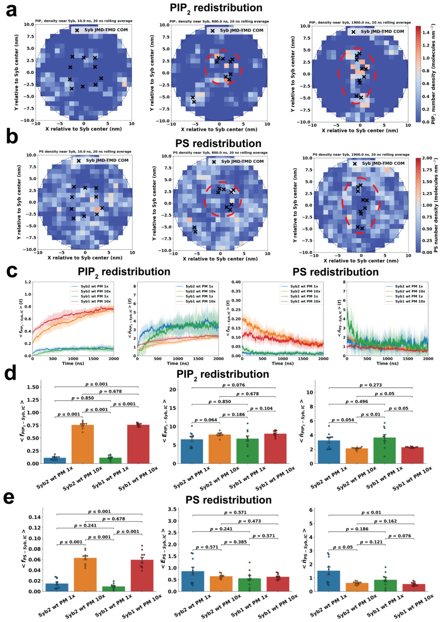
Disclaimer/Publisher’s Note: The statements, opinions and data contained in all publications are solely those of the individual author(s) and contributor(s) and not of MDPI and/or the editor(s). MDPI and/or the editor(s) disclaim responsibility for any injury to people or property resulting from any ideas, methods, instructions or products referred to in the content. |
© 2026 by the authors. Licensee MDPI, Basel, Switzerland. This article is an open access article distributed under the terms and conditions of the Creative Commons Attribution (CC BY) license (http://creativecommons.org/licenses/by/4.0/).
Copyright: This open access article is published under a Creative Commons CC BY 4.0 license, which permit the free download, distribution, and reuse, provided that the author and preprint are cited in any reuse.