
Submitted:
01 September 2026
Posted:
02 September 2026
You are already at the latest version
Abstract
The T cells of the mammalian immune system have a tricky challenge. They must recognize foreign antigen as “other” or “non-self” but in the context of self, while also being tolerant to the body’s own molecules and tissues. In this review, I provide context to the temporal and spatial parameters that make this possible. To do so, I briefly discuss some of the many molecules and cells that work in concert to ensure that our T cells work for us, not against us.
Keywords:
self-tolerance
; thymocyte development
; thymic epithelial cells
1. Introduction—In the Beginning
Vertebrate animals respond to an ever-changing antigenic universe by having an adaptive immune system. It is adaptive for two reasons. Firstly, its constituent B cells and T cells are generated daily. Secondly, these lymphocytes collectively express a diverse set of antigen receptors of different specificities. These unique antigen specificities are embodied in antigen binding sites that are created by seemingly random gene rearrangement mechanisms that occur during B-cell and T-cell development. Consequently, it is possible for a B cell or T cell to express receptors that have affinity for self-molecules. To counteract this danger, during their development, both B cells and T cells undergo selection processes that cull cells bearing autoreactive receptors. In the case of T cells expressing an αβT-cell receptor for antigen, (αβTCR), this occurs in the thymus via a two-step process. Both steps rely on the fact that the ligand for the αβTCR is a Major Histocompatibility Complex (MHC) molecule bearing an antigenic peptide. These two steps—called positive and negative selection—are discussed in detail below.
2. First and Foremost, Know Who You Are
T-cell development occurs in the thymus and begins in the thymic cortex. As stated above, gene rearrangement events occurring in developing T cells (thymocytes) generate TCRs a diverse array of antigen receptors, each with its unique specificity. The gene rearrangement process involves several steps [1]. There are two kinds of TCRs—an αβTCR and a γδTCR. Only αβTCRs recognize MHC/peptide complexes, and so all subsequent discussion in this review focuses on thymocytes expressing αβTCRs. It is not the αβTCR alone that binds to MHC/peptide. Either of two coreceptors—CD4 or CD8—also bind to MHC. These two coreceptors are distinguished by their MHC specificity. CD4 is specific for MHC II, and CD8 binds MHC I. CD4 and CD8 are of paramount importance in positive and negative selection in two ways. Firstly, they increase the avidity of αβTCR binding to its cognate MHC + peptide ligand. Secondly, they help initiate signal transduction events that lead to the survival of a thymocyte or to its death. Prior to TCR gene rearrangement, thymocytes express neither CD4 or CD8. Thymocytes that lack both CD4 and CD8 are said to be double negative (DN, Figure 1). Upon the generation of a properly formed TCR, the thymocytes express both CD4 and CD8 and are said to be double-positive (DP). Two important events happen at the DP stage of thymocyte development: positive selection and lineage commitment, which I will discuss shortly.
Lineage commitment is the developmental decision to become either a CD4+ helper T cell or a CD8+ cytotoxic T cell [2,3]. Until recently, it was believed that signaling through either CD4 or CD8 determines a thymocyte’s lineage choice. However, results from single-cell studies on DP thymocytes showed that engagement of CD4 or CD8 by MHC is not the sole determinant of a thymocyte’s lineage fate [4]. During positive selection, cis-regulatory elements in the cd4 and cd8 genes regulate both the kinetics of the expression of these co-receptor genes and the duration of TCR signaling [5]. Concomitantly, the developing thymocyte commits to its helper or cytotoxic function. The master transcriptional regulators—ThPOK (in the case of CD4) [6,7] and Runx3 (in the case of CD8) [8,9,10,11]—are required both for commitment to expression of either CD4 or CD8 and for lineage function. Robey and her colleagues have provided a comprehensive review detailing the intricacies of the molecular regulation of cd4 and cd8 transcription [12].
Also important during thymocyte development is the co-stimulatory molecule CD28. CD28 is a cell-surface glycoprotein that is expressed by mature T cells, where it plays a critical role in T cell activation [13]. CD28’s value stems from the fact that its binding to its ligand—CD80 or CD86—amplifies signaling through the TCR [14,15,16,17,18,19] . In mature T cells in peripheral lymphoid tissues, the binding of CD28 to either CD80 or CD86 on antigen presenting cells (APCs) delivers a second signal that is required for T-cell activation [20]. This is particularly crucial when low numbers of TCRs are engaged or when the TCRs have weak affinity for their MHC + peptide ligands [21]. Not surprisingly, CD28 is also found on thymocytes. DP thymocytes, DPlo thymocytes (which are developmentally downstream of DP thymocytes; Figure 1), and CD4+ and CD8+ single positive (SP) thymocytes all express CD28 [22]. However, CD80 and CD86 are chiefly expressed by thymic medullary APCs [23,24] and hence, the CD28 co-stimulatory signal is chiefly operative during negative selection.
3. Growing up is hard to do
The steps in thymocyte development and the thymic locations in which these steps occur are outlined briefly in Figure 2. As stated earlier, CD4’s and CD8’s roles in thymocyte development begin at the DP stage. Central to thymocyte development is the binding of αβTCRs on thymocytes to MHC+ peptide complexes occurring on the surfaces of thymic epithelial cells (TECs) and other thymic stromal cells. During interactions with cortical thymic epithelial cells (cTECs), a CD4+CD8+ DP thymocyte that can bind its TCR’s cognate MHC + peptide ligand is positively selected for survival. After positive selection, expression of CD4 and CD8 diminish. The downregulation is not equivalent for both coreceptors [12]. However, for simplicity’s sake, I will refer to this stage as the DPlo thymocyte. The DPlo thymocyte expresses the chemokine receptor, CCR7, which enables it to migrate towards the chemokine CCL21. Since CCL21 is produced by thymic medullary cells, the DPlo thymocyte migrates to the corticomedullary junction [25]. (Interestingly, when migration to the medulla is prevented, as in the case of mice deficient both in CCL21 and in CCR7, SP thymocytes still develop, but they accumulate in the thymic cortex [26,27]).
In normal mice, the DPlo thymocyte interacts with medullary thymic epithelial cells (mTECs) and other medullary APCs such as medullary dendritic cells (mDCs). If the DPlo thymocyte undergoes moderate signaling through its TCR, it becomes a CD4+ or CD8+ SP thymocyte and migrates out to peripheral lymphoid tissues. Conversely, strong signal transduction through the TCR leads to death via apoptosis (negative selection) or to divergent differentiation, as follows: A thymocyte bearing an MHC II-specific TCR that survives negative selection differentiates into a CD4+ regulatory T cell (Treg). A thymocyte bearing an MHC I-specific TCR that escapes negative selection can become an intraepithelial lymphocyte precursor (IEL precursor). However, as described below, not all MHC I-specific thymocytes that survive negative selection become IEL precursors.
4. With a little help from my friends
The TCR is a complex of several polypeptides. In the case of the αβTCR, the antigen-binding properties reside in the α and β chains of the TCR. But they have no signaling capabilities. Instead, the αβTCR is associated with the CD3 complex of polypeptides (shown in Figure 3). The ζ chains of the CD3 complex have long cytoplasmic tails that contain immunoreceptor tyrosine-based activation motifs (ITAMs), which can be phosphorylated by tyrosine kinases. This is where CD4 and CD8 come in. CD4 and CD8 have both extracellular and intracellular moieties and hence, dual functions. Via their extracellular domains, CD4 and CD8 bind to MHC II and MHC I, respectively, and via their cytoplasmic tails, they bind to a tyrosine protein kinase called p56lck [28]. During the binding of MHC + peptide, the αβTCR, CD4, CD8, and other associated molecules cluster at the immunological synapse, which is the junction between the T cell (or thymocyte) and the APC. The coordinated engagement of MHC by both the αβTCR and by CD4 or CD8 launches an enzymatically based signal transduction cascade [29]. Specifically, the binding of CD8 or CD4 and of αβTCR to MHC + peptide facilitates the phosphorylation by p56lck of the ITAMs of CD3ζ [29] (Figure 3). p56lck also phosphorylates Zeta-chain-associated protein kinase 70 (ZAP 70). ZAP70 then phosphorylates the adaptor proteins, Linker for activation of T cells (LAT) and SLP-76 [30,31,32] (Figure 3). These proteins are joined in the immunological synapse by Protein kinase Cθ [33] and phospholipase Cγ (PLCγ) [34] (Figure 3). This completes the assemblage of signal transduction complexes that are essential for efficient T cell activation.
Since CD4 is a single-chain co-stimulatory molecule, all the essential features for ligand binding and signal transduction are vested in the single CD4 polypeptide chain. The situation is more complicated for CD8, which is a dimer that can occur in either of two forms [37]. All T cells (except for IELs) express CD8 as a heterodimer of CD8α and CD8β. IELs express CD8 as a CD8αα homodimer. CD8αβ is much more effective in T-cell activation than is CD8αα [38,39]. This was initially surprising because CD8α can bind to p56lck through its cytoplasmic tail [40] and thus, should be able to promote signal transduction. The signaling prowess of CD8αβ and hence, its efficacy as a co-stimulatory molecule for the TCR, stem from properties that are unique to CD8β. CD8β promotes the interaction of CD8αβ with the αβTCR [32], thereby facilitating the phosphorylation of CD3ζ-chain ITAMs (Figure 3). Also, via CD8β, CD8αβ efficiently recruits ZAP70, thereby enabling its phosphorylation and activation by p56lck [30,31,32].
In DP and DPlo thymocytes, CD8αβ must compete with CD4 for binding to p56lck. CD4 has higher affinity for p56lck than does CD8αβ [28]. Hence, in a thymocyte expressing both CD4 and CD8αβ, the number of p56lck-associated CD8αβ molecules is lower than is the number of p56lck-associated CD4 molecules [41]. This discrepancy is important to consider in the context of negative selection for the following reasons. Any given MHC molecule can bind a diversity of peptides. Therefore, only a small fraction of the MHC + peptide complexes on the surface of a thymic TEC is expected to be bound by a particular αβTCR. Consequently, the MHC/antigenic peptide combination that is a suitable ligand for any given MHC I-specific αβTCR on a developing DPlo thymocyte may be present at suboptimal levels on the surface of the TEC. In this situation, a diminished number of p56lck-associated CD8αβ molecules in the immunological synapse could result in inefficient signaling and failure to undergo negative selection.
As mentioned previously, CD28 binding to CD80 or CD86 amplifies signaling through the αβTCR. However, CD28 seems not to be equally important for negative selection of all thymocytes. Thus, CD28 interaction with CD80/CD86 on medullary epithelial cells invariably promotes either apoptosis of a DPlo thymocyte expressing MHC II-specific αβTCRs or its development into a CD4+ Treg [42]. By contrast, in the case of DPlo thymocytes expressing MHC I-specific αβTCRs, the need for CD28 during negative selection appears to vary with the affinity or avidity of the αβTCR for its MHC I/peptide ligand [43].
5. Location, location, location—lipid rafts and the immunological synapse
The first step in the activation of a T cell involves the binding of the αβTCR to its MHC + peptide ligand and of the relevant coreceptor (CD4 or CD8), CD28, and other molecules (such as the adhesion molecule, LFA-1) to their own ligands (Figure 3). As noted earlier, these interactions occur within the immunological synapse [44]. Integral to the clustering of molecules within the immunological synapse are lipid rafts [45]. Lipid rafts are dynamically changing assemblages of cholesterol and sphingolipids [46]. Associations between T-cell proteins and their ligands on APCs stimulate the lateral mobility of the molecules within the plasma membrane of each cell and their clustering into the lipid rafts that comprise the immunological synapse (Figure 3). For example, αβTCR localization into lipid rafts is promoted by CD28 binding to CD80 or CD86 during T cell activation [47]. The congregation of the αβTCR and other relevant proteins within the immunological synapse (Figure 3) is thought to enable efficient assembly of signaling molecules and thus, to maximize signaling [48].
CDβ—but not CD8α —can promote the migration of CD8αβ into lipid rafts [49]. Palmitoylation of the CD8β cytoplasmic tail enables CD8αβ to partition into lipid rafts [50,51], but this is not the sole determinant of lipid raft localization. In the case of human CD8αβ, an arginine motif in the CD8β cytoplasmic region also contributes to lipid raft localization [49]. These observations help explain the requirement for the CD8β chain for positive selection of MHC I-specific thymocytes and for their development into CD8+ thymocytes [52,53,54]. Furthermore, as discussed below, the abundance of CD8β, and therefore of cell-surface CD8αβ, is essential for the negative selection of thymocytes bearing self-reactive αβTCRs [55,56].
6. Slipping under the radar
In this review, I will focus on the role of CD8αβ in negative selection. As already stated, the prevailing view is that thymocytes bearing αβTCRs that have high affinity for self MHC I/peptide either die by apoptosis or develop into IEL precursors [57,58,59,60]. In unmanipulated mice, there are ~5 million CD8αα+ IELs bearing autoreactive αβTCRs [61]. The genesis of these CD8αα+ IELs from DPlo thymocytes can occur when αβTCR engagement occurs in the absence of CD28 co-stimulation [61]. This finding underscores the multifaceted nature of negative selection: Signaling through the αβTCR alone does not necessarily promote the culling of thymocytes with autoreactive TCRs.
Robey and colleague used a thymic tissue slice experimental system to study in real time the development of thymocytes bearing an MHC I-specific αβTCR [62]. Their model was the OT-I TCR transgenic mouse (which expresses an αβTCR that is specific for H-2Kb + chicken ovalbumin peptide SIINFEKL). They laid OT-I thymocytes upon thymic slices bathed in SIINFEKL peptide and assessed thymocyte development over time. In this system, DP thymocytes downregulated both CD4 and CD8, consistent with previous studies [63]. Some of these DPlo thymocytes survived negative selection and became IEL precursors. In the surviving IEL precursors, CD8β levels remained low, but CD8α levels did not [62]. Hence, these thymocytes were largely CD8αα+.
The low CD8β level of the surviving DPlo thymocytes in these studies brings front and center the role of CD8αβ in signal transduction within the immunological synapse. Specifically, since CD8αβ (but not CD8αα) can partition into lipid rafts, DPlo thymocytes expressing autoreactive αβTCRs and having insufficient numbers of CD8αβ dimers might escape negative selection. My colleagues and I employed a probabilistic gating procedure to examine this possibility in a transgenic mouse αβTCR model [64]. This double transgenic model involves CBA (H-2k) mice expressing allogeneic H-2Kb in the thymic medulla and an anti-H-2Kb TCR. We found that the viability of double transgenic DPlo thymocytes remained high across a range of CD8β/α ratios but then dropped precipitously at a specific CD8β/α ratio [64]. We inferred that in DPlo thymocytes having low CD8β/α ratios, there is a shortage of p56lck-associated CD8αβ molecules in lipid rafts (and hence, in the immunological synapse). We further inferred that this deficiency of p56lck-associated CD8αβ results in inadequate signaling through the anti-H-2Kb TCR, thereby allowing escape from negative selection.
7. Wait—there’s more, and it’s complicated!
Programmed cell death protein 1 (PD-1) is an inhibitory cell-surface protein that is expressed by mature T cells [65]. PD-1 binding to its molecular ligand—either PD-L1 or PD-L2—on APCs results in the attenuation of T-cell activation, thus dampening the immune response [65,66]. PD-1 does this by recruiting SHP-2 phosphatase to its cytoplasmic tail [67]. This results in the suppression of phosphorylation of CD3 chains and of ZAP70 [67]. Thus, in mature T cells, PD-1 plays a key role in regulating signal transduction through the αβTCR. PD-1 also is expressed in the thymus, where it plays a regulatory role during positive selection [68]. But does PD-1 have a role during negative selection? In their thymic slice experiments, the Robey group observed a slight increase in PD-1 expression in OT-I DPlo thymocytes that had been exposed to SIINFEKL peptide [62]. This result was consistent with earlier studies in non-transgenic mice showing that “developmentally diverted” thymocytes that had survived negative selection have elevated levels of PD-1 [61]. However, PD-1 expression is not required for developmental diversion since it occurred in mice in which the pd1 gene had been deleted [61]. Nevertheless, PD-1 may promote the survival of thymocytes expressing a self-reactive αβTCR during negative selection by attenuating signaling through the αβTCR [67,69,70,71,72]. We examined this possibility in our studies. We found that the importance of PD-1 for the survival of autoreactive DPlo thymocytes was conditional [64]. Specifically, PD-1 was important only in autoreactive DPlo thymocytes having intermediate levels of CD8β. At very low levels of CD8β (and therefore low levels of CD8αβ), DPlo thymocytes bearing a self-reactive αβTCR survived, regardless of PD-1 levels. Conversely, DPlo thymocytes bearing a self-reactive αβTCR and having high CD8β levels (and therefore high CD8αβ levels) were apoptotic, even when PD-1 levels were high [64].
8. Let me in—lipid rafts revisited
Why might PD-1 be beneficial at intermediate CD8β/α ratios but not at high CD8β/α ratios? For PD-1 to compromise signal transduction, it either must enter lipid rafts, or it must prevent essential signal transduction molecules from doing so. A recent study reported that PD-1 is recruited into lipid rafts by CD48 [73]. If both CD8αβ and PD-1 can enter lipid rafts, then what determines if CD8αβ promotes signal transduction or if PD-1 squelches it? It occurred to us that the answer might lie in the small size of lipid rafts [74]. It has been estimated that no more than 10-30 proteins can occur in a lipid raft [48,74]. We thus hypothesized that CD8β is more effective in targeting CD8αβ to lipid rafts than CD48 is in recruiting PD-1 to them [64]. If this is true, then PD-1 molecules might be “crowded out” of lipid rafts when CD8αβ is abundant. Then, there wouldn’t be enough PD-1 to suppress signal transduction through the αβTCR, and the strong signaling would lead to cell death by apoptosis. The predictions of this model are illustrated in Figure 4, in which four different scenarios are depicted.
Unfortunately, it will be difficult to test this model. Lipid rafts are too small to be observed with standard light microscopic techniques. Antibodies and/or lectins must be used to promote clustering of lipid raft components to detect them microscopically [75,76]. Thus, it may be difficult to definitively test this hypothesis in a physiologically relevant experiment. Furthermore, different proteins may partition into lipid rafts with different kinetics and thus have different residence time in them. Of course, assembly of T-cell receptor components into multimeric signaling complexes should increase residence time in lipid rafts. But even if PD-1 occurs in lipid rafts, its association with the TCR signaling complex may not be observed under experimental conditions.
9. A simple twist of fate
What happens to a thymocyte bearing a TCR specific for self MHC I + self-peptide that survives negative selection? Several research groups have demonstrated beautifully that MHC I-restricted DPlo thymocytes that survive negative selection become IEL precursors and go on to populate the intestine [57,58,59,60,61,62]. The implication—and in some minds, the assumption—has been that for a thymocyte bearing a TCR specific for self MHC I + self-peptide becoming an IEL precursor is the only alternative to apoptosis. Is this in fact true? Unlike thymocytes destined for peripheral lymphoid tissues [26], type A IEL precursors do not express CCR7 [59]. Our studies demonstrated that in double-transgenic mice that express both allogeneic H-2Kb in the medulla and the anti-H-2Kb TCR, surviving DPlo post-selection thymocytes can give rise both to PD-1+DN thymocytes that express CCR7 and those that do not express CCR7, i.e., PD-1+CCR7+DN and PD-1+CCR7-DN thymocytes [64]. Furthermore, we found transgenic αβTCR+ DN lymphocytes in the peripheral lymph nodes of these mice [64]. These DN lymphocytes are presumed to be derived from αβTCR+PD-1+CCR7+DN thymocytes. These results suggest that surviving thymocytes bearing autoreactive TCRs are not necessarily confined to becoming IELs but also give rise to lymphocytes that populate other peripheral lymphoid tissues.
10. Live and let live—becoming an enforcer of self-tolerance
It is important to remember that the vast majority of thymocytes expressing self-reactive TCRs die during negative selection. To prevent autoimmunity, the surviving thymocytes must be restrained. The immune system has distinct ways of enforcing self-tolerance in the small proportion of potentially autoreactive CD4+ and CD8+ T cells that escape negative selection. As stated previously, surviving thymocytes bearing autoreactive MHC II-specific TCRs become CD4+ Treg cells that enforce tolerance to self [77,78,79]. In the case of the CD8 lineage, thymocytes bearing autoreactive MHC I-specific TCRs give rise to CD8αα+ IELs and DN IELs and to peripheral lymph node DN T cells that are anergic to self. What is the evidence for this assertion? In the intestine, antigen presentation to IELs bearing autoreactive TCRs fails to induce calcium mobilization, proliferation, and production of IL-2 [61,80]. There is also evidence that these IELs suppress inflammation [81,82]. The majority of the transgenic αβTCR+ DN T cells in our studies were PD-1+, and thus, should be anergic [64]. In unpublished studies, we found that double transgenic mice that express both H-2Kb in the medulla and the anti-H-2Kb TCR are tolerant of H-2Kb-bearing skin. Furthermore, it was previously shown that transgenic TCR+ T cells from these double transgenic mice are very inefficient in killing H-2Kb-bearing target cells ex vivo [80,83]. These findings are in keeping with studies by other research groups demonstrating that αβTCR+ DN T cells from lymph nodes of antigen-expressing mice are functionally anergic ex vivo [84]. Finally, the αβTCR+ DN T cells in the model used in the Teh lab [84,85] were found to have immunosuppressive properties both ex vivo and in vivo [85]. Thus, the small number of αβ T cells bearing self-reactive TCRs and populating peripheral lymphoid tissues do not pose an immediate threat to self-tolerance and even can enforce self-tolerance.
11. Me, myself, and I
What are the specific factors in thymocyte survivors of negative selection that promote their differentiation into a tolerant cell type? Not surprisingly, transcriptional control is key. For example, in response to both the cognate ligand of the TCR and the cytokine, IL-15, type A IEL precursors express the transcription factor, T-bet [86,87]. However, the exact cellular interactions that trigger new transcriptional programs remain to be elucidated. Presumably, interactions of the developing thymocytes with TECs initiate differentiation down the alternative “tolerant” pathway. As noted above, in our model, surviving autoreactive DPlo post-selection thymocytes can give rise to PD-1+CCR7+DN and PD-1+CCR7-DN thymocytes [64]. The expression of CCR7 or lack thereof ensures that these two populations become spatially separated and complete their development in the medulla and in the cortex, respectively. There, they are exposed to different kinds of TECs and other stromal cells [88,89,90,91], which provide them with the cytokines and other signals necessary for the development of their final identities.
The development of TECs is beyond the scope of this review and is beautifully presented elsewhere [92]. Here I will focus on the mature TECs—cortical TECs (cTECs) and medullary TECs (mTECs). Many features that distinguish cTECs and mTECs have been elucidated [93]. Best understood are the differences in the antigen presentation properties of these two cell types, which make them ideally suited for their roles in positive and negative selection [94]. The “thymoproteasomes” of cTECs have three specialized subunits, β5t, β1i, and β2i. Proteasomes containing these subunits are necessary for the positive selection of thymocytes destined to become CD8+ T cells [95,96,97,98]. Essential to the positive selection of CD4+ T cells is CD83. CD83 stabilizes cell-surface MHC II molecules [99] by suppressing the ubiquination of MHC II by the E3 ubiquitin ligase, March-1 [100]. In this way, CD83 inhibits the endocytosis of cell-surface MHC II/peptide complexes and their subsequent degradation [100].
Because of its role during negative selection in the medulla, of greater relevance to this review is the expression by mTECs of the transcription autoimmune regulator (AIRE). AIRE facilitates the expression of otherwise “tissue-restricted” antigens [101] by interacting with hypomethylated histone H3 [102] and the ATF7ip-MBD1 protein complex [103]. In so doing, AIRE influences the repertoires not only of CD4+ and CD8+ T cells but also of Foxp3+ CD4+ Tregs [104,105,106], which are essential for repressing autoimmunity [79,107,108]. The chromatin of mTECs is especially accessible to AIRE and to “lineage-defining” transcription factors [109,110]. Hence, chromatin structure, AIRE, and lineage-defining transcription factors work together to promote the ectopic expression of tissue-specific genes. Furthermore, AIRE also deviates autoreactive CD4+ thymocytes away from developing into conventional CD4+ T cells and to becoming Foxp3+ T regs [111]. Finally, AIRE appears also to have roles in homeostatic regulation of the cellular composition of the thymic medulla [112].
12. Meet the great pretenders
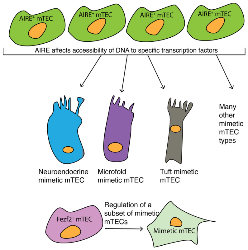
Heretofore, I have discussed mTECs as if they are one homogeneous population. It has long been known that in fact, mTECs are heterogeneous, but the number of different types of mTECs was unknown. A game-changing breakthrough in our appreciation for the different types of mTECs came with the use of high-throughput single-cell RNA-sequencing (scRNA-seq) analysis [113,114,115,116,117]. Eleven types of mTECs have been identified [118], and the number may well increase as techniques for studying them improve. Using single-cell assay for transposase-accessible chromatin with sequencing (scATAC-seq), the Benoist and Mathis group investigated tissue-specific antigen expression in individual mTECs [110]. They confirmed that AIRE and “lineage-defining” transcription factors found in diverse tissues promote expression of a variety of tissue-specific genes. AIRE promotes chromatin accessibility at specific genes in most (but not all) mTECs. By contrast, lineage-defining transcription factors are active in distinct subsets of mTECs [110]. Each cluster is named after its cell counterpart (e.g., keratinocyte, ciliated, secretory/neuroendocrine, microfold, tuft, enterocyte/hepatocyte; Figure 5). These mTEC are regarded as “mimetic cells” because in terms of gene expression, they mimic cell types in non-lymphoid tissues. Perhaps surprisingly, the transcription factors that mediate gene expression in specific peripheral tissues are not involved in tissue-specific gene expression by mimetic mTECs [119].
Expression of specific antigens is sufficient to induce tolerance to that antigen [110]. Significantly, mimetic mTECs (first identified in mice) also occur in the human thymus [110]. The fundamental importance of mimetic mTECs in self-tolerance in humans is underscored by genome-wide association studies (GWAS). Two such studies implicated the lineage-defining transcription factors HNF4α and SPIB in inflammatory bowel disease [122] and primary biliary cirrhosis [123], respectively.
Another transcription factor with far-reaching effects is the previously mentioned Runx3. It plays a crucial role in mTEC development as well as in the expression of tissue-specific genes in mTECs [124]. Deletion of the Runx3 gene in TECs causes a loss of transient amplifying cell thymic epithelial cells and of AIRE+ mTECs [124]. The effect on the transit-amplifying cells is notable because they give rise to AIRE+ mTECs [115]. Thus, the lack of AIRE+ mTECs is likely to be a downstream consequence of its absence on transit-amplifying cells.
Beyond this, lack of Runx3 activity leads to drastically lower expression of tissue-specific genes in mTECs, resulting in diminished negative selection and reduced diversion of thymocytes to a Treg phenotype [124]. Mice lacking Runx3 develop signs of autoimmune disease. In humans, Runx3 polymorphisms correlate with risk of developing the autoimmune disease ankylosing spondylitis [125]. Also, low levels of Runx3 expression are observed in patients with systemic sclerosis [126]. Intriguingly, scRNA-seq data from human thymus tissue indicates that the Runx3 and Aire genes are coordinately expressed [124].
Expression of the zinc-finger transcription factor, Ikaros, also is required for AIRE+ mTEC development and tissue-specific antigen gene expression [127]. Ikaros also regulates the relative proportions of mimetic mTECs and influences their gene expression patterns [127]. Unsurprisingly, ablation of Ikzf1, the gene encoding Ikaros, results in autoimmunity [127].
A more recently described protein that is indispensable for AIRE-independent immune tolerance is Fezf2, a Kruppel-type zinc finger protein [121]. Fezf2 interacts with the chromatin remodeler Chd4 and other proteins to directly induce the expression of tissue-specific genes [128]. Fezf2 action influences the steady state composition of a small number of mimetic mTEC populations (Figure 5), including mimetic tuft cells [129]. Interestingly, Fezf2’s actions result both in the activation of the expression of some tissue specific genes and the repression of the expression of others [128,129]. The absence of Fezf2 in TECs results in a severe reduction in mTEC expression of Pou2f3 and Hnf4α transcription factors, which are involved in the development of tuft and entero-hepato mimetic mTECs, respectively [110,119]. However, deletion of Fezf2 in TECs does not lead to the loss of all mimetic mTECs [129]. Indeed, the number of mimetic mTECs was slightly elevated in mice lacking Fezf2 activity [129].
From all these studies three major points emerge. Firstly, there are several types of mTECs, each of which contributes to the representation in the medulla of a vast swath of the gene products expressed in the various tissues of the body. Secondly, lineage-defining transcription factors are essential for medullary expression of tissue-specific genes. Finally, the functioning of mimetic mTECs hinges both on chromatin remodeling and on transcription factors working in concert. It is important to recall, as stated above, that some transcription factors also affect mimetic mTEC differentiation. As already noted, Fezf2 influences the prevalence of certain mimetic mTECs. Runx3 also imposes retrains on the differentiation of some mimetic mTECS (Sin, et al., 2026). Finally, as stated previously, AIRE is implicated in homeostatic regulation of the thymic medulla [112].
Given the critical roles played by CD28-, CD4-, and CD8-mediated signaling during negative selection, it is advantageous that AIRE promotes expression of CD80 and of MHC molecules by mTECs [89,130]. Thus, collectively, mTECs are armed both with MHC molecules presenting diverse self-peptides and the necessary ligands of TCR-adjacent accessory molecules to maximize the likelihood of efficient negative selection. Finally, some mTECs produce CCL21, which recruits thymocytes and DCs to the thymic medulla [131,132,133]. Thus, together, mTECs fulfill the three critical roles essential for negative selection—the recruitment of positively selected thymocytes and other antigen presenting cells (such as DCs) to the medulla, the display of MHC + self-peptides, and the expression of CD80 to maximize signal transduction through the TCR.
13. The more, the better
Medullary DCs (mDCs) promote negative selection [134] and the diversion of CD4+ cells to a Treg phenotype. They also have been shown to mediate negative selection of MHC I-specific autoreactive thymocytes that otherwise would develop into CD8+ T cells [135,136]. Amongst the antigenic peptides presented by mDCs are peptides that are generated within mTECs and then subsequently are transferred to the mDCs [137]. This mTEC to mDC peptide transfer is important because of the extraordinary array of peptides generated by mimetic mTECs. Like mTECs, mDCs are heterogeneous. A recent study [138] used scRNA-seq and functional assays to characterize mDCs and found that there are seven distinct mDC subsets. These mDC subsets are functionally distinct from each other in terms of the efficiency with which they mediate negative selection versus induction of Tregs [137,138]. Together, mTECs and mDCs greatly curtail the survival of thymocytes expressing self-reactive TCRs.
14. Three kisses and a promise
The integration of signals (via the various components of their signaling machinery) from the TCR complex, from the coreceptor CD4 or CD8, and from CD28 is essential for effective silencing of autoreactive thymocytes. Failure to do so has a high cost: Any autoreactive thymocyte that escapes negative selection and fails to differentiate into a Treg (in the case of a presumptive CD4+ thymocyte) or into an IEL or DN T cell (in the case of a presumptive CD8+ thymocyte) may later cause autoimmune disease. In this regard, the fact that the various mTECs and mDC subsets have non-overlapping capabilities is particularly reassuring. These diverse sets of “self-peptide-presenting” APCs provide multiple opportunities to eliminate—or failing that—to convert an autoreactive thymocyte into a cell that promises self-tolerance.
15. Future directions—circling back to the thymic medulla as a whole
Single-cell technologies have enabled the identification of diverse thymic medullary cells and their individual contributions to self-tolerance, which is indeed wonderful. But single-cell approaches are of limited value in determining how these cells work collectively to effectuate self-tolerance of the thymocytes that are born daily. Now, we must devise experimental systems and tools to determine how the medulla orchestrates all its individual enforcers of self-tolerance. Solving this next mystery promises to be an exciting challenge. I can hardly wait to see how it unfolds.
Acknowledgments
The author thanks Nicholas Fong for contributing to the preparation of Figure 3 and Shahar Dubiner, Stephanie Smith-Berdan, and Angela Deguzman for reading and commenting on the manuscript. This article is dedicated to the memory of Glenn George Capps, PhD, who for 30 years was a constant source of inspiration and insight.
Conflicts of Interest
The author declares on conflicts of interest.
References
- M. S. Krangel, “Mechanics of T cell receptor gene rearrangement,” Current Opinion in Immunology, vol. 21, no. 2, pp. 133–139, Apr. 2009. [CrossRef]
- Taniuchi, “CD4 Helper and CD8 Cytotoxic T Cell Differentiation,” Annu Rev Immunol, vol. 36, pp. 579–601, Apr. 2018. [CrossRef]
- L. Klein, B. Kyewski, P. M. Allen, and K. A. Hogquist, “Positive and negative selection of the T cell repertoire: what thymocytes see (and don’t see),” Nat Rev Immunol, vol. 14, no. 6, pp. 377–391, Jun. 2014. [CrossRef]
- M. M. Karimi et al., “The order and logic of CD4 versus CD8 lineage choice and differentiation in mouse thymus,” Nat Commun, vol. 12, no. 1, p. 99, Jan. 2021. [CrossRef]
- M. Shinzawa et al., “Reversal of the T cell immune system reveals the molecular basis for T cell lineage fate determination in the thymus,” Nat Immunol, vol. 23, no. 5, pp. 731–742, May 2022. [CrossRef]
- X. He et al., “The zinc finger transcription factor Th-POK regulates CD4 versus CD8 T-cell lineage commitment,” Nature, vol. 433, no. 7028, pp. 826–833, Feb. 2005. [CrossRef]
- M. A. Luckey, M. Y. Kimura, A. T. Waickman, L. Feigenbaum, A. Singer, and J.-H. Park, “The transcription factor ThPOK suppresses Runx3 and imposes CD4(+) lineage fate by inducing the SOCS suppressors of cytokine signaling,” Nat Immunol, vol. 15, no. 7, pp. 638–645, Jul. 2014. [CrossRef]
- T. Sato et al., “Dual functions of Runx proteins for reactivating CD8 and silencing CD4 at the commitment process into CD8 thymocytes,” Immunity, vol. 22, no. 3, pp. 317–328, Mar. 2005. [CrossRef]
- R. Setoguchi et al., “Repression of the transcription factor Th-POK by Runx complexes in cytotoxic T cell development,” Science, vol. 319, no. 5864, pp. 822–825, Feb. 2008. [CrossRef]
- Taniuchi et al., “Differential requirements for Runx proteins in CD4 repression and epigenetic silencing during T lymphocyte development,” Cell, vol. 111, no. 5, pp. 621–633, Nov. 2002. [CrossRef]
- T. Egawa, R. E. Tillman, Y. Naoe, I. Taniuchi, and D. R. Littman, “The role of the Runx transcription factors in thymocyte differentiation and in homeostasis of naive T cells,” J Exp Med, vol. 204, no. 8, pp. 1945–1957, Aug. 2007. [CrossRef]
- Z. Steier, E. J. Y. Kim, D. A. Aylard, and E. A. Robey, “The CD4 Versus CD8 T Cell Fate Decision: A Multiomics-Informed Perspective,” Annu Rev Immunol, vol. 42, no. 1, pp. 235–258, Jun. 2024. [CrossRef]
- J. H. Esensten, Y. A. Helou, G. Chopra, A. Weiss, and J. A. Bluestone, “CD28 Costimulation: From Mechanism to Therapy,” Immunity, vol. 44, no. 5, pp. 973–988, May 2016. [CrossRef]
- H. Gmünder and W. Lesslauer, “A 45-kDa human T-cell membrane glycoprotein functions in the regulation of cell proliferative responses,” Eur J Biochem, vol. 142, no. 1, pp. 153–160, Jul. 1984. [CrossRef]
- N. K. Damle, L. V. Doyle, L. S. Grosmaire, and J. A. Ledbetter, “Differential regulatory signals delivered by antibody binding to the CD28 (Tp44) molecule during the activation of human T lymphocytes,” J Immunol, vol. 140, no. 6, pp. 1753–1761, Mar. 1988.
- N. K. Damle and L. V. Doyle, “Stimulation via the CD3 and CD28 molecules induces responsiveness to IL-4 in CD4+CD29+CD45R- memory T lymphocytes,” J Immunol, vol. 143, no. 6, pp. 1761–1767, Sep. 1989.
- J. A. Gross, T. St John, and J. P. Allison, “The murine homologue of the T lymphocyte antigen CD28. Molecular cloning and cell surface expression,” J Immunol, vol. 144, no. 8, pp. 3201–3210, Apr. 1990.
- Shahinian et al., “Differential T cell costimulatory requirements in CD28-deficient mice,” Science, vol. 261, no. 5121, pp. 609–612, Jul. 1993. [CrossRef]
- M. F. Bachmann et al., “T cell responses are governed by avidity and co-stimulatory thresholds,” Eur J Immunol, vol. 26, no. 9, pp. 2017–2022, Sep. 1996. [CrossRef]
- J. S. Boomer and J. M. Green, “An enigmatic tail of CD28 signaling,” Cold Spring Harb Perspect Biol, vol. 2, no. 8, p. a002436, Aug. 2010. [CrossRef]
- M. F. Bachmann et al., “Distinct roles for LFA-1 and CD28 during activation of naive T cells: adhesion versus costimulation,” Immunity, vol. 7, no. 4, pp. 549–557, Oct. 1997. [CrossRef]
- J. A. Gross, E. Callas, and J. P. Allison, “Identification and distribution of the costimulatory receptor CD28 in the mouse,” J Immunol, vol. 149, no. 2, pp. 380–388, Jul. 1992.
- K. Inaba et al., “The tissue distribution of the B7-2 costimulator in mice: abundant expression on dendritic cells in situ and during maturation in vitro,” J Exp Med, vol. 180, no. 5, pp. 1849–1860, Nov. 1994. [CrossRef]
- H. Reiser and E. E. Schneeberger, “The costimulatory molecule B7 is expressed in the medullary region of the murine thymus,” Immunology, vol. 81, no. 4, pp. 532–537, Apr. 1994.
- S. Zuklys, G. Balciunaite, A. Agarwal, E. Fasler-Kan, E. Palmer, and G. A. Holländer, “Normal thymic architecture and negative selection are associated with Aire expression, the gene defective in the autoimmune-polyendocrinopathy-candidiasis-ectodermal dystrophy (APECED).,” J Immunol, vol. 165, no. 4, pp. 1976–1983, Aug. 2000. [CrossRef]
- T. Ueno et al., “CCR7 signals are essential for cortex-medulla migration of developing thymocytes,” J Exp Med, vol. 200, no. 4, pp. 493–505, Aug. 2004. [CrossRef]
- V. Griffith, M. Fallahi, H. Nakase, M. Gosink, B. Young, and H. T. Petrie, “Spatial mapping of thymic stromal microenvironments reveals unique features influencing T lymphoid differentiation,” Immunity, vol. 31, no. 6, pp. 999–1009, Dec. 2009. [CrossRef]
- P. W. Kim, Z.-Y. J. Sun, S. C. Blacklow, G. Wagner, and M. J. Eck, “A zinc clasp structure tethers Lck to T cell coreceptors CD4 and CD8,” Science, vol. 301, no. 5640, pp. 1725–1728, Sep. 2003. [CrossRef]
- N. M. Woessner, V. Uleri, O. Stepanek, and S. Minguet, “The TCR and LCK: foundations for T-cell activation and therapeutic innovation,” Front Immunol, vol. 16, p. 1737013, 2025. [CrossRef]
- P. S. Kabouridis, A. I. Magee, and S. C. Ley, “S-acylation of LCK protein tyrosine kinase is essential for its signalling function in T lymphocytes,” EMBO J, vol. 16, no. 16, pp. 4983–4998, Aug. 1997. [CrossRef]
- W. Zhang, J. Sloan-Lancaster, J. Kitchen, R. P. Trible, and L. E. Samelson, “LAT: the ZAP-70 tyrosine kinase substrate that links T cell receptor to cellular activation,” Cell, vol. 92, no. 1, pp. 83–92, Jan. 1998. [CrossRef]
- R. Bosselut et al., “Role of CD8beta domains in CD8 coreceptor function: importance for MHC I binding, signaling, and positive selection of CD8+ T cells in the thymus,” Immunity, vol. 12, no. 4, pp. 409–418, Apr. 2000. [CrossRef]
- K. Bi et al., “Antigen-induced translocation of PKC-theta to membrane rafts is required for T cell activation,” Nat Immunol, vol. 2, no. 6, pp. 556–563, Jun. 2001. [CrossRef]
- M. C. Verí et al., “Membrane raft-dependent regulation of phospholipase Cgamma-1 activation in T lymphocytes,” Mol Cell Biol, vol. 21, no. 20, pp. 6939–6950, Oct. 2001. [CrossRef]
- H. Shi and B. Shao, “LFA-1 Activation in T-Cell Migration and Immunological Synapse Formation,” Cells, vol. 12, no. 8, p. 1136, Apr. 2023. [CrossRef]
- T. A. Springer and M. L. Dustin, “Integrin inside-out signaling and the immunological synapse,” Curr Opin Cell Biol, vol. 24, no. 1, pp. 107–115, Feb. 2012. [CrossRef]
- R. Zamoyska, “The CD8 coreceptor revisited: one chain good, two chains better,” Immunity, vol. 1, no. 4, pp. 243–246, Jul. 1994. [CrossRef]
- V. Renard, P. Romero, E. Vivier, B. Malissen, and I. F. Luescher, “CD8 beta increases CD8 coreceptor function and participation in TCR-ligand binding,” J Exp Med, vol. 184, no. 6, pp. 2439–2444, Dec. 1996. [CrossRef]
- T. Witte, R. Spoerl, and H. C. Chang, “The CD8beta ectodomain contributes to the augmented coreceptor function of CD8alphabeta heterodimers relative to CD8alphaalpha homodimers,” Cell Immunol, vol. 191, no. 2, pp. 90–96, Feb. 1999. [CrossRef]
- J. M. Turner, M. H. Brodsky, B. A. Irving, S. D. Levin, R. M. Perlmutter, and D. R. Littman, “Interaction of the unique N-terminal region of tyrosine kinase p56lck with cytoplasmic domains of CD4 and CD8 is mediated by cysteine motifs,” Cell, vol. 60, no. 5, pp. 755–765, Mar. 1990. [CrossRef]
- V. Horkova et al., “Dynamics of the Coreceptor-LCK Interactions during T Cell Development Shape the Self-Reactivity of Peripheral CD4 and CD8 T Cells,” Cell Rep, vol. 30, no. 5, pp. 1504-1514.e7, Feb. 2020. [CrossRef]
- M. Watanabe, Y. Lu, M. Breen, and R. J. Hodes, “B7-CD28 co-stimulation modulates central tolerance via thymic clonal deletion and Treg generation through distinct mechanisms,” Nat Commun, vol. 11, no. 1, p. 6264, Dec. 2020. [CrossRef]
- H. S. Teh and S. J. Teh, “The affinity/avidity and length of exposure to the deleting ligand determine dependence on CD28 for the efficient deletion of self-specific CD4+CD8+ thymocytes,” Cell Immunol, vol. 207, no. 2, pp. 100–109, Feb. 2001. [CrossRef]
- M. L. Dustin and J. T. Groves, “Receptor signaling clusters in the immune synapse,” Annu Rev Biophys, vol. 41, pp. 543–556, 2012. [CrossRef]
- D. Filipp, O. Ballek, and J. Manning, “Lck, Membrane Microdomains, and TCR Triggering Machinery: Defining the New Rules of Engagement,” Front Immunol, vol. 3, p. 155, 2012. [CrossRef]
- K. Simons and G. van Meer, “Lipid sorting in epithelial cells,” Biochemistry, vol. 27, no. 17, pp. 6197–6202, Aug. 1988. [CrossRef]
- M. Martin, H. Schneider, A. Azouz, and C. E. Rudd, “Cytotoxic T lymphocyte antigen 4 and CD28 modulate cell surface raft expression in their regulation of T cell function,” J Exp Med, vol. 194, no. 11, pp. 1675–1681, Dec. 2001. [CrossRef]
- K. Simons and D. Toomre, “Lipid rafts and signal transduction.,” Nat Rev Mol Cell Biol, vol. 1, no. 1, pp. 31–39, Oct. 2000. [CrossRef]
- D. J. Pang, A. C. Hayday, and M.-J. Bijlmakers, “CD8 Raft localization is induced by its assembly into CD8alpha beta heterodimers, Not CD8alpha alpha homodimers.,” J Biol Chem, vol. 282, no. 18, pp. 13884–13894, May 2007. [CrossRef]
- Arcaro et al., “Essential role of CD8 palmitoylation in CD8 coreceptor function.,” J Immunol, vol. 165, no. 4, pp. 2068–2076, Aug. 2000. [CrossRef]
- Arcaro et al., “CD8beta endows CD8 with efficient coreceptor function by coupling T cell receptor/CD3 to raft-associated CD8/p56(lck) complexes.,” J Exp Med, vol. 194, no. 10, pp. 1485–1495, Nov. 2001. [CrossRef]
- M. E. Crooks and D. R. Littman, “Disruption of T lymphocyte positive and negative selection in mice lacking the CD8 beta chain.,” Immunity, vol. 1, no. 4, pp. 277–285, Jul. 1994. [CrossRef]
- H. Y. Irie et al., “The cytoplasmic domain of CD8 beta regulates Lck kinase activation and CD8 T cell development.,” J Immunol, vol. 161, no. 1, pp. 183–191, Jul. 1998.
- K. Nakayama et al., “Requirement for CD8 beta chain in positive selection of CD8-lineage T cells.,” Science, vol. 263, no. 5150, pp. 1131–1133, Feb. 1994. [CrossRef]
- M. J. Barnden, W. R. Heath, and F. R. Carbone, “Down-modulation of CD8 beta-chain in response to an altered peptide ligand enables developing thymocytes to escape negative selection.,” Cell Immunol, vol. 175, no. 2, pp. 111–119, Feb. 1997. [CrossRef]
- Chidgey and R. Boyd, “Agonist peptide modulates T cell selection thresholds through qualitative and quantitative shifts in CD8 co-receptor expression.,” Int Immunol, vol. 9, no. 10, pp. 1527–1536, Oct. 1997. [CrossRef]
- J. Leishman et al., “Precursors of functional MHC class I- or class II-restricted CD8alphaalpha(+) T cells are positively selected in the thymus by agonist self-peptides,” Immunity, vol. 16, no. 3, pp. 355–64, Mar. 2002. [CrossRef]
- D. McDonald, J. J. Bunker, I. E. Ishizuka, B. Jabri, and A. Bendelac, “Elevated T cell receptor signaling identifies a thymic precursor to the TCRαβ(+)CD4(-)CD8β(-) intraepithelial lymphocyte lineage,” Immunity, vol. 41, no. 2, pp. 219–229, Aug. 2014. [CrossRef]
- R. Ruscher, R. L. Kummer, Y. J. Lee, S. C. Jameson, and K. A. Hogquist, “CD8αα intraepithelial lymphocytes arise from two main thymic precursors.,” Nat Immunol, vol. 18, no. 7, pp. 771–779, Jul. 2017. [CrossRef]
- S. Mayans et al., “αβT cell receptors expressed by CD4(-)CD8αβ(-) intraepithelial T cells drive their fate into a unique lineage with unusual MHC reactivities,” Immunity, vol. 41, no. 2, pp. 207–218, Aug. 2014. [CrossRef]
- L. A. Pobezinsky et al., “Clonal deletion and the fate of autoreactive thymocytes that survive negative selection,” Nat Immunol, vol. 13, no. 6, pp. 569–578, Jun. 2012. [CrossRef]
- N. S. Kurd et al., “Factors that influence the thymic selection of CD8αα intraepithelial lymphocytes,” Mucosal Immunol, vol. 14, no. 1, pp. 68–79, Jan. 2021. [CrossRef]
- M. A. McGargill and K. A. Hogquist, “Antigen-induced coreceptor down-regulation on thymocytes is not a result of apoptosis,” J Immunol, vol. 162, no. 3, pp. 1237–45, Feb. 1999.
- M. C. Zúñiga et al., “Fine tuning of TCR signaling via CD8αβ and PD-1 and the fate of autoreactive thymocytes during negative selection,” Immunohorizons, vol. 10, no. 3, p. vlaf078, Mar. 2026. [CrossRef]
- Y. Ishida, Y. Agata, K. Shibahara, and T. Honjo, “Induced expression of PD-1, a novel member of the immunoglobulin gene superfamily, upon programmed cell death,” EMBO J, vol. 11, no. 11, pp. 3887–3895, Nov. 1992. [CrossRef]
- G. J. Freeman et al., “Engagement of the PD-1 immunoinhibitory receptor by a novel B7 family member leads to negative regulation of lymphocyte activation,” J Exp Med, vol. 192, no. 7, pp. 1027–1034, Oct. 2000. [CrossRef]
- R. V. Parry et al., “CTLA-4 and PD-1 receptors inhibit T-cell activation by distinct mechanisms,” Mol Cell Biol, vol. 25, no. 21, pp. 9543–9553, Nov. 2005. [CrossRef]
- M. E. Keir, Y. E. Latchman, G. J. Freeman, and A. H. Sharpe, “Programmed death-1 (PD-1):PD-ligand 1 interactions inhibit TCR-mediated positive selection of thymocytes,” J Immunol, vol. 175, no. 11, pp. 7372–7379, Dec. 2005. [CrossRef]
- K.-A. Sheppard et al., “PD-1 inhibits T-cell receptor induced phosphorylation of the ZAP70/CD3zeta signalosome and downstream signaling to PKCtheta.,” FEBS Lett, vol. 574, no. 1–3, pp. 37–41, Sep. 2004. [CrossRef]
- R. V. Parry et al., “CTLA-4 and PD-1 receptors inhibit T-cell activation by distinct mechanisms.,” Mol Cell Biol, vol. 25, no. 21, pp. 9543–9553, Nov. 2005. [CrossRef]
- T. Yokosuka, M. Takamatsu, W. Kobayashi-Imanishi, A. Hashimoto-Tane, M. Azuma, and T. Saito, “Programmed cell death 1 forms negative costimulatory microclusters that directly inhibit T cell receptor signaling by recruiting phosphatase SHP2.,” J Exp Med, vol. 209, no. 6, pp. 1201–1217, Jun. 2012. [CrossRef]
- R. Mizuno et al., “PD-1 Primarily Targets TCR Signal in the Inhibition of Functional T Cell Activation.,” Front Immunol, vol. 10, p. 630, 2019. [CrossRef]
- White et al., “Programmed Cell Death-1 (PD-1) anchoring to the GPI-linked co-receptor CD48 reveals a novel mechanism to modulate PD-1-dependent inhibition of human T cells.,” Mol Immunol, vol. 156, pp. 31–38, Apr. 2023. [CrossRef]
- Pralle, P. Keller, E. L. Florin, K. Simons, and J. K. Hörber, “Sphingolipid-cholesterol rafts diffuse as small entities in the plasma membrane of mammalian cells.,” J Cell Biol, vol. 148, no. 5, pp. 997–1008, Mar. 2000. [CrossRef]
- T. Harder, P. Scheiffele, P. Verkade, and K. Simons, “Lipid domain structure of the plasma membrane revealed by patching of membrane components,” J Cell Biol, vol. 141, no. 4, pp. 929–942, May 1998. [CrossRef]
- P. W. Janes, S. C. Ley, and A. I. Magee, “Aggregation of lipid rafts accompanies signaling via the T cell antigen receptor,” J Cell Biol, vol. 147, no. 2, pp. 447–461, Oct. 1999. [CrossRef]
- S. Sakaguchi, T. Yamaguchi, T. Nomura, and M. Ono, “Regulatory T cells and immune tolerance,” Cell, vol. 133, no. 5, pp. 775–787, May 2008. [CrossRef]
- S. Sakaguchi, K. Wing, Y. Onishi, P. Prieto-Martin, and T. Yamaguchi, “Regulatory T cells: how do they suppress immune responses?,” Int Immunol, vol. 21, no. 10, pp. 1105–1111, Oct. 2009. [CrossRef]
- K. Wing and S. Sakaguchi, “Regulatory T cells exert checks and balances on self tolerance and autoimmunity,” Nat Immunol, vol. 11, no. 1, pp. 7–13, Jan. 2010. [CrossRef]
- M. E. Crooks and D. R. Littman, “Disruption of T lymphocyte positive and negative selection in mice lacking the CD8 beta chain,” Immunity, vol. 1, no. 4, pp. 277–285, Jul. 1994. [CrossRef]
- T. L. Denning et al., “Mouse TCRalphabeta+CD8alphaalpha intraepithelial lymphocytes express genes that down-regulate their antigen reactivity and suppress immune responses,” J Immunol, vol. 178, no. 7, pp. 4230–4239, Apr. 2007. [CrossRef]
- L. W. Peterson and D. Artis, “Intestinal epithelial cells: regulators of barrier function and immune homeostasis.,” Nat Rev Immunol, vol. 14, no. 3, pp. 141–153, Mar. 2014. [CrossRef]
- M. Sponaas et al., “Tolerance induction by elimination of subsets of self-reactive thymocytes,” Int Immunol, vol. 6, no. 10, pp. 1593–604, Oct. 1994. [CrossRef]
- J. Caveno, Y. Zhang, B. Motyka, S. J. Teh, and H. S. Teh, “Functional similarity and differences between selection-independent CD4-CD8- alphabeta T cells and positively selected CD8 T cells expressing the same TCR and the induction of anergy in CD4-CD8- alphabeta T cells in antigen-expressing mice.,” J Immunol, vol. 163, no. 3, pp. 1222–1229, Aug. 1999.
- J. J. Priatel, O. Utting, and H. S. Teh, “TCR/self-antigen interactions drive double-negative T cell peripheral expansion and differentiation into suppressor cells.,” J Immunol, vol. 167, no. 11, pp. 6188–6194, Dec. 2001. [CrossRef]
- S. N. Klose et al., “The transcription factor T-bet is induced by IL-15 and thymic agonist selection and controls CD8αα(+) intraepithelial lymphocyte development,” Immunity, vol. 41, no. 2, pp. 230–243, Aug. 2014. [CrossRef]
- S. N. Klose, J. F. Hummel, L. Faller, Y. d’Hargues, K. Ebert, and Y. Tanriver, “A committed postselection precursor to natural TCRαβ+ intraepithelial lymphocytes,” Mucosal Immunol, vol. 11, no. 2, pp. 333–344, Mar. 2018. [CrossRef]
- Nita, M. Nishiyama, Y. Muto, and K. I. Nakayama, “FBXL12 regulates T-cell differentiation in a cell-autonomous manner,” Genes Cells, vol. 21, no. 5, pp. 517–524, May 2016. [CrossRef]
- M. Irla, “Thymic Crosstalk: An Overview of the Complex Cellular Interactions that Control the Establishment of T-Cell Tolerance,” Adv Exp Med Biol, vol. 1471, pp. 177–197, 2025. [CrossRef]
- N. Kurd and E. A. Robey, “T-cell selection in the thymus: a spatial and temporal perspective.,” Immunol Rev, vol. 271, no. 1, pp. 114–126, May 2016. [CrossRef]
- M. Vobořil, S. Xuan, and K. A. Hogquist, “Thymic Dendritic Cells Revisited,” Immunol Rev, vol. 336, no. 1, p. e70076, Nov. 2025. [CrossRef]
- G. Anderson, E. J. Cosway, K. D. James, I. Ohigashi, and Y. Takahama, “Generation and repair of thymic epithelial cells,” Journal of Experimental Medicine, vol. 221, no. 10, p. e20230894, Oct. 2024. [CrossRef]
- G. Anderson and Y. Takahama, “Thymic epithelial cells: working class heroes for T cell development and repertoire selection,” Trends Immunol, vol. 33, no. 6, pp. 256–263, Jun. 2012. [CrossRef]
- L. Klein and E. Petrozziello, “Antigen presentation for central tolerance induction,” Nat Rev Immunol, vol. 25, no. 1, pp. 57–72, Jan. 2025. [CrossRef]
- S. Murata et al., “Regulation of CD8+ T cell development by thymus-specific proteasomes,” Science, vol. 316, no. 5829, pp. 1349–1353, Jun. 2007. [CrossRef]
- T. Nitta et al., “Thymoproteasome shapes immunocompetent repertoire of CD8+ T cells,” Immunity, vol. 32, no. 1, pp. 29–40, Jan. 2010. [CrossRef]
- K. Takada et al., “TCR affinity for thymoproteasome-dependent positively selecting peptides conditions antigen responsiveness in CD8(+) T cells,” Nat Immunol, vol. 16, no. 10, pp. 1069–1076, Oct. 2015. [CrossRef]
- K. Sasaki et al., “Thymoproteasomes produce unique peptide motifs for positive selection of CD8(+) T cells,” Nat Commun, vol. 6, p. 7484, Jun. 2015. [CrossRef]
- J. von Rohrscheidt et al., “Thymic CD4 T cell selection requires attenuation of March8-mediated MHCII turnover in cortical epithelial cells through CD83,” J Exp Med, vol. 213, no. 9, pp. 1685–1694, Aug. 2016. [CrossRef]
- S. Kaul et al., “CD83 suppresses endogenous March-I-dependent MHC class II ubiquitination, endocytosis, and degradation,” Proc Natl Acad Sci U S A, vol. 122, no. 21, p. e2504077122, May 2025. [CrossRef]
- M. S. Anderson et al., “Projection of an immunological self shadow within the thymus by the aire protein,” Science, vol. 298, no. 5597, pp. 1395–1401, Nov. 2002. [CrossRef]
- S. Koh, R. E. Kingston, C. Benoist, and D. Mathis, “Global relevance of Aire binding to hypomethylated lysine-4 of histone-3,” Proc Natl Acad Sci U S A, vol. 107, no. 29, pp. 13016–13021, Jul. 2010. [CrossRef]
- M. Waterfield et al., “The transcriptional regulator Aire coopts the repressive ATF7ip-MBD1 complex for the induction of immunotolerance,” Nat Immunol, vol. 15, no. 3, pp. 258–265, Mar. 2014. [CrossRef]
- P. Peterson, T. Org, and A. Rebane, “Transcriptional regulation by AIRE: molecular mechanisms of central tolerance,” Nat Rev Immunol, vol. 8, no. 12, pp. 948–957, Dec. 2008. [CrossRef]
- A. Speck-Hernandez et al., “Aire Disruption Influences the Medullary Thymic Epithelial Cell Transcriptome and Interaction With Thymocytes,” Front Immunol, vol. 9, p. 964, 2018. [CrossRef]
- G. A. Passos, C. A. Speck-Hernandez, A. F. Assis, and D. A. Mendes-da-Cruz, “Update on Aire and thymic negative selection,” Immunology, vol. 153, no. 1, pp. 10–20, Jan. 2018. [CrossRef]
- C.-S. Hsieh, H.-M. Lee, and C.-W. J. Lio, “Selection of regulatory T cells in the thymus,” Nat Rev Immunol, vol. 12, no. 3, pp. 157–167, Feb. 2012. [CrossRef]
- S. Z. Josefowicz, L.-F. Lu, and A. Y. Rudensky, “Regulatory T cells: mechanisms of differentiation and function,” Annu Rev Immunol, vol. 30, pp. 531–564, 2012. [CrossRef]
- M. Guerau-de-Arellano, M. Martinic, C. Benoist, and D. Mathis, “Neonatal tolerance revisited: a perinatal window for Aire control of autoimmunity,” J Exp Med, vol. 206, no. 6, pp. 1245–1252, Jun. 2009. [CrossRef]
- A. Michelson, K. Hase, T. Kaisho, C. Benoist, and D. Mathis, “Thymic epithelial cells co-opt lineage-defining transcription factors to eliminate autoreactive T cells,” Cell, vol. 185, no. 14, pp. 2542-2558.e18, Jul. 2022. [CrossRef]
- S. Malchow, D. S. Leventhal, V. Lee, S. Nishi, N. D. Socci, and P. A. Savage, “Aire Enforces Immune Tolerance by Directing Autoreactive T Cells into the Regulatory T Cell Lineage,” Immunity, vol. 44, no. 5, pp. 1102–1113, May 2016. [CrossRef]
- P. Peterson, “AIRE’s Complex Role Beyond Promiscuous Gene Expression,” Immunol Rev, vol. 337, no. 1, p. e70091, Jan. 2026. [CrossRef]
- C. Bornstein et al., “Single-cell mapping of the thymic stroma identifies IL-25-producing tuft epithelial cells,” Nature, vol. 559, no. 7715, pp. 622–626, Jul. 2018. [CrossRef]
- C. N. Miller et al., “Thymic tuft cells promote an IL-4-enriched medulla and shape thymocyte development,” Nature, vol. 559, no. 7715, pp. 627–631, Jul. 2018. [CrossRef]
- K. L. Wells et al., “Combined transient ablation and single-cell RNA-sequencing reveals the development of medullary thymic epithelial cells,” Elife, vol. 9, p. e60188, Nov. 2020. [CrossRef]
- T. Miyao et al., “Integrative analysis of scRNA-seq and scATAC-seq revealed transit-amplifying thymic epithelial cells expressing autoimmune regulator,” Elife, vol. 11, p. e73998, May 2022. [CrossRef]
- M. Matsumoto, H. Yoshida, K. Tsuneyama, T. Oya, and M. Matsumoto, “Revisiting Aire and tissue-restricted antigens at single-cell resolution,” Front Immunol, vol. 14, p. 1176450, 2023. [CrossRef]
- R. Muro and T. Nitta, “Breaking the upper limit of mTEC heterogeneity,” Nat Immunol, vol. 27, no. 7, pp. 1317–1319, Jul. 2026. [CrossRef]
- S. Lammers et al., “Ehf and Fezf2 regulate late medullary thymic epithelial cell and thymic tuft cell development,” Front Immunol, vol. 14, p. 1277365, 2023. [CrossRef]
- M. Giraud and P. Peterson, “The Autoimmune Regulator (AIRE) Gene, The Master Activator of Self-Antigen Expression in the Thymus,” Adv Exp Med Biol, vol. 1471, pp. 199–221, 2025. [CrossRef]
- H. Takaba et al., “Fezf2 Orchestrates a Thymic Program of Self-Antigen Expression for Immune Tolerance,” Cell, vol. 163, no. 4, pp. 975–987, Nov. 2015. [CrossRef]
- UK IBD Genetics Consortium et al., “Genome-wide association study of ulcerative colitis identifies three new susceptibility loci, including the HNF4A region,” Nat Genet, vol. 41, no. 12, pp. 1330–1334, Dec. 2009. [CrossRef]
- X. Liu et al., “Genome-wide meta-analyses identify three loci associated with primary biliary cirrhosis,” Nat Genet, vol. 42, no. 8, pp. 658–660, Aug. 2010. [CrossRef]
- J. H. Sin, S. H. Phillips, A. V. Parent, and M. R. Waterfield, “Runx3 instructs Aire+ mTEC development, TSA gene expression, and central tolerance,” Journal of Autoimmunity, vol. 161, p. 103573, Jun. 2026. [CrossRef]
- H. Liu et al., “RUNX3 Polymorphisms Affect the Risk of Ankylosing Spondylitis,” Med Sci Monit, vol. 26, p. e919528, May 2020. [CrossRef]
- J. Affandi et al., “Low RUNX3 expression alters dendritic cell function in patients with systemic sclerosis and contributes to enhanced fibrosis,” Ann Rheum Dis, vol. 78, no. 9, pp. 1249–1259, Sep. 2019. [CrossRef]
- J. H. Sin et al., “Ikaros is a principal regulator of Aire+ mTEC homeostasis, thymic mimetic cell diversity, and central tolerance,” Sci Immunol, vol. 8, no. 88, p. eabq3109, Oct. 2023. [CrossRef]
- Y. Tomofuji et al., “Chd4 choreographs self-antigen expression for central immune tolerance,” Nat Immunol, vol. 21, no. 8, pp. 892–901, Aug. 2020. [CrossRef]
- Ushio et al., “Functionally diverse thymic medullary epithelial cells interplay to direct central tolerance,” Cell Reports, vol. 43, no. 4, p. 114072, Apr. 2024. [CrossRef]
- J. Gäbler, J. Arnold, and B. Kyewski, “Promiscuous gene expression and the developmental dynamics of medullary thymic epithelial cells,” Eur J Immunol, vol. 37, no. 12, pp. 3363–3372, Dec. 2007. [CrossRef]
- M. Kozai et al., “Essential role of CCL21 in establishment of central self-tolerance in T cells,” J Exp Med, vol. 214, no. 7, pp. 1925–1935, Jul. 2017. [CrossRef]
- Ohigashi, H. Kyuma, E. Otsu, S. Hayashi, T. Takemoto, and Y. Takahama, “Ccl21a, Rather Than Ccl21b, is Essential for Thymocyte Migration in Mouse,” Eur J Immunol, vol. 55, no. 12, p. e70114, Dec. 2025. [CrossRef]
- C. Michalaki, M. Sharjeel, and J. E. Cowan, “From Development to Aging: Dynamic Roles of the Thymic Medulla in T Cell Biology,” Immunological Reviews, vol. 339, no. 1, p. e70118, May 2026. [CrossRef]
- C. Ohnmacht et al., “Constitutive ablation of dendritic cells breaks self-tolerance of CD4 T cells and results in spontaneous fatal autoimmunity,” Journal of Experimental Medicine, vol. 206, no. 3, pp. 549–559, Mar. 2009. [CrossRef]
- L. Wu and K. Shortman, “Heterogeneity of thymic dendritic cells,” Semin Immunol, vol. 17, no. 4, pp. 304–312, Aug. 2005. [CrossRef]
- H. Li et al., “IL-23 promotes TCR-mediated negative selection of thymocytes through the upregulation of IL-23 receptor and RORγt,” Nat Commun, vol. 5, p. 4259, Jul. 2014. [CrossRef]
- J. Březina, M. Vobořil, and D. Filipp, “Mechanisms of Direct and Indirect Presentation of Self-Antigens in the Thymus,” Front Immunol, vol. 13, p. 926625, 2022. [CrossRef]
- J. Srinivasan et al., “The differentiation and function of heterogeneous thymic dendritic cell subsets require signals provided by distinct thymocyte cell types,” Nat Immunol, vol. 27, no. 2, pp. 265–280, Feb. 2026. [CrossRef]
Figure 1.
CD4 and CD8 track the developmental progression of thymocytes expressing αβTCRs. Shown is a flow cytometry plot of mouse thymocyte populations identified by expression of CD4 and CD8. Above the flow cytometry plot are indicated the five major populations of developing thymocytes (DN, DP, DPlo, CD4, and CD8) are marked and their occurrence during thymocyte developmental progression.
Figure 1.
CD4 and CD8 track the developmental progression of thymocytes expressing αβTCRs. Shown is a flow cytometry plot of mouse thymocyte populations identified by expression of CD4 and CD8. Above the flow cytometry plot are indicated the five major populations of developing thymocytes (DN, DP, DPlo, CD4, and CD8) are marked and their occurrence during thymocyte developmental progression.
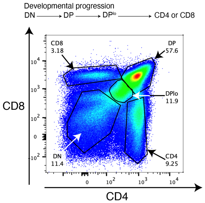
Figure 2.
Cellular interactions and locations of thymocyte development. For clarity purposes, molecules, such as TCR, CD4, CD8, CCR7, CCL21, etc. are not shown. These details are within the text. Small yellow circles represent secreted CCL21. The cell with a large X across it is an apoptotic cell.
Figure 2.
Cellular interactions and locations of thymocyte development. For clarity purposes, molecules, such as TCR, CD4, CD8, CCR7, CCL21, etc. are not shown. These details are within the text. Small yellow circles represent secreted CCL21. The cell with a large X across it is an apoptotic cell.
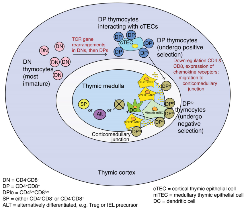
Figure 3.
Signal transduction events required for activation via the TCR occur within the lipid raft that constitutes the immunological synapse. As discussed in the text, binding of CD8αβ to MHC I (illustrated here) or of CD4 binding to MHC II launches a series of phosphorylation events that promote signal transduction through the TCR. The first step in the signal transduction cascade is the phosphorylation of CD3ζ ITAMs by p56lck. p56lck can occur both freely in the cytoplasm and bound to the coreceptor (in this case, CD8αβ). Arrows identify phosphorylation targets. Further details are provided in the text. Not shown are the adhesion molecules Lymphocyte function associated antigen 1(LFA-1, an integrin) [35] on the thymocyte membrane and its ligand, Intercellular Adhesion Molecule I (ICAM-1) [36] on the TEC membrane.
Figure 3.
Signal transduction events required for activation via the TCR occur within the lipid raft that constitutes the immunological synapse. As discussed in the text, binding of CD8αβ to MHC I (illustrated here) or of CD4 binding to MHC II launches a series of phosphorylation events that promote signal transduction through the TCR. The first step in the signal transduction cascade is the phosphorylation of CD3ζ ITAMs by p56lck. p56lck can occur both freely in the cytoplasm and bound to the coreceptor (in this case, CD8αβ). Arrows identify phosphorylation targets. Further details are provided in the text. Not shown are the adhesion molecules Lymphocyte function associated antigen 1(LFA-1, an integrin) [35] on the thymocyte membrane and its ligand, Intercellular Adhesion Molecule I (ICAM-1) [36] on the TEC membrane.
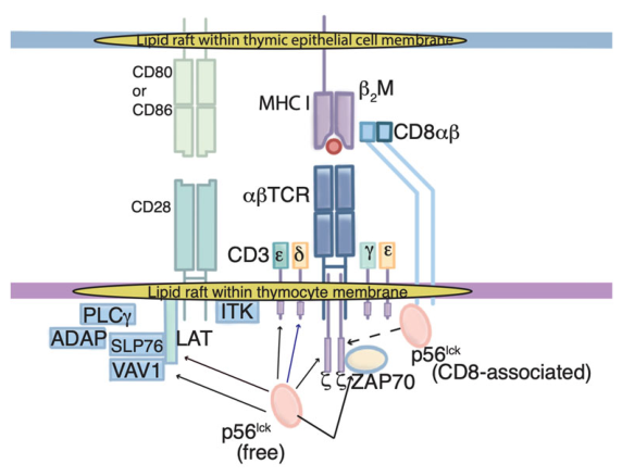
Figure 4.
Roles of CD8αβ and PD-1 in negative selection. Panel A: A high density of CD8αβ promotes strong signal transduction through the αβTCR, resulting in the apoptosis of autoreactive post-selection DPlo thymocytes, regardless of PD-1 levels. Panel B: When there is not enough PD-1 to attenuate signaling, even moderate levels of CD8 αβ sufficiently augment signaling through the αβTCR to promote apoptosis. Panel C: Autoreactive thymocytes can survive when PD-1 levels are high and CD8αβ levels are low to moderate. Panel D: Autoreactive thymocytes can survive when CD8αβ levels are very low regardless of the PD-1 level.
Figure 4.
Roles of CD8αβ and PD-1 in negative selection. Panel A: A high density of CD8αβ promotes strong signal transduction through the αβTCR, resulting in the apoptosis of autoreactive post-selection DPlo thymocytes, regardless of PD-1 levels. Panel B: When there is not enough PD-1 to attenuate signaling, even moderate levels of CD8 αβ sufficiently augment signaling through the αβTCR to promote apoptosis. Panel C: Autoreactive thymocytes can survive when PD-1 levels are high and CD8αβ levels are low to moderate. Panel D: Autoreactive thymocytes can survive when CD8αβ levels are very low regardless of the PD-1 level.
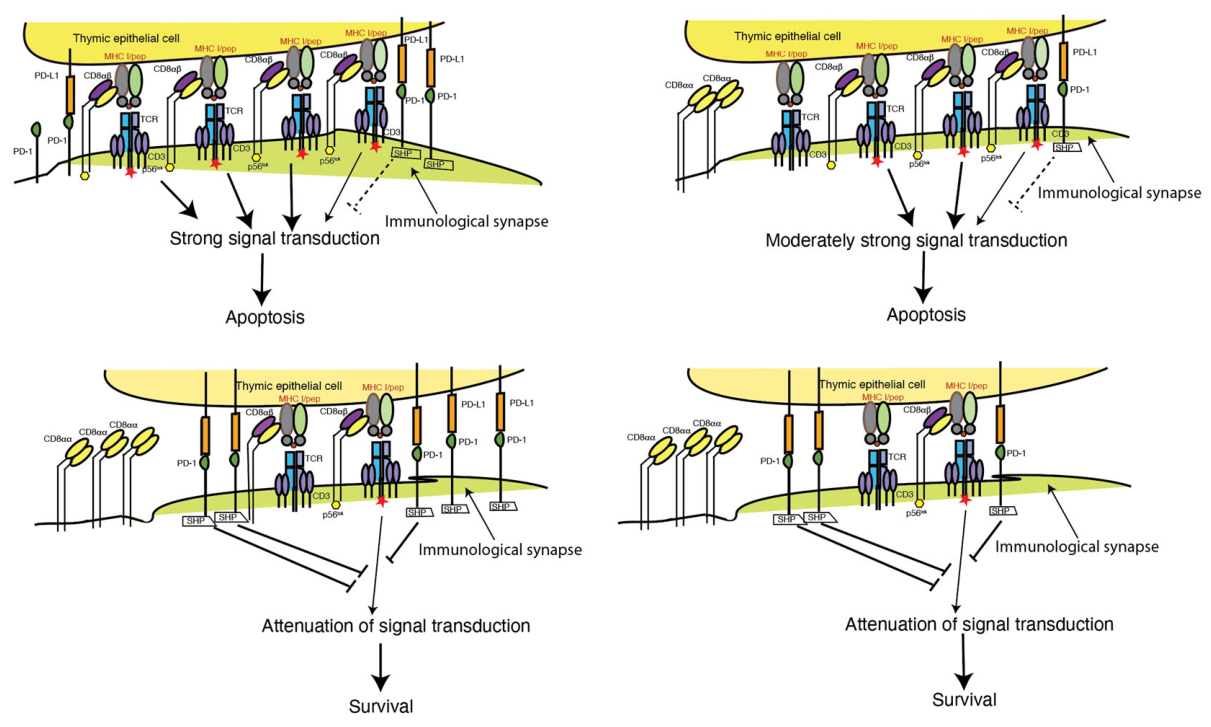
Figure 5.
Diverse mTECs express genes of different cell lineages. As discussed in the text, AIRE promotes the accessibility of otherwise transcriptionally silent genes to tissue-specific transcription factors [120]. In contrast, Fezf2 binds to specific DNA sequences and recruits transcriptional machinery to specific genes [121]. In this regard, it acts like a canonical transcription factor.
Figure 5.
Diverse mTECs express genes of different cell lineages. As discussed in the text, AIRE promotes the accessibility of otherwise transcriptionally silent genes to tissue-specific transcription factors [120]. In contrast, Fezf2 binds to specific DNA sequences and recruits transcriptional machinery to specific genes [121]. In this regard, it acts like a canonical transcription factor.
Disclaimer/Publisher’s Note: The statements, opinions and data contained in all publications are solely those of the individual author(s) and contributor(s) and not of MDPI and/or the editor(s). MDPI and/or the editor(s) disclaim responsibility for any injury to people or property resulting from any ideas, methods, instructions or products referred to in the content. |
© 2026 by the authors. Licensee MDPI, Basel, Switzerland. This article is an open access article distributed under the terms and conditions of the Creative Commons Attribution (CC BY) license (http://creativecommons.org/licenses/by/4.0/).
Copyright: This open access article is published under a Creative Commons CC BY 4.0 license, which permit the free download, distribution, and reuse, provided that the author and preprint are cited in any reuse.