

Submitted:
01 September 2026
Posted:
02 September 2026
You are already at the latest version
Abstract
In this paper, we propose a bioimpedance-based digital twin approach for organ-on-a-chip systems. Organ-on-a-chip platforms integrate microfluidic control, specific cell populations, extracellular matrices, mechanical stimuli and inter-tissue interactions to reproduce selected physiological functions under controlled conditions. Bioimpedance measurements allow non-destructive and repeated monitoring of cellular and tissue behavior. However, the electrical signal measured from an organ-on-a-chip system is not only dependent on the biological state. It is also influenced by the device geometry, electrode configuration, electrode-electrolyte interface, culture conditions and measurement stability. Therefore, an electrical signal cannot be directly considered as a biological state without considering the complete measurement system. We propose that the next development of organ-on-a-chip technology should move from continuous monitoring toward bioimpedance-based digital twins. The physical organ-on-a-chip system, integrated sensing system, measurement model, mechanistic model and data-driven model can be connected to form a continuously updated "experimental-computational" loop. In such a system, the objective is not only to monitor the sensor signal, but to estimate the current biological state, quantify the uncertainty associated with this estimation and predict the future response of the system to experimental perturbations. This approach could provide a transition from descriptive measurements of organ-on-a-chip systems toward predictive and adaptive microphysiological experiments.
Keywords:
bioimpedance
; digital twins
; organs-on-chips
; computational modeling
; bio-state estimation
1. Introduction
Over the past two decades, organ-on-a-chip technology has developed rapidly at the interface of microengineering, tissue engineering, cell biology and computational biomedical engineering [1,2]. The objective of organ-on-a-chip systems is not only to reduce the size of conventional cell culture systems, but to reproduce selected structural, mechanical, biochemical and functional properties of living tissues under experimentally controlled conditions [3]. Microfluidic perfusion allows continuous renewal of the chemical environment and provides controlled mass transport. Mechanical stimulation can be applied as cyclic strain, shear stress or other physical inputs [4,5]. Different cell types can also be organized across membranes or extracellular matrices to reproduce tissue interfaces and cell-cell interactions [6]. Based on these capabilities, organ-on-a-chip systems have been developed to reproduce different organs, including lung, intestine, liver, kidney, heart, vasculature, skin and brain [7,8,9]. These systems provide opportunities for disease modeling, drug development, toxicity testing and personalized medicine.
However, the increasing complexity of organ-on-a-chip systems also increases the requirement for appropriate measurements. Biological systems are continuously changing, while many conventional measurements provide information only at selected time points [10]. Optical imaging is generally performed at defined time points [11,12,13]. Biochemical analysis requires periodic sampling, and molecular characterization often requires fixation, lysis or destruction of the biological sample [14,15]. These methods provide specific biological information, but the temporal evolution of the biological state is difficult to obtain from discrete measurements. This limitation becomes important when the biological process itself is dynamic. For example, a barrier can progressively lose its integrity before a final measurable endpoint is reached [16]. A drug can induce a transient dysfunction followed by recovery. An inflammatory response can originate from small changes in cellular behavior before a large molecular response is detected. In addition, two tissues can have a similar final state while following different trajectories during the experiment. Therefore, when organ-on-a-chip systems are used to reproduce dynamic physiological processes, continuous observation of the biological state becomes important [17,18].
Integrated biosensors provide a possible solution to this problem [19,20]. Sensors integrated within organ-on-a-chip devices allow repeated measurements to be performed without terminating the experiment. Optical, electrochemical, mechanical and electrical sensing methods have therefore become important components of microphysiological systems. Among these approaches, bioimpedance is particularly attractive because electrical measurements can be performed without labeling or destroying the biological sample and can be repeated continuously. Electrical methods have been used to monitor epithelial and endothelial barrier function, cell adhesion and proliferation, as well as changes in the electrical properties of tissues [21,22].
However, continuous sensing does not directly provide biological interpretation. The sensor records an electrical response, while the biological processes producing this response can be only partially known. This problem is particularly important for impedance measurements. The measured impedance depends on the biological properties of the tissue, but also on the device geometry, electrode configuration, electrode-electrolyte interface, temperature, culture medium and stability of the electrical measurement [23]. Therefore, a change in impedance can have several possible origins. It can reflect a biological modification, a change in the measurement condition, or both. Consequently, impedance should not be considered as a direct readout of a specific biological process without considering the complete physical and measurement system.
The concept of a digital twin provides a possible framework to address this problem. A digital twin is not only a computational model of a physical system. The computational representation is continuously connected to the physical system through measurements and is updated when new observations are obtained. In biomedical applications, such computational approaches can be used for state estimation, mechanistic modeling, prediction and uncertainty quantification. Organ-on-a-chip systems are particularly suitable for this type of approach because many experimental parameters can be controlled. Device geometry, flow rate, mechanical stimulation and chemical exposure can be defined, while integrated sensors can provide continuous observations of the biological system. Therefore, the physical organ-on-a-chip system and the computational model can be connected during the experiment [24,25].
In this paper, we propose that the next development of organ-on-a-chip systems should not only focus on increasing the number of sensors, but on establishing a relationship between sensor observations and biological states. Bioimpedance can provide the continuous observation of the system, while computational models can be used to interpret the measurements and estimate the biological state. The updated model can then be used to predict the future response of the system. Such a bioimpedance-based digital twin could provide a framework to connect experimental organ-on-a-chip systems with computational modeling and to support predictive and adaptive experiments.
2. Current Status of Organ-on-a-Chip Systems
2.1. Measurement Gap in Dynamic Organ-on-Chip Systems
A major limitation in organ-on-a-chip research is the difference between the dynamic behavior of biological systems and the temporal resolution of conventional measurements. Cells continuously respond to changes in their environment. Ion transport, cell adhesion, cytoskeletal remodeling, metabolic activity and intercellular communication can change before these modifications become visible in conventional endpoint measurements. Therefore, the capability of organ-on-a-chip systems to reproduce dynamic physiological conditions also requires methods capable of following these changes over time.
Conventional characterization methods remain important because they provide specific biological information. High-resolution imaging provides information regarding tissue morphology and cellular organization. Immunostaining allows the detection of proteins related to differentiation, barrier formation and inflammation. Molecular biology methods provide information regarding gene expression and signaling pathways. Biochemical assays allow quantitative measurements of secreted molecules, metabolites and markers of tissue damage. These methods are complementary to continuous sensing because they provide information that cannot be obtained from an electrical signal alone.
However, these measurements are generally discontinuous. The measurement time points need to be selected before the complete biological progression is known. Therefore, a transient event occurring between two time points can be missed. Increasing the number of time points can reduce this problem, but repeated sampling can consume reagents, modify the microenvironment and increase experimental complexity. Destructive measurements cannot be repeated on the same biological sample.
For example, the response to a drug can involve adaptation, dysfunction and recovery. The sequence of these events can be more informative than the final endpoint alone. A tissue that recovers after a transient dysfunction can have a different biological response from a tissue that reaches the same endpoint after progressive damage. Similarly, disease models can involve progressive changes that are difficult to reconstruct from a limited number of observations. Therefore, the temporal information itself can become an important parameter of the experiment.
Continuous sensing changes the temporal structure of the measurement. Instead of only determining whether a treatment produced a difference at a predefined time point, the experiment can provide information regarding when the response starts, how rapidly it develops, whether it reaches a stable value and whether recovery occurs. However, continuous measurement does not necessarily provide direct information about the biological mechanism. Increasing the measurement frequency produces more data, but does not necessarily increase the biological specificity of the measurement.
Therefore, continuous sensing requires an additional interpretation layer. When the biological system is only partially observable, computational models can combine the available measurements with information regarding the behavior of the system to estimate variables that cannot be directly measured. This process can be considered as biological state estimation.
2.2. Bioimpedance as a Dynamic Observation Layer
Electrical measurements have been used to characterize biological systems because biological structures modify the flow of electrical current. Cell membranes, intercellular junctions, extracellular fluids and cell-substrate interfaces contribute to the electrical response of cells and tissues. Therefore, changes in resistance, capacitance and impedance can reflect modifications of the physical and functional state of the biological system [23].
Transepithelial and transendothelial electrical resistance (TEER) is widely used to evaluate barrier-forming tissues [26,27]. The formation of tight intercellular junctions increases the resistance to current flow across the cell layer. Therefore, a decrease in resistance can indicate a modification of barrier integrity. TEER has been used extensively in conventional cell culture and has also been integrated into organ-on-a-chip systems [28].
However, TEER measurements in microfluidic systems are dependent on the measurement configuration. Electrode position, channel geometry, membrane properties and the size of the tissue interface can all influence the measured resistance. Therefore, direct comparison between different devices can be difficult without appropriate calibration and normalization. In addition, the same change in resistance can have different biological meanings depending on the tissue model and experimental conditions.
Electric Cell-substrate Impedance Sensing (ECIS) provides another method to monitor cellular behavior. In ECIS systems, adherent cells modify the current path between the microelectrodes and the surrounding medium [29]. Changes in cell adhesion, spreading, morphology and intercellular interactions therefore modify the measured impedance. This approach has been used to study cell dynamics, wound healing, barrier function and cellular responses to pharmacological treatment [30,31].
Electrochemical Impedance Spectroscopy (EIS) provides additional information by measuring the frequency-dependent response of the system [32]. Different physical and biological processes can contribute to the electrical response at different frequencies. Therefore, EIS can provide more information than a single-frequency resistance measurement. However, the increased complexity of the electrical response also increases the requirement for interpretation. A complex impedance spectrum does not directly identify a specific biological process. Its interpretation depends on the physical model used to explain the measured response.
Therefore, bioimpedance should be considered as a measurement of a coupled biological and physical system. The measured electrical signal is generated by the interaction between the biological sample and the device. Consequently, a computational model should distinguish the biological state from the measurement process.
For example, a gradual decrease in impedance can be caused by a change in barrier properties, cell morphology or tissue coverage. The same electrical change can also be produced by electrode polarization, temperature variation or a modification of the culture medium. Therefore, an important function of the digital twin is to determine whether the observed electrical variation corresponds to a biological change, a measurement artifact or a combination of these effects.
3. From Sensor-Integrated Chips to Genuine Digital Twins
The term "digital twin" is increasingly used in engineering and biomedical research, but a computational model and a sensor-integrated device should not automatically be considered as a digital twin. A computational simulation describes the behavior of a system, while a sensor provides observations from the physical system. The essential characteristic of a digital twin is the dynamic connection between these two components.
For organ-on-a-chip applications, the computational model should remain connected to the specific physical microphysiological system. Measurements obtained from the physical system should update the computational model. The updated model should then provide information that cannot be obtained directly from the raw measurements. Therefore, the digital twin should not only reproduce the sensor signal, but should estimate the state of the biological system and predict its future behavior [33,34].
Several capabilities are required for this process. The first is observation, in which the physical organ-on-a-chip system provides measurements to the computational model. The second is interpretation, in which the measurements are related to biological and physical states. The third is updating, in which newly acquired measurements modify the computational representation during the experiment. The fourth is prediction, in which the updated model is used to estimate the future response of the physical system.
The digital twin can therefore be considered as a system composed of several layers. The physical layer includes the organ-on-a-chip device, cells, extracellular matrix, culture medium and external inputs. The sensing layer provides continuous or intermittent measurements. The measurement layer describes the relationship between the physical and biological states and the measured electrical signal. The computational layer estimates the biological states and predicts future trajectories. The decision layer can use these predictions to determine subsequent measurements or experimental interventions.
Bioimpedance is particularly useful in this architecture because it can provide continuous observations of the same physical system. In contrast, molecular assays provide more specific biological information but generally cannot be repeated on the same living sample. Therefore, impedance measurements can provide the temporal information, while imaging and biochemical measurements can provide biological validation.
The digital twin should therefore not replace conventional biological measurements. Instead, continuous and intermittent measurements should be combined. Bioimpedance can provide the continuous observation along the time dimension, while imaging and biochemical assays can provide biological anchoring and validation. The combination of these measurements can reduce the ambiguity of impedance-based observations (see Figure 1.).
3.1. Hybrid Mechanistic and Data-Driven Modelling
Mechanistic and data-driven models can provide complementary information in an organ-on-a-chip digital twin [35,36]. Mechanistic models are useful when the physical processes are sufficiently understood. Microfluidic flow, mass transport, diffusion and drug delivery can often be described using physical principles. Such models can therefore predict how flow rate, device geometry or input concentration modify the physical environment of the tissue [37].
However, biological systems contain processes that are difficult to describe completely using a mechanistic model. Cellular adaptation, injury, recovery and intercellular signaling can involve nonlinear responses and can depend on the biological context. Therefore, data-driven methods can be used to identify relationships that are difficult to define a priori.
Purely data-driven models also have limitations. Organ-on-a-chip datasets are generally limited in size compared with datasets used in many other machine learning applications. In addition, the device design, biological sample source and experimental protocol can vary between systems. Therefore, a model trained on one system can learn experimental characteristics that are not generalizable to another system.
Hybrid models can combine the advantages of the two approaches. Mechanistic knowledge can constrain the physically possible behavior of the system, while data-driven components can describe relationships that are not sufficiently understood. For example, a mechanistic model can estimate compound transport within the microfluidic system, while a data-driven model can estimate the biological response of the tissue to the resulting local exposure.
This approach is particularly relevant for bioimpedance-based digital twins because the measurement process itself can be described using physical models, while the relationship between the measured impedance and the biological state can be learned from experimental observations. The combination therefore allows the electrical signal to be interpreted using both physical constraints and biological data.
3.2. Multimodal Sensing as the Biological Foundation of the Digital Twin
Bioimpedance alone is not sufficient to characterize the complete biological state of a complex organ-on-a-chip system [38]. The biological state includes structure, metabolism, inflammation and function, while a single electrical measurement provides only a limited observation of these properties.
The main advantage of bioimpedance is the continuous time-series information. Electrical measurements can be acquired repeatedly during the experiment, while other methods can provide complementary biological information at lower temporal resolution. Imaging can characterize cell morphology, cell density, migration and tissue structure. Optical biosensors can provide information regarding reporter molecules and cellular activity. Oxygen sensors can provide information regarding local metabolic requirements and microenvironmental conditions. Electrochemical sensors can quantify metabolites and secreted molecules [39]. Periodic biochemical assays can provide more specific molecular information.
However, these measurements cannot simply be combined into a single dataset without considering their different characteristics. Different sensing modalities have different sampling frequencies, measurement uncertainties and biological meanings. Therefore, temporal alignment and measurement reliability need to be considered in the construction of the digital twin.
An appropriate approach is to use continuous impedance measurements as an observation stream and intermittent biological measurements as calibration and validation points. For example, a sudden change in impedance can indicate the onset of tissue dysfunction. Imaging or molecular analysis performed subsequently can determine whether the electrical change is associated with disruption of cell junctions, cell loss or another biological modification. The relationship obtained from repeated experiments can then be used to improve the computational model.
Multimodal measurements are also important because an impedance change can have different origins [40,41]. If a decrease in impedance occurs together with increased permeability and cell detachment observed by imaging, the probability of a barrier dysfunction becomes higher. At the opposite, if the impedance change is not supported by biological observations and occurs together with an environmental disturbance, the probability of measurement error or interference should increase.
Therefore, multimodal sensing is not only used to obtain additional information. It also provides a way to distinguish between different possible origins of an electrical signal.
3.3. From Single-Organ Systems to Connected Multiorgan Models
The development of multi-organ organ-on-a-chip systems represents an important direction in microphysiological research. Connected models can reproduce interactions between different tissues, including compound distribution, metabolism and downstream biological effects. With increasing system complexity, the interpretation of multiple interconnected measurements becomes more difficult [42,43,44].
Single-organ systems can already generate several continuous measurements. Multi-organ systems can generate several measurement streams from different tissues at the same time [45]. Therefore, the main challenge is not only the amount of data, but also the relationship between the different data streams.
Bioimpedance provides a possible approach for distributed sensing because electrical measurements can be integrated into different organ modules. Each tissue can provide a local functional observation, while computational models can describe the interactions between modules. For example, a change in one organ can modify compound exposure or the functional state of another organ. The computational model can therefore integrate upstream metabolism, downstream exposure and tissue-specific responses.
The objective should not necessarily be to construct a complete computational replica of the human body. Such a model would contain a very large number of parameters and biological processes that are difficult to validate. A more practical approach is to construct models with a defined application range and a specific predictive objective.
Therefore, modularity is important for connected organ-on-a-chip digital twins. Individual organ modules can first be characterized using validated computational models and can subsequently be connected to describe interactions between tissues. This approach can improve interpretability and allows the complexity of the computational model to be adapted according to the scientific question.
4. Challenges and Opportunities
4.1. Uncertainty as a Core Output Rather Than a Limitation
Biological and experimental variations are intrinsic to organ-on-a-chip systems. Cells from different donors or batches can have different properties. Device fabrication can introduce small geometric variations. Sensors can drift, while temperature and culture conditions can also fluctuate. Therefore, uncertainty cannot simply be removed from the computational model.
The digital twin should provide not only an estimated biological state, but also an estimation of the uncertainty associated with this state. This information is important for experimental decisions. If the model indicates a stable biological state with low uncertainty, additional measurements may provide limited information. At the opposite, if the model cannot distinguish between reversible dysfunction and progressive damage, additional measurements can provide important information.
Therefore, uncertainty can be used as an experimental parameter. Instead of collecting all possible measurements continuously, the system can identify when additional information is required. This can reduce experimental burden while increasing the information obtained from the measurements.
For example, if the impedance signal changes but the computational model has a high uncertainty regarding its biological origin, an imaging or biochemical measurement can be performed to reduce this uncertainty. The new observation can then be incorporated into the computational model. Therefore, uncertainty is not only an output of the digital twin, but can also determine the next experimental measurement.
Uncertainty information can also be important for reproducibility. When the uncertainty of a prediction is reported, researchers can distinguish between a difference strongly supported by the measurements and a difference that remains within the uncertainty of the computational model.
4.2. Standardization and Reproducibility
The development of organ-on-a-chip systems supporting digital twins requires appropriate standardization. A computational model cannot compensate indefinitely for experimental parameters that are not sufficiently characterized. If different laboratories use different electrode geometries, cell sources, culture media or measurement frequencies, direct comparison of raw impedance measurements can become difficult.
Therefore, standardization should begin at the measurement level. Electrical measurements should include information regarding electrode configuration, measurement frequency, calibration procedure and environmental conditions. The biological context should also be described, including cell source, tissue maturity and experimental duration.
Standardization does not mean that all organ-on-a-chip systems should use the same design. Differences in device design and biological models can provide important scientific information. Instead, the objective should be to define the minimum metadata and validation procedures required to determine how these differences influence the interpretation of the measurements and computational results.
Reproducibility is also required for the computational workflow. Longitudinal sensor measurements can be strongly affected by preprocessing. Baseline correction, artifact detection, normalization and feature extraction can modify the resulting interpretation. Therefore, these procedures should be documented. Reproducible software environments and transparent computational workflows can facilitate independent validation.
A digital twin should therefore be evaluated as a complete experimental-computational workflow rather than only as a computational algorithm.
4.3. Toward Closed-Loop and Decision-Guided Experimentation
A further development of bioimpedance-based digital twins is the construction of a closed-loop experimental system. In this configuration, the computational model is not only used after the experiment, but also provides information during the experiment.
Continuous sensor measurements update the digital twin. The computational model estimates the current biological state and the associated uncertainty and predicts possible future trajectories. Based on this information, the system can identify which additional measurements may provide the most useful information.
For example, if an impedance change is observed and the model cannot distinguish between reversible stress and progressive tissue damage, additional imaging can be proposed to identify the biological origin of the signal. Alternatively, the observation period can be extended to determine whether the system returns toward its previous state. The additional information is then used to update the computational model.
This approach changes the experimental strategy. Conventional experiments generally use predefined measurement time points because the timing of the most informative biological transition is not known in advance. With a digital twin, periods of rapid biological change can be identified from the continuous signal, and additional experimental measurements can be concentrated during these periods.
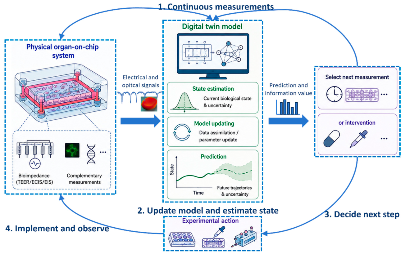
However, closed-loop experimentation requires careful validation. During the initial development, the computational model should be used as a decision-support system rather than as an autonomous controller. Human intervention remains necessary, particularly when the prediction is associated with high uncertainty or when the suggested experimental intervention can modify the biological system (see Figure 2.).
4.4. Challenges to Clinical and Pharmaceutical Translation
Although organ-on-a-chip digital twin technology is currently mainly at the experimental stage, its long-term value depends on its ability to provide useful information for translational applications. In pharmaceutical applications, an important question is whether the system can improve the prediction of human biological responses. Technical integration alone is not sufficient to achieve this objective.
The biological model needs to be appropriate for the intended application. If the microphysiological environment does not reproduce the biological process of interest, a more complex computational model cannot compensate for this limitation. Translational applications also require reproducibility and sufficient throughput.
Highly personalized digital twins can provide detailed mechanistic information, but their complexity can limit their application to large-scale compound screening. Simpler models are easier to scale, but can lose biological specificity. Therefore, a balance between model complexity, biological specificity and throughput is required.
Regulatory acceptance represents another limitation. Computational predictions need to be supported by transparent experimental validation. Therefore, the field needs to demonstrate not only that a digital twin can be constructed, but that its prediction provides information beyond that obtained using existing experimental approaches.
Therefore, the first translational applications should probably focus on specific and well-defined biological questions. Prediction of barrier dysfunction, detection of dynamic toxicity and characterization of recovery after exposure are possible examples. Demonstrating reliable predictive performance for these specific applications may provide a more practical pathway toward translation than attempting to construct a universal digital twin model.
5. Conclusions
The future development of organ-on-a-chip technology will depend increasingly on the capability to interpret dynamic biological changes. The integration of additional sensors alone is unlikely to solve this problem. A system can generate a large amount of sensor data while the biological meaning of these signals remains uncertain.
In this paper, we propose that bioimpedance can be used as a continuous observation layer within a digital twin architecture. The physical organ-on-a-chip system, sensing system, measurement model, biological state estimator and predictive computational model can be considered as interconnected components of an "experimental-computational" system. The objective is not only to monitor the electrical signal, but to estimate the current biological state, quantify the uncertainty and predict the future response of the system.
The transition from sensor-integrated organ-on-a-chip systems toward predictive digital twins requires further development in sensor stability, biological validation, computational modeling, data standardization and experimental design. However, the integration of continuous electrical measurements with biological characterization and computational prediction provides a possible approach to overcome the temporal and interpretational limitations of conventional measurements.
Such systems could progressively move organ-on-a-chip technology from descriptive experiments toward predictive microphysiological models. In this framework, continuous bioimpedance measurements provide the temporal observation, biological measurements provide the interpretation and validation, and computational models provide state estimation and prediction. The combination of these components could ultimately allow organ-on-a-chip experiments to become more adaptive and more directly connected with computational decision-making.
Data Availability Statement
Data sharing is not applicable to this article as no new data were created or analyzed in this study.
Acknowledgements
This work was supported by Young Scientists Fund of the National Natural Science Foundation of China (Ref. 62501375).
References
- C. M. Leung et al., “A guide to the organ-on-a-chip,” Nat. Rev. Methods Prim., vol. 2, no. 1, p. 33, May 2022. [CrossRef]
- Maschmeyer and S. Kakava, “Organ-on-a-Chip,” 2020, pp. 311–342. [CrossRef]
- J. Y. Park, J. Jang, and H.-W. Kang, “3D Bioprinting and its application to organ-on-a-chip,” Microelectron. Eng., vol. 200, pp. 1–11, Nov. 2018. [CrossRef]
- T. M. Maul, D. W. Chew, A. Nieponice, and D. A. Vorp, “Mechanical stimuli differentially control stem cell behavior: morphology, proliferation, and differentiation,” Biomech. Model. Mechanobiol., vol. 10, no. 6, pp. 939–953, Dec. 2011. [CrossRef]
- Y. Nagai, H. Yokoi, K. Kaihara, and K. Naruse, “The mechanical stimulation of cells in 3D culture within a self-assembling peptide hydrogel,” Biomaterials, vol. 33, no. 4, pp. 1044–1051, Feb. 2012. [CrossRef]
- D. Manou et al., “The Complex Interplay Between Extracellular Matrix and Cells in Tissues,” 2019, pp. 1–20. [CrossRef]
- D. Huh, B. D. Matthews, A. Mammoto, M. Montoya-Zavala, H. Y. Hsin, and D. E. Ingber, “Reconstituting Organ-Level Lung Functions on a Chip,” Science (80-. )., vol. 328, no. 5986, pp. 1662–1668, Jun. 2010. [CrossRef]
- Maschmeyer et al., “A four-organ-chip for interconnected long-term co-culture of human intestine, liver, skin and kidney equivalents,” Lab Chip, vol. 15, no. 12, pp. 2688–2699, 2015. [CrossRef]
- M. Boul et al., “A versatile microfluidic tool for the 3D culture of HepaRG cells seeded at various stages of differentiation,” Sci. Rep., vol. 11, no. 1, p. 14075, Jul. 2021. [CrossRef]
- Hilfinger and J. Paulsson, “Separating intrinsic from extrinsic fluctuations in dynamic biological systems,” Proc. Natl. Acad. Sci., vol. 108, no. 29, pp. 12167–12172, Jul. 2011. [CrossRef]
- D. Sheyn et al., “Bone-chip system to monitor osteogenic differentiation using optical imaging,” Microfluid. Nanofluidics, vol. 23, no. 8, p. 99, Aug. 2019. [CrossRef]
- T. Yu, Q. Yang, B. Peng, Z. Gu, and D. Zhu, “Vascularized organoid-on-a-chip: design, imaging, and analysis,” Angiogenesis, vol. 27, no. 2, pp. 147–172, May 2024. [CrossRef]
- N. Araújo-Gomes et al., “Bioluminescence imaging on-chip platforms for non-invasive high-content bioimaging,” Biosens. Bioelectron., vol. 237, p. 115510, Oct. 2023. [CrossRef]
- G. Blankenstein and U. Darling Larsen, “Modular concept of a laboratory on a chip for chemical and biochemical analysis,” Biosens. Bioelectron., vol. 13, no. 3–4, pp. 427–438, Mar. 1998. [CrossRef]
- K. Sato, A. Hibara, M. Tokeshi, H. Hisamoto, and T. Kitamori, “Microchip-based chemical and biochemical analysis systems,” Adv. Drug Deliv. Rev., vol. 55, no. 3, pp. 379–391, Feb. 2003. [CrossRef]
- Z. Chong et al., “Multi-channel microfluidic chip with bilateral stripe for reconstruction of tissue barrier models in vitro,” Biomed. Mater., vol. 21, no. 3, p. 035023, Jun. 2026. [CrossRef]
- Y. S. Zhang et al., “Multisensor-integrated organs-on-chips platform for automated and continual in situ monitoring of organoid behaviors,” Proc. Natl. Acad. Sci., vol. 114, no. 12, Mar. 2017. [CrossRef]
- R. Novak et al., “Robotic fluidic coupling and interrogation of multiple vascularized organ chips,” Nat. Biomed. Eng., vol. 4, no. 4, pp. 407–420, Jan. 2020. [CrossRef]
- J. Wang, W.-B. Jung, R. S. Gertner, H. Park, and D. Ham, “Synaptic connectivity mapping among thousands of neurons via parallelized intracellular recording with a microhole electrode array,” Nat. Biomed. Eng., vol. 9, no. 7, pp. 1144–1154, Feb. 2025. [CrossRef]
- E. Samiei, G. S. Luka, H. Najjaran, and M. Hoorfar, “Integration of biosensors into digital microfluidics: Impact of hydrophilic surface of biosensors on droplet manipulation,” Biosens. Bioelectron., vol. 81, pp. 480–486, Jul. 2016. [CrossRef]
- C. Tiruppathi, A. B. Malik, P. J. Del Vecchio, C. R. Keese, and I. Giaever, “Electrical method for detection of endothelial cell shape change in real time: assessment of endothelial barrier function.,” Proc. Natl. Acad. Sci., vol. 89, no. 17, pp. 7919–7923, Sep. 1992. [CrossRef]
- C. G. Jones and C. Chen, “An arduino-based sensor to measure transendothelial electrical resistance,” Sensors Actuators A Phys., vol. 314, p. 112216, Oct. 2020. [CrossRef]
- Français and B. Le Pioufle, “Single-Cell Electrical Characterization Techniques,” Springer Int. Publ. Switz. Author, pp. 293–326, 2016. [CrossRef]
- K. Graf et al., “Digital twin-enhanced three-organ microphysiological system for studying drug pharmacokinetics in pregnant women,” Front. Pharmacol., vol. 16, Feb. 2025. [CrossRef]
- H. Akbarialiabad, M. S. Seyyedi, S. Paydar, A. Habibzadeh, A. Haghighi, and J. C. Kvedar, “Bridging silicon and carbon worlds with digital twins and on-chip systems in drug discovery,” npj Syst. Biol. Appl., vol. 10, no. 1, p. 150, Dec. 2024. [CrossRef]
- J. Wegener, M. Sieber, and H.-J. Galla, “Impedance analysis of epithelial and endothelial cell monolayers cultured on gold surfaces,” J. Biochem. Biophys. Methods, vol. 32, no. 3, pp. 151–170, Jul. 1996. [CrossRef]
- Y. F. Henry, R. Villenave, M. J. Cronce, W. D. Leineweber, M. A. Benz, and D. E. Ingber, “Organs-on-chips with integrated electrodes for trans-epithelial electrical resistance (TEER) measurements of human epithelial barrier function,” Lab Chip, vol. 17, no. 13, pp. 2264–2271, 2017. [CrossRef]
- N. Renous, M. D. Kiri, R. A. Barnea, R. Rauti, Y. Leichtmann-Bardoogo, and B. M. Maoz, “Spatial trans-epithelial electrical resistance (S-TEER) integrated in organs-on-chips,” Lab Chip, vol. 22, no. 1, pp. 71–79, 2022. [CrossRef]
- H. Moghtaderi et al., “Electric cell-substrate impedance sensing in cancer research: An in-depth exploration of impedance sensing for profiling cancer cell behavior,” Sensors and Actuators Reports, vol. 7, p. 100188, Jun. 2024. [CrossRef]
- P. O. Bagnaninchi and N. Drummond, “Real-time label-free monitoring of adipose-derived stem cell differentiation with electric cell-substrate impedance sensing,” Proc. Natl. Acad. Sci., vol. 108, no. 16, pp. 6462–6467, Apr. 2011. [CrossRef]
- D. M. Lewis, N. Mavrogiannis, Z. Gagnon, and S. Gerecht, “Microfluidic platform for the real time measurement and observation of endothelial barrier function under shear stress,” Biomicrofluidics, vol. 12, no. 4, Jul. 2018. [CrossRef]
- T. Xu et al., “Characterization of red blood cell microcirculatory parameters using a bioimpedance microfluidic device,” Sci. Rep., vol. 10, no. 1, p. 9869, Dec. 2020. [CrossRef]
- P. Barbiero, R. Viñas Torné, and P. Lió, “Graph Representation Forecasting of Patient’s Medical Conditions: Toward a Digital Twin,” Front. Genet., vol. 12, Sep. 2021. [CrossRef]
- K. Sel et al., “Building Digital Twins for Cardiovascular Health: From Principles to Clinical Impact,” J. Am. Heart Assoc., vol. 13, no. 19, Oct. 2024. [CrossRef]
- Y. Zhao et al., “Artificial intelligence-enabled organoids and organ-on-a-chip for pioneering biomedical platforms,” Bio-Design Manuf., vol. 9, no. 4, pp. 756–781, Jul. 2026. [CrossRef]
- J. Li, M. Suvarna, L. Pan, Y. Zhao, and X. Wang, “A hybrid data-driven and mechanistic modelling approach for hydrothermal gasification,” Appl. Energy, vol. 304, p. 117674, Dec. 2021. [CrossRef]
- K. Mu et al., “Toward closed-loop engineering of nanomedicines: integrating AI-enabled design, microfluidic manufacturing, and organ-on-a-chip validation,” J. Nanobiotechnology, Aug. 2026. [CrossRef]
- M. Kim et al., “Multimodal Characterization of Cardiac Organoids Using Integrations of Pressure-Sensitive Transistor Arrays with Three-Dimensional Liquid Metal Electrodes,” Nano Lett., vol. 22, no. 19, pp. 7892–7901, Oct. 2022. [CrossRef]
- S. Li et al., “Single-Cell Electrochemical Aptasensor Array,” ACS Sensors, vol. 8, no. 8, pp. 2921–2926, Aug. 2023. [CrossRef]
- S. Yasotharan, S. Pinto, J. G. Sled, S.-S. Bolz, and A. Günther, “Artery-on-a-chip platform for automated, multimodal assessment of cerebral blood vessel structure and function,” Lab Chip, vol. 15, no. 12, pp. 2660–2669, 2015. [CrossRef]
- X. Hu et al., “Multimodal characterization of cardiomyocytes cell culture using a Thin-Film-Transistor microelectrodes arrays (TFT-MEA),” Sensors Actuators B Chem., vol. 440, p. 137815, Oct. 2025. [CrossRef]
- Skardal et al., “Multi-tissue interactions in an integrated three-tissue organ-on-a-chip platform,” Sci. Rep., vol. 7, no. 1, p. 8837, Aug. 2017. [CrossRef]
- E.-M. Materne et al., “A multi-organ chip co-culture of neurospheres and liver equivalents for long-term substance testing,” J. Biotechnol., vol. 205, pp. 36–46, Jul. 2015. [CrossRef]
- Wagner et al., “A dynamic multi-organ-chip for long-term cultivation and substance testing proven by 3D human liver and skin tissue co-culture,” Lab Chip, vol. 13, no. 18, p. 3538, 2013. [CrossRef]
- Essaouiba et al., “Development of a pancreas-liver organ-on-chip coculture model for organ-to-organ interaction studies,” Biochem. Eng. J., vol. 164, p. 107783, Dec. 2020, doi: 10.1016/j.bej.2020.107783. [CrossRef]
Figure 1.
Conceptual transition from sensor-integrated organ-on-chip systems to bioimpedance-informed digital twins. The organ-on-chip system is continuously connected with an electrical sensing layer for bioimpedance monitoring. The measured impedance signal is affected by both biological properties and non-biological factors associated with the device and measurement conditions. Therefore, a measurement model is used to distinguish the biological contribution from other sources of impedance variation. The impedance measurements are further combined with complementary biological measurements and computational models to estimate the biological state and its changes over time. Based on the estimated state, the model predicts the future biological response and the associated uncertainty. These predictions can be used to determine subsequent measurements or experimental interventions, which are then applied to the physical organ-on-chip system. The resulting measurements are used to further update the computational model, forming a closed experimental-computational loop for continuous monitoring and prediction of the biological response.
Figure 1.
Conceptual transition from sensor-integrated organ-on-chip systems to bioimpedance-informed digital twins. The organ-on-chip system is continuously connected with an electrical sensing layer for bioimpedance monitoring. The measured impedance signal is affected by both biological properties and non-biological factors associated with the device and measurement conditions. Therefore, a measurement model is used to distinguish the biological contribution from other sources of impedance variation. The impedance measurements are further combined with complementary biological measurements and computational models to estimate the biological state and its changes over time. Based on the estimated state, the model predicts the future biological response and the associated uncertainty. These predictions can be used to determine subsequent measurements or experimental interventions, which are then applied to the physical organ-on-chip system. The resulting measurements are used to further update the computational model, forming a closed experimental-computational loop for continuous monitoring and prediction of the biological response.
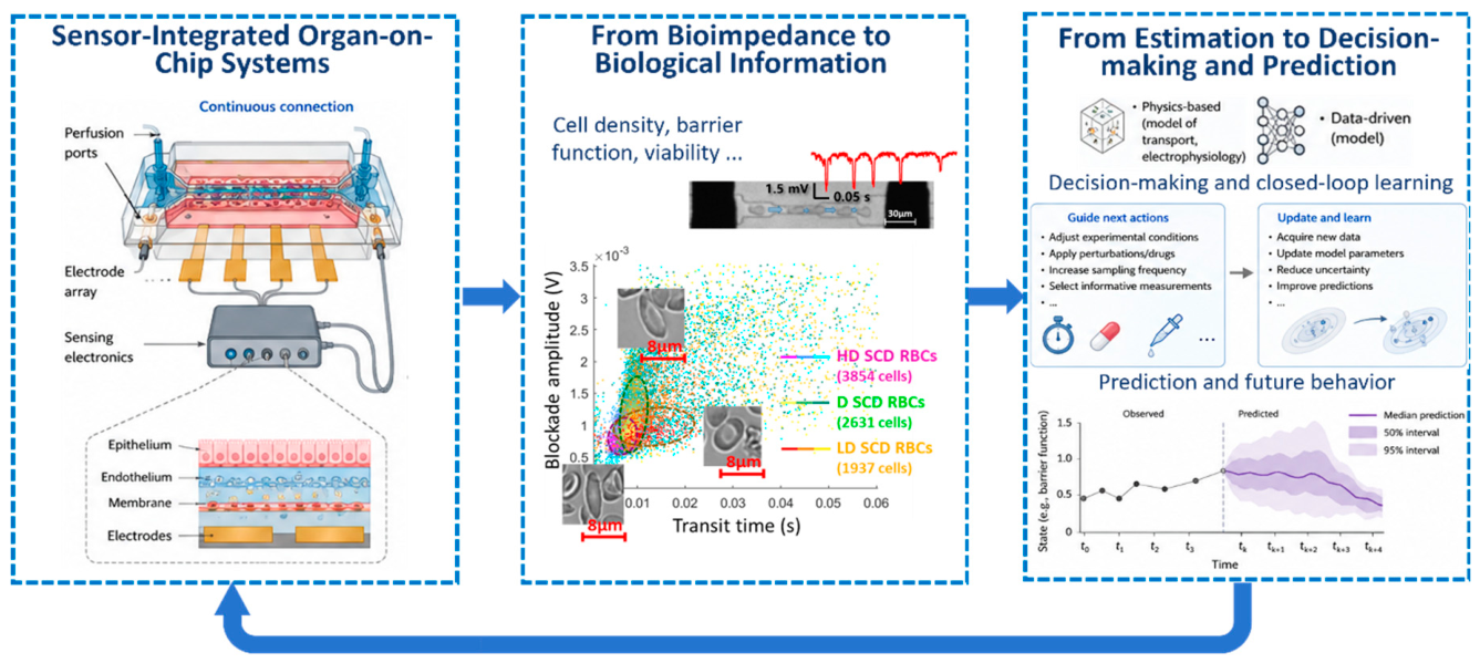
Figure 2.
Closed-loop bioimpedance-informed experimentation. The physical organ-on-chip system provides continuous bioimpedance measurements together with complementary biological measurements. These measurements are used to update the digital twin and estimate the current biological state and the associated uncertainty. Based on the estimated state, the model predicts the future response of the system and determines whether additional measurements or experimental interventions are required. The selected measurements or interventions are then applied to the physical system, and the resulting observations are used to further update the digital twin. This process forms a closed-loop experimental system in which continuous measurements are used to monitor, interpret and predict the biological response.
Figure 2.
Closed-loop bioimpedance-informed experimentation. The physical organ-on-chip system provides continuous bioimpedance measurements together with complementary biological measurements. These measurements are used to update the digital twin and estimate the current biological state and the associated uncertainty. Based on the estimated state, the model predicts the future response of the system and determines whether additional measurements or experimental interventions are required. The selected measurements or interventions are then applied to the physical system, and the resulting observations are used to further update the digital twin. This process forms a closed-loop experimental system in which continuous measurements are used to monitor, interpret and predict the biological response.
Disclaimer/Publisher’s Note: The statements, opinions and data contained in all publications are solely those of the individual author(s) and contributor(s) and not of MDPI and/or the editor(s). MDPI and/or the editor(s) disclaim responsibility for any injury to people or property resulting from any ideas, methods, instructions or products referred to in the content. |
© 2026 by the authors. Licensee MDPI, Basel, Switzerland. This article is an open access article distributed under the terms and conditions of the Creative Commons Attribution (CC BY) license (http://creativecommons.org/licenses/by/4.0/).
Copyright: This open access article is published under a Creative Commons CC BY 4.0 license, which permit the free download, distribution, and reuse, provided that the author and preprint are cited in any reuse.