

Submitted:
01 September 2026
Posted:
02 September 2026
You are already at the latest version
Abstract
Couinaud’s eight-segment model remains the universal language of liver surgery, yet contemporary multidetector computed tomography (CT), its three-dimensional (3D) reconstruction, and minimally invasive surgery have exposed substantial variability in third-order or more peripheral portal branching. This variability becomes clinically important when hepatocellular carcinoma and other liver malignancies are treated with parenchymal-sparing small anatomical resections where third-order or more peripheral portal territories define the oncologic unit at risk.
In this article, the current surgical anatomy for liver cancers was reviewed focusing at third-order portal branches, comparing classical Couinaud segmentation with CT/3D reconstruction-based portal territories. Clinically relevant variability in the right anterior (S5/S8) and right posterior (S6/S7) sections are described while also highlighting that comparable discordance exists in the left hemi-liver. In the right anterior section, craniocaudal, ventrodorsal, trifurcation, and other branching patterns may alter the practical interpretation of S5/S8 anatomy. In the right posterior section, bifurcation and loop-type portal configurations have distinct implications for S6/S7-oriented resection and cone-unit-based anatomical resection. Comparable variability in the left hemiliver, particularly around the umbilical portion and segment 4 branches, further supports individualized preoperative territory mapping.
A pragmatic two-tier surgical strategy is proposed; Couinaud-based resection for first/second-order anatomy and individualized CT-guided cone-unit resection for third-order or more peripheral anatomy. Current status of preoperative simulation and intraoperative implementation using ultrasound, indocyanine green fluorescence, and navigation/fusion technologies are discussed. Finally, emerging directions including automated vessel segmentation and AI-assisted territory analysis are also discussed.
Keywords:
Couinaud’s segmentation
; portal vein
; third-order branches
; cone unit
; 3D CT reconstruction
; anatomical liver resection
; hepatocellular carcinoma
; indocyanine green
; navigation
1. Introduction
Couinaud’s description of functionally independent liver segments provided the conceptual foundation for anatomical liver resection and remains a standard framework in hepatobiliary surgery [1]. The Brisbane 2000 terminology harmonized international nomenclature and reinforced the hierarchical view of liver division (first-order hemi-liver, second-order section, third-order segment) [2,3]. However, Brisbane 2000 did not define how to identify segmental borders in vivo nor how to describe anatomical units smaller than one segment. The Tokyo 2020 update, developed by the Precision Anatomy for Minimally Invasive HBP Surgery (PAM-HBP) consensus, explicitly addressed anatomical resection of segment or less and introduced standardized terminology for peripheral portal territories including the ‘cone unit’ [4].
With modern multidetector computed tomography (CT) and routine three dimensional (3D) reconstruction, surgeons can interrogate portal branching beyond the third-order, and studies have shown that portal territories distal than second order branches frequently diverge from classical Couinaud borders. Intersegmental hepatic veins do not always coincide with true portal ‘watersheds’, especially at third-order or more peripheral levels [5,6,7,8]. This discordance is most consequential in liver cancer management. Hepatocellular carcinoma (HCC) can spread within portal territories via microscopic portal invasion and dissemination, providing the biologic rationale for anatomical resection of the tumor-bearing portal territory [9,10,11]. Since many patients have chronic liver disease, parenchymal-sparing anatomical resection at the subsegmental level is increasingly pursued to balance oncologic clearance with functional preservation [9,10,11,12]. In addition, recent universal spread of minimally invasive liver resection increased the importance of small anatomical resection. Disorientation (caused by loss of three-dimensional view, decreased tactile sensation and loss of overview for whole surgical field= trade-off to local magnified view), often occurs in small but complicated parenchymal sparing laparoscopic resection for deep small tumors. For the situation, the resection of small anatomical territory where the tumor locates inside has been increasingly applied [4,5]. When the exact small anatomical area which contains the small tumor inside is resected, the tumor can be removed without exact localization of the tumor itself.
In this review, we examined the current surgical anatomy for liver cancers, comparing classical Couinaud segmentation with CT/3D reconstruction-based portal territories at third-order portal branches. Clinically relevant variability in the right anterior (S5/S8) and right posterior (S6/S7) sections are described while also highlighting that comparable discordance exists in the left hemi-liver. A pragmatic two-tier surgical strategy is proposed; Couinaud-based resection for first/second-order anatomy and individualized CT-guided cone-unit resection for third-order or more peripheral anatomy. Current status of preoperative simulation and intraoperative implementation using ultrasound, indocyanine green fluorescence, and navigation/fusion technologies are discussed. Finally, we also discuss emerging directions including automated vessel segmentation and AI-assisted territory analysis.
2. Couinaud’s Segmentation and CT/3D Territory-Based Segmentation
Couinaud’s segmentation is robust for planning major hepatectomy and sectionectomy because it aligns closely with first- and second-order portal division in most patients [1,2]. In this context, portal inflow territories and major hepatic veins provide reproducible reference planes. However, Couinaud’s model implicitly assumes that each third-order portal branch supplies a consistent segmental territory with almost equal volume and one main trunk of portal vein except segment 1. Contemporary imaging demonstrates that a Couinaud segment may contain multiple third-order branches of variable size and orientation, and their perfusion territories can cross classical borders [6,13]. This becomes clinically relevant when the operative goal is to remove the tumor-bearing portal territory of segment or less.
Three-dimensional reconstruction using preoperative contrast-enhanced CT image can show liver parenchyma, portal and hepatic venous trees, and tumors. It also enables us to see the shape of target portal perfusion territories and its relationship to surrounding vessel structures besides the area’s quantitative volumetry. Early work demonstrated that virtual hepatectomy can accurately predict resection volumes and tumor margin status, and can also quantify drainage areas relevant to venous congestion risk [13]. 3D simulation as a routine adjunct to complex hepatectomy and living donor transplantation has established, and navigation systems that reproduce preoperative plans intraoperatively are expected [14]. A 2020 guideline-style consensus summarized workflows and evidence suggesting 3D simulation can reduce blood loss and complications in selected settings [15].
For portal anatomy, CT-based series revealed frequent variants of portal branching. CT arterial portography and multidetector CT studies reported portal vein variants in approximately 20–35% of patients, emphasizing the need to recognize variants before complex resection, portal vein embolization, or transplantation [16,17,18,19]. Even when first/second-order anatomy is conventional, variability increases distally, and third-order or more peripheral territories cannot be assumed from a schematic standard atlas of Couinaud’s segmentation.
3. Third-Order or More Peripheral Portal Anatomy in the Liver
3.1. Right Anterior Section
In the branching and territorial configuration of the right anterior section, significant variability exists at the level of the third-order branches and beyond. Although the classical Couinaud framework divides this region into a cranial segment 8 and a caudal segment 5, the actual territory supplied by individual third-order portal pedicles in anterior section does not always follow a simple cranio-caudal arrangement. This uncertainty is clinically important because the border between S5 and S8 lacks a consistently visible anatomical landmark, and limited anatomical resections in this region often require the surgeon to choose between a Couinaud-based craniocaudal concept and a patient-specific portal-territory [1,6,20,21,22,23,24,25].
The ventro-dorsal concept was strongly promoted by Cho, Ryu, and colleagues. In their 3D CT/CTAP-based reevaluation of right-liver portal ramification, all branches arising from the right anterior trunk were divided into right ventral portal branches and right dorsal portal branches, with the anterior fissure vein crossing between these two territories. They therefore proposed that the anterior section could be divided into ventral and dorsal components rather than into S5 and S8 (caudal and cranial ones) and described the anterior fissure as a surgically exploitable plane—the so-called “third door” of the liver [21,22]. In this concept, the right anterior section is divided into the antero-ventral segment and antero-dorsal segment, demarcated by the anterior fissure vein; however, subsequent MDCT evaluation showed that, although the anterior fissure vein could frequently be identified, its use as a reliable boundary was sometimes difficult, particularly when it drained proximally into the middle hepatic vein or when the venous course did not clearly form a parenchymal plane [23].
Later comparative 3D analyses refined this debate rather than replacing one model with the other. Kobayashi et al. reported that the right anterior portal vein showed a craniocaudal pattern in 53% of patients, a ventro-dorsal pattern in 23%, a trifurcation pattern in 13%, and miscellaneous patterns in 11%; V8 was identified in 91% of evaluable patients, but served as a useful ventro-dorsal landmark in only 63%. These findings support the view that ventro-dorsal segmentation is anatomically meaningful in selected patients, but that ventro-dorsal segmentation to every right anterior section cannot be applied universally [24]. Similarly, Ishii et al. classified right anterior tertiary portal ramification into craniocaudal, ventrodorsal, trifurcation, and quadfurcation types, and further highlighted the PV8c branch, identified in 53.6% of patients, as a small but characteristic medial horizontal branch that may complicate strict assignment of peripheral territories to standard S5/S8 or ventral/dorsal labels [25].
From a surgical standpoint, the right anterior section should therefore be treated as a high-variability area in which the operative unit is determined by the individual portal territory rather than by a fixed schematic border. In patients with a clear craniocaudal pattern, S5- or S8-oriented anatomical resection remains reasonable; in patients with a clear ventro-dorsal pattern, ventral or dorsal anatomical resection of the anterior section may better match the true portal drainage territory. Hepatic vein-guided approaches leveraging the anterior fissure vein, middle hepatic vein, right hepatic vein, and V8 can be useful to standardize parenchymal-sparing anatomical resection, but these landmarks should be interpreted in the context of preoperative 3D simulation rather than used as independent substitutes for portal-territory mapping (Figure 1C–D) [21,22,23,24,25,26].
3.2. Right Posterior Section
The third-order anatomy in right posterior section has been discussed frequently. S7 anatomical resection and posterior sectionectomy are technically demanding, especially in minimally invasive procedure [7], and right posterior section graft for living donor transplantation have also been reported recently [27]. Those procedures depend heavily on peripheral portal and venous landmarks during the surgeries. A CT during arterial portography study demonstrated that a Couinaud-type bifurcation into P6 and P7 occurred in only 41% of cases; 50% cases showed a loop-like single trunk issuing sequential branches, and 9% showed trifurcation with an intermediate branch occupying the putative S6/S7 border (Figure 1A–B) [28]. Furthermore, a subsequent 3D CT study proposed intersegmental veins as landmarks between S6 and S7 and reported high identification rates in initial laparoscopic application [29].
We also performed retrospective study analyzing CT-based 3D reconstruction images of portal and hepatic veins [30]. In 45 among 100 cases, the posterior portal vein divides into two major branches to segments 6 and 7 (Bifurcation type) with subsequent peripheral arborization. On the other hand, posterior portal vein forms a loop type trunk issuing sequential small branches (Loop type) in 53 cases. In Bifurcation type, the 3D length of the common stem before the bifurcation measured 8.89±7.87 mm and the volume of Segment 6 (the area supplied by the first branch toward the right caudal direction = Segment 6) corresponds to 43.8% ± 12.6% of the whole right posterior section. In Loop type, the 3D distance from the origin of right posterior portal vein to the first branch measured 17.3±8.36 mm, which is almost double of that in Bifurcation type (p<0.001), and the volume of the first-branch portal territory corresponds to 14.9% ± 8.64%, which is almost 1/3 volume of that in Bifurcation type (p<0.001). Our data show that when the first branch from the posterior trunk is divided earlier, the branching type tends to be Bifurcation type, and the volume of the area supplied by the first branch from the posterior trunk is much larger in Bifurcation type than in Loop type. In our study, hepatic vein anatomy in posterior section was also categorized as HV-type1 (presence of portal fissure-plane forming intersegmental vein), HV-type2 (presence of landmark vein without plane formation), or HV-type3 (neither). The ratios of those HV-types were compared between portal vein branching types for the reference to surgical implication. The ratios of HV-type1: HV-type 2: HV-type 3 in types of portal vein branching pattern were 29:16:0 (64.4%:35.6%:0%) in Bifurcation type and 25:14:14 (47.2%:26.4%:26.4%) in Loop type (p<0.001). In bifurcation type, the IRHV or the distal part of the RHV always lies between segments 6 and 7 as landmarks for segmentectomies, and their tributaries frequently (almost two third) delineate the intersegmental fissure plane between the caudal and cranial parts (S6 and 7). In Loop type, one fourth of the patients do not have the vein as a landmark nor a fissure vein. Wang et al. identified an intersegmental fissure vein (ISV) between S6 and S7 in 82% of patients (derived from the RHV in 92% and IRHV in 8%) and successfully implemented ISV-guided laparoscopic segmentectomies with favorable perioperative outcomes, including R0 rates [29]. In contrast with their data, ISV forming fissure plane was not identified in 35.6% and 52.8% of cases in Bifurcation and Loop types of portal vein branching pattern, respectively, in our study. 26.4% of Loop type cases did not even have landmark (not-plane-forming) veins. Our data show the need to reconsider sub-sectional anatomical resection in the posterior section. Although Couinaud’s classification of segments 6 and 7 can be applied to some part of the patient, there are the other half with Loop type branching (some even without ISV) who do not fall into it. The strategy based on Couinaud’s classification (segments 6 and 7) using the Glissonian approach and/or hepatic vein-guided approach is still valuable for almost half of the patients. However, others do not fit this strategy. In patients with Loop type PV branching without ISV, “cone-unit-based anatomical resection”, defined by peripheral portal territories, based on individualized preoperative planning and intraoperative approach with navigations should be considered. [4,31,32,33,34,35,36]
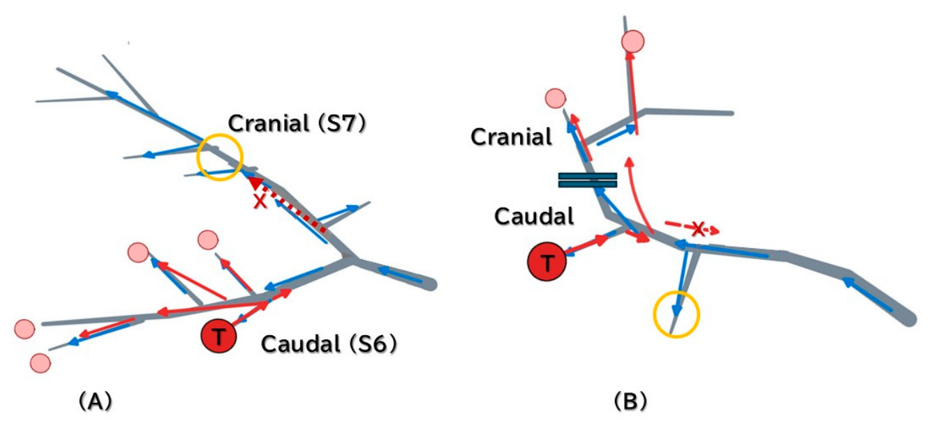
The concept of anatomical resection of the liver originates from the fact that the portal vein sometimes works as a drainage vein when huge HCC tumors compress the hepatic vein and disturb the drainage of blood through it. In this situation, the dissemination of tumor cells can occur in the portal territory. [9,11] In this aspect, strategies for Bifurcation and Loop types should be different. The cranial or caudal area of the bifurcation type (S7 or 6) in the posterior section is a separate independent portal territory in which the tumor cells can be disseminated. However, the cone units in the loop type are separately and directly drained into the posterior main trunk, and tumor cells in one cone unit cannot be drained into the next unit, even if they are both in the same (cranial or caudal) half of the posterior section (Figure 3).
Figure 2.
Two-tier surgical strategy for liver cancer management based on anatomical hierarchy. Conceptual algorithm proposing Couinaud-based resection for first/second-order anatomy and CT-guided cone-unit resection for third-order or more peripheral anatomy, supported by ultrasound, indocyanine green fluorescence, and navigation tools.
Figure 2.
Two-tier surgical strategy for liver cancer management based on anatomical hierarchy. Conceptual algorithm proposing Couinaud-based resection for first/second-order anatomy and CT-guided cone-unit resection for third-order or more peripheral anatomy, supported by ultrasound, indocyanine green fluorescence, and navigation tools.
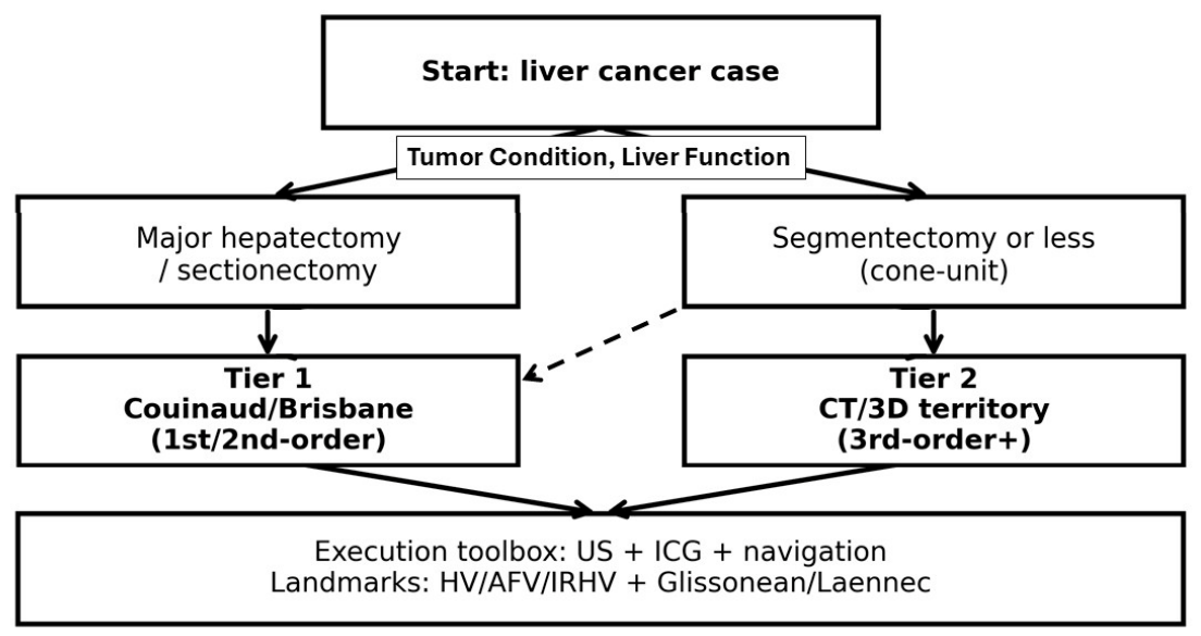
Figure 3.
The Difference of Tumor Spreading between Bifurcation and Loop Types (Reproduced/adapted from Imanaka et al. [30] under the Creative Commons Attribution License). (A): Bifurcation type, (B): Loop type. Red colored-in circle with “T” shows tumor (liver cancer). Pink colored-in circle shows possible tumor dissemination. Blue arrow shows original flow of portal vein. Red arrow shows possible drainage flow from tumor-bearing area. Red interrupted arrow with “X” shows that tumor cell does not spread toward the direction. Yellow circle shows spared area from tumor cell spreading. In bifurcation type, tumor spreading first occurs inside the main third branch area (segment 6=caudal or segment 7=cranial). In loop type, the most central branch of portal vein is spared from tumor cell spreading, even though the branch and the tumor-bearing branch are both in caudal part of posterior section next to each other.
Figure 3.
The Difference of Tumor Spreading between Bifurcation and Loop Types (Reproduced/adapted from Imanaka et al. [30] under the Creative Commons Attribution License). (A): Bifurcation type, (B): Loop type. Red colored-in circle with “T” shows tumor (liver cancer). Pink colored-in circle shows possible tumor dissemination. Blue arrow shows original flow of portal vein. Red arrow shows possible drainage flow from tumor-bearing area. Red interrupted arrow with “X” shows that tumor cell does not spread toward the direction. Yellow circle shows spared area from tumor cell spreading. In bifurcation type, tumor spreading first occurs inside the main third branch area (segment 6=caudal or segment 7=cranial). In loop type, the most central branch of portal vein is spared from tumor cell spreading, even though the branch and the tumor-bearing branch are both in caudal part of posterior section next to each other.
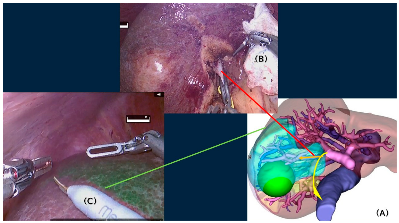
Translating preoperative territory maps into intraoperative planes during surgery, planned with the cone-unit strategy, requires complementary modalities. Reviews have summarized how indocyanine green (ICG) fluorescence can demarcate hepatic segments either by portal injection (positive staining) or intravenous injection after inflow control (negative staining) [32,33] (Figure 4). Prospective evidence suggests that ICG-guided recognition of boundaries is feasible and may reduce major liver-related complications in selected cohorts undergoing anatomical resection [35]. Real-time virtual sonography and fusion navigation systems can co-register intraoperative ultrasound with preoperative CT/3D simulation to navigate the planned planes in real time [36].
3.3. Left Hemiliver
Comparable discordance exists in the left hemiliver, although it is often less emphasized than right-sided variation because the umbilical portion of the left portal vein provides an apparently intuitive intrahepatic landmark. Classical anatomy assigns the left lateral section to S2/S3 and the left medial section to S4, but the branching of the left portal vein—particularly branches to S4, branches arising near the umbilical portion, and cranio-lateral branches—can create portal territories that do not correspond precisely to schematic Couinaud borders [1,2,6,20,37,38,39].
The left lateral section is usually considered more regular than the right anterior or posterior sections; nevertheless, the relationship among P2, P3, the left hepatic vein, and the umbilical portion is not always equivalent to a simple two-segment map. Anatomical summaries of the umbilical portion indicate that P2 is often represented by a single large branch, whereas P3 is relatively constant from the left horn of the umbilical portion, but segment 4 is typically supplied by multiple branches arising from the right horn and nearby junctional areas [38]. In addition, cranio-lateral portal branches arising from the cranial side of the umbilical portion have been reported in a substantial subset of patients, which may be relevant when planning right trisectionectomy, extended left-sided resections, or parenchymal-preserving resections near the left portal trunk [39].
Segment 4 is the most important source of left-sided ambiguity. A CT-based study of S4 portal supply demonstrated that S4 was vascularized by 2–8 portal branches, with most patients having 3–6 branches; a minority of branches arose from the transverse portion of the left portal vein, and rare branches arose from the right portal vein. The same study concluded that S4 portal vascularization varies widely and predominantly originates near the junction between the left portal vein and the round ligament, and that there is no consistent anatomical rationale for rigid subdivision of S4 into S4a and S4b solely on the basis of portal inflow [37]. These findings are consistent with the broader message of this review: third-order or more peripheral portal territories should be reconstructed and measured in each patient rather than inferred from the two-dimensional Couinaud schema.
For left-sided segmentectomy or subsegmentectomy, practical planning should therefore include: (i) identification of the exact origin and number of S4 branches; (ii) separation of the tumor-bearing branch territory from neighboring P2/P3/P4 territories; (iii) assessment of the left hepatic vein and middle hepatic vein as potential, but not absolute, boundary landmarks; and (iv) confirmation of the planned transection plane by intraoperative ultrasound, ICG fluorescence, or fusion/navigation when available. This individualized approach is particularly relevant for small anatomical resections around the umbilical portion, where sacrificing a small unrecognized S4 or cranio-lateral branch may produce either unnecessary ischemia/congestion or insufficient anatomical clearance [6,32,33,34,35,36,37,38,39].
4. Surgical Translation, Two-Tier Strategy for Liver Cancer Management, and Future Perspectives
A tiered view of anatomic concordance is clinically useful. At first- and second-order levels, Couinaud’s segmentation generally coincides with CT territory maps, enabling major hepatectomy and sectionectomy to remain Couinaud-based with minimal compromise [2,3,4]. Discordance increases at the third-order level, where segmental borders may not correspond to Couinaud’s portal segmentations and hepatic veins may not reliably delineate functional boundaries [5,6,7,8]. The PAM-HBP consensus emphasized that accurate border recognition requires integrating portal branching patterns, hepatic veins, and intraoperative modalities (ultrasound, fluorescence, 3D simulation) rather than assuming fixed planes [6].
For tumors requiring major hepatectomy or sectionectomy, Couinaud/Brisbane-based planning remains appropriate because the resection unit corresponds to stable first/second-order portal division and established venous planes [2,3,4]. In these cases, the main value of 3D simulation is risk assessment (future liver remnant, venous drainage, variant recognition) rather than redefining the unit of resection [14,15,16].
For parenchymal-sparing ‘segmentectomy or less’ anatomical resections, we propose a CT-guided cone-unit framework (Figure 2). The Tokyo 2020 terminology defines a ‘cone unit’ as a peripheral portal territory representing the smallest functional unit relevant to anatomical resection [4]. This concept is rooted in Glissonean pedicle-based anatomy [31] and has been popularized in minimally invasive limited liver resection. Although precise tumor-bearing territory identification is needed in safe limited resections, the specific conditions in minimally invasive liver resection, such as less tactile sensation and loss of a bird's-eye view to whole surgical field, easily lead to disorientation of tumors and other important structures. When third-order anatomy deviates from Couinaud borders, “cone-unit” planning, which resects the small portal territory bearing tumor inside, aligns resection with the true portal territory at oncologic risk and may reduce unnecessary loss of non-cancerous parenchyma in cirrhotic patients. When cone-unit based resection is performed, minimally invasive anatomical liver resection increasingly leverages standardized extrahepatic Glissonean pedicle isolation and hepatic vein-guided transection. Laennec’s capsule-based approaches propose that dissection planes between the capsule and surrounding structures can standardize isolation of Glissonean pedicles and hepatic veins, potentially improving safety and reproducibility in the situation [40]. ‘Inter-Laennec’ approaches for tumors near major hepatic veins have also been described to preserve veins and avoid congestion while maintaining surgical margins [41].
Translating preoperative territory maps into intraoperative planes during the surgery of cone-unit strategy requires complementary modalities. Reviews have summarized how indocyanine green (ICG) fluorescence can demarcate hepatic segments either by portal injection (positive staining) or by intravenous injection after inflow control (negative staining) [32,33,34]. Prospective evidence suggests that ICG-guided recognition of boundaries can be feasible and may reduce major liver-related complications in selected cohorts undergoing anatomical resection [35]. Real-time virtual sonography and fusion navigation systems can co-register intraoperative ultrasound with preoperative CT-based 3D simulation to navigate planned planes in real time [36].
Automated vessel segmentation and territory computation, currently used in preoperative simulation, are advancing rapidly, and they represent the technical bridge between descriptive liver anatomy and reproducible precision surgery. 3D-simulation systems automatic extract portal and hepatic venous trees, identify branch hierarchy, calculate peripheral perfusion territories, and estimate functional remnant volume. A two-step AI algorithm has already been reported to automate anatomic virtual liver resection with segmentations in high accuracy for major divisions, potentially reducing the manual workload required for 3D planning and making territory-based planning more accessible outside highly specialized centers [42]. When the system exports these data into intraoperative navigation platforms, AI could be the next logical layer in the evolution, making it capable to interpret and overlay these data dynamically onto real time images combined with the information from various landmarks structures, ICG fluorescence, and intraoperative ultrasonography. Thereafter, those future AI-assisted systems should be evaluated by clinically meaningful endpoints, such as correctness of portal territory-resection, preservation of venous drainage, surgical margin status, concordance of resection planes with ICG-demarcated boundaries, intraoperative variables (operative time, blood loss, conversion), postoperative morbidity (bile leakage, postoperative liver function, re-operation), length of stay, re-admission, and long-term oncological outcomes [13,14,15,32,33,34,35,36,42].
5. Conclusions
Although Couinaud’s segmentation remains indispensable as the universal language of hepatic surgery, it is insufficient to describe third-order or more peripheral portal territories, which are increasingly used as operative units in parenchymal-sparing liver cancer (especially minimally invasive) surgery. CT-based 3D reconstruction reveals patient-specific portal territories throughout the liver, with particularly important clinical implications in the right posterior and anterior sections. It demonstrates that discrepancies between actual portal territories and Couinaud-defined boundaries are often critical when planning segmentectomy or smaller anatomical resections. In the surgical management of liver cancer, a pragmatic two-tier strategy is proposed: Couinaud-based planning should be maintained for first- and second-order anatomical resections, whereas CT-guided cone-unit planning combined with navigation-assisted execution should be adopted for third-order or more peripheral anatomical resections (Figure 2 and Figure 4).
Author Contributions
Conceptualization, T.I. and Z.M.; writing-original draft preparation, T.I.; writing-review and editing, Z.M., H.K., A.H., K.N., T.K., K.M., T.E., K.M., K.K., H.K. and Y.T.; supervision, Z.M. All authors have read and agreed to the published version of the manuscript.
Funding
This research received no external funding.
Institutional Review Board Statement
Not applicable.
Informed Consent Statement
Not applicable.
Data Availability Statement
No new data were created or analyzed in this review. Data sharing is not applicable to this article.
Acknowledgments
Not applicable.
Conflicts of Interest
The authors declare no conflicts of interest.
References
- Couinaud, C. Le Foie: Études Anatomiques et Chirurgicales; Masson: Paris, France, 1957. [Google Scholar]
- Terminology Committee of the International Hepato-Pancreato-Biliary Association. The Brisbane 2000 Terminology of Liver Anatomy and Resections. HPB 2000, 2, 333–339. [Google Scholar] [CrossRef]
- Strasberg, S.M. Nomenclature of hepatic anatomy and resections: A review of the Brisbane 2000 system. J. Hepatobiliary Pancreat. Surg. 2005, 12, 351–355. [Google Scholar] [CrossRef] [PubMed]
- Wakabayashi, G.; Cherqui, D.; Geller, D.A.; Abu Hilal, M.; Berardi, G.; Ciria, R.; Abe, Y.; Aoki, T.; Asbun, H.J.; Chan, A.C.Y.; Chanwat, R.; Chen, K.H.; Chen, Y.; Cheung, T.T.; Fuks, D.; Gotohda, N.; Han, H.S.; Hasegawa, K.; Hatano, E.; Honda, G.; Itano, O.; Iwashita, Y.; Kaneko, H.; Kato, Y.; Kim, J.H.; Liu, R.; López-Ben, S.; Morimoto, M.; Monden, K.; Rotellar, F.; Sakamoto, Y.; Sugioka, A.; Yoshiizumi, T.; Akahoshi, K.; Alconchel, F.; Ariizumi, S.; Benedetti Cacciaguerra, A.; Durán, M.; Garcia Vazquez, A.; Golse, N.; Miyasaka, Y.; Mori, Y.; Ogiso, S.; Shirata, C.; Tomassini, F.; Urade, T.; Wakabayashi, T.; Nishino, H.; Hibi, T.; Kokudo, N.; Ohtsuka, M.; Ban, D.; Nagakawa, Y.; Ohtsuka, T.; Tanabe, M.; Nakamura, M.; Tsuchida, A.; Yamamoto, M. The Tokyo 2020 terminology of liver anatomy and resections: Updates of the Brisbane 2000 system. J. Hepatobiliary Pancreat. Sci. 2022, 29, 6–15. [Google Scholar] [CrossRef] [PubMed]
- Morise, Z. Current status and future perspectives of minimally-invasive redo liver surgery - what can we add with technologies of simulation/navigation and robot? Mini-Invasive Surg. 2024, 8, 34. [Google Scholar] [CrossRef]
- Wakabayashi, T.; Benedetti Cacciaguerra, A.; Ciria, R.; Ariizumi, S.; Durán, M.; Golse, N.; Ogiso, S.; Abe, Y.; Aoki, T.; Hatano, E.; Itano, O.; Sakamoto, Y.; Yoshizumi, T.; Yamamoto, M.; Wakabayashi, G. Study Group of Precision Anatomy for Minimally Invasive Hepato-Biliary-Pancreatic surgery (PAM-HBP surgery). Landmarks to identify segmental borders of the liver: A review prepared for PAM-HBP expert consensus meeting 2021. J. Hepatobiliary Pancreat. Sci. 2022, 29, 82–98. [Google Scholar] [CrossRef] [PubMed]
- Ban, D.; Nara, S.; Takamoto, T.; Mizui, T.; Yoshino, J.; Esaki, M.; Shimada, K. Revisiting the role of the hepatic vein in laparoscopic liver resection. Hepatoma Res. 2021, 7, 13. [Google Scholar] [CrossRef]
- Ogiso, S.; Seo, S.; Ishii, T.; Okumura, S.; Yoh, T.; Nishio, T.; Koyama, Y.; Fukumitsu, K.; Taura, K.; Hatano, E. Anatomy of the Middle Hepatic Vein Tributaries to Promote Safer Hepatic Vein-Guided Liver Resection. J. Gastrointest. Surg. 2022, 26, 122–127. [Google Scholar] [CrossRef] [PubMed]
- Makuuchi, M. Surgical treatment for HCC--special reference to anatomical resection. Int. J. Surg. 2013, 11, S47–9. [Google Scholar] [CrossRef] [PubMed]
- Takamoto, T.; Makuuchi, M. Precision surgery for primary liver cancer. Cancer Biol. Med. 2019, 16, 475–485. [Google Scholar] [CrossRef] [PubMed]
- Sakon, M.; Ogawa, H.; Fujita, M.; Nagano, H. Hepatic resection for hepatocellular carcinoma based on tumor hemodynamics. Hepatol. Res. 2013, 43, 155–64. [Google Scholar] [CrossRef] [PubMed]
- Eguchi, S.; Kanematsu, T.; Arii, S.; Okazaki, M.; Okita, K.; Omata, M.; Ikai, I.; Kudo, M.; Kojiro, M.; Makuuchi, M.; Monden, M.; Matsuyama, Y.; Nakanuma, Y.; Takayasu, K. Liver Cancer Study Group of Japan. Comparison of the outcomes between an anatomical subsegmentectomy and a non-anatomical minor hepatectomy for single hepatocellular carcinomas based on a Japanese nationwide survey. Surgery 2008, 143, 469–75. [Google Scholar] [CrossRef] [PubMed]
- Saito, S.; Yamanaka, J.; Miura, K.; Nakao, N.; Nagao, T.; Sugimoto, T.; Hirano, T.; Kuroda, N.; Iimuro, Y.; Fujimoto, J. A novel 3D hepatectomy simulation based on liver circulation: application to liver resection and transplantation. Hepatology 2005, 41, 1297–304. [Google Scholar] [CrossRef] [PubMed]
- Miyata, A.; Arita, J.; Kawaguchi, Y.; Hasegawa, K.; Kokudo, N. Simulation and navigation liver surgery: an update after 2,000 virtual hepatectomies. Glob. Health Med. 2020, 2, 298–305. [Google Scholar] [CrossRef] [PubMed]
- Fang, C.; An, J.; Bruno, A.; Cai, X.; Fan, J.; Fujimoto, J.; Golfieri, R.; Hao, X.; Jiang, H.; Jiao, L.R.; Kulkarni, A.V.; Lang, H.; Lesmana, C.R.A.; Li, Q.; Liu, L.; Liu, Y.; Lau, W.; Lu, Q.; Man, K.; Maruyama, H.; Mosconi, C.; Örmeci, N.; Pavlides, M.; Rezende, G.; Sohn, J.H.; Treeprasertsuk, S.; Vilgrain, V.; Wen, H.; Wen, S.; Quan, X.; Ximenes, R.; Yang, Y.; Zhang, B.; Zhang, W.; Zhang, P.; Zhang, S.; Qi, X. Consensus recommendations of three-dimensional visualization for diagnosis and management of liver diseases. Hepatol. Int. 2020, 14, 437–453. [Google Scholar] [CrossRef] [PubMed]
- Covey, A.M.; Brody, L.A.; Getrajdman, G.I.; Sofocleous, C.T.; Brown, K.T. Incidence, patterns, and clinical relevance of variant portal vein anatomy. AJR Am. J. Roentgenol. 2004, 183, 1055–64. [Google Scholar] [CrossRef] [PubMed]
- Atasoy, C.; Ozyürek, E. Prevalence and types of main and right portal vein branching variations on MDCT. AJR Am. J. Roentgenol. 2006, 187, 676–81. [Google Scholar] [CrossRef] [PubMed]
- Koç, Z.; Oğuzkurt, L.; Ulusan, S. Portal vein variations: clinical implications and frequencies in routine abdominal multidetector CT. Diagn. Interv. Radiol. 2007, 13, 75–80. [Google Scholar] [PubMed]
- Schmidt, S.; Demartines, N.; Soler, L.; Schnyder, P.; Denys, A. Portal vein normal anatomy and variants: implication for liver surgery and portal vein embolization. Semin Interv. Radiol. 2008, 25, 86–91. [Google Scholar] [CrossRef] [PubMed]
- Liu, Z.; Xia, F.; Guo, B.; Leng, C.; Zhang, E.; Xu, L.; Chen, X.; Zhu, P. Comprehensive evaluation of the ramification patterns of hepatic vascular anatomy based on three-dimensional visualization technology. Updat. Surg. 2025, 77, 2477–2489. [Google Scholar] [CrossRef] [PubMed]
- Cho, A.; Okazumi, S.; Makino, H.; Miura, F.; Shuto, K.; Mochiduki, R.; Tohma, T.; Kudo, H.; Matsubara, K.; Gunji, H.; Yamamoto, H.; Ryu, M.; Ochiai, T. Anterior fissure of the right liver - the third door of the liver. J. Hepatobiliary Pancreat. Surg. 2004, 11, 390–396. [Google Scholar] [CrossRef] [PubMed]
- Cho, A.; Okazumi, S.; Miyazawa, Y.; Makino, H.; Miura, F.; Ohira, G.; Yoshinaga, Y.; Tohma, T.; Kudo, H.; Matsubara, K.; Ryu, M.; Ochiai, T. Proposal for a reclassification of liver based anatomy on portal ramifications. Am. J. Surg. 2005, 189, 195–199. [Google Scholar] [CrossRef] [PubMed]
- Kaneko, T.; Tomiyama, T.; Kiyuna, H.; Machida, T.; Hayashi, H.; Kumita, S. Identification of Ryu's segmentation of the liver using MDCT analysis. J. Nippon Med. Sch. 2010, 77, 244–249. [Google Scholar] [CrossRef] [PubMed]
- Kobayashi, T.; Ebata, T.; Yokoyama, Y.; Igami, T.; Sugawara, G.; Mizuno, T.; Yamaguchi, J.; Nagino, M. Study on the segmentation of the right anterior sector of the liver. Surgery 2017, 161, 1536–1542. [Google Scholar] [CrossRef] [PubMed]
- Ishii, N.; Harimoto, N.; Kogure, K.; Araki, K.; Hagiwara, K.; Tsukagoshi, M.; Igarashi, T.; Watanabe, A.; Kubo, N.; Shirabe, K. Study on the portal ramification pattern of the right anterior sector of the liver and a unique medial branch (PV8c) of the right anterior portal vein. Ann. Gastroenterol. Surg. 2022, 6, 679–687. [Google Scholar] [CrossRef] [PubMed]
- Monden, K.; Sadamori, H.; Iwasaki, T.; Hioki, M.; Takakura, N. Hepatic Vein-Guided Approach in Laparoscopic Anatomic Liver Resection of the Ventral and Dorsal Parts of Segment 8. J. Pers. Med. 2023, 13, 1007. [Google Scholar] [CrossRef] [PubMed]
- Hori, T.; Kirino, I.; Uemoto, S. Right posterior segment graft in living donor liver transplantation. Hepatol. Res. 2015, 45, 1076–1082. [Google Scholar] [CrossRef] [PubMed]
- Gavriilidis, P.; Yuan, S.; Weixia, L.; Limin, C.; Baiyong, S. An Imaging Study of a Ramification Pattern of Right Posterior Portal Vein Branch. J. Cytol. Histol. 2011, 2, 110. [Google Scholar] [CrossRef]
- Wang, J.; Xu, J.; Lei, K.; You, K.; Liu, Z. Prevalence and clinical significance of the Sg6/Sg7 intersegmental veins based on re-evaluation of the Couinaud classification for the right posterior portal vein. Updat. Surg. 2023, 75, 1941–1948. [Google Scholar] [CrossRef] [PubMed]
- Imanaka, T.; Morise, Z.; Kato, H.; Nakamura, K.; Koide, T.; Matsuo, K.; Endo, T.; Morohara, K.; Horiguchi, A.; Katsuno, H. A CT-Based 3D Anatomical Mapping Study of Right Posterior Portal and Hepatic Venous Patterns: Revisiting Liver Segmentation and Its Surgical Implication. J. Clin. Med. 2026, 15, 6093. [Google Scholar] [CrossRef] [PubMed]
- Takasaki, K. Glissonean pedicle transection method for hepatic resection: a new concept of liver segmentation. J. Hepatobiliary Pancreat. Surg. 1998, 5, 286–91. [Google Scholar] [CrossRef] [PubMed]
- Ishizawa, T.; Saiura, A.; Kokudo, N. Clinical application of indocyanine green-fluorescence imaging during hepatectomy. Hepatobiliary Surg. Nutr. 2016, 5, 322–8. [Google Scholar] [CrossRef] [PubMed]
- Aoki, T.; Koizumi, T.; Mansour, D.A.; Tomioka, K.; Murakami, M. Indocyanine green fluorescence imaging technology in minimally invasive liver resection. Laparosc. Surg. 2021, 5, 13. [Google Scholar] [CrossRef]
- Felli, E.; Ishizawa, T.; Cherkaoui, Z.; Diana, M.; Tripon, S.; Baumert, T.F.; Schuster, C.; Pessaux, P. Laparoscopic anatomical liver resection for malignancies using positive or negative staining technique with intraoperative indocyanine green-fluorescence imaging. HPB 2021, 23, 1647–1655. [Google Scholar] [CrossRef] [PubMed]
- Gon, H.; Omiya, S.; Komatsu, S.; Yamasaki, N.; Murakami, S.; Fukushima, K.; Urade, T.; Tsugawa, D.; Yanagimoto, H.; Toyama, H.; Kido, M.; Fukumoto, T. Efficacy and safety of indocyanine green-fluorescence imaging guided liver resection: a single-arm prospective cohort study. Langenbecks Arch. Surg. 2025, 410, 34. [Google Scholar] [CrossRef] [PubMed]
- Satou, S.; Aoki, T.; Kaneko, J.; Sakamoto, Y.; Hasegawa, K.; Sugawara, Y.; Arai, O.; Mitake, T.; Miura, K.; Kokudo, N. Initial experience of intraoperative three-dimensional navigation for liver resection using real-time virtual sonography. Surgery 2014, 155, 255–62. [Google Scholar] [CrossRef] [PubMed]
- Maurer, R.; Rivoire, M.; Basso, V.; Meeus, P.; Peyrat, P.; Dupré, A. Portal supply of segment IV of the liver based on CT-scan. Surg. Radiol. Anat. 2017, 39, 471–476. [Google Scholar] [CrossRef] [PubMed]
- Najah, H.; Ammar, H.; Gupta, R.; et al. Segmental branching pattern of the left portal vein: anatomical characteristics and clinical implications. Clin. Anat. 2018, 31, 1122–1128. [Google Scholar] [CrossRef] [PubMed]
- Minami, T.; Ebata, T.; Yokoyama, Y.; et al. Study of the portal branches arising from the cranial part of the umbilical portion of the left portal vein: implications for anatomic right hepatic trisectionectomy. World J. Surg. 2020, 44, 4231–4235. [Google Scholar] [CrossRef] [PubMed]
- Sugioka, A.; Kato, Y.; Tanahashi, Y. Systematic extrahepatic Glissonean pedicle isolation for anatomical liver resection based on Laennec's capsule: proposal of a novel comprehensive surgical anatomy of the liver. J. Hepatobiliary Pancreat. Sci. 2017, 24, 17–23. [Google Scholar] [CrossRef] [PubMed]
- Monden, K.; Sugioka, A.; Hioki, M.; Sadamori, H.; Takakura, N. The Inter-Laennec Approach for Liver Tumors in Contact with Hepatic Veins in Laparoscopic Liver Resection. Ann. Surg. Oncol. 2024, 31, 7890–7891. [Google Scholar] [CrossRef] [PubMed]
- Kazami, Y.; Kaneko, J.; Keshwani, D.; Kitamura, Y.; Takahashi, R.; Mihara, Y.; Ichida, A.; Kawaguchi, Y.; Akamatsu, N.; Hasegawa, K. Two-step artificial intelligence algorithm for liver segmentation automates anatomic virtual hepatectomy. J. Hepatobiliary Pancreat. Sci. 2023, 30, 1205–1217. [Google Scholar] [CrossRef] [PubMed]
Figure 1.
Variability of third-order portal branching patterns in the right posterior (S6/S7) and right anterior (S5/S8) sections. (A, front view) Couinaud-type P6/P7 bifurcation of the right posterior portal branch. (B, caudal view) Loop/arch-type posterior branching with a single trunk issuing sequential branches. (C, front view) Couinaud-type craniocaudal branching concept in the right anterior section. (D, caudal view) Ventral/dorsal branching concept in the right anterior section.
Figure 1.
Variability of third-order portal branching patterns in the right posterior (S6/S7) and right anterior (S5/S8) sections. (A, front view) Couinaud-type P6/P7 bifurcation of the right posterior portal branch. (B, caudal view) Loop/arch-type posterior branching with a single trunk issuing sequential branches. (C, front view) Couinaud-type craniocaudal branching concept in the right anterior section. (D, caudal view) Ventral/dorsal branching concept in the right anterior section.
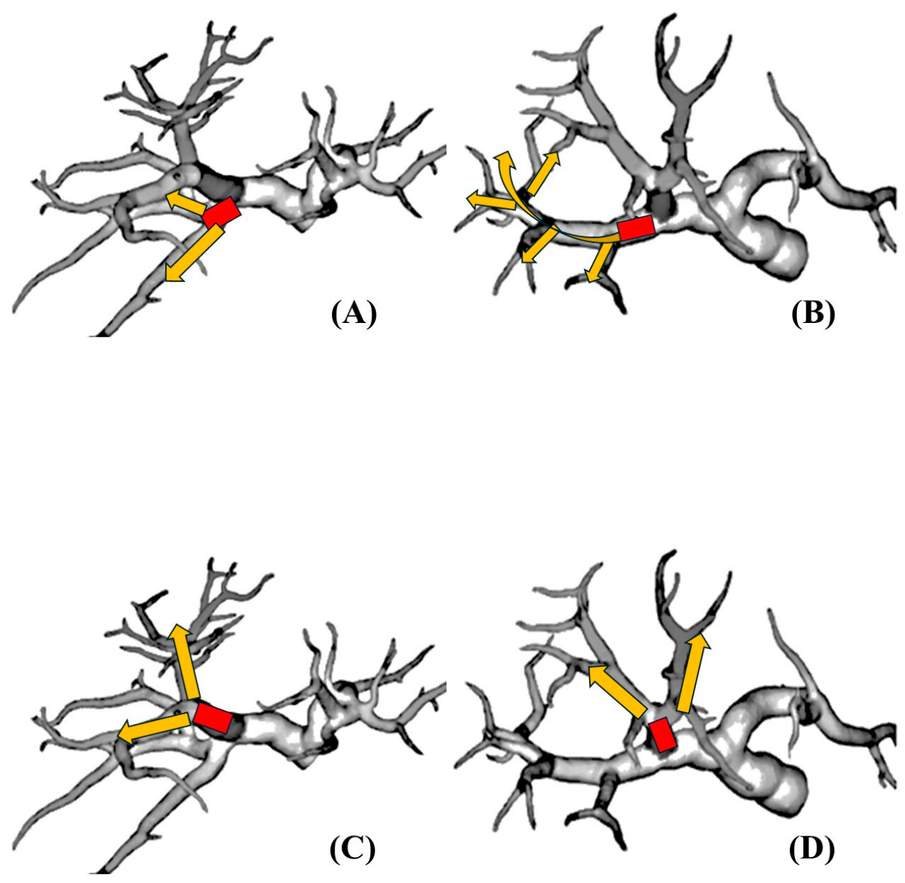
Figure 4.
Simulation and Navigation for Liver Resection (Reproduced/adapted from Imanaka et al. [30] under the Creative Commons Attribution License.) (A): The image of preoperative CT simulation. Purple: Hepatic veins and Vena Cava; Pink: Portal vein; Green: Hepatocellular carcinoma; Curved yellow arrow: Looped posterior portal vein; Ocher arrow: First 3rd order branch from posterior portal vein. The tumor was mainly located inside the first third-order branch territory and robotic resection of extended the territory was planned. (B) Intraoperative findings: The first branch from posterior portal vein was dissected and clamped (Red line). (C) ICG injection after clamping the branch showed clear demarcation line which shows the tumor bearing area.
Figure 4.
Simulation and Navigation for Liver Resection (Reproduced/adapted from Imanaka et al. [30] under the Creative Commons Attribution License.) (A): The image of preoperative CT simulation. Purple: Hepatic veins and Vena Cava; Pink: Portal vein; Green: Hepatocellular carcinoma; Curved yellow arrow: Looped posterior portal vein; Ocher arrow: First 3rd order branch from posterior portal vein. The tumor was mainly located inside the first third-order branch territory and robotic resection of extended the territory was planned. (B) Intraoperative findings: The first branch from posterior portal vein was dissected and clamped (Red line). (C) ICG injection after clamping the branch showed clear demarcation line which shows the tumor bearing area.
Disclaimer/Publisher’s Note: The statements, opinions and data contained in all publications are solely those of the individual author(s) and contributor(s) and not of MDPI and/or the editor(s). MDPI and/or the editor(s) disclaim responsibility for any injury to people or property resulting from any ideas, methods, instructions or products referred to in the content. |
© 2026 by the authors. Licensee MDPI, Basel, Switzerland. This article is an open access article distributed under the terms and conditions of the Creative Commons Attribution (CC BY) license (http://creativecommons.org/licenses/by/4.0/).
Copyright: This open access article is published under a Creative Commons CC BY 4.0 license, which permit the free download, distribution, and reuse, provided that the author and preprint are cited in any reuse.