

Submitted:
31 August 2026
Posted:
01 September 2026
You are already at the latest version
Abstract
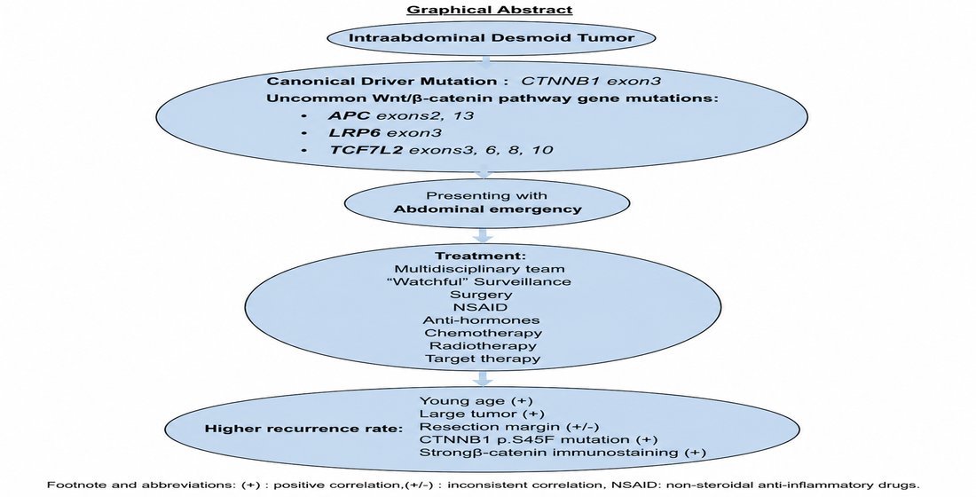
Desmoid tumors (DT) may be sporadic or associated with familial polyposis syndromes including familial adenomatous polyposis (FAP) and its variant Gardner syndrome (GS). DT is pathogenetically related to activation of the Wnt/β-catenin signaling pathway. In sporadic DT, CTNNB1 is most frequently implicated with somatic mutations in exon 3 (T41A, S45F and S45P) being most commonly reported. The APC gene is implicated in FAP/GS associated DT and sometimes in sporadic DT, with the mutation cluster region in exon 15 being most often involved. In our study of 5 sporadic intraabdominal DT, we discovered uncommon somatic mutations in 3 cases, including those in APC (exons 2, 13) and the Wnt/β-catenin pathway genes LRP6 (exon 3) and TCF7L2 (exons 6, 8, 10) which could have mechanistic impacts on the Wnt/β-catenin pathway. One case showed classical canonical somatic CTNNB1 exon 3 mutation and another showed no detectable mutations. All 5 cases were also distinctive in being intraabdominal, an uncommon location of DT, presenting as abdominal emergencies requiring surgical intervention. Intraabdominal DT are rare with reported incidence of 24% compared with 58% for extraabominal DT. The necessity for surgical intervention in abdominal emergencies posed by large intraabdominal DT is a risk factor for DT recurrence.
Keywords:
desmoid
; novel CTNNB1
; uncommon APC
; LRP6
; TCF7L2 mutations
; Wnt/β-catenin pathway
1. Introduction
DT is a non-metastasizing locally recurrent neoplasm of fibroblasts and myofibroblasts. In sporadic DT (SDT), the most common somatic deleterious mutation occur in exon 3 of CTNNB1 (89%) [1,2,3], though non-CTNNB1 mutations of other genes and signaling pathways have been documented [4,5,6]. In one study, 15.2% (31/204) of DT harbored non-CTNNB1 mutations including those of the AKT, ALK, AR, EGFR, ERBB2, IDH2, KIT, RET, SDHA and SDHD genes, which were predicted by in silico tools to be potentially deleterious. These mutations were found to show simultaneous CTNNB1 alterations [4]. In another study, mutations of AKT and BRAF were also detected [5]. Dysfunction or upregulation of the signaling pathways Hedgehog, Notch, JAK/STAT, PI3K3/AKT/mTOR, growth regulatory and estrogen driven pathways are also implicated in DT [6]. DT may also be associated with FAP and its variant GS, with germline mutations in the APC [3,7]. However, somatic mutations of APC have also been demonstrated in FAP associated DT [8,9], and sporadic DT [9,10]. In addition, non-CTNNB1 and non-APC somatic deleterious mutations have been reported in DT including those with wild type CTNNB1 and APC [1,4,5,11,12,13]. In this study, further uncommon somatic mutations in APC and the Wnt/β-catenin pathway genes LRP6 and TCF7L2 were discovered in 3 sporadic intraabdominal SDT. This study is also unique in including only intraabdominal SDT, which are uncommon among all SDT [1]. Their presentation as abdominal emergency is also unusual with only a handful of sporadic reports in the literature [14,15,16,17].
2. Results
2.1. Clinicopathologic and Immunohistochemical Findings
There were 3 females and 2 males without previous or family history of intestinal polyposis or DT. There was history of pregnancy 18 years prior to the abdominal presentation in one female (Case 2). Oral contraceptives were not used in all 3 females. There was no former history of abdominal surgery, radiotherapy or trauma in all 5 patients. All 5 patients presented with intractable abdominal pain and were treated by surgery which revealed large (sizes ranged from 3.5 to 13 cm) obstructive small intestinal tumors originating from the mesentery and involving the intestinal walls extensively up to the mucosa. The tumors feature firm whish tissue with irregular borders. All resection margins were clear of tumor. Case 5 developed recurrence of the original duodenal DT 5 years after surgery. The reccurrent tumors involved the mesentery, duodenum and abdominal wall. The remaining 4 patients were free from tumor recurrence at last follow up, 1-3 years after surgery. Histologically, the tumors showed bland spindle cells arranged in fascicles with manifestations of 7 histological patterns in varying proportions [1] (Table 1 and Figure 1). Four cases contain all 7 histological patterns. There was consistent lack of cytologic atypia or mitotic activity. The growth pattern was uniformly infiltrative, involving the mesentery and intestines almost transmurally, partially involving the mucosa. Immunohistochemically, the spindle tumor cells showed positive nuclear staining for ß-catenin of moderate intensity in all cases (Figure 1). All were positive for α-SMA and 3 for desmin. STAT6 was notably negative. Staining was negative for the remaining antibodies in tested cases .The clinicopathologic and immunohistochemistry findings are summarized in Table 1 and Table 2.
2.2. Genetic Alterations
Case 1 did not show detectable genetic alterations. A summary of the genetic alterations of the remaining 4 cases is presented in Table 3. All were somatic mutations as confirmed by paired genetic testing with normal non-neoplastic intestinal tissue in all cases, where the mutations were detected only in tumor tissues and nor normal intestinal tissues. There was no overlap with genetic alterations available fro gnome AD v 4.1.1 All alterations were missense (MS) mutations.
2.2.1. CTNNB1
A CTNNB1 mutation was detected in Case 3. This is a less common CTNNB1 exon 3 activating mutation p.S45P (Tier 1/2). The variant allele frequency (VAF) was 15%.
2.2.2. APC
A Tier 2/3 MS mutation in exon 13 and a Tier 3 mutation in exon 2 were detected in Case 5 and Case 2 with VAF 6% and 11% respectively. These are distinctly different from the more common mutations in exon 15 at the mutation cluster region (MCR) of the APC gene
2.2.3. Other Wnt/β-Catenin Pathway Genes
2.2.3.1. LRP6
A Tier 3 MS mutation was detected in Case 5 with VAF 10%. This mutation has hitherto not been reported in DT and is of uncertain pathogenetic significance.
2.2.3.2. TCF7L2
Tier 3 MS mutations involving exons 6, 8, 10 were detected in Case 4, with VAF respectively of 7%, 20% and 15%. This mutation has hitherto not been reported in DT and is of uncertain pathogenetic significance.
2.2.4. Tumor Mutation Burden (TMB) and Microsatellite (MS) Stability
All 5 tumors showed low TMB and were MS stable.
2.2.5. USP6 Rearrangement
No USP6 rearrangement in all cases was detected (Figure 1).
3. Discussion
DT/fibromatosis is a rare fibroblastic and myofibroblastic neoplastic proliferation with an incidence of 4-6/1,000,000 population per year, accounting for 0.03% of all tumors and 3% of soft tissue tumors [3]. SDT (accounting for 89% of DT) most commonly harbor somatic activating mutations in the CTNNB1 gene in 3p22.1 [1]. The most frequently reported SDT hotspot somatic CTNNB1 mutations occur in exon 3, namely p.T41A, p.S45P and p.S45F [1,2,3,8,11,12,18,19]. All mutations detected in our cases were somatic as confirmed by paired genetic testing with non-neoplastic intestinal tissue and referencing to gnome AD v 4.1.1. Hereditary DT (HDT) are mostly associated with FAP and harbor germline inactivating mutations of APC in 5q21-22, often involving the MCR in exon 15 [7,8]. Somatic inactivating mutations in exon 15 of the APC gene had also been reported in SDT wildtype for CTNNB1 [3,9,10]. CTNNB1 and APC mutations cause DT through activation of the Wnt/β-catenin signaling pathway, and the mutations are generally mutually exclusive [1,3,13].
3.1. Mechanism of Nuclear β-Catenin Accumulation
In DT, there is decreased β-catenin protein destruction, nuclear β-catenin accumulation and downstream activation of the Wnt/β-catenin pathway. The Wnt/β-catenin pathway is usually triggered through binding of Wnt ligands to the cell surface receptor Frizzled in collaboration with low density lipoprotein receptor-related protein (LRP) 5/6, followed by recruiting Dishevelled to the plasma membrane. The destruction complex (DC) for β-catenin is then sequestered at the plasma membrane, through binding and phosphorylation of LRP by axin and glycogen synthase kinase (GSK)3β in the DC [20,21,22,23,24]. The DC is mostly constituted by 4 major proteins, the scaffold axin and other partner proteins including APC, GSK3β and casein kinase (CK) 1. CK1 and GSK3β are kinases that phosphorylate β-catenin at the serine threonine sites of its N-terminal domain (NTD) when Wnt activation is off [21,22,23]. Phosphorylated β-catenin is recognized by E3 ubiquitin ligase which ubiquitinates β-catenin for proteasomal destruction [22,23]. When Wnt/β-catenin pathway is turned on by Wnt ligand binding, β-catenin phosphorylation by DC and subsequent degradation is abolished with ensuing β-catenin accumulation. CTNNB1 mutation results in an altered β-catenin protein which is not phosphorylated by the DC. APC mutation leads to an altered truncated APC protein which decreases GSK3β function in the DC [6,7], also causing decreased β-catenin phosphorylation, proteasomal destruction and cytoplasmic/nuclear accumulation. In the nucleus, the accumulated β-catenin protein engages the transcription factors T-cell specific transcription factor/lymphoid enhancer factor (TCF/LEF) to activate proto-oncogenes c-myc, cyclin D1, CD44 and IGF1 (insulin-like growth factor-1), thus resulting in cell proliferation, immunosuppression, disruption of metabolic regulation and oncogenesis [22,23,25,26].
3.2. Genetic Alterations
3.2.1. CTNNB1 Mutations
Among the reported exon 3 mutations in SDT, p.T41A accounts for 55-60%, p.S45F for 20-22% and p.S45P for 9-12% [1]. S45F mutation is associated with higher risks of recurrence and predictive of poor response to drug treatment (meloxicam) [18,19,26]. The less common exon 3 p.S45P mutation detected in one of our cases may be less likely to recur. Our patient has remained recurrence free 3 years after surgery.
3.2.2. APC Mutations
Tier 2/3 APC gene mutations were detected in 2 of our cases, involving exons 2 and 13. These mutations are distinctly different from previously reported driver somatic or germline APC mutations that most commonly affect exon 15 particularly MCR [7,8,9,10]. There was, however, a previous report of a rare germline mutation occurring at the extreme 3’ end in exon 15 outside the MCR that is associated with a severe form of multiple DT affecting the axial skeleton or extremities [27]. Rare non-exon 15 mutations in exons 9 and 16 of the APC gene have also been reported previously [28]. The significance of the non-exon 15 MCR mutations in APC of our cases is uncertain and requires further studies.
3.2.3. Other Wnt/β-Catenin Pathway Gene Mutations
3.2.3.1. LRP6
Lipoprotein receptor-relate protein 6 (LRP6) is a single-pass transmembrane protein, which together with LPR5 and Frizzled receptors, act as co-receptors of Wnt ligands in Wnt/β-catenin signaling [29]. LRP6 is frequently overexpressed in colorectal, liver, breast and pancreatic adenocarcinomas in association with increased Wnt/β-catenin signaling. Hyperphosphorylation of LRP6 is also detected in KRAS-mutated cells and colorectal tumors. LRP6 single nucleotide polymorphism and mutations have been observed in non-small cell lung cancer, lung squamous cell carcinoma, urinary bladder carcinoma and colorectal carcinoma (CRC) [30]. However, to the best of our knowledge, LRP6 mutations or overexpression have not been reported in DT. Though the LRP6 mutation discovered in one of our cases is of uncertain significance, its possible pathogenetic role in DT remains to be explored in future studies.
3.2.3.2. TCF7L2
T-cell specific transcription factor (TCF) L2 belongs to a small high-mobility-group box subfamiliy which comprises 4 members: TCF7/TCF1, TCF7L1/TCF3, TCF7L2/TCF4 and LEF-1 [31]. TCF/LEF are intranuclear transcription factors that β-catenin engages to activate downstream oncogenes [22,23,24,25,26]. TCF7L2 is implicated in oncogenesis (colorectal cancer, hepatoblastoma) [32,33]. TCF7L2 mutations have frequently been demonstrated in CRC with a possible tumor suppressor function. The TCF7L2 protein positively regulates the proto-oncogene MYC and represses the cell cycle inhibitors CDKN2C/CDKN2D. TCF7L2 also suppresses the pro-metastatic transcription factor RUNX2 and the expression of cell adhesion molecules. TCF7L2 loss of function mutations may thus be important in oncogenesis of CRC [32]. To the best of our knowledge, however, TCF7L2 mutations have not been documented in DT and their pathogenetic role in DT remains to be further elucidated by further studies.
3.2.4. Epigenetic Mechanisms
In cases where no genomic alterations are detected, it is possible that oncogenesis was driven by epigenetic mechanisms. It has been demonstrated that the balance of TCF7L2 variants with increased TCF7L2 transcription and differential activities in Wnt signaling is regulate by exogenous lithium [31]. Another study showed that differential association of β-catenin with TCF/LEF transcription factors results in differential Wnt/β-catenin signaling [33]. Promoter hypermethylation of Wnt antagonists, tumor microenvironment growth factors, inflammatory mediators and extracellular proteins are also partakers in DT oncogenesis [6,34].
Clinical observation strongly supports a hormonal contribution as suggested by increased incidence of DT during and after pregnancy and female prevalence [1,3,13]. DT has been discovered to express estrogen receptor β (ERβ or ER2), providing scientific support to this time-honored clinical onservation [6]. Increased incidence of DT after previous surgery, irradiation or trauma [1] may also be related to epigenetic mechanisms.This occurred in one of our cases who developed recurrent DT 5 years after initial DT resection.
3.3. Presentation, Diagnosis and Differential Diagnoses
Abdominal emergencies are due frequently to intestinal obstruction (IO), perforation, hemorrhage, acute peritonitis or tumor rupture, which require urgent surgical intervention. IO is commonly caused by fibrous adhesions, hernias, intussusception, intestinal tumors or intraluminal foreign bodies such as gallstones. Abdominal tumors causing these emergencies are usually primary gastrointestinal (GI) cancers or secondary malignancies. Intraabdominal DT is a rare GI tumor [3] and an uncommon cause of abdominal emergencies that usually cannot be confirmed until pathological examination on excised surgical material. Only a handful of isolated intraabdominal DT cases with this presentation were reported in the past 5 years [14,15,16,17], with none being studied genetically. Our intraabdominal DT presenting with abdominal emergencies adds to these rare reports, embellished with full molecular workup.
The histology of DT is myriad, requiring full recognition for diagnosis. There are seven histological patterns [1,35]: conventional with fascicles of bland spindle cells, hyalinized, staghorn vessel (stagv), hypocellular/myxoid, keloidal, nodular fasciitis (NF)-like and hypercellular. Most DT manifest several patterns with the mean number of two. Our cases exhibit multiple patterns in varying combinations, with most expressing all 7 patterns. In all patterns, the spindle-shaped fibroblasts and myofibroblasts have bland pale-staining nuclei, inconspicuous nucleoli with or without mild cytological atypia and minimal mitotic activity. The NF-like and stagv patterns were most problematic, potentially mistaken for NF and solitary fibrous tumor (SFT). DT is distinguishable by nuclear β-catenin expression [1,19], with or without detectable mutations in CTNNB1 [1,18,19,26] or APC [3,4,5,6,7,11,28]. There is correlation of the intensity of β-catenin nuclear staining with prognosis, with DT expressing strong β-catenin staining associated with poorer prognosis [19]. Our cases exhibited β-catenin staining of moderate intensity, which may portend better prognosis. However, β-catenin immunopositivity is not entirely specific for DT [36,37]. The morphologic differences of DT from its mimickers and the different immunophenotypic and genotypic features should enable their distinction. NF is a major differential diagnosis which has not been reported in internal viscera, often characterized histologically by “tissue culture” pattern and extravasated red cells, and is negative for nuclear β-catenin staining immunohistochemically. Further, NF lacks staghorn vessels present in DT. NF is also characterized by USP6 gene rearrangement [38]. USP6 FISH performed on our cases showed no rearrangement, thus differentiating it from NF. SFT may be more problematic as it morphologically may also manifest keloidal fibers, staghorn vessels and 40% may be nuclear β-catenin positive. It is however distinctive by STAT-6 positive immunophenotype [39]. Differential diagnosis from inflammatory fibroblastic/myofibroblastic tumor (IMT) can be made by differences in morphology, immunophenotypes and nuclear β-catenin positivity in DT. IMT may morphologically range from bland to atypical spindle cells with frequent ALK immunopositivity and ALK gene rearrangement, allowing its distinction from DT [40]. Other differential diagnoses from DT encompass various malignant/borderline spindle cell tumors (spindle cell carcinoma and melanoma, dedifferentiated liposarcoma, leiomyosarcoma, malignant peripheral nerve sheath tumor, synovial sarcoma, angiosarcoma, fibrosarcoma and gastrointestinal stromal tumor). These tumors could be distinguished by their characteristic clinical presentations, morphology, immunophenotypes and genotypes [1].
3.4. Treatment and Prognosis
The treatment of asymptomatic DT could be surveillance. However, spontaneous regression is less common in intraabdominal than abdominal DT. The recurrence rate is 20-30% and more common in larger tumors (>70 mm), younger (<37 years) subjects and related to tumor location [1,3,12,14,15,16,17,41]. Higher recurrence rate has been reported in DT with CTNNB1 p.S45F mutations [1,18,44,45]. Strong intensity of β-catenin immunostaining may be associated with worse prognosis [19]. Wide negative resection margins are associated with a lower recurrence rate, compared with positive margins (10% vs 80%), though incomplete resection does not correlate consistently with increased recurrence [1,12,28]. Negative resection margins and moderate β-catenin immunostaining intensity in our patients may portend an expected lower recurrence rate and better prognosis. In a French study, tumor location was identified as the most significant prognostic factor [41]. Other treatment modalities include chemotherapy, radiotherapy or systemic therapy (non-steroidal anti-inflammatory drugs, anti-hormonal, cytotoxic drugs, tyrosine kinase inhibitors, γ-secretase inhibitors therapy), usually requiring a multidisciplinary team [36,42,46,47]. Exploiting increased understanding of the canonical and non-canonical Wnt/β-catenin and other signaling pathways, new modalities of treatment targeting at down regulation of these pathways have been proposed, including negative regulators of Wnt/β-catenin signaling, microRNAs and long non-coding RNAs [11,47].
4. Materials and Methods
4.1. Clinicopathologic and Immunohistochemical Study
Five SDT presenting as abdominal emergencies were identified over a 6 year period (2020-2025). The clinical and imaging records were reviewed. Tumor and non-tumor involved tissues were obtained from resection specimens and processed for formalin fixed paraffin embedded (FFPE) tissue blocks. H & E stained four micron sections were prepared. Immunostaining using an automatic immunostainer (Ventanna, USA) was performed with antibodies including cytokeratin, CD117, S100 protein, β-catenin, desmin, α- smooth muscle actin (SMA), SOX10, Dog 1, CD34, STAT 6, SSX, ALK, PDGFRA and β-catenin (Abcam, Dako, Epitomics, Fouzhou Maxin Biotechnology, Leica and Ventanna).
4.2. Genetic Study
4.2.1. Genomic DNA Preparation
Tumor tissues and non-tumor tissue from all 5 cases were studied. The non-tumor intestinal tissue was used for paired genetic testing to look for germline mutations. Genomic DNA in FFPE tissue was prepared as described before [48].
4.2.2. Whole Genome Sequencing (WGS)
The library construction and sequencing were performed as described before [49]. Briefly, fragmentation of genomic DNA was performed on Covaris E220 following the manufacturer’s instructions. DNA fragments of 100bp-300bp were selected with AMPure XP beads (AGENCOURT), which were repaired and modified for a sticky dATP 3’ end and then ligated to a dTTP tailed adapter sequence. The ligated product was amplified by polymerase chain reaction (PCR) and a single strand circular DNA library was produced by single strand circularization. WGS was conducted on the DNBSEQ-T7 platform. The DNBSEQ basecalling software was employed to process sequencing derived image files as raw data.
4.2.3. Bioinformatics and Mutational Analysis
SOAPnuke (version 2.2.6) was used to filter the raw data, mapped to the UCSC human reference genome by the Burrows-Wheeler Aligner (BWA, version 0.7.17), and converted into binary alignment map format files. Best practices for variant analysis were followed using the Genome Analysis Toolkit (GATK, version 4.4.0.0) and quality sequence data was guaranteed using strict data analysis quality control in the whole pipeline (GATK Base Recalibrator and GATK ApplyBQSR).The detected genetic variants were classified into tiers according to established criteria of the Association for Molecular Pathology, American Society of Clinical Oncology and College of American Pathologists [50]. Reference was also made to gnome AD v4.1.1 for checking germline mutations.
4.2.4. Fluorescent In-Situ Hybridization (FISH) for USP6
The USP6 dual-color break apart probe kit (Guangzhou Anbiping Pharmaceutical, China). The probes target centromere (red fluorescence) and telomere (green fluorescence) of chromosome 17p13.2. Briefly, FFPE tissue sections were deparaffinized, rehydrated, pepsin digested, hybridized with probes.and counterstained with 4,6-diamidino-2-phenylindole. Signals were evaluated under a fluorescence microscope. Two hundred non-overlapping nuclei were counted. Cells showing a separation of red and green signals greater than two signal diameters were considered positive. A proportion of positive cells greater than 5% was interpreted as positive for split rearrangement.
5. Conclusions
In closing, we report 5 molecularly characterized intraabdominal DT presenting as abdominal emergency which required surgery. Other than an activating mutation in exon 3 of the CTNNB1 gene, uncommon mutations in exon 2, 13 of the APC gene and other Wnt/β-catenin pathway genes LRP6 and TCF7L2 were discovered. The significance of these uncommon mutations, however, remains to be explored in DT. Intraabdominal DT presenting as abdominal emergency are rare. Despite inconsistent correlation with completeness of surgical resection, wide surgical excision with negative resection margins may confer a lower recurrence rate, when surgery is unequivocally indicated in cases of emergency. A number of parameters have been proposed for prognostication and tumor location appeared to be a more significant prognostic factor. The tissue diagnosis could be challenging due to the myriad histologic patterns. An integrated approach of clinical, morphologic, immunohistochemical and genetic workup should be useful in making the diagnosis and prognostication. Better understanding the pathogenetic mechanisms in the Wnt/β-catenin pathway lays the ground for developing future new treatment strategies.
A major limitation is the small number of cases studied. The 5 cases studied, however, represented a collection fro 3 major centers over a 5 year period, refecting the rarity of intraabdominal DT [1]. The small case number is further related to the uncommon presentation as abdominal emergencies [14,15,16,17]. Further work on more cases is warranted for better understanding of the genomics and epigenetics of DT.This study is also limited by being a sole genomic study on DT. Exploration into possible epigenetic mechanisms was not performed. Using techniques like whole transcriptome RNA sequencing, DNA methylation, expression profile analysis or micro-RNA studies should be helpful in uncovering possible epigenetic features of DT.
Author Contributions
CSN: Conceptualization, study design, case review, literature search and curation, results interpretation, drafted manuscript, approved final manuscript. JQ: Case review and curation, performed genetic studies, prepared images of figures, approved final manuscript. GYG: reviewed cases and approved final manuscript. All authors agreed to be accountable for all aspects pf this work.
Funding
This work was supported by the Guangzhou Municipal Bureau of Science and Technology 2023 Municipal School (Hospital) Joint Funding (Dengfeng Hospital) Basic Research Project (2023A03J0814).
Institutional Review Board Statement
This work was carried out following the rules of the Declaration of Helsinki of 1975 (revised 2013). It was approved by the Ethics Committee of the First Affiliated Hospital of Guangzhou Medical University, Guangzhou, China (reference number ES-2023-209-01 on December 5, 2023).
Informed Consent Statement
Informed consent was obtained from all subjects involved in the study.
Data Availability Statement
Data is available upon reasonable request.
Acknowledgments
We thank Dr. CH Kan, Chief Pathologist, St.Teresa’s Hospital, Hong Kong for allowing us to include case 2. We are also grateful to Ms Yvonne Chan for assistance in manuscript preparation.
Conflicts of Interest
The authors declare that there is no conflict of interest.
References
- Ajabnoor, R. Different shades of desmoid-type fibrosis (DTF): detection of novel mutations in the clinicopathologic analysis of 32 Cases. Diagnostics 2024, 14, 2161. [Google Scholar] [CrossRef] [PubMed]
- F Prete, F.; Rotell, M.T.; Stella, A.; Calculli, G.; Sgaramella, L.I.; Amati, A.; et al. Intraabdominal sporadic desmoid tumors and inflammation: an updated literature review and presentation and insights on pathogenesis of synchronous sporadic mesenteric desmoid tumors occurring after surgery for necrotizing pancreatitis. Clin. Exp. Med. 2022, 23, 607–617. [Google Scholar] [CrossRef]
- Fritchie, K.J.; HornickL, J.L.; Rossi, S. Desmoid fibromatosis. In Digestive System Tumours, 5th edition; WHO Classification of Tumours Editorial Board, Ed.; IARC: Lyons (France), 2019; pp. pp446–447. [Google Scholar]
- MTrautmann; Rehkamper, J.; Gevensleben, H.; Becker, J.; Wardelmann, E.; Hartmann, W.; et al. Novel pathogenic alterations in pediatric and adult desmoid-type fibromatosis – a systematic analysis of 204 cases. Sci. Rep. 2020, 10, 3368. [Google Scholar] [CrossRef] [PubMed]
- Meazza, C.; Belfiore, A.; Busico, A.; Settanni, G.; Paielli, N.; Cesana, L.; et al. AKT1 and BRAF mutations in pediatric aggressive fibromatosis. Cancer Med. 2016, 5, 1204–1213. [Google Scholar] [CrossRef] [PubMed]
- Timergen, M.J.M.; Smits, R.; Grunhagen, D.J.; Verhoef, C.; Sleifer, S.; Wiemer, E.A.C. Activated Signaling Pathways and Targeted Therapies in Desmoid-Type Fibromatosis: A Literature Review. Front Oncol. 2019, 9, 397. [Google Scholar] [CrossRef] [PubMed]
- Church, J.; Xhileta, X.; LaGuardia, L.; O’Malley, M.; Burke, C.; Kalady, M. Desmoids and genotype in familial adenomatous polyposis. Dis. Colon Rectum 2015, 58, 444–448. [Google Scholar] [CrossRef] [PubMed]
- Iwama, T.; Kuwabara, K.; Ushiama Myoshida, T.; Sugano, K.; Ishida, H. Identification of somatic APC mutations in recurrent desmoid tumors in a patient with familial adenomatous polyposis to determine actual recurrence of original tumor or de novo occurrence. Fam. Cancer 2009, 8, 51–54. [Google Scholar] [CrossRef] [PubMed]
- Giarola, M.; Wells, D.; Mondini, P.; Pilotti, S.; Sala, P.; Azzarelli, A.; Bertario, L.; Pierotti, M.A.; Delhanty, J.D.; Radice, P. Mutations of adenomatous polyposis coli (APC) gene are uncommon in sporadic desmoid tumours. Br. J. Cancer 1998, 78, 582–587. [Google Scholar] [CrossRef] [PubMed]
- Alman, B.; Li, C.; Pajerski, M.E.; Diaz-Cano, S.; Wolfe, H.J. Increased β-catenin protein and somatic APC mutations in sporadic aggressive fibromatosis (desmoiod tumors). Am. J. Pathol. 1997, 151, 329–334. [Google Scholar] [PubMed]
- Liu, J.; Xiao, Q.; Xiao, J.; Niu, C.; Li, Y.; Zhang, X.; et al. Wnt/B-catenin signaling: function, biological mechanisms, and therapeutic opportunities. Sig Transduct. Target 2022, 7, 3. [Google Scholar] [CrossRef]
- Crago, A.M.; Chmielecki, J.; Rosenberg, M.; O’Connor, R.; Byrne, C.; Wilder, F.G.; et al. Near universal detection of alterations in CTNNB1 and Wnt pathway regulators in desmoid-type fibromatosis by whole-exome sequencing and genomic analysis. Genes Chrom Cancer 2015, 54, 606–615. [Google Scholar] [CrossRef] [PubMed]
- Penel, N.; Chibon, F.; Salas, S. Adult desmoid tumors: biology, management and ongoing trials. Curr. Opin. Oncol. 2017, 29, 268–274. [Google Scholar] [CrossRef]
- Zhao, D.; Wang, X. A rare cause of small bowel obstruction caused by duodenum-derived aggressive fibromatosis with β-catenin T41A mutation: A case analysis. Medicine 2024, 103, e38984. [Google Scholar] [CrossRef] [PubMed]
- Morris, D.; Gonzalez, R.; Cook, J.; Metts, J.; Shaw, P. Gastrointestinal complications of intra-abdominal desmoid tumors: a case report and review of the literature. Case Rep. Oncol. 2020, 15, 515–521. [Google Scholar] [CrossRef] [PubMed]
- Das, S.S.; Navlani, S.; Alawa, A.; Abbas, F.M.; Raman, L.G.; Mestha, A. Challenging diagnostic dilemma: mesenteric desmoid tumor masquerading as perforated peritonitis. Cureus 2023, 15, e42946. [Google Scholar] [CrossRef] [PubMed]
- Yoshida, A.; Uchima, Y.; Hosaka, N.; Minaga, K.; Kudo, M. Transverse colon volvulus due to mesenteric fibromatosis: a case report. BMC Gastroenterol. 2021, 21, 11. [Google Scholar] [CrossRef] [PubMed]
- Colombo, C.; Miceli, R.; Lazar, A.J.; Perrone, F.; Pollock, R.E.; Le Cesne, A.; et al. CTNNB1 45F mutation is a molecular prognosticator of increased postoperative primary desmoid tumor recurrence. Cancer 2013, 119, 3696–3702. [Google Scholar] [CrossRef] [PubMed]
- Hamada, S.; Futamura, N.; Ikuta, K.; Urakawa, H.; Kozawa, E.; Ishiguro, N.; et al. CTNNB1 S45F mutation predicts poor efficacy of Meloxicam treatment for desmoid tumors: a pilot study. PLoS ONE 2014, 9, e96391. [Google Scholar] [CrossRef] [PubMed]
- Sharma, M.; Castro-Piedras, I.; Simmons, G.E.; Pruitt, K. Dishevelled: a masterful conductor of complex Wnt signals. Cell Sig 2018, 47, 52–64. [Google Scholar] [CrossRef] [PubMed]
- Liu, C.; Li, Y.; Semenov, M.; Han, C.; Baeg, Gyeong-Hun; Tan, Y.; et al. Control of β-catenin phosphorylation/degradation by a dual-kinase mechanism. Cell 2002, 108, 837–847. [Google Scholar] [CrossRef] [PubMed]
- Gao, C.; Wang, Y.; Broaddus, R.; Sun, L.; Xue, F.; Zhang, W. Exon 3 mutations of CTNNB1 drive tumorigenesis: a review. Oncotarget 2018, 9, 5492–5508. [Google Scholar] [CrossRef] [PubMed]
- Schaefer, K.N.; Pronobis, M.I.; Williams, C.E.; Zhang, S.; Bauer, L.; Goldfarb, D.; et al. Wnt regulation: exploring Axin-Dishevelled interactions and defining mechanisms by which the SCF E3 biquitin ligase is recruited to the destruction complex. Mol. Biol. Cell 2020, 31, 992–1014. [Google Scholar] [CrossRef] [PubMed]
- Ma, W.; Chen, M.; Kang, H.; Steinhart, Z.; Angers, S.; He, X.; et al. Single-molecule dynamics of Dishevelled at the plasma membrane and Wnt pathway activation. PNAS 2020, 117, 16690–16701. [Google Scholar] [CrossRef]
- Korinek, V.; Barker, N.; Morin, P.J.; van Wichen, D.; de Weger, R.; Kinzler, K.W.; et al. Constitutive transcriptional activation by a β-catenin-Tcf complex in APC-/- colon carcinoma. Science 1997, 275, 1784–1787. [Google Scholar] [CrossRef] [PubMed]
- Lazar, A.J.F.; Tuvin, D.; Hajibashi, S.; Habeeb, S.; Bolshakov, S.; Mayordomo-Aranda, E.; et al. Specific Mutations in the β-catenin gene (CTNNB1) correlate with local recurrence in sporadic desmoid tumors. Am. J. Pathol. 2008, 173, 1518–1527. [Google Scholar] [CrossRef] [PubMed]
- Couture, J.; Mitri, A.; Lagace, R.; Smits, R.; Berk, T.; Bouchard, H.-L.; et al. A germline mutation at the extreme 3’ end of the APC gene results in a severe desmoid phenotype and is associated with overexpression of beta-catenin in the desmoid tumor. Clin. Genet 2000, 57, 205–212. [Google Scholar] [CrossRef] [PubMed]
- Wang, J.; Jia, N.; Lin, Q.; Huang, Y.; Li, J.; Jiang, Q.; et al. Clinicopathological and molecular characteristics of abdominal desmoid tumors in the Chinese population: a single-center report of 15 cases. Oncol. Lett. 2019, 18, 6443–6450. [Google Scholar] [CrossRef] [PubMed]
- Ren, Q.; Chen, J.; Liu, Y. LRP5 and LRP6 in Wnt signaling: similaity and divergence. Front Cell Dev. Biol. 2021, 9, 670960. [Google Scholar] [CrossRef] [PubMed]
- Raisch, J.; Cote-Biron, A.; Rivard, N. A Role for the WNT Co-Receptor LRP6 in Pathogenesis and Therapy of Epithelial Cancers. Cancers 2019, 11, 1162. [Google Scholar] [CrossRef] [PubMed]
- Struewing, I.; Boyechko, T.; Barnett, C.; Belldeck, M.; Byers, S.W.; Mao, C.D. The balance of TCF7L2 variants with differential activities in Wnt-signaling is regulated by lithium in a GSK3β-independent manner. Biochem Biophys. Res. Commun. 2010, 399, 245–250. [Google Scholar] [CrossRef]
- Wenzei, J.; Rose, K.; Haghighi, E.B.; Lamprecht, C.; Rauen, G.; Freihen, V.; et al. Loss of the nuclear Wnt pathway effector TCF7L2 promotes migration and invasion of human colorectal cancer cells. Oncogene 2020, 39, 3893–3909. [Google Scholar] [CrossRef] [PubMed]
- Kluvier, T.A.; Nordin, A.; Lu, Y.; Guo, X.; Schubert, S.A.; Yeung, D.; et al. TCF7L2/LEF1 binding specificity shapes cellular and tumor phenotypes. PNAS 2026, 123, e2528450123. [Google Scholar] [CrossRef] [PubMed]
- Enzo, M.V.; Rastrelli, M.; Rossi, C.R.; Hladnik, U.; Segat, D. The Wnt/β-catenin pathway in human fibrotic-like diseases and its eligibility as a therapeutic target. [CrossRef] [PubMed]
- Zreik, R.T.; Fritchie, K.J. Morphologic Spectrum of Desmoid-Type Fibromatosis. Am. J. Clin. Pathol. 2016, 145, 332–40. [Google Scholar] [CrossRef] [PubMed]
- Bhattacharya, B.; Dilworth, H.P.; lacobuzio-Donahue, C.; Ricci, F.; Weber, K.; Furlong, M.A.; et al. Nuclear beta-catenin expression distinguishes deep fibromatosis from other benign and malignant fibroblastic and myofibroblastic lesions. Am. J. Surg. Pathol. 2005, 29, 653–9. [Google Scholar] [CrossRef] [PubMed]
- Ng, T.L.; Gown, A.M.; Barry, T.S.; Cheang, C.U.; Chan, A.K.W.; Turbin, D.A.; et al. Nuclear beta-catenin in mesenchymal tumors. Mod. Pathol. 2005, 18, 68–74. [Google Scholar] [CrossRef]
- Erber, R.; Agaimy, A. Misses and near misses in diagnosing nodular fasciitis and morphologically related reactive myofibroblastic proliferations: experience of a referral center with emphasis on frequency of USP6 gene rearrangements. Virchow Arch. 2018, 473, 351–60. [Google Scholar] [CrossRef] [PubMed]
- Martin-Broto, J.; Mondaza-Hernandez, J.L.; Moura, D.S.; Hindi, N. A comprehensive review on solitary fibrous tumor: new insights for new horizons. Cancers 2021, 23, 2913. [Google Scholar] [CrossRef] [PubMed]
- Simion, K.; Reszec-Gielazin, J.; Kisluk, J.; Roszkowiak, L.; Zak, J.; Korzynska, A. What do we know about inflammatory myofibroblastic tumors? - A systematic review. Adv. Med. Sci. 2022, 67, 129–138. [Google Scholar] [CrossRef]
- Penel, N.; Le Cesne, A.; Bonvalot, S.; Giraud, A.; Bompas, E.; Rios, M.; et al. Surgical versus non-surgical approach in primary desmoid-type fibromatosis patients: a nationwide prospective cohort from the French Sarcoma Group. Eur. J. Cancer 2017, 83, 125–131. [Google Scholar] [CrossRef] [PubMed]
- Kasper, B.; Baumgarten, C.; Garcia, J.; Bonvalot, S.; Haas, R.; Haller, F.; et al. An update on the management of sporadic desmoid-type fibromatosis: a European Consensus Initiative between Sarcoma Patients EuroNet (SPAEN) and European Organization for Research and Treatment of Cancer (EORTC)/Soft Tissue and Bone Sarcoma Group (STBSG). Ann. Oncol. 2017, 28, 2399–2408. [Google Scholar] [CrossRef] [PubMed]
- Salas, S.; Bulard, C.; Terrier, P.; Ranchere-Vince, D.; Neuville, A.; Guillou, L.; et al. Gene expression profiling of desmoid tumors by cDNA microarrays and correlation with progression-free survival. Clin. Cancer Res. 2015, 15, 4194–4200. [Google Scholar] [CrossRef] [PubMed]
- Huss, S.; Nehles, J.; Binot, E.; Wardelmann, E.; Mittler, J.; Kleine, M.A.; et al. β-catenin (CTNNB1) mutations and clinicopathological features of mesenteric desmoid-type fibromatosis. Histopathology 2013, 62, 294–304. [Google Scholar] [CrossRef] [PubMed]
- Carrillo-Garcia, J.; Hindi, N.; Conceicao, M.; Salas, M.A.; Ugalde, A.; Lopez-Pousa, A.; et al. Prognostic impact of tumor location and gene expression profile in sporadic desmoid tumor. Eur. J. Cancer 2024, 209, 114270. [Google Scholar]
- Garbay, D.; Le Cesne, A.; Penel, N.; et al. Chemotherapy in patients with Desmoid Tumors: A Study from the French Sarcoma Group (FSG). Ann. Oncol. 2012, 23, 182–186. [Google Scholar] [CrossRef] [PubMed]
- Ibrahim, R.; Assi, T.; Khoury, R.; Ngo, C.; Faron, M.; Verret, B.; Levy, A.; Honore, C.; Henon, C.; Le Pechoux, C.; et al. Desmoid-Type Fibromatosis: Current Therapeutic Strategic and Future Perspectives. Cancer Treat. Rev. 2024, 123, 102675. [Google Scholar] [CrossRef] [PubMed]
- Shi, S.; Data, R.; Liu, C.; Wu, L.; Zhang, Z.; Cote, R.J.; et al. DNA extraction from archival formalin-fixed, paraffin-embedded tissues: heat-induced retrieval in alkaline solution. Histochem Cell Biol. 2004, 122, 211–218. [Google Scholar] [CrossRef] [PubMed]
- Huang, J.; Liang, Y.; Xuan, Y.; Geng, C.; Li, Y.; Lu, H.; et al. A reference human genome dataset of the BGISEQ-500 sequencer. Gigascience 2017, 6, 1–9. [Google Scholar] [CrossRef] [PubMed]
- Li, M.M.; Datto, M.; Duncavage, E.J.; Kulkarni, S.; Lindeman, N.I.; Roy, S.; et al. Standards and Guidelines for the interpretation and reporting of sequence variants in cancer. A joint consensus recommendation of the Association for Molecular Pathology, American Society of Clinical Oncology, and College of American Pathologists. J. Mol. Diag 2017, 19, 4–23. [Google Scholar] [CrossRef] [PubMed]
Figure 1.
(A)Cut surface of mesenteric tumor of Case 5, featuring firm whitish tumor tissue extending to and involving the small intestinal wall almost transmurally.(B)Low power revealed spindle cell tumor involving the muscularis propria and mesentery. H&E x 40.(C)Low power of mesenteric DT involving adjacent jejunal wall with hemorrhage. H&E x40(D) Conventional pattern of DT. H&E x 200.(E). Nodular fasciitis-like pattern with extravasated red cells and loose stroma. H&E x200.(F). Staghorn vessel pattern. H&E x100.(G). Hyalinize pattern. H&E x 200.(H). Myxoid hypocellular pattern. H&E x200.(I). Keloidal pattern, featuring thick ropy collagen fibers, H&E z200.(J). Hypercellular pattern, with bland spindle cells in fascicles. H&E x200.(K). Immunohistochemical staining for β-catenin showed tumor cell nuclear staining of moderate intensity. X200.(L). USP6 FISH showed no gene rearrangement.
Figure 1.
(A)Cut surface of mesenteric tumor of Case 5, featuring firm whitish tumor tissue extending to and involving the small intestinal wall almost transmurally.(B)Low power revealed spindle cell tumor involving the muscularis propria and mesentery. H&E x 40.(C)Low power of mesenteric DT involving adjacent jejunal wall with hemorrhage. H&E x40(D) Conventional pattern of DT. H&E x 200.(E). Nodular fasciitis-like pattern with extravasated red cells and loose stroma. H&E x200.(F). Staghorn vessel pattern. H&E x100.(G). Hyalinize pattern. H&E x 200.(H). Myxoid hypocellular pattern. H&E x200.(I). Keloidal pattern, featuring thick ropy collagen fibers, H&E z200.(J). Hypercellular pattern, with bland spindle cells in fascicles. H&E x200.(K). Immunohistochemical staining for β-catenin showed tumor cell nuclear staining of moderate intensity. X200.(L). USP6 FISH showed no gene rearrangement.
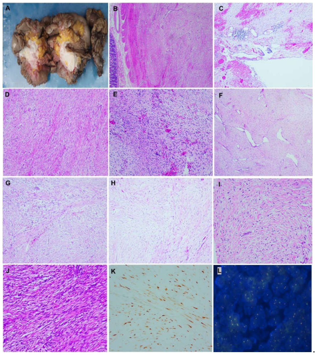

Table 1.
Clinicopathologic findings of 5 new sporadic intraabdominal DT cases.
| Case No. | Age | Sex | Clinical presentation | Involved sites | OC | *Previous pregnancy | Previous surgery or RT | Tumor size | Resection margins |
Histology | ||||||
|---|---|---|---|---|---|---|---|---|---|---|---|---|---|---|---|---|
| C | Stag | N | Hyal | Myx | Kel | hyC | ||||||||||
| 1 | 42 | M | Abdominal pain, IO | Mesentery/Jejunum | NA | NA | No | 13 cm | -ve | + | + | - | ± | ± | ± | ± |
| 2 | 40 | F | Abdominal pain, IO, weight loss | Mesentery/Jejunum | No | 18* | No | 7cm | -ve | + | + | + | ± | ± | ± | ± |
| 3 | 57 | F | Abdominal/ back pain | Mesentery/Jejunum | No | No | No | 4.5cm | -ve | + | + | + | + | + | + | + |
| 4 | 59 | F | Abdominal pain, hematemesis | Mesentery/ Jejunum | No | No | No | 3.5cm | -ve | + | - | + | + | ± | + | + |
| 5 | 32 | M | Abdominal pain, melena | Mesentery/ Duodenum/Abdominal wall | NA | NA | Y | 13cm | -ve | + | - | + | + | ± | + | ± |
Abbreviations: C, conventional; hyal, hyalinized; hyC, hypercellular; IO, intestinal obstruction; kel, keloidal; myx, myxoid; N, nodular fasciitis-like; NA, not applicable; OC, oral contraceptive; RT, radiotherapy; Stag, Staghorn vessel; Y, previous surgery 5 years ago before recurrence; +, >10%; ±, <10%; -, 0%; -ve, not involved; *: years before intraabdominal disease.
Table 2.
Immunohistochemical findings in 5 new sporadic intraabdominal DT.
| Case No. | β-catenin | CK | α-SMA | Desmin | STAT6 | PDGFRA | CD117 | Dog1 | S-100 | SOX10 | SSX-SS18 | ALK(D5F3) | CD34 |
|---|---|---|---|---|---|---|---|---|---|---|---|---|---|
| 1 | + | - | +/- | - | - | ND | - | - | - | ND | ND | - | - |
| 2 | + | - | +/- | - | - | ND | - | - | - | ND | ND | - | - |
| 3 | + | - | + | +/- | - | - | - | - | - | - | - | - | - |
| 4 | + | - | + | +/- | - | - | - | - | - | - | - | - | - |
| 5 | + | - | + | +/- | - | - | - | - | - | - | - | - | - |
Abbreviations: α-SMA, α-smooth muscle actin; ND, not done; PDGFRA, platelet-derived growth factor receptor alpha; +, moderate positive; -, negative; +/-, weak or focal positive.
Table 3.
Genetic alterations in intraabdominal DT cases .
| Gene | Location | Alteration | Type | Tier | Case | VAF(%) | SIG | |
|---|---|---|---|---|---|---|---|---|
| CTNNB1 | exon3 | c.133T>C:p.S45P | MS | 1/2 | 3 | 15 | S/P | |
|
APC LRP6 TCF7L2 |
exon2 | c.76G>A:p.E26K | MS | 3 | 2 | 11 | US | |
| exon13 exon3 exon6 exon8 exon10 |
c.1799G>A:p.G600D c.619C>T:p.H207Y c.727C>T:p.R243W c.849G>T:p.K283N c.1010G>A:p.C337Y |
MS MS MS MS MS |
2/3 3 3 3 3 |
5 5 4 4 4 |
6 10 7 20 15 |
US US US US US |
Abbreviations: MS=missense mutation, SIG=significance, S/P=Strong/Potential significance, US=Unknown significance, VAF=variant allele frequency.
Disclaimer/Publisher’s Note: The statements, opinions and data contained in all publications are solely those of the individual author(s) and contributor(s) and not of MDPI and/or the editor(s). MDPI and/or the editor(s) disclaim responsibility for any injury to people or property resulting from any ideas, methods, instructions or products referred to in the content. |
© 2026 by the authors. Licensee MDPI, Basel, Switzerland. This article is an open access article distributed under the terms and conditions of the Creative Commons Attribution (CC BY) license (http://creativecommons.org/licenses/by/4.0/).
Copyright: This open access article is published under a Creative Commons CC BY 4.0 license, which permit the free download, distribution, and reuse, provided that the author and preprint are cited in any reuse.