

Submitted:
31 August 2026
Posted:
31 August 2026
You are already at the latest version
Abstract
Osteonecrosis of the femoral head (ONFH) is a progressive ischemic bone disorder that may lead to subchondral fracture, femoral-head collapse, and total hip arthroplasty. Impaired regeneration reflects persistent ischemia, endothelial dysfunction, defective angiogenesis and bone remodeling, inflammation, oxidative stress, and dysfunction of endogenous mesenchymal stromal cells (MSCs). Although MSC therapy, particularly with core decompression in precollapse ONFH, is a promising joint-preserving approach, conventional two-dimensional (2D)-expanded MSCs are limited by poor survival and retention, anoikis, disrupted cell-cell and cell-extracellular matrix interactions, and loss of potency during ex vivo expansion.
Three-dimensional MSC spheroids may overcome several of these limitations by preserving intercellular and matrix interactions and enhancing resistance to ischemic stress, paracrine signaling, angiogenic activity, immunomodulation, and osteogenic competence. Hypoxia-responsive signaling, autophagy, trophic-factor secretion, and extracellular vesicle-mediated communication may promote angiogenesis-osteogenesis coupling and restoration of the ischemic bone–vascular niche.
This review summarizes the pathophysiological barriers to ONFH regeneration, current evidence and limitations of conventional MSC therapy, biological mechanisms underlying MSC spheroid activity, and emerging strategies for therapeutic optimization. Importantly, direct evidence demonstrating the superiority of MSC spheroids over conventional MSC preparations specifically in ONFH remains limited. Future translation will require disease-specific comparative studies, standardized GMP-compatible manufacturing, validated potency assays and clinically practical delivery systems.
Keywords:
osteonecrosis of the femoral head
; mesenchymal stromal cells
; MSC spheroids
; angiogenesis
; osteogenesis
; regenerative medicine
; cell therapy
1. Introduction
Osteonecrosis of the femoral head (ONFH), also referred to as avascular necrosis of the femoral head, is a progressive skeletal disorder characterized by compromised blood supply, death of osteocytes and bone marrow cells, and deterioration of subchondral bone architecture [1,2,3]. The disease predominantly affects young and middle-aged adults and may progress from an initially asymptomatic ischemic lesion to subchondral fracture, femoral-head collapse, secondary osteoarthritis, and irreversible loss of hip function [1,2]. Because many patients are relatively young, progression to total hip arthroplasty (THA) represents a substantial clinical burden and may expose them to revision procedures later in life. Accordingly, preservation of the native femoral head before structural collapse remains a major therapeutic objective [1,4].
The pathogenesis of ONFH is multifactorial and involves vascular injury, ischemia and hypoxia, endothelial dysfunction, abnormal bone remodeling, oxidative stress, inflammation, and impaired function of endogenous mesenchymal stromal cells (MSCs) [1,2,3]. Glucocorticoid exposure, excessive alcohol consumption, trauma, coagulation abnormalities, and systemic disorders can initiate or aggravate these processes. Despite differences in etiology, disruption of intraosseous microcirculation and failure to restore an adequate vascular supply represent central events in disease progression. At the same time, impaired osteogenic differentiation, increased adipogenic commitment of bone marrow MSCs, and dysregulated bone resorption progressively weaken the trabecular network. ONFH should therefore be regarded not simply as an ischemic lesion but as a disorder of the entire bone–vascular regenerative niche, in which insufficient angiogenesis and defective osteogenesis mutually reinforce disease progression [1,2,3].
Disease stage strongly influences therapeutic outcome. Once substantial subchondral collapse and secondary degenerative changes have developed, THA remains the most predictable treatment. In contrast, the precollapse and early pericollapse stages provide a critical therapeutic window for joint-preserving interventions [1,4]. Core decompression (CD) remains one of the most commonly used procedures in early-stage ONFH because it can reduce intraosseous pressure and create channels that facilitate reparative tissue ingrowth. However, decompression alone cannot reliably restore the vascular and cellular components lost during osteonecrosis, particularly in larger lesions [1,2,4]. This limitation has stimulated the development of biologically augmented strategies aimed at reconstructing the regenerative capacity of the necrotic femoral head.
Among these approaches, MSC therapy has attracted considerable interest because MSCs combine osteogenic potential with paracrine, proangiogenic, immunomodulatory and cytoprotective activities. Their therapeutic effects are increasingly considered to depend not only on direct differentiation into osteoblasts but also on secretion of trophic mediators that stimulate endothelial cells, recruit endogenous progenitors, regulate inflammatory responses, and support tissue remodeling. Clinical studies and meta-analyses suggest that MSC augmentation of core decompression may improve femoral-head preservation and reduce disease progression in selected patients with early-stage ONFH [5,6,7,8]. Nevertheless, clinical outcomes remain heterogeneous because of differences in disease stage and etiology, lesion size, cell source and dose, manufacturing procedures, and delivery strategies [5,6].
An additional limitation arises from the biological properties of conventional two-dimensional (2D)-expanded MSCs. During monolayer culture, MSCs grow as adherent cells on rigid plastic surfaces and must subsequently be enzymatically detached for transplantation. This process disrupts cell-cell and cell-extracellular matrix (ECM) interactions that contribute to cellular survival and maintenance of therapeutic phenotype. Once delivered into osteonecrotic tissue, dissociated MSCs are exposed to hypoxia, nutrient deprivation, oxidative stress, inflammatory mediators, and limited matrix anchorage. These conditions can result in rapid cell loss through apoptosis and anoikis and may substantially shorten the duration of therapeutic paracrine activity [9]. Thus, improving the ability of transplanted MSCs to survive and remain functionally active within the ischemic microenvironment represents a major challenge for cell-based ONFH therapy.
Three-dimensional organization of MSCs into multicellular spheroids has emerged as a promising strategy to address several of these limitations. MSC spheroids are self-assembled cellular aggregates characterized by extensive cell-cell interactions and retention of endogenous ECM, creating a microenvironment that more closely resembles native tissue organization than conventional monolayer culture [9,10]. Spheroid formation can induce substantial functional reprogramming, including enhanced resistance to environmental stress, increased secretion of cytoprotective and immunomodulatory mediators, preservation of matrix-dependent signaling, and enhanced angiogenic and osteogenic activity [10,11,12,13]. Importantly, spheroid behavior is influenced by aggregate size, oxygen availability, and culture conditions, indicating that therapeutic efficacy depends on controlled three-dimensional organization rather than aggregation alone.
These properties are particularly relevant to ONFH, where successful regeneration requires simultaneous restoration of vascular supply and viable bone. Experimental studies in bone-defect and ischemic models have shown that MSC spheroids can enhance cell survival, VEGF production, vascularization, osteogenic differentiation, and bone regeneration compared with corresponding dissociated MSC preparations [14,15]. Increasing evidence therefore supports a conceptual shift from viewing MSC spheroids as simple aggregates of therapeutic cells toward considering them as bioactive regenerative microtissues. Their biological activity may arise from coordinated interactions among hypoxia-responsive signaling, cell survival pathways, endogenous ECM, paracrine trophic factors, extracellular vesicles, immunomodulatory mediators and osteogenic differentiation. Of particular importance is their potential to promote vascularized tissue regeneration and thereby support angiogenesis–osteogenesis coupling [9,16].
However, an important translational gap remains. Conventional MSC-based approaches have already been evaluated clinically in ONFH, whereas evidence for MSC spheroids is still derived predominantly from bone-defect, ischemic-tissue, and vascularized tissue-regeneration models. Direct head-to-head studies comparing standardized MSC spheroids with equivalent doses of conventional MSC suspensions specifically in ONFH remain limited. Therefore, their superiority in preventing femoral-head collapse cannot yet be considered established. Disease-specific studies are required to determine whether the biological advantages observed in other regenerative models translate into improved revascularization, trabecular reconstruction, and preservation of femoral-head architecture.
This review critically examines MSC spheroids as an emerging regenerative strategy for ONFH. Particular attention is given to the pathophysiological barriers that limit regeneration of the osteonecrotic femoral head, the current evidence and limitations of conventional MSC therapy, the biological changes induced by three-dimensional aggregation, and the mechanisms underlying enhanced cell survival and angiogenesis-osteogenesis coupling. Emerging strategies to further improve therapeutic efficacy including preconditioning, biomaterial-assisted delivery, vascular co-culture, and modulation of the regenerative microenvironment are also discussed together with the key manufacturing and translational challenges that must be addressed before MSC spheroids can advance toward clinical application.
2. Pathophysiological Barriers to Regeneration in ONFH
ONFH is not simply the consequence of an isolated ischemic event but represents a progressive failure of the entire bone-vascular regenerative niche. Interruption of the femoral head microcirculation initiates osteocyte and bone marrow cell death, but subsequent disease progression is determined by the inability of the affected tissue to restore vascular supply, replace necrotic trabeculae with viable bone and maintain mechanical integrity [1,2]. Several mutually reinforcing pathological processes including persistent ischemia and endothelial dysfunction, disruption of the trabecular–vascular architecture, chronic inflammation and oxidative stress, and dysfunction of endogenous MSCs - create an environment that is intrinsically unfavorable for regeneration. Understanding these barriers is particularly important for the development of MSC-based therapies because transplanted cells must remain viable and biologically active within the same hostile microenvironment.
2.1. Ischemic and Hypoxic Microenvironment
Disruption of blood supply is the initiating event and one of the most persistent biological barriers in ONFH. The femoral head is particularly vulnerable to ischemic injury because its intraosseous circulation has limited collateral capacity. Traumatic interruption of retinacular vessels or non-traumatic mechanisms including endothelial injury, intravascular thrombosis, fat embolism, venous stasis, and increased intraosseous pressure can compromise perfusion and initiate ischemic cell death [1,17]. Once microvascular flow falls below the level required to maintain tissue viability, oxygen and nutrient deprivation leads to death of osteocytes, osteoblasts, bone marrow cells, and endothelial cells.
Endothelial dysfunction appears to play a central role in both initiation and persistence of the ischemic state. In non-traumatic ONFH, glucocorticoid exposure, dyslipidemia, inflammation, and coagulation abnormalities can alter the antithrombotic and vasoregulatory properties of vascular endothelial cells, favoring vasoconstriction, platelet activation, microthrombosis, and impaired angiogenesis [17]. Importantly, endothelial dysfunction is not restricted to an upstream vascular insult but persists within the necrotic microenvironment and directly compromises regenerative angiogenesis.
Evidence from patient-derived cells supports this concept. Yu et al. demonstrated that bone microvascular endothelial cells isolated from patients with glucocorticoid-induced ONFH exhibited reduced viability, migration, and tube-forming capacity together with increased apoptosis compared with endothelial cells from control femoral heads [18]. Thus, the vascular compartment required to initiate tissue repair is itself functionally impaired. Recent work has further highlighted apoptosis of bone microvascular endothelial cells as an early event contributing to deterioration of femoral-head microcirculation.
Hypoxia initially activates compensatory signaling, particularly through hypoxia-inducible factor-1α (HIF-1α), which can induce vascular endothelial growth factor (VEGF) and other genes involved in cellular adaptation and angiogenesis [19]. However, in ONFH this endogenous response is generally insufficient to overcome persistent vascular damage and impaired angiogenic capacity [19]. Prolonged oxygen deprivation eventually contributes to mitochondrial dysfunction, oxidative stress, cellular senescence, and loss of osteogenic function [20]. Consequently, the necrotic region enters a state in which the signals demanding vascular regeneration are strong, but the endothelial and progenitor-cell populations required to execute this response are functionally compromised.
This distinction is important for regenerative therapy. Moderate hypoxic signaling can activate adaptive and proangiogenic pathways in MSCs, whereas prolonged and severe ischemia reduces cellular survival and regenerative potency. Therefore, transplanted cells are introduced into an environment in which they must survive long enough to stimulate revascularization before persistent ischemia eliminates their therapeutic activity.
2.2. Disruption of the Trabecular–Vascular Architecture and Defective Bone Remodeling
The structural integrity of the femoral head depends on the close spatial and functional coupling of trabecular bone and the intraosseous vascular network. Following ischemic injury, osteocyte death occurs before gross structural collapse becomes apparent. Necrotic trabeculae may initially retain their mineralized architecture, but the absence of viable osteocytes eliminates normal mechanosensing and impairs coordinated bone remodeling [1,2].
The subsequent repair process is spatially heterogeneous. At the interface between viable and necrotic bone, vascularized reparative tissue attempts to penetrate the necrotic region, accompanied by osteoclastic resorption and new bone deposition. However, repair frequently remains restricted to the reactive interface and fails to replace the entire necrotic volume. In other cases, removal of necrotic bone proceeds more rapidly than formation of mechanically competent new bone, resulting in a period of pronounced structural weakness.
Micro-CT and biomechanical studies of human necrotic femoral heads demonstrate marked regional abnormalities in trabecular structure. Ma et al. identified substantial changes in bone microarchitecture in both necrotic and sclerotic zones and demonstrated a pronounced reduction in mechanical properties within the necrotic region [21]. Similarly, analysis of early-stage human ONFH revealed reduced bone mineral density, trabecular thickness, and trabecular number in necrotic and subchondral regions, together with microfractures and increased osteoclastic activity [22].
These findings indicate that apparent preservation of the macroscopic femoral-head contour during early ONFH does not imply preservation of biomechanical competence. Considerable microstructural deterioration is already present before radiographic collapse becomes obvious. Subchondral fractures tend to arise in regions undergoing active bone resorption, emphasizing that mechanically destabilizing remodeling rather than necrosis alone contributes directly to collapse.
Vascular and trabecular deterioration are closely linked. Loss of intraosseous vessels restricts delivery of oxygen, nutrients, circulating progenitors, and osteogenic signals, while failure of trabecular reconstruction disrupts the physical framework that supports vascular ingrowth. Thus, ONFH develops a self-reinforcing vascular-skeletal defect in which impaired vascularization prevents effective bone formation and defective bone remodeling further limits vascular reconstruction.
For regenerative therapies, restoration of osteogenesis without simultaneous revascularization is therefore unlikely to produce durable repair. Conversely, new vessel formation without reconstruction of mechanically competent trabecular bone cannot prevent structural collapse. Effective regeneration must restore these two compartments in a coordinated manner.
2.3. Chronic Inflammatory Signaling and Oxidative Stress
Although ischemia initiates tissue damage, subsequent sterile inflammation strongly influences the regenerative microenvironment. Necrotic osteocytes and bone marrow cells release damage-associated molecular patterns that activate resident and recruited immune cells. Macrophages are particularly important regulators of this response and contribute to both removal of necrotic tissue and initiation of repair. However, persistent polarization toward a pro-inflammatory phenotype can convert a normally transient reparative response into chronic inflammation [17,23].
Evidence from glucocorticoid-associated ONFH demonstrates increased accumulation of pro-inflammatory M1-like macrophages within the femoral head. These cells produce mediators including TNF-α, IL-1β, and IL-6 that can enhance local cell injury, promote osteoclastogenesis, and inhibit osteogenic activity. Cheng et al. demonstrated increased M1 macrophages in human and experimental glucocorticoid-associated ONFH and showed that M1 macrophage-conditioned medium induced apoptosis of both MSCs and osteocytes through TNF-α/NF-κB signaling. Importantly, early depletion of M1 macrophages reduced cellular apoptosis and slowed disease progression in vivo [23].
Persistent inflammatory signaling also disrupts the balance between bone resorption and formation. TNF-α and related inflammatory mediators promote RANKL-dependent osteoclastogenesis while suppressing osteoblast differentiation, favoring resorption of weakened necrotic trabeculae before sufficient new bone has formed. At the same time, inflammatory interactions with endothelial cells impair angiogenesis, providing a direct link between osteoimmune dysregulation and persistent vascular insufficiency [17].
Oxidative stress represents another closely interconnected component of this pathological environment. Glucocorticoids, ischemia–reperfusion events, mitochondrial dysfunction, lipid accumulation and inflammatory signaling can increase production of reactive oxygen species (ROS). Excessive ROS damages mitochondria, proteins, lipids, and DNA and can promote apoptosis or senescence of endothelial cells, osteoblasts, osteocytes, and MSCs. Oxidative stress also impairs osteogenic differentiation and may further suppress angiogenic signaling [3,24].
Inflammation and oxidative stress therefore form a reciprocal amplification loop. Pro-inflammatory signaling promotes ROS formation, whereas oxidative stress activates stress-sensitive and inflammatory pathways, including NF-κB-related signaling. Rather than functioning independently, these processes progressively convert the necrotic region into a microenvironment that suppresses angiogenesis and osteogenesis while favoring apoptosis, senescence, and bone resorption.
This represents a major challenge for MSC-based regeneration. Transplanted MSCs must not only resist ischemia but also maintain their therapeutic phenotype in the presence of inflammatory cytokines and oxidative stress. Strategies capable of simultaneously improving cellular stress resistance and modulating the inflammatory microenvironment are therefore likely to be more effective than approaches based solely on increasing cell number.
2.4. Reduced Endogenous Regenerative Potential of Mesenchymal Progenitor Cells
Failure of endogenous regeneration in ONFH also reflects profound dysfunction of the local progenitor-cell compartment. Bone marrow-derived mesenchymal stromal cells (BM-MSCs) are responsible for generating osteoblast-lineage cells and contribute to maintenance of the vascular and hematopoietic microenvironment through paracrine signaling. In ONFH, however, both intrinsic alterations in MSCs and persistent pathological signals within the necrotic niche impair these functions.
A particularly important feature is disruption of the balance between osteogenic and adipogenic differentiation. Glucocorticoid exposure and excessive alcohol consumption can suppress osteogenic programs while promoting adipogenic commitment of BM-MSCs. This shift is associated with reduced activity of osteogenic regulators such as RUNX2 and alterations in pathways including Wnt/β-catenin, TGF-β/BMP, and PI3K/AKT, together with increased activity of adipogenic transcription factors, particularly PPARγ and C/EBP family members [3]. Increased marrow adipogenesis not only reduces the number of cells available for osteoblast formation but may also increase lipid accumulation and adversely influence the local vascular environment.
Recent evidence from patient-derived MSCs directly supports the presence of intrinsic cellular dysfunction in ONFH. Chen et al. compared BM-MSCs isolated from necrotic femoral-head regions with paired iliac-crest BM-MSCs from patients with ONFH and found that femoral-head-derived BM-MSCs displayed impaired osteogenic differentiation, enhanced adipogenesis, mitochondrial dysfunction, increased oxidative stress, and cellular senescence [25]. These abnormalities were associated with dysregulation of HIF-1α-dependent metabolic signaling, demonstrating that progenitor dysfunction in ONFH extends beyond simple loss of differentiation capacity.
Cellular senescence represents an additional barrier to endogenous repair. Okamoto et al. identified accumulation of senescence-associated β-galactosidase- and p16(INK4a)-positive cells in the transitional region of human ONFH specimens, together with increased expression of p21 and p53 [26]. Senescent cells may not only lose proliferative and regenerative capacity but also alter surrounding tissue through secretion of senescence-associated inflammatory mediators, potentially amplifying dysfunction in neighboring cells.
Thus, the endogenous MSC compartment in ONFH is affected at several levels: reduced osteogenic commitment, increased adipogenesis, metabolic and mitochondrial dysfunction, oxidative stress and cellular senescence. These abnormalities limit the ability of resident progenitors to replace necrotic osteocytes and osteoblasts and provide a strong biological rationale for regenerative strategies using functionally optimized exogenous MSCs.
However, transplantation of healthy MSCs does not automatically eliminate this barrier. Unless the pathological microenvironment is simultaneously modified, exogenous cells may progressively acquire similar functional deficits. This consideration provides an important rationale for approaches such as MSC spheroids, which aim not only to deliver regenerative cells but also to increase their resistance to environmental stress and preserve their paracrine and osteogenic functions after implantation.
2.5. Convergence of Biological and Mechanical Failure
The major barriers to regeneration in ONFH are therefore highly interconnected rather than independent. These interconnected pathological barriers and their contribution to failure of angiogenesis-osteogenesis coupling are summarized in Figure 1.
Vascular injury produces ischemia and cellular death; ischemia impairs endothelial and progenitor cell function; inflammation and oxidative stress further inhibit angiogenesis and osteogenesis; defective remodeling weakens the trabecular network and continued physiological loading promotes microfracture formation within mechanically compromised bone [21,22,27].
Once subchondral fracture develops, the regenerative problem changes fundamentally. Biological reconstruction must occur within tissue that is progressively losing its ability to withstand mechanical loading, substantially reducing the probability of successful joint preservation. This explains why regenerative approaches generally show their greatest potential during precollapse disease, when the architecture of the femoral head is still sufficiently intact to provide a structural framework for vascular and bone regeneration.
Accordingly, an effective regenerative strategy for ONFH must overcome several barriers simultaneously rather than simply supply additional osteogenic cells. Ideally, it should maintain transplanted-cell viability, restore vascularization, stimulate osteogenesis, suppress excessive inflammation and oxidative stress, recruit endogenous reparative cells and preserve the mechanical integrity of the femoral head. These requirements provide the biological rationale for the development of three-dimensional MSC spheroids and other advanced tissue-engineering approaches designed to reconstruct the damaged bone-vascular niche.
3. Mesenchymal Stromal Cell Therapy for ONFH: Current Evidence and Limitations
MSC therapy has emerged as one of the most promising biological approaches for joint preservation in ONFH, particularly during the early precollapse stages. MSCs may contribute to tissue repair through osteogenic differentiation and, perhaps more importantly, through paracrine stimulation of angiogenesis, immunomodulation, recruitment of endogenous progenitor cells, and remodeling of the damaged bone–vascular microenvironment [28]. Most clinical strategies therefore combine MSC transplantation with CD, which reduces intraosseous pressure while providing direct access to the necrotic region.
3.1. Sources of MSCs Used for ONFH Therapy
Bone marrow-derived MSCs (BM-MSCs) are the most extensively studied cell source and remain the clinical benchmark for MSC-based ONFH therapy. In a randomized trial, Zhao et al. showed that implantation of cultured autologous BM-MSCs after core decompression improved clinical outcomes and reduced treatment failure at 5 years compared with core decompression alone [29]. Similarly, the multicenter ORTHO2 study demonstrated that autologous BM-MSCs were safe and helped preserve the femoral head in most patients with precollapse ONFH during 5 years of follow-up [7]. However, cultured BM-MSCs should be distinguished from bone marrow aspirate concentrate (BMAC), which contains heterogeneous cell populations and relatively few true MSCs. This may partly explain inconsistent clinical results, including the lack of additional benefit of BMAC over core decompression reported by Pepke et al. [30].
Adipose-derived MSCs (AD-MSCs) represent another attractive autologous source because adipose tissue is abundant, readily accessible, and yields cells with high proliferative activity. Nevertheless, clinical evidence in ONFH remains limited. Yoon et al. evaluated autologous AD-MSCs implanted directly into the necrotic region. Although increased vascularity was detected by SPECT/CT in 79% of evaluable hips at 2 years, no significant reduction in necrotic lesion size or improvement in major functional scores was observed [31]. Thus, AD-MSC transplantation appears feasible and relatively safe, but its efficacy compared with BM-MSC therapy remains uncertain.
Umbilical cord-derived MSCs (UC-MSCs) are increasingly attractive as a potential allogeneic source for standardized “off-the-shelf” cell products because they can be obtained non-invasively, extensively expanded, cryopreserved and banked. In a small clinical study of patients with ARCO stage II–IIIa ONFH, intra-arterial administration of UC-MSCs increased tissue perfusion and was associated with a significant reduction in necrotic volume at 12–24 months [32]. Recent mechanistic data also support their strong proangiogenic activity. In steroid-induced ONFH, UC-MSCs improved femoral-head microarchitecture and vascularization and enhanced bone microvascular endothelial-cell function through COL6A2-mediated activation of integrin α1β1–FAK/PI3K/AKT signaling [33]. These properties make UC-MSCs particularly interesting for the development of standardized allogeneic products targeting both vascular and skeletal regeneration.
Other MSC populations remain predominantly at the preclinical stage. Synovial fluid-derived MSCs embedded in alginate beads promoted bone regeneration and helped maintain femoral-head density and sphericity in a rabbit model of steroid-induced ONFH [34]. Human immature dental pulp-derived stromal cells have also demonstrated regenerative activity in an ovine ONFH model [35].
3.2. Routes and Strategies for MSC Delivery
The efficacy of MSC therapy depends not only on cell source and dose but also on efficient delivery, retention, and survival within the necrotic femoral head.
Core decompression combined with local MSC transplantation remains the most established approach. It reduces intraosseous pressure while enabling direct cell delivery into the necrotic region. Long-term studies indicate that adding BM-MSCs to core decompression reduces conversion to total hip arthroplasty, particularly in precollapse ONFH [29,36].
Intra-arterial delivery provides a less invasive alternative and relies on MSC homing to ischemic tissue. UC-MSC administration has been associated with improved femoral head perfusion and reduced necrotic volume [32], although control over cell homing and local retention remains limited.
Scaffold-assisted delivery can improve MSC retention and survival by providing extracellular-matrix-like support. In experimental ONFH, biomaterial systems incorporating MSCs, endothelial progenitor cells, or oxygen-releasing components enhanced angiogenesis, osteogenesis, and grafted-cell survival [37,38,39]. Thus, biomaterials may serve as active regulators of MSC regenerative activity rather than merely as cell carriers.
3.3. Preclinical Evidence
Preclinical studies consistently support the regenerative potential of MSCs in ONFH. In rabbit and other animal models, MSC transplantation improves bone formation, trabecular architecture, vascularization, and preservation of the femoral head [33,34,35,37,38,39].Increasing evidence suggests that restoration of the vascular niche is essential for successful repair. UC-MSCs enhance microvascular density and bone architecture [33], while genetically modified MSCs overexpressing PDGF-BB further stimulate angiogenesis and bone regeneration [40].
These findings highlight the close coupling between angiogenesis and osteogenesis. Because ONFH is characterized by severe ischemia and impaired microcirculation, osteogenic stimulation alone is unlikely to be sufficient. Accordingly, strategies combining MSCs with endothelial cells, oxygen-generating materials, hydrogels, or bioactive scaffolds generally produce stronger regenerative responses [38,39]. MSC therapy therefore appears to act through both osteogenic differentiation and paracrine remodeling of the ischemic bone–vascular microenvironment.
3.4. Clinical Studies of MSC Therapy
Clinical studies have mainly evaluated autologous BM-MSCs combined with core decompression. Overall, available evidence indicates that this approach is safe and may delay progression of early-stage ONFH [7,28,36].
Randomized and long-term studies have shown better femoral-head preservation and lower rates of conversion to total hip arthroplasty following BM-MSC implantation compared with core decompression alone [7,29,36]. The strongest benefits are observed in precollapse ONFH, whereas established structural collapse is unlikely to be reversed by MSC transplantation.
However, clinical outcomes remain heterogeneous because of differences in disease stage and etiology, lesion characteristics, cell source and dose, manufacturing procedures, and delivery strategies. Evidence for AD-MSCs and UC-MSCs remains more limited than for autologous BM-MSCs [31,32]. A recent network meta-analysis of 29 comparative studies involving 2,177 hips found that core decompression combined with cell-based therapy showed favorable effects on structural progression; however, no significant reduction in conversion to total hip arthroplasty was demonstrated, and substantial heterogeneity in cell products and treatment protocols remained [41]. Representative preclinical and clinical studies of MSC-based therapy for ONFH are summarized in Table 1.
Thus, MSC therapy should currently be considered a promising biological adjunct to hip-preserving surgery, while further standardized randomized trials are required to define the optimal cell product and treatment strategy.
3.5. Major Limitations of Conventional 2D-Expanded MSCs
Despite encouraging results, conventional MSC therapy has several important limitations. A major challenge is poor cell survival and retention after transplantation. Enzymatic harvesting of 2D-expanded MSCs disrupts cell-cell and cell-ECM interactions and may induce anoikis, while cells delivered as suspensions can rapidly leave the implantation site [42].
These problems are particularly important in ONFH because transplanted cells encounter a severely ischemic, hypoxic, and nutrient-deficient microenvironment. Experimental oxygen-releasing systems have improved MSC survival, angiogenesis and osteogenesis in ONFH models, demonstrating that the local microenvironment is a major barrier to effective cell therapy [39].
Another limitation is extensive ex vivo expansion. Repeated passages promote replicative senescence, DNA damage, reduced proliferation and differentiation capacity and changes in gene expression, which may decrease therapeutic potency and increase batch-to-batch variability [43,44].
Finally, conventional monolayer culture poorly reproduces the three-dimensional organization of native tissues. In contrast, 3D MSC spheroids restore extensive cell-cell and cell-matrix interactions and can enhance anti-inflammatory activity, resistance to apoptosis, VEGF secretion, and preservation of osteogenic potential [10,11,45].
These advantages are highly relevant to ONFH, where transplanted cells must survive prolonged ischemic stress while simultaneously supporting angiogenesis and osteogenesis. Therefore, MSC spheroids represent a rational next-generation strategy for overcoming several fundamental limitations of conventional 2D-expanded MSC therapy.
4. Mesenchymal Stem Cell Spheroids for the Treatment of ONFH
Although MSC therapy has demonstrated considerable potential for the treatment of ONFH, its therapeutic efficacy is limited by poor cell survival and retention after transplantation and by progressive alterations in MSC phenotype and regenerative potency during conventional 2D expansion. These limitations are particularly relevant to ONFH because transplanted MSCs are introduced into a severely compromised microenvironment characterized by ischemia, limited nutrient supply, oxidative stress, inflammation, and defective vascularization. 3D MSC spheroids have therefore emerged as a promising strategy for improving MSC therapeutic performance. In contrast to dissociated cells expanded as monolayers, spheroids establish extensive cell-cell and cell-ECM interactions and generate spatial gradients of oxygen, nutrients, and metabolites that more closely resemble the native cellular microenvironment [46]. These changes can substantially modify MSC metabolism, survival, secretory activity, stemness and regenerative function.
4.1. Biological Advantages of MSC Spheroids over Conventional 2D-Expanded MSCs
During conventional monolayer expansion, MSCs attach to rigid plastic substrates and acquire a flattened morphology that differs markedly from their organization within native tissues. Repeated expansion additionally alters cytoskeletal organization, cell–matrix interactions, differentiation potential, and paracrine activity. In contrast, spheroid formation promotes cellular condensation, enhances cell-cell communication, and facilitates deposition and retention of endogenous ECM within the aggregate [46]. The major biological and therapeutic differences between conventional 2D-expanded MSCs and 3D MSC spheroids are summarized in Figure 2.
One of the earliest demonstrations of functional reprogramming induced by MSC aggregation was provided by Bartosh et al. [10]. Human MSCs assembled into 3D spheroids exhibited markedly increased expression of TNF-α-stimulated gene/protein 6 (TSG-6), stanniocalcin-1, and other anti-inflammatory and cytoprotective mediators and more efficiently suppressed inflammatory responses than monolayer-expanded MSCs. Thus, spheroid formation does not merely package MSCs into a convenient delivery form but can actively modify their therapeutic phenotype.
Similar observations have been reported for AD-MSCs. Cheng et al. [47] demonstrated that short-term spheroid culture increased the expression of stemness-associated transcription factors, CXCR4, angiogenic growth factors, and matrix metalloproteinases. Spheroid-derived cells displayed greater migration, increased angiogenesis, and improved cellular engraftment in vivo. Collectively, these findings support the concept that 3D aggregation functions as a form of biological preconditioning.
This is particularly relevant to ONFH, where successful regeneration requires more than osteogenic differentiation of transplanted cells. MSCs must survive within the ischemic lesion, stimulate revascularization, regulate inflammatory responses, recruit endogenous reparative cells, and support reconstruction of necrotic trabecular bone. MSC spheroids potentially address several of these requirements simultaneously.
4.2. Enhanced Survival Under Ischemic Conditions
Poor survival of transplanted MSCs is a major obstacle to conventional cell therapy. Following implantation into ischemic tissues, dissociated MSCs are abruptly exposed to oxygen and nutrient deprivation, oxidative stress, and inflammatory mediators. Enzymatic harvesting from culture surfaces additionally disrupts cell–cell and cell–ECM interactions and may increase susceptibility to anoikis and apoptosis.
Spheroid organization provides substantial protection against these stresses. Regmi et al. [48] demonstrated that 3D aggregation strongly induced autophagy in MSCs, reduced reactive oxygen species production, and increased cellular viability. Inhibition of autophagy diminished the survival advantage of spheroid MSCs and increased apoptotic activity, indicating that autophagy-mediated metabolic adaptation is an important mechanism underlying resistance to environmental stress.
Combining spheroid formation with controlled hypoxic preconditioning may further enhance this protective effect. Ho et al. [15] exposed human MSCs to 1% O₂ before spheroid formation and implantation into rat critical-sized femoral defects. Hypoxia-preconditioned spheroids exhibited greater resistance to apoptosis, increased VEGF secretion, preserved osteogenic potential, and significantly greater bone formation than preconditioned individual MSCs. These effects were associated with activation of hypoxia-responsive signaling, including HIF-1α.
The concept has particular relevance to ONFH. Zhao et al. [49] demonstrated directly in a rabbit ONFH model that hypoxic preconditioning increased BMSC viability, growth-factor production, osteogenic activity, and subsequent femoral-head repair following core decompression. Moreover, HIF-1α-overexpressing BM-MSCs significantly reduced osteonecrotic areas and preserved femoral-head structure in a glucocorticoid-induced mouse model [50]. Although these studies did not use MSC spheroids, they provide mechanistic support for the hypothesis that the adaptive hypoxic signaling generated during controlled 3D aggregation may improve MSC function within osteonecrotic tissue.
4.3. Enhanced Angiogenic and Paracrine Activity
Revascularization is a fundamental requirement for successful ONFH treatment. Bone formation cannot be maintained without restoration of oxygen and nutrient delivery, and persistent impairment of microcirculation contributes to osteocyte death and defective remodeling. Enhancement of the proangiogenic MSC secretome therefore represents one of the most important potential advantages of 3D spheroid culture.
Murphy et al. [45] demonstrated that MSC spheroids incorporated into fibrin hydrogels and cultured under serum deprivation and 1% O₂ showed significantly less apoptosis than equivalent numbers of dissociated MSCs. Importantly, spheroids secreted up to approximately 100-fold more VEGF while retaining comparable osteogenic differentiation capacity. These findings indicate that increased survival and enhanced angiogenic signaling can coexist within a single 3D cellular product.
Similar effects have been observed in vivo. Bhang et al. [51] reported that transplantation of cord blood-derived MSC spheroids into ischemic mouse hindlimbs substantially increased graft survival and secretion of VEGF and FGF-2 compared with dissociated cells. This was accompanied by increased microvessel density, improved blood perfusion, and reduced tissue necrosis. Cheng et al. [47] likewise demonstrated enhanced angiogenic-factor expression and neovascularization following short-term spheroid formation of adipose-derived MSCs. More recent evidence further supports the proangiogenic reprogramming induced by 3D MSC organization. Rajendran et al. showed that conditioned medium from UC-MSC spheroids contained significantly higher levels of the proangiogenic miRNAs miR-21-5p, miR-126-5p, and miR-130a-3p than conditioned medium from corresponding 2D cultures. Spheroid-derived conditioned medium enhanced endothelial-cell proliferation, migration, and tube formation, while 3D UC-MSCs promoted greater vascular formation in vivo [52].
These findings support the concept that MSC spheroids may function as transient paracrine regenerative units, releasing a coordinated combination of VEGF, FGF-2, HGF, chemokines, cytokines, miRNAs, and other trophic mediators after transplantation. Such paracrine activity may be therapeutically more important than long-term engraftment or direct differentiation of every transplanted MSCs.
Multicellular spheroids containing MSCs together with vascular progenitor cells represent an additional strategy. Song et al. [53] demonstrated that hybrid MSC/endothelial colony-forming cell (ECFC) spheroids increased ECFC survival, secretion of proangiogenic factors, vascular density, and blood-flow recovery in a murine hindlimb ischemia model. The relevance of simultaneously targeting vascular and bone regeneration is supported directly in ONFH. Xu et al. [38] showed that co-transplantation of BMSCs and endothelial progenitor cells within a 3D scaffold significantly increased both osteogenesis and angiogenesis in steroid-induced rabbit ONFH compared with single-cell-type constructs. Thus, enhancement of vascular regeneration may be one of the principal mechanisms through which spheroid-based MSC products could improve ONFH repair.
4.4. Osteogenic Potential and Bone Regeneration
Restoration of vascularity alone cannot reconstruct the necrotic femoral head. Necrotic trabecular bone must ultimately be replaced by viable, mechanically competent bone. Preservation or enhancement of MSC osteogenic activity is therefore another important requirement for an effective spheroid-based therapeutic product.
Cellular condensation is an important developmental event during skeletogenesis, and 3D MSC aggregation can partially reproduce this biological process. Spheroid culture promotes endogenous ECM accumulation and maintains cell–cell and integrin-mediated signaling that is largely lost when monolayer-expanded cells are enzymatically dissociated.
Yamaguchi et al. [14] reported accelerated osteogenic differentiation and calcium deposition in MSC spheroids compared with monolayer MSCs. When transplanted into rat calvarial defects, spheroids produced significantly greater bone regeneration, demonstrating that the 3D aggregate can function as an intrinsically active osteoregenerative construct. Suenaga et al. [54] similarly generated scaffold-free human BM-MSC spheroids using rotational culture. Implantation into critical-sized rat calvarial defects resulted in formation of new full-thickness bone, indicating that spheroids can promote bone regeneration without requiring a synthetic carrier.
An important mechanistic explanation for preservation of osteogenic activity was subsequently provided by Murphy et al. [11]. Osteogenically induced MSC spheroids retained their phenotype more efficiently than dissociated cells after withdrawal of soluble osteogenic stimuli. This effect was mediated in part through α2β1-integrin interactions with cell-secreted collagen, demonstrating that endogenous ECM within the spheroid provides sustained osteogenic signaling.
More recently, dynamic 3D culture has been used to restore cellular properties that deteriorate during conventional expansion. Ohori-Morita et al. [55] demonstrated that MSC spheroids generated using a shaking-culture approach exhibited enhanced stem-cell characteristics, efficient transplantation, and increased bone regeneration in a rat femoral defect model.
Nevertheless, the superiority of spheroids should not be assumed in every experimental system. Bone-regeneration outcomes depend strongly on MSC source, passage number, spheroid size and formation method, osteogenic preconditioning, implantation site, biomaterial properties, and administered cell dose. Therefore, the therapeutic product should be defined by measurable biological properties rather than by 3D morphology alone.
4.5. Relevance of MSC Spheroids to ONFH
Direct head-to-head evidence demonstrating superiority of MSC spheroids over equivalent doses of conventional MSC suspensions specifically in ONFH remains limited. Most available evidence originates from bone-defect, ischemic-tissue, and regenerative-angiogenesis models. Nevertheless, these models reproduce several major biological barriers present in the osteonecrotic femoral head.
The potential advantages of MSC spheroids for ONFH can be summarized through four complementary mechanisms. First, aggregation improves cellular resistance to hypoxia, nutrient deprivation, and apoptosis [15,47,48]. Second, spheroids exhibit enhanced secretion of VEGF and other trophic mediators capable of promoting revascularization [45,47,51]. Third, endogenous ECM and sustained integrin signaling can preserve osteogenic activity [14,54,55]. Fourth, 3D aggregation enhances anti-inflammatory and immunomodulatory signaling, including increased TSG-6 production [10]. Importantly, direct ONFH studies independently demonstrate the therapeutic importance of several of these mechanisms. Hypoxia-preconditioned BMSCs enhance angiogenesis and osteogenesis in experimental femoral-head osteonecrosis [49], HIF-1α-overexpressing MSCs improve preservation of necrotic femoral heads [50], and simultaneous transplantation of mesenchymal and endothelial progenitor cells enhances vascular and bone regeneration [38]. These studies strongly support the concept that successful ONFH therapy requires coordinated restoration of the vascular and skeletal compartments.
Thus, these findings provide a strong biological rationale for the use of MSC spheroids in ONFH, particularly during the early precollapse stages, when the femoral-head architecture remains sufficiently preserved to support vascular and bone regeneration. However, dedicated ONFH studies are still required to determine whether the advantages observed in bone-defect and ischemic models translate into improved revascularization, trabecular regeneration, and preservation of femoral-head architecture. Representative preclinical studies supporting the therapeutic potential of MSC spheroids for ONFH are summarized in Table 2.
4.6. Combining MSC Spheroids with Biomaterial Delivery Systems
Despite their advantages over dispersed cells, free MSC spheroids may still exhibit inadequate retention after implantation and may not efficiently fill irregularly shaped osteonecrotic lesions. Integration of spheroids with injectable biomaterials represents a logical strategy for overcoming these limitations.
Hydrogels are particularly attractive because they can be delivered through minimally invasive approaches, conform to irregular defects, and provide a temporary ECM-like environment. Murphy et al. [45] demonstrated that MSC spheroids incorporated into fibrin hydrogels retained greater viability and proangiogenic activity than dissociated MSCs under ischemia-like conditions.
The surrounding matrix itself can further regulate spheroid behavior. Ho et al. [56] compared MSC spheroids incorporated into RGD-modified adhesive alginate with non-adhesive alginate. Spheroids within RGD-containing hydrogels demonstrated improved survival and higher VEGF secretion, and the constructs supported greater mineralization in vivo. Thus, biomaterials should not be considered merely passive cell carriers; appropriate adhesive and mechanical cues can actively regulate spheroid function.
Hypoxic preconditioning combined with spheroid formation and hydrogel delivery may provide additional synergy. In the study by Ho et al. [15], hypoxia-preconditioned MSC spheroids incorporated into RGD-modified alginate significantly increased repair of critical-sized femoral defects compared with individual MSCs.
For ONFH, an injectable spheroid-loaded hydrogel could simultaneously localize cells within the decompressed necrotic region, limit rapid cell loss, protect aggregates from mechanical stress, provide cell-adhesive signals, and serve as a depot for the spheroid secretome. Such matrices could additionally incorporate angiogenic or osteogenic factors, potentially creating multifunctional constructs capable of simultaneously stimulating vascular regeneration and bone reconstruction.
4.7. Spheroid Size and Manufacturing Considerations
Spheroid size is a critical determinant of biological activity. Increasing spheroid diameter modifies diffusion distances for oxygen and nutrients and alters cellular metabolism, viability, senescence, secretory activity, and ECM organization.
Direct measurements performed by Murphy et al. [12] demonstrated size-dependent oxygen gradients within human MSC spheroids. Interestingly, spheroids containing 15,000-60,000 cells did not necessarily develop a completely anoxic central core under the tested conditions; nevertheless, cellular metabolism decreased as spheroid size increased. This finding emphasizes that the biological effects of spheroid size cannot be reduced to a simple “hypoxic core” model.
Functional differences between spheroid sizes are also evident at the level of the secretome. Rovere et al. [57] compared MSC spheroids of approximately 200 and 300 μm diameter and demonstrated size-dependent differences in cellular senescence, soluble-factor composition, extracellular-vesicle secretion, and angiogenic activity. Smaller spheroids exhibited lower senescence and their soluble and extracellular-vesicle fractions showed stronger proangiogenic effects on endothelial cells. More recently, Yuan et al. demonstrated that spheroid size can be deliberately optimized to generate moderate physiological hypoxia while avoiding excessive oxygen deprivation. Using an oxygen-transfer model, the authors identified an intermediate spheroid configuration that provided an optimal balance between cell viability and proangiogenic activity and enhanced vascularized tissue regeneration in vivo [58]. These findings further support the view that spheroid size is a functional quality attribute that directly influences regenerative potency.
Therefore, spheroid diameter, cell number, and manufacturing method represent critical product attributes rather than merely technical parameters. Clinical translation will require control of cell number per spheroid, size distribution, morphology, viability, formation time, MSC passage, culture medium, oxygen conditions, differentiation status, and biological potency. Large-scale production must additionally be reproducible, GMP-compatible, and preferably based on xeno-free or defined culture systems.
For ONFH, these requirements are particularly important because treatment may require implantation of large numbers of spheroids into a defined necrotic volume. The optimal therapeutic product is therefore unlikely to be the largest possible spheroid but rather a reproducible aggregate whose dimensions provide an appropriate balance between survival, adaptive signaling, angiogenic potency, osteogenic capacity, and injectability.
5. Mechanisms Underlying the Enhanced Therapeutic Activity of MSC Spheroids in ONFH
The enhanced therapeutic potential of MSC spheroids in ONFH is likely mediated by several interconnected mechanisms rather than by a single signaling pathway. Three-dimensional aggregation increases cell–cell and cell–ECM interactions and generates spatial gradients of oxygen, nutrients, and metabolites. The magnitude of these gradients depends strongly on spheroid size and culture conditions [1]. Taken together, these changes induce adaptive responses that improve MSC survival, modify their secretory phenotype, enhance angiogenic and osteogenic activity, and strengthen immunomodulatory functions. These properties are particularly relevant to ONFH, where transplanted cells must remain functional within an ischemic and metabolically compromised microenvironment while simultaneously supporting revascularization and bone regeneration.
5.1. Hypoxic Adaptation and HIF-1α Signaling
A major consequence of MSC aggregation is the establishment of an oxygen microenvironment that differs from conventional monolayer culture. Direct measurements have demonstrated size-dependent changes in oxygen availability and cellular metabolism within MSC spheroids, although appropriately sized spheroids do not necessarily develop a completely anoxic core [12]. Thus, the biological effects of 3D aggregation are more appropriately viewed as resulting from spatial oxygen gradients and hypoxia-responsive adaptation rather than severe central hypoxia.
The magnitude of this response is strongly influenced by spheroid size. Rovere et al. demonstrated substantially higher HIF-1α expression in larger MSC spheroids than in smaller spheroids and conventional 2D cultures, directly linking aggregate geometry to activation of hypoxia-responsive pathways [57]. More recently, Yuan et al. showed that size-dependent physiological hypoxia can optimize the balance between cell viability and proangiogenic activity, whereas excessive intraspheroidal hypoxia may compromise regenerative performance [58]. These findings indicate that moderate hypoxic adaptation may be beneficial, whereas excessive oxygen deprivation should be avoided.
HIF-1α is a central regulator of cellular adaptation to reduced oxygen availability and controls glycolytic metabolism, cell survival, and angiogenesis, particularly through induction of VEGF. Hypoxic preconditioning before spheroid formation enhances HIF-1α-associated responses, increases VEGF secretion, improves resistance to apoptosis, and accelerates bone regeneration in vivo [15].
Importantly, the relevance of this pathway has also been demonstrated directly in ONFH. Fan et al. showed that culturing BM-MSCs from rabbits with osteonecrosis under 2% O₂ increased growth-factor secretion, enhanced cell viability, and reduced apoptosis. Following implantation after core decompression, hypoxia-preconditioned BM-MSCs promoted both angiogenesis and osteogenesis more effectively than conventionally cultured cells [59]. Similarly, transplantation of HIF-1α-overexpressing BM-MSCs reduced osteonecrotic areas and enhanced bone repair in glucocorticoid-induced ONFH in mice [50].
Therefore, although spheroid-induced hypoxic adaptation is not identical to conventional hypoxic preconditioning, controlled activation of the HIF-1α–VEGF axis may partially pre-adapt MSCs to the ischemic microenvironment of the necrotic femoral head and contribute to their enhanced therapeutic activity.
5.2. Enhanced Cell Survival, Resistance to Apoptosis, and Autophagy
Poor post-transplantation survival is a major limitation of conventional MSC therapy. Enzymatic detachment of 2D-expanded MSCs disrupts cell-cell and cell-ECM interactions and exposes isolated cells to anoikis, oxidative stress, nutrient deprivation, and abrupt changes in oxygen availability. Spheroid formation partially preserves endogenous cell contacts and ECM organization and thereby creates a more protective microenvironment.
Several studies demonstrate activation of prosurvival mechanisms following MSC aggregation. In adipose-derived MSC spheroids, Cho et al. observed increased BCL-2 expression, an increased BCL-2/BAX ratio, and increased VEGF and FGF-2 levels compared with dissociated cells [60]. MSC spheroids also exhibit enhanced survival under ischemia-like conditions and can maintain secretion of trophic factors for longer periods than dispersed cells [45,51].
Autophagy represents another important adaptive mechanism. Regmi et al. demonstrated markedly increased autophagic activity and reduced reactive oxygen species production in 3D MSC spheroids compared with monolayer cultures. Pharmacological inhibition of autophagy decreased spheroid-cell viability and increased apoptosis, demonstrating that autophagy actively contributes to the enhanced stress resistance produced by 3D aggregation [48].
This mechanism may be particularly relevant to ONFH, where transplanted MSCs initially encounter severe limitations in oxygen and nutrient availability. Increased survival during this early post-transplantation period may prolong the production of angiogenic, cytoprotective, and immunomodulatory mediators even when permanent cellular engraftment remains limited. However, excessive spheroid size may produce detrimental metabolic stress. Therefore, optimization of spheroid dimensions is critical for balancing adaptive responses against senescence or cell death [12].
5.3. Enhancement of the Proangiogenic Secretome
Restoration of blood supply is one of the principal requirements for successful ONFH regeneration. Persistent microvascular insufficiency contributes to osteocyte death and prevents efficient bone remodeling. Enhancement of the proangiogenic secretome is therefore likely to represent one of the most therapeutically important consequences of MSC spheroid formation.
MSC spheroids produce increased levels of several trophic and angiogenic factors. Murphy et al. demonstrated that human MSC spheroids maintained in fibrin hydrogels under ischemia-like conditions exhibited improved survival and markedly increased VEGF secretion compared with equivalent numbers of dissociated MSCs [45]. Similarly, transplantation of cord blood-derived MSCs as spheroids increased VEGF and FGF-2 production, graft survival, microvascular density, and blood perfusion in experimental hindlimb ischemia [51].
These effects are likely mediated by a network of complementary trophic factors rather than VEGF alone. VEGF stimulates endothelial proliferation, migration, and new-vessel formation, whereas FGF-2 supports both endothelial and mesenchymal-cell proliferation. HGF provides additional cytoprotective and proangiogenic activity. Consequently, MSC spheroids may function as temporary local proangiogenic bioreactors, continuously delivering multiple paracrine signals to endothelial and other resident cells.
The importance of simultaneously improving endothelial and MSC function is directly supported by ONFH studies. Xu et al. showed that cotransplantation of BM-MSCs and endothelial progenitor cells in a three-dimensional scaffold significantly enhanced both vascularization and bone regeneration in steroid-induced rabbit ONFH compared with constructs containing either cell population alone [38]. Thus, the enhanced proangiogenic phenotype of MSC spheroids may directly address one of the major pathological components of ONFH.
5.4. Preservation of Osteogenic Activity
Restoring vascularity alone is insufficient for successful ONFH repair because necrotic trabecular bone must ultimately be replaced by viable and mechanically competent tissue. Preservation of MSC osteogenic capacity therefore represents another important potential advantage of spheroid culture.
Three-dimensional aggregation promotes deposition and retention of endogenous ECM, including collagen and other adhesion molecules. This self-generated matrix provides persistent biological signals that are largely disrupted when conventionally expanded MSCs are enzymatically detached and transplanted as individual cells.
Murphy et al. demonstrated that osteogenically preconditioned MSC spheroids maintained their osteogenic phenotype more effectively than dissociated MSCs following withdrawal of soluble osteogenic stimuli [11]. Mechanistically, this effect depended in part on binding of α2β1 integrin to cell-secreted collagen, demonstrating that endogenous ECM within spheroids can sustain osteogenic signaling after transplantation. The study therefore supports the concept that MSC spheroids function as self-organized microtissues rather than simple cellular aggregates.
Cellular condensation and enhanced cell-cell communication may additionally influence osteogenic pathways involving RUNX2 and downstream bone-matrix proteins. Nevertheless, the osteogenic response to 3D aggregation varies according to tissue source, spheroid size, passage number, differentiation status, culture conditions, and surrounding biomaterials. Consequently, spheroid formation should not be assumed to enhance osteogenesis under all experimental conditions.
For ONFH, preservation of osteogenic competence is particularly important because resident bone marrow progenitor populations may already be impaired by ischemia and glucocorticoid exposure. A cellular product that maintains osteogenic function while simultaneously stimulating vascular regeneration could therefore address two fundamental pathological components of ONFH.
5.5. Angiogenesis-Osteogenesis Coupling
The therapeutic action of MSC spheroids in ONFH may be best understood through the concept of angiogenesis–osteogenesis coupling. Vascular and skeletal regeneration are biologically interdependent processes. Newly forming vessels supply oxygen, nutrients, growth factors, and progenitor cells to regenerating bone, whereas osteogenic cells produce signals that influence vascular development.
Experimental ONFH studies strongly support the importance of targeting both processes simultaneously. Hypoxia-preconditioned BM-MSCs enhance both angiogenesis and osteogenesis in rabbit ONFH [59], whereas HIF-1α-overexpressing BM-MSCs enhance bone repair and preservation of the femoral-head structure [60]. Similarly, combined transplantation of BM-MSCs and endothelial progenitor cells produces greater osteogenic and angiogenic repair than transplantation of either cell type individually [38].
MSC spheroids are particularly attractive in this context because a single three-dimensional cellular product may combine enhanced proangiogenic paracrine activity with preserved osteogenic competence. MSC spheroids may promote this coupling through adaptive HIF-1α signaling and enhanced secretion of VEGF and other trophic factors, thereby stimulating neovascularization, improving tissue oxygenation, and supporting sustained osteogenic activity and viable bone formation (Figure 3). This positive feedback between vascular restoration and skeletal regeneration may be more important for ONFH treatment than direct differentiation of transplanted MSCs into osteoblasts alone.
5.6. Immunomodulatory Activity
Although impaired blood supply represents a central feature of ONFH, inflammatory signaling and secondary tissue injury can further compromise the regenerative microenvironment. MSC spheroid formation substantially alters the immunomodulatory phenotype of MSCs.
A seminal study by Bartosh et al. demonstrated that aggregation of human MSCs into spheroids strongly increased expression of TNF-α-stimulated gene/protein 6 (TSG-6/TNFAIP6) together with other inflammation-modulating and cytoprotective molecules [10]. The spheroid-derived MSCs exhibited greater anti-inflammatory activity than conventionally cultured MSCs, demonstrating that 3D aggregation can actively reprogram the MSC secretome rather than simply preserve cells in a different physical configuration.
Such immunomodulatory activity could potentially benefit ONFH by reducing local inflammatory injury and generating a microenvironment more permissive for vascular and bone regeneration. However, direct evidence demonstrating that TSG-6 is a major mediator of MSC spheroid therapy specifically in ONFH is currently lacking. This mechanism should therefore be regarded as biologically plausible and supported by general MSC spheroid biology rather than as an established ONFH-specific pathway.
5.7. Extracellular Vesicle-Mediated Signaling
An increasing body of evidence indicates that a considerable proportion of MSC therapeutic activity is mediated by EVs, including small EVs commonly referred to as exosomes. These vesicles transfer proteins, lipids, mRNAs, microRNAs, and other regulatory molecules to endothelial, osteogenic and immune cells.
Three-dimensional MSC culture can alter both EV production and biological activity. Rovere et al. demonstrated that spheroid size influenced cellular senescence, soluble secretome composition, EV secretion, and angiogenic potency. Three-dimensional culture increased EV production, while EVs derived from smaller spheroids exhibited particularly strong proangiogenic activity in endothelial-cell assays [57]. These findings indicate that spheroid geometry can directly influence EV-mediated signaling.
The relevance of EV-mediated mechanisms to ONFH is supported by direct experimental evidence. Yuan et al. demonstrated that exosomes derived from hypoxia-preconditioned BM-MSCs more strongly promoted endothelial proliferation, migration, VEGF expression, and tube formation than exosomes derived from normoxic MSCs. In a rat model of steroid-induced ONFH, these exosomes increased femoral-head vessel volume and reduced bone loss [61].
Similarly, Liu et al. demonstrated that exosomes from human induced pluripotent stem cell-derived MSCs increased microvessel density and reduced bone loss in steroid-induced ONFH. The exosomes promoted endothelial proliferation, migration, and tube formation through activation of the PI3K/AKT pathway, and pharmacological inhibition of this pathway substantially attenuated their angiogenic effects [62].
More recently, a systematic review and meta-analysis of 12 preclinical studies demonstrated that stem cell-derived exosomes significantly improved bone mineral density, trabecular microarchitecture, and vascular parameters in experimental steroid-induced ONFH [63]. These findings provide broader evidence that EV-mediated signaling can simultaneously promote angiogenesis and osteogenesis within the osteonecrotic femoral head. However, all studies included in the analysis were performed in rat models, underscoring the need for further validation in large-animal models and clinical studies.
5.8. Integrated Mechanism of MSC Spheroid-Mediated Repair in ONFH
Taken together, available evidence suggests that MSC spheroids act through a coordinated network of mutually reinforcing mechanisms rather than through a single dominant pathway. Three-dimensional aggregation modifies oxygen availability, metabolism, cell-cell communication, and ECM organization, activating adaptive pathways including HIF-1α signaling and autophagy [12,15,48]. These responses enhance resistance to environmental stress and improve cell survival after transplantation.
Spheroid formation also modifies the MSC secretome, increasing proangiogenic and cytoprotective signaling through VEGF, FGF-2, HGF, and potentially extracellular vesicles [45,48,51]. Enhanced vascularization improves oxygen and nutrient delivery, thereby supporting MSC survival, endogenous repair, and osteogenesis. Preservation of cell-secreted ECM and integrin signaling further maintains the osteogenic phenotype [11], while anti-inflammatory mediators such as TSG-6 may reduce secondary tissue injury [10].
These interconnected mechanisms form an integrated regenerative response in which adaptive HIF-1α and autophagy signaling improves MSC survival, enhanced paracrine activity promotes neovascularization, and restoration of oxygen and nutrient supply supports ECM-integrin-mediated osteogenesis and trabecular bone regeneration (Figure 3).
However, the complete mechanistic cascade has not yet been demonstrated in a controlled ONFH study directly comparing MSC spheroids with equivalent 2D-expanded MSCs. Such studies will be essential to identify the mechanisms most strongly associated with therapeutic efficacy and to establish relevant potency markers for future clinical-grade MSC spheroid products.
6. Strategies to Enhance the Therapeutic Potential of MSC Spheroids for ONFH
Although 3D MSC spheroids show improved survival, paracrine activity, angiogenic potential, and cell-cell/ECM interactions compared with single-cell preparations, spheroid formation alone may not fully overcome the hostile ONFH microenvironment characterized by ischemia, hypoxia, oxidative stress, inflammation, poor vascularization, and limited mechanical support. Their therapeutic efficacy may therefore be enhanced through biological preconditioning, biomaterial-assisted delivery, controlled growth-factor release, vascular co-culture, and optimization of spheroid manufacturing. These strategies aim to simultaneously improve MSC survival, angiogenesis, and osteogenesis, thereby promoting restoration of the bone-vascular niche before femoral-head collapse.
6.1. Hypoxic Preconditioning of MSC Spheroids
Hypoxic preconditioning represents one of the most mechanistically justified strategies for enhancing MSC spheroid function in ONFH. Transplanted MSCs are generally expanded under atmospheric oxygen conditions and are then suddenly exposed to the markedly lower oxygen tension of ischemic tissue. This abrupt environmental transition can induce oxidative stress, mitochondrial dysfunction, and apoptosis. Short-term exposure to controlled hypoxia before transplantation can instead activate adaptive pathways involving HIF-1α and downstream cytoprotective and proangiogenic genes.
Ho et al. demonstrated that MSCs preconditioned at 1% O₂ before spheroid formation exhibited increased resistance to apoptosis and enhanced VEGF secretion while maintaining osteogenic potential. When incorporated into alginate hydrogels and implanted into critical-sized femoral defects, hypoxia-preconditioned MSC spheroids induced significantly greater bone healing than preconditioned individual MSCs or acellular controls [15].
This strategy is highly relevant to ONFH because the therapeutic importance of hypoxic adaptation has already been demonstrated using conventional MSC preparations in experimental osteonecrosis. Thus, combining controlled hypoxic preconditioning with spheroid formation could provide a synergistic effect: hypoxia activates HIF-dependent survival and angiogenic signaling, whereas aggregation preserves cell–cell interactions and sustains the activated cellular phenotype.
However, the intensity and duration of hypoxic exposure require careful optimization. Excessive or prolonged hypoxia may impair MSC proliferation and osteogenic differentiation. A translational strategy should therefore aim for controlled preconditioning rather than severe hypoxic stress, followed by implantation of spheroids with preserved viability and enhanced proangiogenic potency.
6.2. Optimization of Spheroid Size and Cellular Organization
Spheroid size represents a fundamental determinant of MSC therapeutic activity. Increasing the number of cells per spheroid alters oxygen and nutrient diffusion, metabolic activity, apoptosis, proliferation, and growth-factor secretion. Murphy et al. demonstrated that relatively small MSC spheroids showed greater metabolic activity and reduced apoptosis, while spheroid formation markedly enhanced VEGF production under hypoxic conditions [45].
Accordingly, increasing spheroid diameter cannot be assumed to improve therapeutic efficacy. Recent evidence further supports this concept. Yuan et al. demonstrated that regulation of spheroid size can establish a moderate physiological hypoxic state that preserves cell viability while enhancing proangiogenic activity. In contrast, excessive hypoxia associated with larger aggregates may promote central cellular dysfunction and reduce regenerative performance [58]. Thus, spheroid optimization should aim to achieve a balance between beneficial hypoxia-responsive signaling and adequate oxygen and nutrient diffusion rather than simply maximize aggregate size. Large aggregates may provide stronger cell-cell interactions but also develop unfavorable nutrient and oxygen gradients and potentially non-viable central regions. Conversely, very small aggregates may lose some of the biological advantages associated with 3D cellular condensation.
For ONFH, optimization should consider not only individual spheroid viability but also the ability to distribute multiple spheroids throughout the necrotic region. Rather than implanting a limited number of large aggregates, delivery of numerous standardized small-to-intermediate spheroids may provide more uniform coverage of the lesion and a larger effective surface area for interaction with host tissue.
Other manufacturing parameters including MSC tissue source, passage number, number of cells per spheroid, aggregation time, oxygen tension, culture medium, and formation method should also be standardized. These parameters may influence VEGF secretion, osteogenic differentiation, extracellular-vesicle production, and resistance to transplantation-associated stress. Consequently, spheroid size and composition should ultimately be considered critical quality attributes of the cellular product rather than merely technical culture parameters.
6.3. Biomaterial-Assisted Delivery of MSC Spheroids
One of the most promising approaches is the combination of MSC spheroids with injectable or implantable biomaterials. Although spheroids improve cellular organization, they do not inherently guarantee retention within the necrotic lesion. Biomaterials can prevent spheroid displacement, provide temporary mechanical and biological support, reproduce aspects of native ECM signaling, and regulate migration of cells from the aggregates.
Fibrin hydrogel represents an attractive clinically relevant carrier. Murphy et al. showed that MSC spheroids incorporated into fibrin hydrogels displayed substantially lower apoptosis and secreted up to approximately 100-fold more VEGF under severe hypoxic conditions than equivalent numbers of dissociated MSCs, while retaining osteogenic potential [45].
Engineered alginate systems provide even greater control over the cellular microenvironment. Ho et al. demonstrated that incorporation of MSC spheroids into RGD-modified adhesive alginate increased cell survival and VEGF secretion compared with non-adhesive alginate and promoted mineralization in vivo [56].
Importantly, the mechanical properties of the hydrogel can actively control spheroid function. Whitehead et al. found that viscoelastic, stress-relaxing alginate hydrogels enhanced osteogenic differentiation and bone formation by entrapped MSC spheroids compared with more elastic matrices [64]. These findings indicate that the scaffold should not be regarded simply as a passive carrier but as an active regulator of MSC behavior.
For ONFH, injectable hydrogels are particularly attractive because they could be administered directly through the tract produced during core decompression. A hydrogel containing MSC spheroids could fill irregular areas of necrotic bone, retain cells within the target region, and create a temporary regenerative niche. Reviews of hydrogel technologies for ONFH increasingly support this concept, emphasizing their potential for simultaneous control of angiogenesis, osteogenesis, inflammation, and local drug delivery.
6.4. Prolongation of the MSC Spheroid Secretome
The therapeutic effects of MSCs are increasingly attributed to their secretome rather than solely to long-term engraftment and direct osteogenic differentiation. Consequently, retaining MSC-derived growth factors within the lesion may substantially prolong therapeutic activity.
Gionet-Gonzales et al. developed sulfated alginate hydrogels capable of binding endogenous heparin-binding factors secreted by MSC spheroids. These hydrogels prolonged retention of the MSC secretome while preserving its biological activity toward endothelial cells [65].
This strategy has clear relevance to ONFH. Instead of relying on continuous survival of all implanted cells, a biomaterial could locally sequester VEGF, FGF-2, HGF, and other trophic molecules released during the initial post-transplantation period. Such a system could extend paracrine activity even as the number of surviving donor MSCs gradually decreases.
Thus, an optimized ONFH platform could combine enhanced trophic-factor secretion by MSC spheroids with biomaterial-mediated sequestration and prolonged local presentation of the secretome. This approach may provide more sustained biological signaling than administration of either spheroids or soluble growth factors alone.
6.5. Oxygen-Generating and Microenvironment-Modulating Biomaterials
Because severe ischemia is one of the major causes of transplanted-cell loss in ONFH, temporary restoration of local oxygen availability represents another rational strategy. Wang et al. developed CaO₂/gelatin oxygen-releasing microspheres incorporated into a 3D-printed PCL/nano-hydroxyapatite scaffold containing BMSCs. The system released oxygen for approximately 19 days and increased MSC survival under 1% O₂ conditions. In a rabbit ONFH model, oxygen-generating scaffolds reduced transplanted-cell apoptosis and significantly enhanced angiogenesis and osteogenesis [39].
Although this study used conventional BMSCs rather than MSC spheroids, the principle is directly applicable to spheroid-based therapy. MSC spheroids are relatively resistant to hypoxia, but an oxygen-generating carrier could protect them from extreme ischemia during the critical period before sufficient neovascularization develops.
The ideal system would require carefully controlled oxygen release. Excessive oxygen generation can increase reactive oxygen species and negate the beneficial adaptive effects associated with moderate hypoxia. Therefore, oxygen delivery should support early cellular survival without completely suppressing HIF-mediated angiogenic signaling.
Other strategies can target inflammatory components of the ONFH microenvironment. For example, Fu et al. recently developed an injectable hydrogel that modulated immune infiltration, particularly Th17-associated responses, and improved endogenous stem-cell function in steroid-induced ONFH [66]. Such findings suggest that future MSC spheroid carriers could be designed not only to deliver cells but also to actively convert the pathological ONFH niche into a pro-regenerative environment.
6.6. Controlled Delivery of Angiogenic and Osteogenic Factors
A particularly attractive strategy is to combine MSC spheroids with controlled delivery of bioactive factors that reinforce angiogenesis–osteogenesis coupling.
Among candidate molecules, FGF-2 is especially relevant to ONFH. Local delivery of recombinant human FGF-2 in a gelatin hydrogel has already progressed to clinical evaluation. A pilot study demonstrated feasibility and bone regeneration in precollapse ONFH, and subsequent studies reported encouraging long-term outcomes [67]. In the multicenter Phase II TRION study involving patients with precollapse ONFH, percutaneous administration of 800 μg rhFGF-2 in gelatin hydrogel was associated with prolonged joint preservation, radiological bone regeneration, and improved clinical scores [68]. Five-year follow-up of the initial cohort also supported persistent joint preservation and reduction of osteonecrotic lesion size [69].
These clinical observations make FGF-2 particularly interesting for combination with MSC spheroids. FGF-2 could potentially stimulate endothelial-cell proliferation and early vascularization while simultaneously supporting MSC proliferation and tissue regeneration. Importantly, however, the combination of MSC spheroids + controlled FGF-2 delivery has not yet been clinically validated in ONFH, and this approach remains a rational future strategy rather than an established treatment.
Other potential factors include VEGF and BMPs. VEGF is particularly suitable for early stimulation of neovascularization, whereas BMPs can subsequently enhance osteogenic differentiation and bone matrix formation. Sequential rather than simultaneous delivery may be advantageous because vascularization should ideally precede extensive mineralization. Controlled delivery systems that release VEGF/FGF-2 early and osteogenic BMP signaling later have demonstrated the feasibility of such temporal control in bone regeneration models.
Recent ONFH-specific evidence further supports the controlled delivery of osteogenic factors. Lv et al. developed a thermoresponsive chitosan-based hydrogel incorporating demineralized bone matrix for sustained release of recombinant human BMP9 (rhBMP9). The system maintained prolonged growth-factor release and enhanced the proliferation and migration of both BMSCs and endothelial cells, while promoting osteogenesis and angiogenesis in vitro and in vivo [70]. These findings indicate that biomaterial-mediated sustained delivery of osteogenic signals can simultaneously support bone formation and vascular regeneration in the osteonecrotic femoral head.
A potential therapeutic platform could therefore be designed to provide temporally coordinated proangiogenic and osteogenic signaling. Early proangiogenic signaling through VEGF or FGF-2 could promote restoration of vascular supply, followed by delayed or sustained osteogenic signaling through BMPs to support bone reconstruction.
6.7. Prevascularization and MSC-Endothelial Cell Co-Spheroids
Because vascular failure represents a central pathological component of ONFH, direct inclusion of endothelial cells or endothelial progenitor cells within a spheroid-based construct may accelerate vascular regeneration.
The biological rationale is supported by ONFH-specific evidence. Zhao et al. co-transplanted angiotensin II-pretreated MSCs with endothelial cells following core decompression in a rabbit model of early steroid-induced ONFH. Co-transplantation promoted both angiogenesis and bone regeneration, supporting the concept that simultaneous targeting of endothelial and osteogenic compartments is more effective than focusing on MSCs alone [71].
This concept can be extended to heterotypic or vascularized spheroids, in which MSCs are aggregated together with endothelial cells. Such constructs may promote rapid formation of microvascular networks while MSCs provide trophic support and osteogenic potential.
Recent advances strengthen this approach. Son et al. developed prevascularized grafts containing spatially organized MSC spheroids together with engineered microvascular structures. Optimization of spheroid arrangement increased VEGF secretion and host capillary infiltration, while implantation into a critical limb ischemia model enhanced neovascularization and tissue perfusion [72]. Although not ONFH model, this study demonstrates the potential of spatially organized MSC spheroids to support therapeutic vascularization in severely ischemic tissues.
For ONFH, a clinically practical alternative to complex prevascularized grafts may be the generation of MSC - endothelial co-spheroids that can be injected through the core-decompression channel within an appropriate hydrogel.
6.8. Bioactive Scaffolds and Mechanical Support
An important limitation of purely cellular or hydrogel-based therapies is their limited capacity to restore the mechanical integrity of a structurally weakened femoral head. This becomes increasingly important in larger necrotic lesions approaching subchondral collapse.
A recent study provides particularly relevant proof of concept. Guo et al. combined a 3D-printed bioactive magnesium alloy scaffold with BM-MSC-loaded 3D microspheres in a steroid-induced rabbit model of femoral-head osteonecrosis. The magnesium scaffold provided mechanical support while releasing Mg²⁺ ions, whereas the BM-MSC-containing 3D structures provided a protective cellular microenvironment. The combined system enhanced osteogenesis and angiogenesis through pathways involving ECM organization, focal adhesion, and PI3K–AKT signaling and significantly promoted femoral-head repair [73].
Although BM-MSC-loaded microspheres are not identical to scaffold-free MSC spheroids, this study is particularly important because it directly demonstrates in ONFH that combining a 3D cell-delivery compartment with a mechanically supportive bioactive scaffold can improve the osteogenic-angiogenic microenvironment.
This concept suggests that different lesion stages may require different spheroid-delivery strategies. Small precollapse lesions might be suitable for injectable spheroid–hydrogel systems, whereas large lesions with substantial loss of trabecular support may require spheroids combined with osteoconductive or mechanically reinforcing scaffolds. The strategies for enhancing the therapeutic efficacy of MSC spheroids in ONFH, together with their mechanisms, expected benefits, limitations, and translational readiness, are summarized in Table 3.
6.9. Toward an Integrated Therapeutic Platform for ONFH
No single enhancement strategy is likely to address all pathological components of ONFH. The most effective future approach may therefore involve an integrated construct combining multiple complementary functions.
For early precollapse ONFH, a potential integrated therapeutic platform could combine core decompression, standardized MSC spheroids, an injectable ECM-mimicking hydrogel, and controlled proangiogenic signaling. A more advanced construct could additionally incorporate oxygen-regulating materials, endogenous secretome sequestration, FGF-2 or VEGF delivery, and endothelial cells. For larger lesions requiring mechanical stabilization, MSC spheroids could instead be combined with porous osteoconductive or bioactive scaffolds.
Importantly, increasing technological complexity does not necessarily translate into superior clinical applicability. Every additional cell population, recombinant factor, engineered biomaterial, or genetic modification increases manufacturing complexity, regulatory requirements, cost, and potential safety concerns. Therefore, strategies should be prioritized according to the magnitude of biological benefit relative to translational complexity.
From this perspective, hypoxic preconditioning, optimization of spheroid size, and incorporation of spheroids into an injectable hydrogel appear to represent the most immediately translatable approaches. Addition of controlled FGF-2 delivery is also attractive because local FGF-2 hydrogel therapy already has clinical evidence in precollapse ONFH. More complex strategies including endothelial co-spheroids, oxygen-generating biomaterials, engineered EVs, gene-modified MSCs, and multifunctional 3D-printed scaffolds offer substantial biological potential but currently remain primarily at the preclinical stage.
Ultimately, successful MSC spheroid therapy for ONFH will require coordinated control of cell survival, local retention, angiogenesis, osteogenesis, immunomodulation and mechanical stability. Rather than functioning solely as a source of osteogenic progenitors, optimized MSC spheroids should be viewed as biologically active regenerative units capable of remodeling the ischemic bone-vascular microenvironment. Combining this intrinsic activity with carefully selected preconditioning and biomaterial strategies may provide a rational pathway toward clinically effective next-generation hip-preserving therapies.
7. Translational Challenges and Roadmap to Clinical Application
Despite the promising biological properties of MSC spheroids, their translation into clinically applicable products for ONFH requires further standardization of manufacturing, potency assessment, delivery, and disease-specific validation. Spheroid size is particularly important because it influences cellular senescence, secretome composition, extracellular-vesicle production, and angiogenic activity [57]. Accordingly, clinical-grade spheroids should be defined by reproducible critical quality attributes, including MSC source and passage, cell number per spheroid, diameter and size distribution, morphology, viability, and biological potency. Large-scale production should also be compatible with GMP requirements, preferably using defined or serum-free culture conditions and scalable manufacturing platforms [74].
Potency testing should reflect the proposed mechanism of action rather than rely solely on conventional MSC identity markers. Because MSC spheroids exert multiple complementary effects, a matrix of functional assays may be more informative than a single marker. For ONFH, particularly relevant parameters include resistance to ischemic stress, proangiogenic activity, trophic-factor secretion, immunomodulatory capacity, and preservation of osteogenic competence. Recent developments in MSC potency-assay validation emphasize the importance of selecting disease-relevant functional readouts and establishing their relationship with product efficacy [75].
A major translational gap remains the lack of controlled ONFH studies directly comparing standardized MSC spheroids with equivalent doses of conventional 2D-expanded MSCs. Future disease-specific studies should assess early cell survival and retention, vascular perfusion, neovascularization, osteogenesis, trabecular reconstruction, biomechanical preservation, and prevention of femoral-head collapse. Recent clinical studies with culture-expanded MSCs confirm the feasibility of manufacturing and locally delivering defined MSC products for ONFH, providing an important foundation for future spheroid-based trials [76,77]. Initial clinical development should probably focus on precollapse disease, where preservation of femoral-head architecture provides a more favorable environment for vascular and bone regeneration.
Delivery strategy will be equally important. Injectable hydrogels are particularly attractive for ONFH because they can be introduced through the core-decompression tract, improve local cell retention, conform to irregular lesions, and provide an ECM-like regenerative environment [78]. Recent ONFH studies further demonstrate that advanced injectable hydrogels can modulate the pathological microenvironment and improve endogenous stem-cell function, highlighting the potential of biomaterials as active regulators rather than passive cell carriers [66].
Overall, early clinical translation should prioritize relatively simple, reproducible, and scalable approaches. A practical starting strategy may combine core decompression with standardized MSC spheroids and an injectable ECM-mimicking hydrogel, whereas additional growth factors, endothelial cells, oxygen-generating systems, or mechanically supportive scaffolds should be introduced only when their added therapeutic benefit justifies increased manufacturing and regulatory complexity. Successful translation will ultimately depend on integrating standardized manufacturing, mechanism-related potency testing, disease-specific comparative validation, clinically practical delivery, and appropriate patient selection.
8. Conclusions
Osteonecrosis of the femoral head remains a challenging disorder because successful joint preservation requires coordinated restoration of vascular supply, viable bone, and structural integrity. Although conventional MSC-based therapies, particularly in combination with core decompression, have shown encouraging results in precollapse ONFH, their therapeutic efficacy may be limited by poor cell survival and retention, anoikis, loss of cell–cell and cell–ECM interactions, and functional deterioration during ex vivo expansion.
Three-dimensional MSC spheroids offer a promising approach for overcoming several of these limitations. Spheroid formation preserves intercellular and ECM interactions, enhances resistance to ischemic stress, activates adaptive survival pathways, and promotes paracrine, proangiogenic, immunomodulatory, and osteogenic activity. These properties are particularly relevant to ONFH, where regeneration depends on restoration of the damaged bone–vascular niche. Rather than acting primarily through direct osteogenic differentiation, MSC spheroids may exert their therapeutic effects by simultaneously supporting neovascularization and bone regeneration and thereby restoring angiogenesis–osteogenesis coupling.
However, the therapeutic superiority of MSC spheroids over equivalent conventional 2D-expanded MSC preparations specifically in ONFH has not yet been established. Most supporting evidence currently derives from bone-defect, ischemic-tissue, and regenerative-angiogenesis models, whereas direct disease-specific studies remain limited. Therefore, MSC spheroids should currently be considered a biologically well-supported but still emerging regenerative strategy rather than an established treatment for ONFH.
Overall, MSC spheroids should be viewed not simply as aggregates of therapeutic cells, but as bioactive regenerative microtissues capable of modulating the ischemic bone–vascular microenvironment and supporting coordinated vascular and skeletal regeneration. Establishing their disease-specific efficacy, reproducible clinical-grade manufacture, and practical delivery will determine whether this approach can ultimately become an effective hip-preserving therapy for early precollapse ONFH.
Author Contributions
Conceptualization, Writing-original draft preparation: V.O., S.M., M.S., D.S. and M.I.; Writing-review and editing: V.O., S.M., M.I., D.S., A.N.; Supervision: V.O., and N.A.; Funding acquisition: V.O. All authors have read and agreed to the published version of the manuscript.
Funding
This research was funded by the Science Committee of the Ministry of Science and High Education of the Republic of Kazakhstan, grant number AP26103353.
Data Availability Statement
No new data were created or analyzed in this study. Data sharing is not applicable to this article.
Conflicts of Interest
The authors declare no conflicts of interest.
References
- Hines, J.T.; Jo, W.L.; Cui, Q.; Mont, M.A.; Koo, K.H.; Cheng, E.Y.; Goodman, S.B.; Ha, Y.C.; Hernigou, P.; Jones, L.C.; et al. Osteonecrosis of the femoral head: an updated review of ARCO on pathogenesis, staging and treatment. J. Korean Med. Sci. 2021, 36, e177. [Google Scholar] [CrossRef]
- Ko, Y.S.; Ha, J.H.; Park, J.W.; Lee, Y.K.; Kim, T.Y.; Koo, K.H. Updating osteonecrosis of the femoral head. Hip Pelvis 2023, 35, 147–156. [Google Scholar] [CrossRef]
- Zhang, S.; Wang, H.; Meng, Q.; Lee, W.Y.W.; Li, Z.; Sun, S. Recent advances in osteonecrosis of the femoral head: a focus on mesenchymal stem cells and adipocytes. J. Transl. Med. 2025, 23, 592. [Google Scholar] [CrossRef]
- Zhang, Q.Y.; Li, Z.R.; Gao, F.Q.; Sun, W. Pericollapse stage of osteonecrosis of the femoral head: a last chance for joint preservation. Chin. Med. J. 2018, 131, 2589–2598. [Google Scholar]
- Li, M.; Chen, D.; Ma, Y.; Zheng, M.; Zheng, Q. Stem cell therapy combined with core decompression versus core decompression alone in the treatment of avascular necrosis of the femoral head: a systematic review and meta-analysis. J. Orthop. Surg. Res. 2023, 18, 560. [Google Scholar] [CrossRef]
- Wang, X.; Hu, L.; Wei, B.; Wang, J.; Hou, D.; Deng, X. Regenerative therapies for femoral head necrosis in the past two decades: a systematic review and network meta-analysis. Stem Cell Res. Ther. 2024, 15, 21. [Google Scholar] [CrossRef]
- Gómez-Barrena, E.; Padilla-Eguiluz, N.G.; Rosset, P.; Hernigou, P.; Baldini, N.; Ciapetti, G.; Gonzalo-Daganzo, R.M.; Avendaño-Solá, C.; Rouard, H.; Giordano, R.; et al. Osteonecrosis of the femoral head safely healed with autologous, expanded, bone marrow-derived mesenchymal stromal cells in a multicentric trial with minimum 5 years follow-up. J. Clin. Med. 2021, 10, 508. [Google Scholar] [CrossRef]
- Blanco, J.F.; Garcia-Garcia, F.J.; Villarón, E.M.; da Casa, C.; Fidalgo, H.; López-Parra, M.; Santos, J.A.; Sánchez-Guijo, F. Long-term results of a Phase I/II clinical trial of autologous mesenchymal stem cell therapy for femoral head osteonecrosis. J. Clin. Med. 2023, 12, 2117. [Google Scholar] [CrossRef]
- Griffin, K.H.; Fok, S.W.; Leach, J.K. Strategies to capitalize on cell spheroid therapeutic potential for tissue repair and disease modeling. npj Regen. Med. 2022, 7, 70. [Google Scholar] [CrossRef]
- Bartosh, T.J.; Ylöstalo, J.H.; Mohammadipoor, A.; Bazhanov, N.; Coble, K.; Claypool, K.; Lee, R.H.; Choi, H.; Prockop, D.J. Aggregation of human mesenchymal stromal cells into 3D spheroids enhances their antiinflammatory properties. Proc. Natl. Acad. Sci. USA 2010, 107, 13724–13729. [Google Scholar] [CrossRef]
- Murphy, K.C.; Hoch, A.I.; Harvestine, J.N.; Zhou, D.; Leach, J.K. Mesenchymal stem cell spheroids retain osteogenic phenotype through α2β1 signaling. Stem Cells Transl. Med. 2016, 5, 1229–1237. [Google Scholar] [CrossRef]
- Murphy, K.C.; Hung, B.P.; Browne-Bourne, S.; Zhou, D.; Yeung, J.; Genetos, D.C.; Leach, J.K. Measurement of oxygen tension within mesenchymal stem cell spheroids. J. R. Soc. Interface 2017, 14, 20160851. [Google Scholar] [CrossRef]
- Murphy, K.C.; Whitehead, J.; Falahee, P.C.; Zhou, D.; Simon, S.I.; Leach, J.K. Multifactorial experimental design to optimize the anti-inflammatory and proangiogenic potential of mesenchymal stem cell spheroids. Stem Cells 2017, 35, 1493–1504. [Google Scholar] [CrossRef]
- Yamaguchi, Y.; Ohno, J.; Sato, A.; Kido, H.; Fukushima, T. Mesenchymal stem cell spheroids exhibit enhanced in-vitro and in-vivo osteoregenerative potential. BMC Biotechnol. 2014, 14, 105. [Google Scholar] [CrossRef]
- Ho, S.S.; Hung, B.P.; Heyrani, N.; Lee, M.A.; Leach, J.K. Hypoxic preconditioning of mesenchymal stem cells with subsequent spheroid formation accelerates repair of segmental bone defects. Stem Cells 2018, 36, 1393–1403. [Google Scholar] [CrossRef]
- Kang, Y.; Na, J.; Karima, G.; Amirthalingam, S.; Hwang, N.S.; Kim, H.D. Mesenchymal stem cell spheroids: a promising tool for vascularized tissue regeneration. Tissue Eng. Regen. Med. 2024, 21, 673–693. [Google Scholar] [CrossRef]
- Shao, W.; Wang, P.; Lv, X.; Wang, B.; Gong, S.; Feng, Y. Unraveling the role of endothelial dysfunction in osteonecrosis of the femoral head: a pathway to new therapies. Biomedicines 2024, 12, 664. [Google Scholar] [CrossRef]
- Yu, H.; Liu, P.; Zuo, W.; Sun, X.; Liu, H.; Lu, F.; Guo, W.; Zhang, Q. Decreased angiogenic and increased apoptotic activities of bone microvascular endothelial cells in patients with glucocorticoid-induced osteonecrosis of the femoral head. BMC Musculoskelet. Disord. 2020, 21, 277. [Google Scholar] [CrossRef]
- Zhu, W.; Xu, Z.; Zhou, D.; Xu, J.; He, Y.; Li, Z.A. Bioengineering strategies targeting angiogenesis: Innovative solutions for osteonecrosis of the femoral head. J. Tissue Eng. 2025, 16, 20417314241310541. [Google Scholar] [CrossRef]
- Wu, Y.; Ma, R.; Jiang, H. Oxidative stress in steroid-induced necrosis of femoral head: a review revealing new insights into pathogenic mechanisms. Front. Musculoskelet. Disord. 2026, 4, 1788598. [Google Scholar] [CrossRef]
- Ma, J.X.; He, W.W.; Zhao, J.; Kuang, M.J.; Bai, H.H.; Sun, L.; Lu, B.; Tian, A.X.; Wang, Y.; Dong, B.C.; et al. Bone microarchitecture and biomechanics of the necrotic femoral head. Sci. Rep. 2017, 7, 13345. [Google Scholar] [CrossRef]
- Wang, C.; Meng, H.; Wang, Y.; Zhao, B.; Zhao, C.; Sun, W.; Zhu, Y.; Han, B.; Yuan, X.; Liu, R.; et al. Analysis of early stage osteonecrosis of the human femoral head and the mechanism of femoral head collapse. Int. J. Biol. Sci. 2018, 14, 156–164. [Google Scholar] [CrossRef]
- Cheng, Y.; Chen, H.; Duan, P.; Zhang, H.; Yu, Y.; Yu, J.; Yu, Z.; Zheng, L.; Ye, X.; Pan, Z. Early depletion of M1 macrophages retards the progression of glucocorticoid-associated osteonecrosis of the femoral head. Int. Immunopharmacol. 2023, 122, 110639. [Google Scholar] [CrossRef]
- Li, L.; Zhao, S.; Leng, Z.; Chen, S.; Shi, Y.; Shi, L.; Li, J.; Mao, K.; Tang, H.; Meng, B.; et al. Pathological mechanisms and related markers of steroid-induced osteonecrosis of the femoral head. Ann. Med. 2024, 56, 2416070. [Google Scholar] [CrossRef]
- Chen, Q.; Yi, L.; Nie, P.; Wang, J.; Zhu, J.; Peng, J.; Weng, T. DMOG pretreatment restores osteogenic-adipogenic balance and mitochondrial function in ONFH BMSCs through the HIF-1α/Homer3 pathway. Stem Cell Res. Ther. 2026, 17, 204. [Google Scholar] [CrossRef]
- Okamoto, M.; Nakashima, H.; Sakai, K.; Takegami, Y.; Osawa, Y.; Watanabe, J.; Ito, S.; Hibi, H.; Imagama, S. Cellular senescence is associated with osteonecrosis of the femoral head while mesenchymal stem cell conditioned medium inhibits bone collapse. Sci. Rep. 2024, 14, 3329. [Google Scholar] [CrossRef]
- Hamada, H.; Takao, M.; Sakai, T.; Sugano, N. Subchondral fracture begins from the bone resorption area in osteonecrosis of the femoral head: a micro-computerised tomography study. Int. Orthop. 2018, 42, 1479–1484. [Google Scholar] [CrossRef]
- Jeyaraman, M.; Muthu, S.; Jain, R.; Khanna, M. Autologous bone marrow derived mesenchymal stem cell therapy for osteonecrosis of femoral head: A systematic overview of overlapping meta-analyses. J. Clin. Orthop. Trauma 2021, 13, 134–142. [Google Scholar] [CrossRef]
- Zhao, D.; Cui, D.; Wang, B.; Tian, F.; Guo, L.; Yang, L.; Liu, B.; Yu, X. Treatment of early stage osteonecrosis of the femoral head with autologous implantation of bone marrow-derived and cultured mesenchymal stem cells. Bone 2012, 50, 325–330. [Google Scholar] [CrossRef]
- Pepke, W.; Kasten, P.; Beckmann, N.A.; Janicki, P.; Egermann, M. Core decompression and autologous bone marrow concentrate for treatment of femoral head osteonecrosis: a randomized prospective study. Orthop. Rev. 2016, 8, 6162. [Google Scholar] [CrossRef]
- Yoon, P.W.; Kang, J.Y.; Kim, C.H.; Lee, S.J.; Yoo, J.J.; Kim, H.J.; Kang, S.K.; Min, J.H.; Yoon, K.S. Culture-expanded autologous adipose-derived mesenchymal stem cell treatment for osteonecrosis of the femoral head. Clin. Orthop. Surg. 2021, 13, 37–46. [Google Scholar] [CrossRef]
- Chen, C.; Qu, Z.; Yin, X.; Shang, C.; Ao, Q.; Gu, Y.; Liu, Y. Efficacy of umbilical cord-derived mesenchymal stem cell-based therapy for osteonecrosis of the femoral head: A three-year follow-up study. Mol. Med. Rep. 2016, 14, 4209–4215. [Google Scholar] [CrossRef]
- Chen, J.; Jin, W.; Zhong, C.; Cai, W.; Huang, L.; Zhou, J.; Peng, H. Human umbilical cord mesenchymal stem cells promote steroid-induced osteonecrosis of the femoral head repair by improving microvascular endothelial cell function. Aging 2024, 16, 7928–7945. [Google Scholar] [CrossRef]
- Chen, Y.P.; Chen, W.C.; Wang, K.C.; Chen, C.H. Effectiveness of synovial fluid mesenchymal stem cells embedded in alginate beads for treatment of steroid-induced avascular necrosis of the femoral head. J. Orthop. Sci. 2014, 19, 657–666. [Google Scholar] [CrossRef]
- Feitosa, M.L.T.; Fadel, L.; Beltrão-Braga, P.C.B.; Wenceslau, C.V.; Kerkis, I.; Kerkis, A.; Birgel Júnior, E.H.; Martins, J.F.P.; dos Santos Martins, D.; Miglino, M.A.; et al. Successful transplant of mesenchymal stem cells in induced osteonecrosis of the ovine femoral head: preliminary results. Acta Cir. Bras. 2010, 25, 416–422. [Google Scholar] [CrossRef]
- Kang, J.S.; Suh, Y.J.; Moon, K.H.; Park, J.S.; Roh, T.H.; Park, M.H.; Ryu, D.J. Clinical efficiency of bone marrow mesenchymal stem cell implantation for osteonecrosis of the femoral head: a matched pair control study with simple core decompression. Stem Cell Res. Ther. 2018, 9, 274. [Google Scholar] [CrossRef]
- Vélez, R.; Hernández-Fernández, A.; Caminal, M.; Vives, J.; Soldado, F.; Fernández, A.; Pla, A.; Aguirre, M. Treatment of femoral head osteonecrosis with advanced cell therapy in sheep. Arch. Orthop. Trauma Surg. 2012, 132, 1611–1618. [Google Scholar] [CrossRef]
- Xu, H.; Wang, C.; Liu, C.; Peng, Z.; Li, J.; Jin, Y.; Wang, Y.; Guo, J.; Zhu, L. Cotransplantation of mesenchymal stem cells and endothelial progenitor cells for treating steroid-induced osteonecrosis of the femoral head. Stem Cells Transl. Med. 2021, 10, 781–796. [Google Scholar] [CrossRef]
- Wang, C.; Xu, H.; Liu, C.; Peng, Z.; Min, R.; Zhang, Z.; Li, J.; Jin, Y.; Wang, Y.; Li, Z.; et al. CaO2/gelatin oxygen slow-releasing microspheres facilitate tissue engineering efficiency for the osteonecrosis of femoral head by enhancing the angiogenesis and survival of grafted bone marrow mesenchymal stem cells. Biomater. Sci. 2021, 9, 3005–3018. [Google Scholar] [CrossRef]
- Guzman, R.A.; Maruyama, M.; Moeinzadeh, S.; Lui, E.; Zhang, N.; Storaci, H.W.; Tam, K.; Huang, E.E.; Utsunomiya, T.; Rhee, C.; et al. The effect of genetically modified platelet-derived growth factor-BB over-expressing mesenchymal stromal cells during core decompression for steroid-associated osteonecrosis of the femoral head in rabbits. Stem Cell Res. Ther. 2021, 12, 503. [Google Scholar] [CrossRef]
- Niu, Q.; Yang, X.; Liu, X.; Yang, F.; He, W. Comparative efficacy and safety of core decompression, cell-based therapy, hyperbaric oxygen therapy, extracorporeal shock wave therapy, and combined regimens for osteonecrosis of the femoral head: a network meta-analysis. Front. Cell Dev. Biol. 2026, 14, 1876711. [Google Scholar] [CrossRef]
- Xu, Y.; Chen, Y.; Wang, Y.; Yang, Z.; Ruan, Y.; Li, J.; Huang, Y.; Ming, X.; Zhao, J.; Zhang, Y.; et al. Artificially constructed collagen-targeting receptors on mesenchymal stromal cells promote anoikis resistance and tissue repair. Proc. Natl. Acad. Sci. USA 2026, 123, e2525881123. [Google Scholar] [CrossRef]
- Wagner, W.; Horn, P.; Castoldi, M.; Diehlmann, A.; Bork, S.; Saffrich, R.; Benes, V.; Blake, J.; Pfister, S.; Eckstein, V.; et al. Replicative senescence of mesenchymal stem cells: a continuous and organized process. PLoS ONE 2008, 3, e2213. [Google Scholar] [CrossRef]
- Alves, H.; Munoz-Najar, U.; de Wit, J.; Renard, A.J.S.; Hoeijmakers, J.H.J.; Sedivy, J.M.; van Blitterswijk, C.; de Boer, J. A link between the accumulation of DNA damage and loss of multi-potency of human mesenchymal stromal cells. J. Cell. Mol. Med. 2010, 14, 2729–2738. [Google Scholar] [CrossRef]
- Murphy, K.C.; Fang, S.Y.; Leach, J.K. Human mesenchymal stem cell spheroids in fibrin hydrogels exhibit improved cell survival and potential for bone healing. Cell Tissue Res. 2014, 357, 91–99. [Google Scholar] [CrossRef]
- Jauković, A.; Abadjieva, D.; Trivanović, D.; Stoyanova, E.; Kostadinova, M.; Pashova, S.; Kestendjieva, S.; Kukolj, T.; Jeseta, M.; Kistanova, E.; et al. Specificity of 3D MSC spheroids microenvironment: impact on MSC behavior and properties. Stem Cell Rev. Rep. 2020, 16, 853–875. [Google Scholar] [CrossRef]
- Cheng, N.C.; Chen, S.Y.; Li, J.R.; Young, T.H. Short-term spheroid formation enhances the regenerative capacity of adipose-derived stem cells by promoting stemness, angiogenesis, and chemotaxis. Stem Cells Transl. Med. 2013, 2, 584–594. [Google Scholar] [CrossRef]
- Regmi, S.; Raut, P.K.; Pathak, S.; Shrestha, P.; Park, P.H.; Jeong, J.H. Enhanced viability and function of mesenchymal stromal cell spheroids is mediated via autophagy induction. Autophagy 2021, 17, 2991–3010. [Google Scholar] [CrossRef]
- Zhao, H.; Yeersheng, R.; Xia, Y.; Kang, P.; Wang, W. Hypoxia enhanced bone regeneration through the HIF-1α/β-catenin pathway in femoral head osteonecrosis. Am. J. Med. Sci. 2021, 362, 78–91. [Google Scholar] [CrossRef]
- Zhang, X.X.; Liang, X.; Li, S.R.; Guo, K.J.; Li, D.F.; Li, T.F. Bone marrow mesenchymal stem cells overexpressing HIF-1α prevented the progression of glucocorticoid-induced avascular osteonecrosis of femoral heads in mice. Cell Transplant. 2022, 31, 09636897221082687. [Google Scholar] [CrossRef]
- Bhang, S.H.; Lee, S.; Shin, J.Y.; Lee, T.J.; Kim, B.S. Transplantation of cord blood mesenchymal stem cells as spheroids enhances vascularization. Tissue Eng. Part A 2012, 18, 2138–2147. [Google Scholar] [CrossRef]
- Rajendran, R.L.; Gangadaran, P.; Oh, J.M.; Hong, C.M.; Ahn, B.C. Engineering Three-Dimensional Spheroid Culture for Enrichment of Proangiogenic miRNAs in Umbilical Cord Mesenchymal Stem Cells and Promotion of Angiogenesis. ACS Omega 2024, 9, 40358–40367. [Google Scholar] [CrossRef]
- Song, Y.C.; Park, G.T.; Moon, H.J.; Choi, E.B.; Lim, M.J.; Yoon, J.W.; Lee, N.; Kwon, S.M.; Lee, B.J.; Kim, J.H. Hybrid spheroids containing mesenchymal stem cells promote therapeutic angiogenesis by increasing engraftment of co-transplanted endothelial colony-forming cells in vivo. Stem Cell Res. Ther. 2023, 14, 193. [Google Scholar] [CrossRef]
- Suenaga, H.; Furukawa, K.S.; Suzuki, Y.; Takato, T.; Ushida, T. Bone regeneration in calvarial defects in a rat model by implantation of human bone marrow-derived mesenchymal stromal cell spheroids. J. Mater. Sci. Mater. Med. 2015, 26, 254. [Google Scholar] [CrossRef]
- Ohori-Morita, Y.; Niibe, K.; Limraksasin, P.; Nattasit, P.; Miao, X.; Yamada, M.; Mabuchi, Y.; Matsuzaki, Y.; Egusa, H. Novel mesenchymal stem cell spheroids with enhanced stem cell characteristics and bone regeneration ability. Stem Cells Transl. Med. 2022, 11, 434–449. [Google Scholar] [CrossRef]
- Ho, S.S.; Murphy, K.C.; Binder, B.Y.K.; Vissers, C.B.; Leach, J.K. Increased survival and function of mesenchymal stem cell spheroids entrapped in instructive alginate hydrogels. Stem Cells Transl. Med. 2016, 5, 773–781. [Google Scholar] [CrossRef]
- Rovere, M.; Reverberi, D.; Arnaldi, P.; Palamà, M.E.F.; Gentili, C. Spheroid size influences cellular senescence and angiogenic potential of mesenchymal stromal cell-derived soluble factors and extracellular vesicles. Front. Bioeng. Biotechnol. 2023, 11, 1297644. [Google Scholar] [CrossRef]
- Yuan, X.; Wang, S.; Yuan, Z.; Wan, Z.; Zhang, L.; Song, R.; Ge, L.; Zhao, Y. Boosting the angiogenesis potential of self-assembled mesenchymal stem cell spheroids by size mediated physiological hypoxia for vascularized pulp regeneration. Acta Biomater. 2025, 198, 102–114. [Google Scholar] [CrossRef]
- Fan, L.; Zhang, C.; Yu, Z.; Shi, Z.; Dang, X.; Wang, K. Transplantation of hypoxia preconditioned bone marrow mesenchymal stem cells enhances angiogenesis and osteogenesis in rabbit femoral head osteonecrosis. Bone 2015, 81, 544–553. [Google Scholar] [CrossRef]
- Cho, R.J.; Kim, Y.S.; Kim, J.Y.; Oh, Y.M. Human adipose-derived mesenchymal stem cell spheroids improve recovery in a mouse model of elastase-induced emphysema. BMB Rep. 2017, 50, 79–84. [Google Scholar] [CrossRef]
- Yuan, N.; Ge, Z.; Ji, W.; Li, J. Exosomes secreted from hypoxia-preconditioned mesenchymal stem cells prevent steroid-induced osteonecrosis of the femoral head by promoting angiogenesis in rats. Biomed. Res. Int. 2021, 2021, 6655225. [Google Scholar] [CrossRef]
- Liu, X.; Li, Q.; Niu, X.; Hu, B.; Chen, S.; Song, W.; Ding, J.; Zhang, C.; Wang, Y. Exosomes secreted from human-induced pluripotent stem cell-derived mesenchymal stem cells prevent osteonecrosis of the femoral head by promoting angiogenesis. Int. J. Biol. Sci. 2017, 13, 232–244. [Google Scholar] [CrossRef]
- Ma, Y.; Zhang, W.; Yang, Z.; Hu, L.; Lu, F.; Hu, Y. Significance of stem cell-derived exosomes in femoral head necrosis: a systematic review and meta-analysis of preclinical studies. Stem Cell Rev. Rep. 2026, 22, 989–1006. [Google Scholar] [CrossRef]
- Whitehead, J.; Griffin, K.H.; Gionet-Gonzales, M.; Vorwald, C.E.; Cinque, S.E.; Leach, J.K. Hydrogel mechanics are a key driver of bone formation by mesenchymal stromal cell spheroids. Biomaterials 2021, 269, 120607. [Google Scholar] [CrossRef]
- Gionet-Gonzales, M.; Casella, A.; Diloretto, D.; Ginnell, C.; Griffin, K.H.; Bigot, A.; Leach, J.K. Sulfated alginate hydrogels prolong the therapeutic potential of MSC spheroids by sequestering the secretome. Adv. Healthc. Mater. 2021, 10, e2101048. [Google Scholar] [CrossRef]
- Fu, Z.; Xu, Y.; Xu, F.; Zhou, H.; Lin, N.; Zhang, N.; Lin, F. Injectable hydrogel regulates immune infiltration through physical and chemical synergy in the treatment of steroid-induced osteonecrosis of the femoral head. Mater. Today Bio 2025, 31, 101511. [Google Scholar] [CrossRef]
- Kuroda, Y.; Matsuda, S.; Akiyama, H. A pilot study of regenerative therapy using controlled release of recombinant human fibroblast growth factor for patients with pre-collapse osteonecrosis of the femoral head. Int. Orthop. 2016, 40, 1747–1754. [Google Scholar] [CrossRef]
- Kuroda, Y.; Tanaka, T.; Miyagawa, T.; Hamada, H.; Abe, H.; Ito-Ihara, T.; Asada, R.; Fujimoto, Y.; Takahashi, D.; Tetsunaga, T.; et al. Recombinant human FGF-2 for the treatment of early-stage osteonecrosis of the femoral head: TRION, a single-arm, multicenter, Phase II trial. Regen. Med. 2021, 16, 535–548. [Google Scholar] [CrossRef]
- Kuroda, Y.; Ito-Ihara, T.; Abe, H.; Nankaku, M.; Okuzu, Y.; Kawai, T.; Goto, K.; Matsuda, S. Recombinant human FGF-2 therapy for osteonecrosis of the femoral head: 5-year follow-up. Regen. Med. 2020, 15, 2261–2271. [Google Scholar] [CrossRef]
- Lv, Y.; Wang, Z.; Wei, Y.; Sun, C.; Chen, M.; Qin, R.; Qin, H.; Ma, C.; Ren, Y.; Wang, S. Thermoresponsive dual-network chitosan-based hydrogels with demineralized bone matrix for controlled release of rhBMP9 in the treatment of femoral head osteonecrosis. Carbohydr. Polym. 2025, 352, 123197. [Google Scholar] [CrossRef]
- Zhao, J.; He, W.; Zheng, H.; Zhang, R.; Yang, H. Bone regeneration and angiogenesis by co-transplantation of angiotensin II-pretreated mesenchymal stem cells and endothelial cells in early steroid-induced osteonecrosis of the femoral head. Cell Transplant. 2022, 31, 09636897221086965. [Google Scholar] [CrossRef]
- Son, J.; Naren, A.; Mohamed, H.J.; Ahn, M.; Ha, W.; Kim, M.K.; Jeon, S.; Kim, B.S.; Cho, Y.K.; Takeuchi, S.; et al. Prevascularized grafts with spatially organized MSC spheroids to accelerate therapeutic angiogenesis in ischemic disease. Angiogenesis 2026, 29, 38. [Google Scholar] [CrossRef]
- Guo, M.; Xu, H.; Pei, Z.; Qi, B.; Meng, C.; Chen, X.; Luo, H.; Ren, J.; Wang, Z.; Wang, L.; et al. A bioactive magnesium alloy scaffold integrated with BMSCs-loaded 3D microspheres synergistically promotes femoral head osteonecrosis repair by improving the osteogenic-angiogenic microenvironment. Bioact. Mater. 2026, 63, 73–96. [Google Scholar] [CrossRef]
- Strecanska, M.; Sekelova, T.; Smolinska, V.; Kuniakova, M.; Nicodemou, A. Automated manufacturing processes and platforms for large-scale production of clinical-grade mesenchymal stem/stromal cells. Stem Cell Rev. Rep. 2025, 21, 372–389. [Google Scholar] [CrossRef]
- Sadeghi, S.; Nimtz, L.; Niebergall-Roth, E.; Norrick, A.; Hägele, S.; Vollmer, L.; Esterlechner, J.; Frank, M.H.; Ganss, C.; Scharffetter-Kochanek, K.; et al. Potency assay to predict the anti-inflammatory capacity of a cell therapy product for macrophage-driven diseases: overcoming the challenges of assay development and validation. Cytotherapy 2024, 26, 512–523. [Google Scholar] [CrossRef]
- Yoon, S.D.; Shim, B.J.; Baek, S.H.; Kim, S.Y. Implantation of culture-expanded bone marrow derived mesenchymal stromal cells for treatment of osteonecrosis of the femoral head. Tissue Eng. Regen. Med. 2024, 21, 929–941. [Google Scholar] [CrossRef]
- Gómez-Barrena, E.; Padilla-Eguiluz, N.G.; Cabello-Blanco, J.; Pozo-Kreilinger, J.J.; Mozo-Del-Castillo, Y.; Martínez-Muñoz, M.E.; Martín-Donaire, T.; Zafra, R.; Duarte, R.F.; Velasco-Iglesias, A.; et al. Safety, feasibility, and preliminary efficacy of allogeneic MSCs to treat advanced femoral head osteonecrosis (ALOFEM): a pilot study in young onco-hematological patients. Stem Cells Int. 2026, 2026, 1986839. [Google Scholar] [CrossRef]
- Bal, Z.; Takakura, N. Hydrogel use in osteonecrosis of the femoral head. Gels 2024, 10, 544. [Google Scholar] [CrossRef]
Figure 1.
Pathophysiological barriers to regeneration in ONFH ONFH involves interconnected vascular, inflammatory, cellular, and structural abnormalities that impair tissue repair. Persistent ischemia, hypoxia, endothelial dysfunction, and impaired angiogenesis limit revascularization and promote cell death. Chronic inflammation and oxidative stress enhance apoptosis and senescence, while defective bone remodeling causes reduced osteogenesis, microfractures, and subchondral weakness. Endogenous MSC dysfunction further contributes through impaired osteogenic differentiation, increased adipogenesis, and mitochondrial dysfunction. Together, these processes disrupt angiogenesis–osteogenesis coupling, impair restoration of the bone–vascular niche, and promote femoral-head collapse.
Figure 1.
Pathophysiological barriers to regeneration in ONFH ONFH involves interconnected vascular, inflammatory, cellular, and structural abnormalities that impair tissue repair. Persistent ischemia, hypoxia, endothelial dysfunction, and impaired angiogenesis limit revascularization and promote cell death. Chronic inflammation and oxidative stress enhance apoptosis and senescence, while defective bone remodeling causes reduced osteogenesis, microfractures, and subchondral weakness. Endogenous MSC dysfunction further contributes through impaired osteogenic differentiation, increased adipogenesis, and mitochondrial dysfunction. Together, these processes disrupt angiogenesis–osteogenesis coupling, impair restoration of the bone–vascular niche, and promote femoral-head collapse.
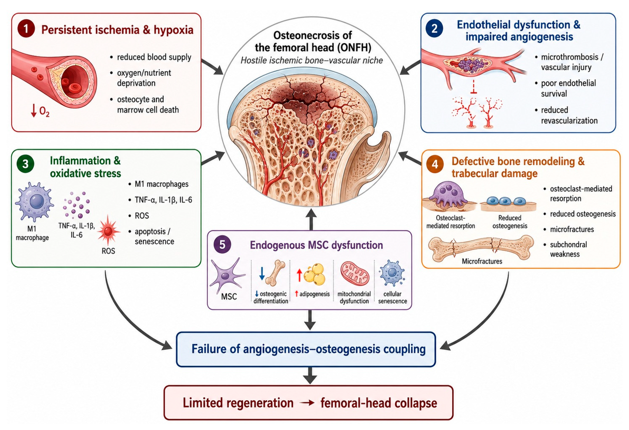
Figure 2.
Biological and therapeutic differences between conventional 2D-expanded MSCs and 3D MSC spheroids. Conventional 2D-expanded MSCs lose cell-cell and ECM interactions after enzymatic detachment, resulting in anoikis, poor retention, reduced survival, and limited therapeutic persistence. In contrast, 3D MSC spheroids preserve cell-cell/ECM signaling, enhance stress resistance and paracrine activity, and increase secretion of VEGF, FGF-2, HGF, EVs, and TSG-6, thereby promoting angiogenesis, osteogenesis, immunomodulation, and regeneration of the ischemic bone-vascular niche.
Figure 2.
Biological and therapeutic differences between conventional 2D-expanded MSCs and 3D MSC spheroids. Conventional 2D-expanded MSCs lose cell-cell and ECM interactions after enzymatic detachment, resulting in anoikis, poor retention, reduced survival, and limited therapeutic persistence. In contrast, 3D MSC spheroids preserve cell-cell/ECM signaling, enhance stress resistance and paracrine activity, and increase secretion of VEGF, FGF-2, HGF, EVs, and TSG-6, thereby promoting angiogenesis, osteogenesis, immunomodulation, and regeneration of the ischemic bone-vascular niche.
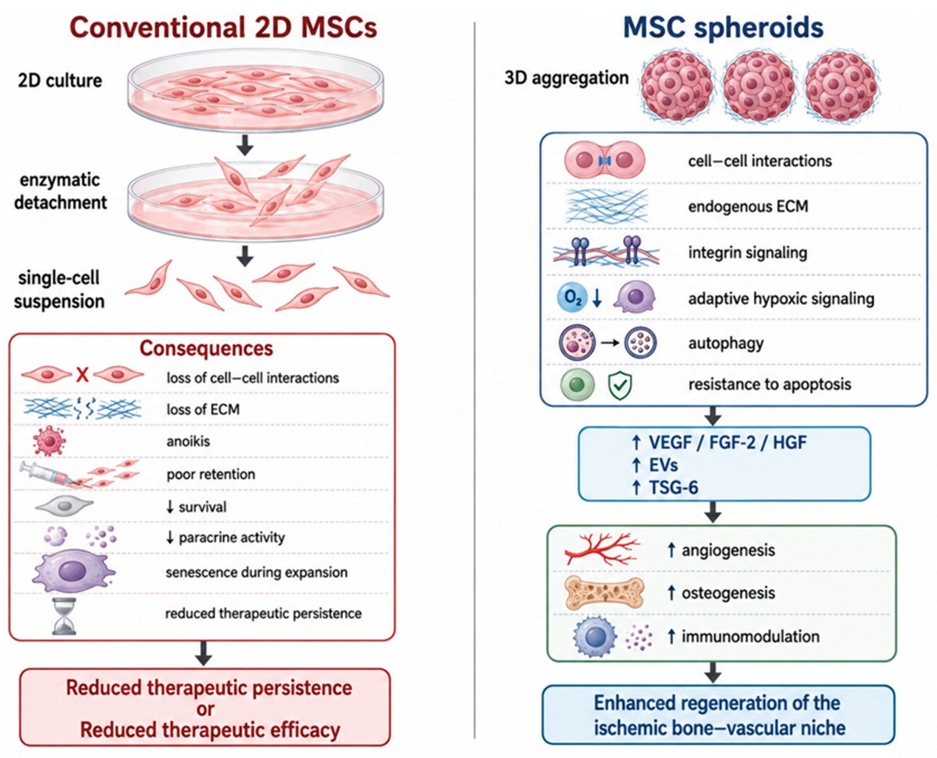
Figure 3.
Proposed mechanisms underlying the enhanced therapeutic activity of MSC spheroids in ONFH. MSC spheroids enhance therapeutic activity through coordinated hypoxic adaptation, improved cell survival, paracrine signaling, ECM–integrin interactions, immunomodulation, and extracellular vesicle-mediated communication. These mechanisms promote angiogenesis–osteogenesis coupling, leading to neovascularization, restoration of the bone–vascular niche, regeneration of viable trabecular bone, and preservation of femoral-head architecture.
Figure 3.
Proposed mechanisms underlying the enhanced therapeutic activity of MSC spheroids in ONFH. MSC spheroids enhance therapeutic activity through coordinated hypoxic adaptation, improved cell survival, paracrine signaling, ECM–integrin interactions, immunomodulation, and extracellular vesicle-mediated communication. These mechanisms promote angiogenesis–osteogenesis coupling, leading to neovascularization, restoration of the bone–vascular niche, regeneration of viable trabecular bone, and preservation of femoral-head architecture.
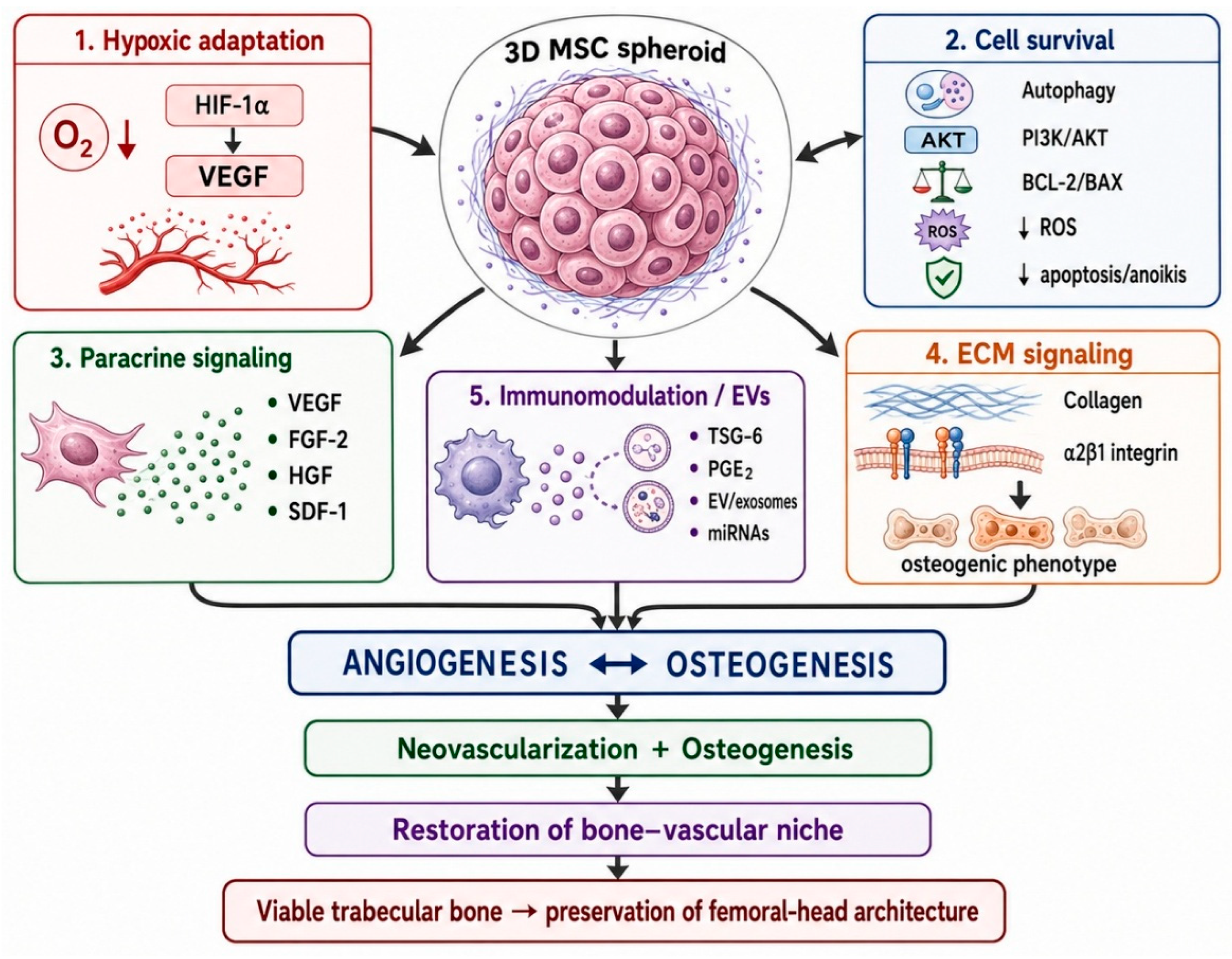

Table 1.
Representative preclinical and clinical studies of MSC-based therapy for ONFH.
| Study | Cell type | Study design / ONFH model | Delivery strategy | Main findings | Main limitations |
| Zhao et al. [29] | Autologous BM-MSCs | Randomized clinical study; 100 patients with early-stage ONFH | CD + local implantation of 2 × 10⁶ cells | Improved clinical outcomes and femoral head survival and reduced treatment failure compared with CD alone | Single-center study; heterogeneous ONFH etiology |
| Gómez-Barrena et al. [7] | Autologous BM-MSCs | Multicenter ORTHO2 clinical study; early/precollapse ONFH; ≥5-year follow-up | Intraosseous administration after CD; 140 × 10⁶ cells | Majority of evaluable femoral heads remained preserved; favorable long-term safety with no severe cell-product-related adverse events | Non-randomized study; relatively limited cohort; selected early-stage patients |
| Pepke et al. [30] | Autologous BMAC | Prospective randomized study; 24 patients/25 hips; ARCO II | CD + 10 mL BMAC into necrotic region | BMAC administration was safe, but no significant advantage over CD alone in clinical outcome, lesion volume, or femoral-head survival at 2 years | Small cohort; BMAC is heterogeneous and contains relatively few MSCs; short follow-up |
| Kang et al. [36] | Autologous BM- MSC | Matched-pair clinical study; 100 patients/106 hips | CD + intraosseous bone marrow cell implantation | THA conversion was lower with cell therapy than with CD alone (28.3% vs 49%); strongest benefit in precollapse ARCO I–II disease | Retrospective design; treatment did not significantly prevent ARCO stage progression |
| Yoon et al. [31] | Autologous AD-MSCs | Phase I/IIa clinical study; 18 hips with lesions ≥30% | 6-mm drilling + local implantation; 1 × 10⁸ cells | Increased vascularity was detected in 79% of evaluable hips at 2 years; treatment was feasible without major treatment-related adverse events | No significant reduction in necrotic lesion size or improvement in major functional scores; small uncontrolled cohort |
| Chen et al. [32] | Allogeneic UC-MSCs | Clinical study; 9 patients/9 hips; ARCO II–IIIa | Intra-arterial infusion | Increased early tissue perfusion and significant reduction in necrotic volume at 12–24 months; no obvious safety abnormalities | Very small cohort; no control group; limited control over MSC homing and retention |
| Chen et al. [33] | hUC-MSCs | Preclinical study; steroid-induced ONFH model | CD + MSC-loaded pluronic F127 hydrogel | Improved femoral-head microarchitecture and vascularization; enhanced bone microvascular endothelial-cell function through COL6A2/integrin α1β1–FAK/PI3K/AKT signaling | Mechanism and efficacy require clinical validation |
| Chen et al. [34] | Synovial fluid-derived MSCs | Preclinical study; steroid-induced rabbit ONFH model | CD + MSC-loaded alginate beads | Preserved femoral-head bone density and sphericity and promoted bone regeneration | Short 6-week follow-up; biomaterial contribution cannot be completely separated from MSC effects |
| Xu et al. [38] | BM-MSCs + endothelial progenitor cells (EPCs) | Preclinical study; steroid-induced rabbit ONFH model | CD + 3D CMC/alginate scaffold containing 5 × 10⁶ BMSCs + 5 × 10⁶ EPCs | Significantly enhanced osteogenesis and angiogenesis and reduced adipogenesis compared with single-cell-type and scaffold controls | Complex multicellular construct; contribution of individual components difficult to isolate; preclinical model |
| Wang et al. [39] | BM-MSCs in oxygen-generating tissue-engineered scaffold | Preclinical study; steroid-induced rabbit ONFH model | CD + BM-MSCs + PCL/nHA scaffold + hydrogel + CaO₂/gelatin microspheres | Sustained oxygen release for ~19 days; reduced grafted-cell apoptosis and enhanced MSC survival, angiogenesis, and osteogenesis | Complex biomaterial system; manufacturing and regulatory complexity; preclinical evidence only |
| Guzman et al. [40] | PDGF-BB-overexpressing MSCs | Preclinical study; steroid-induced rabbit ONFH | CD + collagen/alginate hydrogel containing genetically modified MSCs | Reduced histological osteonecrosis and increased angiogenesis; evidence of enhanced regenerative activity compared with unmodified MSCs | Gene-modified cell product; short-term animal study; substantial translational and regulatory complexity |
Abbreviations: AD-MSCs, adipose-derived mesenchymal stromal cells; BMAC, bone marrow aspirate concentrate; BM-MSCs, bone marrow-derived mesenchymal stromal cells; CD, core decompression; CMC, carboxymethyl chitosan; EPCs, endothelial progenitor cells; hUC-MSCs, human umbilical cord-derived mesenchymal stromal cells; ONFH, osteonecrosis of the femoral head; PCL/nHA, polycaprolactone/nano-hydroxyapatite; PDGF-BB, platelet-derived growth factor-BB; THA, total hip arthroplasty.
Table 2.
Representative preclinical studies supporting the therapeutic potential of MSC spheroids for ONFH.
Table 2.
Representative preclinical studies supporting the therapeutic potential of MSC spheroids for ONFH.
| Study | MSC source | Spheroid strategy | Experimental model | Main findings | Relevance to ONFH | |
| Bartosh et al. [10] | Human BM-MSCs | 3D aggregation into spheroids | In vitro and in vivo inflammatory models | Spheroid formation markedly increased TSG-6, stanniocalcin-1, and other anti-inflammatory/cytoprotective mediators and enhanced anti-inflammatory activity compared with 2D MSCs. | Supports immunomodulation and cytoprotection within the inflammatory ONFH microenvironment | |
| Murphy et al. [45] | Human BM-MSCs | Hanging-drop spheroids; 15,000 cells/spheroid selected for subsequent experiments; fibrin hydrogel | Ischemia-like in vitro conditions: serum deprivation + 1% O₂ | Spheroids showed lower apoptosis and up to ~100-fold higher VEGF secretion than equivalent dissociated MSCs while maintaining osteogenic potential. | Strong mechanistic support for simultaneous ischemic survival + angiogenic signaling + osteogenic competence | |
| Bhang et al. [51] | Human cord blood-derived MSCs | Hanging-drop MSC spheroids | Mouse hindlimb ischemia | Spheroids improved transplanted-cell survival, increased VEGF and FGF-2, increased microvessel density and mature vessels, improved perfusion, and reduced tissue necrosis. | Highly relevant to the ischemic component of ONFH and restoration of microvascular supply | |
| Yamaguchi et al. [14] | Rat BM-MSCs | Spheroids generated in low-binding plates | Rat calvarial bone-defect model | Enhanced osteogenic gene/protein expression and calcium deposition in vitro; significantly increased bone regeneration in vivo versus monolayer MSCs. | Supports enhanced osteogenic differentiation and reconstruction of necrotic trabecular bone | |
| Murphy et al. [11] | Human BM-MSCs | Osteogenically preconditioned MSC spheroids in collagen hydrogel | In vitro osteogenic model | Spheroids maintained osteogenic phenotype better than dissociated cells after withdrawal of osteogenic stimuli; effect mediated partly by collagen-α2β1 integrin signaling. | Supports preservation of osteogenic competence through endogenous ECM-integrin signaling after transplantation | |
| Ho et al. [15] | Human BM-MSCs | Hypoxic preconditioning followed by spheroid formation and RGD-alginate hydrogel delivery | Critical-sized segmental femoral defect in rats | Hypoxia-preconditioned spheroids showed increased resistance to apoptosis and VEGF secretion while preserving osteogenesis; significantly enhanced vascularization and bone healing compared with preconditioned individual MSCs. | One of the strongest models supporting combined hypoxic adaptation, angiogenesis-osteogenesis coupling, and bone regeneration | |
| Ohori-Morita et al. [55] | Human BM-MSCs | Spheroids generated using neurosphere medium and continuous shaking culture | Critical-sized rat femoral bone defect | Spheroids recovered/maintained stem-cell characteristics, showed high transplantation efficiency, and enhanced bone regeneration compared with adherent MSCs and conventional spheroids. | Supports manufacturing optimization, post-transplantation persistence, and regeneration of large bone defects | |
| Song et al. [53] | Human MSCs + endothelial colony-forming cells (ECFCs) | Hybrid MSC/ECFC spheroids | Murine hindlimb ischemia | Hybrid spheroids improved ECFC engraftment and proangiogenic signaling, increased vascular density and blood-flow recovery, and reduced ischemic tissue damage. | Supports prevascularization and MSC-endothelial co-spheroids for rapid restoration of the vascular niche |
Abbreviations: BM-MSCs, bone marrow-derived mesenchymal stromal cells; ECM, extracellular matrix; ECFCs, endothelial colony-forming cells; EVs, extracellular vesicles; FGF-2, fibroblast growth factor-2; ONFH, osteonecrosis of the femoral head; RGD, arginine–glycine–aspartic acid; TSG-6, TNF-α-stimulated gene/protein 6; VEGF, vascular endothelial growth factor.
Table 3.
Strategies for enhancing the therapeutic efficacy of MSC spheroids in ONFH.
| Strategy | Mechanism | Expected therapeutic benefit | Representative evidence | Major limitations | Translational readiness |
| Hypoxic preconditioning | Activation of HIF-1α-dependent cytoprotective and proangiogenic signaling | ↑ survival, ↓ apoptosis, ↑ VEGF, enhanced angiogenesis and osteogenesis |
Hypoxia-preconditioned MSC spheroids improved vascularization and bone repair [15] | Optimal O₂ level and exposure time require standardization; excessive hypoxia may impair osteogenesis | Relatively high |
| Optimization of spheroid size and cellular organization | Control of oxygen/nutrient gradients, metabolism, apoptosis, secretome, and ECM organization | Improved viability, angiogenic potency, injectability, and product reproducibility | Size-dependent effects also demonstrated in MSC spheroid studies [45] | Optimal size for ONFH remains undefined; manufacturing variability | Relatively high |
| Injectable hydrogel-assisted delivery | Local retention, protection from mechanical stress, ECM-like signaling, and controlled cell migration | ↑ retention and survival, prolonged paracrine activity, enhanced angiogenesis and osteogenesis | Fibrin and RGD-alginate systems improved MSC spheroid survival, VEGF secretion, and bone formation [45,56,64] | Hydrogel composition, degradation, mechanics, and spheroid release require optimization | Relatively high |
| Oxygen-generating and microenvironment-modulating biomaterials | Temporary oxygen supplementation and/or modulation of the inflammatory niche | ↑ early cell survival, ↓ apoptosis, improved angiogenesis and osteogenesis |
Oxygen-releasing CaO₂ system improved MSC survival and ONFH repair [6]; Immunomodulatory hydrogel improved the ONFH niche [66] | Excessive O₂ may increase ROS; studies did not directly use MSC spheroids; increased material complexity | Moderate / preclinical |
| Controlled delivery of angiogenic and osteogenic factors | Early VEGF/FGF-2 signaling followed by sustained osteogenic signaling such as BMPs | Restoration of vascular supply followed by enhanced bone formation | Local FGF-2 hydrogel therapy has clinical evidence in precollapse ONFH [67,68,69] | MSC spheroid + FGF-2/VEGF/BMP combination has not been clinically validated; dose and release kinetics must be optimized | Moderate–high |
| MSC–endothelial co-spheroids / prevascularization | Direct interaction between MSCs and endothelial cells; accelerated microvascular-network formation | Rapid neovascularization and enhanced angiogenesis–osteogenesis coupling | MSC/endothelial co-transplantation improved ONFH repair [71]; Spatially organized MSC spheroids enhanced therapeutic angiogenesis [72] | Additional cell type increases manufacturing, safety, and regulatory complexity | Moderate / preclinical |
| Bioactive and mechanically supportive scaffolds | Structural support combined with osteoconductive, angiogenic, and cell-protective signals | Maintenance of femoral-head architecture and enhanced vascularized bone regeneration | BMSC-loaded 3D microspheres + bioactive Mg scaffold enhanced repair in rabbit ONFH [73] | More invasive; complex manufacturing; most relevant to larger lesions; not yet validated with true MSC spheroids | Preclinical |
Disclaimer/Publisher’s Note: The statements, opinions and data contained in all publications are solely those of the individual author(s) and contributor(s) and not of MDPI and/or the editor(s). MDPI and/or the editor(s) disclaim responsibility for any injury to people or property resulting from any ideas, methods, instructions or products referred to in the content. |
© 2026 by the authors. Licensee MDPI, Basel, Switzerland. This article is an open access article distributed under the terms and conditions of the Creative Commons Attribution (CC BY) license (http://creativecommons.org/licenses/by/4.0/).
Copyright: This open access article is published under a Creative Commons CC BY 4.0 license, which permit the free download, distribution, and reuse, provided that the author and preprint are cited in any reuse.