

Submitted:
29 August 2026
Posted:
31 August 2026
You are already at the latest version
Abstract
Plant stress biology has traditionally relied on the analysis of snapshot measurements—including hormone levels, reactive oxygen species, transcripts, and physiological traits—which primarily characterize the current state of the cell, whereas the physical consequences of previous stress exposure remain considerably less accessible to direct measurement. This distinction may be particularly important under natural conditions, where plants experience recurrent, sequential, and combined stresses. This raises a fundamental question: can a cell, after physiological recovery, retain a measurable residual physical state that reflects aspects of its previous stress history and influences its response to subsequent stress? Here, we propose Environmental Phase Imprinting (EPI) as a testable biophysical hypothesis according to which environmental stress may leave a measurable imprint on the physical state of biomolecular condensates that persists after cessation of the initial exposure. EPI is not proposed as a new form of biological information or as an established mechanism of stress memory, but rather as a potential physical substrate, correlate, or consequence of previously described forms of cellular stress memory. To operationalize this hypothesis, we introduce the Plant Condensate Code (PCC), a multidimensional conceptual framework designed to move from a static “snapshot” of cellular state toward a time-resolved physical trajectory. PCC integrates complementary characteristics of condensate populations, including morphology, dynamics, molecular mobility, material state, and molecular composition, across a sequence of states encompassing baseline, stress, adaptation, recovery, and the post-stress state.We propose that the trajectory of condensate states, rather than any single measurement, may contain information about cellular stress history and may help explain differences in responses to recurrent or combined stress. Multimodal approaches, including live-cell imaging, fluorescence recovery after photobleaching (FRAP), molecular mobility analysis, microrheology, Brillouin microscopy, quantitative phase imaging, and molecular profiling, could provide complementary measurements of this physical state. PCC is further positioned within our broader conceptual research program encompassing Cytoplasmic Phase Homeostasis, Cytoplasmic Phase Sensing, and the Plant Threat Matrix (PTM)—a proposed six-state framework for describing plant physiological states under stress. Within this framework, physical measurements of condensate and cytoplasmic states may represent one possible approach for defining and quantitatively characterizing cellular physiological states. Finally, we discuss the potential application of this conceptual framework to stress-resilience phenotyping, evaluation of biostimulants, and selection for stress tolerance, while clearly distinguishing experimentally established phenomena from hypotheses and conceptual proposals. The proposed framework may provide a foundation for the development of a new direction in biophysical phenotyping of plant stress resilience, complementing molecular and physiological approaches to the study of stress memory.
Keywords:
plant stress
; biomolecular condensates
; liquid-liquid phase separation
; environmental phase imprinting
; stress memory
; cytoplasmic phase homeostasis
; condensate-state trajectory
; plant cell biophysics
; stress phenotyping
; Plant Threat Matrix
1. Introduction: Environmental History as an Unresolved Dimension of Cellular State
1.1. Current State Versus Environmental History
A plant cell responds to what is happening to it right now: the current water potential, the current temperature, the current light intensity. This is the implicit assumption behind most stress physiology - that a measurement taken at a given moment tells us about the plant’s present condition, and that this condition is a function of present, not past, environmental input. Yet plants growing outside a growth chamber rarely experience a single stress in isolation; they experience sequences and combinations of stresses across a season, and their physiological response to the last stress in a sequence is often not the same as their response to that stress in isolation [1,2,3]. This raises a simple, largely unaddressed question: does a plant cell’s present physical state depend only on its present environment, or can it also carry a measurable trace of what happened to it earlier? Our own recent work on plant immunity as a multilevel signal-metabolic network illustrates why this question is not merely academic: even the network-level description of how plants integrate pathogen and abiotic threats already presupposes that the cell’s regulatory state depends on a history of prior inputs rather than on the current threat alone [4].
1.2. Limitations of Snapshot-Based Stress Phenotyping
Much of plant stress biology is, by necessity, built on snapshot measurements: hormone titres, ROS levels, Ca²⁺ transients, transcript abundance, or photosynthetic efficiency measured at one or a few time points [5,6]. Each of these readouts is informative about the state of the plant at the moment of sampling. None of them, on their own, is designed to reveal whether the cell’s current baseline has itself shifted because of an earlier stress episode that has, by conventional physiological criteria, already been “recovered from.”
1.3. The Missing Physical Dimension of Cellular History
This leads to the specific, largely unexamined question at the centre of this article: what physical information about a previous stress remains inside the cell after the stress itself has been removed and the plant looks physiologically recovered? Biomolecular condensates are a plausible place to look for an answer, because they are known to physically reorganise in response to several of the same stressors - hydration, temperature, osmotic pressure - that drive whole-plant physiological responses [7,8]. Whether that reorganisation, or some residue of it, outlasts the stress that caused it is an empirical question that, to our knowledge, has not been directly tested in plants.
1.4. Environmental Phase Imprinting Hypothesis
We propose the term Environmental Phase Imprinting (EPI) for this candidate phenomenon: a measurable, residual alteration in the physical state of a plant cell’s condensate populations that persists, at least partially, after an environmental stress has been removed and conventional physiological indicators have returned toward baseline. EPI is a hypothesis about physical state, not a claim about a newly discovered memory mechanism. Section 5 develops this definition in full; Section 6, Section 10 and Section 11 turn it into concrete, testable comparisons.
1.5. Scope and Epistemic Status
We emphasize from the outset the nature and scope of this article. This is a conceptual, hypothesis-generating article, rather than a report of new experimental data. Its primary purpose is to formulate a specific and falsifiable hypothesis—Environmental Phase Imprinting (EPI)—to propose an operational framework for measuring the physical variables relevant to this hypothesis, termed the Plant Condensate Code (PCC), and to define concrete experimental approaches capable of supporting or refuting the proposed model.
Throughout the article, we distinguish explicitly among four categories of statements: (i) phenomena and mechanisms established in the existing literature; (ii) reasonable inferences derived from the available evidence; (iii) the original hypotheses and conceptual propositions introduced here; and (iv) questions that remain unresolved and require direct experimental testing.
Accordingly, statements concerning biomolecular condensates, phase behavior, and plant stress responses that are supported by previous studies should be understood as literature-based observations or interpretations. In contrast, EPI and the Plant Condensate Code represent the conceptual framework proposed in this article and should not be interpreted as experimentally established mechanisms. The central objective is therefore not to claim that EPI already constitutes a demonstrated form of cellular memory, but to define a measurable physical hypothesis and a rigorous experimental strategy through which its validity can be tested.
2. Biomolecular Condensates in Plant Stress: What Is Established?
2.1. Stress-Responsive Condensates
Biomolecular condensates are dynamic, membraneless cellular compartments formed through liquid-liquid phase separation (LLPS), typically driven by multivalent interactions among proteins - often ones containing intrinsically disordered regions - and, in many cases, RNA [9,10]. Condensates organise biochemical reactions locally without a lipid boundary and can assemble, dissolve, or change their physical properties as cellular conditions change [9,10,11]. Because condensate assembly is sensitive to the same physicochemical variables that stress perturbs - temperature, hydration, ionic strength, molecular crowding - condensates have become a natural candidate site at which physical stress signals might be converted into biological response [12,13]. The founding observation for the field, that P granules in the nematode germline behave as liquid droplets that localise through controlled dissolution and condensation, was made outside plants [14], but the underlying physics applies broadly.
2.2. Plant Condensate Systems
In plants specifically, a small set of condensate-forming proteins now has direct experimental support as physical sensors of environmental variables. FLOE1 is a prion-like protein that undergoes hydration-dependent phase separation in Arabidopsis seeds and has been proposed to help gate germination in response to water availability [15]. ELF3 contains a prion-like domain whose propensity to phase-separate changes with temperature, and has been characterised as a thermosensor that links temperature to circadian and photomorphogenic output [16]. Phytochrome B (phyB) integrates light and temperature information through the dynamics of nuclear condensates known as photobodies [17]. SEUSS (SEU), a transcriptional co-regulator, rapidly forms nuclear condensates in response to hyperosmotic stress, in a manner proposed to reflect molecular crowding, and this condensation is required for the induction of osmotic-stress-tolerance genes [18]. FRIGIDA forms cold-induced nuclear condensates that contribute to the vernalisation-associated repression of FLOWERING LOCUS C [19]. Reviews of plant LLPS more broadly converge on the same picture: a growing but still molecule-by-molecule catalogue of condensate systems, each tied to a specific stress input [20,21,22]. Related, if mechanistically distinct, evidence comes from desiccation biology: intrinsically disordered late-embryogenesis-abundant (LEA) proteins and tardigrade-specific disordered proteins undergo hydration-dependent condensation or glass formation that protects cells during extreme water loss [23,26], reinforcing the general principle that hydration-sensitive phase behaviour is a recurring theme across plant and non-plant systems alike, and one directly relevant to the drought scenario used throughout this article.
2.3. Condensate Remodeling During Stress
Beyond these individually characterised nuclear sensors, cytoplasmic ribonucleoprotein condensates - stress granules and P-bodies - assemble and remodel in response to a broad range of abiotic and biotic stresses in plants, with documented roles in mRNA triage, translational control, and the transition into and out of a stressed state [27,28,29]. Outside plants, stress granule assembly has been described as an adaptive, evolutionarily tuned response to stress rather than a passive by-product of cellular damage [30], and yeast prion-protein phase separation has similarly been linked to improved cellular fitness under stress [31]. At the same time, stress granules can undergo an aberrant physical transition when triggered by misfolded protein, one that chaperone activity normally prevents [32], and pathological, non-adaptive demixing within stress granules has recently been described in a mammalian disease context [33]. These findings, taken together, establish that condensate remodeling under stress is a real, physically grounded, and biologically consequential phenomenon - but also that not every condensate-state change is adaptive, a distinction the framework proposed here has to keep in view.
2.4. What Is Already Known Versus What Remains Unresolved
What the literature already establishes, reasonably firmly, is: (i) several specific plant proteins undergo stress-triggered phase separation; (ii) this phase separation is tied, mechanistically, to specific physical variables (hydration, temperature, crowding); and (iii) condensate remodeling participates functionally in at least some stress responses, rather than being a passive correlate. What the literature has not, to our knowledge, directly addressed is whether the joint physical state of these condensate populations - considered as a multidimensional, time-resolved trajectory rather than a single before/after snapshot - retains information about stress history after the stress itself has ended.
2.5. The Knowledge Gap
We want to state this gap carefully, because overstating novelty would misrepresent an active field. Condensates in the context of recurrent stress, priming, and cellular memory are increasingly discussed in the broader (largely non-plant) LLPS literature, and plant biology already has a substantial, independent literature on stress memory operating through transcriptional, chromatin and RNA-turnover mechanisms [34,35]. What has not been done, specifically, is to treat the physical state of plant condensate populations itself - their morphology, dynamics, mobility, material properties and composition, considered jointly - as a candidate carrier of environmental history, and to propose a concrete experimental programme for testing whether that physical state actually differs depending on what happened to the cell earlier, under matched present-day conditions. That is the specific, narrow gap this article addresses.
2.6. From Established Biology to Condensate-State Trajectories
If condensate populations reorganise in response to present stress (Section 2.1, Section 2.2 and Section 2.3), and if some plant memory phenomena are already known to persist physically at other levels of cellular organisation (chromatin, transcript pools), then it becomes reasonable to ask whether condensate physical state itself might behave as a trajectory that depends on more than the present moment, rather than as a state fully determined by present conditions alone. Section 3 develops this shift from single measurements to trajectories.
3. From Condensate Responses to Condensate-State Trajectories
A snapshot describes where the cell is; a trajectory describes how it got there. This distinction is the conceptual centre of this article, and it does not require complicated mathematics to state.
3.1. Baseline State
Before any stress, a condensate population has some resting physical state - a baseline number, size distribution, exchange rate and material property, specific to cell type, tissue and developmental stage.
3.2. Stress-Induced Remodeling
As stress begins, condensates typically change: they may increase in number, grow, become less dynamic, or recruit different molecular partners, tracking the specific physical perturbation involved (Section 2.2).
3.3. Adaptation and Peak Stress
As stress continues or intensifies, the condensate population may reach a new, stress-adapted configuration - a different, but not necessarily pathological, physical regime.
3.4. Recovery
When the stress is removed, condensates typically begin to return toward their pre-stress properties - dissolving, shrinking, or regaining faster molecular exchange.
3.5. Residual State
The open, and to our knowledge untested, question is whether this return is ever complete. If the condensate population’s physical state after recovery differs measurably from its pre-stress baseline, that difference is, by definition, a residual state - and its systematic study is what we mean by Environmental Phase Imprinting.
3.6. Why Trajectories Matter More than Snapshots
A single measurement - say, condensate number at one time point - cannot distinguish a cell that is actively remodeling from one that has stabilised in a new configuration, nor can it tell us anything about where the cell was an hour, a day, or a week earlier. Only a time-resolved trajectory, sampled across baseline, stress, and recovery, can support any claim about history-dependence. This is why the remainder of this article is organised around trajectories rather than single time-point comparisons.
4. The Multidimensional Physical State of Condensates
No single physical variable fully describes a condensate population’s state. We organise the relevant variables into five domains, each with an established measurement basis (developed further in Section 8), plus a sixth contextual layer that is not itself part of condensate state but strongly shapes its interpretation.
4.1. Morphology
How many condensates are present, how large they are, what shape they have, what fraction of the relevant cellular volume they occupy, and where they are located.
4.2. Dynamics
How condensates form, dissolve, fuse with one another, or split apart over time, and how long an individual condensate persists.
4.3. Molecular Mobility
How freely molecules move within a condensate, exchange with the surrounding cytoplasm or nucleoplasm, or become transiently confined or immobilised.
4.4. Material State
Whether a condensate behaves more like a freely flowing liquid, a viscoelastic gel, or an increasingly rigid, glass-like solid - a property that can change with stress and age even when morphology looks similar [36,37,38,39]. Material-state shifts of this kind are closely tied to macromolecular crowding, which itself is increasingly recognised as a physiologically sensed and actively regulated cellular variable rather than a passive by-product of stress [40,41].
4.5. Molecular Composition
4.6. Spatial and Cellular Context
Condensates do not exist in isolation; their behaviour is shaped by, and in turn shapes, their surrounding context - proximity to the nucleus, endoplasmic reticulum, chloroplast, mitochondria, cytoskeletal elements, membranes, and organelle contact sites. We treat this spatial context as important background rather than as a sixth domain of the condensate state itself, because it describes the condensate’s environment rather than the condensate’s own physical properties.
Together, domains 4.1-4.5 define what we mean, operationally, by “condensate state” at a given moment; Section 7 explains how the Plant Condensate Code assembles these domains into a single working framework, and Section 8 explains, method by method, how each domain can actually be measured (Figure 1).
5. Environmental Phase Imprinting
5.1. Definition
We define Environmental Phase Imprinting (EPI) as a measurable, residual alteration in the physical state of a plant cell’s condensate populations - in one or more of the domains described in Section 4 - that persists after an environmental stress has been removed and conventional physiological markers have returned toward their pre-stress values.
5.2. Baseline and Reference State
Any claim about EPI requires a well-characterised, biologically matched baseline state (Section 3.1), collected under the same growth conditions, developmental stage and tissue type as the subsequent stress and recovery measurements. Without a rigorous baseline, “residual” has no defined reference point.
5.3. Stress-Induced Phase Remodeling
As described in Section 2 and Section 3, stress drives measurable changes across the five condensate-state domains. This step is already well supported by existing literature for several specific systems (Section 2.2) and is not itself part of the new hypothesis.
5.4. Recovery and Residual Deviation
Following stress removal, condensate-state variables typically move back toward baseline. The candidate signature of EPI is a residual deviation: a measurable difference between the post-recovery state and the pre-stress baseline, in one or more domains, after a defined recovery interval.
5.5. Persistence and Reversibility
An imprint, if real, need not be permanent. It could plausibly persist for minutes, hours, days, or across a developmental transition, and it could be complete, partial, delayed, or ultimately fully reversible. All of these are open empirical possibilities, not alternative theories in competition; distinguishing among them requires exactly the kind of longitudinal, multi-timepoint measurement described in Section 3.6 and Section 10.
5.6. Magnitude of the Imprint
Where an imprint is detectable, its magnitude - how far the residual state departs from baseline, in which domains, and for how long - is itself a variable of biological interest, potentially informative about stress severity, stress type, or the cell’s underlying resilience (developed further in Section 13).
5.7. EPI Versus Molecular Stress Memory
Plant stress memory is already documented at other levels of cellular organisation, particularly transcriptional priming and chromatin-based mechanisms [34,35]. EPI is not proposed as a replacement for, or a new example of, these established mechanisms. It describes a physical-state variable that may or may not relate to them mechanistically.
5.8. EPI as Correlate, Consequence or Contributor
We deliberately leave open, and consider it essential to leave open, which of several relationships EPI might have to plant physiology: it could be a passive marker that correlates with an underlying memory process implemented elsewhere; a downstream consequence of stress-induced changes in pH, hydration, or metabolism that has no further functional role; or a contributor that itself influences how the cell responds to a subsequent stress. Distinguishing these possibilities is an empirical task, not something the definition in Section 5.1 can settle by itself; it is precisely this openness that keeps EPI falsifiable rather than a foregone conclusion, and Section 6, Section 10 and Section 11 describe how it could be tested (Figure 2).
6. History-Dependent Condensate States
The value of the EPI hypothesis stands or falls on a specific type of comparison: does the same present-day stress produce a different condensate-state trajectory depending on what happened to the cell earlier? We describe the relevant comparisons through concrete stress scenarios rather than abstract notation.
6.1. Single Stress
As a reference case, a single, well-characterised stress (for example, progressive drought) is applied to previously unstressed plants, and the condensate-state trajectory - baseline, stress, recovery - is measured (Scenario A, Section 8.11).
6.2. Recurrent Stress: A → Recovery → A
The same stress is applied twice, separated by a defined recovery interval. If the condensate-state trajectory during the second exposure differs from the first - for example, a faster or more pronounced remodeling response - this is consistent with, though not sufficient proof of, a retained physical trace of the first exposure.
6.3. Sequential Stress: A → Recovery → B
A different stress follows the first, after recovery. Comparing the condensate-state trajectory under B in this sequence to the trajectory under B applied to a naive, previously unstressed plant is, in our view, the single most informative comparison the framework offers.
6.4. Combined Stress: A + B
Two stresses are applied simultaneously (for example, heat and drought together, a combination of clear field relevance [7,8]). We note, without invoking a formal null model, that the joint condensate-state response to combined stress need not be a simple sum of the two individual responses; this is a biological expectation grounded in the interconnectedness of ABA, Ca²⁺, and energy-sensing signalling [44,45,46], not a claim we treat as already demonstrated at the condensate level.
6.5. Multiple Stress Sequences
In principle, longer, more field-realistic sequences (for example, heat wave → recovery → drought → recovery → a second heat wave) can be constructed from the same basic building blocks; we treat these as a natural extension of the paired comparisons in 6.2-6.4 rather than as requiring separate theoretical treatment.
6.6. The Critical Comparison: A → Recovery → B Versus B
The central falsifiable comparison of this article is simple to state: take two groups of biologically matched plants. Expose one group only to stress B. Expose the other group to stress A, allow recovery, then expose it to the same stress B under matched conditions. If the condensate-state trajectories under B differ measurably and reproducibly between the two groups, this constitutes direct, testable evidence of history-dependence in the physical state of plant condensates - independent of whether that history-dependence is ultimately explained by EPI, by some other mechanism, or by a combination of both (Figure 3).
7. The Plant Condensate Code as an Operational Measurement Framework
7.1. Why No Single Measurement Is Sufficient
Confocal imaging alone tells us about morphology; FRAP alone tells us about local molecular exchange; Brillouin microscopy alone tells us about local mechanical properties. None of these, individually, describes “the” condensate state, because condensate state is inherently multidimensional (Section 4). A framework is needed simply to keep track of which measurement addresses which physical question, and to combine them into a coherent trajectory.
7.2. Five-Dimensional Representation
We use the Plant Condensate Code (PCC) as a name for the practical task of organising measurements from the five domains introduced in Section 4 - morphology, dynamics, molecular mobility, material state, and molecular composition - into a shared, time-resolved description of condensate state. This is a bookkeeping and integration framework, not a claim about a new type of biological information.
7.3. Condensate-State Trajectories
Within the PCC, a “trajectory” is simply the same set of measurements, repeated at defined time points across baseline, stress, adaptation, recovery and (where relevant) a second stress exposure, as introduced in Section 3.
7.4. Code Versus Biological Mechanism
We want to state this as plainly as possible: the Plant Condensate Code is not proposed as an established biological code, analogous to the genetic code, nor as a memory mechanism in its own right. It is an operational framework for integrating multidimensional measurements of condensate organisation, dynamics, molecular mobility, material state, and composition into a form that can be compared across conditions, time points, and biological replicates.
7.5. Quantifying Residual Phase Imprint
Within this framework, a residual phase imprint (Section 5.4) can be described in ordinary, largely qualitative or semi-quantitative terms - for example, “condensate number and FRAP mobile fraction remained different from baseline 48 hours after stress removal, while material-state measurements had returned to baseline” - without requiring a single composite index. Where a compact, formal distance measure between trajectories is useful for statistical comparison, standard multivariate approaches (for example, normalised Euclidean or Mahalanobis distances, or dimensionality-reduction techniques such as principal component analysis or UMAP [47]) are available and are discussed briefly, as analytical options rather than as the conceptual core of the framework, in Section 9.7.
8. How Can the Physical State of Cytoplasmic Condensates Actually Be Measured?
This section is intentionally structured for an interdisciplinary readership, including researchers without specialized training in biophysics. For each method, we systematically address four key questions: what the method directly measures; which physical property of the cellular or condensate state can be inferred from the measurement; what biological question the method can help answer; and which conclusions cannot be drawn from the measurement alone. This approach is intended to clearly distinguish between the directly measured parameter, its biophysical interpretation, and the resulting biological inference. Following the methodological overview, we provide a practical example of how these approaches can be applied to a drought-stress scenario, which serves as a cross-cutting example throughout this article.
8.1. Live-Cell Confocal and Spinning-Disk Microscopy
What we see: the number, size, shape and subcellular location of fluorescently labelled condensates, and, with time-lapse acquisition, their formation, fusion and dissolution. Physical property: morphology and dynamics (Section 4.1 and Section 4.2). Biological question it answers: does the condensate population change in a stress- or genotype-dependent way? What it does not prove: the appearance of a fluorescent punctum is not, by itself, evidence that the structure is a bona fide phase-separated condensate; independent criteria such as fusion behaviour, shape relaxation after fusion, concentration dependence, and reversible dissolution should be sought wherever feasible, and unconfirmed structures should be described as condensate-like rather than assumed to be LLPS-derived [10,11]. Practical example: after progressive drought, condensate number and average size increase relative to baseline; after rehydration and a defined recovery period, number and size decline but do not fully return to baseline values - a first, purely morphological candidate signature of EPI.
8.2. FRAP (Fluorescence Recovery After Photobleaching)
FRAP asks a simple physical question: how quickly can fluorescent molecules replace those that have been locally photobleached inside a condensate? Physical property: molecular exchange kinetics (Section 4.3). Biological question it answers: is the condensate still exchanging molecules readily with its surroundings, or has exchange slowed? Fast recovery generally indicates higher molecular exchange; slow or incomplete recovery generally indicates reduced exchange, which can reflect stronger internal interactions, increased viscosity, or a shift toward a more solid-like material state [48]. What it does not prove: FRAP recovery reflects a mixture of diffusion and binding kinetics, and slow recovery on its own does not distinguish between these; nor does FRAP recovery, by itself, prove that a structure is a liquid-like phase-separated condensate at all [48]. Practical example: FRAP mobile fraction inside stress-responsive condensates drops during drought and, in our proposed experimental design (Section 10), is tested for whether it returns fully to its pre-stress value after recovery or remains partially reduced.
8.3. FCS (Fluorescence Correlation Spectroscopy) and Single-Particle Tracking (SPT)
FCS measures fluctuations in fluorescence intensity within a small observation volume over time, from which diffusion coefficients and local concentration can be estimated; SPT follows the position of individual labelled molecules or particles over time, revealing whether their motion is freely diffusive, confined, or transiently trapped [49]. Biological question they answer: how mobile are individual molecules inside and around a condensate, and does that mobility change with stress or stress history? What they do not prove: both methods characterise molecular motion, not phase identity or biochemical function, and results depend on labelling strategy and acquisition parameters. Practical example: after drought, diffusion coefficients measured by FCS inside condensate-associated regions decrease; in plants previously exposed to drought and then recovered, a second drought exposure could plausibly produce a faster or slower change in diffusion than in naive plants - the specific comparison introduced in Section 6.3.
8.4. Microrheology8.4. Microrheology
Microrheology infers the local mechanical, viscoelastic behaviour of the cellular environment from the motion of tracked particles (which can be naturally occurring granules or introduced probes) under thermal or applied forces, an approach that has revealed glass-like and actively tuned mechanical behaviour of the cytoplasm in non-plant systems [50,51]. Physical property: apparent viscosity and viscoelastic modulus (Section 4.4). Biological question it answers: has the physical consistency of the cytoplasm or a specific condensate shifted toward a more fluid or more rigid regime? This method is particularly important for the EPI hypothesis, because a plausible physical mechanism for an incomplete recovery is a shift toward increased viscoelasticity that slows molecular rearrangement even once conventional stress markers have normalised - potentially a direct physical component of an imprint. What it does not prove: the inferred mechanical properties depend on probe size, probe-environment interactions, and the model used to relate particle motion to viscoelasticity, so results should be interpreted as apparent, model-dependent estimates rather than absolute material constants.
8.5. Brillouin Microscopy
Brillouin microscopy uses the interaction of light with spontaneous, thermally driven acoustic waves in a material to obtain spatially resolved information about its local mechanical and acoustic properties, without added labels [52]. In plants, this approach has been used to map subcellular mechanical properties in intact tissue [53]. Biological question it answers: does the local mechanical environment around a condensate change with stress or recovery? What it does not prove, and this needs to be stated clearly: the Brillouin frequency shift is not a direct measurement of viscosity. Its relationship to viscosity depends on the longitudinal elastic modulus, density, and hydration state of the sample, and interpreting a Brillouin signal as “viscosity” without accounting for these factors is a common and avoidable overreach [52,53].
8.6. Fluorescence Anisotropy
This method measures the degree to which emitted fluorescence retains the polarisation of the exciting light, which depends on how freely the fluorophore (or the molecule it is attached to) can rotate during its excited-state lifetime. Biological question it answers: has the local rotational freedom or packing environment of a labelled molecule changed, for example becoming more constrained within a denser or more rigid condensate? What it does not prove: anisotropy changes can reflect binding, crowding, or genuine viscosity changes, and these contributions are not automatically separable from the anisotropy signal alone.
8.7. Quantitative Phase Imaging (QPI)
QPI is a label-free method that measures how much a cell or subcellular structure delays the phase of transmitted light, which relates to local dry-mass density and refractive index. Biological question it answers: has the overall optical density or mass distribution within a cell region changed, consistent with altered crowding or hydration? What it does not prove: QPI is not a direct condensate detector; a change in local optical density is consistent with, but not specific to, condensate remodeling, and should be interpreted alongside, not instead of, the condensate-specific methods in Section 8.1, Section 8.2, Section 8.3, Section 8.4, Section 8.5 and Section 8.6.
8.8. Molecular Profiling (Proteomics, Phosphoproteomics, Interactomics, RNA Analysis)
These methods identify and quantify which proteins, post-translational modifications, interaction partners and RNA species are present in or associated with condensates under a given condition, typically via biochemical fractionation, proximity labeling, or related approaches [42,43]. Biological question they answer: what changed, molecularly, inside the condensate? This complements the biophysical methods above by identifying candidate molecular drivers of any observed physical change. What it does not prove: compositional data are typically destructive, population-averaged snapshots, and by themselves do not establish the dynamic, single-condensate physical behaviour that FRAP, FCS, SPT and microrheology address directly.
8.9. Physiological Sensors (pH, Ca²⁺, ROS, Redox, ATP/ADP, Hydration, Temperature)
Genetically encoded or chemical biosensors and standard physiological assays provide readouts of the cellular physiological context in which condensates exist [45,54,55,56]. Energy-status sensing through SnRK1 and TOR signalling is a particularly relevant physiological covariate, since both pathways are tightly linked to hydration and crowding-sensitive cellular states [46,57,58]. Role in this framework: these are contextual measurements that help interpret condensate-state changes - for example, distinguishing a pH-driven material-state shift [36] from one driven by a different mechanism - rather than being measurements of condensate state themselves.
8.10. Measurement Limitations and Orthogonal Validation
No single method in this list is sufficient on its own to establish a claim about EPI. Reporter-protein fusions used for visualisation can themselves alter condensation behaviour and should, where feasible, be validated against native-locus tagging or orthogonal, label-free methods. Any inference about material-state change from one modality (for example, FRAP) should ideally be cross-checked against at least one independent modality (for example, microrheology or Brillouin microscopy) before being treated as robust. Table 1 summarises this section for quick reference.
8.11. A Practical Experimental Workflow
To make these methods concrete, consider a minimal drought-recovery workflow (Scenario A, developed further in Section 10): (1) establish a condensate-state baseline in unstressed, biologically matched plants; (2) apply progressive drought as a defined, monitored stress; (3) perform live-cell confocal imaging at defined time points to track morphology and dynamics; (4) perform FRAP at the same time points to track molecular mobility; (5) perform FCS or SPT, where feasible, for an independent mobility estimate; (6) perform microrheology or Brillouin microscopy for material-state information; (7) rehydrate and allow a defined recovery interval; (8) repeat the full measurement panel during recovery; (9) apply a second stress exposure to a subset of plants; (10) compare the resulting trajectories - most importantly, stress B alone versus A → Recovery → B, as introduced in Section 6.6 (Figure 4, Table 1).
9. Multimodal Integration: From Measurements to Environmental Phase Imprint
9.1. Raw Measurements
Each method in Section 8 produces its own raw output - an image stack, a recovery curve, a correlation function, a mechanical spectrum, a list of enriched proteins.
9.2. Physical Features
From each raw output, a small number of interpretable physical features are extracted (for example, condensate number, FRAP mobile fraction, an apparent viscosity value).
9.3. Multidomain Integration
These features, taken together across the five domains of Section 4, are compiled - for a given plant, tissue, and time point - into that time point’s condensate-state description, in the sense defined in Section 7.2.
9.4. Condensate-State Trajectory Reconstruction
Repeating this compilation across baseline, stress, adaptation and recovery time points (Section 3) yields the trajectory for a given experimental condition.
9.5. Recovery-State Estimation
Comparing the trajectory’s recovery-phase values against its own baseline values, domain by domain, yields the residual-deviation description introduced in Section 5.4 and Section 7.5.
9.6. EPI Quantification
Where a residual deviation is observed, its magnitude, its persistence over time (Section 5.5 and Section 5.6), and its relationship to a subsequent stress response (Section 6.6) together constitute the empirical content of an EPI measurement for that experiment.
9.7. Statistical and AI-based Analysis as a Secondary Tool
For studies with sufficient replication, standard multivariate statistical or machine-learning tools - principal component analysis, clustering, UMAP-based visualisation [47], or supervised classifiers such as random forests [59], evaluated with appropriate cross-validation [60] - can help summarise and visualise trajectories across many biological replicates and conditions. We treat these as useful secondary analytical tools for exploring and summarising data that has already been collected through the methods in Section 8, not as evidence in their own right for the underlying biological hypothesis; a classifier that discriminates conditions well tells us the physical measurements differ, not why they differ or what that difference means biologically (Figure 5).
10. Experimental Validation and Falsification
We propose a staged, cumulative programme of seven experiments, each building on the last, using the drought-centred workflow of Section 8.11 as a template that can be adapted to other stressors.
Experiment 1 - Baseline. Characterise condensate-state variability (Section 4) in unstressed, biologically matched plants, to establish the natural range of baseline measurements before any stress comparison is attempted.
Experiment 2 - Single stress. Apply a single, well-defined stress (for example, progressive drought) and track the condensate-state trajectory through to peak stress.
Experiment 3 - Recovery. Following stress removal, track the condensate-state trajectory through a defined recovery interval and quantify any residual deviation from baseline (Section 5.4).
Experiment 4 - Recurrent stress (A → Recovery → A). Repeat the same stress after recovery and compare the second trajectory to the first (Section 6.2).
Experiment 5 - History dependence (A → Recovery → B versus B). Apply a different stress after recovery from the first, and compare the resulting trajectory under the second stress to the trajectory observed when the same second stress is applied to naive plants (Section 6.6) - the central comparison of this framework.
Experiment 6 - Combined stress (A + B). Apply two stresses together and compare the resulting trajectory qualitatively to the trajectories obtained for each stress individually (Section 6.4), without presupposing a specific quantitative additivity rule.
Experiment 7 - Long-term persistence. Extend the recovery interval across days to a developmental transition, to test whether any residual deviation detected in Experiment 3 persists, diminishes, or disappears over longer timescales (Section 5.5).
Each experiment should include biologically matched controls, sufficient independent replication for the statistical comparisons in Section 9.7, and, wherever feasible, at least one orthogonal measurement per condensate-state domain (Section 8.10) before a residual difference is treated as a genuine physical signal rather than a measurement artefact (Figure 6).
11. Four Central Falsifiable Predictions
We deliberately limit the framework to four core predictions, each stated in plain terms and each directly tied to one or more of the experiments in Section 10.
Prediction 1 - Stress-specific trajectories. Different environmental stresses (for example, drought, heat, cold, osmotic stress) produce distinguishable condensate-state trajectories, detectable through the multimodal measurements described in Section 8. Falsification: if trajectories under clearly different stressors cannot be reproducibly distinguished using the domains in Section 4, across independent biological replicates, this prediction fails.
Prediction 2 - History-dependent states. The condensate-state trajectory under a given stress B differs, reproducibly and under matched conditions, depending on whether the plant was previously exposed to a different stress A and allowed to recover (A → Recovery → B ≠ B; Section 6.6). Falsification: if no reproducible difference is found between these two conditions across independent replicates (Experiment 5), this prediction fails.
Prediction 3 - Persistent environmental phase imprint. Following recovery from a stress, the condensate-state trajectory can remain measurably different from the pre-stress baseline in at least one domain, for a defined period after conventional physiological recovery markers have normalised (Section 5.4). Falsification: if condensate-state measurements consistently return fully to baseline as soon as, or before, conventional physiological markers normalise, across replicated experiments, this prediction fails.
Prediction 4 - Predictive value for future response or recovery. The magnitude or persistence of a residual imprint (Section 5.6) is associated with how the cell subsequently responds to a new stress, or with how completely it recovers from it. Falsification: if imprint magnitude and persistence show no relationship to subsequent stress response or recovery outcomes, across independent replicates, this prediction fails.
We emphasise that a negative result for any of these predictions would be a substantive and useful scientific outcome, not a failure of the framework: it would indicate that condensate physical state, whatever its other roles, does not retain exploitable information about environmental history under the conditions tested.
12. Alternative Explanations, Limitations and Causal Challenges
We consider this section as scientifically important as the predictions themselves.
12.1. Consequence Versus Cause
An observed residual condensate-state deviation could be a downstream consequence of some other lingering physiological change (for example, incompletely restored hydration or metabolic state) rather than a phenomenon with any independent physical or functional significance. Distinguishing these possibilities requires the causal, perturbation-based experiments of Section 10, not correlational observation alone.
12.2. Puncta Versus Bona Fide Condensates
As noted in Section 8.1, a visualised fluorescent punctum is not automatically evidence of a phase-separated structure. Independent criteria - fusion, shape relaxation, concentration dependence, reversible dissolution - should be sought, and unconfirmed structures should be described as condensate-like rather than assumed to be LLPS-derived.
12.3. FRAP Interpretation
As noted in Section 8.2, FRAP recovery reflects a mixture of diffusion and binding processes and does not, by itself, establish phase identity or prove a liquid-like state.
12.4. Brillouin Interpretation
As noted in Section 8.5, the Brillouin signal should not be equated directly with viscosity; its relationship to mechanical properties depends on modulus, density and hydration and requires careful, calibrated interpretation.
12.5. Metabolic and Ionic Confounders
Stress and recovery are accompanied by changes in pH, ionic strength, osmolarity and metabolite pools, any of which can independently alter condensate physical properties [36,61]; these need to be measured and, where possible, controlled for, rather than assumed to be constant across baseline, stress and recovery.
12.6. Reporter Artefacts
Fluorescent-protein fusions used to visualise condensates can themselves alter condensation propensity; wherever feasible, findings should be cross-validated using native-locus tagging or label-free methods (Section 8.10).
12.7. Cell- and Tissue-Type Specificity
Condensate-state trajectories are plausibly cell-type- and tissue-specific, and findings in one tissue or developmental stage should not be assumed to generalise without direct testing.
12.8. Batch Effects
Imaging sessions, growth-chamber batches, and reporter-expression variability can introduce systematic differences that mimic genuine biological signal; experiments should include matched batch controls and, where statistical classification is used (Section 9.7), explicit tests for batch-driven rather than biology-driven discrimination.
12.9. Causality
Even a robust, reproducible statistical association between condensate-state history and subsequent stress response would not, by itself, establish that the condensate physical state causally contributes to that response, as opposed to being a correlated marker of some other underlying process. We regard this as an open question the framework is designed to help investigate, not one it can resolve by definition. None of the limitations above are reasons to abandon the hypothesis; they are the specific reasons the hypothesis is scientifically useful; a framework this concretely falsifiable is, by construction, one that real experiments can actually challenge.
13. From Condensate-State Recovery to Cellular Resilience
13.1. Recovery as a Measurable Physical Process
Section 3.4 and Section 3.5 already treats recovery as a trajectory phase in its own right, not merely as “the stress ending.” This framing allows recovery itself - how completely, and how quickly, condensate-state variables return toward baseline - to become a measurable object of study.
13.2. Phase Plasticity
We use the term phase plasticity, consistent with the broader Cytoplasmic Phase Homeostasis framework [62], to describe a cell’s capacity to remodel its condensate physical state under stress and then substantially restore that state once stress is removed.
13.3. Phase Rigidity
Where recovery is incomplete or delayed - where a residual imprint (Section 5.4) persists longer or more strongly than in comparable cells - we describe the underlying condition as reduced phase plasticity, or phase rigidity, again following the terminology of our broader framework [62,63].
13.4. Condensate-State Recovery as a Potential Resilience Marker
We suggest, as a hypothesis rather than an established fact, that the completeness and speed of condensate-state recovery could serve as a candidate early biophysical indicator of cellular resilience - potentially detectable before conventional yield or visible-damage phenotypes appear. This connects directly to Prediction 4 (Section 11) and would need to be tested using the experimental design in Section 10, particularly Experiments 3 and 7. The broader dynamical-systems literature on early-warning signals preceding critical transitions in complex systems [64,65,66] offers a useful, general analogy here - not as a claim that plant cells are formally equivalent to the systems studied in that literature, but as a reminder that a system approaching a threshold can, in principle, show measurable precursor signals before the threshold is crossed, which is exactly the kind of early signal a persistent condensate-state deviation would need to represent to be practically useful. This resilience framing also connects to hormonal and energy-sensing switches that govern the balance between stress response and growth resumption, a balance our own work has discussed under the concept of regulatory agronomy [67].
13.5. Linking EPI to Future Stress Response
If Prediction 4 is supported by experiment, EPI would provide a physical, mechanistically grounded link between a plant’s stress history and its future stress response - a link that is currently inferred indirectly, if at all, from physiological or yield outcomes rather than measured directly at the level of cellular physical state.
14. Integration with Cytoplasmic Phase Homeostasis, Cytoplasmic Phase Sensing, and the Plant Threat Matrix
This article is the third step of a broader research programme, and it is worth being explicit about how its parts relate rather than presenting them as competing ideas.
14.1. Phase Homeostasis
Our Cytoplasmic Phase Homeostasis framework proposes that regulated cytoplasmic phase plasticity - the capacity to remodel and then restore cytoplasmic physical organisation - may be a candidate universal principle of stress adaptation across living systems, with a parallel treatment of its relationship to aging and phase rigidity [62,63]. This is the level at which “what is the physical target of adaptation?” is posed.
14.2. Phase Sensing
Our Cytoplasmic Phase Sensing framework asks how physical cytoplasmic state itself might participate in the perception of stress - a distributed, physicochemical complement to receptor-based sensing mechanisms [68]. This is the level at which “how does physical state participate in perception?” is posed.
14.3. Phase Imprinting
The present article asks a third, distinct question: given that condensate populations remodel with stress (Section 2) and that this remodeling can plausibly be tracked as a trajectory (Section 3), does any part of that trajectory persist as a residual physical trace after the stress has ended, and does that trace carry information about environmental history (Section 5 and Section 6)? This is a question about physical memory of past states, not about the physical target of adaptation or the perceptual mechanism itself.
14.4. A Unified Physical-State Framework, and Its Connection to the Plant Threat Matrix
Together, these three levels - homeostasis, sensing, and imprinting - describe a single physical-state research programme, addressing, respectively, what is being maintained, how disturbance to it is sensed, and whether its history leaves a measurable trace. We further connect this programme to the Plant Threat Matrix (PTM), a six-state framework describing plant stress physiology as transitions among discrete physiological configurations - active-growth homeostasis (S1), a pre-stress warning window (PSW) in which the regulatory network is metastable and condensate-primed but still reversible, early/moderate adaptive stress (S2), progressive immune-metabolic exhaustion (S3), collapse (CS), and recovery (R). Within this integration, condensate-state trajectories and any detected environmental phase imprint are proposed as one candidate, physically grounded source of evidence for inferring which PTM state a plant occupies - particularly for the PSW state, which is defined as preceding overt physiological stress markers and might, in principle, be detectable earlier through condensate-state measurements than through conventional physiological readouts. We stress that this integration is a proposed connection between two conceptual frameworks, not a demonstrated diagnostic capability (Figure 7).
15. Implications for Plant Stress Biology and Resilience
15.1. Why This Matters for Global Agriculture
Global agriculture increasingly has to contend not with single, isolated stresses but with sequences and combinations - drought following flooding, repeated heat waves within a season, salinity compounding nutrient limitation [1,7]. A framework that treats environmental history, not just present conditions, as a variable worth measuring is, in principle, better matched to this reality than one built entirely around single-stress snapshots.
15.2. Early Diagnosis of Hidden or Sublethal Stress
We propose, as a future application requiring direct experimental validation, that condensate-state measurements could eventually contribute to detecting stress before visible symptoms appear: a plant can look outwardly healthy - green, turgid leaves - while its condensate populations show reduced molecular mobility, increased viscoelasticity, and incomplete recovery from a recent stress episode. Such a discrepancy between visible appearance and underlying physical state would constitute a candidate early-warning phenotype, but this remains a hypothesis to be tested, not a diagnostic tool that currently exists (Figure 8).
15.3. Evaluating Biostimulants and Stress-Protective Treatments
The trajectory framework in Section 3 suggests a natural comparative experimental design: track condensate-state trajectories in control plants (baseline → stress → recovery) against treated plants (baseline → stress → treatment → recovery), using the measurement panel in Section 8. If a treatment moves the post-recovery physical state closer to baseline than in untreated controls, this would be a candidate physical indicator of restored phase plasticity - useful information for evaluating a biostimulant or other stress-protective treatment, though we deliberately avoid describing this as “therapy” for plants and stress that it, too, requires direct testing before any practical claim is made.
15.4. Breeding for Resilience
Two genotypes exposed to the same stress could, in principle, show different condensate-state trajectories - one with strong initial remodeling and slow recovery, another with more moderate remodeling and faster recovery. If Prediction 4 (Section 11) holds, condensate-state recovery could become a candidate new biophysical phenotype for screening stress resilience in breeding programmes, complementing existing physiological and yield-based selection criteria. This, again, is a hypothesis requiring validation, not an established selection method.
15.5. Toward Precision, Phase-Resolved Phenotyping
Combining live-cell imaging, mobility measurements (FRAP/FCS/SPT), and physiological sensing, potentially supported by the statistical and machine-learning tools discussed in Section 9.7, could eventually contribute to a more physically resolved form of plant phenotyping. We present this as a plausible future direction rather than a current capability.
15.6. Why Could Environmental Phase Imprinting Matter Beyond Plant Biology?
We note, cautiously, that the underlying question - whether cellular physical state retains a measurable trace of environmental history - is not specific to plants; it touches on general questions of cellular stress history, phase plasticity, recovery, and resilience that recur across biological systems, including in the context of cellular aging and stress accumulation more broadly [69,70,71,72]. We raise this connection only to indicate why the underlying physical question may be of broader interest; we explicitly do not extrapolate the plant-specific hypothesis and experiments proposed here to aging biology or human medicine, which would require an entirely separate, dedicated line of investigation (Figure 9).
16. Conclusions
Plant biomolecular condensates are now well established as stress-responsive physical structures, with several specific systems - FLOE1, ELF3, phytochrome B, SEUSS, FRIGIDA, stress granules and P-bodies - characterised as sensors or participants in the response to hydration, temperature, osmotic and translational stress. What has not been directly tested is whether the physical state of these condensate populations, considered as a time-resolved trajectory rather than a single measurement, retains a measurable trace of environmental history after the stress itself has ended. We have proposed Environmental Phase Imprinting (EPI) as a testable hypothesis for this residual physical trace, explicitly distinguished it from established forms of molecular stress memory, and described the Plant Condensate Code as an operational - not a biological - framework for integrating morphological, dynamic, mobility, material-state and compositional measurements into a single trajectory description. We have laid out, in plain physical terms, what nine complementary measurement approaches can and cannot tell us about this trajectory, proposed a staged programme of seven experiments built around a single critical comparison (a stress applied alone versus the same stress applied after a prior stress and recovery), and stated four falsifiable predictions whose failure would be as scientifically informative as their success. We have connected this framework to our broader Cytoplasmic Phase Homeostasis and Cytoplasmic Phase Sensing work and to the Plant Threat Matrix, and outlined - carefully, as hypotheses requiring validation - where such a framework might eventually matter for stress phenotyping, biostimulant evaluation, and breeding for resilience. The central claim of this article is deliberately modest in form and, we think, substantial in content: plant cells may retain a measurable physical imprint of environmental history in the dynamic state of their biomolecular condensates, this imprint can be investigated experimentally using existing biophysical and imaging methods, and whether it exists, and matters, is now a question that can be put to a direct experimental test.
Author Contributions
Conceptualization, S.H.K.; methodology, S.H.K. and L.M.B.; formal analysis, S.H.K.; investigation, S.H.K., Y.V.K., V.M.S. and Y.A.A.; writing-original draft preparation, S.H.K.; writing-review and editing, all authors; visualization, S.H.K.; supervision, S.H.K. All authors have read and agreed to the published version of the manuscript.
Funding
This research received no external funding.
Institutional Review Board Statement
Not applicable.
Informed Consent Statement
Not applicable.
Data Availability Statement
No new experimental data were generated in this conceptual article; all cited literature is publicly available via the DOIs and identifiers listed in the References.
Acknowledgments
During the preparation of this manuscript, the author(s) used Claude (Anthropic, 2025–2026) for grammar checking, style editing, and English-language formulation. The authors have reviewed and edited the output and take full responsibility for the content of this publication.
Conflicts of Interest
The authors declare no conflict of interest.
References
- Kopecka, R.; Kameniarova, M.; Cerny, M.; Brzobohaty, B.; Novak, J. Abiotic stress in crop production. Int. J. Mol. Sci. 2023, 24, 6603. [Google Scholar] [CrossRef] [PubMed]
- Zhang, H.; Zhu, J.; Gong, Z.; Zhu, J.K. Abiotic stress responses in plants. Nat. Rev. Genet. 2022, 23, 104–119. [Google Scholar] [CrossRef] [PubMed]
- Zhu, J.K. Abiotic stress signaling and responses in plants. Cell 2016, 167, 313–324. [Google Scholar] [CrossRef] [PubMed]
- Khablak, S.H.; Bondareva, L.M.; Dolia, M.M.; Spychak, V.M.; Lykholat, T.Y.; Sklyar, T.V.; Lykholat, Y.V. Integrative model of plant immunity to pathogens and stresses: a multilevel signal-metabolic network. Regul. Mech. Biosyst. 2025, 16, e25175. [Google Scholar] [CrossRef]
- Mittler, R. Abiotic stress, the field environment and stress combination. Trends Plant Sci. 2006, 11, 15–19. [Google Scholar] [CrossRef] [PubMed]
- Suzuki, N.; Rivero, R.M.; Shulaev, V.; Blumwald, E.; Mittler, R. Abiotic and biotic stress combinations. New Phytol. 2014, 203, 32–43. [Google Scholar] [CrossRef] [PubMed]
- Zandalinas, S.I.; Mittler, R. Plant responses to multifactorial stress combinations. Trends Plant Sci. 2022, 27, 836–848. [Google Scholar] [CrossRef] [PubMed]
- Rivero, R.M.; Mittler, R.; Blumwald, E.; Zandalinas, S.I. Developing climate-resilient crops: improving plant responses to stress combination. Plant J. 2022, 109, 373–389. [Google Scholar] [CrossRef] [PubMed]
- Banani, S.F.; Lee, H.O.; Hyman, A.A.; Rosen, M.K. Biomolecular condensates: organizers of cellular biochemistry. Nat. Rev. Mol. Cell Biol. 2017, 18, 285–298. [Google Scholar] [CrossRef] [PubMed]
- Hyman, A.A.; Weber, C.A.; Julicher, F. Liquid-liquid phase separation in biology. Annu. Rev. Cell Dev. Biol. 2014, 30, 39–58. [Google Scholar] [CrossRef] [PubMed]
- Alberti, S.; Gladfelter, A.; Mittag, T. Considerations and challenges in studying liquid-liquid phase separation and biomolecular condensates. Cell 2019, 176, 419–434. [Google Scholar] [CrossRef] [PubMed]
- Alberti, S.; Hyman, A.A. Biomolecular condensates at the nexus of cellular stress, protein aggregation disease and ageing. Nat. Rev. Mol. Cell Biol. 2021, 22, 196–213. [Google Scholar] [CrossRef] [PubMed]
- Mittag, T.; Pappu, R.V. A conceptual framework for understanding phase separation and addressing open questions and challenges. Mol. Cell 2022, 82, 2201–2214. [Google Scholar] [CrossRef] [PubMed]
- Brangwynne, C.P.; Eckmann, C.R.; Courson, D.S.; Rybarska, A.; Hoege, C.; Gharakhani, J.; Julicher, F.; Hyman, A.A. Germline P granules are liquid droplets that localize by controlled dissolution/condensation. Science 2009, 324, 1729–1732. [Google Scholar] [CrossRef] [PubMed]
- Dorone, Y.; Boeynaems, S.; Flores, E.; Jin, B.; Hateley, S.; Bossi, F.; Lazarus, E.; Pennington, J.G.; Michiels, E.; De Decker, M.; et al. A prion-like protein regulator of seed germination undergoes hydration-dependent phase separation. Cell 2021, 184, 4284–4298.e27. [Google Scholar] [CrossRef] [PubMed]
- Jung, J.H.; Barbosa, A.D.; Hutin, S.; Kumita, J.R.; Gao, M.; Derwort, D.; Silva, C.S.; Lai, X.; Pierre, E.; Geng, F.; et al. A prion-like domain in ELF3 functions as a thermosensor in Arabidopsis. Nature 2020, 585, 256–260. [Google Scholar] [CrossRef] [PubMed]
- Chen, D.; Lyu, M.; Kou, X.; Li, J.; Yang, Z.; Gao, L.; Li, Y.; Fan, L.M.; Shi, H.; Zhong, S. Integration of light and temperature sensing by liquid-liquid phase separation of phytochrome B. Mol. Cell 2022, 82, 3015–3029.e6. [Google Scholar] [CrossRef] [PubMed]
- Wang, B.; Zhang, H.; Huai, J.; Peng, F.; Wu, J.; Lin, R.; et al. Condensation of SEUSS promotes hyperosmotic stress tolerance in Arabidopsis. Nat. Chem. Biol. 2022, 18, 1361–1369. [Google Scholar] [CrossRef] [PubMed]
- Zhu, P.; Lister, C.; Dean, C. Cold-induced Arabidopsis FRIGIDA nuclear condensates for FLC repression. Nature 2021, 599, 657–661. [Google Scholar] [CrossRef] [PubMed]
- Cuevas-Velazquez, C.L.; Dinneny, J.R. Organization out of disorder: liquid-liquid phase separation in plants. Curr. Opin. Plant Biol. 2018, 45, 68–74. [Google Scholar] [CrossRef] [PubMed]
- Emenecker, R.J.; Holehouse, A.S.; Strader, L.C. Emerging roles for phase separation in plants. Dev. Cell 2020, 55, 69–83. [Google Scholar] [CrossRef] [PubMed]
- Liu, Q.; Liu, W.; Niu, Y.; Wang, T.; Dong, J. Liquid-liquid phase separation in plants: advances and perspectives from model species to crops. Plant Commun. 2024, 5, 100663. [Google Scholar] [CrossRef] [PubMed]
- Boothby, T.C.; Tapia, H.; Brozena, A.H.; Piszkiewicz, S.; Smith, A.E.; Giovannini, I.; Rebecchi, L.; Pielak, G.J.; Koshland, D.; Goldstein, B. Tardigrades use intrinsically disordered proteins to survive desiccation. Mol. Cell 2017, 65, 975–984.e5. [Google Scholar] [CrossRef] [PubMed]
- Hincha, D.K.; Thalhammer, A. LEA proteins: IDPs with versatile functions in cellular dehydration tolerance. Biochem. Soc. Trans. 2012, 40, 1000–1003. [Google Scholar] [CrossRef] [PubMed]
- Buitink, J.; Leprince, O. Glass formation in plant anhydrobiotes: survival in the dry state. Cryobiology 2004, 48, 215–228. [Google Scholar] [CrossRef] [PubMed]
- Ginsawaeng, O.; Heise, C.; Sangwan, R.; Karcher, D.; Hernandez-Sanchez, I.E.; Sampathkumar, A.; Zuther, E. Subcellular localization of seed-expressed LEA_4 proteins reveals liquid-liquid phase separation for LEA9 and for LEA48 homo- and LEA42-LEA48 heterodimers. Biomolecules 2021, 11, 1770. [Google Scholar] [CrossRef] [PubMed]
- Chantarachot, T.; Bailey-Serres, J. Polysomes, stress granules, and processing bodies: a dynamic triumvirate controlling cytoplasmic mRNA fate and function. Plant Physiol. 2018, 176, 254–269. [Google Scholar] [CrossRef] [PubMed]
- Jang, G.J.; Jang, J.C.; Wu, S.H. Dynamics and functions of stress granules and processing bodies in plants. Plants 2020, 9, 1122. [Google Scholar] [CrossRef] [PubMed]
- Chodasiewicz, M.; Jang, J.C.; Gutierrez-Beltran, E. Editorial: Biology of stress granules in plants. Front. Plant Sci. 2022, 13, 938654. [Google Scholar] [CrossRef] [PubMed]
- Riback, J.A.; Katanski, C.D.; Kear-Scott, J.L.; Pilipenko, E.V.; Rojek, A.E.; Sosnick, T.R.; Drummond, D.A. Stress-triggered phase separation is an adaptive, evolutionarily tuned response. Cell 2017, 168, 1028–1040.e19. [Google Scholar] [CrossRef] [PubMed]
- Franzmann, T.M.; Jahnel, M.; Pozniakovsky, A.; Mahamid, J.; Holehouse, A.S.; Nuske, E.; Richter, D.; Baumeister, W.; Grill, S.W.; Pappu, R.V.; et al. Phase separation of a yeast prion protein promotes cellular fitness. Science 2018, 359, eaao5654. [Google Scholar] [CrossRef] [PubMed]
- Mateju, D.; Franzmann, T.M.; Patel, A.; Kopach, A.; Boczek, E.E.; Maharana, S.; Lee, H.O.; Carra, S.; Hyman, A.A.; Alberti, S. An aberrant phase transition of stress granules triggered by misfolded protein and prevented by chaperone function. EMBO J. 2017, 36, 1669–1687. [Google Scholar] [CrossRef] [PubMed]
- Yan, X.; Kuster, D.; Mohanty, P.; Nijssen, J.; Pombo-García, K.; Garcia Morato, J.; Rizuan, A.; Franzmann, T.M.; Sergeeva, A.; Ly, A.M.; Liu, F.; Passos, P.M.; George, L.; Wang, S.H.; Shenoy, J.; Danielson, H.L.; Ozguney, B.; Honigmann, A.; Ayala, Y.M.; Fawzi, N.L.; Dickson, D.W.; Rossoll, W.; Mittal, J.; Alberti, S.; Hyman, A.A. Intra-condensate demixing of TDP-43 inside stress granules generates pathological aggregates. Cell 2025, 188(15), 4123–4140.e18. [Google Scholar] [CrossRef] [PubMed]
- Crisp, P.A.; Ganguly, D.; Eichten, S.R.; Borevitz, J.O.; Pogson, B.J. Reconsidering plant memory: intersections between stress recovery, RNA turnover, and epigenetics. Sci. Adv. 2016, 2, e1501340. [Google Scholar] [CrossRef] [PubMed]
- Lamke, J.; Baurle, I. Epigenetic and chromatin-based mechanisms in environmental stress adaptation and stress memory in plants. Genome Biol. 2017, 18, 124. [Google Scholar] [CrossRef] [PubMed]
- Munder, M.C.; Midtvedt, D.; Franzmann, T.; Nuske, E.; Otto, O.; Herbig, M.; Ulbricht, E.; Muller, P.; Taubenberger, A.; Maharana, S.; et al. A pH-driven transition of the cytoplasm from a fluid- to a solid-like state promotes entry into dormancy. eLife 2016, 5, e09347. [Google Scholar] [CrossRef] [PubMed]
- Parry, B.R.; Surovtsev, I.V.; Cabeen, M.T.; O’Hern, C.S.; Dufresne, E.R.; Jacobs-Wagner, C. The bacterial cytoplasm has glass-like properties and is fluidized by metabolic activity. Cell 2014, 156, 183–194. [Google Scholar] [CrossRef] [PubMed]
- Nishizawa, K.; Fujiwara, K.; Ikenaga, M.; Nakajo, N.; Yanagisawa, M.; Mizuno, D. Universal glass-forming behavior of in vitro and living cytoplasm. Sci. Rep. 2017, 7, 15143. [Google Scholar] [CrossRef] [PubMed]
- Persson, L.B.; Ambati, V.S.; Brandman, O. Cellular control of viscosity counters changes in temperature and energy availability. Cell 2020, 183, 1572–1585. [Google Scholar] [CrossRef] [PubMed]
- Tabaka, M.; Kalwarczyk, T.; Szymanski, J.; Hou, S.; Holyst, R. The effect of macromolecular crowding on mobility of biomolecules, association kinetics, and gene expression in living cells. Front. Phys. 2014, 2, 54. [Google Scholar] [CrossRef]
- Subramanya, A.R.; Boyd-Shiwarski, C.R. Molecular crowding: physiologic sensing and control. Annu. Rev. Physiol. 2024, 86, 429–452. [Google Scholar] [CrossRef] [PubMed]
- Markmiller, S.; Soltanieh, S.; Server, K.L.; Mak, R.; Jin, W.; Fang, M.Y.; Luo, E.-C.; Krach, F.; Yang, D.; Sen, A.; et al. Context-dependent and disease-specific diversity in protein interactions within stress granules. Cell 2018, 172, 590–604.e13. [Google Scholar] [CrossRef] [PubMed]
- Youn, J.-Y.; Dunham, W.H.; Hong, S.J.; Knight, J.D.R.; Bashkurov, M.; Chen, G.I.; Bagci, H.; Rathod, B.; MacLeod, G.; Eng, S.W.M.; Angers, S.; Morris, Q.; Fabian, M.; Côté, J.-F.; Gingras, A.-C. High-Density Proximity Mapping Reveals the Subcellular Organization of mRNA-Associated Granules and Bodies. Mol. Cell 2018, 69, 517–532.e11. [Google Scholar] [CrossRef] [PubMed]
- Cutler, S.R.; Rodriguez, P.L.; Finkelstein, R.R.; Abrams, S.R. Abscisic acid: emergence of a core signaling network. Annu. Rev. Plant Biol. 2010, 61, 651–679. [Google Scholar] [CrossRef] [PubMed]
- Dong, Q.; Wallrad, L.; Almutairi, B.O.; Kudla, J. Ca2+ signaling in plant responses to abiotic stresses. J. Integr. Plant Biol. 2022, 64, 287–300. [Google Scholar] [CrossRef] [PubMed]
- Baena-Gonzalez, E.; Rolland, F.; Thevelein, J.M.; Sheen, J. A central integrator of transcription networks in plant stress and energy signalling. Nature 2007, 448, 938–942. [Google Scholar] [CrossRef] [PubMed]
- McInnes, L.; Healy, J.; Melville, J. UMAP: Uniform Manifold Approximation and Projection for Dimension Reduction. arXiv 2018, arXiv:1802.03426. [Google Scholar] [CrossRef]
- Taylor, N.O.; Wei, M.T.; Stone, H.A.; Brangwynne, C.P. Quantifying dynamics in phase-separated condensates using fluorescence recovery after photobleaching. Biophys. J. 2019, 117, 1285–1300. [Google Scholar] [CrossRef] [PubMed]
- Manzo, C.; Garcia-Parajo, M.F. A review of progress in single particle tracking: from methods to biophysical insights. Rep. Prog. Phys. 2015, 78, 124601. [Google Scholar] [CrossRef] [PubMed]
- Delarue, M.; Brittingham, G.P.; Pfeffer, S.; Surovtsev, I.V.; Pinglay, S.; Kennedy, K.J.; Schaffer, M.; Gutierrez, J.I.; Sang, D.; Poterewicz, G.; et al. mTORC1 controls phase separation and the biophysical properties of the cytoplasm by tuning crowding. Cell 2018, 174, 338–349. [Google Scholar] [CrossRef] [PubMed]
- Molines, A.T.; Lemiere, J.; Gazzola, M.; Steinmark, I.E.; Edrington, C.H.; Hsu, C.-T.; Real-Calderon, P.; Suhling, K.; Goshima, G.; Holt, L.J.; et al. Physical properties of the cytoplasm modulate the rates of microtubule polymerization and depolymerization. Dev. Cell 2022, 57, 466–479.e6. [Google Scholar] [CrossRef] [PubMed]
- Prevedel, R.; Diz-Munoz, A.; Ruocco, G.; Antonacci, G. Brillouin microscopy: an emerging tool for mechanobiology. Nat. Methods 2019, 16, 969–977. [Google Scholar] [CrossRef] [PubMed]
- Elsayad, K.; Werner, S.; Gallemi, M.; Kong, J.; Sanchez Guajardo, E.R.; Zhang, L.; Jaillais, Y.; Greb, T.; Belkhadir, Y. Mapping the subcellular mechanical properties of live cells in tissues with fluorescence emission-Brillouin imaging. Sci. Signal. 2016, 9, rs5. [Google Scholar] [CrossRef] [PubMed]
- Mittler, R.; Zandalinas, S.I.; Fichman, Y.; Van Breusegem, F. Reactive oxygen species signalling in plant stress responses. Nat. Rev. Mol. Cell Biol. 2022, 23, 663–679. [Google Scholar] [CrossRef] [PubMed]
- Hamilton, E.S.; Schlegel, A.M.; Haswell, E.S. United in diversity: mechanosensitive ion channels in plants. Annu. Rev. Plant Biol. 2015, 66, 113–137. [Google Scholar] [CrossRef] [PubMed]
- Feng, W.; Kita, D.; Peaucelle, A.; Cartwright, H.N.; Doan, V.; Duan, Q.; Liu, M.C.; Maman, J.; Steinhorst, L.; Schmitz-Thom, I.; et al. The FERONIA receptor kinase maintains cell-wall integrity during salt stress through Ca2+ signaling. Curr. Biol. 2018, 28, 666–675.e5. [Google Scholar] [CrossRef] [PubMed]
- Dobrenel, T.; Caldana, C.; Hanson, J.; Robaglia, C.; Vincentz, M.; Veit, B.; Meyer, C. TOR signaling and nutrient sensing. Annu. Rev. Plant Biol. 2016, 67, 261–285. [Google Scholar] [CrossRef] [PubMed]
- Xiong, Y.; McCormack, M.; Li, L.; Hall, Q.; Xiang, C.; Sheen, J. Glucose-TOR signalling reprograms the transcriptome and activates meristems. Nature 2013, 496, 181–186. [Google Scholar] [CrossRef] [PubMed]
- Breiman, L. Random forests. Mach. Learn. 2001, 45, 5–32. [Google Scholar] [CrossRef]
- Kohavi, R. A Study of Cross-Validation and Bootstrap for Accuracy Estimation and Model Selection. In Proceedings of the 14th International Joint Conference on Artificial Intelligence (IJCAI); Morgan Kaufmann: San Francisco, CA, USA, 1995; Volume 2, pp. 1137–1143. [Google Scholar]
- Rivas, G.; Minton, A.P. Macromolecular crowding in vitro, in vivo, and in between. Trends Biochem. Sci. 2016, 41, 970–981. [Google Scholar] [CrossRef] [PubMed]
- Khablak, S.H.; Bondareva, L.M.; Kolomiiets, Y.V.; Lykholat, T.Y.; Sklyar, T.V.; Lykholat, Y.V.; Spychak, V.M.; Abdullaieva, Y.A. The phase homeostasis theory of cellular survival: Regulated cytoplasmic phase plasticity as a universal principle of stress adaptation in living organisms. Regul. Mech. Biosyst. 2026, 17(9), e26156. [Google Scholar] [CrossRef]
- Khablak, S.H.; Bondareva, L.M.; Kolomiiets, Y.V.; Spychak, V.M.; Abdullaieva, Y.A. Biophysical theory of aging and stress: a theoretical model based on phase plasticity. Preprints 2026, 2026080736. [Google Scholar] [CrossRef]
- Scheffer, M.; Bascompte, J.; Brock, W.A.; et al. Early-warning signals for critical transitions. Nature 2009, 461, 53–59. [Google Scholar] [CrossRef] [PubMed]
- Scheffer, M.; Carpenter, S.R.; Lenton, T.M.; et al. Anticipating critical transitions. Science 2012, 338, 344–348. [Google Scholar] [CrossRef] [PubMed]
- Strogatz, S.H. Nonlinear Dynamics and Chaos: With Applications to Physics, Biology, Chemistry, and Engineering, 2nd ed.; CRC Press: Boca Raton, FL, USA, 2015. [Google Scholar] [CrossRef]
- Khablak, S.H.; Bondareva, L.M.; Abdullaieva, Y.A.; Spychak, V.M. From stress to growth dominance: hormonal switches, energy sensing, and the concept of regulatory agronomy in plants. Plant Horm. 2026, 2, e017. [Google Scholar] [CrossRef]
- Khablak, S.H.; Bondareva, L.M.; Kolomiiets, Y.V.; Spychak, V.M.; Abdullaieva, Y.A. Cytoplasmic phase sensing: a new biophysical concept linking environmental stress perception to adaptive reorganization of the plant cell. Preprints 2026, 2026081141. [Google Scholar] [CrossRef]
- Lopez-Otin, C.; Blasco, M.A.; Partridge, L.; Serrano, M.; Kroemer, G. The hallmarks of aging. Cell 2013, 153, 1194–1217. [Google Scholar] [CrossRef] [PubMed]
- Lopez-Otin, C.; Blasco, M.A.; Partridge, L.; Serrano, M.; Kroemer, G. Hallmarks of aging: an expanding universe. Cell 2023, 186, 243–278. [Google Scholar] [CrossRef] [PubMed]
- Hipp, M.S.; Kasturi, P.; Hartl, F.U. The proteostasis network and its decline in ageing. Nat. Rev. Mol. Cell Biol. 2019, 20, 421–435. [Google Scholar] [CrossRef] [PubMed]
- Campisi, J.; d’Adda di Fagagna, F. Cellular senescence: when bad things happen to good cells. Nat. Rev. Mol. Cell Biol. 2007, 8, 729–740. [Google Scholar] [CrossRef] [PubMed]
Figure 1.
Five physical dimensions of the plant condensate state. The condensate state is conceptualized as a multidimensional physical entity defined by morphology, dynamics, molecular mobility, material state, and molecular composition. Morphology describes condensate number, size, shape, spatial distribution, and localisation. Dynamics captures condensate assembly, dissolution, fusion, fission, and persistence over time. Molecular mobility reflects the movement, exchange, confinement, and transient immobilisation of molecules within and around condensates. Material state describes the physical regime of condensates, ranging from more liquid-like and viscoelastic states to gel-like and increasingly rigid or glass-like states. Molecular composition represents the proteins, RNAs, and other interaction partners enriched or depleted within condensates. Together, these five dimensions define the measurement space for describing time-resolved condensate-state trajectories and provide the physical basis for the proposed Plant Condensate Code.
Figure 1.
Five physical dimensions of the plant condensate state. The condensate state is conceptualized as a multidimensional physical entity defined by morphology, dynamics, molecular mobility, material state, and molecular composition. Morphology describes condensate number, size, shape, spatial distribution, and localisation. Dynamics captures condensate assembly, dissolution, fusion, fission, and persistence over time. Molecular mobility reflects the movement, exchange, confinement, and transient immobilisation of molecules within and around condensates. Material state describes the physical regime of condensates, ranging from more liquid-like and viscoelastic states to gel-like and increasingly rigid or glass-like states. Molecular composition represents the proteins, RNAs, and other interaction partners enriched or depleted within condensates. Together, these five dimensions define the measurement space for describing time-resolved condensate-state trajectories and provide the physical basis for the proposed Plant Condensate Code.
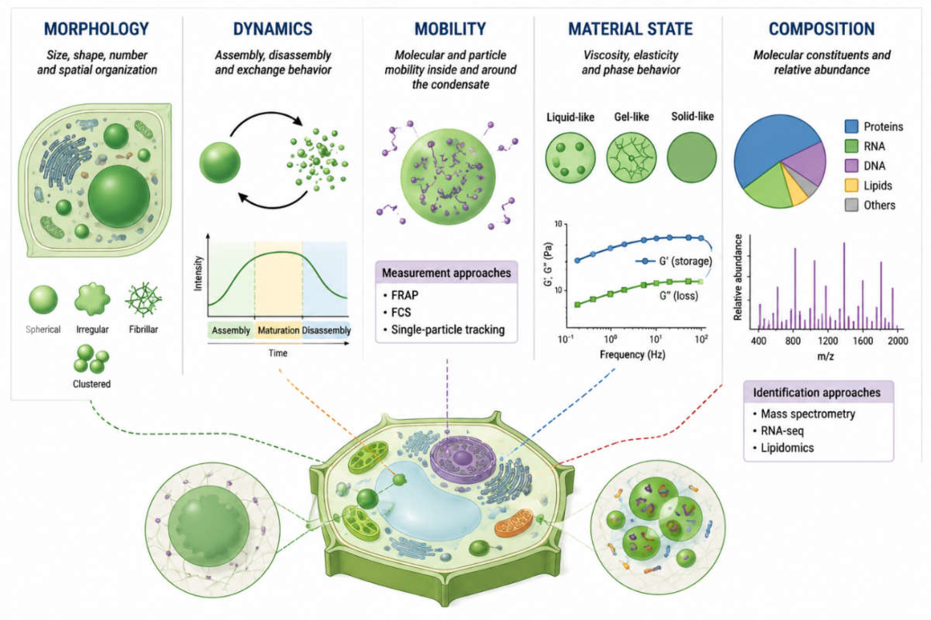
Figure 2.
Environmental phase imprinting in plant cells through dynamic biomolecular condensates. Environmental stresses induce changes in the physical state of biomolecular condensates, including alterations in condensate formation, size, material properties, molecular exchange, and dissolution dynamics. These stress-dependent changes can persist beyond the initial stimulus and may therefore encode aspects of stress intensity, duration, sequence, and recovery history. The proposed framework considers condensates as dynamic physical records that continuously evolve during stress and recovery rather than as static molecular archives. Multimodal measurements such as live-cell imaging, FRAP, FCS, fluorescence anisotropy, and complementary biophysical approaches could be used to characterize these state-dependent trajectories and test whether condensate dynamics retain predictive information about subsequent cellular responses.
Figure 2.
Environmental phase imprinting in plant cells through dynamic biomolecular condensates. Environmental stresses induce changes in the physical state of biomolecular condensates, including alterations in condensate formation, size, material properties, molecular exchange, and dissolution dynamics. These stress-dependent changes can persist beyond the initial stimulus and may therefore encode aspects of stress intensity, duration, sequence, and recovery history. The proposed framework considers condensates as dynamic physical records that continuously evolve during stress and recovery rather than as static molecular archives. Multimodal measurements such as live-cell imaging, FRAP, FCS, fluorescence anisotropy, and complementary biophysical approaches could be used to characterize these state-dependent trajectories and test whether condensate dynamics retain predictive information about subsequent cellular responses.
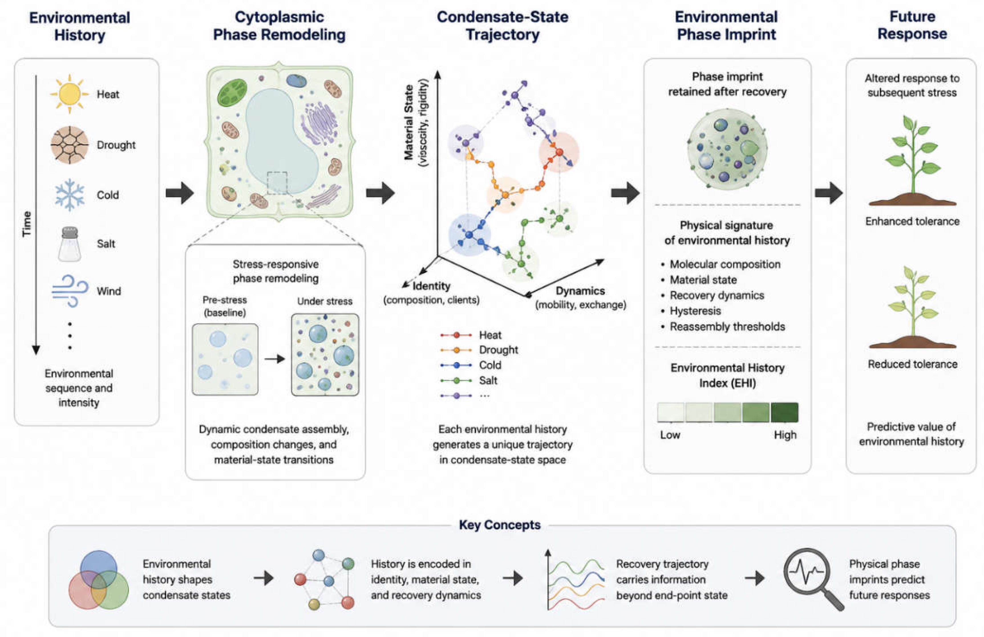
Figure 3.
History-dependent trajectories of condensate states. Schematic representation of condensate-state trajectories across five physical dimensions for three scenarios: A → B (stress B applied without prior stress history), A → Recovery → B (stress B applied after prior stress A and recovery), and A → Recovery → A (the same stress A reapplied after recovery). Each point represents a condensate state described across five physical dimensions—morphology, dynamics, molecular mobility, material state, and molecular composition. The solid line represents the observed state trajectory, whereas the dashed line indicates the reference trajectory toward the baseline state. The schematic illustrates the hypothesis that prior stress exposure may alter the subsequent trajectory of the condensate state.
Figure 3.
History-dependent trajectories of condensate states. Schematic representation of condensate-state trajectories across five physical dimensions for three scenarios: A → B (stress B applied without prior stress history), A → Recovery → B (stress B applied after prior stress A and recovery), and A → Recovery → A (the same stress A reapplied after recovery). Each point represents a condensate state described across five physical dimensions—morphology, dynamics, molecular mobility, material state, and molecular composition. The solid line represents the observed state trajectory, whereas the dashed line indicates the reference trajectory toward the baseline state. The schematic illustrates the hypothesis that prior stress exposure may alter the subsequent trajectory of the condensate state.
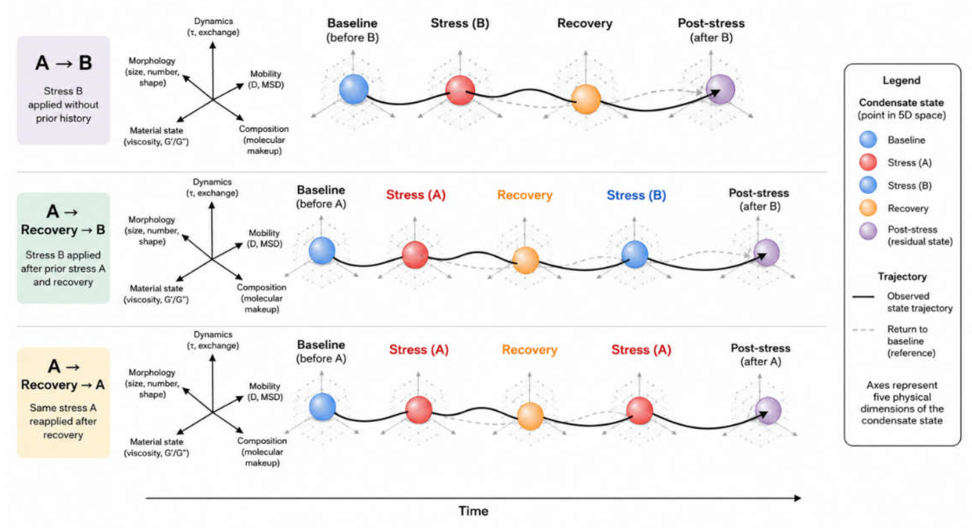
Figure 4.
Multimodal measurement architecture for reconstructing the plant condensate state. Complementary experimental modalities provide orthogonal information on different dimensions of condensate organisation. Live-cell confocal microscopy captures condensate morphology and spatiotemporal dynamics; fluorescence recovery after photobleaching (FRAP) assesses molecular exchange kinetics; fluorescence correlation spectroscopy (FCS) and single-particle tracking (SPT) provide information on molecular mobility and diffusion; microrheology and Brillouin microscopy probe local mechanical and viscoelastic properties; quantitative phase imaging (QPI) provides label-free information on optical density, dry-mass distribution, and refractive-index-related changes; molecular profiling characterises proteins, RNAs, post-translational modifications, and interaction partners; and physiological sensors provide contextual information on variables such as Ca²⁺, ROS, pH, hydration, temperature, and cellular energy status. Integration of these complementary measurements generates a multidimensional, time-resolved condensate-state representation, enabling comparison of condensate trajectories across baseline, stress, adaptation, recovery, and residual states within the proposed Plant Condensate Code framework.
Figure 4.
Multimodal measurement architecture for reconstructing the plant condensate state. Complementary experimental modalities provide orthogonal information on different dimensions of condensate organisation. Live-cell confocal microscopy captures condensate morphology and spatiotemporal dynamics; fluorescence recovery after photobleaching (FRAP) assesses molecular exchange kinetics; fluorescence correlation spectroscopy (FCS) and single-particle tracking (SPT) provide information on molecular mobility and diffusion; microrheology and Brillouin microscopy probe local mechanical and viscoelastic properties; quantitative phase imaging (QPI) provides label-free information on optical density, dry-mass distribution, and refractive-index-related changes; molecular profiling characterises proteins, RNAs, post-translational modifications, and interaction partners; and physiological sensors provide contextual information on variables such as Ca²⁺, ROS, pH, hydration, temperature, and cellular energy status. Integration of these complementary measurements generates a multidimensional, time-resolved condensate-state representation, enabling comparison of condensate trajectories across baseline, stress, adaptation, recovery, and residual states within the proposed Plant Condensate Code framework.
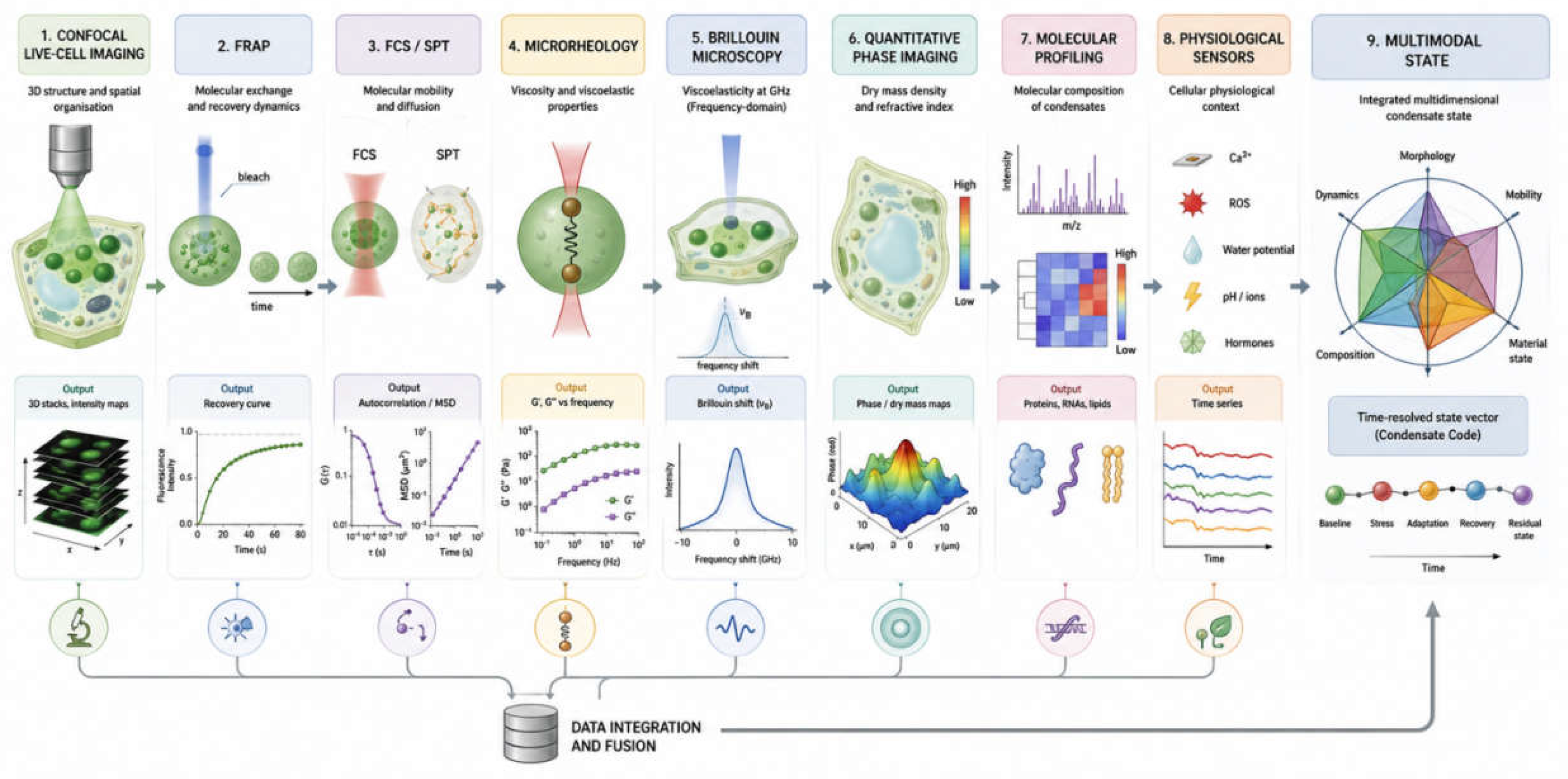
Figure 5.
Environmental phase imprint: trajectories of condensate-state recovery. Schematic representation of possible biomolecular condensate-state trajectories following environmental stress: complete recovery, partial recovery, and persistent imprint. The trajectories follow the sequence baseline → stress → recovery → residual state. Complete recovery represents a return of the condensate physical state to the pre-stress range; partial recovery represents an incomplete return with a measurable residual deviation; and persistent imprint represents a stable residual deviation that remains after recovery. The schematic illustrates the testable hypothesis that a previous stress exposure may leave a measurable physical trace in condensates after conventional physiological indicators have returned toward baseline.
Figure 5.
Environmental phase imprint: trajectories of condensate-state recovery. Schematic representation of possible biomolecular condensate-state trajectories following environmental stress: complete recovery, partial recovery, and persistent imprint. The trajectories follow the sequence baseline → stress → recovery → residual state. Complete recovery represents a return of the condensate physical state to the pre-stress range; partial recovery represents an incomplete return with a measurable residual deviation; and persistent imprint represents a stable residual deviation that remains after recovery. The schematic illustrates the testable hypothesis that a previous stress exposure may leave a measurable physical trace in condensates after conventional physiological indicators have returned toward baseline.
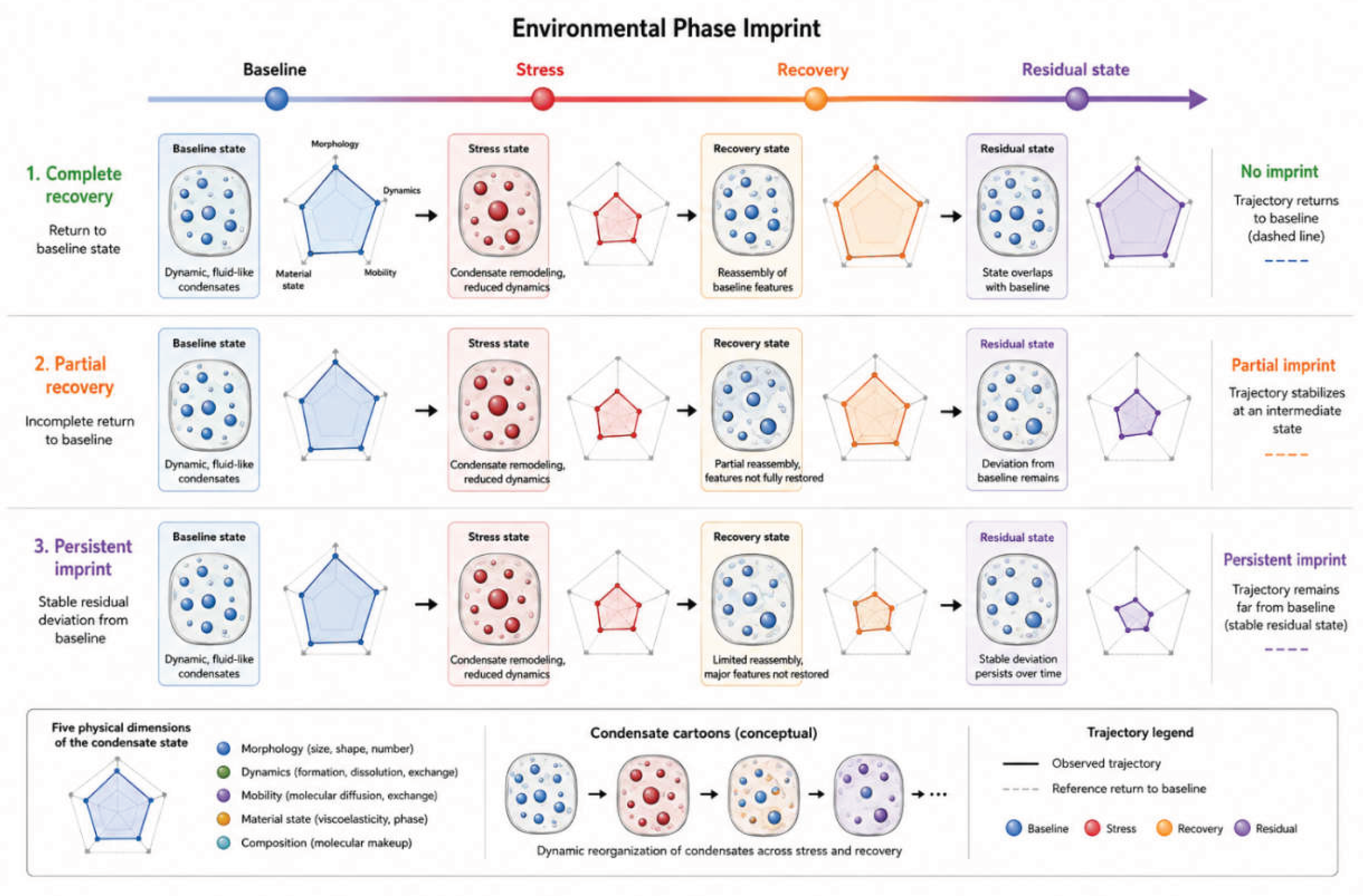
Figure 6.
Experimental framework for validating cytoplasmic phase resilience and stress memory. Sequential experimental design linking baseline cytoplasmic state, stress-induced phase transition, recovery, repeated stress exposure, and physiological performance. Measurements of cytoplasmic phase state (CPS) at each stage allow quantification of phase perturbation, reversibility, and the magnitude of the response to repeated stress. A reduced or altered phase shift during the second stress exposure provides an operational measure of stress memory, while the relationship between CPS dynamics and physiological performance enables evaluation of cytoplasmic phase resilience.
Figure 6.
Experimental framework for validating cytoplasmic phase resilience and stress memory. Sequential experimental design linking baseline cytoplasmic state, stress-induced phase transition, recovery, repeated stress exposure, and physiological performance. Measurements of cytoplasmic phase state (CPS) at each stage allow quantification of phase perturbation, reversibility, and the magnitude of the response to repeated stress. A reduced or altered phase shift during the second stress exposure provides an operational measure of stress memory, while the relationship between CPS dynamics and physiological performance enables evaluation of cytoplasmic phase resilience.
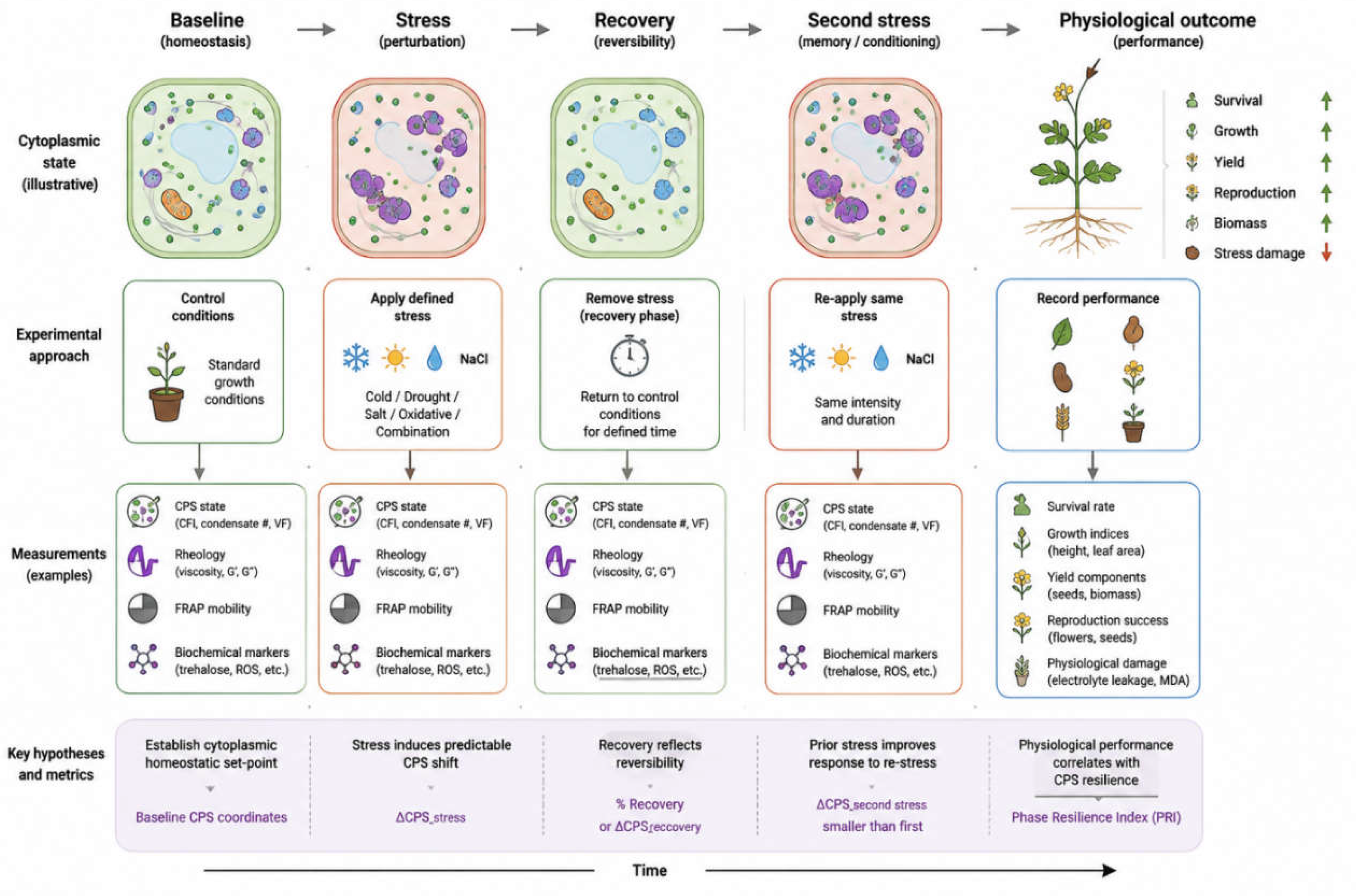
Figure 7.
Multidimensional Biophysical Profiling of the Cellular Phase-State Trajectory from Homeostasis to Recovery. Conceptual trajectory of plant cellular phase states from S1 (Homeostasis Baseline State) through PSW (Pre-Stress Warning State), S2 (Early Stress Adaptation State), S3 (Progressive Stress State), and CS (Collapse State) to R (Recovery State). Each state is proposed to exhibit a distinct multidimensional biophysical signature that can be characterized using complementary measurement modalities: FRAP for molecular mobility and fluorescence-recovery kinetics; FCS for molecular diffusion dynamics; Brillouin microscopy for spatially resolved mechanical and viscoelastic properties; quantitative phase imaging (QPI) for optical-path-length and structural features; fluorescence imaging for molecular localization and reporter-state changes; thermal imaging for thermal-state distribution; and impedance spectroscopy for cellular electrical properties. The trajectory represents a conceptual model of progressive changes in cellular physical organization during stress and their potential normalization during recovery. Integration of these orthogonal measurements is proposed as a basis for constructing a multidimensional Plant Condensate State Vector and testing whether individual, combined, and recurrent stresses generate distinct and history-dependent biophysical signatures.
Figure 7.
Multidimensional Biophysical Profiling of the Cellular Phase-State Trajectory from Homeostasis to Recovery. Conceptual trajectory of plant cellular phase states from S1 (Homeostasis Baseline State) through PSW (Pre-Stress Warning State), S2 (Early Stress Adaptation State), S3 (Progressive Stress State), and CS (Collapse State) to R (Recovery State). Each state is proposed to exhibit a distinct multidimensional biophysical signature that can be characterized using complementary measurement modalities: FRAP for molecular mobility and fluorescence-recovery kinetics; FCS for molecular diffusion dynamics; Brillouin microscopy for spatially resolved mechanical and viscoelastic properties; quantitative phase imaging (QPI) for optical-path-length and structural features; fluorescence imaging for molecular localization and reporter-state changes; thermal imaging for thermal-state distribution; and impedance spectroscopy for cellular electrical properties. The trajectory represents a conceptual model of progressive changes in cellular physical organization during stress and their potential normalization during recovery. Integration of these orthogonal measurements is proposed as a basis for constructing a multidimensional Plant Condensate State Vector and testing whether individual, combined, and recurrent stresses generate distinct and history-dependent biophysical signatures.
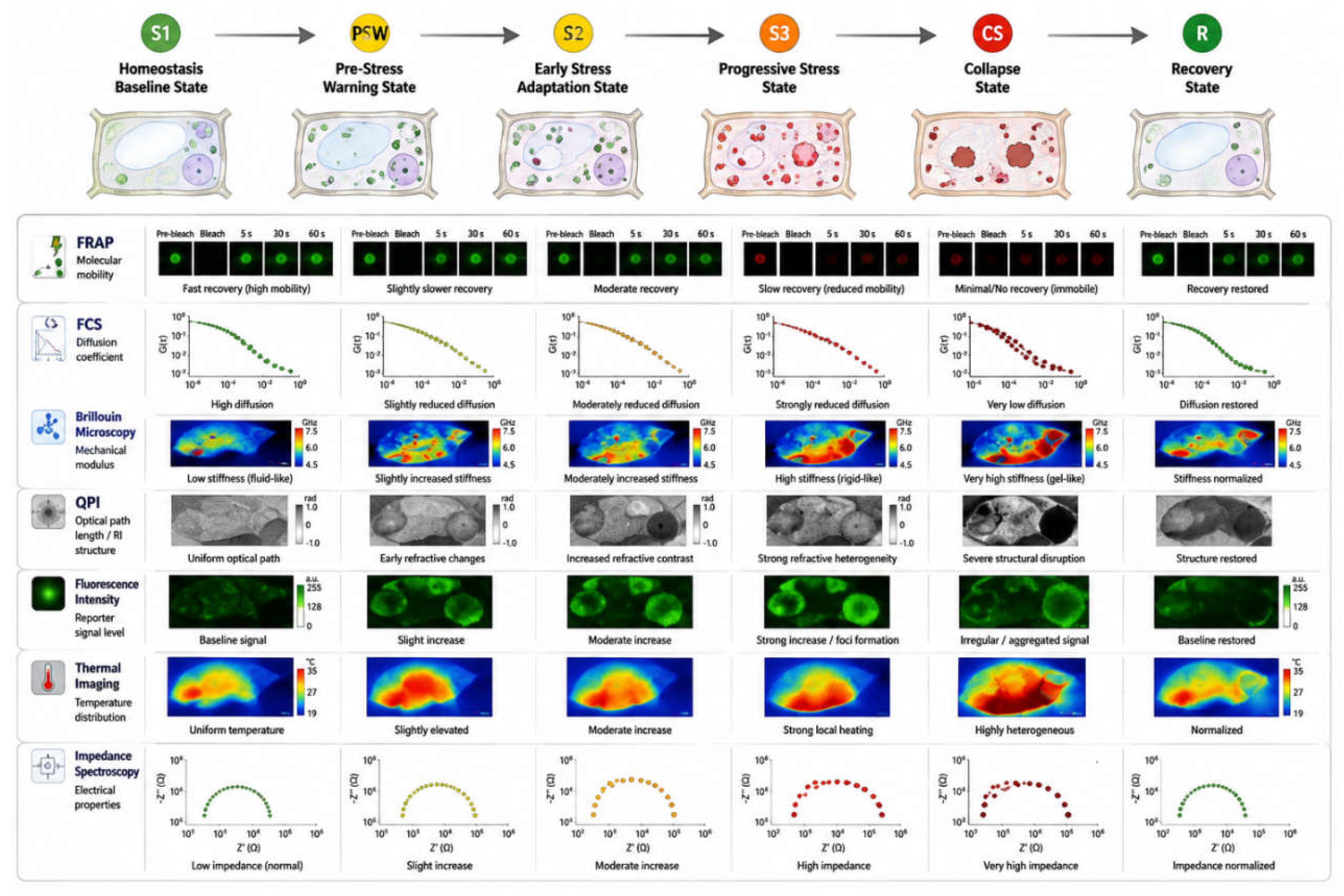
Figure 8.
Conceptual model of stress-induced cytoplasmic phase remodeling and condensate-code formation. Environmental stressors induce progressive changes in the physical state of the cytoplasm, reflected in condensate properties including number, size, composition, localization, diffusion, viscosity, lifetime, reversibility, and recovery capacity. These features generate stress-specific and combined-stress signatures that may encode temporal information about prior stress exposure and influence cellular recovery trajectories, ranging from full recovery to phase rigidity and collapse.
Figure 8.
Conceptual model of stress-induced cytoplasmic phase remodeling and condensate-code formation. Environmental stressors induce progressive changes in the physical state of the cytoplasm, reflected in condensate properties including number, size, composition, localization, diffusion, viscosity, lifetime, reversibility, and recovery capacity. These features generate stress-specific and combined-stress signatures that may encode temporal information about prior stress exposure and influence cellular recovery trajectories, ranging from full recovery to phase rigidity and collapse.
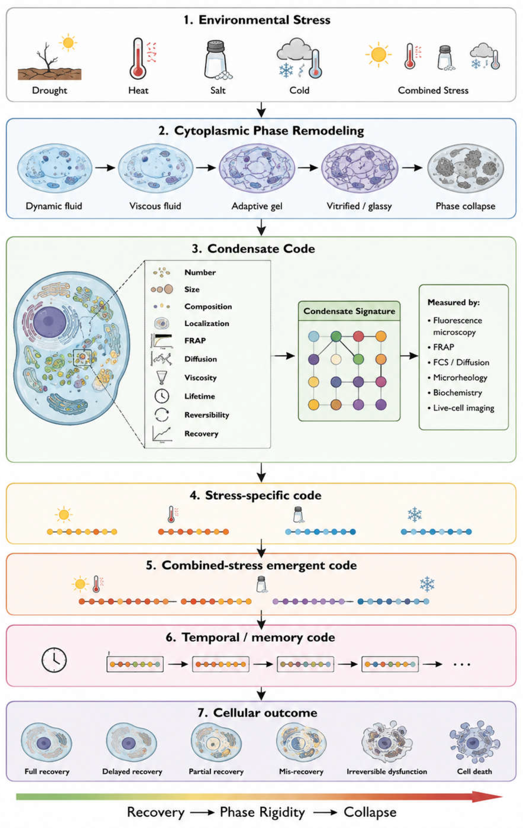
Figure 9.
A multidimensional framework for quantifying the plant condensate state and stress memory. Environmental stressors induce physicochemical perturbations that are reflected in distinct responses of multiple biomolecular condensate populations. The proposed condensate code, CC(t), integrates five complementary dimensions: morphology, dynamics, material properties, molecular composition, and molecular mobility. These dimensions can be quantified using live-cell imaging, FRAP, fluorescence correlation spectroscopy, single-particle tracking, microrheology, Brillouin microscopy, proteomics, RNA profiling, and proximity-labeling approaches. The resulting multidimensional state vector can be used to derive quantitative descriptors of condensate-state deviation, combined-stress interactions, and residual stress memory. The lower panels illustrate hypothetical trajectories of condensate states during heat stress and recovery, stress-specific memory, and combined-stress exposure. This framework is conceptual and hypothesis-driven; the proposed condensate code and derived indices require experimental validation.
Figure 9.
A multidimensional framework for quantifying the plant condensate state and stress memory. Environmental stressors induce physicochemical perturbations that are reflected in distinct responses of multiple biomolecular condensate populations. The proposed condensate code, CC(t), integrates five complementary dimensions: morphology, dynamics, material properties, molecular composition, and molecular mobility. These dimensions can be quantified using live-cell imaging, FRAP, fluorescence correlation spectroscopy, single-particle tracking, microrheology, Brillouin microscopy, proteomics, RNA profiling, and proximity-labeling approaches. The resulting multidimensional state vector can be used to derive quantitative descriptors of condensate-state deviation, combined-stress interactions, and residual stress memory. The lower panels illustrate hypothetical trajectories of condensate states during heat stress and recovery, stress-specific memory, and combined-stress exposure. This framework is conceptual and hypothesis-driven; the proposed condensate code and derived indices require experimental validation.
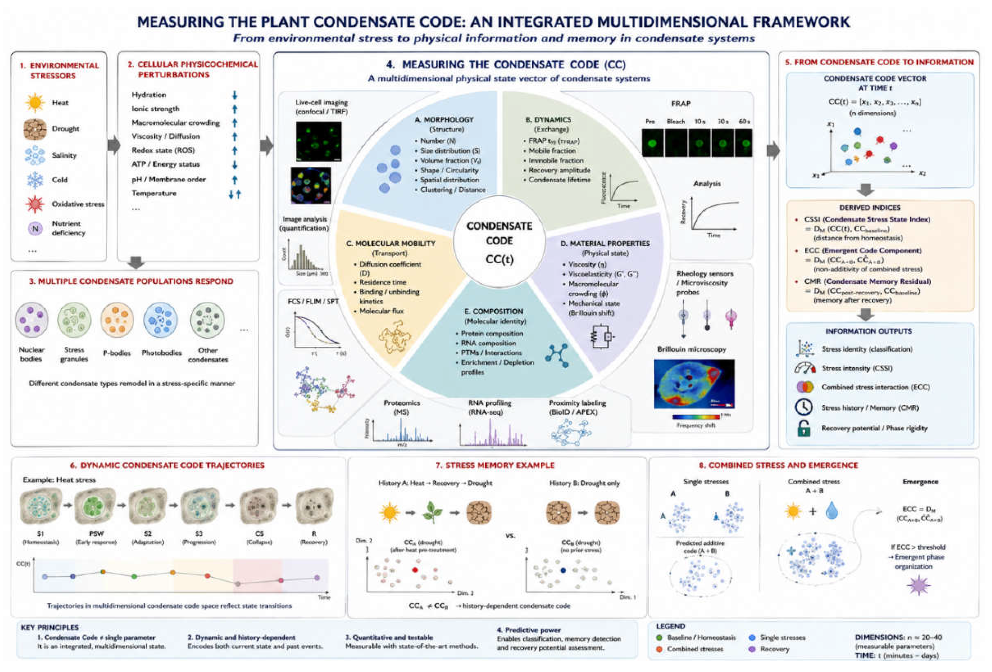

Table 1.
What each measurement tells us about condensate physical state.
| Method | What is measured | Physical property revealed | Biological interpretation | Main limitation |
| Confocal / spinning-disk microscopy | Number, size, shape, localisation | Morphology, dynamics | Population-level organisation | Puncta alone do not prove LLPS |
| FRAP | Fluorescence recovery after photobleaching | Molecular exchange | Mobility, tentative material-state signal | Diffusion/binding ambiguity; not proof of LLPS |
| FCS | Fluorescence intensity fluctuations | Diffusion coefficient, local concentration | Molecular mobility | Requires suitable fluorescent signal |
| SPT | Individual molecule/particle trajectories | Diffusion mode, confinement | Molecular behaviour | Labeling and tracking constraints |
| Microrheology | Tracked-particle motion | Viscoelasticity | Material state | Probe-dependent, model-dependent |
| Brillouin microscopy | Spectral shift/linewidth of scattered light | Mechanical-acoustic properties | Local material state | Not a direct measurement of viscosity |
| Fluorescence anisotropy | Polarisation of emitted light | Rotational mobility, local packing | Molecular crowding/binding | Binding and viscosity effects not separable |
| QPI | Optical phase delay | Dry-mass density, refractive index | Cellular physical context | Not condensate-specific |
| Proteomics / interactomics / RNA profiling | Molecular composition | Molecular identity | Compositional remodeling | Destructive, population-averaged |
| Physiological sensors | pH, Ca²⁺, ROS, redox, ATP/ADP, hydration, temperature | Cellular physiological context | Mechanistic context | Not a direct condensate measurement |
Disclaimer/Publisher’s Note: The statements, opinions and data contained in all publications are solely those of the individual author(s) and contributor(s) and not of MDPI and/or the editor(s). MDPI and/or the editor(s) disclaim responsibility for any injury to people or property resulting from any ideas, methods, instructions or products referred to in the content. |
© 2026 by the authors. Licensee MDPI, Basel, Switzerland. This article is an open access article distributed under the terms and conditions of the Creative Commons Attribution (CC BY) license (http://creativecommons.org/licenses/by/4.0/).
Copyright: This open access article is published under a Creative Commons CC BY 4.0 license, which permit the free download, distribution, and reuse, provided that the author and preprint are cited in any reuse.