
Submitted:
30 August 2026
Posted:
31 August 2026
You are already at the latest version
Abstract
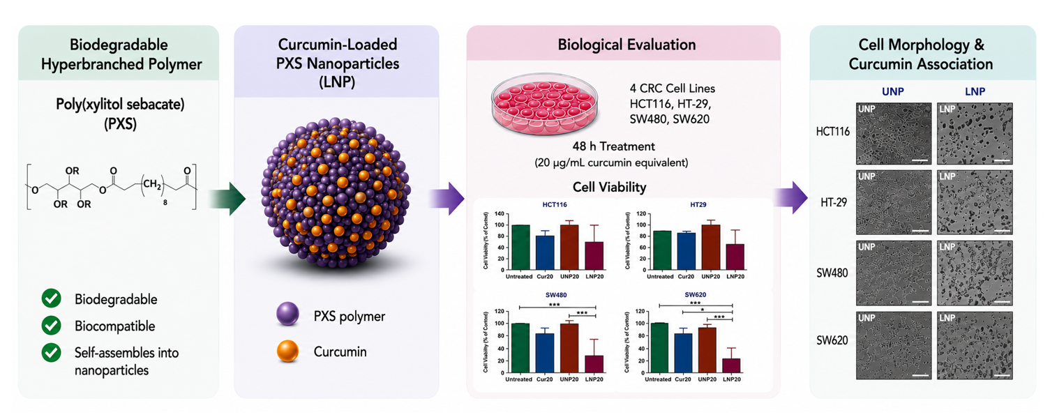
Background/Objectives: Poly(xylitol sebacate) (PXS) is a biodegradable, biocompatible hyperbranched polyester capable of self-assembling into nanoparticles for hydrophobic drug delivery. Building on our prior physicochemical characterization of PXS nanoparticles, this study evaluated whether curcumin-loaded PXS nanoparticles (LNP) can deliver curcumin to colorectal cancer (CRC) cells and enhance its anticancer activity relative to free curcumin. Methods: Unloaded (UNP) and curcumin-loaded PXS nanoparticles were fabricated by nanoprecipitation and characterized by dynamic light scattering (DLS) and scanning electron microscopy (SEM). Four CRC cell lines (HCT116, HT29, SW480, SW620) were treated with free curcumin, UNP, or LNP at an equivalent curcumin concentration of 20 μg/mL for 48 h; viability was measured by CellTiter-Blue® assay, and cell morphology and curcumin cellular association were assessed by phase-contrast and fluorescence microscopy. Results: Nanoparticles fell within the target 100–200 nm range (LNP: 171.04 nm; UNP: 212.40 nm at 24 h) and remained colloidally stable for 48 h (159–203 nm). SEM confirmed spherical morphology for both formulations, and UNP were not cytotoxic at 20 μg/mL. LNP produced substantially greater viability reduction than free curcumin across all lines: SW480 and SW620 decreased to ~30% viability (~70% reduction), HT29 to ~60% (~40% reduction, despite minimal response to free curcumin), and HCT116, the most resistant line, to ~70% (~30% reduction). Fluorescence imaging suggested closer cellular association of LNP-delivered curcumin, although the signal was modest and not quantified. Conclusions: PXS nanoparticles are an effective, biodegradable carrier that enhances the cellular delivery and anticancer activity of curcumin in CRC cells, supporting further development of this hyperbranched polyester platform for cancer drug delivery.
Keywords:
poly(xylitol sebacate)
; hyperbranched polyester
; nanoparticle drug delivery
; curcumin
; colorectal cancer
1. Introduction
Colorectal cancer (CRC) is the third most common cancer diagnosed in the United States, with an estimated 158,850 new cases and 55,230 deaths projected in 2026 [1]. Polymeric nanoparticles are among the most versatile drug-delivery platforms because they improve solubility, protect payloads from premature degradation, and enable sustained or targeted release, improving efficacy while reducing systemic toxicity of poorly bioavailable therapeutics [2,3]. Biodegradable polyesters are particularly attractive carriers because they degrade into non-toxic byproducts while offering tunable degradation behavior and excellent biocompatibility [4,5]; although poly(lactic-co-glycolic acid) (PLGA) remains the most widely studied example, continued efforts target alternative polymer systems with greater synthetic versatility and architectural control [2,3,4,5,6].
Poly(xylitol sebacate) (PXS) is a biodegradable hyperbranched polyester synthesized by melt polycondensation of xylitol and sebacic acid. The five hydroxyl groups on xylitol drive extensive branching, producing a three-dimensional architecture with high terminal functional-group density that favors nanoparticle self-assembly, hydrophobic drug encapsulation, and future surface modification [4,5,7,8,9,10,11,12,13,14,15]. PXS also shows favorable biodegradation, biocompatibility, and a reduced inflammatory response relative to PLGA [10,11].
Our previous work established the synthesis, physicochemical characterization, and nanoparticle fabrication of hyperbranched PXS, identifying 15 h melt polymerization as optimal for reproducible nanoparticle formation. Curcumin-loaded PXS nanoparticles from that study had diameters of approximately 112 nm, strongly negative surface charge, 23.9% curcumin encapsulation efficiency, and sustained curcumin release under physiological conditions [10], establishing the materials foundation for biological evaluation.
Building on that platform, the present study evaluates whether PXS nanoparticles can effectively deliver curcumin to colorectal cancer cells and enhance its anticancer activity. Curcumin was selected as the model therapeutic for its well-documented antioxidant, anti-inflammatory, and anticancer activity, including suppression of proliferation and induction of apoptosis in CRC [2,3]; however, its clinical use is limited by poor water solubility, rapid degradation, and low bioavailability [4,5,6]. Polymeric nanoparticle encapsulation is a well-established strategy for overcoming these limitations [2,3,4,5,6]. Unloaded and curcumin-loaded PXS nanoparticles were therefore evaluated in four human CRC cell lines (HCT116, HT29, SW480, SW620) using cell-viability assays, phase-contrast microscopy, and fluorescence imaging. This work provides the first biological validation of PXS as a drug-delivery platform and represents the next step toward hyperbranched xylitol-based polyesters for anticancer applications.
2. Materials and Methods
2.1. Materials
Poly(xylitol sebacate) (PXS) was synthesized in-house by melt polycondensation of xylitol and sebacic acid under nitrogen at 150 °C, following Culpepper et al. [10]; the 15 h polymerization was used, as previously identified as optimal for nanoparticle fabrication. Curcumin (natural), dimethyl sulfoxide (DMSO), methanol, and Pluronic® F-127 were purchased from MilliporeSigma (St. Louis, MO, USA). Dulbecco’s Modified Eagle Medium (DMEM), Roswell Park Memorial Institute (RPMI-1640) medium, fetal bovine serum (FBS), penicillin streptomycin, phosphate buffered saline (PBS), paraformaldehyde, CellTiter-Blue® Cell Viability Assay reagent (Promega, Madison, WI, USA), and DAPI (Fisher Scientific, Waltham, MA, USA) were used as received. All aqueous solutions were prepared using deionized (DI) water.
2.2. Preparation of Curcumin-Loaded PXS Nanoparticles
Curcumin-loaded (LNP) and unloaded (UNP) PXS nanoparticles were prepared by nanoprecipitation, modified from Culpepper et al. [10] (Figure 1). Briefly, 0.03 - 0.05 g of PXS was dissolved in DMSO (LNP formulations additionally contained 1.2 mg curcumin) and added dropwise, under constant stirring, to 30 mL of an aqueous surfactant solution containing 1% w/v Pluronic® F-127 in a 1:2 (v/v) ratio of DI water to methanol. The suspension was probe sonicated using alternating one-minute on/off cycles for 20 minutes. One-mL aliquots were dispensed into 1.7-mL centrifuge tubes and centrifuged at 20,000 × g and 16 °C for 13 minutes; each pellet was washed twice more by resuspension in DI water followed by centrifugation (20,000 × g, 16 °C, 5 min). Washed pellets were pooled and resuspended in the final volume of 5 mL DI water. This reduced final suspension volume relative to our previously published procedure [10] increased nanoparticle and curcumin concentration per unit volume and reduced the risk of hypotonic effects during cell treatment (Section 3.3). Unloaded nanoparticles were prepared identically, omitting curcumin.
2.3. Nanoparticle Characterization
Nanoparticle morphology was evaluated by scanning electron microscopy (SEM; Phenom XL, High-Performance Materials Institute) after sputter-coating with 1 nm platinum (Cressington sputter coater). Hydrodynamic particle diameter was determined by dynamic light scattering (DLS; Lifesizer DLS 700) at 25 °C, and additionally after incubation at 37 °C for 24 and 48 h to confirm stability under the conditions used for the cell-viability assay. Curcumin loading was quantified using a Tecan NanoQuant Infinite M200 Pro plate reader; absorbance spectra (350 - 490 nm, average of 8 scans) were compared against a calibration curve generated from curcumin standards of known concentration (Figure S3) to calculate curcumin-equivalent nanoparticle doses. Zeta potential and encapsulation efficiency were not re-measured for the present batches; previously reported values [10] are used only as historical characterization of the PXS platform.
2.4. Cell Culture and Treatment Optimization
Human colorectal cancer cell lines HCT116, HT29, SW480, and SW620 were cultured at 37 °C in a humidified 5% CO₂ incubator in their recommended growth media supplemented with 10% FBS and 1% penicillin–streptomycin. Preliminary optimization in HCT116 cells (Figure S3) showed that nanoparticle treatment volumes equivalent to 30 μg/mL curcumin produced a hypotonic reduction in viability that was independent of nanoparticle toxicity, as confirmed using an equal-volume DI water control; 20 μg/mL was therefore selected as the working concentration, and the fabrication protocol (Section 2.2) was concentrated accordingly. Seeding density and exposure time were further optimized to 10,000 cells/well and 48 h. All four cell lines were subsequently treated under these optimized conditions.
2.5. Cell Viability, Morphology, and Fluorescence Imaging
Cell viability was evaluated using the CellTiter-Blue® assay. Cells (1 × 10⁴/well, black-wall 96-well plates) were treated with free curcumin, UNP, or LNP at an equivalent curcumin concentration of 20 μg/mL for 48 h, with untreated cells as controls; fluorescence was read on a Synergy H1 Multimode plate reader (Agilent Technologies) relative to untreated controls (n ≥ 3 independent replicates per condition). Phase-contrast images were collected after the 48 h treatment period to assess morphology. For fluorescence imaging, cells (2.5 × 10⁴/well, eight well chamber slides) were treated with free curcumin or LNP (20 μg/mL curcumin-equivalent, 24 h), fixed with 4% paraformaldehyde, and stained with DAPI (1:5000) prior to imaging on an Echo Revolve fluorescence microscope.
2.6. Statistical Analysis
Data are presented as mean ± standard deviation (n ≥ 3 independent biological replicates). Comparisons among treatment groups were performed by one-way ANOVA with Tukey’s multiple comparison test (GraphPad Prism v6, GraphPad Software, San Diego, CA, USA); p < 0.05 was considered significant, with exact p-values reported in figure legends.
3. Results and Discussion
3.1. Physicochemical Confirmation of the PXS Platform
Twenty-minute probe sonication produced homogeneous PXS nanoparticle suspensions, confirmed by DLS. Because this study extends the PXS platform previously established by Culpepper et al. [10], polymer identity was reconfirmed by ¹³C NMR, FTIR, TGA, and DSC (Figure S1). ¹³C NMR showed the expected sebacate methylene (~25 - 34 ppm), oxygenated xylitol (~61 - 73 ppm), and ester carbonyl (~173 - 175 ppm) resonances; FTIR showed residual hydroxyl (~3400 cm⁻¹), aliphatic –CH₂ (~2900 cm⁻¹), and ester C=O (~1700 - 1750 cm⁻¹) absorptions; TGA showed limited mass loss below ~200 °C followed by the principal degradation event; and DSC showed a subambient thermal transition without a prominent melting endotherm, consistent with an amorphous, hyperbranched structure. These results confirm that the PXS used here retains the chemical and thermal signature previously reported [10], allowing the present study to focus on nanoparticle behavior and biological performance.
3.2. Nanoparticle Size, Morphology, and Stability
SEM confirmed predominantly spherical morphology for both UNP and LNP, indicating that curcumin loading did not alter overall particle shape (Figure S2). DLS showed average hydrodynamic diameters of 212.40 nm (UNP) and 171.04 nm (LNP) at 24 h/25 °C (Figure S2); because DLS measures hydrated particles in suspension while SEM images dried particles, the two techniques were treated as complementary rather than directly equivalent. After incubation at 37 °C, the temperature and duration used for cell treatment, LNP measured 200.3 nm (24 h) and 162.10 nm (48 h), while UNP measured 202.50 nm (24 h) and 159.35 nm (48 h) (Table 1). Neither formulation showed the progressive size increase expected from aggregation, supporting nanoscale stability throughout the 48-h treatment window. Curcumin loading was quantified against a linear calibration curve (y = 0.0540x + 0.1258, R² = 0.9989; Figure S3) generated from 1–10 μg/mL curcumin standards, allowing curcumin-equivalent nanoparticle doses to be normalized across treatment groups.
3.3. Selection of a Non-Cytotoxic Working Concentration
Unloaded PXS nanoparticles were first tested at volumes equivalent to 10, 20, and 30 μg/mL curcumin-loaded nanoparticles in HCT116 cells. The 30 μg/mL volume produced a marked reduction in viability and crater-like morphology (Figure S4); an equal volume of DI water produced a comparable response, indicating a hypotonic effect of the treatment volume itself rather than nanoparticle cytotoxicity. Based on this, the nanoparticle fabrication protocol was concentrated (Section 2.2) and 20 μg/mL was adopted as the working concentration; at this dose, UNP produced no substantial reduction in viability or morphological change across all four cell lines (Figure S5), establishing that the PXS carrier itself is well tolerated and providing an appropriate baseline for evaluating curcumin-loaded nanoparticles.
3.4. Curcumin-Loaded Nanoparticles Increase Sensitivity of CRC Cells to Curcumin
At the 20 μg/mL curcumin equivalent dose, LNP produced a substantially greater reduction in viability than free curcumin across all four cell lines (Figure 2). Free curcumin alone produced only a modest (~20%) reduction in HCT116, SW480, and SW620, and essentially no reduction in HT29. In contrast, LNP reduced viability to ~30% (a ~70% reduction) in SW480 and SW620; a matched primary/metastatic cell-line pair from the same patient to ~60% (~40% reduction) in HT29 despite its limited response to free drug, and to ~70% (~30% reduction) in the most resistant line, HCT116. These viability changes were mirrored by phase-contrast morphology, where LNP treatment produced pronounced cell rounding and reduced cell density relative to free curcumin or UNP, while UNP produced no discernible morphological change in any line. Together, these results indicate that nanoparticle encapsulation markedly improves the cellular delivery and anticancer efficacy of curcumin in CRC cells, consistent with the physicochemical feasibility established previously for this platform [10], and that encapsulation can restore curcumin sensitivity in an otherwise poorly responsive line (HT29).
3.5. Fluorescence Imaging of Cellular Association
Fluorescence imaging in all four cell lines showed curcumin fluorescence (green) as sparse, punctuate signal under both free curcumin and LNP treatment (Figure 3). Free curcumin puncta were broadly distributed with limited apparent cell association, whereas some LNP associated puncta appeared closer to, or overlapping with, cell boundaries. However, signal intensity and puncta density were modest and qualitatively similar between conditions, and no co-localization analysis, flow cytometry, or dedicated internalization assay was performed. These images are therefore suggestive of, rather than definitive evidence for, enhanced cellular association of nanoparticle delivered curcumin, and are reported as a qualitative complement to the viability and morphology data.
4. Conclusions
This study provides the first biological validation of curcumin-loaded PXS nanoparticles as a delivery platform for colorectal cancer. Nanoparticles were reproducibly fabricated with the target size, spherical morphology, and 48-h colloidal stability required for cellular delivery and consistently outperformed free curcumin across four CRC cell lines. SW480 and SW620 — primary and metastatic lines from the same patient — were most sensitive; HT29 and HCT116 showed more modest but significant responses; and nanoparticle encapsulation restored sensitivity to curcumin in HT29, which was otherwise minimally responsive to the free compound. These findings are broadly consistent with reports of enhanced efficacy for other polymeric nanoparticle-delivered anticancer agents, including curcumin and other polyphenols [16,17] and 5-fluorouracil in colorectal and other cancer models [18,19]. Because this study did not include an apoptosis assay or cytoskeletal imaging, the mechanism underlying the observed morphological changes remains to be defined; future work incorporating apoptosis markers (e.g., Annexin V/caspase activity), cytoskeletal imaging, and in vivo efficacy studies is needed to further establish PXS as a biodegradable platform for anticancer drug delivery.
Supplementary Materials
The following supporting information can be downloaded at the website of this paper posted on Preprints.org, Figure S1: Physicochemical characterization of the PXS polymer (¹³C NMR, FTIR, TGA, DSC); Figure S2: Morphological and hydrodynamic size characterization of PXS nanoparticles (SEM and DLS); Figure S3: Curcumin calibration curve used for dose normalization; Figure S4: Effect of unloaded PXS nanoparticles (UNP) on CRC cell viability during treatment-condition optimization; Figure S5: Comparative effect of curcumin-loaded PXS nanoparticles (LNP) at 10 and 20 μg/mL across all four CRC cell lines.
Author Contributions
Conceptualization, J.P. and N.A.; Methodology, X.C. and J.P.; Formal Analysis, J.P. and N.A.; Investigation, X.C.; Writing — Original Draft Preparation, X.C., J.P., and N.A.; Writing — Review and Editing, N.A. and J.P.; Supervision, N.A. and J.P.; Funding Acquisition, J.P. and N.A. All authors have read and agreed to the published version of the manuscript.
Funding
This research was supported by the National Cancer Institute, Center for Reducing Cancer Health Disparities of the National Institutes of Health under award numbers U54CA233396, U54CA233444, and U54CA233465 (Florida-California Cancer Research, Education and Engagement [CaRE²] Health Center), and P30CA247796 and P30CA014089 (University of Florida Health Cancer Institute and University of Southern California Norris Comprehensive Cancer Center, respectively). Additional support was provided by NSF PREM SEED (Award No. 2122142) and Virginia Tech GlycoMIP. The content is solely the responsibility of the authors and does not necessarily represent the official views of the National Institutes of Health.
Institutional Review Board Statement
Not applicable. This study did not involve humans or animals; all experiments used established, commercially sourced human colorectal cancer cell lines.
Informed Consent Statement
Not applicable.
Data Availability Statement
The data presented in this study are available from the corresponding author upon reasonable request.
Acknowledgments
The authors thank the FAMU Departments of Biology and Chemistry, FAMU-FSU College of Engineering, FAMU Center for Plasma Science and Technology, High-Performance Materials Institute (HPMI), and FAMU Pharmacy for access to characterization equipment and support.
Conflicts of Interest
The authors declare no conflicts of interest.
References
- Siegel, R.L.; Wagle, N.S.; Star, J.; Kratzer, T.B.; Smith, R.A.; Jemal, A. Colorectal Cancer Statistics, 2026. CA Cancer J. Clin. 2026. [Google Scholar] [CrossRef] [PubMed]
- Langer, R. Drug Delivery and Targeting. Nature 1998, 392, 5–10. [Google Scholar] [PubMed]
- Duncan, R. Polymer Therapeutics as Nanomedicines: New Perspectives. Nat. Rev. Drug Discov. 2003, 2, 347–360. [Google Scholar] [PubMed]
- Kamaly, N.; Yameen, B.; Wu, J.; Farokhzad, O.C. Degradable Controlled-Release Polymers and Polymeric Nanoparticles: Mechanisms of Controlling Drug Release. Chem. Rev. 2016, 116, 2602–2663. [Google Scholar] [CrossRef] [PubMed]
- Mitchell, M.J.; Billingsley, M.M.; Haley, R.M.; Wechsler, M.E.; Peppas, N.A.; Langer, R. Engineering Precision Nanoparticles for Drug Delivery. Nat. Rev. Drug Discov. 2021, 20, 101–124. [Google Scholar] [CrossRef] [PubMed]
- Anselmo, A.C.; Mitragotri, S. Nanoparticles in the Clinic. Bioeng. Transl. Med. 2016, 1, 10–29. [Google Scholar] [CrossRef] [PubMed]
- Gao, C.; Yan, D. Hyperbranched Polymers: From Synthesis to Applications. Prog. Polym. Sci. 2004, 29, 183–275. [Google Scholar] [CrossRef]
- Voit, B.I. Hyperbranched Polymers—All Problems Solved after 15 Years of Research? J. Polym. Sci. Part A Polym. Chem. 2005, 43, 2679–2699. [Google Scholar] [CrossRef]
- Fréchet, J.M.J. Functional Polymers and Dendrimers: Reactivity, Molecular Architecture, and Interfacial Energy. Science 1994, 263, 1710–1715. [Google Scholar] [CrossRef] [PubMed]
- Culpepper, K.M.; Tindal, J.B.; Arnett, N.Y. Effect of Reaction Time on the Properties of Poly(xylitol Sebacate) for Nanoparticle Formation. J. Polym. Res. 2024, 31, 289. [Google Scholar] [CrossRef]
- Culpepper, K.M. Synthesis and Characterization of Xylitol-Based Polyesters for Targeted Drug Delivery. Ph.D. Dissertation, Florida Agricultural and Mechanical University, Tallahassee, FL, USA, 2023. [Google Scholar]
- Sperling, L.H. Introduction to Physical Polymer Science, 4th ed.; Wiley-Interscience: Hoboken, NJ, USA, 2006. [Google Scholar]
- Khandare, J.; Calderón, M.; Dagia, N.M.; Haag, R. Multifunctional Dendritic Polymers in Nanomedicine. Chem. Soc. Rev. 2012, 41, 2824–2848. [Google Scholar] [CrossRef] [PubMed]
- Lee, C.C.; MacKay, J.A.; Fréchet, J.M.J.; Szoka, F.C. Designing Dendrimers for Biological Applications. Nat. Biotechnol. 2005, 23, 1517–1526. [Google Scholar] [CrossRef] [PubMed]
- Blanco, E.; Shen, H.; Ferrari, M. Principles of Nanoparticle Design for Overcoming Biological Barriers to Drug Delivery. Nat. Biotechnol. 2015, 33, 941–951. [Google Scholar] [CrossRef] [PubMed]
- Hosseinzadeh, S.; et al. Polyethylene Glycol Triggers the Anti-Cancer Impact of Curcumin Nanoparticles in SW-1736 Thyroid Cancer Cells. Sci. Rep. 2021. [Google Scholar] [CrossRef] [PubMed]
- Ben-Zichri, S.; et al. Synergistic Activity of Anticancer Polyphenols Embedded in Polymeric Nanoparticles. ACS Appl. Polym. Mater. 2022. [Google Scholar] [CrossRef]
- Singh, D.; et al. Development and Evaluation of Optimized Molecularly Imprinted Polymeric Nanoparticles of 5-FU. J. Mol. Recognit. 2025. [Google Scholar]
- Anjum, S.; et al. Enhanced Targeted Treatment of Cervical Cancer Using Nanoparticle-Based Doxycycline Delivery System. Sci. Rep. 2024. [Google Scholar] [CrossRef] [PubMed]
Figure 1.
Schematic overview of the modified nanoprecipitation procedure used to fabricate unloaded and curcumin-loaded PXS nanoparticles (see Section 2.2).
Figure 1.
Schematic overview of the modified nanoprecipitation procedure used to fabricate unloaded and curcumin-loaded PXS nanoparticles (see Section 2.2).
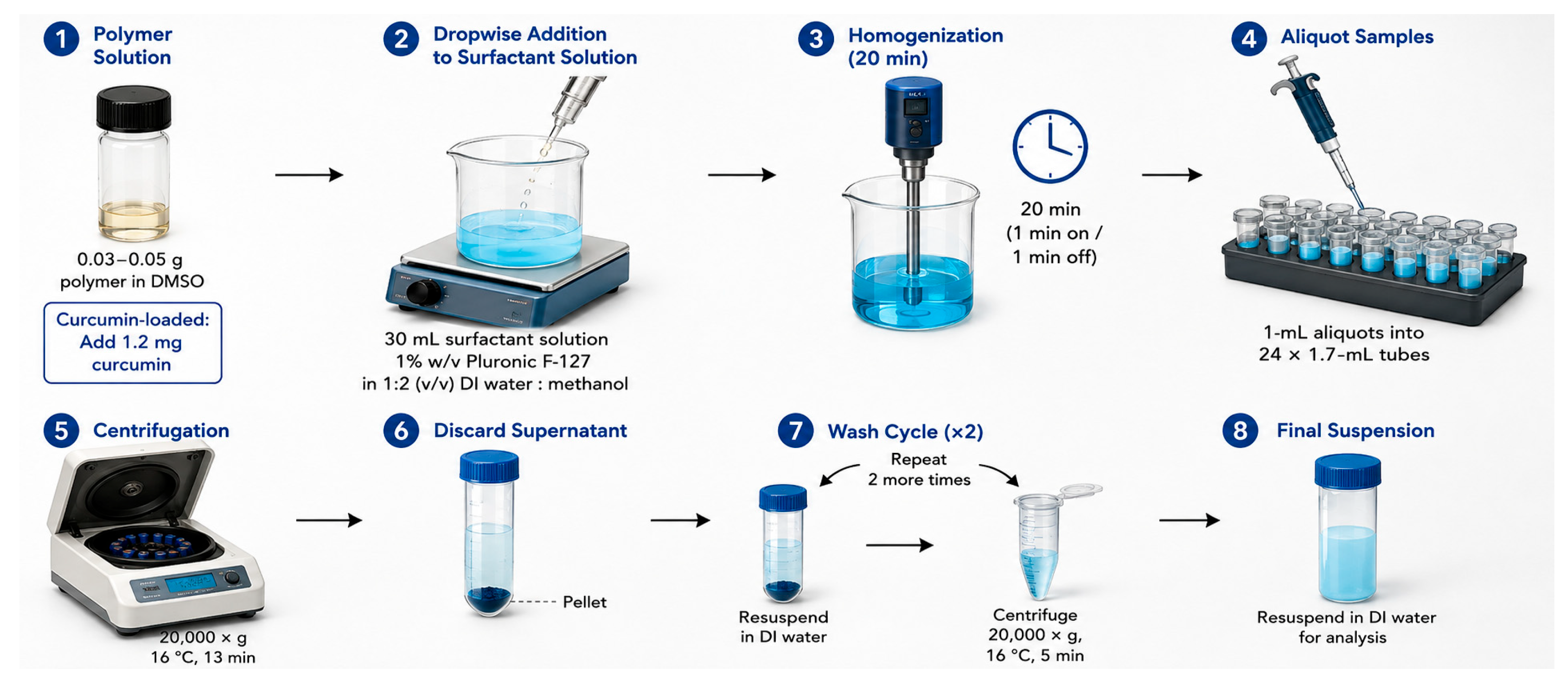
Figure 2.
Curcumin-loaded PXS nanoparticles increase sensitivity to curcumin in colorectal cancer cells. Effect of PXS nanoparticles at a 20 μg/mL curcumin-equivalent dose in HCT116 (A), HT29 (B), SW480 (C), and SW620 (D) cell lines on cell viability and cellular morphology. * p < 0.05, ** p < 0.01, *** p < 0.001.
Figure 2.
Curcumin-loaded PXS nanoparticles increase sensitivity to curcumin in colorectal cancer cells. Effect of PXS nanoparticles at a 20 μg/mL curcumin-equivalent dose in HCT116 (A), HT29 (B), SW480 (C), and SW620 (D) cell lines on cell viability and cellular morphology. * p < 0.05, ** p < 0.01, *** p < 0.001.
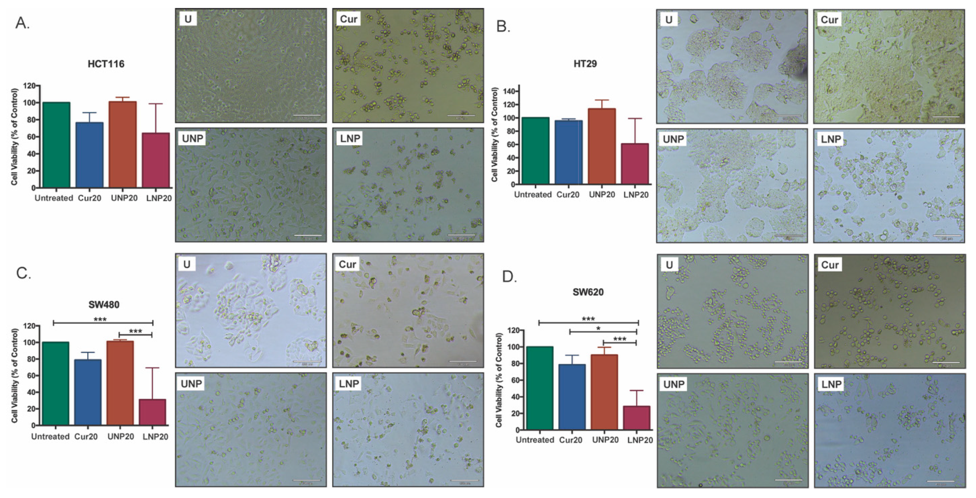
Figure 3.
Fluorescence imaging of curcumin association with HCT116, HT29, SW480, and SW620 cells following treatment with free curcumin (Cur) or curcumin-loaded PXS nanoparticles (LNP). Blue: DAPI (nuclei); green: curcumin. Scale bars, 20 μm. Images are representative of a single imaging session per cell line; no quantitative image analysis was performed (see Section 3.5).
Figure 3.
Fluorescence imaging of curcumin association with HCT116, HT29, SW480, and SW620 cells following treatment with free curcumin (Cur) or curcumin-loaded PXS nanoparticles (LNP). Blue: DAPI (nuclei); green: curcumin. Scale bars, 20 μm. Images are representative of a single imaging session per cell line; no quantitative image analysis was performed (see Section 3.5).
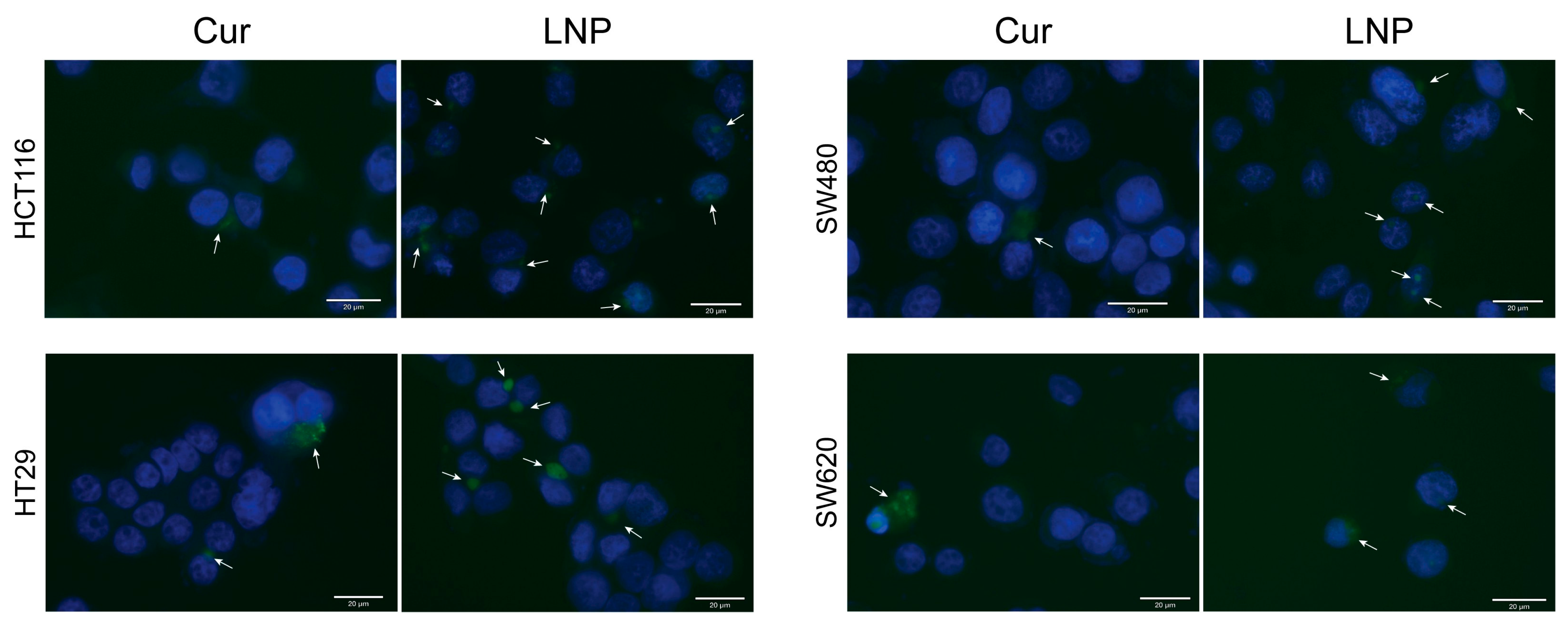

Table 1.
Dynamic light scattering (DLS) particle size analysis of unloaded (UNP) and curcumin-loaded (LNP) PXS nanoparticles, freshly prepared and following incubation at 37 °C.
Table 1.
Dynamic light scattering (DLS) particle size analysis of unloaded (UNP) and curcumin-loaded (LNP) PXS nanoparticles, freshly prepared and following incubation at 37 °C.
| Sample | Average Diameter (nm) |
|---|---|
| UNP (24 h, 25 °C) | 212.40 |
| LNP (24 h, 25 °C) | 171.04 |
| LNP incubated at 37 °C (24 h) | 200.3 |
| LNP incubated at 37 °C (48 h) | 162.10 |
| UNP incubated at 37 °C (24 h) | 202.50 |
| UNP incubated at 37 °C (48 h) | 159.35 |
Disclaimer/Publisher’s Note: The statements, opinions and data contained in all publications are solely those of the individual author(s) and contributor(s) and not of MDPI and/or the editor(s). MDPI and/or the editor(s) disclaim responsibility for any injury to people or property resulting from any ideas, methods, instructions or products referred to in the content. |
© 2026 by the authors. Licensee MDPI, Basel, Switzerland. This article is an open access article distributed under the terms and conditions of the Creative Commons Attribution (CC BY) license (http://creativecommons.org/licenses/by/4.0/).
Copyright: This open access article is published under a Creative Commons CC BY 4.0 license, which permit the free download, distribution, and reuse, provided that the author and preprint are cited in any reuse.