

Submitted:
28 August 2026
Posted:
31 August 2026
You are already at the latest version
Abstract
The use of antimicrobial peptides (AMPs) to combat Herpes Simplex Virus Type 1 (HSV-1) infection has surged amidst the rise of HSV-1 drug-resistant strains. In contrast to most first-line drugs that target DNA replication, AMPs offer an unconventional approach to combat HSV-1 infection by exerting their activity on multiple phases of the viral life cycle, while exhibiting low rates of resistance emergence. In this study, an integrated in silico and in vitro experimental approach was used to evaluate six proprietary Cationic lipo-oligo peptides (CLOPs) (P226, P359, P577, P581, OB1105, and OB1111) to identify candidates with potential antiviral activity against HSV-1. We hypothesized that CLOPs would be non-cytotoxic, exhibit high affinity for HSV-1 glycoproteins, and effectively inhibit HSV-1 growth in eukaryotic cells. Computational tools were employed to predict various pharmacokinetic parameters of CLOPs and to assess their molecular docking and binding affinities for HSV-1 glycoproteins. Next, in vitro cytotoxicity and viral plaque reduction assays were performed in eukaryotic cells to evaluate the cytotoxicity of CLOPs and their antiviral activity against HSV-1. Our findings showed that CLOPs conform to Pfizer’s rule and are predicted to exhibit antiviral activity against HSV-1, with OB1111 showing the most robust antiviral activity. All CLOPs were predicted to bind to major HSV-1 glycoproteins, with energies ranging from -7.5 to -3.2 Kcal/mol, suggesting potential inhibitory activity against HSV-1. Cytotoxicity and plaque reduction assays confirmed that CLOPs were non-toxic and inhibited HSV-1 plaque formation and viral titers in eukaryotic cells. Notably, CLOPs altered the morphology of plaques with visually smaller plaques than in the HSV-1 control. Among the tested CLOPs, OB1111 emerged as the most potent peptide, consistently demonstrating strong antiviral activity across both computational and experimental platforms. Of utmost significance, OB1111 exerted a significant antiviral effect at the earliest stage of infection in cells. Overall, the in silico and in vitro data demonstrate potent antiviral activity of CLOPs against HSV-1, suggesting their potential therapeutic relevance.
Keywords:
antimicrobial peptides
; herpes simplex virus- type 1
; glycoproteins
; ADMET
; molecular docking
; ligand
; viral plaque assay
; cytotoxicity
1. Introduction
Herpes simplex virus Type 1 (HSV-1) infection is highly prevalent worldwide, affecting approximately 67% of the global population under 50 years of age in 2016 [1,2]. HSV-1 is an enveloped, double-stranded DNA virus belonging to the Alphaherpesvirinae subfamily that can severely undermine human health by causing a spectrum of neurological diseases [3,4]. Its genome is ~152 kb long and comprises an envelope, a proteinaceous tegument, and a nucleocapsid that protects the viral genome. HSV-1 primarily infects mucosal epithelial cells and establishes lytic infection; after which the progeny virions travel along axons to infect sensory neurons in the trigeminal ganglia, thereby establishing persistent latent infections [5]. To date, HSV-1 infections have no cure, but antivirals like acyclovir (ACV) and its derivatives effectively manage the infection by reducing the rate of recurrent episodes, lesion formation, and their duration and severity. However, the reliance of these drugs on active viral replication limits their efficacy to lytic infections. To exacerbate this, mutations in viral thymidine kinase and DNA polymerase genes result in drug-resistant HSV-1 strains, which are commonly seen in immunocompromised hosts [4,6,7]. Hence, there is an urgent need for alternative treatments to control HSV-1 infections.
Drug development efforts to combat HSV-1 have primarily focused on lytic infection, with specific priorities given to the initial infection or cell-to-cell spread, which is considered the most promising target for novel antiviral development [8]. HSV-1 has numerous glycoproteins (g), giving it leverage over most enveloped viruses, which typically possess only one or two. Glycoproteins facilitate the entry of HSV-1 into a variety of host cell types through different routes, conferring it significant biological benefits [9]. Four major glycoproteins drive attachment, entry, and spread, respectively, the receptor binding mediator (gD), the heterodimer fusion regulators (gH/gL), and the viral fusogen (gB) [1]. There is considerable evidence for interactions among glycoproteins, suggesting that they are novel therapeutic targets in HSV-1 drug development. The benefits of targeting HSV-1 glycoproteins extend beyond expanding current antiviral options to combat infection at its initial stages [10]. A few examples include reports by Awasthi and colleagues (2008) who demonstrated that a gD mutant HSV-1 strain reduced cellular viral entry and minimized disease in mice [11]. Similarly, Li et al. (2020) demonstrated that Myricetin, a plant-derived flavonoid, interacts with gD and blocks HSV-1 adsorption and membrane fusion [12]. Another study showed that four peptides designed from HSV-1 gH regions inhibited HSV-1 entry, fusion, and infectivity by membrane insertion and aggregation [13]. Correspondingly, Caesappanin C was found to bind gB and inhibit entry, fusion, replication, and inflammatory response [14]. gBh1m, a cholesterol-tagged peptide derived from HSV-1 gB, aggregated and interacted with viral cell membranes, inhibiting viral entry. The presence of the cholesterol content and engineering sequences inhibited gB conformational rearrangements and enhanced gB binding affinity, respectively [15]. These studies exemplify the positive effects of limiting HSV-1 infection at the earliest stages by interfering with the function of essential glycoproteins.
Peptide-based therapeutics are emerging alternatives to small molecules that offer several advantages, including high specificity, affordable production rates, and ease of production and optimization [16]. Antimicrobial peptides (AMPs) can destabilize virions, obstruct attachment, entry, replication, and viral egress [17]. Numerous studies have demonstrated their ability to bind to HSV-1 glycoproteins [17,18,19,20,21]. While this is promising, HSV-1 infection remains incurable, and achieving complete viral suppression remains a challenge, warranting continued investigations of alternative peptide candidates.
In this study, we evaluated the potential of six Cationic lipo-oligopeptides (CLOPs) as alternative therapeutics against HSV-1 using an integrated in silico and in vitro experimental approach. We hypothesized that CLOPs would be non-cytotoxic, exhibit high affinity for HSV-1 glycoproteins, and effectively inhibit HSV-1 growth in eukaryotic cells. To test our hypothesis, we conducted comprehensive in silico modeling studies followed by in vitro cytotoxicity and anti-HSV1 experiments. We present our findings supporting the potential of CLOPs as being novel, safe, and effective alternative antivirals against HSV-1.
2. Materials and Methods
2.1. In Silico Peptide Selection and Preparation
The proprietary CLOPs used in this study were provided by Owen Biosciences, Inc. (Baton Rouge, LA). Six CLOPs (P226, P359, P577, P581, OB1105, and OB1111) were selected from a larger group of CLOPs for screening based on their computational antiviral predictions and structural characteristics deemed conducive to binding to enveloped viruses. They are amphipathic and contain highly cationic amino acid residues, particularly Lys and Arg, which favor electrostatic interactions with negatively charged viral membranes, capsids, and proteins, thereby disrupting virions [22]. The presence of the lipid moiety was also considered as an important criterion, as it is known to improve stability, half-life [23], specificity, permeability, and efficacy of AMPs [24]. The molecular structures of these CLOPs were optimized using specific computational tools as described below. The list of tools used in the in silico analysis is presented in Supplementary Table S1.
2.1.1. Lipinski Rule, Pfizer Rule, and Pharmacokinetics
The pharmacokinetic characteristics and drug-likeness score of CLOPs were obtained using SwissADME (https://www.swissadme.ch/) and AMDETlab 3.0 (https://admetlab3.scbdd.com/). These parameters were used as hallmarks to predict the bioavailability and drug-likeness of CLOPs. The Lipinski rule was used to assess the tendencies of CLOPs to conform to conventional drug evaluation guidelines. On the other hand, the Pfizer 3/75 rule was used to evaluate the potential risk of CLOPs-related toxicity. Taken together, the measured parameters included their molecular weight (MW), logP (log of the partition coefficient), number of hydrogen-bond donors (nHBD), number of hydrogen-bond acceptors (nHBA), and TPSA (topological polar surface area), among others. All physicochemical, drug-likeness, and ADMET calculations were performed on the complete CLOP chemical structures, including the N-terminal lipid moiety and C-terminal amidation; therefore, the reported values correspond to the intact lipo-oligopeptides rather than to the peptide backbone alone.
2.1.2. ADMET (Absorption, Distribution, Metabolism, Excretion, and Toxicity) Analysis
The ADMET properties of CLOPs were predicted using the software tool ADMET Predictor (http://admet.bioai-global.com/predict). Key parameters, including absorption potential, distribution volume, metabolic stability, excretion rates, and toxicity (including hepatotoxicity and cardiotoxicity), were evaluated.
2.1.3. Generation of CLOPs 2D Chemical Structures
The CLOPs under study are proprietary and thus are unavailable in public databases. To easily visualize and interpret the antiviral properties of CLOPs, their amino acid sequences were converted into 2D structures and canonical SMILES using DataWarrior (https://openmolecules.org/name2structure.html) and Open Parser for Systematic IUPAC Nomenclature (OPSIN) (https://www.ebi.ac.uk/opsin) software programs. DataWarrior, a data analysis and visualization program, was used to extract hidden knowledge from the CLOPs. For instance, this data filter was used to focus on specific data subsets or to make chemical intelligence work seamlessly with the program. It was used to filter data by various structural motives, view chemical structures from a chemistry aware perspective, and to predict the molecular properties of CLOPs. DataWarrior supports numerous file types and can merge data from sources or databases. The OPSIN software was used to interpret the International Union of Pure and Applied Chemistry (IUPAC) nomenclature for the structures of various lipopeptides. It converted these structures into three different chemical groups: CML (Chemical Markup Language), InChI (International Chemical Identifier), and SMILES (Simplified Molecular Input Line Entry System). Thus, each CLOP was run through these two programs to generate its 2D structure and canonical SMILES for downstream computational investigations.
2.1.4. Model Training Steps
The antiviral potency modeling workflow was implemented as a regression problem because the response variable was continuous IC₅₀/pIC₅₀. Hyperparameters for the support vector regression (SVR) and other regression models were optimized within the training data, and model performance was evaluated using correlation and error-based metrics appropriate for continuous outcomes. The final validation framework comprised ten-fold cross-validation, an independent external validation dataset, and leave-one-out cross-validation (LOOCV), as described in Section 2.1.9, Section 2.1.10, Section 2.1.11 and Section 2.1.12. Classification metrics such as accuracy, precision, recall, F1-score, ROC, and AUC were therefore not used for the final IC₅₀ prediction analysis.
2.1.5. Prediction of CLOPs Activity
For quantitative antiviral potency prediction, experimentally validated antiviral peptides with reported IC₅₀ values were used as the modeling dataset. After preprocessing and duplicate removal, sequence derived descriptors were generated and used to train regression algorithms for prediction of pIC₅₀/IC₅₀. The proprietary CLOPs were encoded using the same descriptor pipeline and submitted to the trained models. Support vector regression (SVR), Random Forest regression, XGBoost, Gradient Boosting, and Ridge regression were evaluated, with model performance assessed by PCC, Spearman correlation, R²/Q², RMSE, and MAE. Details of dataset composition, descriptor generation, model development, and validation are provided in Section 2.1.9, Section 2.1.10, Section 2.1.11 and Section 2.1.12.
2.1.6. Retrieval, Homology Modeling, and Preparation of HSV-1 Glycoprotein Structures
To conduct the in silico modeling analyses, the 3D structures of HSV-1 glycoproteins used for molecular docking were obtained from a combination of experimentally resolved structures deposited in the Protein Data Bank (PDB) and homology modeling approaches. A detailed search of the Protein Data Bank (PDB) was first conducted to identify available crystal or NMR structures of HSV-1 glycoproteins involved in viral attachment, membrane fusion, entry, and immune evasion. This was followed by the identification of experimentally resolved structures for glycoprotein B (gB; PDB ID: 3NW8), glycoprotein D (gD; PDB ID: 2C36), and glycoprotein H (gH; PDB ID: 2LQY). These were downloaded directly from PDB and used as receptor structures for the molecular docking studies. However, some experimentally determined structures were unavailable for several HSV-1 glycoproteins; thus, the amino acid sequences corresponding to glycoproteins gC, gE, gG, gI, gJ, gK, gL, gM, and gN were retrieved from the National Center for Biotechnology Information (NCBI) GenBank database using their respective accession numbers as indicated in Supplementary Table S2. The retrieved sequences were subjected to homology modeling using the Swiss-Model server (https://swissmodel.expasy.org). The 3D protein models were generated using the highest-ranking templates and evaluated using the Global Model Quality Estimation (GMQE) and QMEAN scoring metrics from the Swiss-Model platform (Supplementary Table S4). These highest quality models were selected for downstream molecular docking analyses. Following model selection, both experimentally resolved and homology modeled proteins underwent structural preparation and optimization. Crystallographic water molecules, co-crystallized ligands, ions, and other nonessential heteroatoms were removed using Discovery Studio Visualizer (Dassault Systèmes BIOVIA) and PyMOL. Hydrogen atoms were added, bond orders were verified, and protein structures were visually inspected for structural inconsistencies. Where necessary, structural refinement and energy minimization were performed to improve geometric stability and remove unfavorable steric interactions. The prepared protein structures were subsequently converted into docking-compatible formats, and Kollman charges and polar hydrogens were assigned prior to molecular docking simulations using AutoDock Tools (version 1.5.7).
2.1.7. Molecular Docking and Binding Studies
Molecular docking to identify possible interaction of CLOPs against HSV-1 glycoproteins essential viral attachment, entry, immune evasion, and spread was conducted. The final docking panel comprised gB, gC, gD, gE, gG, gH, gI, gJ, gK, and gL. The modeled 3D structures of the CLOPs were docked to these glycoproteins using PyRx/AutoDock Vina, with receptor and ligand preparation performed as described above [25,26]. Ligand structures were energy-minimized and converted to PDBQT format, while receptor preparation included addition of polar hydrogens and assignment of appropriate charges. Each receptor grid box was generated in PyRx using the Vina Wizard, and docking was performed with an exhaustiveness of 8. Nine poses were generated for each ligand-receptor pair, and the best-scoring pose was retained for residue interaction analysis in BIOVIA Discovery Studio. RMSD values reported in this study were calculated from the set of docking poses generated for each ligand-receptor pair and therefore describe docking-pose dispersion/consistency; they were not derived from a molecular dynamics trajectory and should not be interpreted as time-resolved structural stability.
2.1.8. Antiviral Peptide Prediction and Validation Workflow
Figure 1.
Overall workflow of the antiviral peptide IC₅₀ prediction and validation pipeline. The workflow consisted of dataset acquisition, preprocessing, duplicate sequence removal, conversion of experimental IC₅₀ values to pIC₅₀, extraction of 578 sequence-derived descriptors, machine learning model development, internal and external validation, and statistical performance evaluation. Sequence descriptors included amino acid composition (AAC), dipeptide composition (DPC), molecular weight, net charge, isoelectric point, aromaticity, GRAVY hydrophobicity, instability index, aliphatic index, Boman index, sequence entropy, and composition-transition-distribution (CTD) descriptors. Model performance was evaluated using Pearson correlation coefficient (PCC), Spearman correlation coefficient, coefficient of determination (R²/Q²), root mean square error (RMSE) and mean absolute error (MAE).
Figure 1.
Overall workflow of the antiviral peptide IC₅₀ prediction and validation pipeline. The workflow consisted of dataset acquisition, preprocessing, duplicate sequence removal, conversion of experimental IC₅₀ values to pIC₅₀, extraction of 578 sequence-derived descriptors, machine learning model development, internal and external validation, and statistical performance evaluation. Sequence descriptors included amino acid composition (AAC), dipeptide composition (DPC), molecular weight, net charge, isoelectric point, aromaticity, GRAVY hydrophobicity, instability index, aliphatic index, Boman index, sequence entropy, and composition-transition-distribution (CTD) descriptors. Model performance was evaluated using Pearson correlation coefficient (PCC), Spearman correlation coefficient, coefficient of determination (R²/Q²), root mean square error (RMSE) and mean absolute error (MAE).
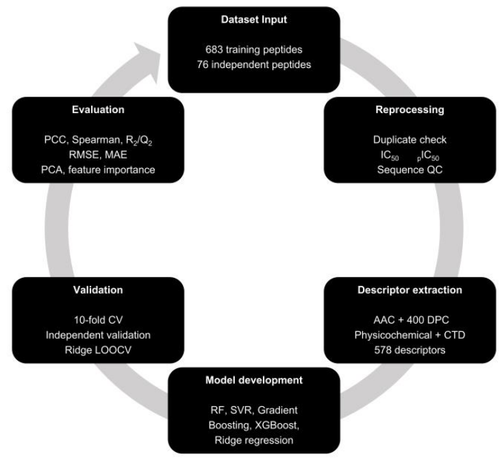
2.1.9. Dataset Collection and preprocessing
The antiviral peptide datasets used in this study were obtained from the AVP-IC50Pred dataset. The dataset comprised 683 peptides used for model development and an independent validation dataset of 76 peptides. To ensure data integrity, both datasets were merged during preprocessing and screened for duplicate peptide sequences. Duplicate entries were identified using identical peptide sequences and removed, resulting in a final dataset consisting of 683 unique antiviral peptides. No conflicting IC₅₀ values were detected among duplicate entries, indicating complete consistency within the dataset. Because IC₅₀ values span several orders of magnitude, experimental IC₅₀ values (μM) were transformed into pIC₅₀ values to normalize the response variable according to the formula:
Basic sequence statistics, including peptide length, amino acid composition, and pIC₅₀, as well as the data quality assessment details can be seen in Table 3A and 3B respectively.
2.1.10. Sequence Descriptor Generation
A comprehensive panel of sequence-derived descriptors was generated for each peptide to capture compositional and physicochemical characteristics relevant to antiviral activity. The descriptor set included: amino acid composition (AAC), dipeptide composition (400 descriptors), molecular weight, net charge, isoelectric point (pI), aromaticity, GRAVY hydrophobicity index, instability index, aliphatic index, Boman index, sequence entropy, Composition-Transition-Distribution (CTD) descriptors. Collectively, these features generated a total of 578 numerical descriptors for each peptide.
2.1.11. Machine Learning Model Development
Five regression algorithms were evaluated for antiviral activity prediction. These include Random Forest (RF), Support Vector Regression (SVR), Gradient Boosting Regression, Extreme Gradient Boosting (XGBoost), and Ridge Regression. Following this the model performance was assessed using three complementary validation strategies which included ten-fold cross-validation, independent external validation, and LOOCV. The LOOCV analysis was performed using Ridge regression because exhaustive LOOCV of nonlinear ensemble models would require repeated fitting of hundreds of computationally intensive models.
2.1.12. Model Evaluation
Model performance was assessed using Pearson correlation coefficient (PCC), Spearman’s rank correlation coefficient (ρ), coefficient of determination (R²), predictive squared correlation coefficient (Q²), root mean square error (RMSE), and mean absolute error (MAE). Detailed formulas and the statistical metrics used to evaluate the model’s performance can be found in Supplementary Table S3.
2.2. In Vitro Validation of CLOPs’ Antiviral Activity
2.2.1. Cell Line and CLOPs
Vero cells (African Green Monkey Kidney cells) were kindly provided by Dr. Francois Villinger (MD Anderson Cancer Center, Bastrop, TX, USA). HEp-2 cells (Human epithelial cells) (ATCC CCL-23) were obtained from the American Type Culture Collection (ATCC, Manassas, VA, USA). Cells were cultured in Eagle’s Minimal Essential Medium (EMEM) (ATCC) supplemented with 10% heat-inactivated Fetal Bovine Serum (FBS), and 1 μg/mL antibiotic and antimycotic (ThermoFisher Scientific (Waltham, MA, USA)). Both cell lines were maintained at 37°C in a humidified incubator with 5% CO2. Proprietary CLOPs were provided by Owen Biosciences Inc (see above), and acyclovir (ACV) was purchased from Sigma-Aldrich (St. Louis, MO, USA). CLOPs used in this study were 5-6 amino acids in length, synthesized by Owen Biosciences, Inc., and have a purity grade of >85% as determined by HPLC (unpublished data). Lyophilized CLOPs were dissolved in distilled water, aliquoted, and stored at -80 °C. Aliquots of 10,000 mg/mL were made and stored at -20 ºC until used.
2.2.2. Infectivity
HSV-1 strain F (ATCC #VR-733) was obtained from ATCC and propagated in Vero cells monolayers in EMEM supplemented with 2% FBS for 1 h (hour) to establish infection. The cells were shaken periodically to ensure optimal absorption. The infectious inoculum was removed from cells, followed by treatment with CLOPs, which were maintained at 37 °C in a humidified incubator with 5% CO2.
2.2.3. Cytotoxicity Studies
The cytotoxicity of CLOPs and ACV to HEp-2 and Vero cells was evaluated by the 3-(4,5-dimethyl-thiazol-2-yl)-2,5-diphenyl-tetrazoliumbromide (MTT) dye reduction assay using an MTT Cell Proliferation Assay kit (ATCC #30-1010K). Cells (5 104) were seeded into each well of a 96-well plate in 100 µL of EMEM for 24 h, and incubated at 37 °C in a humidified 5% CO2 atmosphere. The culture media were replaced with 100 µL of fresh media containing various concentrations of CLOPs and ACV (0.9 to 250 µg/mL), and incubated for 24 and 48 h. MTT dye solution (10 µL) was added to each well, and incubated for 1.5 and 1 h, respectively, for HEp-2 and Vero cell lines. The detergent reagent (100 µL) was added to each well to dissolve the formazan crystals, followed by incubation overnight at 37 °C in a humidified 5% CO2 atmosphere. The absorbance at 570 nm was measured using the Multiskan SkyHigh microplate reader (ThermoFisher Scientific), and cell viability (%) was assessed by comparing the absorbance of CLOPs-treated cells to that of untreated control cells.
2.2.4. Plaque Reduction Assay
The plaque reduction assay was performed using Vero cells, a model cell line for evaluating HSV-1 plaque formation [27]. Vero cells were seeded at a density of 1 106 cells/well in 6-well plates for 24 h. The medium was removed, and cells were infected with HSV-1 at a multiplicity of infection (MOI) of 0.001 for 1 h. The inoculum was removed and replaced with 1.2% carboxymethylcellulose (Sigma-Aldrich) alone or containing ACV or CLOPs, according to the experimental design, for 48 h, and the culture was maintained at 37 °C in a humidified incubator with 5% CO2. Cells were fixed with 4% paraformaldehyde (ThermoFisher Scientific) and stained with 1% crystal violet (ThermoFisher Scientific) for plaque visualization and enumeration.
2.2.5. Viral Attachment Assay
An assay was conducted to evaluate the effect of OB1111 on HSV-1 attachment to cells. Vero cells were seeded at a density of 1 × 10⁶ cells/well in 6-well plates for 24 h. Cells, HSV-1, and 2% FBS media were precooled at 4 °C for 30 min. Cells were infected and treated simultaneously with HSV-1 at a MOI of 0.001 and maintained at 4 °C for 1 h. The inoculum was removed, replaced with 2% FBS media, and cells were maintained at 37 °C in a humidified incubator with 5% CO2 for 1 h to permit entry. The media was removed and replaced with 1.2% carboxymethylcellulose and cultures were maintained at 37 °C in a humidified incubator with 5% CO2 for 48 h. Cells were fixed with 4% paraformaldehyde and stained with 1% crystal violet to visualize and enumerate plaques. Plaque inhibition percentages were determined using the formula: Viral inhibition (%) = [ 1- (number of plaques) inhibitor/ (number of plaques) control] 100 [28].
2.3. Statistical Analysis
Data from the molecular docking analysis, machine learning algorithms, and CLOPs/glycoproteins binding affinity calculations were generated and analyzed using Prism 10 (GraphPad Software, Inc., San Diego, CA, USA). Cell viability data were analyzed using two-way analysis of variance (ANOVA). HSV-1 virus titers resulting from the plaque reduction assay were analyzed using one-way ANOVA followed by Šidák multiple comparison test. Data from the viral attachment assay was analyzed using one-way ANOVA followed by Bartlett’s tests using Prism 10. P values < 0.001 were considered significant.
3. Results
3.1. CLOPs 2D Chemical Structure and Molecular Features
The amino acid sequences of CLOPs were used to generate their 2D chemical structures using the DataWarrior and OPSIN software (Figure 2). All CLOPs used in this study were lipidated to improve cellular uptake, delivery, and stability.
The molecular features of CLOPs, including their composition, net charges, structural features, and modifications, which influence their biological activity, are tabulated in Table 1. CLOPs are positively charged with values ranging from (+3) to (+5), suggesting they can readily bind to negatively charged membranes and cellular surfaces. P226, P359, P577, and P581 are pentapeptides with a net positive charge of (+3), which is mainly attributed to the presence of Lys residues. Likewise, OB1105 is a pentapeptide but differs from is preceeding counterparts in charge and amino acid composition. The (+4) net charge results from its Lys/Arg amino acid composition. OB1111 is a slightly more cationic hexapeptide (+5) that is rich in Lys and Arg. Cationic charge directly correlates with biological activity, where a higher charge results in enhanced activity, and vice versa [29]. On this premise, OB1111 and OB1105, with charges of (+5) and (+4), respectively, have a greater tendency to bind to membranes than the other CLOPs. A defining feature of all CLOPs used in this study is their amphipathic nature, encompassing their hydrophobic and hydrophilic domains. Amphipathicity is a crucial structural property of many antiviral peptides, enabling simultaneous interactions with aqueous environments and lipid-rich viral envelopes, thus facilitating binding to viral membranes, membrane-associated glycoproteins, and protein-binding pockets [30,31,32]. Such properties have been shown to enhance peptide-mediated disruption of viral attachment, membrane fusion, and entry processes [33].
The physicochemical properties and drug-likeness of CLOPs were assessed utilizing SwissADME, AMDETlab 3.0, and ADMET Predictor (Table 2). The Lipinski and Pfizer rules were used to determine the drug-likeness with respect to size, solubility, and permeability. The Lipinski Rule predicts bioavailability if a compound’s MW is ≤ 500, nHBA ≤ 10, nHBD ≤ 5, logP (logarithm of the partition coefficient), and TPSA (topological polar surface area) are ≤ 5 and ≤ 140 Ų, respectively [34]. Poor bioavailability is predicted if a compound does not adhere to at least four of these rules. All CLOPs MWs exceeded the Lipinski rule, with the lowest predicted MW being 781.15 Daltons (Da) and nHBA above acceptable limits. P226 and P359 numbers of hydrogen bond acceptors are within the permissible standard limits, indicating good probability for increased solubility. Additionally, the TPSA ranged from 235.59 to 441.44, suggesting that these peptides will be absorbed at low concentrations in the systemic circulation. Collectively, these findings suggest that all CLOPs, rather than being administered orally, may require other routes to fully realize their therapeutic potential.
On the contrary, all CLOPs were in conformity with the Pfizer 3/75 rule, which associates toxicity to compounds with logP > 3 and TPSA < 75 Ų [34]. Regarding absorption, the predictions for both parameters favor poor solubility and absorption. This, in addition to the MW, accounts for poor predictions of BBB (blood-brain barrier) penetration. The VD (Volume of Distribution) analysis in ADMETlab 3.0 indicates that all CLOP values are within the optimal range (0.04-20 L/kg), which is important and directly impacts dosing, safety, and efficacy. Notably, we observed that CLOPs are not inhibitors of the major liver enzymes CYP1A2 and CYP3A4, which are responsible for metabolizing many drugs [35], suggesting minimal potential of drug-drug interactions or co-administration issues with other drugs. The predicted excretion report also revealed that CLOPs had low plasma clearance rates and short half-lives, similar to what has been reported for ACV [36,37]. Although compounds of this nature may require frequent dosing, they generally have reduced toxicity and a lower tendency to accumulate in organs, making them safer for immunocompromised patients who suffer most from HSV-1-related complications. Lastly, the toxicity assessment showed that CLOPs are non-carcinogenic and do not induce kidney damage, thus widening their therapeutic potentials.
3.3. CLOPs Displayed HSV-1 Neutralization Capabilities and Structural Stability
Using an SVR learning algorithm, we obtained the predicted antiviral activities of CLOPs as shown in Figure 3A. Antiviral activity was determined using IC50 values, the predicted concentration at which CLOPs inhibit 50% of HSV-1 activity, with lower IC50 values suggesting higher potency. The IC50 values for P226, P359, P577, P581, and OB1105 predict moderate antiviral activity against HSV-1. OB1111 shows the greatest potential for therapeutic application among those tested, with an IC50 of 0.01 µM, strongly indicating high potency against HSV-1 and a low concentration to achieve significant antiviral effects.
To further validate the reliability of the antiviral prediction, the RF algorithm was employed to predict the antiviral activities of CLOPs. This model predicted higher IC50 values for P226, P359, and OB1105 (Figure 3B) than the SVR, indicating that CLOPs will exhibit weak antiviral activity. On the contrary, the potencies of P577 and P581 were predicted to be relatively higher, as indicated by their lower IC50 values of 39.67 and 20.95 µM, respectively, suggesting that lower concentrations are required to achieve the same antiviral effect, as predicted by the SVR. Interestingly, the OB1111 IC50 predictions from both models aligned, providing strong support for its potential as an effective anti-HSV-1 agent. The discrepancy between the two machine learning algorithms suggests that one model may consider certain structural or physicochemical features of CLOPs that the other does not, resulting in differences in predicted IC50 values.
Next, we evaluated docking-pose consistency using root-mean-square deviation (RMSD) values calculated across the nine poses generated for each CLOP-receptor docking run. As shown in Figure 3C, these RMSD distributions describe the degree of variation among predicted docking poses and do not represent molecular-dynamics trajectories. OB1105 exhibited the lowest RMSD variability, indicating a comparatively consistent predicted binding orientation, whereas P226 showed the greatest pose dispersion. OB1111 showed lower docking-pose variability than P581, P577, and P226. Accordingly, these data are interpreted as differences in docking-pose consistency rather than as direct evidence of dynamic or time-resolved complex stability.
3.4. Dataset Quality Assessment
The predictive performance of the machine learning algorithms (SVR and RF) was assessed to determine their accuracy. Data preprocessing was carried out and 683 duplicate peptide entries were removed from the merged dataset, yielding 683 unique antiviral peptide sequences. No conflicting IC₅₀ measurements were identified among duplicate sequences, confirming excellent dataset consistency. The peptides ranged from 8 to 38 amino acids in length, with an average length of 22.07 amino acids and an average pIC₅₀ of 5.471 (Table 3). Sequence characterization generated 578 numerical descriptors, including amino acid composition, dipeptide composition, physicochemical properties, and CTD descriptors. Importantly, no peptide sequence overlap was detected between the training and independent validation datasets, demonstrating the independence of the external validation set.

Table 3.
Summary statistics of the (A) antiviral peptide dataset and (B) data quality assessment following preprocessing.
Table 3.
Summary statistics of the (A) antiviral peptide dataset and (B) data quality assessment following preprocessing.
| (A) | |
| Metric | Result |
| Total records | 1,366 |
| Unique peptide sequences | 683 |
| Duplicate sequences removed | 683 |
| Conflicting duplicate IC<sub>50</sub> values | 0 |
| Mean peptide length | 22.07 aa |
| Median peptide length | 20 aa |
| Minimum peptide length | 8 aa |
| Maximum peptide length | 38 aa |
| Mean IC₅₀ | 37.334 μM |
| Mean pIC₅₀ | 5.471 |
| Abbreviations: aa: amino acid residues. | |
| (B) | |
| Check | Result |
| Training peptides | 683 |
| Independent validation peptides | 76 |
| Shared sequences | 0 |
| Conflicting duplicates | 0 |
| Total descriptors | 578 |
3.5. Ten-Fold Cross-Validation Performance
The predictive performance of four machine learning algorithms was first evaluated using ten-fold cross-validation (Table 4). Among the evaluated models, Random Forest exhibited the strongest internal predictive performance, achieving a PCC of 0.751, Spearman correlation of 0.690, R²/Q² of 0.561, RMSE of 0.883, and MAE of 0.672. SVR demonstrated comparable performance (PCC = 0.743), whereas Gradient Boosting showed the lowest predictive accuracy.
3.6. Independent External Validation
Model generalizability was subsequently assessed using an independent dataset of 76 antiviral peptides. In contrast to the internal validation results, SVR demonstrated the highest predictive performance, achieving a PCC of 0.692, Spearman correlation of 0.572, RMSE of 0.839, and MAE of 0.651 (Table 5). Although Random Forest produced the highest cross-validation performance, its external PCC decreased to 0.625, suggesting a moderate degree of model overfitting. Overall, SVR exhibited the greatest robustness and generalization capability.
3.7. Leave-One-Out Cross-Validation
To further evaluate model robustness, LOOCV was performed using Ridge regression with the identical descriptor-processing pipeline. Ridge regression achieved a PCC of 0.671, Spearman correlation of 0.591, R²/Q² of 0.434, RMSE of 1.002, and MAE of 0.766 (Table 6). Exhaustive LOOCV was not performed for the nonlinear ensemble models because it would require repeated fitting of 683 independent models, making the computation prohibitively expensive. Therefore, shuffled ten-fold cross-validation and independent external validation were considered more appropriate for evaluating ensemble learning algorithms. Additional validation details can be found in Supplementary Figures S3–S8 and Supplementary Table 3.
3.8. Molecular Docking Against HSV-1 Glycoproteins
Docking studies were conducted to determine the binding affinities of CLOPs to several HSV-1 glycoproteins (gB, gC, gD, gE, gG,gH, gI, gJ, gK, gL) utilizing AutoDock wizard (with multiple search algorithms) to predict binding affinities, coupled with AutoDock Tools (ADT) for efficient data analysis [26]. A total of 9 poses for each CLOP against a glycoprotein were predicted, and the best CLOP/glycoprotein complex with the highest relative free binding energy was selected for analysis. Our results, as shown in Figure 4, revealed that all CLOPs bind to all tested glycoproteins, with docking scores ranging from -7.5 to -3.2 Kcal/mol. P226 preferentially binds more potently to gB (-7.5), gL (-6.0), gI (-5.9), and gD (-5.8). P359 most potent binding affinities were seen during its interaction with gB (-6.5), gD (-5.8), gI (-5.7), and gL (-5.5). P577 binds strongly with most of the glycoproteins; however, the highest affinities were observed in gB (-6,9), gI (-6.3), gL (-6.3), and gD (-5.9). P581, like P226, binds more potently to gB (-6.9), gL (-6.5), gI (-6.1), and gD (-6.0). OB1105 highest binding affinities were with gL (-7.0), gB (-6.7), gD (-6.3), and gI (-6.1). Lastly, OB1111 exhibits its highest docking scores within the gL (-7.1), gI (-6.4), gB (-6.3), and gD (-6.3) docking complexes. Overall, our results show favorable CLOPs/glycoprotein interactions, with preferential binding to gB, gD, gI, and gL, which mediate attachment, fusion, and cell-to-cell spread, suggesting that they may be more effective at interfering with the early stages of infection. Docking energies ranging from -3 to -4 kcal/mol range were interpreted as weak-to-modest predicted interactions rather than “strong” binding; docking scores were used comparatively within this computational screen and were not treated as experimental binding free energies.
CLOPs were subjected to further docking analysis to assess the binding orientation and residue interactions at the docking complex, as presented in the 2D and 3D structural models. HSV-1 gB and gD, both of which are important drivers of HSV-1 attachment and entry, were the preferred selected receptors. We focused our analysis on CLOPs that exhibited the strongest binding affinity to gB (P226 and P577) and gD (OB1105 and OB1111). Our results show the involvement of key interactions, such as alkyl interactions and individually weak interactions, that exert a substantial effect when acting collectively, typically stabilizing molecular structures [38]. Hydrogen bonds, which contribute to the overall stability of the docked complex [39], and electrostatic interactions, the type of complex interaction exerted by charged ligands that aids with repulsion and attraction [40] were also observed at the docking complex.
Specifically, we show in Figure 5 that P226 interacts with gB via alkyl interactions with Tyr296, Tyr314, Leu714, Pro348, Lys349, a carbon-hydrogen bond with Glu187, and conventional hydrogen bonds with Asp713, Glu187, Ile185, Val170, Met183, Gln172. When P226 was docked to gD, we observed alkyl interactions with Leu203, Leu220, Ala201, and Ile217, as well as conventional hydrogen bonds with Tyr137, Ser140, and Asp139.
P577/gB interaction involves alkyl interactions with Tyr380, Tyr142, Leu376, conventional hydrogen bonds with Leu678, Gln140, Arg515, Asn511, and Ser387, and unfavorable donor-donor bonds with Gly139. When docked to gD, we observed alkyl interactions with Leu220, Leu203, Tyr137, Ala207, Val212, and Ile296, and conventional hydrogen bonds with Asp139, Ser200, Ser204, Gln210, and Gly211 (Figure 6).
The 2D and 3D pose of the OB1105 interaction with gB shows alkyl interactions with key amino acid residues, including Val553, Arg558, Ala549, and conventional hydrogen bonds with Asn533, Asn620, Arg661, Tyr647, and Asp566, as well as an unfavorable donor-donor bond with Phe127. Alkyl interactions with Arg196, Pro194, Pro288, Ala201, and Ile217, conventional hydrogen bonds with Arg196, Arg222, Tyr137, and Asn136, as well as an unfavorable donor-donor bond with Ser200 were observed in OB1105 interaction with gD (Figure 7).
As depicted in Figure 8, OB1111 interacts with gB via multiple essential amino acid residues. Key amongst them includes an attractive charge with Glu619, Glu622, and Glu530, an alkyl interaction with Arg558, a carbon-hydrogen bond with Ser552, and conventional hydrogen bonds with Asn546, Val553, Pro130, Asn540, Asn533, and Arg131. On the contrary, the unfavorable donor-donor bonds observed with Thr537 and His534 residues indicate possible electrostatic repulsion and a reduction in stability of the residues. We expanded our evaluation to include OB1111 interaction with gD and found an attractive charge with Asp139, an alkyl interaction with Arg196, a carbon-hydrogen bond with Asn136, and conventional hydrogen bonds with Pro288 and Gly218. Notably, an unfavorable donor-donor bond was observed with Arg222.
To assess whether the strength of the binding affinity reflects the number of favorable interactions with amino acid residues, we evaluated the remaining CLOPs, P359 and P581. As observed in Supplementary Figure S1, P359/gB interaction involves alkyl interactions with ARG691, His692, Lys695, Ile694, conventional hydrogen bonds with Asp701, Thr703, Leu700, Glu704, and unfavorable donor-donor bonds with Tyr702. Contrastingly, when P359 interacted with gD, there were alkyl interactions with Pro198, Tyr137, Arg196, Ala201, Ile217, and Ile290, conventional hydrogen bonds with Asp139, Ser200, Ser140, and Arg222, and a carbon-hydrogen bond with Gly218. We observed alkyl interactions with Leu714, Ile719, Pro348, Lys349, a carbon-hydrogen bond with Asp713, and conventional hydrogen bonds with Glu187, Phe186, Met183 in P581/gB interaction. We also observed alkyl interactions with Pro288 and Pro198, along with the conventional hydrogen bonds with Asp139, Tyr137, Pro194, Arg196, Gly218, and Ser140 when P581 interacts with gD (Supplementary Figure S2).
We next correlated binding affinities (Figure 4) with the number of interactions and interacting amino acid residues at the CLOP/glycoprotein docking complexes (Figure 4, Figure 5, Figure 6 and Figure 7 and Supplementary Figures S1 and S2) and found that CLOPs with fewer interactions and interacting amino acid residues did not necessarily have the lowest binding affinities and vice versa. Collectively, the findings from the docking score suggest that each CLOP docked to each glycoprotein is dependent on its amino acid sequence, as well as the quality, position, and strength of the interactions.
3.9. In Vitro Assessment of CLOPs Cytotoxicity
Assessing the cytotoxicity of therapeutics is critical to ensure their safety in a host. Thus, the cytotoxicity of CLOPs was evaluated using HEp-2 and Vero cells (Figure 9A-9D) to determine their effect on the viability of distinct epithelial cells. Cells were incubated with increasing concentrations of CLOPs or ACV (0.9-250 µg/mL) and subjected to the MTT assay for 24 and 48 h. As shown in Figure 9A, HEp-2 cells retained ≥ 90% viability after 24 h of treatment with ACV, P226, P359, P577, OB1105, and OB1111 at concentrations below 250 µg/mL. P581 exerted a concentration-dependent effect on the cells’ viability. Cell viability, which was ~90% at the lowest concentration (0.9 µg/mL), progressively declined to ~60% at the highest concentrations (62.5-250 µg/mL). At the 48-hour time-point, slight decreases in viability were observed with P226, P359, and OB1105 treatments, whereas cell viability remained consistent after ACV, P577, and P581 treatments. Notably, the viability of HEp-2 cells was enhanced after 48 h of exposure to OB1111 (Figure 9B). Vero cells retained ≥ 90% viability after 24 h of treatment with P226, P359, P577, and OB1111 at concentrations below 250 µg/mL. Cells maintained a consistent viability of ≥ 80% at all tested concentrations after 24 h of exposure to ACV and P581. Similar cell viability was observed at concentrations (0.9 – 62.5 µg/mL) after OB1105 treatment, but progressively declined to ~20% at 250 µg/mL (Figure 9C). Notably, at concentrations below 250 µg/mL, cell viability was slightly decreased after 48 h of exposure to ACV but increased after 48 h of exposure to CLOPs (Figure 9D).
3.10. CLOPs Displayed Anti-HSV-1 Activity
The in silico modeling predictions of CLOPs possessing antiviral activity warranted testing their anti-HSV-1 effects using the plaque reduction assay. The primary objective of this study was to screen CLOPs at a single concentration to identify the most promising candidate for subsequent studies. Optimization studies were first conducted to select the optimal MOI, CLOPs concentration, and time point for plaque formation. Vero cells were infected with HSV-1 (MOI of 0.001) and treated with 25 μg/mL of each CLOP. Plaque formation was visualized and enumerated at 48 h post-infection. As expected, the HSV-1 positive control had the highest number of plaques (Figure 10A). The positive control, ACV, almost completely inhibited plaque formation, resulting in 99% inhibition. When infected cells were exposed to P581, plaque size remained unaltered and was similar to that of the untreated HSV-1 control; however, plaque counts were decreased. CLOPs P226, OB1105, P577, P359, and OB1111 inhibited plaque formation, and plaque sizes were visually smaller than those formed in the untreated HSV-1 control. Ranked in ascending order, CLOP P581, P226, OB1105, P577, and P359 reduced HSV-1 plaque counts, resulting in approximately 19%, 20%, 24%, 46%, and 54% reductions in viral titer, respectively, compared to the untreated HSV-1 control (Figure 10B). OB1111 showed the greatest plaque inhibition rate, resulting in ~60% reduction in viral titer compared with the untreated HSV-1 control.
Because this experiment was designed as a single concentration screening assay, it was not used to experimentally determine CLOP IC₅₀ values or to validate the absolute IC₅₀ rankings predicted computationally in Figure 3A and B. Instead, it served to prioritize a lead candidate for attachment studies and subsequent mechanistic studies. The plaque reduction assay provided qualitative support for the computational prioritization of antiviral candidates, particularly the selection of OB1111; however, the in vitro rank did not completetly match the model-derived IC₅₀ ranking. Therefore, this assay data should not be interpreted as direct experimental validation of predicted IC₅₀ values. The differences likely reflect both model limitations and the complexity and dynamic nature of biological interactions in vitro, and full concentration response measurements for all CLOPs will be required for direct quantitative validation.
OB1111 was selected as our lead candidate with the highest therapeutic value. This decision was based on its ability to demonstrate the strongest inhibitory effect on HSV-1 plaque formation (Figure 10A and B). Coupled with its potent antiviral prediction by the SVR and RF machine learning algorithm (Figure 3A and B) and its ability to exhibit the strongest predicted interactions against seven out of the ten HSV-1 glycoproteins evaluated, with the exception of gB, gE, and gH (Figure 4). The predicted interaction of OB1111 and essential HSV-1 entry glycoproteins prompted further investigation of its activity at the early stage of infection. Thus, to begin to understand its mechanistic action, we conducted a viral attachment assay. The initial steps of this experiment were performed at 4 °C, a condition that favors attachment but not cell entry. The results show that the effect of OB1111 in this scenario was dose-dependent, with the highest percentage of HSV-1 inhibition observed at the highest concentrations (Figure 11A and B), providing evidence of possible interference with HSV-1 attachment to host cells. This assay evaluates plaque-forming infectivity after synchronized low-temperature attachment followed by temperature shift to permit entry; it does not directly demonstrate physical binding of OB1111 to a specific glycoprotein. Thus, the result is interpreted as functional evidence that OB1111 interferes with an early stage of HSV-1 infection, consistent with, but not proving the docking-based mechanism.
4. Discussion
HSV-1, a well-understood human pathogen, remains incurable, constantly infecting new hosts. It relies on glycoproteins for entry into host cells, and for other essential processes that facilitate its propagation and persistence in humans [9]. This makes them attractive targets for the development of novel alternative therapeutics. AMPs have emerged as promising alternative therapeutics for treating HSV-1 infections, and several have demonstrated activity against HSV-1 glycoproteins [8,18,41,42]. In our study, we employed in silico computational and in vitro experimental approaches to assess the antiviral effects of six novel CLOPs against HSV-1. To accomplish this, we used the DataWarrior & OPSIN software to generate the 2D structures of CLOPs. CLOPs used in this study are cationic, primarily due to the presence of highly cationic amino acid residues, particularly Lys and Arg, which promote electrostatic interactions with negatively charged viral membranes, capsids, and proteins, leading to pore formation, reduced infectivity, and possibly virion inactivation/destabilization. CLOPs are amphipathic, comprising a N-terminal lipid moiety, which is strongly hydrophobic, and a C-terminal amidation, which confers its hydrophilicity (Table 1). Hydrophobic residues are integral to AMPs’ specificity and membrane insertion [43]. The hydrophilic nature of CLOPs is essential for maintaining their solubility in aqueous solution.
In silico ADMET and drug-likeness evaluations provide pharmacology and toxicology understanding of compounds, ultimately reducing the costs, time, and failure rates associated with drug development. The Lipinski’s rule, an infamous rule used for solubility and bioavailability [44] was a determinant of CLOPs’ drug-likeness. We observed that CLOPs violated at least three of the five criteria in this rule, indicating poor oral bioavailability. The majority of orally available peptides have a MW of 500-1200 Da, exceeding the limits for membrane permeability set by the Lipinski rule [45]. While this is not promising, it is worth noting that the criteria associated with this rule were originally designed for small-molecule drugs, not for peptides or natural or semisynthetic natural product drugs. A notable fraction of orally administered FDA-approved small-molecule drugs violate at least one criterion of this rule [46]. Roskoski et al (2023) successfully outlined Dabrafenib, along with 29 other FDA-approved drugs that violate Lipinski’s rule despite maintaining relevant clinical bioavailability [47]. More practically, the MW of Rybelsus®, an orally administered FDA-approved peptide-based drug used to treat type 2 diabetes, exceeds the standards of the Lipinski rule by ~ 3,613.58 Da [48,49]. This means that while this rule sets the foundational framework for initial drug screening, its violation does not always indicate poor bioavailability or clinical relevance. When these rules are violated and bioavailability is compromised, alternative routes of administration could be explored for compounds in this category. Topical application is an ideal approach because it delivers the treatment directly to the infection site, resulting in rapid therapeutic action [50]. This approach is particularly relevant for infections such as HSV-1, which causes multifocal mucosal lesions [51]. They are most effective at minimizing lesion duration and discomfort when used at the onset of the infection [50] and are typically formulated as topical applications [52], providing individuals with the flexibility of selecting a formulation most suitable for their skin type. An evident display of the therapeutic potential of peptide-based topicals can be seen in a study by Jose et al (2013), which demonstrates peptide TAT-Cd0 significantly reduced HSV-1 viral titers when administered shortly after infection [53]. Topical drug delivery offers a convenient route that bypasses the gastrointestinal tract and minimizes possible adverse reactions, avoiding factors like hepatic first-pass effect and gastrointestinal irritation, which are linked to poor oral absorption of drugs [54,55,56]. This approach provides a means for CLOPs and other compounds that violate the Lipinski rule to overcome the major constraints associated with oral drug delivery.
The Pfizer 3/75 rule was the second key determinant used for drug-likeness rating. All CLOPs complied with this rule, indicating favorable toxicity predictions, as validated by the MTT cytotoxicity assay (Figure 9). CLOPs ADMET profiling displays favorable predictions for parameters such as volume of distribution, metabolism, and toxicity. Absorption, BBB permeability, and excretion predictions were poor (Table 2B). However, counteractive techniques such as transformative oral delivery, the use of absorption and permeation enhancers, exploration of alternative administration routes, and chemical modification of peptides using molecular techniques can address these limitations [57,58].
An interesting finding in the computational docking study was that all tested CLOPs were able to bind to essential HSV-1 glycoproteins (Figure 4), implying multitarget interference with the HSV-1 life cycle. CLOPs had the highest binding affinities for gD, gB, gL, and gI, indicating potential respective impact on virus entry, fusion, and spread. A closer evaluation of the CLOP/glycoprotein complexes revealed the involvement of several key amino acids that may contribute to the high binding scores observed (Figure 4). We observed that the docking score was not dependent on the number of intermolecular interactions formed between CLOPs and glycoproteins in the docking complex; rather, these scores might have been determined by the quality, orientation, and strength of their interactions. For example, OB1111 had a lower affinity to gB than P359 (Figure 4A), despite its interaction with more amino acid residues than P359 (Figure 8A and Supplementary Figure S1A). Another scenario involves the interaction of OB1105 and OB1111 with gD, in which both had the same docking scores (Figure 4C) despite fewer interactions and amino acid residues in the OB1105/gD interaction than in the OB1111/gD interaction (Figure 7C and 8C). Collectively, attractive charge, alkyl interactions, carbon-hydrogen bonds, and conventional hydrogen bonds are favorable interactions that enhance CLOPs’ stability and specificity for HSV-1 glycoproteins. On the contrary, the unfavorable donor-donor bonds observed at the docking complex with some CLOPs indicate possible electrostatic repulsion and a reduction in stability of these residues. Importantly, docking scores are model-dependent relative estimates and do not establish biochemical binding or inhibition. Values near -3 to -4 kcal/mol were treated as weak-to-modest predicted interactions; mechanistic confirmation will require direct binding and functional entry/fusion assays.
Our cytotoxicity results in HEp-2 and Vero cells after 24 h of CLOPs exposure show that none of the CLOPs were cytotoxic at concentrations below 250 µg/mL. Interestingly, we observed enhanced cell viability after 48 hours of CLOPs treatment, demonstrating that they are well tolerated by cells and have potential for broad application. Most of these CLOPs had better safety profiles than ACV, positioning them as potential safer alternative treatment options. This is promising and may prove beneficial for long-term drug development, especially regarding safety, dosing concentration, and side effects.
In the present study, all CLOPs were found to inhibit HSV-1 infection, with a range of IC50 values across two independent machine learning algorithms, as shown in Figure 3A and B. Because variability was observed in the ranking of CLOPs’ antiviral efficacy between the SVR and RF machine learning algorithms, model validation was conducted to evaluated the predictive performance and robustness of the models. Both machine learning algorithms demonstrated a moderate-to-strong relationship between peptide sequence descriptors and antiviral activity. RF achieved the highest internal predictive performance during cross-validation, whereas SVR exhibited superior generalization on the independent dataset. The reduction in RF performance from cross-validation (PCC = 0.751) (Table 4) to external validation (PCC = 0.625) (Table 5) suggests a modest degree of overfitting, while the relatively stable performance of SVR (PCC = 0.743 vs. 0.692) indicates greater robustness for predicting antiviral peptide activity as shown in Table 4 and Table 5. These findings demonstrate that sequence-derived descriptors provide substantial predictive information for peptide antiviral potency and support the use of SVR as the preferred model for external prediction. The RF model offers nonlinear feature interaction capture and strong internal fit, whereas SVR showed better external generalization in this dataset. Nevertheless, the moderate external correlations and prediction errors indicate that these models should be viewed as prioritization tools rather than substitutes for experimentally determined dose–response IC₅₀ values.
In vitro studies demonstrated CLOPs ability to reduce the virus titers (~19%-60%) and alter HSV-1 morphology (Figure 10A and 10B). Consistent with the predictions of the SVR and RF in silico machine learning algorithms, CLOP OB1111 displayed the highest antiviral potency against HSV-1, reducing plaque counts by ~60%. Moreso, it has one of the best safety profiles and maintained consistent cell viability in HEp-2 and Vero cells over time. A deeper analysis of OB1111’s effect on viral attachment resulted in a significant reduction in HSV-1 infectivity. Other studies have evaluated peptides as alternatives to current HSV-1 drugs, such as temporin B, which exhibited in vitro antiviral activity against HSV-1, possibly by impairing gB expression, which may restrict viral spreading [59]. Another study by Zaczyńska et al. (2022) assessed the antiviral properties of the 4B8M peptide in HSV-1-infected A-549 cells, which markedly reduced the HSV-1 virus titer. Similar to our results, 4B8M demonstrated lower cytotoxicity than cidofovir and ACV, although these drugs displayed higher potency against the virus [60]. Two other peptides, MEL-AM and MEL-AF, had an inhibitory effect on HSV-1 viral attachment of less than 50%. Both peptides had no effect on HSV-1 plaque size, despite suppressing cytopathic effects, as evidenced by reduced cell rounding, aggregation, and nuclear enlargement. The lack of effect on plaque size with these peptides, as observed in five of the six CLOPs we tested in our study, might be due to the peptides’ effect on viral spread, and their presence or absence may indicate differences in the mechanistic action of peptides [61].
Contrary to our results, LL-37, a human cathelicidin peptide, failed to block HSV-1 activity in infected human corneal epithelial cells after post-infection administration [62]. Another peptide, Eval418, displayed weak antiviral activity against HSV-1 following pretreatment and post-treatment applications, and strong antiviral activity when administered during the attachment phase [63]. Similarly, TL and TL1 exerted negligible inhibitory effects on HSV-1 in pre-treatment and post-treatment assays, but significant inhibitory effects when administered prior to or during infection, suggesting a tendency for prophylactic use [17]. This provides evidence for AMP’s ability to target multiple stages of the HSV-1 life cycle. While we acknowledge that direct comparisons cannot be made because we did not evaluate the antiviral activity of CLOPs using the time–of–addition assay, our findings suggest that CLOPs may function as an effective therapeutic agent. A major distinction between our CLOPs and those mentioned in this paper is the length of their amino acid sequences; ours are short, consisting of 5-6 amino acids. This may be an added advantage, as smaller peptides can more readily cross the membrane and exhibit higher bioavailability than their larger counterparts [64]. Additionally, our CLOPs displayed a higher safety threshold before inducing cytotoxicity at higher concentrations, indicating greater cellular tolerance. For example, the cytotoxicity of MEL-AM and MEL-AF was evaluated at concentrations of 2.5 to 20 µg/mL in RAW 264.7 cells, with a significant reduction in cell viability at 20 µg/mL observed after 24 h. Contrastingly, the cytotoxicity of CLOPs in our study was evaluated at concentrations (0.9 to 250 µg/mL) in Vero and HEp-2 cells, and a significant reduction in cell viability was observed at 250 µg/mL following 24 hours of CLOP exposure.
Host-defense peptides constitute a crucial component of the innate immune response against viral pathogens, including herpes simplex viruses. These naturally occurring host defense peptides include, among others, α-defensins and β-defensins, which have been reported to exhibit antiviral activity against HSV-1 and HSV-2 through numerous mechanisms, including direct viral neutralization, inhibition of viral attachment and entry, disruption of envelope integrity, and modulation of host immune responses [65]. The human neutrophil peptides (HNP-1, HNP-2, and HNP-3), which are members of the α-defensin family, reportedly bind viral particles and reduce HSV infectivity by interfering with early stages of infection. Similarly, human β-defensins contribute to antiviral defense at epithelial surfaces and have been implicated in restricting herpesvirus replication [66,67,68]. In addition to defensins, whey acidic protein domain-containing proteins, including secretory leukocyte protease inhibitor and elafin (peptidase inhibitor 3), have demonstrated antiviral activities against several enveloped viruses through mechanisms involving inhibition of viral entry, interference with virus-host interactions, and modulation of inflammatory responses [69]. These naturally occurring host-defense molecules further illustrate the therapeutic potential of cationic and amphipathic peptides as antiviral agents. In our study, CLOPs share numerous key properties with endogenous antiviral peptides, including a net positive charge, amphipathic structure, and predicted membrane-interacting properties. Such physicochemical features are known to facilitate electrostatic interactions with viral envelopes and glycoproteins, thereby contributing to antiviral activity [31,70,71]. CLOPs can be rationally engineered to optimize antiviral capacity, target specificity, stability, and pharmacological properties. Our lead peptide, OB1111, demonstrated a high positive charge (+5), favorable cell-penetrating peptide characteristics, strong docking interactions with HSV-1 glycoproteins, and significant in vitro antiviral activity. Such interesting revelations indicate that synthetic CLOPs may represent a promising class of next-generation antiviral agents that emulate the protective functions of endogenous host-defense peptides while potentially overcoming limitations related to proteolytic instability, manufacturing complexity, and limited pharmacokinetic optimization often associated with natural peptides [70,71]. We further believe that, because resistance to conventional nucleoside analogs such as ACV and related anti-herpetic drugs has been increasingly reported, particularly among immunocompromised patients, alternative therapeutic approaches targeting viral attachment, fusion, or entry should be given considerable attention, as reported in previous publications [72,73]. In this context, the next phase of our study will involve a detailed mechanistic investigation to understand the mode of action of OB1111.
5. Conclusions
In this study, we employed an integrated in silico and in vitro experimental approach to evaluate six candidate CLOPs as potential alternative therapeutics for HSV-1. Our results demonstrate the consistent antiviral properties of CLOPs, both computationally and experimentally. Computational analyses revealed strong binding affinities between CLOPs and major viral glycoproteins, supported by stable interaction profiles, favorable molecular properties, and high predicted antiviral potency. Most CLOPs bind favorably to HSV-1 glycoproteins and display good safety profiles, highlighting their potential therapeutic advantage. Plaque-reduction assays revealed significant inhibition of viral infectivity and titers, evidenced by a major reduction in plaque counts and sizes by CLOPS. Among the CLOPs candidates, OB1111 emerged as the most potent peptide, consistently demonstrating strong antiviral activity across both computational and experimental platforms. Collectively, these findings support the potential of CLOPs as novel therapeutic candidates for HSV-1. However, further studies are needed to fully elucidate the molecular mechanisms underlying their antiviral effects, thereby offering a clear path towards therapeutic advancement.
Supplementary Materials
The following supporting information can be downloaded at the website of this paper posted on Preprints.org, Figure S1: Diagrams of 2D and 3D structures of CLOP P359 interactions with HSV-1 glycoproteins; Figure S2: Diagrams of 2D and 3D structures of CLOP P581 interactions with HSV-1 glycoproteins; Figure S3A&B: Distribution of peptide length and experimental pIC50 values after reprocessing; Figure S4: PCA of the 578 sequence- derived descriptors; Figure S5: RF: ten-fold cross-validation; Figure S6: SVR: independent external validation; Figure S7A&B: Residuals of: RF ten-fold cross-validation and SVR independent external validation; Figure S8: Top 10 RF feature- importance rankings; Table S1: List of computational tools; Table S2: Summary of the HSV-1 glycoproteins, accession numbers, and structure acquisition methods; and Table S3: statistical metrics used to evaluate the model performance; Table S4: Summary of the structural quality metrics of the predicted three-dimensional models for HSV glycoproteins generated using the SWISS-MODEL server.
Author Contributions
OAF: Literature search, Methodology, Investigation – in vitro Data curation, Formal analysis, Validation, Writing – original draft, Writing – review & editing. VAD: Conceptualization, Methodology, Resources, Funding acquisition, Supervision, Validation, Writing – review & editing. JAA: Methodology, Investigation – in silico data curation, Formal analysis, Validation, Writing – original draft, Writing – review & editing. RS: Methodology, Supervision, Software, Formal analysis, Writing – review & editing. ACNS: Investigation – in vitro Data curation, Writing – review & editing. DRO: Resources, Writing – review & editing.
Funding
This research was supported by the National Science Foundation NSF-HBCU-UP (1911660) grant, the National Institute of Health (NIH)-NIGMS-RISE (1R25GM106995-01) grant, and the Ph.D. Program in Microbiology at Alabama State University.
Institutional Review Board Statement
Not applicable.
Informed Consent Statement
Not applicable.
Data Availability Statement
The data presented in this study are available upon request from the corresponding author.
Acknowledgments
The authors would like to acknowledge Amanda Lilly, Yvonne Williams, and LaShaundria Lucas for their exceptional administrative assistance.
Conflicts of Interest
DRO is the President and CEO of Owen BioSciences Inc. and is holding, or currently applying for, patent related to the content of the manuscript. The authors declare no other known potential conflict of interest.
Abbreviations
The following abbreviations are used in this manuscript:
| aa | Amino acid residues |
| AAC | Amino Acid Composition |
| ACV | Acyclovir |
| ADMET | Absorption, Distribution, Metabolism, Excretion, and Toxicity |
| Ala | Alanine |
| AMPs | Antimicrobial Peptides |
| APD | Antimicrobial Peptide Database |
| Arg | Arginine |
| Asn | Asparagine |
| AUC | Area Under the Curve |
| AVPdb | Antiviral Peptide Database |
| BBB | Blood-brain barrier passage |
| Carc.: | Carcinogenicity |
| CLOPs | Cationic lipo-oligo peptides |
| CLplasma | Plasma clearance |
| CML | Chemical Markup Language |
| CTD | Composition-transition-distribution |
| CYP1A2 | Cytochrome P450 1A2 |
| CYP3A4 | Cytochrome P450 3A4; |
| Da | Dalton |
| DPC | Dipeptide Composition |
| G | Glycoproteins |
| Gln | Glutamine |
| Glu | Glutamate |
| Gly | Glycine |
| GMQE | Global Model Quality Estimation |
| His | Histidine |
| HSV-1 | Herpes Simplex Virus Type 1 |
| Ile | Isoleucine |
| InChI | International Chemical Identifier |
| IUPAC | International Union of Pure and Applied Chemistry |
| Leu | Leucine |
| LogP | Logarithm of the partition coefficient |
| Log S | Logarithm of solubility; |
| LOOCV | Leave-one-out cross-validation |
| Lys | Lysine |
| MAE | Mean absolute error |
| Met | Methionine |
| MW | Molecular weight; |
| NCBI | National Center for Biotechnology Information |
| Nephrotox. | Nephrotoxicity |
| nHBA | Number of hydrogen bond acceptors; |
| nHBD | Number of hydrogen bond donors |
| nROT | Number of rotatable bonds |
| PCC | Pearson correlation coefficient |
| PDB | Protein Data Bank |
| Pgp | P-glycoprotein |
| Phe | Phenylalanine |
| Pro | Proline |
| RF | Random Forest |
| RMSE | Root mean square error |
| ROC | Receiver Operating Characteristic |
| Ser | Serine |
| SMILES | Simplified Molecular Input Line Entry System |
| SVR | Support Vector Regression |
| Thr | Threonine |
| TPSA | Topological polar surface area |
| Tyr | Tyrosine |
| Val | Valine |
| VD | Volume of distribution |
| XGBoost | Extreme Gradient Boosting |
References
- Lee, C.V.; Viadiu, H.; Kalamkar, A.; Bernstein, D.I.; Pae, A.; Yu, X.; Wong, S.; Bravo, F.J.; Ding, S.; Seto, E.; et al. Identification and engineering of potent bispecific antibodies that protect against herpes simplex virus recurrent disease. Cell Rep. 2025, 44, 116063. [Google Scholar] [CrossRef] [PubMed]
- Su, D.; Han, L.; Shi, C.; Li, Y.; Qian, S.; Feng, Z.; Yu, L. An updated review of hsv-1 infection-associated diseases and treatment, vaccine development, and vector therapy application. Virulence 2024, 15, 2425744. [Google Scholar] [CrossRef] [PubMed]
- Pennisi, R.; Trischitta, P.; Tamburello, M.P.; Barreca, D.; Mandalari, G.; Sciortino, M.T. Mechanistic understanding of the antiviral properties of pistachios and zeaxanthin against hsv-1. Viruses 2023, 15. [Google Scholar] [CrossRef] [PubMed]
- Jiang, L.; Yu, Y.; Li, Z.; Gao, Y.; Zhang, H.; Zhang, M.; Cao, W.; Peng, Q.; Chen, X. Bms-265246, a cyclin-dependent kinase inhibitor, inhibits the infection of herpes simplex virus type 1. Viruses 2023, 15. [Google Scholar] [CrossRef] [PubMed]
- Dogrammatzis, C.; Waisner, H.; Kalamvoki, M. “Non-essential” proteins of hsv-1 with essential roles in vivo: A comprehensive review. Viruses 2020, 13. [Google Scholar] [CrossRef] [PubMed]
- Guo, X.; An, Y.; Tan, W.; Ma, L.; Wang, M.; Li, J.; Li, B.; Hou, W.; Wu, L. Cathelicidin-derived antiviral peptide inhibits herpes simplex virus 1 infection. Front Microbiol. 2023, 14, 1201505. [Google Scholar] [CrossRef] [PubMed]
- Sadegh Ehdaei, B.; Pirouzmand, A.; Shabani, M.; Mirzaei, A.; Moghim, S. Cellular mir-101-1 reduces efficiently the replication of hsv-1 in hela cells. Intervirology 2021, 64, 88–95. [Google Scholar] [CrossRef] [PubMed]
- Rahangdale, R.; Birangal, S.; Shenoy, G.; Mohammad, F.S.; Pasupuleti, M.; Hariharapura, R.C. Uncovering the anti-herpetic activity of anionic peptides derived from the cytoplasmic domain of nectin-1. Bioinform. Biol. Insights 2025, 19, 11779322251344130. [Google Scholar] [CrossRef] [PubMed]
- Hilterbrand, A.T.; Heldwein, E.E. Go go gadget glycoprotein!: Hsv-1 draws on its sizeable glycoprotein tool kit to customize its diverse entry routes. PLoS Pathog. 2019, 15, e1007660. [Google Scholar] [CrossRef] [PubMed]
- Antoine, T.E.; Park, P.J.; Shukla, D. Glycoprotein targeted therapeutics: A new era of anti-herpes simplex virus-1 therapeutics. Rev. Med. Virol. 2013, 23, 194–208. [Google Scholar] [CrossRef] [PubMed]
- Awasthi, S.; Lubinski, J.M.; Eisenberg, R.J.; Cohen, G.H.; Friedman, H.M. An hsv-1 gd mutant virus as an entry-impaired live virus vaccine. Vaccine 2008, 26, 1195–1203. [Google Scholar] [CrossRef] [PubMed]
- Li, W.; Xu, C.; Hao, C.; Zhang, Y.; Wang, Z.; Wang, S.; Wang, W. Inhibition of herpes simplex virus by myricetin through targeting viral gd protein and cellular egfr/pi3k/akt pathway. Antivir. Res. 2020, 177, 104714. [Google Scholar] [CrossRef] [PubMed]
- Galdiero, S.; Falanga, A.; Vitiello, M.; D’Isanto, M.; Cantisani, M.; Kampanaraki, A.; Benedetti, E.; Browne, H.; Galdiero, M. Peptides containing membrane-interacting motifs inhibit herpes simplex virus type 1 infectivity. Peptides 2008, 29, 1461–1471. [Google Scholar] [CrossRef] [PubMed]
- Yang, J.; Wang, J.; Yan, H.; Xu, Z.; Hao, C.; Wang, W. Inhibition of herpes simplex virus infection by caesappanin c targeting virus gb protein and host hsp90beta. Phytomedicine 2026, 151, 157802. [Google Scholar] [CrossRef] [PubMed]
- Lombardi, L.; Falanga, A.; Del Genio, V.; Palomba, L.; Galdiero, M.; Franci, G.; Galdiero, S. A boost to the antiviral activity: Cholesterol tagged peptides derived from glycoprotein b of herpes simplex virus type i. Int. J. Biol. Macromol. 2020, 162, 882–893. [Google Scholar] [CrossRef] [PubMed]
- Tyagi, A.; Kapoor, P.; Kumar, R.; Chaudhary, K.; Gautam, A.; Raghava, G.P. In silico models for designing and discovering novel anticancer peptides. Sci. Rep. 2013, 3, 2984. [Google Scholar] [CrossRef] [PubMed]
- Zannella, C.; Chianese, A.; Palomba, L.; Marcocci, M.E.; Bellavita, R.; Merlino, F.; Grieco, P.; Folliero, V.; De Filippis, A.; Mangoni, M.; et al. Broad-spectrum antiviral activity of the amphibian antimicrobial peptide temporin l and its analogs. Int. J. Mol. Sci. 2022, 23. [Google Scholar] [CrossRef] [PubMed]
- Bourgade, K.; Frost, E.H.; Dupuis, G.; Witkowski, J.M.; Laurent, B.; Calmettes, C.; Ramassamy, C.; Desroches, M.; Rodrigues, S.; Fulop, T., Jr. Interaction mechanism between the hsv-1 glycoprotein b and the antimicrobial peptide amyloid-beta. J. Alzheimers Dis. Rep. 2022, 6, 599–606. [Google Scholar] [CrossRef] [PubMed]
- Maruzuru, Y.; Fujii, H.; Oyama, M.; Kozuka-Hata, H.; Kato, A.; Kawaguchi, Y. Roles of p53 in herpes simplex virus 1 replication. J. Virol. 2013, 87, 9323–9332. [Google Scholar] [CrossRef] [PubMed]
- Cetina-Corona, A.; Lopez-Sanchez, U.; Salinas-Trujano, J.; Mendez-Tenorio, A.; Barron, B.L.; Torres-Flores, J. Peptides derived from glycoproteins h and b of herpes simplex virus type 1 and herpes simplex virus type 2 are capable of blocking herpetic infection in vitro. Intervirology 2016, 59, 235–242. [Google Scholar] [CrossRef] [PubMed]
- Marcocci, M.E.; Jackowska, B.G.; Prezioso, C.; Protto, V.; De Angelis, M.; Di Leva, F.S.; Casciaro, B.; Carotenuto, A.; Mangoni, M.L.; Palamara, A.T.; et al. The inhibition of DNA viruses by the amphibian antimicrobial peptide temporin g: A virological study addressing hsv-1 and jpcyv. Int. J. Mol. Sci. 2022, 23. [Google Scholar] [CrossRef] [PubMed]
- Neghabi Hajigha, M.; Hajikhani, B.; Vaezjalali, M.; Samadi Kafil, H.; Kazemzadeh Anari, R.; Goudarzi, M. Antiviral and antibacterial peptides: Mechanisms of action. Heliyon 2024, 10, e40121. [Google Scholar] [CrossRef] [PubMed]
- Todaro, B.; Ottalagana, E.; Luin, S.; Santi, M. Targeting peptides: The new generation of targeted drug delivery systems. Pharmaceutics 2023, 15. [Google Scholar] [CrossRef] [PubMed]
- Monroe, M.K.; Wang, H.; Anderson, C.F.; Jia, H.; Flexner, C.; Cui, H. Leveraging the therapeutic, biological, and self-assembling potential of peptides for the treatment of viral infections. J. Control Release 2022, 348, 1028–1049. [Google Scholar] [CrossRef] [PubMed]
- Dallakyan, S.; Olson, A.J. Small-molecule library screening by docking with pyrx. In Chemical Biology; Humana Press: New York, NY, 2015; pp. 243–250. [Google Scholar]
- Vieira, T.F.; Sousa, S.F. Comparing autodock and vina in ligand/decoy discrimination for virtual screening. Appl. Sci. 2019, 9. [Google Scholar] [CrossRef]
- Shurtleff, A.C.; Biggins, J.E.; Keeney, A.E.; Zumbrun, E.E.; Bloomfield, H.A.; Kuehne, A.; Audet, J.L.; Alfson, K.J.; Griffiths, A.; Olinger, G.G.; et al. Standardization of the filovirus plaque assay for use in preclinical studies. Viruses 2012, 4, 3511–3530. [Google Scholar] [CrossRef] [PubMed]
- Yin, Y.; Li, J.; Su, L.; Ou, Z.; Lv, Q.; Xiao, M.; Wang, C.; Zeng, D.; Gu, Y.; Yang, F.; et al. Screening and verification of antiviral compounds against hsv-1 using a method based on a plaque inhibition assay. BMC Infect. Dis. 2023, 23, 890. [Google Scholar] [CrossRef] [PubMed]
- Ma, X.; Wang, X.; Ren, K.; Xu, T.; Zhang, Z.; Xu, M.; Zhiming, R.; Zhang, X. A review of antimicrobial peptides: Structure, mechanism of action, and molecular optimization strategies. Fermentation 2024, 10, 540. [Google Scholar] [CrossRef]
- Brogden, K.A. Antimicrobial peptides: Pore formers or metabolic inhibitors in bacteria? Nat. Rev. Microbiol. 2005, 3, 238–250. [Google Scholar] [CrossRef] [PubMed]
- Jenssen, H.; Hamill, P.; Hancock, R.E. Peptide antimicrobial agents. Clin. Microbiol. Rev. 2006, 19, 491–511. [Google Scholar] [CrossRef] [PubMed]
- Wang, G.; Li, X.; Wang, Z. Apd3: The antimicrobial peptide database as a tool for research and education. Nucleic Acids Res. 2016, 44, D1087–D1093. [Google Scholar] [CrossRef] [PubMed]
- Mulder, K.C.; Lima, L.A.; Miranda, V.J.; Dias, S.C.; Franco, O.L. Current scenario of peptide-based drugs: The key roles of cationic antitumor and antiviral peptides. Front. Microbiol. 2013, 4, 321. [Google Scholar] [CrossRef] [PubMed]
- Nicolov, M.; Cocora, M.; Buda, V.; Danciu, C.; Duse, A.O.; Watz, C.; Borcan, F. Hydrosoluble and liposoluble vitamins: New perspectives through admet analysis. Medicina 2021, 57. [Google Scholar] [CrossRef] [PubMed]
- Sobsey, C.A.; Mady, N.; Richard, V.R.; LeBlanc, A.; Zakharov, T.; Borchers, C.H.; Jagoe, R.T. Measurement of cyp1a2 and cyp3a4 activity by a simplified geneva cocktail approach in a cohort of free-living individuals: A pilot study. Front Pharmacol. 2024, 15, 1232595. [Google Scholar] [CrossRef] [PubMed]
- Spector, S.; Connor, J.; Hintz, M.; Quinn, R.; Blum, R.; Keeney, R. Single-dose pharmacokinetics of acyclovir. Antimicrob. Agents Cemotherapy 1981, Vol. 19(No. 4), 608–612. [Google Scholar] [CrossRef] [PubMed]
- Laskin, O.; Longstreth, J.; Saral, R.; Miranda, P.; Keeney, R.; Lietman, P. Pharmacokinetics and tolerance of acyclovir, a new antiherpesvirus agent, in humans. Antimicrob. Agents Chemother. 1982, Vol. 21(No. 3), 393–398. [Google Scholar] [CrossRef] [PubMed]
- Giese, M.; Albrecht, M. Alkyl-alkyl interactions in the periphery of supramolecular entities: From the evaluation of weak forces to applications. Chempluschem 2020, 85, 715–724. [Google Scholar] [CrossRef] [PubMed]
- Prabhakaran, P.; Hebbani, A.V.; Menon, S.V.; Paital, B.; Murmu, S.; Kumar, S.; Singh, M.K.; Sahoo, D.K.; Desai, P.P.D. Insilico generation of novel ligands for the inhibition of sars-cov-2 main protease (3cl(pro)) using deep learning. Front Microbiol. 2023, 14, 1194794. [Google Scholar] [CrossRef] [PubMed]
- Sun, X.; Wu, B.; Li, N.; Liu, B.; Li, S.; Ma, L.; Zhang, H. Influence of electrostatic interactions on the self-assembly of charged peptides. Gels 2025, 11. [Google Scholar] [CrossRef] [PubMed]
- Wang, S.; Jaggi, U.; Yu, J.; Ghiasi, H. Blocking hsv-1 glycoprotein k binding to signal peptide peptidase reduces virus infectivity in vitro and in vivo. PLoS Pathog. 2021, 17, e1009848. [Google Scholar] [CrossRef] [PubMed]
- Allen, S.J.; Mott, K.R.; Matsuura, Y.; Moriishi, K.; Kousoulas, K.G.; Ghiasi, H. Binding of hsv-1 glycoprotein k (gk) to signal peptide peptidase (spp) is required for virus infectivity. PLoS ONE 2014, 9, e85360. [Google Scholar] [CrossRef] [PubMed]
- Lai, Z.; Chen, H.; Yuan, X.; Tian, J.; Dong, N.; Feng, X.; Shan, A. Designing double-site lipidated peptide amphiphiles as potent antimicrobial biomaterials to combat multidrug-resistant bacteria. Front Microbiol. 2022, 13, 1074359. [Google Scholar] [CrossRef] [PubMed]
- Gheidari, D.; Mehrdad, M.; Bayat, M. Synthesis, docking, md simulation, admet, drug likeness, and dft studies of novel furo[2,3-b]indol-3a-ol as promising cyclin-dependent kinase 2 inhibitors. Sci. Rep. 2024, 14, 3084. [Google Scholar] [CrossRef] [PubMed]
- Lamers, C. Overcoming the shortcomings of peptide-based therapeutics. Future Drug Discov. 2022, 4. [Google Scholar] [CrossRef]
- Zhang, M.Q.; Wilkinson, B. Drug discovery beyond the ‘rule-of-five’. Curr. Opin. Biotechnol. 2007, 18, 478–488. [Google Scholar] [CrossRef] [PubMed]
- Roskoski, R., Jr. Rule of five violations among the fda-approved small molecule protein kinase inhibitors. Pharmacol. Res. 2023, 191, 106774. [Google Scholar] [CrossRef] [PubMed]
- Arumugam, A.; Lakshmi, G.; Gopineedu, S.; N., R. Comparative bioavailability study of rybelsus (semaglutide) 3 mg tablets of novo nordisk versus ozempic (semaglutide) 2 mg/1.5 ml (1.34 mg/ ml) solution for injection of novo nordisk under fasting condition in healthy subjects. J. Bioequiv Availab. 2024, 16. [Google Scholar]
- Asano, D.; Takakusa, H.; Nakai, D. Oral absorption of middle-to-large molecules and its improvement, with a focus on new modality drugs. Pharmaceutics 2023, 16. [Google Scholar] [CrossRef] [PubMed]
- Mancini, A.; Inchingolo, A.M.; Marinelli, G.; Trilli, I.; Sardano, R.; Pezzolla, C.; Inchingolo, F.; Palermo, A.; Dipalma, G.; Inchingolo, A.D. Topical and systemic therapeutic approaches in the treatment of oral herpes simplex virus infection: A systematic review. Int. J. Mol. Sci. 2025, 26. [Google Scholar] [CrossRef] [PubMed]
- Shohael, A.M.; Moin, A.T.; Chowdhury, M.A.-b.; Riana, S.H.; Ullah, M.A.; Araf, Y.; Sarkar, B. An updated overview of herpes simplex virus-1 infection: Insights from origin to mitigation measures. Electron. J. General. Med. 2021, 18. [Google Scholar] [CrossRef] [PubMed]
- Barnes, T.M.; Mijaljica, D.; Townley, J.P.; Spada, F.; Harrison, I.P. Vehicles for drug delivery and cosmetic moisturizers: Review and comparison. Pharmaceutics 2021, 13. [Google Scholar] [CrossRef] [PubMed]
- Jose, G.G.; Larsen, I.V.; Gauger, J.; Carballo, E.; Stern, R.; Brummel, R.; Brandt, C.R. A cationic peptide, tat-cd degrees, inhibits herpes simplex virus type 1 ocular infection in vivo. Invest Ophthalmol. Vis. Sci. 2013, 54, 1070–1079. [Google Scholar] [CrossRef] [PubMed]
- Choi, E.; Nahm, F.S.; Han, W.K.; Lee, P.B.; Jo, J. Topical agents: A thoughtful choice for multimodal analgesia. Korean J. Anesthesiol. 2020, 73, 384–393. [Google Scholar] [CrossRef] [PubMed]
- Zhao, L.; Chen, J.; Bai, B.; Song, G.; Zhang, J.; Yu, H.; Huang, S.; Wang, Z.; Lu, G. Topical drug delivery strategies for enhancing drug effectiveness by skin barriers, drug delivery systems and individualized dosing. Front Pharmacol. 2023, 14, 1333986. [Google Scholar] [CrossRef] [PubMed]
- Hsiao, W.K.; Herbig, M.E.; Newsam, J.M.; Gottwald, U.; May, E.; Winckle, G.; Birngruber, T. Opportunities of topical drug products in a changing dermatological landscape. Eur. J. Pharm. Sci. 2024, 203, 106913. [Google Scholar] [CrossRef] [PubMed]
- Renukuntla, J.; Vadlapudi, A.D.; Patel, A.; Boddu, S.H.; Mitra, A.K. Approaches for enhancing oral bioavailability of peptides and proteins. Int. J. Pharm. 2013, 447, 75–93. [Google Scholar] [CrossRef] [PubMed]
- Ganesh, A.N.; Heusser, C.; Garad, S.; Sánchez-Félix, M.V. Patient-centric design for peptide delivery: Trends in routes of administration and advancement in drug delivery technologies. Med. Drug Discov. 2021, 9. [Google Scholar] [CrossRef]
- M. E. Marcocci, D.A.; Villa, S.; Casciaro, B.; Aimola, P.; Franci, G.; Grieco, P.; Galdiero, M.; Palamara, A.T.; Mangoni, M. L.; Nencioni, L. The amphibian antimicrobial peptide temporin b inhibits in vitro herpes simplex virus 1 infection. 2018. [Google Scholar] [CrossRef] [PubMed]
- Zaczynska, E.; Kaczmarek, K.; Zabrocki, J.; Artym, J.; Zimecki, M. Antiviral activity of a cyclic pro-pro-beta(3)-hophe-phe tetrapeptide against hsv-1 and hadv-5. Molecules 2022, 27. [Google Scholar] [CrossRef] [PubMed]
- Praphawilai, P.; Kaewkod, T.; Suriyaprom, S.; Panya, A.; Disayathanoowat, T.; Tragoolpua, Y. Anti-herpes simplex virus and anti-inflammatory activities of the melittin peptides derived from apis mellifera and apis florea venom. Insects 2024, 15. [Google Scholar] [CrossRef] [PubMed]
- Lee, C.J.; Buznyk, O.; Kuffova, L.; Rajendran, V.; Forrester, J.V.; Phopase, J.; Islam, M.M.; Skog, M.; Ahlqvist, J.; Griffith, M. Cathelicidin ll-37 and hsv-1 corneal infection: Peptide versus gene therapy. Transl. Vis. Sci. Technol. 2014, 3, 4. [Google Scholar] [CrossRef] [PubMed]
- Zeng, Z.; Zhang, R.; Hong, W.; Cheng, Y.; Wang, H.; Lang, Y.; Ji, Z.; Wu, Y.; Li, W.; Xie, Y.; et al. Histidine-rich modification of a scorpion-derived peptide improves bioavailability and inhibitory activity against hsv-1. Theranostics 2018, 8, 199–211. [Google Scholar] [CrossRef] [PubMed]
- Agoni, C.; Fernandez-Diaz, R.; Timmons, P.B.; Adelfio, A.; Gomez, H.; Shields, D.C. Molecular modelling in bioactive peptide discovery and characterisation. Biomolecules 2025, 15. [Google Scholar] [CrossRef] [PubMed]
- Hazrati, E.; Galen, B.; Lu, W.; Wang, W.; Ouyang, Y.; Keller, M.J.; Lehrer, R.I.; Herold, B.C. Human α-and β-defensins block multiple steps in herpes simplex virus infection. J. Immunol. 2006, 177, 8658–8666. [Google Scholar] [CrossRef] [PubMed]
- Ding, J.; Chou, Y.-Y.; Chang, T.L. Defensins in viral infections. J. Innate Immun. 2009, 1, 413–420. [Google Scholar] [CrossRef] [PubMed]
- Sinha, S.; Cheshenko, N.; Lehrer, R.I.; Herold, B.C. Np-1, a rabbit α-defensin, prevents the entry and intercellular spread of herpes simplex virus type 2. Antimicrob. Agents Chemother. 2003, 47, 494–500. [Google Scholar] [CrossRef] [PubMed]
- Bin Hafeez, A.; Jiang, X.; Bergen, P.J.; Zhu, Y. Antimicrobial peptides: An update on classifications and databases. Int. J. Mol. Sci. 2021, 22, 11691. [Google Scholar] [CrossRef] [PubMed]
- Sallenave, J.-M. Secretory leukocyte protease inhibitor and elafin/trappin-2: Versatile mucosal antimicrobials and regulators of immunity. Am. J. Respir. Cell Mol. Biol. 2010, 42, 635–643. [Google Scholar] [PubMed]
- Fjell, C.D.; Hiss, J.A.; Hancock, R.E.; Schneider, G. Designing antimicrobial peptides: Form follows function. Nat. Rev. Drug Discov. 2012, 11, 37–51. [Google Scholar] [CrossRef] [PubMed]
- Mahlapuu, M.; Björn, C.; Ekblom, J. Antimicrobial peptides as therapeutic agents: Opportunities and challenges. Crit. Rev. Biotechnol. 2020, 40, 978–992. [Google Scholar] [CrossRef] [PubMed]
- Piret, J.; Boivin, G. Resistance of herpes simplex viruses to nucleoside analogues: Mechanisms, prevalence, and management. Antimicrob. Agents Chemother. 2011, 55, 459–472. [Google Scholar] [CrossRef] [PubMed]
- Bacon, T.H.; Levin, M.J.; Leary, J.J.; Sarisky, R.T.; Sutton, D. Herpes simplex virus resistance to acyclovir and penciclovir after two decades of antiviral therapy. Clin. Microbiol. Rev. 2003, 16, 114–128. [Google Scholar] [CrossRef] [PubMed]
Figure 2.
Depiction of the 2D chemical structures of six CLOPs. The amino acid sequences of CLOPs were used to generate their 2D chemical structures using DataWarrior and OPSIN softwares.
Figure 2.
Depiction of the 2D chemical structures of six CLOPs. The amino acid sequences of CLOPs were used to generate their 2D chemical structures using DataWarrior and OPSIN softwares.
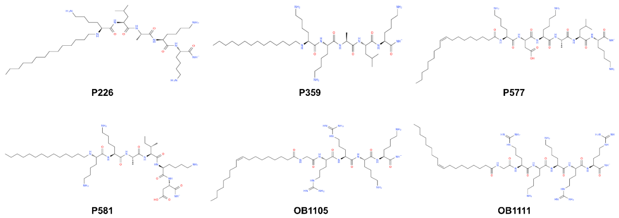
Figure 3.
Predicted anti-HSV-1 potency of CLOPs using Support Vector Regression (A) and Random Forest regression (B). Panel C shows RMSD distributions across docking poses generated for CLOP-HSV-1 receptor pairs; these values represent docking-pose dispersion and were not obtained from molecular-dynamics simulations.
Figure 3.
Predicted anti-HSV-1 potency of CLOPs using Support Vector Regression (A) and Random Forest regression (B). Panel C shows RMSD distributions across docking poses generated for CLOP-HSV-1 receptor pairs; these values represent docking-pose dispersion and were not obtained from molecular-dynamics simulations.
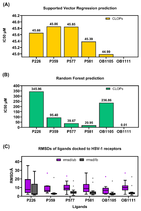
Figure 4.
Histograms displaying the binding affinities of CLOPs to HSV-1 glycoproteins. The binding affinities were determined using AutoDock wizard (with multiple search algorithms) to predict binding affinities, coupled with AutoDock Tools (ADT) for efficient data analysis.
Figure 4.
Histograms displaying the binding affinities of CLOPs to HSV-1 glycoproteins. The binding affinities were determined using AutoDock wizard (with multiple search algorithms) to predict binding affinities, coupled with AutoDock Tools (ADT) for efficient data analysis.
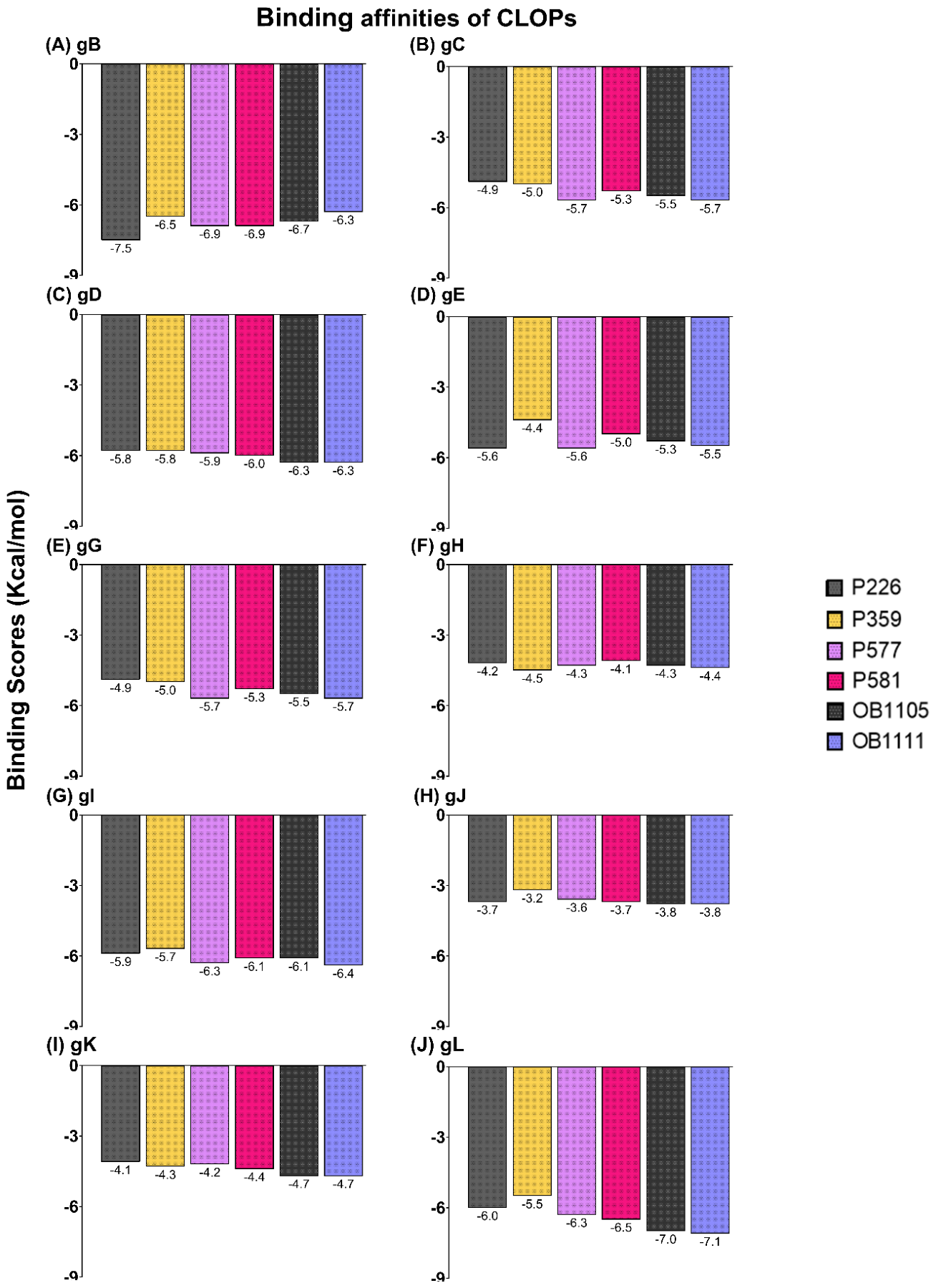
Figure 5.
Diagrams of the 2D and 3D structures of CLOP P226, along with its interactions with HSV-1 glycoproteins B (A & B) and D (C & D). Diagrams are uniformly colored and display the interactions, amino acid residues, and CLOP/glycoprotein complex. The red ligand represents P226, the blue represents the glycoprotein, and the yellow rectangle frames the docking complex.
Figure 5.
Diagrams of the 2D and 3D structures of CLOP P226, along with its interactions with HSV-1 glycoproteins B (A & B) and D (C & D). Diagrams are uniformly colored and display the interactions, amino acid residues, and CLOP/glycoprotein complex. The red ligand represents P226, the blue represents the glycoprotein, and the yellow rectangle frames the docking complex.
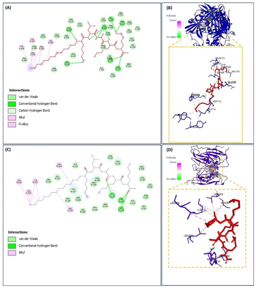
Figure 6.
Diagrams of 2D and 3D structures of P577 interactions with HSV-1 glycoproteins B (A & B) and D (C & D). The uniform color scheme displays the interactions, amino acid residues, and CLOP/glycoprotein complex. The red ligand represents P577, the blue represents the glycoprotein, and the yellow rectangle frames the docking complex.
Figure 6.
Diagrams of 2D and 3D structures of P577 interactions with HSV-1 glycoproteins B (A & B) and D (C & D). The uniform color scheme displays the interactions, amino acid residues, and CLOP/glycoprotein complex. The red ligand represents P577, the blue represents the glycoprotein, and the yellow rectangle frames the docking complex.
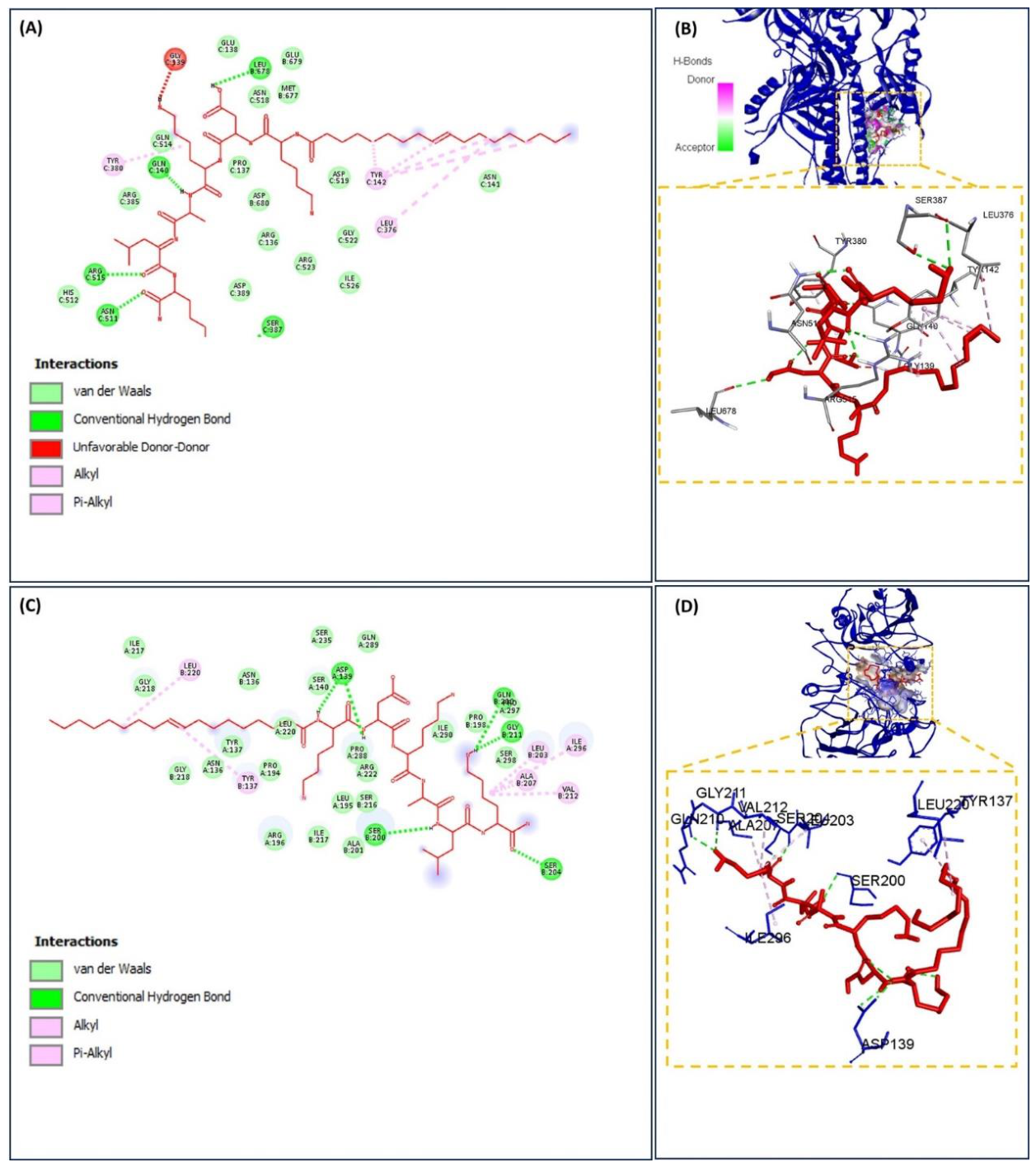
Figure 7.
Diagrams of 2D and 3D structures of OB1105 interactions with HSV-1 glycoproteins B (A & B) and D (C & D). Diagrams are uniformly colored and display the interactions, amino acid residues, and CLOP/glycoprotein complex. The yellow rectangle frames the docking complex, which consists of OB1105 (red) and the glycoprotein (blue).
Figure 7.
Diagrams of 2D and 3D structures of OB1105 interactions with HSV-1 glycoproteins B (A & B) and D (C & D). Diagrams are uniformly colored and display the interactions, amino acid residues, and CLOP/glycoprotein complex. The yellow rectangle frames the docking complex, which consists of OB1105 (red) and the glycoprotein (blue).
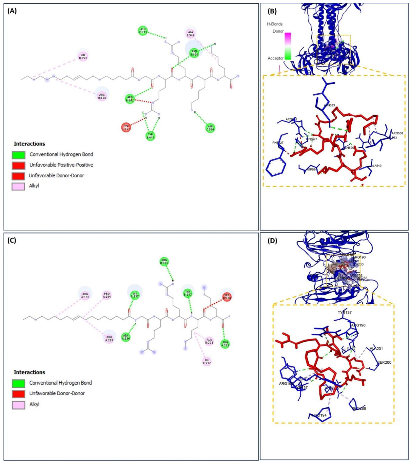
Figure 8.
Diagrams of 2D and 3D structures of OB1111 interactions with HSV-1 glycoproteins B (A & B) and D (C & D). Diagrams are uniformly colored and display the interactions, amino acid residues, and CLOP/glycoprotein complex. The red ligand represents OB1111, the blue represents the glycoprotein, and the yellow rectangle frames the docking complex.
Figure 8.
Diagrams of 2D and 3D structures of OB1111 interactions with HSV-1 glycoproteins B (A & B) and D (C & D). Diagrams are uniformly colored and display the interactions, amino acid residues, and CLOP/glycoprotein complex. The red ligand represents OB1111, the blue represents the glycoprotein, and the yellow rectangle frames the docking complex.
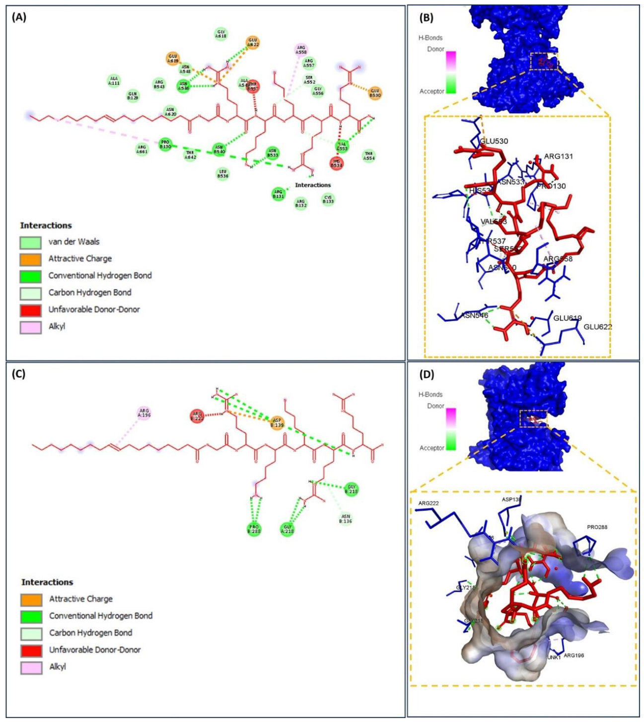
Figure 9.
Cytotoxic effects of six CLOPs on eukaryotic cells. HEp-2 cells (5 x 10⁴/well) were treated with increasing concentrations of each CLOP (0.9 to 250 µg/mL), and cell viability was measured at 24 h (A) and 48 h (B) using the MTT assay (see methods section). The effects of various concentrations of CLOPs (0.9 to 250 µg/mL) on the viability of Vero cells (5 x 10⁴/well) were also measured at 24 h (C) and 48 h (D) using the MTT assay. Acyclovir (ACV) served as the positive control. Data represents the mean ± SD from one of three separate experiments, each run in quadruplicate.
Figure 9.
Cytotoxic effects of six CLOPs on eukaryotic cells. HEp-2 cells (5 x 10⁴/well) were treated with increasing concentrations of each CLOP (0.9 to 250 µg/mL), and cell viability was measured at 24 h (A) and 48 h (B) using the MTT assay (see methods section). The effects of various concentrations of CLOPs (0.9 to 250 µg/mL) on the viability of Vero cells (5 x 10⁴/well) were also measured at 24 h (C) and 48 h (D) using the MTT assay. Acyclovir (ACV) served as the positive control. Data represents the mean ± SD from one of three separate experiments, each run in quadruplicate.
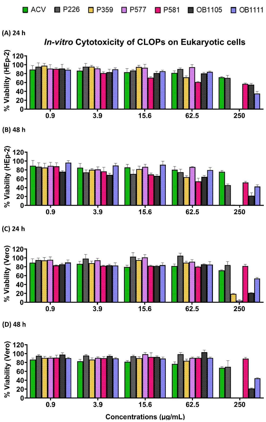
Figure 10.
CLOPs inhibited HSV-1 plaque formation in Vero cells. Vero cells (1 x 10⁶/mL) were infected with HSV-1 (MOI of 0.001), followed by treatment with CLOPs (25 µg/mL), and subjected to the plaque reduction assay. Plaque visualization and enumeration were conducted at 48 h. Representative images of Vero cells exposed to HSV-1 and treated with CLOPs (A). Acyclovir (ACV) served as the positive control. The mean HSV-1 virus titers from three separate experiments were calculated and graphed (B). Data is presented as the mean ± SD. Each experiment was performed 3 times. Statistical significance was performed using one-way ANOVA, followed by Šidák multiple comparison test at ****p <0.0001, ***p <0.001, **p <0.01, *p <0.05.3.11 OB1111 suppresses HSV-1 attachment.
Figure 10.
CLOPs inhibited HSV-1 plaque formation in Vero cells. Vero cells (1 x 10⁶/mL) were infected with HSV-1 (MOI of 0.001), followed by treatment with CLOPs (25 µg/mL), and subjected to the plaque reduction assay. Plaque visualization and enumeration were conducted at 48 h. Representative images of Vero cells exposed to HSV-1 and treated with CLOPs (A). Acyclovir (ACV) served as the positive control. The mean HSV-1 virus titers from three separate experiments were calculated and graphed (B). Data is presented as the mean ± SD. Each experiment was performed 3 times. Statistical significance was performed using one-way ANOVA, followed by Šidák multiple comparison test at ****p <0.0001, ***p <0.001, **p <0.01, *p <0.05.3.11 OB1111 suppresses HSV-1 attachment.
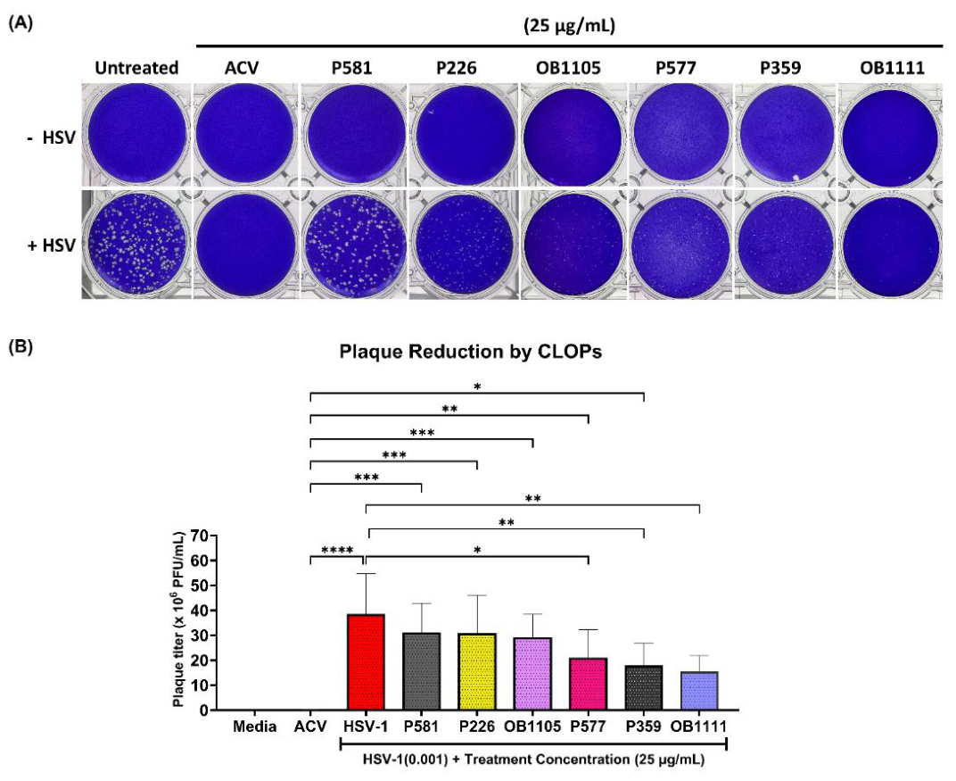
Figure 11.
Concentration-dependent effect of OB1111 on HSV-1 plaque-forming infectivity during an early-stage attachment assay. Vero cells (1 x 10⁶/mL) were subjected to simultaneous OB1111(12.5 - 100 µg/mL)/ HSV-1(MOI of 0.001) administration at 4 °C, followed by a temperature shift to 37 °C to permit entry. Plaques were visualized and enumerated after 48 h. Data are presented as the mean ± SD. One-way ANOVA was used to evaluate treatment effects; Bartlett’s test was used to assess homogeneity of variances. at ****p <0.0001, ***p <0.001, **p <0.01, *p <0.05.
Figure 11.
Concentration-dependent effect of OB1111 on HSV-1 plaque-forming infectivity during an early-stage attachment assay. Vero cells (1 x 10⁶/mL) were subjected to simultaneous OB1111(12.5 - 100 µg/mL)/ HSV-1(MOI of 0.001) administration at 4 °C, followed by a temperature shift to 37 °C to permit entry. Plaques were visualized and enumerated after 48 h. Data are presented as the mean ± SD. One-way ANOVA was used to evaluate treatment effects; Bartlett’s test was used to assess homogeneity of variances. at ****p <0.0001, ***p <0.001, **p <0.01, *p <0.05.
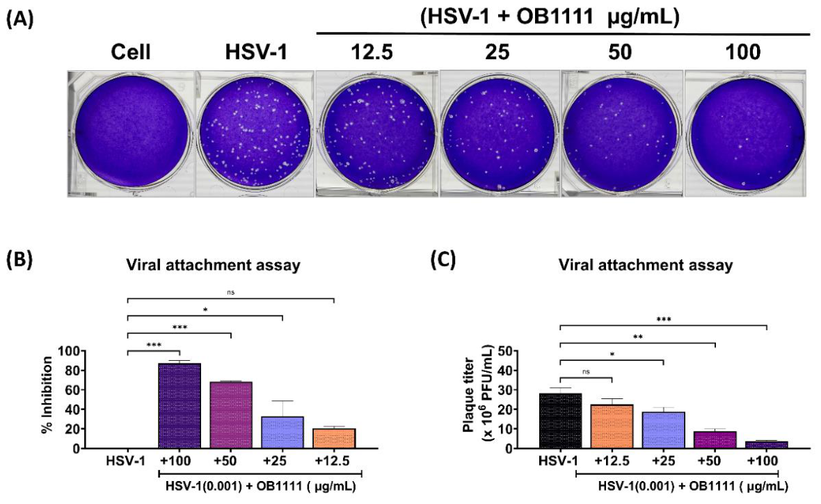
Table 1.
Tabulated display of the molecular features of CLOPs.
| CLOP | Composition | Net Charge | Structural features | Terminal modifications |
|---|---|---|---|---|
|
P226 P359 P577 P581 |
Pentapeptides containing three Lys, one Leu, and one Ala |
+ 3 (from Lys residues) |
Amphipathic, α helical |
N-terminal lipidation (hydrophobic); C-terminal amide (hydrophilic) |
| OB1105 | Pentapeptide containing two Lys, two Arg, and one Gly | +4 (from Lys and Arg residues) | Amphipathic | |
| OB1111 | Hexapeptide containing two Lys, three Arg, and one Gly | +5 (from Lys and Arg residues) | Amphipathic |
Abbreviations: Lys: lysine; Leu:leucine; Ala : alanine ; Arg : arginine ; Gly : glycine 3.2 Molecular properties determining pharmacokinetics and drug-likeness.
Table 2.
Summary of the (A) physiochemical and drug-likeness of CLOPs and (B) ADMET analysis results of CLOPs.
Table 2.
Summary of the (A) physiochemical and drug-likeness of CLOPs and (B) ADMET analysis results of CLOPs.
| (A) | ||||||||||||||||||
| CLOP | MW (Da) | nROT | nHBA | nHBD | LogP | TPSA | Drug-likeness | |||||||||||
| Lipinski | Pfizer | |||||||||||||||||
| P226 | 781.15 | 41 | 10 | 9 | 3.434 | 235.59 | No | Yes | ||||||||||
| P359 | 781.15 | 41 | 10 | 9 | 3.436 | 235.59 | No | Yes | ||||||||||
| P577 | 964.31 | 49 | 13 | 11 | 3.157 | 319.06 | No | Yes | ||||||||||
| P581 | 896.23 | 46 | 13 | 11 | 2.801 | 301.99 | No | Yes | ||||||||||
| OB1105 | 906.24 | 48 | 11 | 14 | 2.131 | 364.43 | No | Yes | ||||||||||
| OB1111 | 1062.42 | 56 | 13 | 18 | 1.214 | 441.44 | No | Yes | ||||||||||
| CLOP | MW (Da) | nROT | nHBA | nHBD | LogP | TPSA | Drug-likeness | |||||||||||
| Lipinski | Pfizer | |||||||||||||||||
| P226 | 781.15 | 41 | 10 | 9 | 3.434 | 235.59 | No | Yes | ||||||||||
| P359 | 781.15 | 41 | 10 | 9 | 3.436 | 235.59 | No | Yes | ||||||||||
| P577 | 964.31 | 49 | 13 | 11 | 3.157 | 319.06 | No | Yes | ||||||||||
| P581 | 896.23 | 46 | 13 | 11 | 2.801 | 301.99 | No | Yes | ||||||||||
| OB1105 | 906.24 | 48 | 11 | 14 | 2.131 | 364.43 | No | Yes | ||||||||||
| OB1111 | 1062.42 | 56 | 13 | 18 | 1.214 | 441.44 | No | Yes | ||||||||||
| Abbreviations: CLOP: cationic lipo-oligopeptides; MW: molecular weight; nROT: number of rotatable bonds; nHBA: number of hydrogen bond acceptors; nHBD: number of hydrogen bond donors; LogP: logarithm of the partition coefficient; TPSA: topological polar surface area. | ||||||||||||||||||
| (B) | ||||||||||||||||||
| CLOP | Absorption | Distribution | Metabolism | Excretion | Toxicity | |||||||||||||
| Log S | Pgp substrate | BBB -PS | VD (L/kg) | CYP1A2 | CYP3A4 | Half-life | CL plasma | Carc. | Nephrotox. | |||||||||
| P226 | -3.2 | Yes | No | 14.117 | No | No | 1.0008 | 4.513 | 0.028 | 0.544 | ||||||||
| P359 | -3.2 | Yes | No | 12.042 | No | No | 1.038 | 4.379 | 0.023 | 0.467 | ||||||||
| P577 | -3.621 | Yes | No | 1.260 | No | No | 1.337 | 3.062 | 0.005 | 0.461 | ||||||||
| P581 | -4.148 | Yes | No | 1.640 | No | No | 0.446 | 5.045 | 0.195 | 0.255 | ||||||||
| OB1105 | -3.299 | Yes | No | 1.821 | No | No | 0.979 | 4.043 | 0.033 | 0.92 | ||||||||
| OB1111 | -3.13 | Yes | No | 1.716 | No | No | 1.364 | 3.455 | 0.032 | 0.99 | ||||||||
| Abbreviations: CLOP: cationic lipo-oligopeptides; Log S: logarithm of solubility; Pgp: P-glycoprotein; BBB: blood-brain barrier passage; VD: volume of distribution; CYP1A2: cytochrome P450 1A2; CYP3A4: cytochrome P450 3A4; CL plasma: plasma clearance; Carc.: Carcinogenicity; Nephrotox.: Nephrotoxicity. | ||||||||||||||||||
Table 4.
Ten-fold cross-validation performance of machine learning regression models.
| Model | PCC | Spearman | Q²/R² | RMSE | MAE |
|---|---|---|---|---|---|
| Random Forest | 0.751 | 0.690 | 0.561 | 0.883 | 0.672 |
| SVR | 0.743 | 0.683 | 0.545 | 0.898 | 0.649 |
| XGBoost | 0.740 | 0.680 | 0.544 | 0.899 | 0.695 |
| Gradient Boosting | 0.714 | 0.650 | 0.501 | 0.940 | 0.729 |
Table 5.
Performance of machine learning models on the independent validation dataset.
| Model | PCC | Spearman | Q²/R² | RMSE | MAE |
|---|---|---|---|---|---|
| SVR | 0.692 | 0.572 | 0.432 | 0.839 | 0.651 |
| XGBoost | 0.628 | 0.592 | 0.388 | 0.871 | 0.670 |
| Random Forest | 0.625 | 0.539 | 0.374 | 0.881 | 0.658 |
| Gradient Boosting | 0.612 | 0.545 | 0.371 | 0.883 | 0.691 |
Table 6.
Performance of Ridge regression during leave-one-out cross-validation.
| Model | PCC | Spearman | Q²/R² | RMSE | MAE |
|---|---|---|---|---|---|
| Ridge Regression (LOOCV) | 0.671 | 0.591 | 0.434 | 1.002 | 0.766 |
Disclaimer/Publisher’s Note: The statements, opinions and data contained in all publications are solely those of the individual author(s) and contributor(s) and not of MDPI and/or the editor(s). MDPI and/or the editor(s) disclaim responsibility for any injury to people or property resulting from any ideas, methods, instructions or products referred to in the content. |
© 2026 by the authors. Licensee MDPI, Basel, Switzerland. This article is an open access article distributed under the terms and conditions of the Creative Commons Attribution (CC BY) license (http://creativecommons.org/licenses/by/4.0/).
Copyright: This open access article is published under a Creative Commons CC BY 4.0 license, which permit the free download, distribution, and reuse, provided that the author and preprint are cited in any reuse.