

Submitted:
28 August 2026
Posted:
31 August 2026
You are already at the latest version
Abstract
Current paradigms of plant stress response fail to explain how diverse physical perturbations—such as hydration shifts, macromolecular crowding, temperature variations, and ionic dynamics—integrate into unified metabolic outcomes. Here, we present a novel conceptual framework asserting that the physical state of the cytoplasm acts as a master intermediate variable coupling environmental cues to cellular signaling and recovery. We propose that distinct physical sensing events (exemplified by FLOE1, DCP5, and ELF3) conceptually converge onto the central SnRK1–TOR metabolic switch. Within this architecture, we identify the FCS-LIKE ZINC FINGER (FLZ) protein family as a key molecular state buffer that modulates the activation threshold, amplitude, and reversibility of SnRK1–TOR signaling. Leveraging established FLZ–SnRK1 interactions and ATG8-dependent FLZ degradation, we formalize a self-reinforcing positive-feedback loop driven by autophagy that stabilizes metabolic transitions. Crucially, while avoiding premature claims of FLZ proteins as direct phase sensors, we define the FLZ–SnRK1–TOR–autophagy axis as a critical biophysical-to-metabolic interface. This work unifies traditionally fragmented biological tiers into a single mechanistic cascade spanning environmental stress factors, cytoplasmic biophysics, biomolecular condensate dynamics, molecular signaling, SnRK1–TOR metabolic switching, autophagy, and ultimate cellular adaptation. Ultimately, this state-centric paradigm shifts crop biotechnology from conventional gene-expression maximization toward precise biophysical threshold engineering and stress-recovery optimization.
Keywords:
SnRK1
; TOR
; FLZ proteins
; autophagy
; liquid–liquid phase separation
; cytoplasmic phase state
; molecular crowding
; stress hysteresis
; Plant Threat Matrix
; phase homeostasis
1. Introduction: From Signal-Centric to State-Centric Models of Plant Stress
Plant stress biology has been built, with considerable success, around a linear signal-transduction logic: an external perturbation is perceived by a dedicated receptor or sensor, transmitted through a defined kinase or second-messenger cascade, and resolved as a change in gene expression or metabolism [1,2]. This framework has correctly identified many of the molecular components involved in drought, salinity, temperature and nutrient-stress responses, and it remains the operational basis for most experimental designs in the field. It is, however, a framework built primarily around discrete, one-input–one-output modules, and it is increasingly strained by several recurring observations that do not follow naturally from a simple cascade model.
First, genotypes exposed to numerically identical stress treatments frequently differ in the severity and reversibility of their response, in ways that are not explained by differences in a single receptor or transcription factor [3]. Second, the SnRK1 energy-sensing kinase, a central hub of low-energy signaling, does not activate at a single fixed threshold; its activation state depends on the prior physiological history of the tissue, on sugar and hormone status, and on the combination of stresses experienced, not on any one input in isolation [1,3]. Third, recovery from stress is not the mirror image of stress onset: plants that have entered an adaptive or defensive state frequently do not return to the pre-stress state as soon as the stress is nominally removed, and the point at which recovery becomes possible does not coincide with the point at which the stress state was entered [4,5,6]. Fourth, combinations of stresses — for example simultaneous drought and heat — generate physiological states that are qualitatively distinct from, rather than additive combinations of, the responses to either stress alone [7].
None of these observations requires abandoning the signal-transduction framework. But they do suggest that an intermediate variable exists between “stress is present” and “the transcriptional/metabolic program is executed” — a variable that carries information about the intensity, duration and combination of stress, and that determines how readily the cell moves between physiological states. We and others have proposed that the physical, or phase, state of the cytoplasm is a strong candidate for this intermediate variable [4,5,6]. Changes in hydration, macromolecular crowding, viscosity, ionic strength and redox state alter the physical environment in which every subsequent signaling reaction occurs, and a growing set of experimentally validated plant proteins — FLOE1, DCP5 and ELF3 among them — show that such physical changes can be converted directly into a phase transition with a defined biological output [8,9,10]. We term this intermediate layer the cytoplasmic physical/phase state and treat it, provisionally, as sitting upstream of, and modulating, classical signal-transduction cascades rather than replacing them.
The central question this article addresses is: if the cytoplasmic physical state can modulate the conditions under which downstream signaling networks operate, which molecular regulators determine the threshold, amplitude and reversibility of the metabolic-state transitions that follow? We focus on the SnRK1–target of rapamycin (TOR) axis, because SnRK1 and TOR are the best-characterized antagonistic integrators of plant metabolic state, sitting at the interface between growth and stress adaptation [3,7,8,9]. We propose that a plant-specific family of small regulatory proteins, the FCS-LIKE ZINC FINGER (FLZ) proteins, is a strong candidate for this role — not as a primary sensor and not as a “master regulator,” but as a molecular state buffer that modulates the coupling between upstream physical/metabolic perturbation and downstream SnRK1–TOR switching, with autophagy providing feedback control over the transition.
We deliberately restrict the scope of this hypothesis. We do not claim that FLZ proteins are an established, direct sensor of cytoplasmic phase state, and we do not claim that FLZ-driven LLPS has been experimentally demonstrated. Both possibilities are biologically plausible given the structural properties of FLZ proteins, and we discuss why, but at present they remain testable hypotheses rather than established mechanisms. Throughout this article we distinguish, for every conceptual step, between (i) established evidence, (ii) emerging evidence with a partial but incomplete experimental basis, (iii) hypothesis proposed here, and (iv) specific testable prediction. Where direct evidence is lacking, we state this explicitly rather than allow the surrounding narrative to imply otherwise.
This framework extends a series of prior theoretical proposals from our group. We have previously argued that regulated cytoplasmic phase plasticity constitutes a universal biophysical principle of stress adaptation across living systems, formalized as the Cytoplasmic Phase Homeostasis Theory (CPHT) [4], that this plasticity underlies a general biophysical model of aging and stress resistance [5], and that a cytoplasmic phase sensor concept links environmental stress perception to adaptive cellular reorganization specifically in plants [6]. We have also proposed an integrative, multilevel signal-metabolic model of plant immunity [7] and a jasmonate–ethylene metabolic defense module within that model [8], and we have argued, in the concept of Regulatory Agronomy, that hormonal and energy-sensing switches determine the balance between stress response and growth dominance in crops [9]. The present article does not repeat these arguments but builds on them, asking specifically how a defined molecular protein family — FLZ — might implement, at the SnRK1–TOR–autophagy level, the state-buffering behavior that CPHT predicts should exist at the biophysical level.
2. Cytoplasmic Physical State as an Upstream Layer of Stress Information
Before any specific signaling protein is activated, an environmental perturbation changes the physical properties of the cytoplasm itself. Six physical variables are particularly relevant to plant cells.
Hydration and water activity. Water loss reduces cell volume and increases the effective concentration of every macromolecule in the cytoplasm; this is not a side effect of stress but one of its most immediate physical consequences, occurring before any transcriptional response is possible [2].
Molecular crowding. The cytoplasm is not a dilute solution; macromolecules occupy a substantial fraction of its volume under normal conditions, and this crowded state has well-documented, non-trivial effects on protein folding, protein–protein association, and diffusion-limited reaction rates that cannot be inferred from dilute-solution biochemistry [10,11]. When cell volume decreases under osmotic or dehydrative stress, crowding increases further, and this increase is itself sufficient to alter macromolecular assembly states, independent of any specific receptor [10,11]. In non-plant systems, the TOR pathway itself has been shown to actively tune cytoplasmic crowding and thereby control the phase-separation behavior of the cytoplasm, indicating a direct, evolutionarily conserved link between growth-promoting kinase signaling and the physical state of the cell interior [12].
Molecular mobility, diffusion and viscosity. Changes in crowding and hydration necessarily change the diffusion coefficients of proteins and metabolites and the collision frequency that underlies bimolecular reactions; increased viscosity and reduced mobility slow enzymatic and signaling reactions independently of any change in enzyme abundance or activity.
Biomolecular condensates and phase separation. A growing body of work shows that many plant proteins with intrinsically disordered regions (IDRs) undergo liquid–liquid phase separation (LLPS) in response to physical or chemical perturbation, forming reversible, membraneless condensates that concentrate specific proteins and RNAs and can rapidly reorganize cellular biochemistry without new protein synthesis [13,14,15,16,17]. LLPS is now recognized as a general organizing principle of eukaryotic cell biology [13,14], and the past five years have produced the first systematic reviews establishing that plant cells use the same principle, while noting that direct experimental characterization in plants remains comparatively limited [16,17].
Redox and ionic environment. Reactive oxygen species (ROS) and calcium ions constitute two of the fastest and most extensively characterized signaling systems in plant cells, each capable of propagating information across the whole plant within minutes [18,19,20]. Redox state and ionic strength also directly influence the conformational and phase behavior of individual proteins.
Mechanical state. The plant cell wall constrains cell volume and generates turgor-dependent membrane tension; a family of mechanosensitive ion channels transduces mechanical perturbation, including that generated by osmotic volume change, into ion flux [21].
Each of these physical variables can, in principle, alter the same categories of biochemical event: the equilibrium and kinetics of protein–protein interactions, the rate of diffusion-limited reactions, the assembly and disassembly of macromolecular complexes, the nucleation and dissolution of condensates, and the accessibility of regulatory complexes to their substrates and partners. The central claim of this section — that physical state changes are causally upstream of, and can modulate, subsequent molecular signaling — is not itself new; it follows directly from the general biophysics of crowding and phase separation [10,11,12,13,14]. What is less established is which specific plant proteins convert a given physical variable into a specific, reproducible biological output, and it is to this question that we turn next.
3. Distributed Cytoplasmic Physical Sensing
A recurring simplification in early discussions of this topic — including some of our own prior work — is to speak of “a cytoplasmic sensor,” as though a single molecular species were responsible for converting physical state into signal. The available evidence does not support this. Instead, several structurally unrelated plant proteins have each been shown, independently, to convert a specific physical variable into a phase transition with a defined downstream consequence. We propose that these proteins should be considered components of a distributed physical sensing network rather than instances of one universal mechanism (Figure 1).
FLOE1 and hydration. FLOE1 is a prion-like, intrinsically disordered Arabidopsis seed protein that is diffuse under low-water conditions and undergoes reversible phase separation into cytoplasmic condensates upon hydration, both in vivo and when reconstituted from purified protein in vitro [22]. Natural variation in FLOE1 phase behavior is associated with different germination strategies across accessions and species, directly linking condensate formation to an adaptive, ecologically relevant phenotype [22]. FLOE1 is, at present, the clearest example of a plant protein that behaves as a bona fide, hydration-tunable phase-separation switch with a demonstrated physiological output.
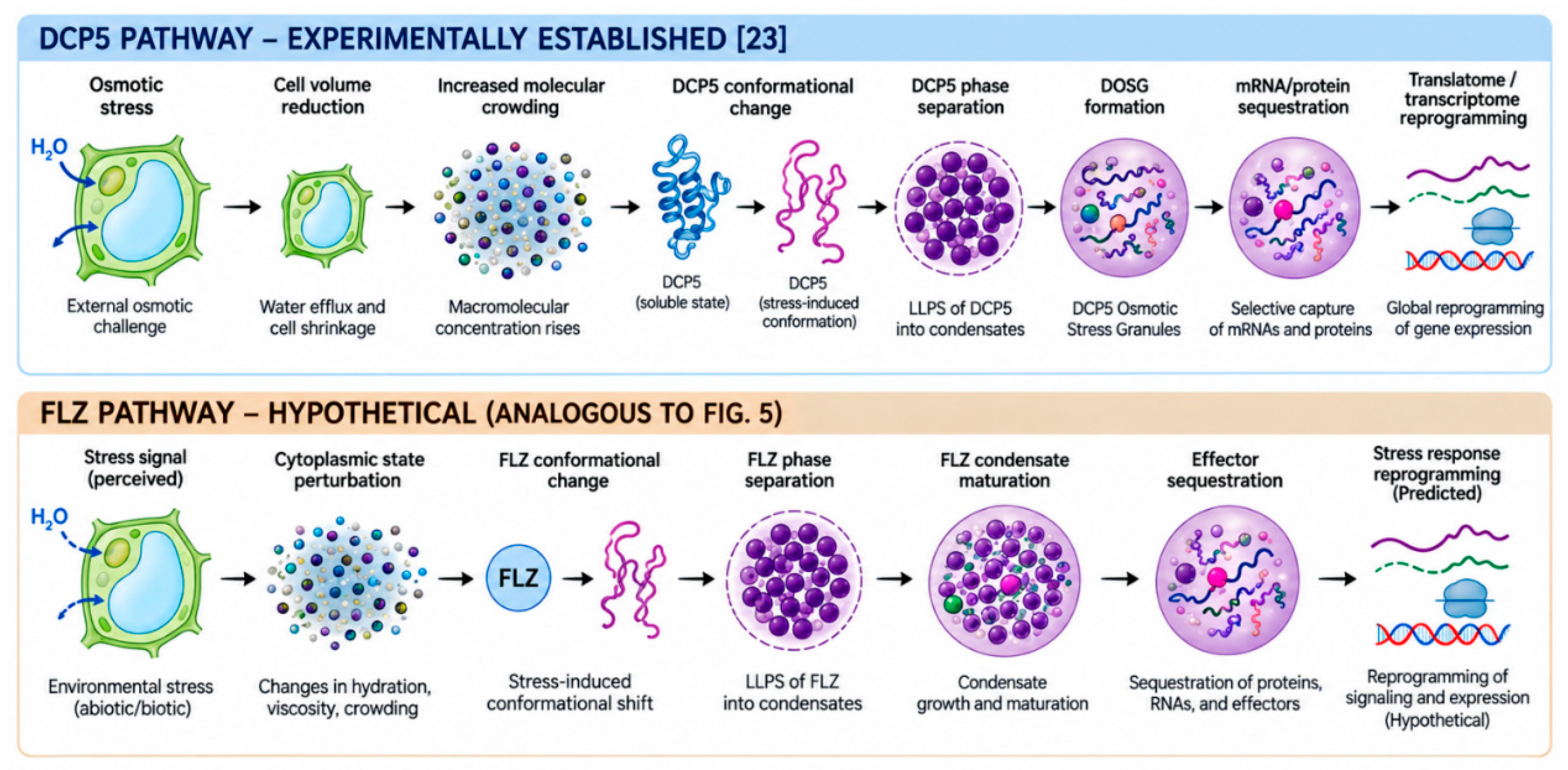
DCP5 and molecular crowding. DECAPPING 5 (DCP5) is a cytoplasmic mRNA-decapping activator that has recently been shown to function as a multifunctional osmosensor in Arabidopsis [23]. DCP5 contains a plant-specific intramolecular crowding-sensing region that undergoes a conformational change and drives phase separation specifically in response to the increase in molecular crowding produced by hyperosmotic cell shrinkage, independently of the identity of the osmoticum used [23]. This crowding-triggered phase separation produces DCP5-enriched osmotic stress granules (DOSGs) that sequester specific mRNAs and regulatory proteins and thereby reprogram both the transcriptome and the translatome within minutes of osmotic challenge [23]. We treat DCP5 as the strongest available proof-of-principle that a physical variable — crowding — can be converted directly into a phase transition with a specific, physiologically consequential biological output, and we return to it in detail in Section 9.
ELF3 and temperature. EARLY FLOWERING 3 (ELF3) is a core circadian-clock component whose prion-like domain undergoes reversible, temperature-dependent phase separation, shifting ELF3 between an active, soluble state at lower temperature and a condensed, transcriptionally inactive state at higher temperature [24]. The length of a polyglutamine tract within this domain varies naturally across Arabidopsis accessions and quantitatively tunes the temperature sensitivity of the phase transition, providing a second example in which condensate material properties are directly coupled to an adaptive, genetically variable phenotype [24].
Mechanosensitive channels. The MscS-like (MSL), Mid1-complementing activity (MCA) and two-pore potassium (TPK) channel families each convert membrane tension — generated by touch, gravity, or the same osmotically driven volume changes that alter crowding — into ion flux, providing a route by which mechanical perturbation of the physical state is converted into a conventional ionic signal [21].
Redox-sensitive and Ca²⁺-dependent proteins. A substantial number of plant regulatory proteins carry cysteine residues subject to reversible oxidative modification, and systemic reactive-oxygen-species and Ca²⁺ waves propagate stress information across the whole plant within minutes, often through mutually reinforcing feedback [18,19,20]. These systems are not phase-separation mechanisms in the same sense as FLOE1, DCP5 or ELF3, but they illustrate that physical/chemical state (here, redox and ionic environment) is routinely converted into signaling output by dedicated, well-characterized plant proteins.
Table 1 summarizes the level of evidence supporting each of these physical sensing modules. The pattern that emerges is that specific physical variables are read out by specific, non-redundant proteins, and that these proteins converge, functionally, on the same class of downstream consequence: altered protein–protein interaction, altered macromolecular assembly, and altered accessibility of regulatory complexes. We propose that this distributed layer of physical sensing feeds, directly or indirectly, into the SnRK1–TOR metabolic switch, the system to which we turn next.
4. SnRK1/TOR: From Energy Sensors to Metabolic State Switches
SnRK1 (SNF1-related protein kinase 1) and TOR (target of rapamycin) are the two central, evolutionarily conserved kinase complexes that integrate nutrient and energy status with plant growth and stress physiology [1,25]. Their activities are triggered by broadly opposite conditions and their global transcriptional and metabolic outputs are, in large part, antagonistic [3,26]: TOR is activated under nutrient- and energy-replete conditions and promotes ribosome biogenesis, translation, and anabolic, growth-supporting metabolism, whereas SnRK1 is activated under energy deficit and promotes catabolism, energy conservation, stress-responsive gene expression and autophagy [1,3,25]. SnRK1 has been shown to directly restrain TOR activity, for example through phosphorylation of the TOR-complex component RAPTOR, establishing at least a partial mechanistic basis for their antagonism [3].
It is tempting, given this antagonism, to describe SnRK1 and TOR as a simple binary “yin–yang” switch. We consider this description useful as a first approximation but insufficient as a complete model. SnRK1 activity is not triggered at a single, fixed energy threshold; it integrates sugar status (partly through the signaling metabolite trehalose-6-phosphate, which inhibits SnRK1 in a manner tied to sucrose availability) [27], hormonal input (abscisic acid, ABA, potentiates SnRK1 signaling through the core PYR/PYL/RCAR–PP2C–SnRK2 pathway) [28,29], developmental stage, and — we argue — the physical state of the cytoplasm in which the SnRK1 complex operates. Moreover, TOR and SnRK1 are not obligately confined to mutually exclusive subcellular domains or cell populations; both kinases can be present, and potentially differentially active, within different compartments of the same cell or different cells of the same tissue [30]. Whether TOR and SnRK1 can be simultaneously active in different subcellular domains of a single stressed cell — rather than acting as a simple population-level switch — is, to our knowledge, not resolved, and we return to this question directly in Section 12.
We therefore propose to describe SnRK1 and TOR not as a static antagonistic pair, but as the effector arm of a dynamic metabolic-state switch, whose position along the growth-to-survival continuum is jointly determined by cellular energy status, nutrient availability, hormonal input, redox state, and — as the hypothesis developed below proposes — the physical/phase state of the cytoplasm (Figure 2). Under this description, a cell moves through a graded sequence of states: a growth-dominant state (TOR-high, SnRK1-low), through an intermediate transition state in which both activities are present at intermediate levels, toward an adaptive survival state (SnRK1-high, TOR-low, autophagy engaged), and, if stress persists or intensifies, toward a state of chronic SnRK1 dominance associated with sustained growth suppression and, ultimately, the risk of irreversible damage. This sequence of states forms the metabolic backbone onto which we now map the FLZ protein family.
5. FLZ Proteins: The Missing Regulatory Layer
The FCS-LIKE ZINC FINGER (FLZ) proteins are a land-plant-specific family defined by a single conserved FLZ domain (also referred to by the Pfam designation DUF581), a compact zinc-finger-like module of roughly 70 amino acids that mediates protein–protein interaction rather than DNA or RNA binding [31,32]. The domain is absent from algae and appears to have originated in bryophytes; MpFLZ1 from the liverwort Marchantia polymorpha already interacts with the SnRK1 ortholog MpSnRK1.1, indicating that the FLZ–SnRK1 module itself is an ancient, conserved feature of land-plant biology rather than a recent innovation [33,34]. The family subsequently expanded through whole-genome duplication events across land-plant evolution, generating 18 members in Arabidopsis thaliana and larger, lineage-specific expansions in crop genomes [34,35].
Structurally, FLZ proteins combine the compact, folded FLZ domain with substantial intrinsically disordered regions (IDRs) at the N- and C-termini [36]. Detailed biochemical dissection of the Arabidopsis FLZ–SnRK1 interaction has shown that the FLZ domain itself mediates binding to the catalytic SnRK1α subunit, whereas the flanking IDRs mediate additional interactions with the regulatory β and βγ subunits of the SnRK1 heterotrimer and also mediate FLZ–FLZ dimerization [36]. FLZ–SnRK1 complexes are not diffusely distributed through the cytoplasm but are confined to discrete cytoplasmic foci that co-localize with the endoplasmic reticulum [36], and in maize, FLZ–SnRK1 co-localization has independently been observed on comparable cytoplasmic aggregates. On the basis of this evidence, FLZ proteins have been proposed to act as a promiscuous, IDR-enabled scaffold that broadens the interaction repertoire of the SnRK1 hub, providing a framework for cell- and stimulus-type-specific SnRK1 signalling [34,35,36].
Functionally, individual FLZ members have been shown to modulate SnRK1 signaling directly. FLZ6 and FLZ10 act as starvation-induced repressors of SnRK1: their loss of function increases the abundance and T-loop phosphorylation of the SnRK1α1 catalytic subunit, increases measured SnRK1 kinase activity, and produces a compromised-growth phenotype resembling that of SnRK1α1 overexpression [37]; the same two genes have subsequently been shown to also modulate osmotic stress responses [38,39]. FLZ genes as a family are transcriptionally responsive to sugar availability, cellular energy status and abiotic stress, with distinct FLZ members induced or repressed by carbon starvation, indicating an actively regulated, rather than constitutive, layer of SnRK1 modulation [33]. A connection to ABA signaling exists but is heterogeneous across the family: the founding FLZ-domain protein, MEDIATOR OF ABA-REGULATED DORMANCY1 (MARD1/FLZ9), was identified through its role in ABA-mediated seed dormancy [32], the maize protein ZmFLZ25 positively regulates ABA sensitivity and physically interacts with several PYL ABA receptors [40], and FLZ4/IRM1 confers resistance to aphid herbivory when overexpressed, at a measurable cost to vegetative growth [41]. These observations establish that FLZ function is not restricted to a single pathway but is embedded, in different family members, within multiple hormonal and metabolic contexts.
We emphasize what this body of evidence does, and does not, establish. It is established that FLZ proteins physically interact with SnRK1 subunits, that this interaction is spatially confined to discrete cytoplasmic structures, and that at least two family members quantitatively repress SnRK1 activity in a manner that is itself regulated by energy status. It is not established that FLZ IDRs drive, or are required for, any phase-separation behavior of the FLZ–SnRK1 complex, nor is it established that FLZ abundance or localization changes the biophysical — as opposed to purely biochemical — state of the SnRK1 complex or its surrounding cytoplasm. The structural features of FLZ proteins (a folded interaction domain flanked by IDRs, confinement to discrete cytoplasmic foci, promiscuous multivalent interaction) are precisely the features associated with LLPS-competent proteins in other systems [13,14,15,16,17], which is why we consider the hypothesis developed below plausible; but plausibility on structural grounds is not evidence, and we treat it as such throughout.
Table 2 summarizes, for the principal aspects of FLZ biology, what is established, what remains unknown, and the specific hypothesis this article proposes to extend the established biology.
6. FLZ as a Molecular State Buffer
his section formulates the central conceptual contribution of this study. We formally define molecular state buffering as the capacity of a regulatory protein network to delay, stabilize, or reverse transitions between cellular metabolic and physical states in response to environmental perturbations and stress.
Under this definition, FLZ proteins are not proposed as a primary sensor of environmental change, and they are not proposed as a master regulator that dictates cell fate. We propose instead that FLZ proteins constitute a state-buffering layer: a set of small, IDR-containing, SnRK1-associated proteins whose abundance, post-translational modification, and interaction state respond to upstream physical and metabolic perturbation, and which in turn set — rather than simply transmit — the threshold, amplitude and reversibility of SnRK1 activation and, downstream of that, of TOR–SnRK1 state switching.
The proposed causal chain (Figure 3) runs as follows. An environmental perturbation first produces a change in cytoplasmic physical and/or metabolic state (Section 2 and Section 3). This change alters FLZ abundance, post-translational modification, or protein–protein interaction state — established, for at least a subset of FLZ genes, at the transcriptional level [33] and, for FLZ6/FLZ10 protein, at the level of steady-state abundance [37]. The resulting change in FLZ state alters the configuration of the FLZ–SnRK1 complex, which we propose determines the effective activation threshold of SnRK1 — a proposal directly consistent with the demonstrated repressive effect of FLZ6/FLZ10 on SnRK1α1 phosphorylation and activity [37], but which has not, to our knowledge, been formally quantified as a threshold-shifting rather than simply activity-modulating effect. This SnRK1 activation state then determines the balance of the TOR–SnRK1 metabolic switch (Section 4), driving a shift toward catabolic, stress-adaptive metabolism and, ultimately, toward recovery once the perturbation subsides — or, if the perturbation is severe or sustained, toward the persistent-stress and phase-rigidity states discussed in Section 11.
We want to be precise about the status of each link in this chain. The FLZ–SnRK1 physical interaction is established [34,36]. The regulation of FLZ transcription and, for specific members, FLZ protein abundance by energy status is established [33,37]. The consequence of FLZ loss-of-function for SnRK1 phosphorylation, activity and downstream growth phenotype is established, for FLZ6/FLZ10 [37,38]. What is not established, and what we identify as the hypothesis proper, is (i) that FLZ state changes the threshold of SnRK1 activation in a way that is separable from simply changing its steady-state activity level, (ii) that this threshold effect is coupled to the physical, rather than only the biochemical, state of the surrounding cytoplasm, and (iii) that this coupling contributes to the hysteresis and recovery-window phenomena discussed in Section 11. Each of these three claims is, in our assessment, a plausible and testable extension of the established biology, not a restatement of it.
7. The ATG8–FLZ–SnRK1–Autophagy Feedback Loop
Of all the links in the model proposed here, the connection between autophagy and FLZ-mediated SnRK1 regulation rests on the strongest direct experimental foundation, and it is worth presenting in detail because it demonstrates that a feedback architecture of the type this article proposes is not merely plausible but already partially documented.
A defined clade of Arabidopsis FLZ proteins, localized to the mitochondrial surface and containing an ATG8-interacting motif (AIM), has been identified as a novel class of ATG8-interacting partners [39,40]. These AIM-containing FLZ proteins inhibit SnRK1 signaling by repressing T-loop phosphorylation of the SnRK1α catalytic subunit, thereby restraining carbon-starvation-induced autophagy under energy-replete conditions [39]. Under energy limitation, transcription of these FLZ genes is repressed and, in parallel, the FLZ proteins themselves are degraded through a selective, ATG8-dependent autophagic pathway; degradation of FLZ removes its repression of SnRK1, allowing full SnRK1 activation [39,40,41,42]. Because SnRK1 activation itself promotes autophagy [3,43], this produces a documented positive-feedback loop: energy limitation lowers FLZ, lowering FLZ activates SnRK1, activated SnRK1 promotes further autophagy, and further autophagy degrades remaining FLZ, reinforcing SnRK1 activation until the cell has committed to a catabolic, autophagy-engaged state [39,40].
Phylogenetic analysis places the origin of this AIM-containing FLZ clade in gymnosperms, and the interaction of AIM-containing FLZ proteins with ATG8 has been experimentally confirmed for both maize (ZmFLZ14) and tomato (SlFLZ14) orthologs, indicating that the ATG8–FLZ–SnRK1 axis is conserved across seed-plant evolution rather than being an Arabidopsis-specific curiosity [39]. Functionally, in maize, loss of ZmFLZ14 confers enhanced tolerance to energy deprivation, while ZmFLZ14 overexpression reduces this tolerance — the expected phenotype if ZmFLZ14 functions as a repressive buffer whose removal permits full SnRK1-dependent stress adaptation [39].
We summarize the two limiting regimes of this loop as follows (Figure 4). Under energy sufficiency, FLZ abundance is high, SnRK1 is restrained below its full activation potential, and the cell remains in a growth-compatible, TOR-permissive state. Under energy limitation, FLZ transcription and protein abundance fall, SnRK1 is released from repression and activates, and autophagy increases; the autophagy-dependent degradation of any remaining FLZ protein then reinforces this transition through the positive-feedback mechanism described above.
We treat this feedback loop as an established mechanism, not a hypothesis, for the specific AIM-containing FLZ clade and the specific tissues and stress conditions in which it has been characterized. What we add to this established mechanism, as hypothesis rather than established fact, is the proposal that this loop constitutes a general template for state switching that extends, by analogy, to the non-AIM-containing FLZ members discussed in Section 5 (whose regulation may occur through transcriptional or non-autophagic proteolytic routes rather than through direct ATG8 binding), and the proposal that the phase-organization consequences of this switch — as opposed to its purely biochemical consequences for SnRK1 phosphorylation — remain to be characterized.
8. FLZ–LLPS: A Testable but Unresolved Connection
This section requires particular caution, because it is the point at which the model proposed here moves furthest from directly demonstrated mechanism. We state explicitly what we are not proposing: we are not proposing that FLZ proteins regulate LLPS, and we are not proposing that FLZ-containing condensates have been observed or characterized. Direct experimental evidence for FLZ-driven, or FLZ-dependent, phase separation is, at present, insufficient to support either claim.
What we propose instead is a narrower, explicitly hedged statement: FLZ proteins may represent candidate coupling factors between SnRK1 signaling and stress-associated condensate dynamics. We consider this hypothesis plausible, rather than arbitrary, for four structural and cell-biological reasons that are themselves established. First, FLZ proteins contain substantial IDRs at both termini, and these IDRs are the structural feature most consistently associated with LLPS competence in other, better-characterized systems [13,15,17,36]. Second, FLZ proteins engage in extensive, promiscuous protein–protein interactions with multiple SnRK1 subunits and with other FLZ proteins through dimerization [34,36] — multivalency of this kind is a further structural correlate of phase-separation propensity [13,15]. Third, FLZ–SnRK1 complexes are not diffuse but form discrete cytoplasmic foci associated with the endoplasmic reticulum [36], a subcellular pattern consistent with, though not diagnostic of, condensate formation. Fourth, SnRK1 itself operates within a cellular context — carbon starvation and osmotic/energy stress — in which stress granule formation and condensate reorganization are independently well documented [41,44,45,46,47].
None of these four observations constitutes direct evidence that FLZ proteins undergo phase separation. What is missing, specifically, is: live-cell imaging showing FLZ condensate formation under physiologically relevant stress; fluorescence recovery after photobleaching (FRAP) data distinguishing liquid-like from solid or diffuse FLZ behavior; reconstitution of purified FLZ protein, alone or with SnRK1 subunits, under defined crowding and ionic conditions in vitro; demonstration of a concentration threshold for FLZ assembly; and comparison of full-length FLZ with IDR-deletion constructs to establish whether the IDRs are required for any observed condensation behavior. Until these experiments are performed, the FLZ–condensate connection should be considered a hypothesis rather than an established mechanism, and we return to the specific experiments required in Section 15.
We propose, schematically, three stages through which stress-associated condensates might in principle progress — a liquid, freely exchanging signaling environment; an adaptive, reversible condensate; and a persistent, more rigid condensate associated with loss of reversibility — and we propose FLZ as a candidate, but unconfirmed, coupling factor that could in principle influence transitions between these stages (Figure 5). This schema is itself a simplification of the material-state continuum documented for biomolecular condensates generally [13,15], applied speculatively to the FLZ–SnRK1 system; it should be read as an experimentally motivated framework for future work, not as a description of demonstrated FLZ behavior.
9. DCP5 as an Experimental Proof-of-Principle for Phase-Based Stress Sensing
We return here to DCP5, introduced briefly in Section 3, because its experimental characterization is, at present, the strongest available demonstration that the general logic of this article — physical state change leading to a phase transition leading to a specific, adaptive biological output — operates as a real mechanism in plant cells, rather than as a plausible analogy imported from animal or yeast systems [23].
The DCP5 pathway proceeds as follows. Hyperosmotic stress produces cell shrinkage, which intensifies molecular crowding in the cytoplasm. This increase in crowding is directly sensed by a plant-specific intramolecular crowding-sensing region within DCP5, which undergoes a conformational change. This conformational change drives DCP5 phase separation, producing DCP5-enriched osmotic stress granules (DOSGs). These granules sequester specific mRNAs and translational and transcriptional regulatory proteins, and this sequestration reprograms both the translatome and the transcriptome within the timescale of the initial physical perturbation, well before any new transcription driven by that reprogramming could itself contribute [23]. The process is rapid and reversible, tracking cell-volume recovery [23].
We emphasize that DCP5 is mechanistically unrelated to FLZ: it is an mRNA-decapping factor, not an SnRK1-associated scaffold, and its crowding-sensing region is structurally distinct from the FLZ domain. We do not extend the DCP5 mechanism to FLZ by analogy or imply that FLZ operates through the same structural motif. What DCP5 demonstrates, and what we do carry forward into the remainder of this article, is a general principle: that a defined physical variable (crowding), acting through a defined plant protein, can be converted into a phase transition that produces a specific, physiologically meaningful, and rapidly reversible change in cellular biochemistry, without requiring new gene expression as an intermediate step. This is the general class of mechanism we hypothesize, but have not established, for FLZ.
Figure 6.
DCP5 as experimental evidence for sensing the physical state of the cytoplasm. The experimentally established response cascade to osmotic stress: osmotic stress → cell volume reduction → increased molecular crowding → conformational change of DCP5 → DCP5 phase separation → formation of DCP5 osmotic stress granules (DOSGs) → sequestration of mRNAs and proteins → translational and transcriptomic reprogramming. Below, a hypothetical FLZ pathway is presented for comparison, proposed by analogy with the model shown in Figure 5. The experimentally validated DCP5 mechanism and the hypothetical FLZ mechanism are clearly distinguished according to their respective levels of evidence.
Figure 6.
DCP5 as experimental evidence for sensing the physical state of the cytoplasm. The experimentally established response cascade to osmotic stress: osmotic stress → cell volume reduction → increased molecular crowding → conformational change of DCP5 → DCP5 phase separation → formation of DCP5 osmotic stress granules (DOSGs) → sequestration of mRNAs and proteins → translational and transcriptomic reprogramming. Below, a hypothetical FLZ pathway is presented for comparison, proposed by analogy with the model shown in Figure 5. The experimentally validated DCP5 mechanism and the hypothetical FLZ mechanism are clearly distinguished according to their respective levels of evidence.
10. From Phase Transition to Metabolic State Switching
We now integrate the preceding sections into a single conceptual framework (Figure 7), which we consider the central schema of this article. Environmental perturbation changes the physical state of the cytoplasm (Section 2). This physical state is read out by a distributed sensing network of specific plant proteins, of which FLOE1, DCP5 and ELF3 are established examples (Section 3). We hypothesize, without direct evidence at this stage, that the resulting composite physical state contributes to setting the FLZ state — the abundance, modification and interaction status of FLZ proteins (Section 6) — alongside the established transcriptional regulation of FLZ by sugar and energy status [33]. FLZ state, in turn, sets the configuration and, we propose, the activation threshold of the SnRK1 complex (Section 5 and Section 6), which determines the position of the TOR–SnRK1 metabolic switch (Section 4). Autophagy provides feedback control over this switch, both as a downstream consequence of SnRK1 activation and, through the ATG8-dependent degradation of FLZ, as a mechanism that reinforces the transition once initiated (Section 7). The resulting metabolic reprogramming produces either recovery, if the cytoplasmic phase state can be restored, or — if phase plasticity is lost — a state of phase rigidity and cellular damage, developed in the next section.
We stress that only some of the arrows in this integrated schema correspond to established mechanism (physical state → distributed sensors; FLZ–SnRK1 interaction; SnRK1–TOR antagonism; SnRK1–autophagy–FLZ feedback), while others (physical state → FLZ state, specifically via a phase-sensing rather than purely transcriptional route; FLZ state → SnRK1 activation threshold, as distinct from activation level) are the hypothesis this article proposes. Figure 7 uses solid lines for the former and dashed lines for the latter, and we ask that this distinction be preserved in any subsequent representation of the model.
11. Phase-State Hysteresis and the Recovery Window
A recurring, and in our view under-analyzed, feature of plant stress physiology is that the level of stress required to move a plant into an adaptive or defensive physiological state is not the same as the level of stress reduction required to move it back out again [4,5,6]. We propose to formalize this asymmetry using the concept of hysteresis, a well-established property of biological switches built on positive and double-negative feedback [48,49,50,51,52].
We define an entry threshold: the stress intensity at which a cell transitions from a growth-dominant to an adaptive stress state, and a recovery threshold: the stress intensity below which a cell already in the adaptive state can return to growth. If these two thresholds coincide, the system is simply reversible and tracks the instantaneous stress level. If, as we hypothesize occurs in the FLZ–SnRK1–TOR–autophagy system, the recovery threshold lies below the entry threshold, the system exhibits hysteresis: for a given intermediate stress intensity, the cell’s state depends on its history — whether it arrived at that stress level from a lower or a higher one — rather than on the instantaneous stress level alone. Feedback architectures of exactly the kind documented for the ATG8–FLZ–SnRK1 loop (Section 7) are a canonical source of biological hysteresis in general systems-biology theory [48,49], which is why we consider this hypothesis mechanistically motivated rather than purely descriptive; but we have not, at present, identified direct experimental measurement of an entry/recovery threshold asymmetry specifically for the FLZ–SnRK1 system, and this remains a testable prediction rather than an established finding.
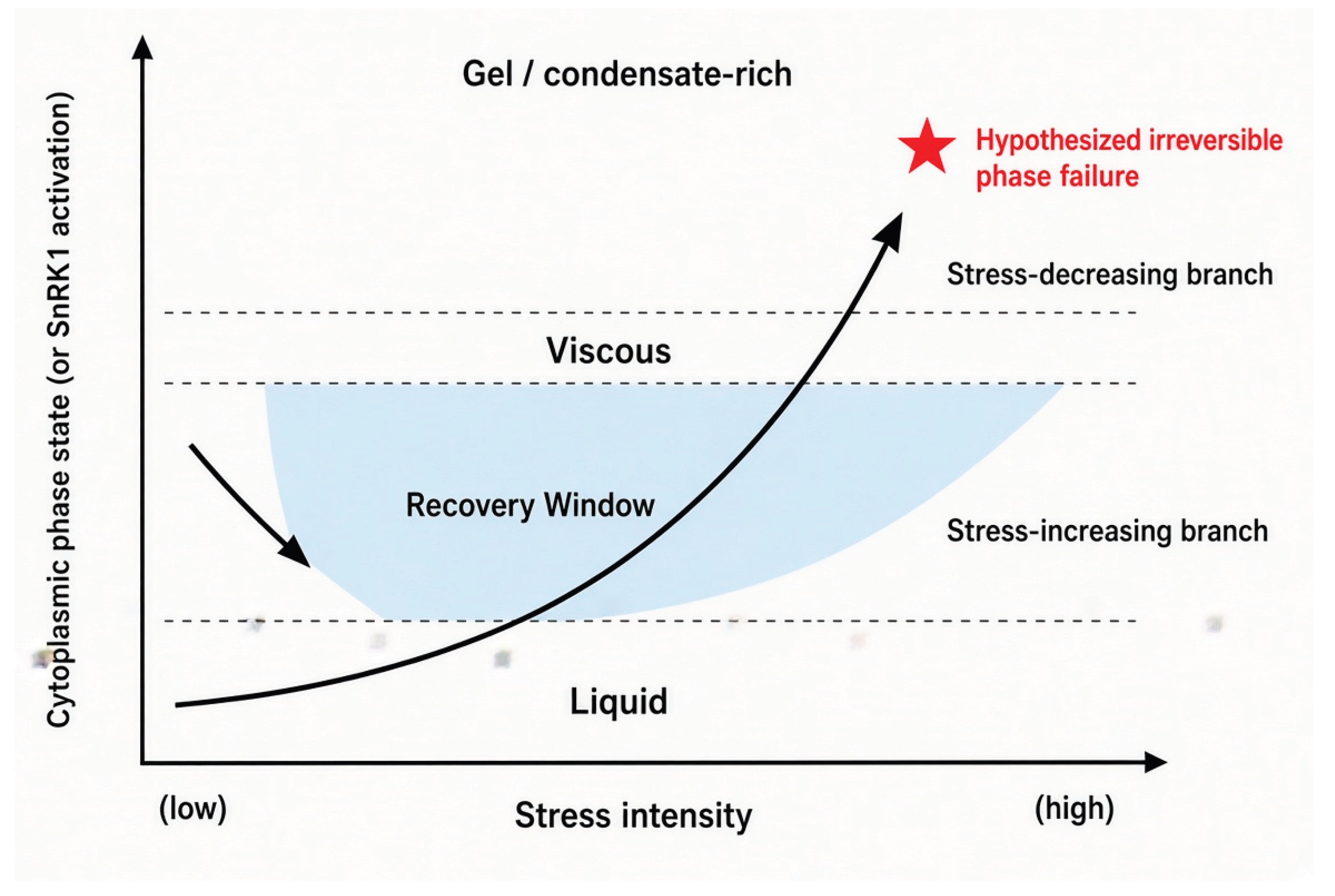
We introduce the term Recovery Window to denote the range of cell states, between the recovery threshold and the point of irreversible phase collapse, within which either a natural decline in stress intensity or an active intervention (for example, an antistress compound, a change in irrigation, or a change in nutrient supply) can still restore cytoplasmic phase plasticity and permit the cell to return to a growth-compatible state. Beyond this window, we hypothesize that phase plasticity is lost irreversibly, and the cell proceeds to phase rigidity, sustained growth suppression, or death. Table 3 sketches the predicted correspondence between physiological state, cytoplasmic physical properties, TOR–SnRK1–autophagy configuration, and condensate organization across this trajectory; we emphasize that the specific physical descriptors used (moderate crowding, increased viscosity, and so on) are hypothesized correlates consistent with the general biophysics of crowding and phase separation [10,11,12,13,14,15], not measured values for this specific system.
Figure 8.
Phase-state hysteresis creates a distinct window of recovery. Hysteresis loop plotting cytoplasmic phase state (or SnRK1 activation) against stress intensity. The stress-increasing branch (liquid → viscous → gel/condensate-rich) does not retrace the stress-decreasing branch. The Recovery Window and the point of hypothesized irreversible phase failure are marked explicitly.
Figure 8.
Phase-state hysteresis creates a distinct window of recovery. Hysteresis loop plotting cytoplasmic phase state (or SnRK1 activation) against stress intensity. The stress-increasing branch (liquid → viscous → gel/condensate-rich) does not retrace the stress-decreasing branch. The Recovery Window and the point of hypothesized irreversible phase failure are marked explicitly.
12. Phase Plasticity as the Determinant of Resilience
We connect the hysteresis concept of Section 11 directly to the Cytoplasmic Phase Homeostasis Theory (CPHT) proposed previously [4]. Within that theory, phase plasticity is defined as the capacity of a cell to (i) form adaptive physical states in response to stress, (ii) reorganize those states as conditions change, and (iii) return to a homeostatic physical state once the perturbation is resolved. The framework developed in this article proposes that FLZ-mediated state buffering of the SnRK1–TOR–autophagy axis is one candidate molecular mechanism through which phase plasticity, as defined by CPHT, could be implemented at the level of a specific, tractable signaling module — without claiming that FLZ is the sole or even the principal determinant of phase plasticity generally, which almost certainly also depends on the chaperone network, membrane lipid composition, and the distributed sensing systems described in Section 3.
We propose, as a conceptual and explicitly provisional metric, a Phase Resilience Index (PRI) — a composite score intended to capture, for a given genotype or condition, the width of the Recovery Window relative to the distance between the entry and recovery thresholds, potentially incorporating measurable correlates such as SnRK1 recovery kinetics, autophagic flux reversibility, and condensate dissolution rate after stress release. We introduce PRI purely as a conceptual placeholder to motivate future quantitative work; it has no validated experimental basis at present, and we explicitly do not present it as a working metric that can be applied to existing datasets.
13. Crop-Specific FLZ State Buffering
If FLZ proteins act as state buffers whose properties influence the threshold and reversibility of stress-state transitions, then differences in the size, sequence, and regulatory properties of the FLZ repertoire between crop species and genotypes become directly relevant to applied stress physiology. We summarize the available evidence for three major cereals, without extending it beyond what has been demonstrated.
Wheat. The hexaploid wheat genome encodes an expanded family of 120 TaFLZ genes [53]. Among these, TaFLZ54D is the most strongly NaCl-induced member and, when overexpressed, increases salt tolerance, elevates antioxidant enzyme activity and soluble sugar content, and reduces both the Na⁺/K⁺ ratio and malondialdehyde accumulation relative to wild-type plants; TaFLZ54D achieves this effect through direct interaction with TaSGT1 and TaPP2C rather than through a demonstrated SnRK1-dependent mechanism [53]. A separate wheat FLZ gene, TaSRHP, improves salt and drought tolerance when expressed ectopically in Arabidopsis [52].
Maize. Thirty-seven ZmFLZ genes have been identified genome-wide, several of which physically interact with the maize SnRK1 catalytic subunit ZmKIN10 and partially co-localize with it in cytoplasmic aggregates [51]. ZmFLZ25 overexpression confers ABA hypersensitivity through direct interaction with multiple PYL ABA receptors [51], and, as discussed in Section 7, ZmFLZ14 is functionally required, as part of the conserved ATG8–FLZ–SnRK1 axis, for the normal tolerance threshold to energy deprivation [39].
Rice. A genome-wide survey identified 29 rice FLZ genes, eight of which interact with the rice SnRK1 subunit SnRK1A; OsFLZ18 physically binds SnRK1A and antagonizes its transcriptional activation of the α-amylase gene αAmy3, and OsFLZ18 overexpression retards early seedling growth and shortens coleoptile elongation specifically under submergence [54,55,56]. This places OsFLZ18 within the broader, independently characterized network of submergence-tolerance regulators in rice that converge on SnRK1A activity.
Given this evidence base, we consider it premature to describe FLZ biology in these crops as generally under-characterized; the more accurate description is that species-specific FLZ functions are individually documented but have not yet been assembled into an integrated, mechanistic model connecting FLZ state to stress-state threshold or reversibility. The central open question we propose for this section is therefore not whether crop FLZ genes matter — they demonstrably do, for the specific phenotypes tested — but whether different crop-specific FLZ repertoires encode different thresholds for stress-state transitions, a hypothesis that would require directly comparable, cross-species measurements of SnRK1 activation kinetics under matched stress regimes, which do not yet exist. We present this as a promising direction for future comparative work rather than as an established conclusion (Figure 9).

Table 4.
FLZ-associated stress responses across major crop species.
| Species | Representative FLZ gene(s) | SnRK1/partner interaction | Documented phenotype | Reference |
| Arabidopsis thaliana | FLZ6, FLZ10 | SnRK1α (repression) | Compromised growth phenotype on loss of function; altered ABA and osmotic sensitivity | [37,38] |
| Arabidopsis thaliana | FLZ4/IRM1 | Not SnRK1-mediated (growth trade-off) | Increased aphid resistance, reduced growth | [50] |
| Arabidopsis thaliana | FLZ9/MARD1 | Not directly SnRK1-mediated | ABA-mediated seed dormancy | [32] |
| Zea mays | ZmFLZ14 | ATG8–FLZ–SnRK1 axis | Loss of function enhances energy-deprivation tolerance | [39 , 44-50] |
| Zea mays | ZmFLZ25 | ABA receptors PYL4/8/12/13 | Enhanced ABA sensitivity on overexpression | [51] |
| Triticum aestivum | TaFLZ54D | TaSGT1, TaPP2C | Enhanced salt tolerance on overexpression | [53] |
| Triticum aestivum | TaSRHP | Not characterized (heterologous expression) | Enhanced salt/drought tolerance in transgenic Arabidopsis | [52] |
| Oryza sativa | OsFLZ18 | SnRK1A (repression of αAmy3 induction) | Reduced coleoptile elongation under submergence on overexpression | [54,55] |
14. FLZ and Molecular State Memory
A further, deliberately conservative, extension of the framework concerns whether FLZ-linked processes could contribute to short-term cellular memory of a prior stress episode. We restrict this discussion strictly to plausible, near-term hypotheses and explicitly exclude claims about transgenerational or epigenetic inheritance, for which no evidence bearing on the FLZ system currently exists.
Because FLZ abundance is regulated by both transcription and ATG8-dependent proteolysis (Section 5 and Section 7), and because both regulatory routes operate on timescales of minutes to hours rather than instantaneously, a cell that has recently experienced an episode of SnRK1 activation may retain a depleted FLZ pool for some period after the initiating stress has subsided. If FLZ pool size genuinely sets the SnRK1 activation threshold, as hypothesized in Section 6, then this transient FLZ depletion would itself constitute a simple form of cellular hysteresis or short-term molecular state memory — a cell recently stressed would show a lower threshold for re-activating SnRK1 than a cell with no recent stress history, independent of any change in gene expression. We propose this specifically as a within-cell-cycle, non-epigenetic mechanism, analogous in logic (though not in molecular identity) to the priming phenomena documented for other stress-responsive systems, and we identify it as directly testable by measuring the SnRK1 re-activation kinetics of cells with experimentally depleted versus restored FLZ pools. Any extension of this idea to condensate persistence, or to longer-term or heritable memory, remains speculative and is noted here only as a direction for future work, not as part of the present hypothesis (Figure 10).
15. Experimental Roadmap
The hypothesis developed in this article is only useful to the extent that it generates experiments capable of rejecting it. We outline six complementary experimental directions.
15.1. Live-cell imaging. FLZ-GFP fusion lines, imaged alongside SnRK1 activity reporters and, where feasible, generic condensate markers, under defined osmotic, thermal and carbon-starvation regimes, with FRAP applied to any observed FLZ puncta to determine liquid-like versus solid-like behavior.
15.2. IDR-deletion analysis. Direct comparison of wild-type FLZ with IDR-deletion (ΔIDR) constructs for (i) SnRK1-complex assembly and localization, already partially characterized [36], and (ii) any condensation behavior observed under 15.1, to test whether the IDRs specifically are required.
15.3. Reconstituted in vitro phase-behavior assays. Purified FLZ protein, alone and in combination with purified SnRK1 subunits, tested for phase-separation behavior across a range of crowding-agent concentrations, osmotic conditions, salt concentrations and pH, following established in vitro LLPS assay methodology [15].
15.4. Autophagy manipulation. Comparison of SnRK1 activation kinetics, FLZ abundance, and — where condensation is observed — condensate dynamics in ATG8/autophagy-deficient backgrounds versus wild type, extending the existing ATG8–FLZ–SnRK1 characterization [39,40] to explicitly test the phase-organization consequences of this feedback loop.
15.5. Genetic dissection. Single and combinatorial FLZ knockout and overexpression lines, including IDR-specific point or deletion mutants, assayed for SnRK1 activation threshold (not only activity level), recovery kinetics after stress release, and — where relevant — hysteresis between stress-entry and stress-recovery thresholds.
16. Key Testable Predictions
Table 5 lists ten specific, falsifiable predictions that follow from the hypothesis developed in this article, each linked to the established mechanism it extends, the specific prediction, a feasible experiment, and the result expected if the hypothesis is correct.
17. Computational FLZ Phase-State Prediction
A further, near-term and comparatively low-cost avenue is computational: systematic comparison of FLZ family members across Arabidopsis, maize, wheat and rice for the sequence features associated with phase-separation propensity in other systems — the length and composition of intrinsically disordered regions, net charge and charge patterning, hydrophobicity, the density of low-complexity regions, aromatic-residue content, and predicted structural disorder, alongside evolutionary conservation of these features across the family [34,36]. Such analysis could, in principle, identify specific FLZ members as higher- or lower-priority candidates for the wet-laboratory experiments outlined in Section 15, and could generate a testable ranking of crop FLZ orthologs by predicted phase-separation propensity. We stress, however, that computational disorder or phase-separation propensity prediction is not, and cannot substitute for, experimental confirmation of condensation behavior; at most it can prioritize candidates for the experiments in Section 15.
18. FLZ as a Target for Crop Stress-State Engineering
If the hypothesis developed here is correct even in part, it implies a shift in how FLZ genes — and, more broadly, state-buffering components generally — might be approached as breeding or engineering targets. Conventional stress-tolerance breeding typically aims to increase resistance to a given stress, generally by maximizing a specific protective trait. The framework proposed here suggests, as an additional and distinct objective, that the threshold and reversibility of the underlying metabolic-state transition may be an independently tunable, and agronomically relevant, property.
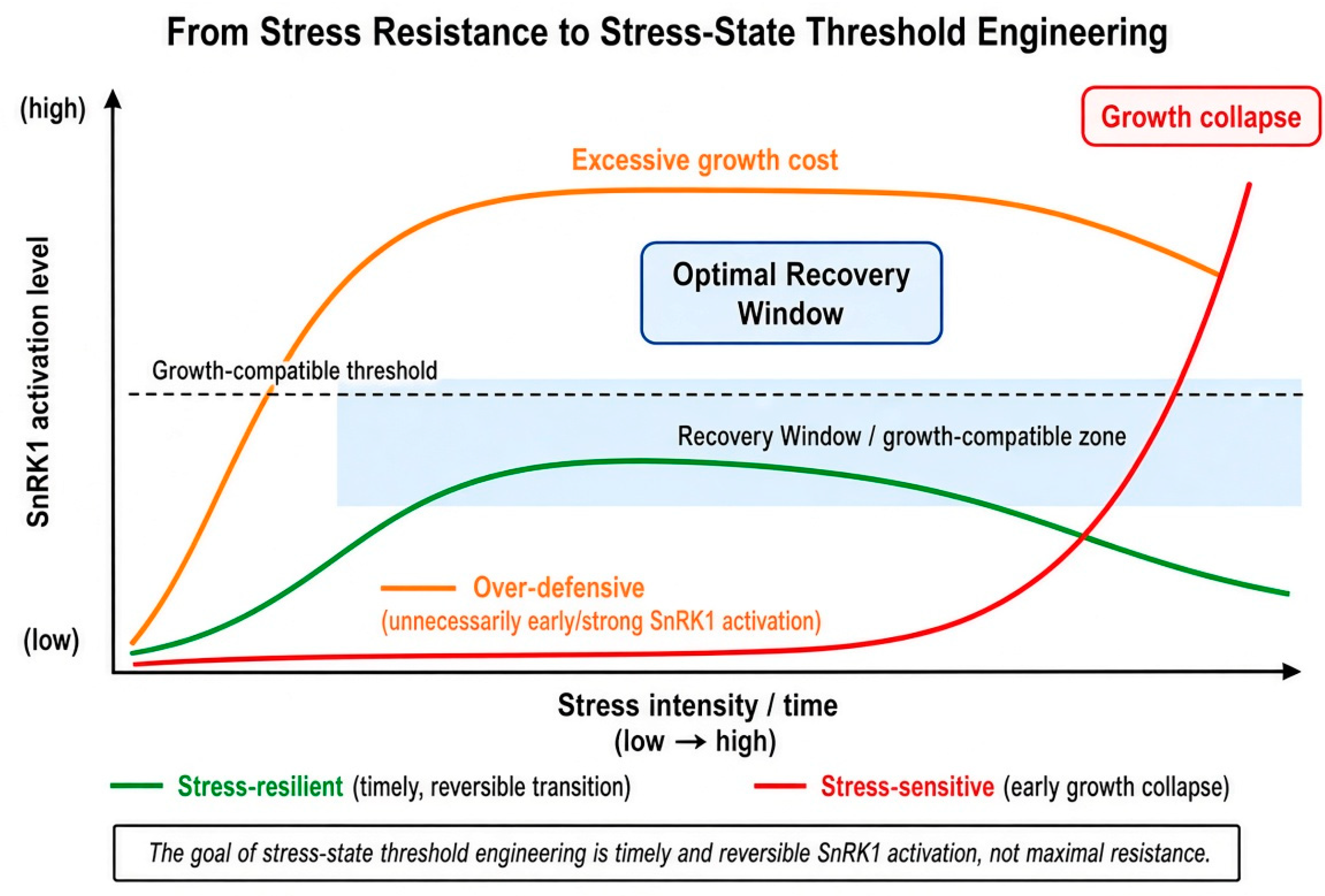
Concretely, we propose that the optimal outcome for a crop genotype may not be a maximally early or maximally strong SnRK1 activation, since this would suppress growth under stress that the plant could otherwise tolerate without a full metabolic shutdown, nor a maximally delayed activation, since this would risk irreversible damage before adaptive metabolism engages. Instead, we propose that an optimal genotype might be one in which the TOR-dominant-to-SnRK1-dominant transition occurs early enough to provide effective protection but late enough, and reversibly enough, to preserve growth and yield potential once the stress subsides — a genotype-specific optimization of transition timing rather than of resistance magnitude alone. Because FLZ proteins are proposed here as one of the molecular determinants of this transition threshold, they become a plausible, though still hypothetical, target for what we term stress-state engineering, distinct from conventional stress-resistance engineering. We emphasize that this application is presented as a long-term, hypothesis-dependent direction, contingent on the experimental validation outlined in Section 15 and Section 16, and not as an actionable breeding recommendation at present.
Figure 11.
From stress resistance to stress-state threshold engineering. Three schematic genotypes — stress-sensitive (early growth collapse), stress-resilient (timely, reversible transition), and over-defensive (unnecessarily early or strong SnRK1 activation with excessive growth cost) — plotted as SnRK1 activation trajectories against stress intensity and time, illustrating the proposed distinction between resistance and threshold optimization.
Figure 11.
From stress resistance to stress-state threshold engineering. Three schematic genotypes — stress-sensitive (early growth collapse), stress-resilient (timely, reversible transition), and over-defensive (unnecessarily early or strong SnRK1 activation with excessive growth cost) — plotted as SnRK1 activation trajectories against stress intensity and time, illustrating the proposed distinction between resistance and threshold optimization.
19. Integration with the Plant Threat Matrix (PTM)
The framework developed here is intended to integrate with, rather than duplicate, a previously proposed model of how plants respond to combined biotic and abiotic stress: the Plant Threat Matrix (PTM). PTM was developed to explain the integrated physiological response of plants to the simultaneous action of multiple stress factors — drought, heat, cold, salinity, nutrient deficiency, oxidative stress, pathogen infection, herbivore damage and mechanical injury — and it rests on the observation that plants do not respond to each stress factor independently but integrate diverse environmental and biotic signals into a single, coordinated adaptive response; the combined action of drought and heat, for example, produces a distinct physiological program that cannot be predicted from the sum of the drought and heat responses individually [7]. Under PTM, increasing overall threat level moves a plant through a defined and, in principle, reversible sequence of physiological states, denoted S1 (baseline) → PSW (a physiological switching/warning state) → S2 → S3 → CS (chronic stress) [4].
The Cytoplasmic Phase Homeostasis Theory (CPHT) developed in our previous work is proposed as a logical extension of this multilevel framework: where PTM describes the physiological state of the whole plant, CPHT proposes the universal biophysical principle underlying the transitions between those states, interpreting changes in physiological state as manifestations of regulated cytoplasmic phase transitions that either support stress adaptation or, if cytoplasmic phase plasticity is lost irreversibly, culminate in phase collapse and cell death [4]. Under this reading, PTM is a physiological-state framework, while CPHT is the biophysical framework explaining the transitions between those states, and we hypothesize that the S1 → PSW → S2 → S3 → CS sequence corresponds to a sequence of distinct cytoplasmic physical states, SnRK1/TOR activity ratios, autophagy levels, condensate organization states, and FLZ states, broadly analogous to the trajectory sketched in Table 3.
The present article adds a third layer to this two-part architecture: FLZ–SnRK1–TOR–autophagy as a candidate molecular/metabolic mechanism potentially connecting cytoplasmic physical state (CPHT) to physiological state switching (PTM). We regard this three-layer integration — physiological description (PTM), biophysical principle (CPHT), and candidate molecular implementation (the present FLZ hypothesis) — as internally consistent but only partially validated: PTM and CPHT are proposed conceptual frameworks in their own right [4,6,7], and the molecular bridge proposed here between them and the FLZ–SnRK1–TOR system has not been directly tested and should be evaluated on the basis of the specific, falsifiable predictions in Table 5, not assumed on the basis of conceptual consistency alone.
20. Plant Digital Twin Perspective
A longer-term, applied motivation for this framework is its potential contribution to a Plant Digital Twin (PDT) — a computational model that represents the physiological state of an individual plant, or genotype, in near-real time on the basis of environmental and physiological input data (stress exposure, nutrient status, temperature, light, soil moisture, pathogen pressure), and that generates predictions of physiological trajectory, yield outcome, and the optimal timing of intervention. We propose that the state-centric logic developed here — a defined sequence of cytoplasmic, metabolic and physiological states, connected by thresholds that may be crossed reversibly within the Recovery Window or irreversibly beyond it — provides a natural conceptual scaffold for such a model, in the form of a proposed inference chain: cytoplasmic state → PTM physiological state → SnRK1/TOR/autophagy metabolic state → stress trajectory → recovery probability → yield risk, with each step, in principle, informed by remote-sensing and physiological measurement inputs. We emphasize strongly that PDT, as described here, is a future application of the conceptual framework, not a system that presently exists in any implemented or validated form; nothing in this section should be read as describing an existing tool.
21. Contribution to Novelty
This article does not present itself as a further review of FLZ, SnRK1, TOR or autophagy biology in isolation; each of these topics has been reviewed individually elsewhere [1,3,25,26,41,42,44,45]. Its intended contribution is the integration of these established literatures into a single, explicitly hedged conceptual model, formalized as five specific novelty claims. First, the introduction of FLZ-mediated molecular state buffering as a defined concept, distinct from FLZ’s established role as a SnRK1-interacting scaffold. Second, the reframing of FLZ–SnRK1–TOR biology from a set of individual protein interactions into a proposed metabolic state-switching system with a threshold and a reversibility property. Third, the integration of the experimentally established ATG8–FLZ–SnRK1 feedback loop [39,40] into this broader state-switching model. Fourth, the explicit formulation of FLZ–condensate coupling as a testable hypothesis, clearly separated from the established FLZ–SnRK1 biochemistry. Fifth, the introduction of phase-state hysteresis and the Recovery Window as a conceptual explanation for the empirically observed difference between reversible stress adaptation and irreversible stress damage.
In its most concise form, the proposed concept can be represented as: Environmental stress → Cytoplasmic physical state → Distributed physical sensing → FLZ state buffering → SnRK1/TOR metabolic switching → Autophagy feedback → Recovery or phase collapse. Importantly, it is fundamentally critical that the central axis—FLZ–physical state → SnRK1 threshold → phase organization—remains a hypothesis that this paper does not present as an established mechanism. It is precisely this clear boundary between established biology and testable hypothesis that significantly enhances the conceptual credibility of the work.
22. Limitations
We close by stating the principal limitations of this framework directly. First, the central mechanistic proposal — that FLZ state changes the physical, threshold-setting properties of SnRK1 activation, as distinct from simply changing SnRK1 activity level — has not been directly tested and rests on an extension, by analogy, of established FLZ–SnRK1 biochemistry. Second, the FLZ–LLPS hypothesis, while structurally plausible, currently has no direct supporting data specific to FLZ proteins, and should not be treated as established even provisionally. Third, the integration with PTM and CPHT connects three conceptual frameworks that are each, individually, still under development, and the three-layer integration proposed in Section 19 has not itself been experimentally validated. Fourth, the crop-level extrapolations in Section 13 are based on species-specific phenotypic and interaction data that have not yet been assembled into directly comparable, cross-species threshold measurements. We regard each of these limitations not as a weakness to be minimized but as the specific, actionable content of the research program this article proposes.
23. Conclusions: A State-Centric Architecture of Plant Stress Biology
We have argued that plant stress physiology is not fully captured by a model in which discrete molecular receptors detect discrete stresses and transduce them, one at a time, into transcriptional outputs. We propose, instead, that stress is in part experienced by the cell as a multidimensional change in cytoplasmic physical state — hydration, crowding, viscosity, ionic environment — before it is resolved into any specific molecular signal, and that this physical state constitutes a genuine intermediate regulatory layer, upstream of and modulating classical signal transduction rather than replacing it.
Building on the established biology of FLZ proteins — their conserved, land-plant-specific interaction with the SnRK1 energy sensor, their IDR-mediated promiscuous complex assembly, their transcriptional regulation by energy status, and the documented ATG8-dependent feedback loop that couples their degradation to SnRK1 activation and autophagy — we have proposed that FLZ proteins constitute a molecular state-buffering layer, coupling changes in cellular physical and metabolic state to the threshold, amplitude and reversibility of TOR–SnRK1 switching. We have proposed, as a distinct and more speculative extension, that FLZ proteins may act as candidate coupling factors between SnRK1 signaling and stress-associated condensate dynamics, without claiming that this coupling, or FLZ-driven phase separation itself, has been established. We have introduced phase-state hysteresis and the Recovery Window as concepts that may explain why stress adaptation is reversible in some circumstances and culminates in phase rigidity and irreversible damage in others, and we have connected this molecular framework to the previously proposed Plant Threat Matrix and Cytoplasmic Phase Homeostasis Theory, and to a long-term application in Plant Digital Twin modeling.
The final integration we propose is the following sequence: stress produces a cytoplasmic physical change; this physical change is organized, by a distributed sensing network, into a defined phase-organization state; this state is proposed to be buffered by FLZ-mediated regulation of the SnRK1–TOR switch; this switch is reinforced by autophagy; the resulting metabolic adaptation leads either to recovery, within the bounds of the Recovery Window, or, if phase plasticity is exceeded, to phase rigidity and collapse (Figure 12).
We have tried throughout this article to keep this sequence honest about its evidentiary status. The upstream and downstream ends of the chain — physical sensing by FLOE1, DCP5 and ELF3; SnRK1–TOR antagonism; the ATG8–FLZ–SnRK1 autophagy loop — rest on direct experimental evidence. The proposed central link — that FLZ state translates physical perturbation into a shift in SnRK1 activation threshold, and that this translation has a physical, condensate-relevant dimension in addition to its established biochemical dimension — is, at present, a hypothesis, logically consistent with the surrounding evidence but not itself demonstrated. We have not attempted to prove this hypothesis from the literature alone, because it cannot be proved that way; what we have tried to do is show that it follows logically from what is already known, state plainly where the evidence for each link ends, and specify the experiments in Section 15 and Section 16 that could support or reject it (Table 6).
Author Contributions
Conceptualization, S.H.K.; writing — original draft preparation, S.H.K.; writing — review and editing, L.M.B., Yu.V.K., V.M.S., Yа.A.A.; supervision, S.H.K. All authors have read and agreed to the published version of the manuscript.
Funding
This research received no external funding.
Data Availability Statement
No new data were created in this conceptual/hypothesis article. All sources discussed are cited in the References section.
Conflicts of Interest
The authors declare no conflict of interest.
References
- Baena-González, E.; Rolland, F.; Thevelein, J.M.; Sheen, J. A central integrator of transcription networks in plant stress and energy signalling. Nature 2007, 448, 938–942. [Google Scholar] [CrossRef] [PubMed]
- Zhu, J.K. Abiotic stress signaling and responses in plants. Cell 2016, 167, 313–324. [Google Scholar] [CrossRef] [PubMed]
- Margalha, L.; Confraria, A.; Baena-González, E. SnRK1 and TOR: modulating growth-defense trade-offs in plant stress responses. J. Exp. Bot. 2019, 70, 2261–2274. [Google Scholar] [CrossRef] [PubMed]
- Khablak, S.H.; Bondareva, L.M.; Kolomiiets, Y.V.; Lykholat, T.Y.; Sklyar, T.V.; Lykholat, Y.V.; Spychak, V.M.; Abdullaieva, Y.A. Theory of phase homeostasis of cellular survival: regulated cytoplasmic phase plasticity as a universal principle of stress adaptation in living organisms. Regul. Mech. Biosyst. 2026, 17(9), e26156. [Google Scholar]
- Khablak, S.H.; Bondareva, L.M.; Kolomiiets, Y.V.; Spychak, V.M.; Abdullaieva, Y.A. Biophysical theory of aging and stress: a theoretical model based on phase plasticity. 2026, 2026080736. [Google Scholar]
- Khablak, S.H.; Bondareva, L.M.; Kolomiiets, Y.V.; Spychak, V.M.; Abdullaieva, Y.A. Cytoplasmic phase sensor: a new biophysical concept linking environmental stress perception to adaptive reorganization of the plant cell. 2026, 2026081141. [Google Scholar]
- Khablak, S.H.; Bondareva, L.M.; Dolia, M.M.; Spychak, V.M.; Lykholat, T.Y.; Sklyar, T.V.; Lykholat, Y.V. Integrative model of plant immunity to pathogens and stresses: a multilevel signal-metabolic network. Regul. Mech. Biosyst. 2025, 16(4), e25175. [Google Scholar] [CrossRef]
- Khablak, S.H.; Bondareva, L.M.; Lykholat, T.Y.; Sklyar, T.V.; Lykholat, Y.V.; Spychak, V.M.; Abdullaieva, Y.A. Jasmonate-ethylene (JA/ET) signaling - metabolic defense system in the integrative model of plant immunity against phytophagous insects. Regul. Mech. Biosyst. 2026, 17(1), e26013. [Google Scholar] [CrossRef]
- Khablak, S.H.; Bondareva, L.M.; Abdullaieva, Y.A.; Spychak, V.M. From stress to growth dominance: hormonal switches, energy sensing, and the concept of Regulatory Agronomy in plants. Plant Horm. 2026, 2, e017. [Google Scholar] [CrossRef]
- Ellis, R.J. Macromolecular crowding: obvious but underappreciated. Trends Biochem Sci. 2001, 26, 597–604. [Google Scholar] [CrossRef] [PubMed]
- Zimmerman, S.B.; Minton, A.P. Macromolecular crowding: biochemical, biophysical, and physiological consequences. Annu Rev. Biophys. Biomol. Struct. 1993, 22, 27–65. [Google Scholar] [CrossRef] [PubMed]
- Delarue, M.; Brittingham, G.P.; Pfeffer, S.; Surovtsev, I.V.; Pinglay, S.; Kennedy, K.J.; Schaffer, M.; Gutierrez, J.I.; Sang, D.; Poterewicz, G.; Chung, J.K.; Plitzko, J.M.; Groves, J.T.; Jacobs-Wagner, C.; Engel, B.D.; Holt, L.J. mTORC1 controls phase separation and the biophysical properties of the cytoplasm by tuning crowding. Cell 2018, 174, 338–349. [Google Scholar] [CrossRef] [PubMed]
- Hyman, A.A.; Weber, C.A.; Julicher, F. Liquid-liquid phase separation in biology. Annu Rev. Cell Dev. Biol. 2014, 30, 39–58. [Google Scholar] [CrossRef] [PubMed]
- Banani, S.F.; Lee, H.O.; Hyman, A.A.; Rosen, M.K. Biomolecular condensates: organizers of cellular biochemistry. Nat. Rev. Mol. Cell Biol. 2017, 18, 285–298. [Google Scholar] [CrossRef] [PubMed]
- Alberti, S.; Gladfelter, A.; Mittag, T. Considerations and challenges in studying liquid-liquid phase separation and biomolecular condensates. Cell 2019, 176, 419–434. [Google Scholar] [CrossRef] [PubMed]
- Emenecker, R.J.; Holehouse, A.S.; Strader, L.C. Emerging roles for phase separation in plants. Dev. Cell 2020, 55, 69–83. [Google Scholar] [CrossRef] [PubMed]
- Emenecker, R.J.; Holehouse, A.S.; Strader, L.C. Biological phase separation and biomolecular condensates in plants. Annu Rev. Plant Biol. 2021, 72, 17–46. [Google Scholar] [CrossRef] [PubMed]
- Dodd, A.N.; Kudla, J.; Sanders, D. The language of calcium signalling. Annu Rev. Plant Biol. 2010, 61, 593–620. [Google Scholar] [CrossRef] [PubMed]
- Kudla, J.; Becker, D.; Grill, E.; Hedrich, R.; Hippler, M.; Kummer, U.; Parniske, M.; Romeis, T.; Schumacher, K. Advances and current challenges in calcium signaling. New Phytol. 2018, 218, 414–431. [Google Scholar] [CrossRef] [PubMed]
- Mittler, R.; Zandalinas, S.I.; Fichman, Y.; Van Breusegem, F. Reactive oxygen species signalling in plant stress responses. Nat. Rev. Mol. Cell Biol. 2022, 23, 663–679. [Google Scholar] [CrossRef] [PubMed]
- Hamant, O.; Haswell, E.S. Life behind the wall: sensing mechanical cues in plants. BMC Biol. 2017, 15, 59. [Google Scholar] [CrossRef] [PubMed]
- Dorone, Y.; Boeynaems, S.; Flores, E.; Jin, B.; Hateley, S.; Bossi, F.; Lazarus, E.; Pennington, J.G.; Michiels, E.; De Decker, M.; Vints, K.; Baatsen, P.; Bassel, G.W.; Otegui, M.S.; Holehouse, A.S.; Exposito-Alonso, M.; Sukenik, S.; Gitler, A.D.; Rhee, S.Y. A prion-like protein regulator of seed germination undergoes hydration-dependent phase separation. Cell 2021, 184, 4284–4298. [Google Scholar] [CrossRef] [PubMed]
- Wang, Z.; Yang, Q.; Zhang, D.; Lu, Y.; Wang, Y.; Pan, Y.; Qiu, Y.; Men, Y.; Yan, W.; Xiao, Z.; Sun, R.; Li, W.; Huang, H.; Guo, H. A cytoplasmic osmosensing mechanism mediated by molecular crowding-sensitive DCP5. Science 2024, 386, eadk9067. [Google Scholar] [CrossRef] [PubMed]
- Jung, J.H.; Barbosa, A.D.; Hutin, S.; Kumita, J.R.; Gao, M.; Derwort, D.; Silva, C.S.; Lai, X.; Pierre, E.; Geng, F.; Kim, S.B.; Baek, S.; Zubieta, C.; Jaeger, K.E.; Wigge, P.A. A prion-like domain in ELF3 functions as a thermosensor in Arabidopsis. Nature 2020, 585, 256–260. [Google Scholar] [CrossRef] [PubMed]
- Baena-González, E.; Hanson, J. Shaping plant development through the SnRK1-TOR metabolic regulators. Curr. Opin. Plant Biol. 2017, 35, 152–157. [Google Scholar] [CrossRef] [PubMed]
- Crepin, N.; Rolland, F. SnRK1 activation, signaling, and networking for energy homeostasis. Curr. Opin. Plant Biol. 2019, 51, 29–36. [Google Scholar] [CrossRef] [PubMed]
- Figueroa, C.M.; Lunn, J.E. A tale of two sugars: trehalose 6-phosphate and sucrose. Plant Physiol. 2016, 172, 7–27. [Google Scholar] [CrossRef] [PubMed]
- Cutler, S.R.; Rodriguez, P.L.; Finkelstein, R.R.; Abrams, S.R. Abscisic acid: emergence of a core signaling network. Annu Rev. Plant Biol. 2010, 61, 651–679. [Google Scholar] [CrossRef] [PubMed]
- Chen, K.; Li, G.J.; Bressan, R.A.; Song, C.P.; Zhu, J.K.; Zhao, Y. Abscisic acid dynamics, signaling, and functions in plants. J. Integr. Plant Biol. 2020, 62, 25–54. [Google Scholar] [CrossRef] [PubMed]
- Jamsheer, K.M.; Jindal, S.; Laxmi, A. Evolution of TOR-SnRK dynamics in green plants and its integration with phytohormone signaling networks. J. Exp. Bot. 2019, 70, 2239–2259. [Google Scholar] [CrossRef] [PubMed]
- Jamsheer, K.M.; Laxmi, A. DUF581 is plant specific FCS-like zinc finger involved in protein-protein interaction. PLoS ONE 2014, 9, e99074. [Google Scholar] [CrossRef] [PubMed]
- He, Y.; Gan, S. A novel zinc-finger protein with a proline-rich domain mediates ABA-regulated seed dormancy in Arabidopsis. Plant Mol. Biol. 2004, 54, 1–9. [Google Scholar] [CrossRef] [PubMed]
- Jamsheer, K.M.; Laxmi, A. Expression of Arabidopsis FCS-Like Zinc finger genes is differentially regulated by sugars, cellular energy level, and abiotic stress. Front Plant Sci. 2015, 6, 746. [Google Scholar] [CrossRef] [PubMed]
- Jamsheer, K.M.; Mannully, C.T.; Gopan, N.; Laxmi, A. Comprehensive evolutionary and expression analysis of FCS-like zinc finger gene family yields insights into their origin, expansion and divergence. PLoS ONE 2015, 10, e0134328. [Google Scholar] [CrossRef] [PubMed]
- Nietzsche, M.; Schießl, I.; Börnke, F. The complex becomes more complex: protein-protein interactions of SnRK1 with DUF581 family proteins provide a framework for cell- and stimulus type-specific SnRK1 signaling in plants. Front Plant Sci. 2014, 5, 54. [Google Scholar] [CrossRef] [PubMed]
- Shukla, B.N.; Jindal, S.; Gopan, N.; Mannully, C.T.; Laxmi, A. The FCS-like zinc finger scaffold of the kinase SnRK1 is formed by the coordinated actions of the FLZ domain and intrinsically disordered regions. J. Biol. Chem. 2018, 293, 13134–13150. [Google Scholar] [CrossRef] [PubMed]
- Jamsheer, K.M.; Sharma, M.; Singh, D.; Mannully, C.T.; Jindal, S.; Shukla, B.N.; Laxmi, A. FCS-like zinc finger 6 and 10 repress SnRK1 signalling in Arabidopsis. Plant J. 2018, 94, 232–245. [Google Scholar] [CrossRef] [PubMed]
- Jamsheer, K.M.; Singh, D.; Sharma, M.; Sharma, M.; Jindal, S.; Mannully, C.T.; Laxmi, A. The FCS-LIKE ZINC FINGER 6 and 10 are involved in regulating osmotic stress responses in Arabidopsis. Plant Signal Behav. 2019, 14, 1592535. [Google Scholar] [CrossRef] [PubMed]
- Yang, C.; Li, X.; Chen, S.; Yang, L.; Liao, J.; Li, K.; Zhou, J.; Shen, W.; Zhuang, X.; Bassham, D.C.; Gao, C. A positive feedback regulation of SnRK1 signaling by autophagy in plants. Mol. Plant 2023, 16, 1192–1211. [Google Scholar] [CrossRef] [PubMed]
- Yang, C.; Li, X.; Zhou, J.; Gao, C. Autophagy contributes to positive feedback regulation of SnRK1 signaling in plants. Autophagy 2023, 19, 3248–3250. [Google Scholar] [CrossRef] [PubMed]
- Signorelli, S.; Tarkowski, Ł.P.; Van den Ende, W.; Bassham, D.C. Linking autophagy to abiotic and biotic stress responses. Trends Plant Sci. 2019, 24, 413–430. [Google Scholar] [CrossRef] [PubMed]
- Marshall, R.S.; Vierstra, R.D. Autophagy: the master of bulk and selective recycling. Annu Rev. Plant Biol. 2018, 69, 173–208. [Google Scholar] [CrossRef] [PubMed]
- Soto-Burgos, J.; Bassham, D.C. SnRK1 activates autophagy via the TOR signaling pathway in Arabidopsis thaliana. PLoS ONE 2017, 12, e0182591. [Google Scholar] [CrossRef] [PubMed]
- Liu, Y.; Bassham, D.C. Autophagy: pathways for self-eating in plant cells. Annu Rev. Plant Biol. 2012, 63, 215–237. [Google Scholar] [CrossRef] [PubMed]
- Yagyu, M.; Yoshimoto, K. New insights into plant autophagy: molecular mechanisms and roles in development and stress responses. J. Exp. Bot. 2024, 75, 1234–1251. [Google Scholar] [CrossRef] [PubMed]
- Maruri-López, I.; Figueroa, N.E.; Hernández-Sánchez, I.E.; Chodasiewicz, M. Plant stress granules: trends and beyond. Front Plant Sci. 2021, 12, 722643. [Google Scholar] [CrossRef] [PubMed]
- Kosmacz, M.; Gorka, M.; Schmidt, S.; Luzarowski, M.; Moreno, J.C.; Szlachetko, J.; Leniak, E.; Sokolowska, E.M.; Sofroni, K.; Schnittger, A.; Skirycz, A. Protein and metabolite composition of Arabidopsis stress granules. New Phytol. 2019, 222, 1420–1433. [Google Scholar] [CrossRef] [PubMed]
- Angeli, D.; Ferrell, J.E., Jr.; Sontag, E.D. Detection of multistability, bifurcations, and hysteresis in a large class of biological positive-feedback systems. Proc. Natl. Acad. Sci. USA 2004, 101, 1822–1827. [Google Scholar] [CrossRef] [PubMed]
- Ferrell, J.E., Jr. Self-perpetuating states in signal transduction: positive feedback, double-negative feedback and bistability. Curr. Opin. Cell Biol. 2002, 14, 140–148. [Google Scholar] [CrossRef] [PubMed]
- Chen, X.; Zhang, Z.; Visser, R.G.F.; Broekgaarden, C.; Vosman, B. Overexpression of IRM1 enhances resistance to aphids in Arabidopsis thaliana. PLoS ONE 2013, 8, e70914. [Google Scholar] [CrossRef] [PubMed]
- Chen, S.; Li, X.; Yang, C.; Yan, W.; Liu, C.; Tang, X.; Gao, C. Genome-wide identification and characterization of FCS-like zinc finger (FLZ) family genes in maize (Zea mays) and functional analysis of ZmFLZ25 in plant abscisic acid response. Int. J. Mol. Sci. 2021, 22, 3529. [Google Scholar] [CrossRef] [PubMed]
- Hou, X.; Liang, Y.; He, X.; Shen, Y.; Huang, Z. A novel ABA-responsive TaSRHP gene from wheat contributes to enhanced resistance to salt stress in Arabidopsis thaliana. Plant Mol. Biol. Rep. 2013, 31, 791–801. [Google Scholar] [CrossRef]
- Qin, Y.; Zhang, B.; Cui, S.; Qin, X.; Li, G. TaFLZ54D enhances salt stress tolerance in wheat by interacting with TaSGT1 and TaPP2C. J. Integr. Agric. 2025, 24(3), 1017–1029. [Google Scholar]
- Ma, Y.; Zhao, J.; Fu, H.; Yang, T.; Dong, J.; Yang, W.; Chen, L.; Zhou, L.; Wang, J.; Liu, B.; Zhang, S.; Edwards, D. Genome-wide identification, expression and functional analysis reveal the involvement of FCS-like zinc finger gene family in submergence response in rice. Rice 2021, 14, 76. [Google Scholar] [CrossRef] [PubMed]
- Cosgrove, D.J. Growth of the plant cell wall. Nat. Rev. Mol. Cell Biol. 2005, 6, 850–861. [Google Scholar] [CrossRef] [PubMed]
- Steinhorst, L.; Kudla, J. Calcium and reactive oxygen species rule the waves of signaling. Plant Physiol. 2013, 163, 471–485. [Google Scholar] [CrossRef] [PubMed]
Figure 1.
Distributed Physical Sensing Layer of the Plant Cell. Schematic representation showing how external environmental factors — water potential, temperature, mechanical stress, salinity and nutrient availability — converge onto a set of cytoplasmic physical variables (hydration, molecular crowding, viscosity, diffusion, redox state and ionic state). Each physical variable is monitored by dedicated molecular sensors (FLOE1, DCP5, ELF3, mechanosensitive channels, redox-sensitive proteins and Ca²⁺-decoding systems). The resulting signals are integrated into a unified cytoplasmic phase state that transmits information to the major signalling hubs TOR, SnRK1, ABA, Ca²⁺ and ROS.
Figure 1.
Distributed Physical Sensing Layer of the Plant Cell. Schematic representation showing how external environmental factors — water potential, temperature, mechanical stress, salinity and nutrient availability — converge onto a set of cytoplasmic physical variables (hydration, molecular crowding, viscosity, diffusion, redox state and ionic state). Each physical variable is monitored by dedicated molecular sensors (FLOE1, DCP5, ELF3, mechanosensitive channels, redox-sensitive proteins and Ca²⁺-decoding systems). The resulting signals are integrated into a unified cytoplasmic phase state that transmits information to the major signalling hubs TOR, SnRK1, ABA, Ca²⁺ and ROS.
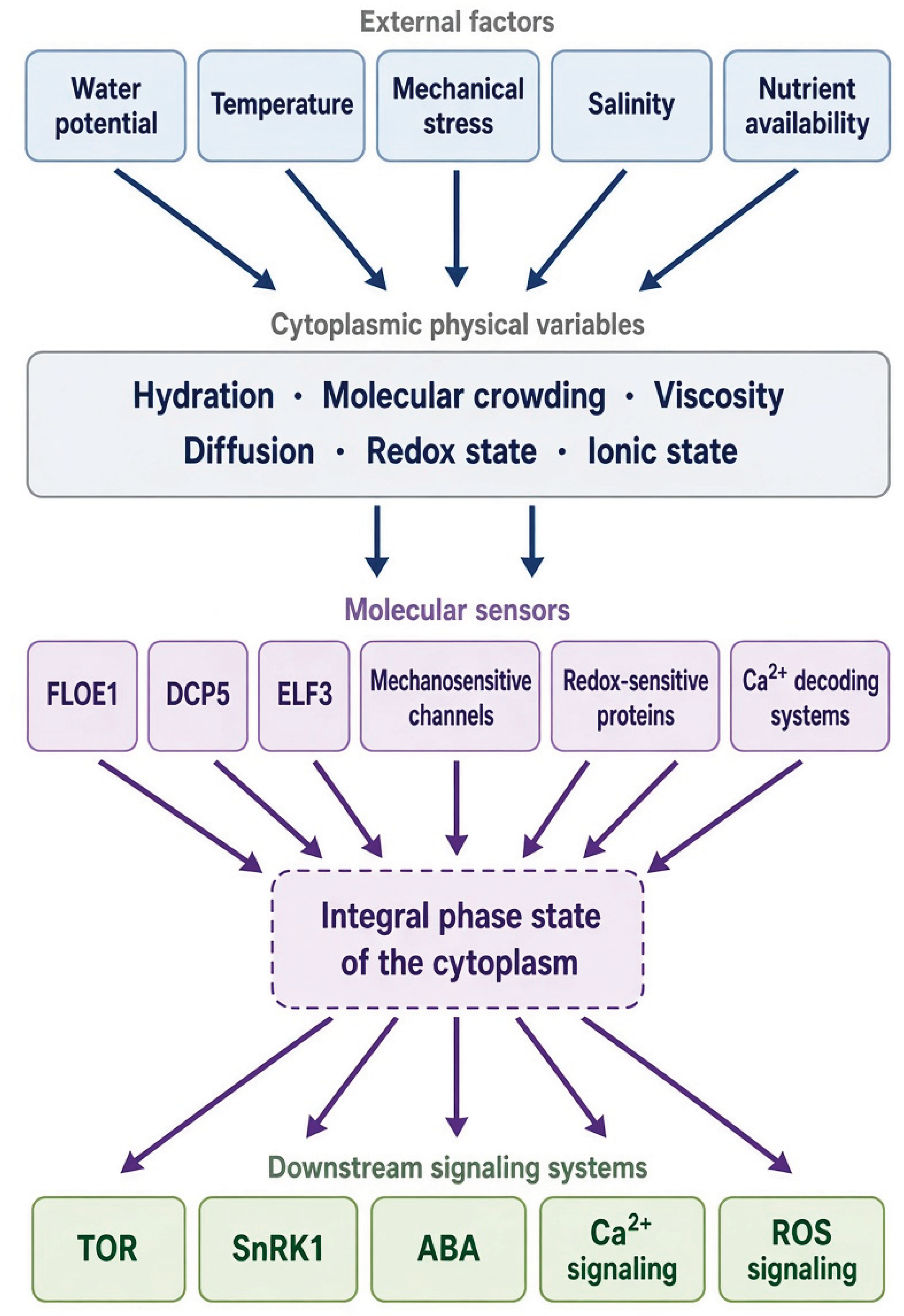
Figure 2.
Context-dependent switching between TOR–SnRK1 states: from metabolic antagonism to adaptive cellular-state reorganization. Conceptual framework illustrating four functional cellular states: the growth state (high TOR / low SnRK1), transition state (TOR and SnRK1 activity), adaptive survival state (high SnRK1 / low TOR, with autophagy activation), and prolonged-stress state characterized by chronic energy deficiency, enhanced degradation, risk of cytoplasmic phase rigidity, and growth suppression. Arrows between states indicate potentially reversible transitions, whereas the overall direction represents increasing stress intensity and decreasing energy availability.
Figure 2.
Context-dependent switching between TOR–SnRK1 states: from metabolic antagonism to adaptive cellular-state reorganization. Conceptual framework illustrating four functional cellular states: the growth state (high TOR / low SnRK1), transition state (TOR and SnRK1 activity), adaptive survival state (high SnRK1 / low TOR, with autophagy activation), and prolonged-stress state characterized by chronic energy deficiency, enhanced degradation, risk of cytoplasmic phase rigidity, and growth suppression. Arrows between states indicate potentially reversible transitions, whereas the overall direction represents increasing stress intensity and decreasing energy availability.
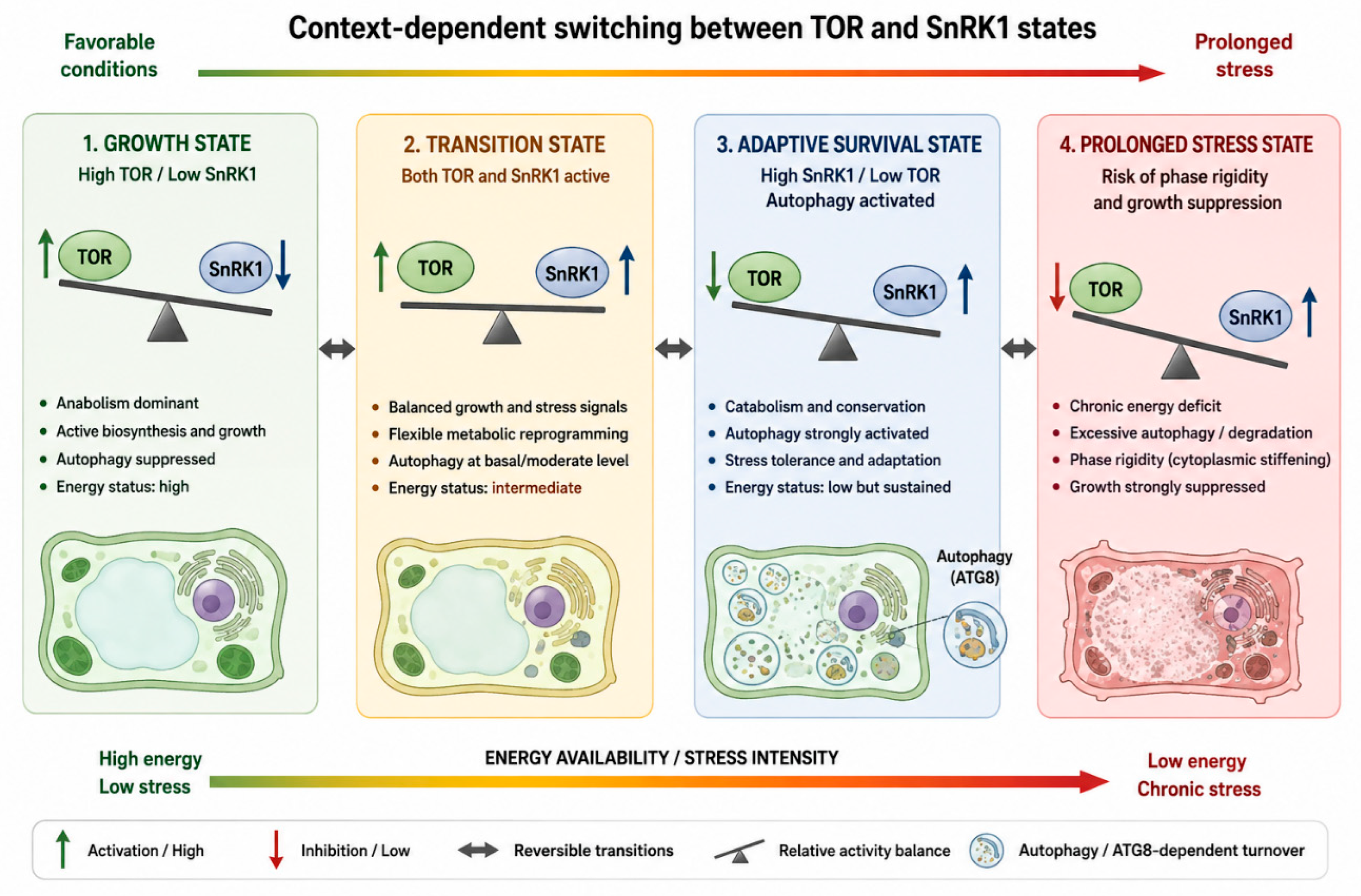
Figure 3.
Model of FLZ-mediated molecular state buffering. Conceptual model of a causal chain in which a physical or metabolic perturbation alters FLZ abundance, post-translational state, conformation, and molecular interaction profile. The altered FLZ state may, in turn, influence SnRK1 complex configuration and its activation threshold, thereby directing context-dependent switching between TOR–SnRK1 states. Subsequent metabolic reprogramming promotes cellular adaptation and functional recovery. The dashed arrow denotes a hypothesized feedback mechanism involving FLZ turnover, autophagy, and regulation of the SnRK1–TOR axis.
Figure 3.
Model of FLZ-mediated molecular state buffering. Conceptual model of a causal chain in which a physical or metabolic perturbation alters FLZ abundance, post-translational state, conformation, and molecular interaction profile. The altered FLZ state may, in turn, influence SnRK1 complex configuration and its activation threshold, thereby directing context-dependent switching between TOR–SnRK1 states. Subsequent metabolic reprogramming promotes cellular adaptation and functional recovery. The dashed arrow denotes a hypothesized feedback mechanism involving FLZ turnover, autophagy, and regulation of the SnRK1–TOR axis.
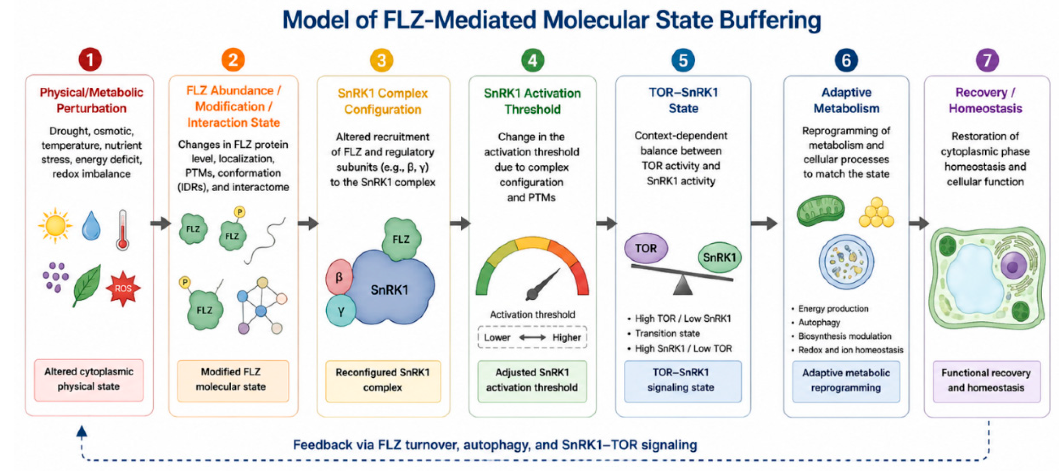
Figure 4.
Autophagy-dependent removal of FLZ as a positive feedback switch for SnRK1 activation. Cyclic scheme under energy limitation: energy restriction triggers transcriptional repression of FLZ and ATG8-dependent FLZ degradation → loss of SnRK1 repression → SnRK1 activation → induction of autophagy → further FLZ degradation, thereby closing a positive feedback loop. The parallel pathway under sufficient energy supply maintains high FLZ levels, keeps SnRK1 activity limited, and supports a growth-compatible cellular state. FLZ functions as a molecular switch between the two states.
Figure 4.
Autophagy-dependent removal of FLZ as a positive feedback switch for SnRK1 activation. Cyclic scheme under energy limitation: energy restriction triggers transcriptional repression of FLZ and ATG8-dependent FLZ degradation → loss of SnRK1 repression → SnRK1 activation → induction of autophagy → further FLZ degradation, thereby closing a positive feedback loop. The parallel pathway under sufficient energy supply maintains high FLZ levels, keeps SnRK1 activity limited, and supports a growth-compatible cellular state. FLZ functions as a molecular switch between the two states.
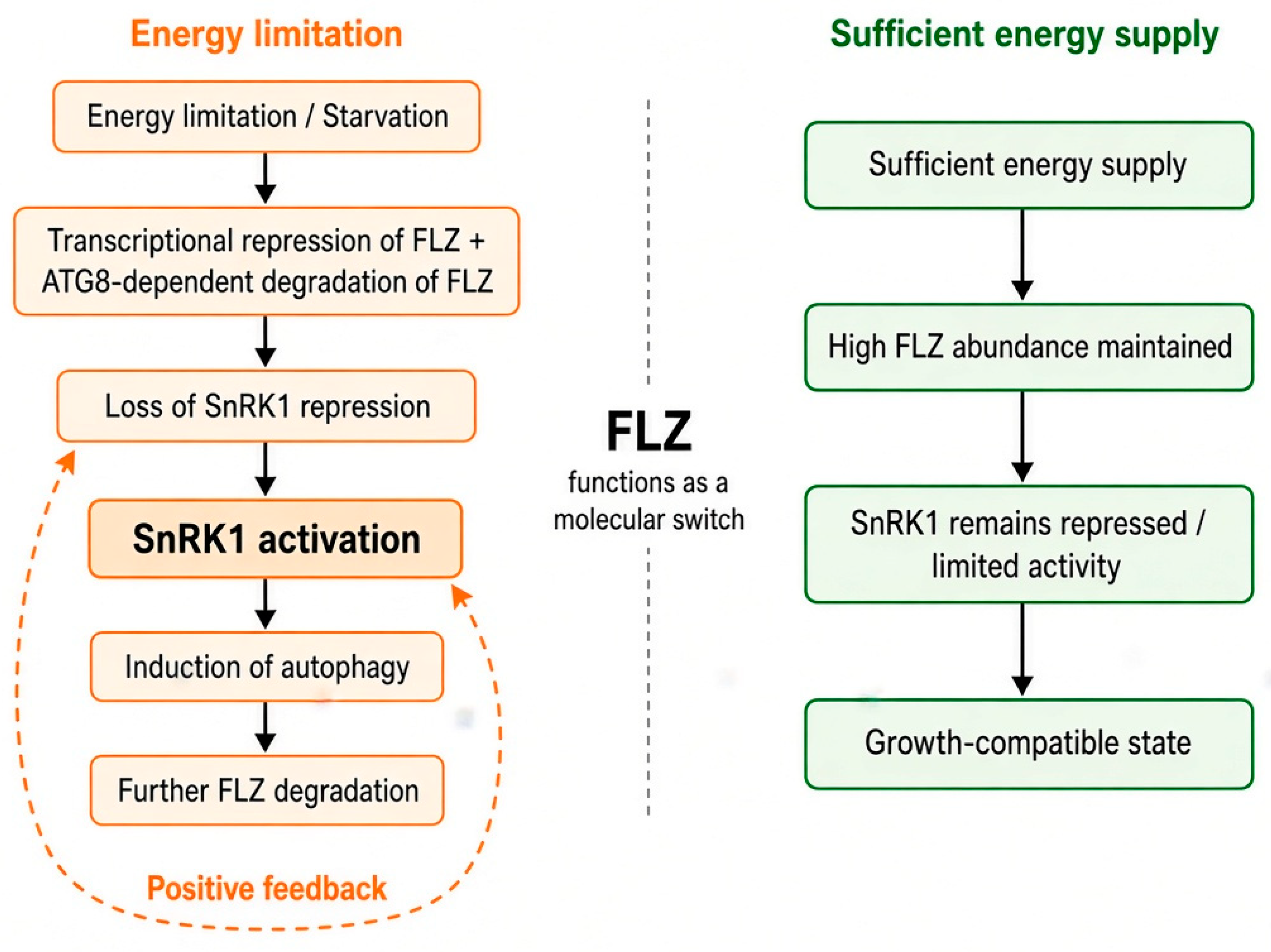
Figure 5.
Hypothetical link between FLZ–SnRK1 complexes and stress-induced biomolecular condensates. Schematic representation of three progressive material states of condensates: liquid-like signaling environment → adaptive reversible condensate → stable/rigid condensate. Solid arrows indicate the experimentally established FLZ–SnRK1 interaction. Dashed lines represent the hypothesized role of FLZ as a candidate factor that may couple the organization of the SnRK1 complex with the material state of the condensate under increasing stress.
Figure 5.
Hypothetical link between FLZ–SnRK1 complexes and stress-induced biomolecular condensates. Schematic representation of three progressive material states of condensates: liquid-like signaling environment → adaptive reversible condensate → stable/rigid condensate. Solid arrows indicate the experimentally established FLZ–SnRK1 interaction. Dashed lines represent the hypothesized role of FLZ as a candidate factor that may couple the organization of the SnRK1 complex with the material state of the condensate under increasing stress.
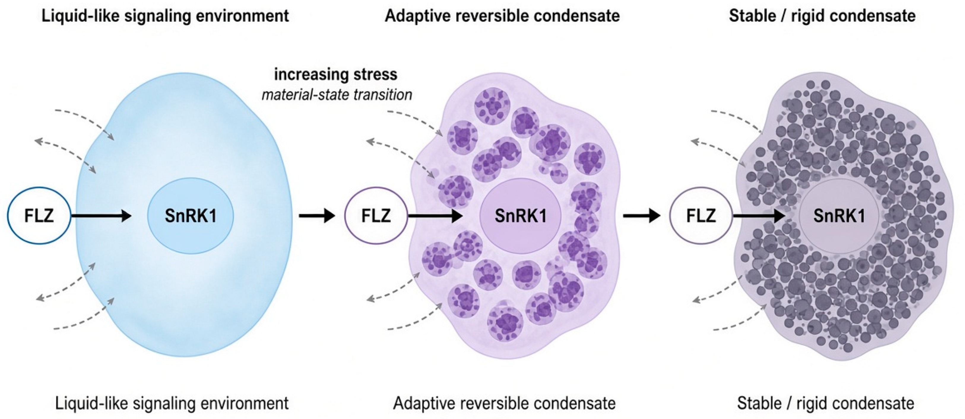
Figure 7.
The FLZ–SnRK1–TOR–Autophagy Phase-State Switching Framework. Schematic model depicting the integrated signaling pathway from environmental cues to cellular metabolic outcomes. Environmental stress alters the cytoplasmic physical state, triggering distributed physical sensing by FLOE1, DCP5, ELF3, and mechanosensors. Physical sensing is hypothesized to couple to the FLZ molecular state, which subsequently regulates the SnRK1 complex configuration and activation threshold. Downstream signaling operates via the TOR–SnRK1 metabolic switch, promoting autophagy. Autophagy drives both ATG8-dependent FLZ degradation (established for the AIM-containing clade) and broader metabolic reprogramming, ultimately directing cellular fate toward recovery, persistent stress, or phase rigidity. Solid lines indicate experimentally established biochemical and metabolic transitions; dashed lines denote hypothesized biophysical couplings and regulatory thresholds.
Figure 7.
The FLZ–SnRK1–TOR–Autophagy Phase-State Switching Framework. Schematic model depicting the integrated signaling pathway from environmental cues to cellular metabolic outcomes. Environmental stress alters the cytoplasmic physical state, triggering distributed physical sensing by FLOE1, DCP5, ELF3, and mechanosensors. Physical sensing is hypothesized to couple to the FLZ molecular state, which subsequently regulates the SnRK1 complex configuration and activation threshold. Downstream signaling operates via the TOR–SnRK1 metabolic switch, promoting autophagy. Autophagy drives both ATG8-dependent FLZ degradation (established for the AIM-containing clade) and broader metabolic reprogramming, ultimately directing cellular fate toward recovery, persistent stress, or phase rigidity. Solid lines indicate experimentally established biochemical and metabolic transitions; dashed lines denote hypothesized biophysical couplings and regulatory thresholds.
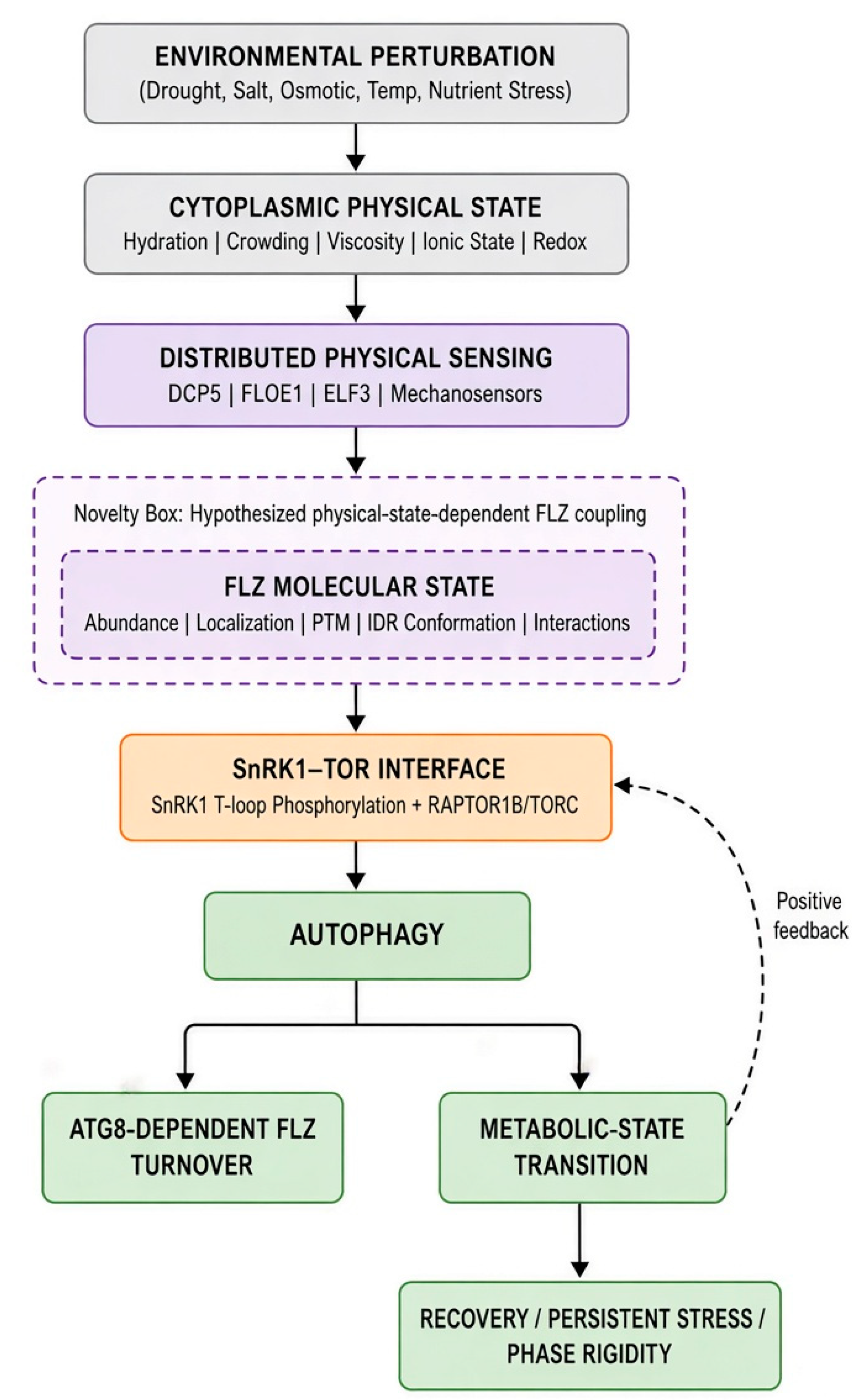
Figure 9.
Evolutionary and Biophysical Architecture of FLZ Proteins Across Crops. A schematic comparison of the conserved FLZ domain against the variable, lineage-specific IDR content of representative FLZ proteins from Arabidopsis, maize, wheat and rice, with an explicit annotation that variable IDR architecture is a candidate, not confirmed, basis for differential SnRK1/condensate interaction.
Figure 9.
Evolutionary and Biophysical Architecture of FLZ Proteins Across Crops. A schematic comparison of the conserved FLZ domain against the variable, lineage-specific IDR content of representative FLZ proteins from Arabidopsis, maize, wheat and rice, with an explicit annotation that variable IDR architecture is a candidate, not confirmed, basis for differential SnRK1/condensate interaction.
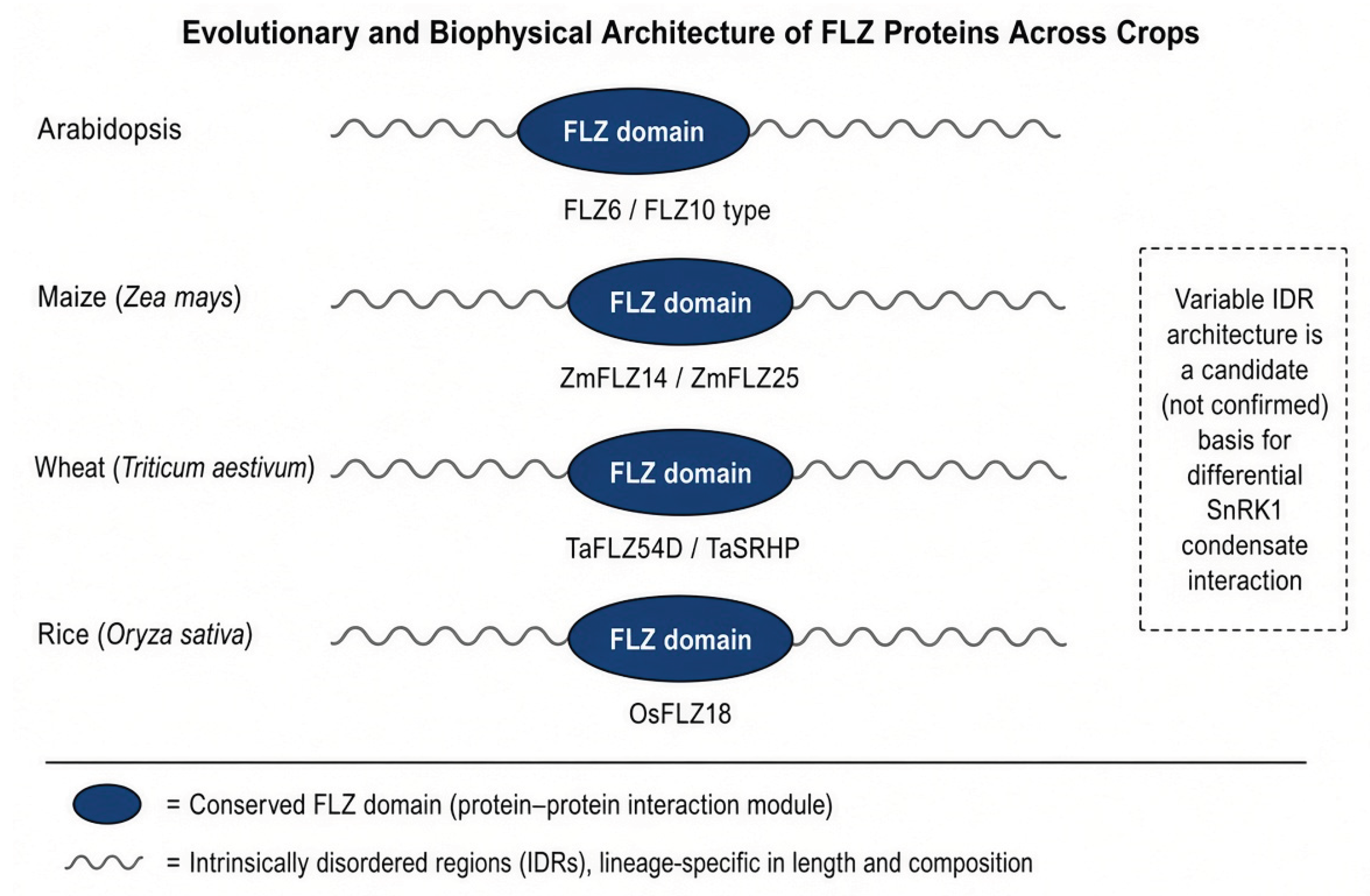
Figure 10.
Phase plasticity determines whether stress adaptation remains reversible. Two trajectories through the states presented in Table 3: one that returns to homeostasis within the Recovery Window, and another that proceeds past the point of irreversible phase failure. The scheme indicates the regions where, according to the proposed hypothesis, depletion and recovery of the FLZ pool may influence trajectory position.
Figure 10.
Phase plasticity determines whether stress adaptation remains reversible. Two trajectories through the states presented in Table 3: one that returns to homeostasis within the Recovery Window, and another that proceeds past the point of irreversible phase failure. The scheme indicates the regions where, according to the proposed hypothesis, depletion and recovery of the FLZ pool may influence trajectory position.
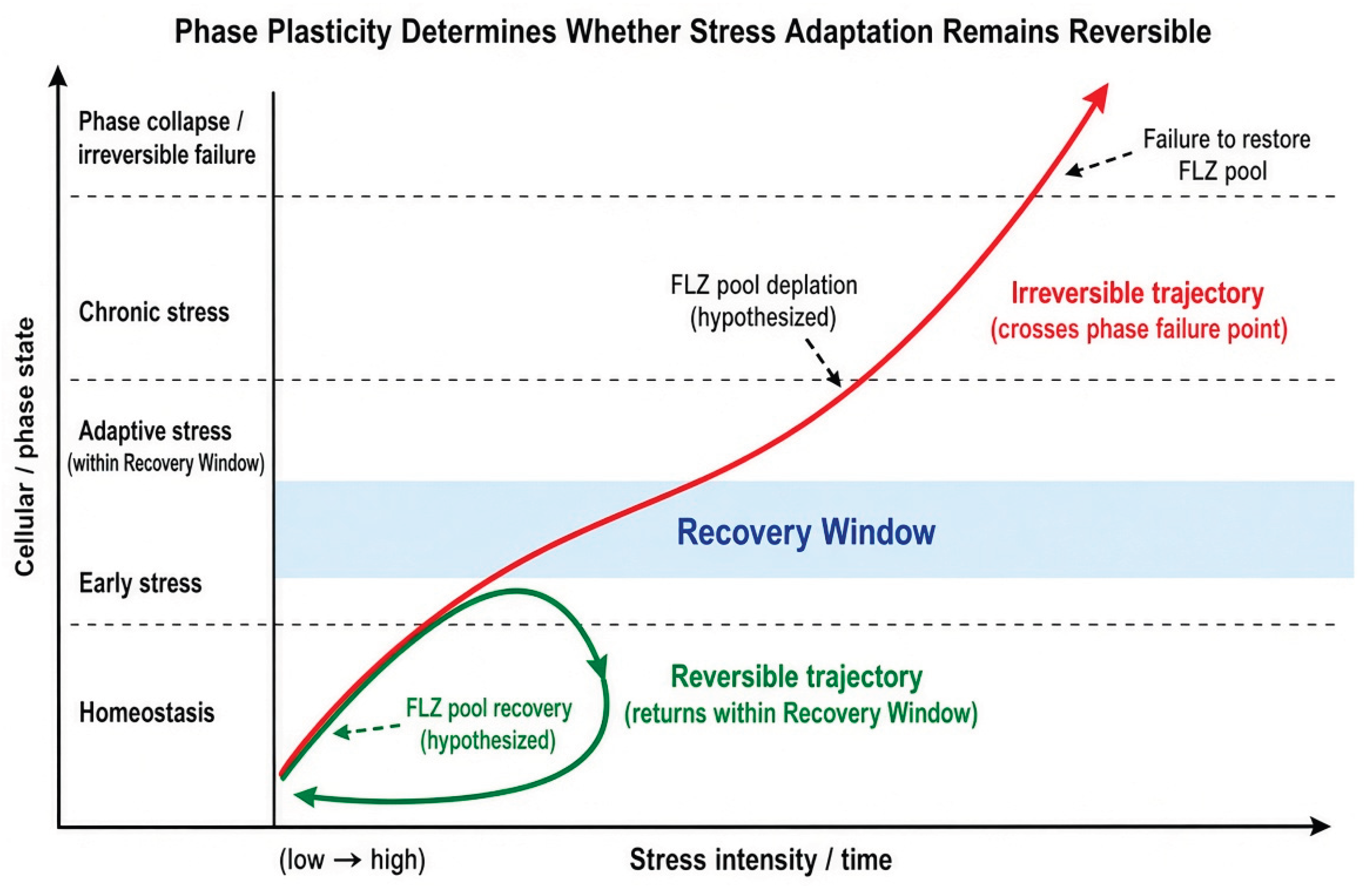
Figure 12.
Integrated FLZ–SnRK1–TOR–Autophagy model of plant stress-state transitions. Environmental stress branches into water/crowding/energy–redox inputs, each read by a distinct physical sensor (FLOE1, DCP5, metabolic sensors), converging on cytoplasmic phase state, which feeds into the FLZ state buffer. FLZ state sets the TOR–SnRK1 balance; SnRK1/TOR signalling feeds into autophagy; autophagy degrades FLZ, closing a positive feedback loop back to SnRK1 activation. The resulting adaptive state branches into recovery/homeostasis or phase rigidity/damage. Established pathway segments are shown with solid lines; hypothesized segments (FLZ–physical-state and FLZ–condensate links) are shown with dashed lines.
Figure 12.
Integrated FLZ–SnRK1–TOR–Autophagy model of plant stress-state transitions. Environmental stress branches into water/crowding/energy–redox inputs, each read by a distinct physical sensor (FLOE1, DCP5, metabolic sensors), converging on cytoplasmic phase state, which feeds into the FLZ state buffer. FLZ state sets the TOR–SnRK1 balance; SnRK1/TOR signalling feeds into autophagy; autophagy degrades FLZ, closing a positive feedback loop back to SnRK1 activation. The resulting adaptive state branches into recovery/homeostasis or phase rigidity/damage. Established pathway segments are shown with solid lines; hypothesized segments (FLZ–physical-state and FLZ–condensate links) are shown with dashed lines.
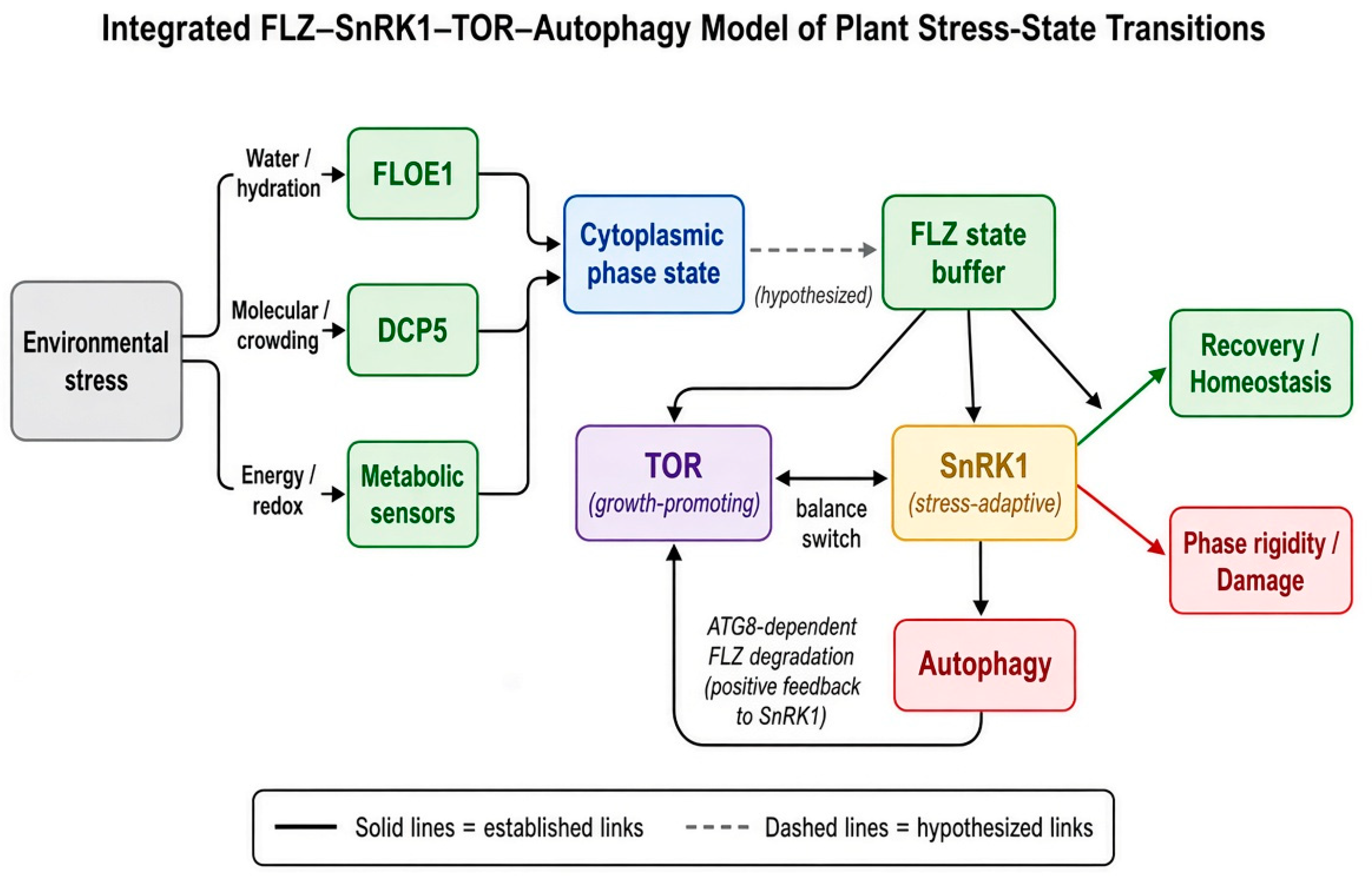
Table 1.
Established and candidate molecular systems involved in cytoplasmic physical-state sensing.
Table 1.
Established and candidate molecular systems involved in cytoplasmic physical-state sensing.
| Physical variable | Molecular candidate | Type of mechanism | Level of evidence | Significance for the model |
| Hydration | FLOE1 | Hydration-dependent phase separation | Experimentally established in vivo and in vitro [22] | Converts water status into an adaptive developmental output |
| Molecular crowding | DCP5 | Crowding-sensitive conformational change and phase separation | Strong, in planta and in vitro [23] | Proof-of-principle for crowding-triggered osmosensing |
| Temperature | ELF3 | Temperature-dependent condensation of a prion-like domain | Experimentally established [24] | Converts temperature into a transcriptional/developmental switch |
| Mechanical tension | MSL / MCA / TPK channels | Membrane-tension-gated ion flux | Experimentally established [21] | Converts turgor/volume change into ionic signal |
| Ca²⁺ | CBL–CIPK, CDPK and related decoding systems | Stimulus-specific Ca²⁺ signature decoding | Experimentally established for individual components [18,19] | Rapid, stimulus-specific systemic signaling |
| Redox / ROS | Cysteine-based redox-sensitive proteins; RBOH-dependent ROS wave | Post-translational oxidative modification; systemic propagation | Experimentally established for specific components [20] | Integrates oxidative and osmotic stress information |
| Cytoplasmic viscosity | Condensate-associated proteins (general) | Physical-state-dependent assembly | Hypothesis, inferred from crowding/LLPS biophysics | Candidate general mechanism for phase-state sensing |
| Cytoplasmic macromolecular mobility | IDR/condensate-forming proteins (general) | Diffusion- and interaction-kinetics-dependent signaling | Hypothesis | Candidate link between physical state and downstream kinetics |
Table 2.
FLZ proteins: established functions, emerging evidence, knowledge gaps and proposed roles.
| Level | Established | Remaining knowledge gap | Hypothesis proposed here |
| FLZ–SnRK1 interaction | FLZ domain binds SnRK1α; IDRs bind β/βγ subunits [34,36] | How the complex composition changes over time and under different stresses | FLZ sets a variable threshold for SnRK1 switching |
| Intrinsically disordered regions | IDRs mediate protein–protein interaction and FLZ dimerization [36] | Whether IDRs confer any phase-separation propensity | IDRs may enable state-dependent molecular assembly |
| Autophagy connection | ATG8–FLZ–SnRK1 feedback demonstrated for a defined FLZ clade [39,40,41,42] | Downstream consequences for cytoplasmic phase organization | FLZ degradation reorganizes cellular physical state |
| Maize FLZ | ZmFLZ14 functionally linked to energy-deprivation tolerance [39]; ZmFLZ25 linked to ABA signaling | Mechanism under field-relevant combined stress | Cereal FLZ repertoires set stress-state thresholds |
| Wheat FLZ | TaFLZ54D increases salt tolerance via TaSGT1/TaPP2C; TaSRHP improves salt/drought tolerance in transgenic Arabidopsis | Relationship to SnRK1/TOR switching in wheat | FLZ proteins act as crop-specific state buffers |
| LLPS | Direct evidence of FLZ-driven LLPS is currently insufficient | Mechanism of any FLZ–condensate coupling | FLZ may modulate condensate-associated signaling |
Table 3.
Predicted cytoplasmic phase states and corresponding TOR–SnRK1–autophagy configurations across a stress trajectory.
Table 3.
Predicted cytoplasmic phase states and corresponding TOR–SnRK1–autophagy configurations across a stress trajectory.
| State | Hypothesized cytoplasmic properties | TOR | SnRK1 | Autophagy | Condensate organization | Physiological outcome |
| Homeostasis | High molecular mobility | High | Basal | Basal | Dynamic, liquid-like | Growth |
| Early stress | Moderate crowding increase | Declining | Rising | Rising | Reversible condensates forming | Adaptation |
| Adaptive stress | Increased viscosity | Low | High | High | Dynamic but stress-associated condensates | Survival, within the Recovery Window |
| Chronic stress | High, sustained crowding | Low | High | High | Persistent condensates | Growth suppression, approaching the Recovery Window’s edge |
| Phase collapse | Loss of phase plasticity | Suppressed | Dysregulated | Dysregulated/impaired | Persistent, non-dynamic aggregate | Irreversible damage or death |
Table 5.
Key testable predictions of the FLZ–SnRK1–TOR–autophagy hypothesis.
| # | Established mechanism | Hypothesis | Prediction | Experiment | Expected result if hypothesis holds |
| 1 | FLZ represses SnRK1 activity [37] | FLZ sets the SnRK1 activation threshold, not only its level | FLZ loss-of-function lowers the stress intensity required for half-maximal SnRK1 activation | Dose–response SnRK1 activation assay across FLZ genotypes | Leftward shift of the activation curve in flz mutants |
| 2 | FLZ abundance is stress-regulated [33,37] | FLZ recovery kinetics set the recovery threshold | flz mutants show altered (likely delayed or hastened) recovery of TOR activity after stress release | Time-course TOR/SnRK1 reporter assay after stress removal | Genotype-dependent difference in recovery kinetics |
| 3 | FLZ IDRs mediate SnRK1-subunit binding [36] | IDRs additionally affect SnRK1 complex dynamics under physical perturbation | ΔIDR FLZ shows altered SnRK1-complex assembly specifically under crowding stress | Co-immunoprecipitation/imaging under varied osmotic conditions, WT vs ΔIDR | Condition-dependent difference specific to ΔIDR |
| 4 | ATG8-dependent FLZ degradation activates SnRK1 [39] | Autophagy manipulation changes FLZ abundance and downstream state transitions predictably | Autophagy-deficient lines show blunted FLZ turnover and delayed SnRK1 activation | FLZ abundance and SnRK1 activity in atg mutants under starvation | Reduced FLZ turnover and reduced/delayed SnRK1 activation |
| 5 | FLZ localizes to discrete cytoplasmic foci [36] | FLZ relocalizes under physical stress | FLZ-GFP shows altered focus number/size/mobility under osmotic or thermal stress | Live imaging time course | Stress-dependent change in FLZ punctum properties |
| 6 | FLZ structural features resemble LLPS-competent proteins [36] | FLZ influences condensate dynamics | Purified FLZ undergoes phase separation under defined crowding/salt conditions in vitro | Reconstituted in vitro phase-separation assay | Concentration- and condition-dependent droplet formation |
| 7 | Hysteresis is a generic property of feedback-based switches [48,49,50] | FLZ contributes to stress-state hysteresis | Entry and recovery thresholds for SnRK1 activation differ measurably, and this difference is reduced in flz mutants | Paired entry/recovery threshold measurement, WT vs flz | Narrower or absent hysteresis gap in flz mutants |
| 8 | Crop FLZ genes are individually characterized [39,51,52,53,54] | Crop FLZ variants set different stress thresholds | Wheat/maize FLZ alleles show quantitatively different SnRK1 activation thresholds under matched stress | Cross-genotype dose–response comparison | Allele-dependent threshold variation |
| 9 | ATG8–FLZ–SnRK1 feedback is established [39,40] | FLZ manipulation changes autophagic flux, not only SnRK1 activity | FLZ overexpression suppresses, and FLZ loss enhances, autophagic flux under matched starvation | Autophagic flux assay (e.g., autophagosome quantification) across FLZ genotypes | Genotype-dependent flux consistent with FLZ’s repressive role |
| 10 | FLZ–SnRK1 interaction is established [34,36] | This coupling is physical-state dependent | FLZ–SnRK1 co-localization/interaction strength varies with crowding/osmotic state independent of SnRK1 abundance | Quantitative co-localization or interaction assay across osmotic conditions | Condition-dependent change in interaction/co-localization |
Table 6.
Established evidence, emerging concepts, knowledge gaps and testable predictions for each component of the model.
Table 6.
Established evidence, emerging concepts, knowledge gaps and testable predictions for each component of the model.
| Component | Established | Emerging | Unknown | Testable prediction |
| Cytoplasmic crowding | Influences protein folding, assembly and diffusion [10,11] | May act as a general stress-sensing variable in plants | How crowding integrates quantitatively with metabolic signaling | Crowding-dependent SnRK1/TOR activation kinetics |
| DCP5 | Crowding-sensitive osmosensing, established in planta [23] | May represent a general physical-sensing principle | Whether analogous crowding sensors exist beyond DCP5/SEU | Discovery of additional crowding-sensitive plant sensors |
| FLZ–SnRK1 | Physical protein–protein interaction [34,36] | Candidate state-buffering function | Mechanism of any threshold-setting effect | FLZ genotype shifts the SnRK1 activation threshold |
| FLZ–ATG8 | Feedback loop demonstrated for AIM-containing clade [39,40] | Candidate general state-switch mechanism | Relationship to cytoplasmic condensate dynamics | Autophagy manipulation alters FLZ-linked phase organization |
| FLZ–LLPS | Direct evidence currently insufficient | Structurally plausible coupling (IDRs, multivalency) [36] | Molecular mechanism, if any, entirely unknown | FRAP and in vitro reconstitution assays |
| TOR–SnRK1 | Central, antagonistic metabolic regulators [1,3,25,26] | Dynamic, context-dependent state switch | Spatial/temporal co-existence within single cells | Single-cell dual-reporter imaging |
| Autophagy | Feedback regulator of SnRK1 [39,40,43] | Candidate state-reset mechanism | Recovery-phase dynamics of autophagic flux | Manipulation of autophagy alters hysteresis width |
| Phase plasticity | Conceptually supported by CPHT [4,5,6] | Candidate resilience variable | Quantitative metric (e.g., PRI) | Recovery kinetics predict genotype-level stress tolerance |
Disclaimer/Publisher’s Note: The statements, opinions and data contained in all publications are solely those of the individual author(s) and contributor(s) and not of MDPI and/or the editor(s). MDPI and/or the editor(s) disclaim responsibility for any injury to people or property resulting from any ideas, methods, instructions or products referred to in the content. |
© 2026 by the authors. Licensee MDPI, Basel, Switzerland. This article is an open access article distributed under the terms and conditions of the Creative Commons Attribution (CC BY) license (http://creativecommons.org/licenses/by/4.0/).
Copyright: This open access article is published under a Creative Commons CC BY 4.0 license, which permit the free download, distribution, and reuse, provided that the author and preprint are cited in any reuse.