

Submitted:
28 August 2026
Posted:
31 August 2026
You are already at the latest version
Abstract
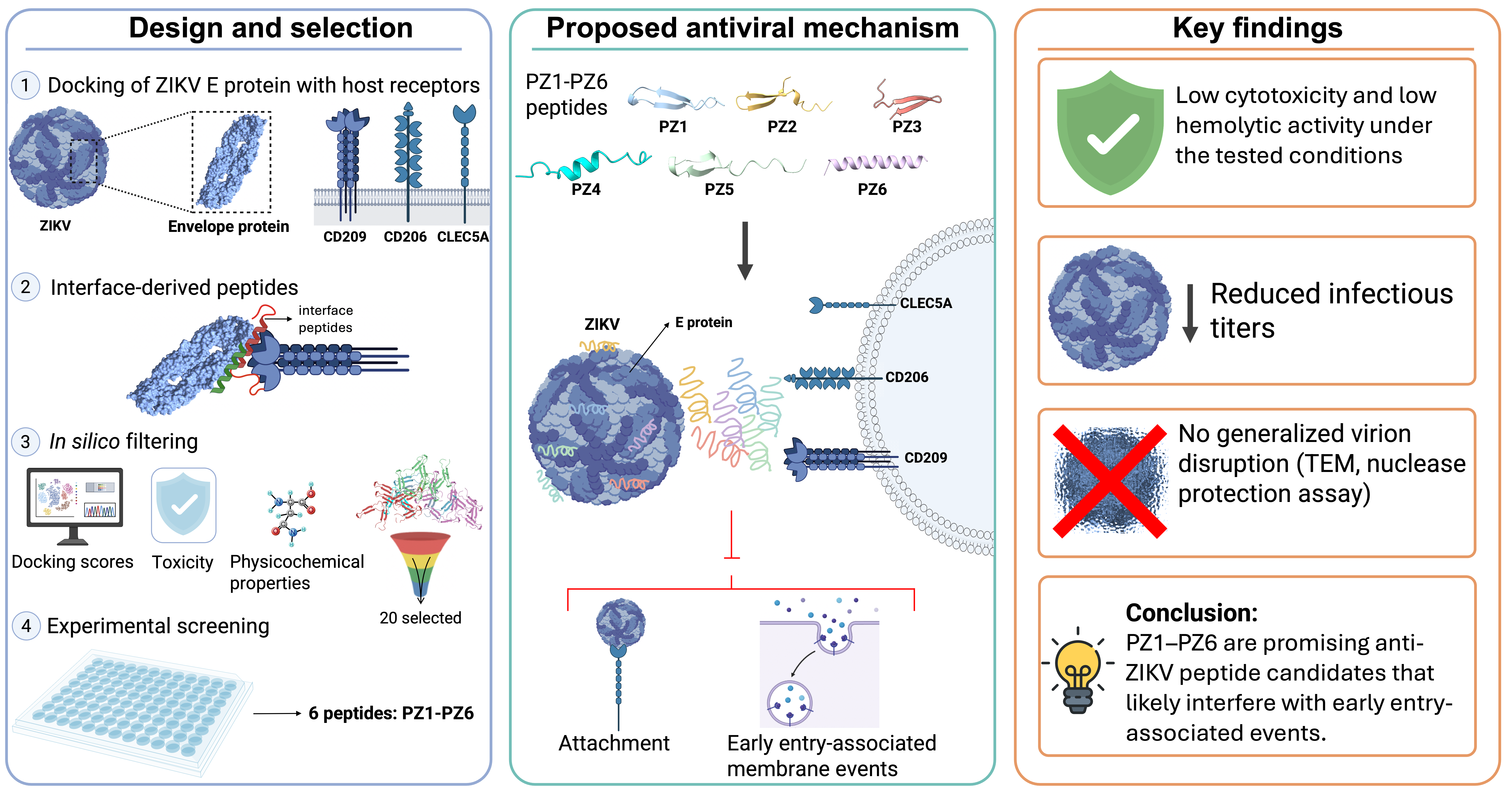
Zika virus (ZIKV) remains a public health concern because of its association with neurological complications and congenital disease, yet no specific antiviral therapy has been approved. Since the envelope (E) protein mediates viral attachment and early entry-associated membrane events, it represents an attractive antiviral target. Here, we used a protein-protein interaction-guided strategy to design peptides to interfere with predicted interfaces between the ZIKV E protein and cellular receptors involved in attachment. After in silico filtering and preliminary screening, six peptides (PZ1-PZ6) were selected. These peptides showed heterogeneous physicochemical profiles, low cytotoxicity and hemolytic activity under the tested conditions, and predicted binding to functional E protein regions. In HTR-8/SVneo cells, antiviral activity depended on the timing of exposure, with greater reductions in infectious titers during co-treatment and post-treatment. All peptides also reduced titers in anti-adsorption assays, supporting interference with viral attachment or early entry-associated events. Consistently, DiOC18 fluorescence signals were reduced in a peptide-specific manner, particularly for PZ3 and PZ6. Nuclease protection assays showed no extensive exposure of the viral genome, whereas transmission electron microscopy revealed ultrastructural alterations without generalized particle disruption. Overall, PZ1–PZ6 are promising protein–protein interaction-guided anti-ZIKV peptide candidates with activity against early viral entry-associated events.
Keywords:
Zika virus
; antiviral agents
; antimicrobial peptides
; computational biology
; viral fusion proteins
; virus internalization
1. Introduction
Zika virus (ZIKV) is an enveloped, positive-sense single-stranded RNA arbovirus of the family Flaviviridae and genus Orthoflavivirus, transmitted mainly by Aedes mosquitoes [1]. Although initially considered of limited clinical relevance after its identification in Uganda in 1947, outbreaks in Yap Island, French Polynesia, and the Americas revealed its association with neurological complications and congenital abnormalities [1,2,3]. Despite the decline in global incidence after 2017, ZIKV continues to circulate in tropical and subtropical regions, where vector expansion, urbanization, human mobility, climate change, and diagnostic cross-reactivity with dengue and other flaviviruses complicate surveillance and control [3,4]. Clinically, ZIKV infection is often mild or asymptomatic, but it can cause febrile illness and has been associated with Guillain-Barré syndrome and other neurological complications [5,6,7]. During pregnancy, ZIKV can infect placental cells and neural progenitors, leading to fetal growth restriction, microcephaly, and other manifestations of congenital Zika syndrome, including postnatal neurodevelopmental alterations [8,9,10].
The ZIKV genome is approximately 10.7 kb and encodes a single polyprotein that is processed into three structural proteins (capsid, premembrane/membrane, and envelope (E)) and seven non-structural proteins [1]. The E protein mediates viral attachment, pH-dependent conformational rearrangements, and membrane fusion, making it a relevant target for entry-directed antiviral strategies [11,12]. No specific antiviral treatment or vaccine has been approved for ZIKV infection; therefore, clinical management remains supportive [1,3,11,13,14]. Although monoclonal antibodies, immunoglobulins, repurposed drugs, and viral protein inhibitors have been explored, none has become standard therapy [11,15,16]. In this context, antiviral peptides are promising candidates because they can interfere with protein-protein interactions, bind functional viral regions, or disrupt envelope-associated steps such as attachment and membrane fusion. Peptides derived from conserved viral regions, host proteins, or antimicrobial molecules have shown inhibitory activity against ZIKV and related flaviviruses [11,17,18,19]. Based on this rationale, we evaluated peptides designed from predicted ZIKV E protein-host receptor interfaces and explored their ability to interfere with early viral entry-associated events.
2. Materials and Methods
2.1. In Silico Peptide Design
2.1.1. Modeling of ZIKV Envelope Protein Interactions with Human Cellular Receptors
X-ray crystallographic structures with resolution better than 3 Å were retrieved from the Protein Data Bank (PDB). The ZIKV E protein structure (PDB: 5JHM) was used as the viral ligand, and a neutralizing anti-ZIKV antibody bound to E domain III (PDB: 5VIG) was included as a reference for inhibitory interfaces [15]. Representative C-type lectin receptors involved in flavivirus attachment [DC-SIGN/CD209 (PDB: 1XPH), MR/CD206 (PDB: 5XTS), and CLEC5A/MDL-1 (PDB: 2YHF)] were selected because they provide defined receptor-envelope interfaces, whereas TAM and TIM receptors were excluded because they primarily mediate entry through phosphatidylserine-associated ligands rather than direct E protein binding [12]. Glycan chains were not modeled, and simulations focused on protein-protein contacts involving the ZIKV E ectodomain. Docking was performed using ClusPro 2.0 [20], and balanced models were selected for interface analysis and peptide candidate identification.
2.1.2. Selection of Inhibitory Peptides Derived from Protein-Protein Interactions
PeptiDerive from the Rosetta Online Server was used to identify peptide fragments derived from receptor-virus interaction interfaces [21,22,23]. From the candidate peptide pool generated from the predicted interfaces, peptides with interface energy scores below −10 Rosetta Energy Units (REU) and high relative interface score contributions were prioritized; toxic sequences predicted by ToxinPred [24] were excluded, and the remaining candidates were evaluated for hydropathicity and predicted solubility using ProtParam [25] and PepCalc. Based on these criteria, twenty peptides were selected for structural modeling with AlphaFold2 [26] and peptide–E protein docking using ClusPro 2.0. Balanced models with the lowest binding energies were analyzed and visualized using UCSF ChimeraX [27]. The same twenty peptides were synthesized and subjected to preliminary antiviral screening.
2.2. Peptide Synthesis
Peptides were custom-synthesized by Peptides 2.0 Inc. using standard solid-phase peptide synthesis, purified to >95% by RP-HPLC, and verified by mass spectrometry.
2.3. Cell Lines and Virus Propagation
Vero cells were used for preliminary antiviral screening and plaque assay, C6/36 mosquito cells for ZIKV propagation, and HTR-8/SVneo trophoblast cells as a human placental model relevant to ZIKV infection. Vero, C6/36, and HTR-8/SVneo cells were cultured in DMEM, Leibovitz L-15, and RPMI-1640, respectively. Growth media were supplemented with 10% fetal bovine serum (FBS), 2 mM L-glutamine, and 1× penicillin-streptomycin (PS); maintenance media contained 2% FBS with the same supplements. C6/36 medium was additionally supplemented with 10% tryptose phosphate broth (TPB). Vero and HTR-8/SVneo cells were maintained at 37 ºC with 5% CO₂, whereas C6/36 cells were incubated at 28 ºC.
ZIKV stocks were propagated in C6/36 cells at a multiplicity of infection (MOI) of 0.1 for 5-6 days at 28 ºC using maintenance medium. Viral supernatants were clarified by centrifugation at 5000 × g, filtered through a 0.22 µm membrane, and stored at -80 ºC until use. RT-qPCR confirmed viral identity.
2.4. Cell Viability and Hemolysis Assays
HTR-8/SVneo cells (1×10⁴ cells/well) were seeded in 96-well plates 24 h before the assay. Cells were washed with PBS, and 100 µL of maintenance medium containing two-fold serial dilutions of the peptides (25 to 400 µM) were added. Untreated cells were used as the viability control, while cells treated with 0.1% Triton X-100 served as a cell death control. Twenty-four hours post-incubation, cell viability was determined using the MTT assay [3-(4,5-dimethylthiazol-2-yl)-2,5-diphenyltetrazolium bromide] (Thermo-Scientific), following the manufacturer’s instructions; Absorbance was measured at 570 nm, with background correction at 630 nm.
For the hemolysis assays, human blood samples from healthy volunteers were collected into EDTA-K₃ Vacutainer tubes, following institutional ethical guidelines and informed consent. Samples were centrifuged at 400 × g for 5 min, and erythrocytes were separated, diluted 1:20 in PBS, and washed four times. Erythrocyte suspensions were dispensed into 96-well polypropylene plates (90 µL/well) and incubated with two-fold serial dilutions of peptides (50-400 µM) for 24 h at 37 ºC. The peptide vehicle was used as the negative control, and 0.1% Triton X-100 served as the hemolysis control. After centrifugation at 400 × g for 5 min, supernatant absorbance was measured at 541 nm using an Epoch BioTek spectrophotometer. Hemolysis (%) was calculated as [(A_sample − A_negative control)/(A_Triton X-100 − A_negative control)] × 100.
2.5. Plaque Assay, Virus Neutralization Screening, and Anti-Adsorption Assay
Viral titers were determined by plaque assay in Vero cells seeded in 24-well plates at 1×10⁵ cells/well 24 h before infection. Viral inoculum from treated samples and controls were 10-fold serially diluted in DMEM, and 200 µL of each dilution was added to the cells. After 2 h of adsorption at 37 ºC with gentle rocking every 15 min, the inoculum was removed and replaced with 1 mL of overlay medium containing DMEM, 2% carboxymethylcellulose, 2% FBS, and 1× PS. Cells were incubated for 5 days, fixed with 4% formaldehyde in PBS, stained with crystal violet, and plaques were counted. Viral titers were expressed as plaque-forming units per milliliter (PFU/mL).
For preliminary antiviral screening, Vero cells were seeded as described above. ZIKV (MOI=1) was preincubated with peptides at different concentrations (200-5 µM) for 1 h at room temperature (RT) to allow virus-peptide interaction. Virus-peptide mixtures were then added to Vero cell monolayers for 1 h at 37 ºC. After adsorption, cells were washed with PBS, overlaid, incubated for 5 days, fixed, and stained as described above.
For anti-adsorption assays, Vero cells were seeded in 48-well plates at 2.5×10⁴ cells/well 24 h before infection. Cells were incubated with ZIKV (MOI=0.2) and peptides (100 µM) at 4 ºC for 1 h to allow viral attachment while limiting entry. Cells were then washed twice with PBS to remove unbound virus and peptides, overlaid, incubated for 5 days at 37 ºC, fixed, and stained as described above.
2.6. Time-of-Addition Antiviral Assay
To investigate the viral cycle stage affected by the peptides, pre-, co-, and post-infection treatments were performed in HTR-8/SVneo cells seeded in 24-well plates at 1×10⁵ cells/well 24 h before infection. For pre-treatment, cells were incubated with peptides at 100 µM for 1 h at 37 ºC, washed, and then infected with ZIKV (MOI=1) for 1 h. For co-treatment, peptides were mixed with ZIKV (MOI=1) and immediately added to cells for 1 h at 37 ºC. For post-treatment, cells were first infected with ZIKV (MOI=1) for 1 h, after which the inoculum was removed, and peptides were added at 100 µM. After each treatment, cultures were maintained in 500 µL of maintenance medium. At 24 h post-infection, supernatants were collected and stored at -80 ºC for viral titration (plaque assay), while cell monolayers were processed for total RNA extraction using the Isolate II RNA Mini Kit (BioLine) for subsequent RT-qPCR analysis. Mock-infected and untreated infected cells served as controls.
2.7. DiOC18 Fluorescence Assay
HTR-8/SVneo cells were seeded on sterile round coverslips in 24-well plates at 1×10⁵ cells/well 24h before the assay. ZIKV stock (1×10⁸ PFU/mL) was labeled with 0.2 µM DiOC18 and purified using Amicon Ultra-15 centrifugal filters with a 100 kDa molecular weight cutoff (Millipore), with three RPMI washes to remove unincorporated dye. Labeled virus was incubated with each peptide at 100 µM or with ammonium chloride (NH₄Cl, 20 mM) as an inhibitory control of low-pH-dependent entry-associated events. Virus-peptide mixtures were added to cells at 4 ºC for 30 min to allow viral binding, followed by incubation at 37 ºC for 30 min to allow temperature-dependent internalization and early entry-associated membrane events. Cells were counterstained with Hoechst, and images were acquired using an Axiolab V fluorescence microscope (Zeiss). DiOC18-positive signals were quantified using ImageJ [28] by measuring integrated green fluorescence intensity in 40X microscopic fields acquired under identical settings. Peptide effects were assessed relative to infected, mock-infected, and NH₄Cl-treated controls.
2.8. Nuclease Protection Assay
To assess whether peptide treatment exposes the viral genome to nuclease digestion, approximately 5×10⁶ PFU-equivalents of ZIKV were incubated with peptides at 100 µM for 1 h at RT. Micrococcal nuclease (150 U; New England BioLabs) was then added for 45 min at 37 ºC, and the reaction was stopped with 10 mM EDTA. Control conditions included untreated virus, virus treated with nuclease alone, and virus treated with Triton X-100 as a positive control for envelope disruption. Viral RNA was extracted and analyzed by RT-qPCR as described below.
2.9. Transmission Electron Microscopy (TEM)
ZIKV stock containing 2.25×10⁸ PFU was concentrated and buffer-exchanged into HBS buffer (20 mM HEPES, 150 mM NaCl, pH 7.4) using Amicon Ultra-15 centrifugal filters with a 100 kDa molecular weight cutoff (Millipore) at 4000 × g for 10 min. Eight conditions were prepared before fixation: six peptide-treated samples (100 µM), one virus-only control, and one acid+heat control. The acid + heat control consisted of virus incubated in acidified HBS (pH 5.6-5.8) followed by 20 min at 37 ºC and 20 min at 45 ºC to induce flaviviral envelope rearrangements. All conditions were incubated for 60 min at RT without post-treatment washing to preserve potential virus-peptide complexes. Samples were fixed with 2% glutaraldehyde in HBS and transported at RT to the Transmission Electron Microscopy Facility at Universidad El Bosque, Colombia.
2.10. RNA Extraction, Reverse Transcription, and Quantitative PCR
Total RNA was extracted from cell culture supernatants using the PureLink Viral DNA/RNA Mini Kit (Invitrogen) and from corresponding infected cell monolayers using the Isolate II RNA Mini Kit (BioLine), after removal of the supernatant. Relative ZIKV RNA levels were determined by one-step SYBR Green RT-qPCR using Luna Universal One-Step RT-qPCR Mix (New England Biolabs) on a Bio-Rad CFX96 Opus instrument. ZIKV was amplified using the primers Forward 5′-CCGCTGCCCAACACAAG-3′ and Reverse 5′-CCACTAACGTTCTTTTGCAGACAT-3′. For cell-associated RNA, levels were normalized to hGAPDH, using primers Forward 5′-TGTTGCCATCAATGACCCCTT-3′ and Reverse 5′-CTCCACGACGTACTCAGCG-3′.
2.11. Statistical Analysis
Unless otherwise specified, experiments were performed in three independent biological replicates. Technical replicates were averaged within each independent experiment before statistical analysis. Specific technical replicate numbers are indicated in the corresponding figure legends. Data are presented as mean ± SD. Normality was assessed using the Shapiro-Wilk test. Within each treatment condition, groups were compared by repeated-measures one-way ANOVA followed by Dunnett’s test against the corresponding reference control. RT-qPCR analyses used ΔCt values, while relative RNA levels are displayed as 2^-ΔΔCt. GraphPad Prism 10 was used; p<0.05 indicated significance.
3. Results
3.1. Characterization and Docking Analysis of Selected Anti-ZIKV Peptides
Twenty peptides were synthesized for preliminary in vitro screening after in silico prioritization. Although most showed acceptable cytotoxicity and hemolysis profiles under the tested conditions, only six significantly reduced infectious viral titers and were selected for further characterization and named PZ1-PZ6. These peptides displayed heterogeneous physicochemical profiles, with molecular weights ranging from 2561.91 to 3103.41 g/mol. At pH 7.0, PZ1, PZ5, and PZ6 were positively charged, PZ3 was negatively charged, and PZ2/PZ4 were near-neutral. GRAVY scores indicated hydrophilic profiles for PZ1, PZ2, PZ5, and PZ6, and comparatively more hydrophobic profiles for PZ3 and PZ4. All six peptides were predicted to be non-toxic and were soluble under the experimental conditions (Table 1).
Docking analysis predicted peptide-specific binding patterns across the ZIKV E protein. PZ1 and PZ5 were predicted to bind regions involving domain II, PZ2, PZ4, and PZ6 mapped near domain I/domain II and fusion-loop-proximal interfaces; and PZ3 was predicted to interact predominantly with domain I/domain III-associated surfaces. (Figure 1 and Table 2). Heavy-atom contact mapping further revealed distinct interaction profiles with the E protein dimer; Figure 1 shows representative residues, and Supplementary Table S1 provides complete residue- and atom-level contacts.
3.2. Candidate Peptides Showed Concentration-Dependent Cytotoxicity and Hemolysis Profiles
MTT and hemolysis assays showed an acceptable safety profile for PZ1-PZ6. Most peptides maintained cell viability above the 70% threshold, with only PZ1 reducing viability at the highest concentration tested (Figure 2A). Hemolysis remained low at 50 and 100 µM, although PZ1, PZ2, PZ5, and PZ6 showed concentration-dependent increases at higher concentrations, whereas PZ3 and PZ4 displayed minimal hemolytic activity (Figure 2B). These results supported the use of 100 µM for subsequent antiviral assays.
3.3. Time-of-Addition Assays Revealed Treatment-Dependent Antiviral Activity
Pre-treatment elicited limited effects on infectious virus production, with only PZ6 showing a modest but significant reduction in viral titer. In contrast, co-treatment markedly reduced infectious titers for several peptides, particularly PZ2 and PZ6, while post-treatment reduced titers for all peptides, indicating that peptide activity was not restricted to the initial virus-cell contact step (Figure 3A). Cell-associated viral RNA levels showed a treatment-dependent pattern that did not fully mirror infectious virus production, with reductions observed for selected peptides in each treatment condition and significant increases for PZ1 and PZ4 during co-treatment (Figure 3B). Together, these data indicate that peptide effects depend on the timing of exposure and differ between infectious particle production and cell-associated viral RNA accumulation.
3.4. Peptides Interfered with Early ZIKV Entry-Associated Events
Anti-adsorption and DiOC18-based assays further supported peptide effects on early ZIKV entry-associated events. In the anti-adsorption assay, all candidate peptides significantly reduced infectious titers relative to the infection control, with PZ5 showing the strongest effect (Figure 4A). DiOC18 fluorescence analysis showed peptide-specific reductions in entry-associated membrane signals: PZ3 and PZ6 produced the strongest decreases, PZ2 and PZ5 also markedly reduced fluorescence, PZ1 showed a partial reduction, and PZ4 displayed a weaker and more variable effect (Figure 4B). NH₄Cl strongly reduced the DiOC18-associated signal, consistent with inhibition of low-pH-dependent flavivirus entry [29,30]. Representative fluorescence and bright-field overlay micrographs supported the quantitative analysis, showing reduced green DiOC18-associated signal in several peptide-treated conditions compared with the infection control (Figure 4C).
3.5. Peptide-Treated ZIKV Particles Retained Nuclease-Protected RNA but Displayed Altered Ultrastructural Features
Nuclease protection assays showed that peptide-treated samples did not exhibit a generalized loss of nuclease-resistant viral RNA, unlike the Triton X-100 control, which nearly abolished the protected RNA signal. PZ1, PZ4, PZ5, and PZ6 showed viral RNA levels close to or above the Virus+MNase control, whereas PZ2 and PZ3 showed lower and more variable levels (Figure 5A). TEM analysis revealed particle-like structures in control samples and heterogeneous ultrastructural changes in peptide-treated conditions, including smaller particle-like structures, electron-dense aggregates, and altered morphologies. Together, these findings suggest that peptide treatment can alter the ultrastructural appearance of ZIKV-containing samples without causing generalized particle disruption or extensive exposure of the viral genome to nuclease digestion (Figure 5B).
4. Discussion
Zika virus remains a public health concern in tropical and subtropical regions where Aedes mosquitoes are established. Although the major epidemic wave in the Americas has declined, ZIKV persists in the region, maintaining the risk of sporadic outbreaks and congenital infections [4,31]. In the absence of a licensed vaccine or specific antiviral therapy, identifying antiviral candidates remains a research priority [10,32]. Peptides designed to disrupt or mimic protein-protein interaction interfaces represent an attractive strategy to interfere with viral entry [33,34]. Here, we combined computational peptide design, docking, and in vitro antiviral assays to identify peptides with anti-ZIKV activity. Of the twenty peptides synthesized after in silico prioritization, only six significantly reduced infectious viral titers in preliminary screening, highlighting the need for experimental validation of computationally predicted antiviral candidates [35].
The physicochemical diversity of the six peptides suggests that antiviral activity is not driven solely by charge, hydrophobicity, or predicted solubility [36]. Although PZ1, PZ5, and PZ6 were positively charged, the activity of negatively charged PZ3 and near-neutral PZ2 and PZ4 indicates that global cationicity is not strictly required [37,38]. Instead, antiviral activity may depend more on local residue composition, peptide conformation, binding orientation, and compatibility with functional E protein regions than on global physicochemical properties alone. Because the E protein mediates receptor attachment, low-pH-triggered rearrangements, and membrane fusion, peptide binding to these regions may interfere with viral entry through non-mutually exclusive mechanisms [15]. In particular, interactions with domain II or fusion-loop-proximal regions may restrict conformational changes required for fusion-loop exposure and membrane insertion [12,15,39]. However, these predictions should be interpreted as mechanistic hypotheses, since docking alone does not confirm binding under physiological conditions or define the exact inhibited step.
Further insights emerged from time-of-addition assays, which indicated that peptide activity depends on the timing of exposure relative to infection [40]. The limited effect observed during pre-treatment suggests that the peptides do not primarily act by stably modifying the cell surface or blocking cellular receptors before viral exposure. In contrast, the stronger activity during co-treatment supports a mechanism requiring simultaneous interaction among peptide, virus, and target cell, consistent with direct virion binding, steric interference with virus–cell attachment, or disruption of early E protein-mediated conformational events. Post-treatment activity further indicates that peptide effects are not restricted to initial attachment and may involve post-attachment events, including internalization, endosomal trafficking, low-pH-triggered E protein rearrangements, fusion, or the production/release of infectious particles.
Effects on virion maturation, including prM–E rearrangement or furin-mediated prM cleavage, cannot be excluded, but this possibility was not directly assessed in the present study [12,15,41]. The incomplete agreement between infectious titers and RT-qPCR-based viral RNA levels suggests that some peptides may reduce particle infectivity or productive infection without proportionally decreasing viral RNA entry or accumulation [42,43,44]. Thus, plaque assay and RT-qPCR should be interpreted as complementary rather than interchangeable antiviral readouts.
Complementary to these findings, the anti-adsorption and DiOC18 assays further support peptide effects on early entry-associated events. In the anti-adsorption assay, all six peptides reduced infectious titers under conditions that favor viral binding while limiting internalization. Consistently, several peptides reduced DiOC18 fluorescence associated with labeled virion membrane association and/or early membrane-mixing events, with PZ3 and PZ6 showing the strongest effects. Because DiOC18-labeled virions were first allowed to bind at 4 ºC and then shifted to 37 ºC for only 30 min, this assay likely emphasizes attachment and very early entry-associated events rather than the full kinetics of productive internalization and endosomal fusion [45,46]. However, residual infectivity detected by plaque assay or RT-qPCR suggests that peptide treatment limits, but does not fully abolish, productive infection [46,47].
Finally, results from the nuclease protection assay and TEM analysis suggest that the peptides do not act mainly as nonspecific membranolytic agents. None of the peptide-treated conditions produced a loss of nuclease-protected viral RNA comparable to the Triton X-100 control, indicating that peptide treatment did not cause generalized envelope disruption or extensive genome exposure. Nevertheless, TEM revealed heterogeneous ultrastructural changes in peptide-treated ZIKV-containing samples, including altered particle-like morphologies and electron-dense aggregates. These findings suggest that the peptides may induce subtle or partial structural perturbations that affect infectivity or entry efficiency without causing complete particle disassembly.
Together, these findings suggest that the peptides reduce ZIKV infection through a multifactorial entry-associated mechanism rather than broad virion disruption. Their activity may involve altered virion–cell interactions, constrained E protein conformational changes, or reduced particle infectivity, and appears to depend on peptide properties, local residue interactions, and compatibility with functional E protein regions. As a proof of concept, this study supports the feasibility of a protein-protein interaction-guided strategy to identify anti-ZIKV peptide candidates with activity against early entry-associated events. Docking analyses suggest that these peptides may interact with functional regions of the ZIKV E protein, but direct binding and precise molecular mechanisms remain to be confirmed. This highlights their potential as starting points for further optimization toward therapeutic development.
Although these findings support the antiviral potential of PZ1–PZ6, some limitations remain. Direct peptide-E protein binding was not experimentally confirmed; therefore, docking results should be interpreted as structural hypotheses rather than direct evidence of E protein targeting. In addition, formal antiviral dose–response curves, IC50 values, and selectivity indices were not established for the selected peptides. The anti-adsorption and time-of-addition assays did not include reference antiviral compounds for each step of the viral cycle, which limits direct comparison with established entry or post-entry inhibitors. The DiOC18 assay supports effects on early entry-associated membrane events but does not distinguish attachment, internalization, endosomal trafficking, and fusion. Finally, TEM observations were qualitative and should be complemented by quantitative morphometry and direct assays of virion integrity or peptide–virion binding. Future studies should address these points using biophysical binding assays, dose–response antiviral analyses, reference entry inhibitors, peptide stability assays, and additional human cell models.
Declaration of generative AI and AI-assisted technologies in the manuscript preparation process
During the preparation of this work, the authors used ChatGPT (OpenAI) to assist with English-language editing and to improve the clarity, conciseness, and organization of the manuscript. The authors reviewed, verified, and edited all AI-assisted output as needed and take full responsibility for the content of the published article.
Supplementary Materials
The following supporting information can be downloaded at the website of this paper posted on Preprints.org: Table S1, complete residue-level and atom-to-atom contact datasets for peptide–ZIKV E protein docking complexes.
Author Contributions
Conceptualization, C.A.R.-S., D.P.R.-R. and M.I.G.; methodology, C.A.R.-S. and M.I.G.; investigation, C.A.R.-S., S.L.-J. and C.L.-C.; formal analysis, C.A.R.-S., S.L.-J. and C.L.-C.; data curation, C.A.R.-S.; visualization, C.A.R.-S.; validation, M.I.G.; resources, D.P.R.-R.; writing—original draft preparation, C.A.R.-S.; writing—review and editing, D.P.R.-R., M.I.G., S.L.-J. and C.L.-C.; supervision, D.P.R.-R.; project administration, C.A.R.-S. and D.P.R.-R.; funding acquisition, C.A.R.-S., D.P.R.-R. and M.I.G. All authors have read and agreed to the published version of the manuscript.
Funding
This research was funded by Corporación Universitaria Empresarial Alexander von Humboldt, under Approval No. D.I.M.02-2023, awarded to C.A.R.-S. and D.P.R.-R. M.I.G. was supported by the Building Interdisciplinary Research Careers in Women’s Health Program (BIRCWH), grant number K12HD052023.
Institutional Review Board Statement
The study was conducted in accordance with the Declaration of Helsinki and approved by the Bioethics Committee of Corporación Universitaria Empresarial Alexander von Humboldt (approval code D.I.M.02-2023, approval date 25 January 2023).
Informed Consent Statement
Informed consent was obtained from all subjects involved in the study.
Data Availability Statement
The data presented in this study are available within the article and Supplementary Materials. Additional raw data are available from the corresponding author upon reasonable request.
Acknowledgments
The authors thank the Molecular Biology and Virology Laboratory, the Faculty of Medicine and Health Sciences, and the Research Office of Corporación Universitaria Empresarial Alexander von Humboldt for institutional support. The authors also thank the Transmission Electron Microscopy Facility at Universidad El Bosque for technical support.
Conflicts of Interest
The authors declare no conflicts of interest.
Abbreviations
The following abbreviations are used in this manuscript:
| ZIKV | Zika virus |
| E | Envelope protein |
| PZ | Peptide Zika |
| prM | Premembrane protein |
| DC-SIGN/CD209 | Dendritic cell-specific intercellular adhesion molecule-3-grabbing non-integrin |
| MR/CD206 | Mannose receptor |
| CLEC5A/MDL-1 | C-type lectin domain family 5 member A/myeloid DAP12-associating lectin-1 |
| PDB | Protein Data Bank |
| REU | Rosetta Energy Units |
| MOI | Multiplicity of infection |
| MTT | 3-(4,5-dimethylthiazol-2-yl)-2,5-diphenyltetrazolium bromide |
| PFU | Plaque-forming units |
| RT-qPCR | Reverse transcription quantitative polymerase chain reaction |
| DiOC18 | 3,3′-dioctadecyloxacarbocyanine perchlorate |
| NH₄Cl | Ammonium chloride |
| MNase | Micrococcal nuclease |
| TEM | Transmission electron microscopy |
| HBS | HEPES-buffered saline |
| UC | Untreated control |
| DC | Death control |
| HC | Hemolysis control |
| IC | Infected control |
| VC | Virus + MNase control |
| GRAVY | Grand average of hydropathicity |
References
- de Jong, H.K.; Grobusch, M.P. Zika virus: an overview update. Curr. Opin. HIV AIDS 2025, 20(3), 294–302. [Google Scholar] [CrossRef] [PubMed]
- Tajik, S.; Farahani, A.V.; Ardekani, O.S.; Seyedi, S.; Tayebi, Z.; Kami, M.; et al. Zika virus tropism and pathogenesis: understanding clinical impacts and transmission dynamics. Virol. J. 2024, 21(1), 271. [Google Scholar] [CrossRef] [PubMed]
- Islam, MdR; Akash, S.; Sharma, R. The recent resurgence of Zika virus: current outbreak, epidemiology, transmission, diagnostic, prevention, treatment and complications – correspondent. Ann. Med. Surg. 2023, 85(4), 1331–3. [Google Scholar] [CrossRef] [PubMed]
- McCain, K.; Vicco, A.; Morgenstern, C.; Rawson, T.; Naidoo, T.M.; Bhatia, S.; et al. A systematic review and meta-analysis of Zika virus epidemiology. Nat. Health 2026, 1(3), 355–67. [Google Scholar] [CrossRef] [PubMed]
- Metzler, A.D.; Tang, H. Zika Virus Neuropathogenesis—Research and Understanding. Pathogens 2024, 13(7), 555. [Google Scholar] [CrossRef] [PubMed]
- Hassaan, N.A.; Xing, L. The mechanisms of Zika virus-induced neuropathogenesis. Braz. J. Microbiol. 2024, 55(4), 3933–43. [Google Scholar] [CrossRef] [PubMed]
- del Carpio Orantes, L.; Cerda Méndez, C.I.; Jiménez García, R.; Garma García, A.N.; López Cabrera, Y.; Flores Salguero, L.S.; et al. Síndrome de Guillain Barré asociado a los brotes de Zika, de Brasil a México. Neurol. Argent. 2020, 12(3), 147–52. [Google Scholar] [CrossRef]
- de Noronha, L.; Zanluca , C.; Azevedo , M.L.V.; Luz, K.G.; dos Santos, C.N.D. Zika virus damages the human placental barrier and presents marked fetal neurotropism. Mem. Inst. Oswaldo Cruz 2016, 111(5), 287–93. [Google Scholar] [CrossRef] [PubMed]
- Wong, S.C.S.; Fung, J.; Hau, P.T.; Guo, Y.; Chiu, P.C.N.; Yung, H.W.; et al. Zika virus and the fetal-maternal interface: deciphering the mechanisms of placental infection and implications for pregnancy outcomes. Emerg. Microbes Infect. 2025, 14(1). [Google Scholar] [CrossRef] [PubMed]
- Giraldo, M.I.; Gonzalez-Orozco, M.; Rajsbaum, R. Pathogenesis of Zika Virus Infection. Annu Rev. Pathol. 2023, 18(1), 181–203. [Google Scholar] [CrossRef] [PubMed]
- Roy, A.; Liu, Q.; Yang, Y.; Debnath, A.K.; Du, L. Envelope Protein-Targeting Zika Virus Entry Inhibitors. Int. J. Mol. Sci. 2024, 25(17), 9424. [Google Scholar] [CrossRef] [PubMed]
- Roozitalab, A.; Zhang, J.; Zhang, C.; Tang, Q.; Zhao, R.Y. The Evolving Role of Zika Virus Envelope Protein in Viral Entry and Pathogenesis. Viruses 2025, 17(6), 817. [Google Scholar] [CrossRef] [PubMed]
- Saiz, J.C.; Martín-Acebes, M.A. The Race To Find Antivirals for Zika Virus. Antimicrob. Agents Chemother. 2017, 61(6), e00411-17. [Google Scholar] [CrossRef] [PubMed]
- Gorshkov, K.; Shiryaev, S.A.; Fertel, S.; Lin, Y.W.; Huang, C.T.; Pinto, A.; et al. Zika Virus: Origins, Pathological Action, and Treatment Strategies. Front Microbiol. 2019, 9, 3252. [Google Scholar] [CrossRef] [PubMed]
- Dai, L.; Song, J.; Lu, X.; Deng, Y.Q.; Musyoki, A.M.; Cheng, H.; et al. Structures of the Zika Virus Envelope Protein and Its Complex with a Flavivirus Broadly Protective Antibody. Cell Host Microbe 2016, 19(5), 696–704. [Google Scholar] [CrossRef] [PubMed]
- Yang, C.; Zeng, F.; Gao, X.; Zhao, S.; Li, X.; Liu, S.; et al. Characterization of two engineered dimeric Zika virus envelope proteins as immunogens for neutralizing antibody selection and vaccine design. J. Biol. Chem. 2019, 294(27), 10638–48. [Google Scholar] [CrossRef] [PubMed]
- Chen, L.; Liu, Y.; Wang, S.; Sun, J.; Wang, P.; Xin, Q.; et al. Antiviral activity of peptide inhibitors derived from the protein E stem against Japanese encephalitis and Zika viruses. Antivir. Res. 2017, 141, 140–9. [Google Scholar] [CrossRef] [PubMed]
- Zoladek, J.; Burlaud-Gaillard, J.; Chazal, M.; Desgraupes, S.; Jeannin, P.; Gessain, A.; et al. Human Claudin-Derived Peptides Block the Membrane Fusion Process of Zika Virus and Are Broad Flavivirus Inhibitors. Microbiol. Spectr. 2022, 10(5), e02989-22. [Google Scholar] [CrossRef] [PubMed]
- de Castro-Amarante, M.F.; Pereira, S.S.; Pereira, L.R.; Santos, L.S.; Venceslau-Carvalho, A.A.; Martins, E.G.; et al. The Anti-Dengue Virus Peptide DV2 Inhibits Zika Virus Both In Vitro and In Vivo. Viruses 2023, 15(4), 839. [Google Scholar] [CrossRef] [PubMed]
- Kozakov, D.; Hall, D.R.; Xia, B.; Porter, K.A.; Padhorny, D.; Yueh, C.; et al. The ClusPro web server for protein–protein docking. Nat. Protoc. 2017, 12(2), 255–78. [Google Scholar] [CrossRef] [PubMed]
- London, N.; Raveh, B.; Cohen, E.; Fathi, G.; Schueler-Furman, O. Rosetta FlexPepDock web server—high resolution modeling of peptide–protein interactions. Nucleic Acids Res. 2011, 39 (suppl_2), W249–53. [Google Scholar] [CrossRef] [PubMed]
- Lyskov, S.; Chou, F.C.; Conchúir, S.Ó.; Der, B.S.; Drew, K.; Kuroda, D.; et al. Serverification of Molecular Modeling Applications: The Rosetta Online Server That Includes Everyone (ROSIE). PLoS ONE 2013, 8(5), e63906. [Google Scholar] [CrossRef] [PubMed]
- Sedan, Y.; Marcu, O.; Lyskov, S.; Schueler-Furman, O. Peptiderive server: derive peptide inhibitors from protein–protein interactions. Nucleic Acids Res. 2016, 44(W1), W536–41. [Google Scholar] [CrossRef] [PubMed]
- Gupta, S.; Kapoor, P.; Chaudhary, K.; Gautam, A.; Kumar, R.; Raghava, G.P.S. In Silico Approach for Predicting Toxicity of Peptides and Proteins. PLoS ONE 2013, 8(9). [Google Scholar] [CrossRef] [PubMed]
- Gasteiger, E.; Hoogland, C.; Gattiker, A.; Duvaud, S.; Wilkins, M.R.; Appel, R.D.; et al. Protein Identification and Analysis Tools on the ExPASy Server. In The Proteomics Protocols Handbook; Walker, J.M., Ed.; Humana Press: Totowa, NJ, 2005; pp. 571–607. [Google Scholar] [CrossRef]
- Mirdita, M.; Schütze, K.; Moriwaki, Y.; Heo, L.; Ovchinnikov, S.; Steinegger, M. ColabFold: making protein folding accessible to all. Nat. Methods 2022, 19(6), 679–82. [Google Scholar] [CrossRef] [PubMed]
- Pettersen, E.F.; Goddard, T.D.; Huang, C.C.; Meng, E.C.; Couch, G.S.; Croll, T.I.; et al. UCSF ChimeraX: Structure visualization for researchers, educators, and developers. Protein Sci. 2021, 30(1), 70–82. [Google Scholar] [CrossRef] [PubMed]
- Schneider, C.A.; Rasband, W.S.; Eliceiri, K.W. NIH Image to ImageJ: 25 years of image analysis. Nat. Methods 2012, 9(7), 671–5. [Google Scholar] [CrossRef] [PubMed]
- Recalde-Reyes, D.P.; Rodríguez-Salazar, C.A.; Castaño-Osorio, J.C.; Giraldo, M.I. PD1 CD44 antiviral peptide as an inhibitor of the protein-protein interaction in dengue virus invasion. Peptides (NY);PubMed 2022, 153. [Google Scholar] [CrossRef] [PubMed]
- Giraldo, M.I.; Xia, H.; Aguilera-Aguirre, L.; Hage, A.; van Tol, S.; Shan, C.; et al. Envelope protein ubiquitination drives entry and pathogenesis of Zika virus. Nature 2020, 585(7825), 414–9. [Google Scholar] [CrossRef] [PubMed]
- Segura, N.A.; Muñoz, A.L.; Losada-Barragán, M.; Torres, O.; Rodríguez, A.K.; Rangel, H.; et al. Minireview: Epidemiological impact of arboviral diseases in Latin American countries, arbovirus-vector interactions and control strategies. Pathog. Dis. 2021, 79(7). [Google Scholar] [CrossRef] [PubMed]
- Peng, Z.Y.; Yang, S.; Lu, H.Z.; Wang, L.M.; Li, N.; Zhang, H.T.; et al. A review on Zika vaccine development. Pathog. Dis. 2024, 82, 82. [Google Scholar] [CrossRef] [PubMed]
- Düzgüneş, N.; Fernandez-Fuentes, N.; Konopka, K. Inhibition of Viral Membrane Fusion by Peptides and Approaches to Peptide Design. Pathogens 2021, 10(12), 1599. [Google Scholar] [CrossRef] [PubMed]
- Lu, H.; Zhou, Q.; He, J.; Jiang, Z.; Peng, C.; Tong, R.; et al. Recent advances in the development of protein–protein interactions modulators: mechanisms and clinical trials. Signal Transduct. Target Ther. 2020, 5(1), 213. [Google Scholar] [CrossRef] [PubMed]
- Hargrove, H.; Tong, B.; Elkabanny, A.H.; Zhang, X.F. Machine Learning in Preclinical Development of Antiviral Peptide Candidates: A Review of the Current Landscape. Viruses 2026, 18(2), 260. [Google Scholar] [CrossRef] [PubMed]
- Vilas Boas, L.C.P.; Campos, M.L.; Berlanda, R.L.A.; de Carvalho Neves, N.; Franco, O.L. Antiviral peptides as promising therapeutic drugs. Cell Mol. Life Sci. 2019, 76(18), 3525–42. [Google Scholar] [CrossRef] [PubMed]
- Zakaryan, H.; Chilingaryan, G.; Arabyan, E.; Serobian, A.; Wang, G. Natural antimicrobial peptides as a source of new antiviral agents. J. Gen. Virol. 2021, 102(9). [Google Scholar] [CrossRef] [PubMed]
- Neghabi Hajigha, M.; Hajikhani, B.; Vaezjalali, M.; Samadi Kafil, H.; Kazemzadeh Anari, R.; Goudarzi, M. Antiviral and antibacterial peptides: Mechanisms of action. Heliyon 2024, 10(22), e40121. [Google Scholar] [CrossRef] [PubMed]
- Liang, H.; Yang, R.; Liu, Z.; Li, M.; Liu, H.; Jin, X. Recombinant Zika virus envelope protein elicited protective immunity against Zika virus in immunocompetent mice. PLoS ONE 2018, 13(3), e0194860. [Google Scholar] [CrossRef] [PubMed]
- Aoki-Utsubo, C.; Chen, M.; Hotta, H. Time-of-addition and Temperature-shift Assays to Determine Particular Step(s) in the Viral Life Cycle that is Blocked by Antiviral Substance(s). Bio Protoc. 2018, 8(9). [Google Scholar] [CrossRef] [PubMed]
- Sirohi, D.; Kuhn, R.J. Zika Virus Structure, Maturation, and Receptors. J. Infect. Dis. 2017, 216 (suppl_10), S935–44. [Google Scholar] [CrossRef] [PubMed]
- Fukuta, M.; Nguyen, C.T.; Nguyen, T.T.T.; Nguyen, T.T.N.; Vu, T.B.H.; Takemura, T.; et al. Discrepancies in Infectivity of Flavivirus and SARS-CoV-2 Clinical Samples: An Improved Assay for Infectious Virus Shedding and Viremia Assessment. Int. J. Environ. Res. Public Health 2021, 18(18), 9845. [Google Scholar] [CrossRef] [PubMed]
- Bae, H.G.; Nitsche, A.; Teichmann, A.; Biel, S.S.; Niedrig, M. Detection of yellow fever virus: a comparison of quantitative real-time PCR and plaque assay. J. Virol. Methods 2003, 110(2), 185–91. [Google Scholar] [CrossRef] [PubMed]
- Sano, Y.; Al-Alawi, H.; Himeno, M.; Majima, R.; Haga, K.; Kyaw, M.T.; et al. Assessment of flavivirus RNA stability and infectivity in various water environments. Trop. Med. Health 2025, 53(1), 11. [Google Scholar] [CrossRef] [PubMed]
- Persaud, M.; Martinez-Lopez, A.; Buffone, C.; Porcelli, S.A.; Diaz-Griffero, F. Infection by Zika viruses requires the transmembrane protein AXL, endocytosis and low pH. Virology 2018, 518, 301–12. [Google Scholar] [CrossRef] [PubMed]
- Tesla, B.; Powers, J.S.; Barnes, Y.; Lakhani, S.; Acciani, M.D.; Brindley, M.A. Temperate Conditions Limit Zika Virus Genome Replication. J. Virol. 2022, 96(10), e00165-22. [Google Scholar] [CrossRef] [PubMed]
- Nour, A.M.; Li, Y.; Wolenski, J.; Modis, Y. Viral Membrane Fusion and Nucleocapsid Delivery into the Cytoplasm are Distinct Events in Some Flaviviruses. PLoS Pathog. 2013, 9(9), e1003585. [Google Scholar] [CrossRef] [PubMed]
Figure 1.
Predicted binding sites and intermolecular contacts between candidate peptides and the ZIKV E protein. Surface representations show two opposite orientations of the same E-protein dimer. Enlarged panels show the predicted peptide-protein interfaces for PZ1-PZ6. Light-green dashed lines indicate peptide-E-protein heavy-atom contacts within 4.0 Å, whereas dark-green dashed lines indicate predicted hydrogen bonds. Only selected representative E-protein residues are labeled for visual clarity. The complete residue-level and atom-to-atom contact datasets are provided in Supplementary Table S1. PZ1-PZ6 are displayed according to the indicated color code; the fusion loop is shown in orange and the E-protein surface in gray.
Figure 1.
Predicted binding sites and intermolecular contacts between candidate peptides and the ZIKV E protein. Surface representations show two opposite orientations of the same E-protein dimer. Enlarged panels show the predicted peptide-protein interfaces for PZ1-PZ6. Light-green dashed lines indicate peptide-E-protein heavy-atom contacts within 4.0 Å, whereas dark-green dashed lines indicate predicted hydrogen bonds. Only selected representative E-protein residues are labeled for visual clarity. The complete residue-level and atom-to-atom contact datasets are provided in Supplementary Table S1. PZ1-PZ6 are displayed according to the indicated color code; the fusion loop is shown in orange and the E-protein surface in gray.
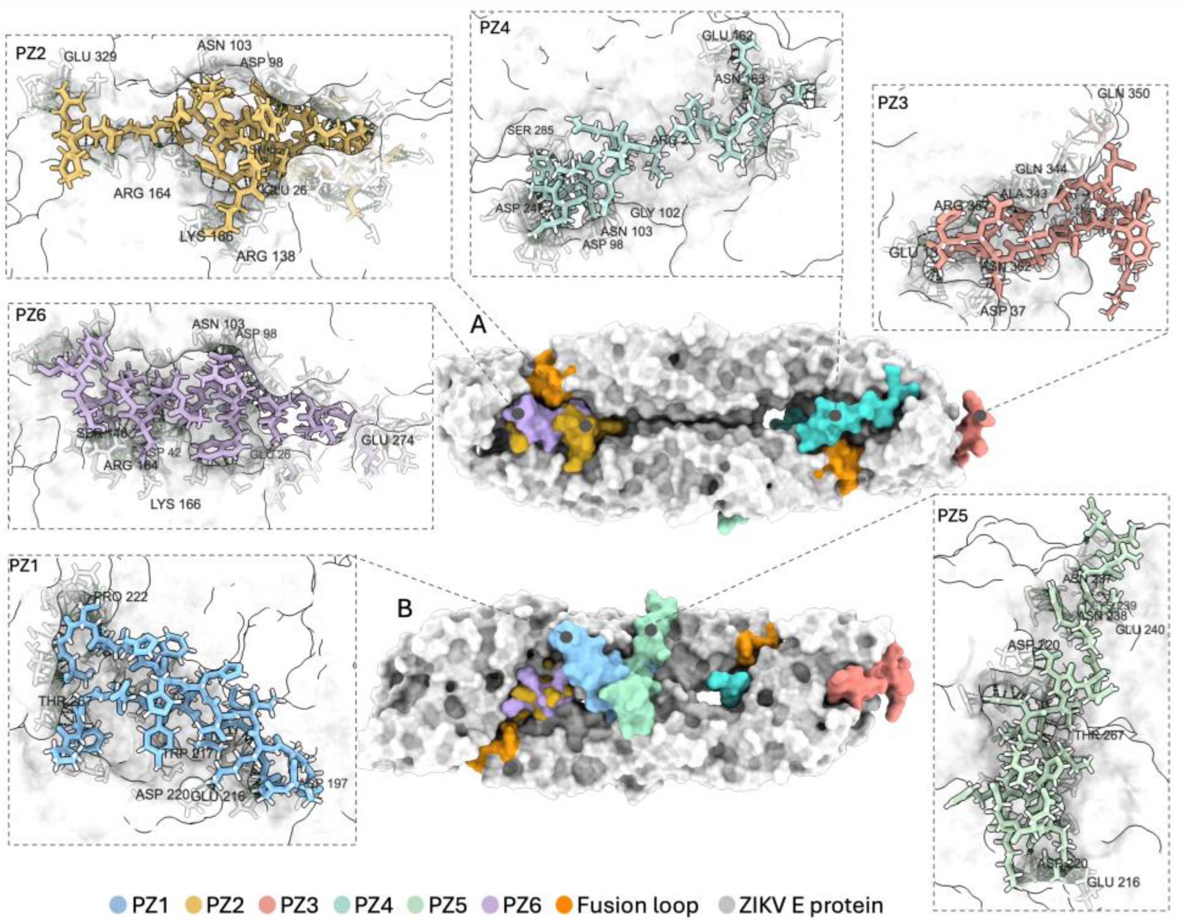
Figure 2.
In vitro cytotoxicity and hemolytic activity of candidate anti-ZIKV peptides. (A) Cell viability of peptide-treated cells determined by MTT assay. PZ1-PZ6 were evaluated at the indicated concentrations, and viability was normalized to untreated control cells (UC). DC denotes the positive death control used in the viability assay. The dashed line indicates the predefined 70% cell viability threshold. (B) Hemolytic activity of candidate peptides evaluated at the indicated concentrations. Hemolysis was normalized to the Triton X-100 hemolysis control (HC). The dashed line indicates the predefined 5% hemolysis threshold. For both panels, each symbol represents the mean of one independent experiment performed in technical triplicate; horizontal lines and error bars indicate the mean ± SD of three independent experiments. Asterisks indicate statistically significant differences relative to UC in panel A or to the negative hemolysis control in panel B; ****p < 0.0001.
Figure 2.
In vitro cytotoxicity and hemolytic activity of candidate anti-ZIKV peptides. (A) Cell viability of peptide-treated cells determined by MTT assay. PZ1-PZ6 were evaluated at the indicated concentrations, and viability was normalized to untreated control cells (UC). DC denotes the positive death control used in the viability assay. The dashed line indicates the predefined 70% cell viability threshold. (B) Hemolytic activity of candidate peptides evaluated at the indicated concentrations. Hemolysis was normalized to the Triton X-100 hemolysis control (HC). The dashed line indicates the predefined 5% hemolysis threshold. For both panels, each symbol represents the mean of one independent experiment performed in technical triplicate; horizontal lines and error bars indicate the mean ± SD of three independent experiments. Asterisks indicate statistically significant differences relative to UC in panel A or to the negative hemolysis control in panel B; ****p < 0.0001.
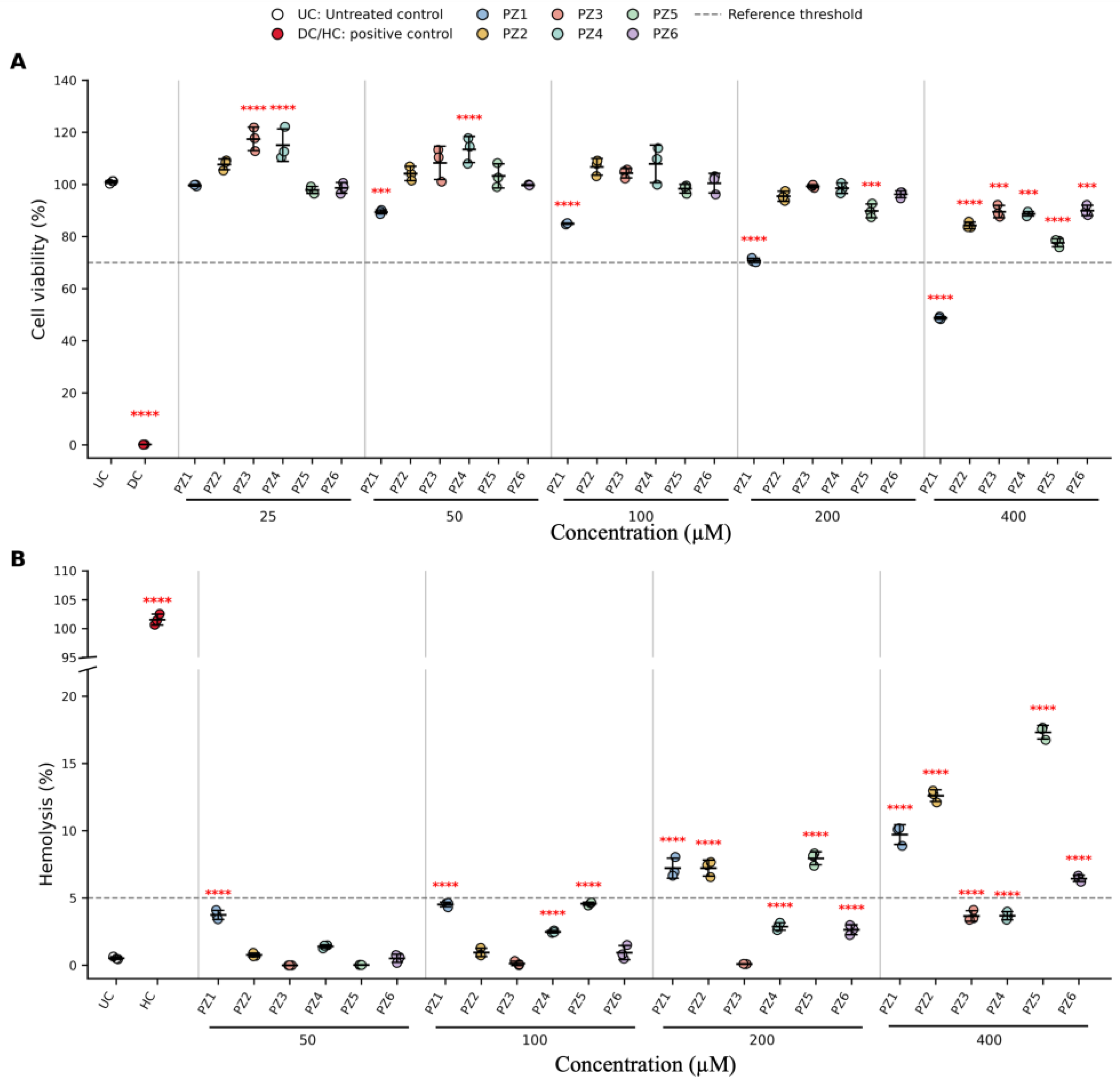
Figure 3.
Time-of-addition antiviral activity of candidate peptides in HTR-8/SVneo trophoblast cells. HTR-8/SVneo cells were treated with candidate peptides at 100 µM before infection (pre-treatment), during infection (co-treatment), or after viral adsorption (post-treatment). (A) Infectious ZIKV titers were quantified by plaque assay and expressed as log₁₀ PFU/mL. (B) Cell-associated ZIKV RNA levels were quantified by RT-qPCR and expressed as relative RNA levels using the 2^−ΔΔCt method, normalized to the infected cell control (IC); the dashed line indicates the IC baseline. GAPDH was used as the reference gene. Data represent three independent biological experiments; each symbol represents one independent experiment, and horizontal lines with error bars indicate mean ± SD. Asterisks indicate statistically significant differences relative to IC. In panel B, red asterisks indicate significant reductions in viral RNA, whereas black asterisks indicate significant increases above the IC baseline. *p < 0.05, **p < 0.01, ***p < 0.001, ****p < 0.0001.
Figure 3.
Time-of-addition antiviral activity of candidate peptides in HTR-8/SVneo trophoblast cells. HTR-8/SVneo cells were treated with candidate peptides at 100 µM before infection (pre-treatment), during infection (co-treatment), or after viral adsorption (post-treatment). (A) Infectious ZIKV titers were quantified by plaque assay and expressed as log₁₀ PFU/mL. (B) Cell-associated ZIKV RNA levels were quantified by RT-qPCR and expressed as relative RNA levels using the 2^−ΔΔCt method, normalized to the infected cell control (IC); the dashed line indicates the IC baseline. GAPDH was used as the reference gene. Data represent three independent biological experiments; each symbol represents one independent experiment, and horizontal lines with error bars indicate mean ± SD. Asterisks indicate statistically significant differences relative to IC. In panel B, red asterisks indicate significant reductions in viral RNA, whereas black asterisks indicate significant increases above the IC baseline. *p < 0.05, **p < 0.01, ***p < 0.001, ****p < 0.0001.
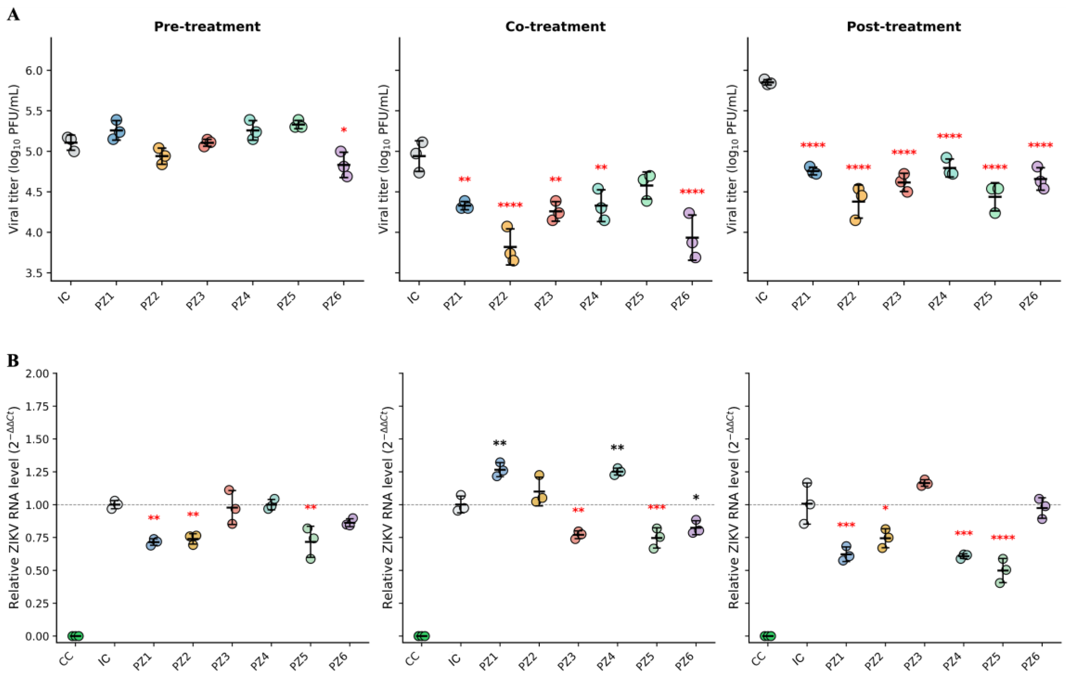
Figure 4.
Candidate peptides interfere with early ZIKV entry-associated events. (A) Anti-adsorption activity of candidate peptides in Vero cells. Infectious viral titers were determined by plaque assay and expressed as log10 PFU/mL. (B) Quantification of DiOC18-associated fluorescence in HTR-8/SVneo cells using DiOC18-labeled ZIKV. Fluorescence values were normalized to the infection control (IC). (C) Representative fluorescence and bright-field overlay micrographs showing DiOC18-associated signal under the following conditions: 1: infection control, 2: mock-infected cells, 3: NH₄Cl-treated control, 4: PZ1, 5: PZ2, 6: PZ3, 7: PZ4, 8: PZ5, and 9: PZ6. Green indicates DiOC18-associated viral signal, and cyan indicates nuclei. Scale bars: 50 µm. In panels A and B, horizontal lines with error bars indicate mean ± SD. In panel A, symbols represent technical replicates from three independent experiments; in panel B, each symbol represents the mean fluorescence value from one independent experiment. Asterisks indicate statistically significant differences versus IC: *p < 0.05, **p < 0.01, ***p < 0.001, ****p < 0.0001.
Figure 4.
Candidate peptides interfere with early ZIKV entry-associated events. (A) Anti-adsorption activity of candidate peptides in Vero cells. Infectious viral titers were determined by plaque assay and expressed as log10 PFU/mL. (B) Quantification of DiOC18-associated fluorescence in HTR-8/SVneo cells using DiOC18-labeled ZIKV. Fluorescence values were normalized to the infection control (IC). (C) Representative fluorescence and bright-field overlay micrographs showing DiOC18-associated signal under the following conditions: 1: infection control, 2: mock-infected cells, 3: NH₄Cl-treated control, 4: PZ1, 5: PZ2, 6: PZ3, 7: PZ4, 8: PZ5, and 9: PZ6. Green indicates DiOC18-associated viral signal, and cyan indicates nuclei. Scale bars: 50 µm. In panels A and B, horizontal lines with error bars indicate mean ± SD. In panel A, symbols represent technical replicates from three independent experiments; in panel B, each symbol represents the mean fluorescence value from one independent experiment. Asterisks indicate statistically significant differences versus IC: *p < 0.05, **p < 0.01, ***p < 0.001, ****p < 0.0001.
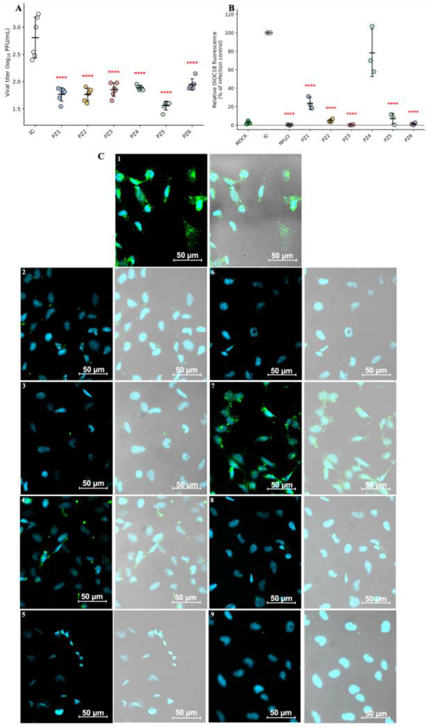
Figure 5.
Peptide-treated ZIKV particles exhibit altered ultrastructural features while largely retaining nuclease-protected viral RNA. (A) Nuclease protection assay of peptide-treated ZIKV particles. Protected viral RNA was quantified by RT-qPCR and expressed relative to the Virus + MNase control (VC, dashed line). Triton X-100 + MNase was included as a positive disruption control. Symbols represent independent experiments, and horizontal lines with error bars indicate mean ± SD. (B) Representative TEM micrographs of ZIKV-containing samples: I, untreated ZIKV control; II, ZIKV exposed to HBS pH 5.6-5.8; and III–VIII, ZIKV treated with PZ1–PZ6, respectively. Red arrows indicate representative particle-like structures, electron-dense aggregates, or altered morphologies observed under each condition. White lines indicate representative diameter measurements. Scale bars: 100 nm.
Figure 5.
Peptide-treated ZIKV particles exhibit altered ultrastructural features while largely retaining nuclease-protected viral RNA. (A) Nuclease protection assay of peptide-treated ZIKV particles. Protected viral RNA was quantified by RT-qPCR and expressed relative to the Virus + MNase control (VC, dashed line). Triton X-100 + MNase was included as a positive disruption control. Symbols represent independent experiments, and horizontal lines with error bars indicate mean ± SD. (B) Representative TEM micrographs of ZIKV-containing samples: I, untreated ZIKV control; II, ZIKV exposed to HBS pH 5.6-5.8; and III–VIII, ZIKV treated with PZ1–PZ6, respectively. Red arrows indicate representative particle-like structures, electron-dense aggregates, or altered morphologies observed under each condition. White lines indicate representative diameter measurements. Scale bars: 100 nm.
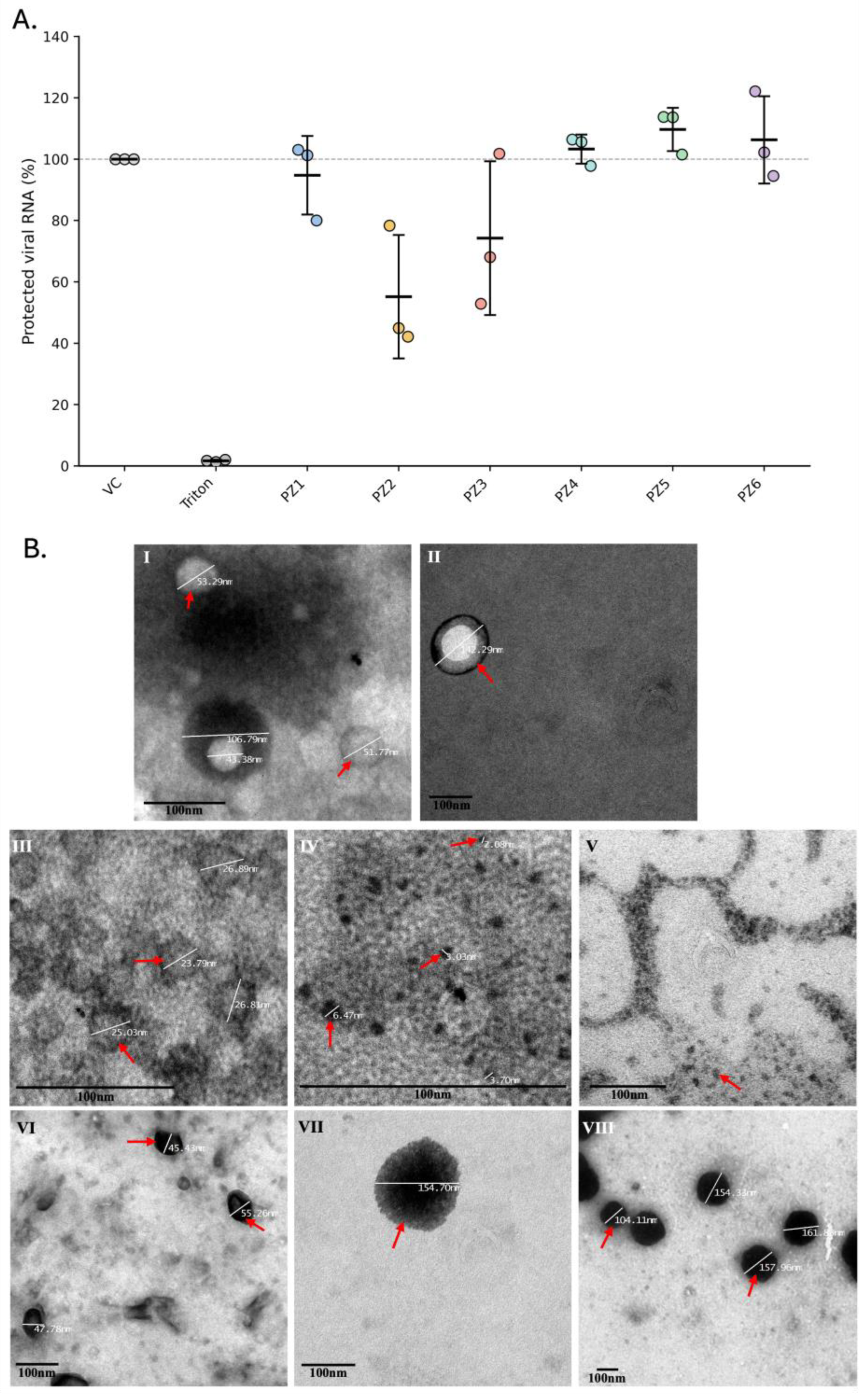

Table 1.
Theoretical physicochemical properties and predicted toxicity of candidate peptides.
| Peptide name |
Sequence | MW (g/mol) | pI | Net charge at pH 7.0 |
GRAVY | Predicted toxicity |
|---|---|---|---|---|---|---|
| PZ1 | PFGDSYIVIGVGEKKITHHWHRSGS | 2808.11 | 9.97 | 1.3 | -0.508 | Non-toxic |
| PZ2 | PRTGLDFSDLYYLTMNNKHWLVHKE | 3078.46 | 7.99 | 0.2 | -0.792 | Non-toxic |
| PZ3 | VEGMSGGTWVDVVLEHGGCVTVMAQ | 2561.91 | 3.69 | -3 | 0.592 | Non-toxic |
| PZ4 | TDGPCKVPAQMAVDMQTLTPVGRLI | 2642.13 | 6.05 | -0.1 | 0.18 | Non-toxic |
| PZ5 | GLIYHREEKRWRWINNSVFNGNVTN | 3103.41 | 10.44 | 2.1 | -1.06 | Non-toxic |
| PZ6 | MKSRVIRAAWSWRESAAHEGTRDYT | 2965.27 | 10.36 | 2.1 | -1.03 | Non-toxic |
MW: Molecular weight, pI: Isoelectric Point, GRAVY: Grand average of hydropathicity.
Table 2.
Predicted binding regions and selected interfacial residues of candidate peptides in docking complexes with the ZIKV E protein.
Table 2.
Predicted binding regions and selected interfacial residues of candidate peptides in docking complexes with the ZIKV E protein.
| Peptide | Predicted E-protein region | Representative interfacial residues* | Potential mechanistic implication** |
|---|---|---|---|
| PZ1 | Domain II | Asp197-A, Glu216-A, Trp217-A, Asp220-A, Pro222-B, Thr267-B | Binding to DII, including residues Glu216 and Asp220, could perturb local E-protein stability or the conformational rearrangements required for membrane fusion. |
| PZ2 | Domains I-III; fusion-loop-proximal region | Asn8-A, Glu26-A, Arg138-A, Arg164-A, Lys166-A, Glu329-A, Asp98-B, Asn103-B | Binding across multiple domains and near the fusion loop could restrict fusion-loop exposure or insertion and interfere with the low-pH-induced rearrangements required for viral membrane fusion. |
| PZ3 | Domains I and III | Glu13-B, Asp37-B, Thr309-B, Ala343-B, Gln344-B, Gln350-B, Arg357-B, Asn362-B | Binding at DI/DIII surfaces could alter interdomain organization or accessibility of regions potentially involved in virion attachment; this mechanism remains less directly supported than fusion-loop interference. |
| PZ4 | Domains I and II; fusion-loop-proximal region | Arg2-B, Asp98-A, Gly102-A, Asn103-A, Glu162-B, Asn163-B, Asp247-A, Ser285-B | Contacts involving the fusion loop and adjacent DI/DII regions could sterically hinder fusion-loop exposure or membrane insertion during viral entry. |
| PZ5 | Domain II | Glu216-B, Asp220-A, Asp220-B, Asn237-A, Asn238-A, Lys239-A, Glu240-A, Thr267-B | Predicted binding to DII could affect local dimer stability or restrict the conformational transition of E from the prefusion to the fusion-active state. |
| PZ6 | Domains I and II; fusion-loop-proximal region | Glu26-A, Asp42-A, Asp98-B, Asn103-B, Ser146-A, Arg164-A, Lys166-A, Glu274-A | Binding near the fusion loop and DI/DII interface could constrain hinge-associated movements and fusion-loop deployment required for endosomal membrane fusion. |
* Representative E-protein residues were selected from contacts detected within 4.0 Å based on contact frequency, chemical relevance, and visual clarity. ** Potential mechanistic implications are inferred from predicted docking locations and require experimental validation.
Disclaimer/Publisher’s Note: The statements, opinions and data contained in all publications are solely those of the individual author(s) and contributor(s) and not of MDPI and/or the editor(s). MDPI and/or the editor(s) disclaim responsibility for any injury to people or property resulting from any ideas, methods, instructions or products referred to in the content. |
© 2026 by the authors. Licensee MDPI, Basel, Switzerland. This article is an open access article distributed under the terms and conditions of the Creative Commons Attribution (CC BY) license (http://creativecommons.org/licenses/by/4.0/).
Copyright: This open access article is published under a Creative Commons CC BY 4.0 license, which permit the free download, distribution, and reuse, provided that the author and preprint are cited in any reuse.