

Submitted:
28 August 2026
Posted:
28 August 2026
You are already at the latest version
Abstract
In July 2026, an outbreak of a cattle illness characterized by high fever, diarrhoea and reduced milk production, often affecting whole herds, was detected across several regions of northern Switzerland. To investigate the cause of the outbreak a broad range of diagnostic methods was applied, including pathogen specific RT-qPCRs, ELISA, serum neutralization test, virus isolation and metagenomic next generation sequencing. A Shamonda virus (SHAV) was detected in samples from affected cattle, followed by successful virus isolation and complete genome sequencing of all three segments. Viral RNA was also detected in tissue samples from aborted calves, including brain and spleen, supporting the need for further investigation of the virus in the context of reproductive disease. These findings document the first detection and molecular characterization of a SHAV in Switzerland and demonstrate its widespread circulation in cattle during the 2026 outbreak.
Keywords:
Orthobunyavirus
; emerging virus
; virus pathogen
; Shamonda virus
1. Introduction
Viruses of the genus Orthobunyavirus are arthropod-borne viruses within the family Peribunyaviridae of the order Bunyavirales, characterized by a segmented, negative-sense RNA genome [1]. The genome has a large (L), a medium (M) and a small (S) segment, encoding the RNA-dependent RNA polymerase, the glycoproteins, and the nucleocapsid protein, respectively. The segmented genome allows genetic reassortment, when two orthobunyaviruses co-infect the same host or vector, potentially contributing to viral evolution and emergence. The major antigenic group within the genus is the unofficial Simbu serogrup that comprises in clade B viruses of considerable veterinary relevance [2,3,4]. These include, among others, Akabane virus (AKAV), Schmallenberg virus (SBV), Aino Virus (AINOV), Shuni Virus (SHUV) and Shamonda virus (SHAV). Several members of this group have been associated with reproductive diseases, congenital abnormalities, abortions and stillbirth in cattle, small ruminants and, in case of SHUV, also with neurological disease in horses. All are primarily transmitted by Culicoides biting midges, and their epidemiology is therefore linked to vector abundance, seasonal activity and geographic distribution, which facilitate the introduction and establishment of new orthobuynaviruses in susceptible ruminant population.
SBV represents a particularly important example of the ability of Orthobunyaviruses to emerge in previously unaffected geographical regions. The virus was detected for the first time in Germany in 2011 in plasma samples from cattle with fever and reduced milk production [5]. This provided a notable case for the discovery of a novel member of the Simbu serogroup virus via metagenomic sequencing. Following initial detection, SBV spread rapidly across Europe, causing mild or transient clinical symptoms in adult cattle, whereas infection of fetuses during critical stage of gestation resulted in congenital abnormalities including arthrogryposis, hydranencephaly, abortion and stillbirths. SBV virus has been postulated as the likely ancestor of SHAV [6]. SHAV was originally detected and isolated from cattle and bitting midges in Nigeria in the 1960s, subsequently detected after 40 years in bitting midges and blood samples from cattle Japan in 2002 and serological evidence of a Shamonda-like virus has been reported from New South Wales, Australia [7,8]. Despite this close evolutionary relationship, the epidemiology and geographic distribution of SHAV and Shamonda-like viruses remain unknown.
In the second week of July 2026, the Cattle Health Service Switzerland (Rindergesundheit Schweiz RGS) recorded a significant rise in reports of fever and diarrhoea accompanied by a simultaneous decline in milk yield in cattle, predominantly in dairy cows. The first reports came from the western part of Switzerland and were followed by reports across central Switzerland as far as the eastern canton of Thurgau. Due to the rapid and apparently epidemic spread of clinical symptoms, uncertainty grew amongst both veterinarians and farmers. Similar clinical signs, initially thought to be related to the intensive heatwave, were observed in southern Germany and eastern France. Initial virological diagnostic testing did not identify any known viral pathogen as cause of the observed disease. SBV was initally considered, however, samples from few animals submitted for SBV diagnostics were all tested negative by SBV RT-qPCR. Taking into consideration the short viraemic period of SBV infections, targeted serological investigations alongside sampling of recent acutely ill animals were undertaken on three selected farms with notable disease rates in adult dairy cows. Indeed, high seroprevalence of SBV antibodies was detected on all three farms from geographically distinct regions. Further investigation of antibody-negative animals or low antibody-positive acute clinical animals in these herds using a broad range pan-Simbu RT-qPCR revealed several animals positive for viral genome. The subsequent metagenomic sequencing of four samples from two affected farms identified an Orthobunyavirus. Complete genome sequences of all three segments were obtained from all four samples and demonstrated that the virus is most closely related to SHAV but is generally distinct from previously characterized sequences. The Swiss sequences showed high genomic similarity with the virus concurrently sequenced in Germany, indicating the circulation of a previously uncharacterized Shamonda-like Orthobunyavirus in Western Europe.
Following the emergence of SBV about 15 years ago, the identification of another Simbu serogroup Orthobunyavirus associated with disease in cattle in several European countries raises concerns of the constant establishment of this virus and, with SBV still circulating, of potential reassortments between them [9].
The present study describes the detection, molecular characterization and phylogenetic classification of a novel Shamonda-like virus and provides the first evidence of its circulation in Switzerland. Virus isolation and whole genome sequencing were utilized to characterize the virus at the genomic level and to establish its phylogenetic relatedness to SHAV, SBV and other veterinary-relevant members of the order Bunyavirales based on all three segments and assess its potential geographic distribution and possible impact on animal health.
2. Materials and Methods
2.1. Laboratory Timeline
During the week of 20–24 July, an increasing number of inquiries were received concerning dairy cattle presenting fever, diarrhea, and a decrease in milk production. Schmallenberg virus (SBV) infection was repeatedly considered based on the clinical presentation. However, initial samples were mainly submitted from single animals with fully developed clinical signs, such as diarrhea, for several days, and all samples tested negative by SBV-specific RT-qPCR [10]. Given that SBV viremia is reported to be relatively short, with viral RNA generally detectable for approximately six days only, it was considered possible that samples had been collected after the viremic phase and that the optimal diagnostic window had therefore been missed. To further elucidate if SBV needed to be considered as a possible cause of the observed disease, primary veterinary practitioners in association with the Cattle Health Service Switzerland (Rindergesundheit Schweiz RGS) carried out census sampling of adult cattle on three farms each with high incidence of disease and representing the regions of western, central and eastern Switzerland.
On Friday, 24th July serum samples from 27 adult dairy cows of a herd in the canton of Jura (western Switzerland) were sent, followed the following week by serum samples of 5 calves born in 2026. On Tuesday the 28th of July samples from a farm in the canton of Thurgau (eastern Switzerland) taken from 29 dairy cows including four acutely affected animals plus three young calves, were received. Finally on Friday the 31st of July samples from 38 dairy cows including three acutely ill animals as well as samples from 10 calves born in 2026 were sent from a farm in the canton of Luzern (central Switzerland). All samples were analyzed for SBV specific antibodies as described below.
2.2. Serological Analysis
Serum samples of the animals from the three herds were analyzed with a commercially available indirect ELISA (ID Screen® Schmallenberg Virus Multi-species, IDvet, France), according to the manufacturer’s instructions. To further investigate the specificity of the antibody response, a SBV serum neutralization test (SNT), established for routine diagnostic at IVI was performed on a subset of 10 serum samples representing different ELISA reactivity levels. However, due to lack of other Orthobunyaviruses serum samples were only tested for their ability to neutralize SBV (Isolate BH80/11-4 originally isolated from cattle serum and received from H. Schirrmeier, Friedrich-Loeffler Institute). Briefly, neutralization assay was carried on serial two-fold dilution of sera from 1:4 until 1:256 in four wells with a total volume of 50 μl per well. Then, 50 μl virus diluted in medium to contain approximately 200 TCID50 (confirmed by back-titration) was added to each well containing serum. The plates were incubated for 72 hours in 37°C with 5% CO2, presence/absence of CPE was recorded and neutralization titers calculated using the Spearman–Karber formula. Neutralizing titres were reported as the highest dilution at which neutralization was observed.
2.3. RT-qPCRs
Nucleic acid from selected serum/EDTA blood samples was extracted using NucleoMag® VET Kit (Macherey Nagel, Düren, Germany) on a KingFisher automated extraction system (ThermoFisher Scientifc, Waltham, USA), according to the manufacturers instructions. Following RNA extraction all selected samples were tested for SBV virus RNA using published SBV-specific RT-qPCR targeting the S segment. However, SBV-S RNA was not detected in any of the tested animals, including the clinically affected animals and those with negative or doubtful SBV antibody results. Therefore, the samples were further analyzed using a pan-Simbu RT-qPCR (targeting S segment), and SBV-specific RT-qPCRs targeting the conserved L and the M segments according to published assays [10,11]. Furthermore, four pan-Simbu RT-qPCR positive samples were selected for whole-genome sequencing (WGS), while seven samples were taken forward for virus isolation to further characterize the detected virus.
2.4. Virus Isolation
Six sera positive in pan-Simbu RT-qPCR and one EDTA-Blood with CT-values between 20.9 and 27.8 were used for an attempt of isolating the virus using SK-6 and BHK-21 cells. Cells were cultured in 25 cm² cell culture flasks. Both BHK-21 and SK-6 were inoculated with 2 mL of the diluted 1:4 sera/blood in GMEM or MEM medium plus antibiotics (Gibco, LuBio Science GmbH, Luzern, Switzerland) for BHK-21 and SK6 respectively.
The cells were incubated for 2 hours at 37°C. To cell cultures inoculated with serum samples 8 ml of the respective media plus antibiotics and 2% fetal bovine serum in BHK-21 or 2% horse serum in SK6 cells (Gibco, LuBio Science GmbH, Luzern, Switzerland) was added with the inoculum remaining. The EDTA inoculum was carefully removed, the cells washed once and 10 ml of the respective media were added as described above. All flaks were incubated at 37°C with 5% CO2. The cells were microscopically controlled daily for CPE.
Due to timing constrains flasks were already frozen at −70°C after 48 hours and freeze-thawed twice. Supernatants were clarified by low-speed centrifugation, stored in aliquots at −70°C, and used for an additional passage P2. Inoculation of BHK-21 and SK6 was carried out using 2ml of P1-supernatant directly as inoculum, incubated for 72 hours at 37°C with 5% CO2 and harvested as described above.
Three P2 cell cultures which had shown the most promising CPE on BHK cells were carried forward for virus titration Titers expressed as TCID50/mL in BHK-21 cells were determined for virus stocks. Furthermore, the expected increase of viral RNA upon successful virus amplification was analyzed in the P1 cell culture passage by testing harvested supernatants using the pan-Simbu RT-qPCR assay.
2.5. Next Generation Sequencing
First, the EDTA blood sample (CT 21.2) was gently mixed by inverting the tube. A volume of 1.5 mL was transferred to a new tube and centrifuged for 10 min at 1,600 × g. The buffy coat was then carefully pipetted off together with a small volume of plasma. Three serum samples (CT 20.9–22.9) and one EDTA blood sample were relatively enriched for virus particles as previously described [12]. The EDTA Blood sample 261216_36 and serum sample 261216-18 were taken from the same animal. Subsequently, the samples were extracted using TRIzol™ Reagent (Thermo Fisher Scientific) at a sample-to-reagent ratio of 1:3, according to the manufacturer’s instructions, β-mercaptoethanol (1%) was added to the TRIzol™ reagent to inactivate nucleases used during the previous step. Reverse transcription, second strand synthesis, and random amplification was performed as previously described using the SISPA (sequence-independent single-primer-amplification) method [12]. The DNA was divided into two aliquots, one aliquot was sheared by sonication (E220 Ultrasonicator, Covaris, USA) at the NGS Platform in Bern, targeting fragments of 500bp length, the other aliquot was used directly for library preparation. Sequencing libraries were made using the NEBNext Ultra II DNA Library Prep Kit for Illumina (New England Biolabs, Ipswich, MA, USA) and barcoded using NEBNext Multiplex Oligos (96 Unique Dual Index Primer Pairs; New England Biolabs, Ipswich, Massachusetts, USA). Finally, libraries were sequenced using NovaSeq™ X Series 1.5B Reagent Kit (300 Cyc), on an Illumina NovaSeq X Benchtop sequencer with loading concentration of 160pM (Illumina, San Diego, CA, USA) at the NGS Platform, Bern.
2.6. Data Analysis
To identify the best matching reference and to consider possible reassortments, all available Orthobunyavirus sequences of all three segments were downloaded from NCBI GenBank (15th August 2026). To reduce redundancies, the sequences were clustered using cd-hit-est (v.4.8.1) using the options -c 0.99 -n 10 -d 0 -M 500000 [13]. The raw reads were mapped to the resulting clustered reference sequences using bowtie2 (v. 2.5.5) with options -k 20 [14]. The most appropriate reference for each segment was identified using samtools coverage (v.1.23, [15]), resulting in the accession numbers OZ502471.1 – OZ502473.1. Consequently, the raw reads were mapped again to the newly identified accession using bowtie2 (v. 2.5.5) with option –very-sensitive-local. [14]. Subsequently new majority consensus sequences were called with samtools consensus (v.1.23) using options --show-ins no --show-del no --mode simple --call-fract 0 -f fasta [15]. Reads from high coverage samples were subsampled using samtools view (v.1.23,[15]
Sequence specific alignments were made using mafft (v.7.490) with --op 5 options [16]). The subsequent approximately-maximum-likelihood phylogenetic trees were inferred using fasttree (v.2.1.11) with default parameters [17].
Pairwise distances were calculate using famsa -dist_export (v.2.4.1, [18]) on alignments trimmed with trimal (v.1.5, [19]) with options -gt 0.9. Genetic distances between samples and publicly available Shamonda and Schmallenberg viruses were tested using a linear mixed model implemented in lme4 (v.1.1.37, [20]). Briefly, a model was fitted with genetic distance as a response variable with the interaction between segment and virus group as predictor and the individual samples as a random effect. Subsequent pairwise comparisons were Tukey adjusted using the emmeans package (v.1.11.2, [21]). Visualization of phylogenetic trees was made with the ggtree package (v.3.10.1) using a circular layout and branch length defined as "none" [22]. Rooting of the tree was made using Simbu serogroup clade B sequences from the ICTV taxonomy (MSL #41, http://ictv.global/taxonomy).
2.7. Geographic Distribution of Positive Samples
3. Results
3.1. Serological Analysis
Serological analysis revealed high levels of anti-SBV antibodies across all three affected farms. On the farm from canton Jura, all 27 adult dairy cows were seropositive, corresponding to a seroprevalence of 100% within the sampled group. No acutely ill adult animal was present anymore at time of sampling. Additionally, five calves born in 2026 were also tested of which 4 were SBV antibody positive, while one calf remained negative for SBV antibodies.
In Thurgau, 26 out of 29 sampled dairy cows were seropositive, two had indeterminate results, and one was seronegative, corresponding to an overall seroprevalence of approximately 90%. Three calves born in 2026, however, were seronegative, despite two being born to antibody positive dams. The four acute clinical animals were all SBV antibody positive.
In Luzern, 36 out of 38 sampled cows were positive in the SBV ELISA, corresponding to a seroprevalence of approximately 95%. Among the 10 calves, two were seropositive and eight negative. Several antibody-negative calves younger than 6 months were offspring of antibody-positive dams. Of the three acute clinical animals one animal was still SBV antibody negative, while the other two had detectable levels of SBV antibodies.
The total absence of maternal antibodies in some young calves born to SBV antibody positive mothers suggested that the dams were very recently infected (after calving) and, since all farms had a direct dam-calf colostrum regime provided the first tentative evidence for recent circulation of SBV, or a related virus.
The SBV ELISA results of 10 sera showed considerable variation in antibody reactivity, with S/P values ranging from 10 to 222%. Corresponding SBV SNT titres ranged from <1:4 to >1:256. (Table 1) All samples but one, which were classed positive for SBV Antibodies by ELISA, also had detectable neuralization titers against SBV, albeit with a marked range in SNT titers not always aligned to the S/P% values obtained. However, sample 261239.44 which was positive for SBV antibodies by ELISA without a detectable SNT titre had been taken from a calf born in 2026 hence, it cannot be excluded that maternal antibodies were detected. Importantly, the serological assays used in this study cannot reliably differentiate antibodies against closely related Orthobunyaviruses and were therefore interpreted in conjunction with the molecular and virological findings. Given the concurrent detection of a novel SHA-like virus in the affected animals, the observed SBV ELISA and SNT reactivity was therefore considered most likely from serological cross-reactivity with the newly identified virus rather than SBV infection.

Table 1.
Comparison of SBV ELISA and serum neutralization test results. The table shows the comparison between SBV-specific antibody detection by ELISA and serum neutralization testing in 10 selected serum samples. ELISA results are expressed as the sample-to-positive (S/P) percentage, while SNT results are reported as the serum dilution corresponding to the neutralization endpoint, calculated using the Spearman–Kärber method.
| Probe ID | ELISA SBV S/P % | SNT Titer |
|---|---|---|
| 1. 261239.20 | 207 | >1:256 |
| 2. 261239.43* | 10 | <1:4 |
| 3. 261239.44* | 124 | <1:4 |
| 4. 261232.1 | 213 | 1:108 |
| 5. 261219.1 | 213 | 1:64 |
| 6. 261216.16 | 188 | 1:38 |
| 7. 261208.1 | 162 | 1:4 |
| 8. 261197.2 | 222 | 1:108 |
| 9. 261197.4 | 205 | 1:215 |
| 10. 261197.9 | 213 | 1:16 |
*Samples from young calves.
3.2. RT-qPCRs
Overall, the high seroprevalence observed across the three geographically distinct herds, together with high antibody levels indicated widespread exposure to SBV. However, this finding was not consistent with the lack of SBV RNA detection – even in clinically affected animals. Therefore, the possibility of the presence of an alternative Orthobunyavirus was taken into consideration and prompted further molecular investigation.
Initial molecular investigation of 16 animals, selected from either clinically affected or animals with weak/negative results in the SBV ELISA, revealed that 6 animals were positive by pan-Simbu RT-qPCR and by the SBV L-segment RT-qPCR, while remaining negative in RT-qPCR assays targeting the SBV M and S segments. Of these six animals, five were clinically affected animals for which EDTA-blood and sera samples were available. Four out of six animals were already weak SBV-antibody positive, and overall, ELISA reactivity values were 9%, 40%, 72%, 83%, 88%, and 125% respectively. The discordant PCR pattern, with detection of the conserved L segment but absence of the M and S segments signals, underpinned the suspicion evoked by the serological results of the presence of an SBV-related Orthobunyavirus and initiated whole genome sequencing.
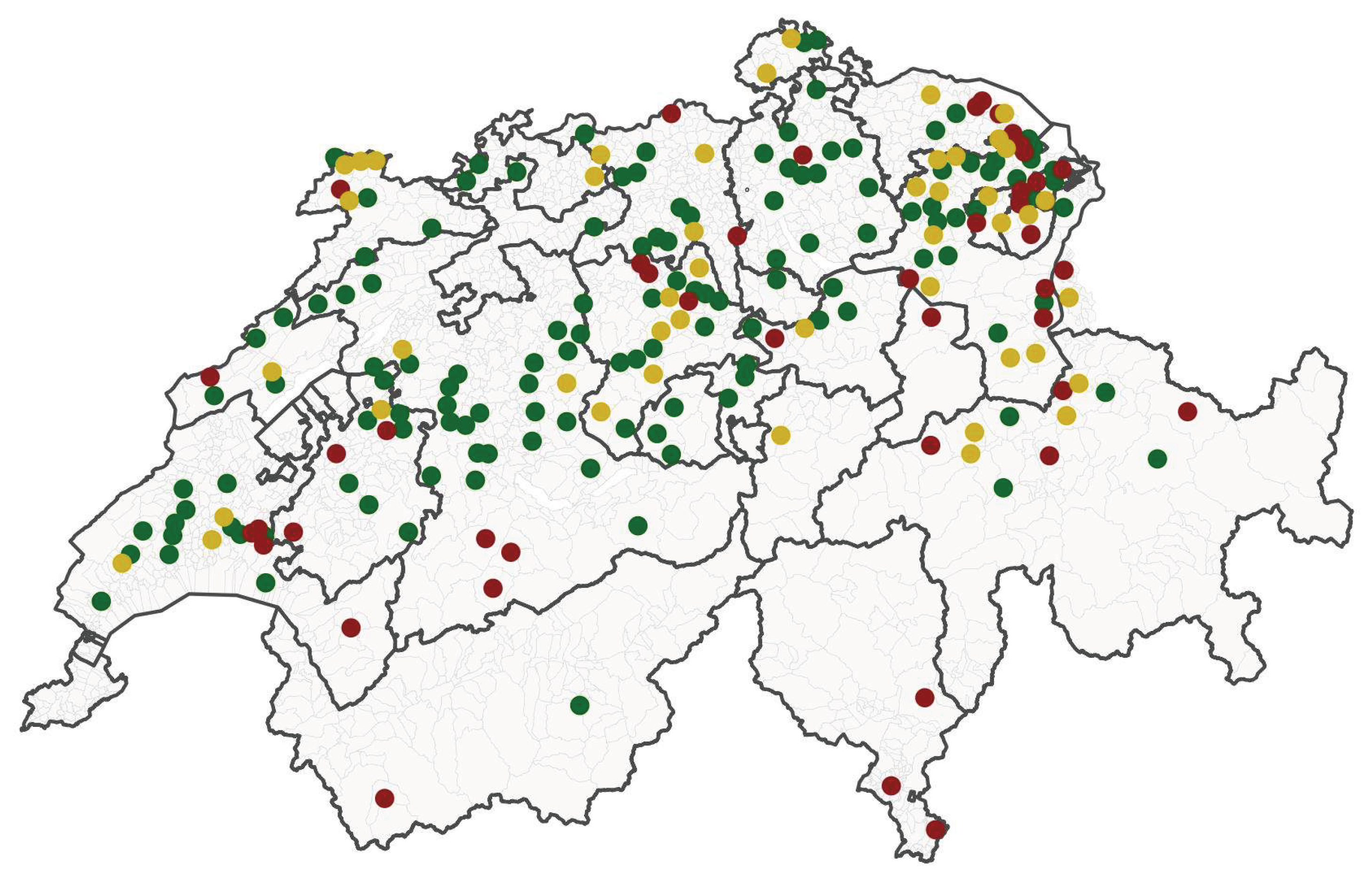
As of 25 August 2026, a total of 155 animals from 110 herds have been tested positive for the novel Orthobunyavirus by panSimbu RT-qPCR, while all samples were tested negative by specific SBV-S segment RT-qPCR results. Positive herds were identified across 21 Swiss cantons (Figure 5), demonstrating a rapid and geographically widespread emergence of the virus through Switzerland. Additionally, to date, 22 aborted calves have been tested positive either in spleen, brain or both, providing further evidence of association with reproductive disease in cattle.
3.3. Virus Isolation
After 48 hours of incubation, cytopathic effect was not visible either on BHK-21 or SK-6 cells. However, all harvested cell culture supernatants were confirmed to be positive by panSimbu RT-qPCR, with all samples inoculated onto BHK cells showing a clear decrease in CT values, indicating successful viral replication. In contrast, viral replication in SK-6 cells was less efficient even in passage 2 and these cultures were excluded from further testing and production. Following the second passage, the three samples exhibiting the most pronounced CPE (approximately 80–100%) in BHK cells were chosen for virus titration. Virus titres obtained on BHK cells ranged from 105.8 to 106.2 logTCID50/mL.
3.4. Next Generation Sequencing
A total of 880 million raw reads were generated in the sequencing run, ranging from 158 to 325 million reads per sample. Of these, 223 million reads were mapped to the SHAV genome, with the mapped reads ranging from 33,490 reads in the EDTA-blood sample TG_261216_36 to 146 million reads in the serum sample LU_261239_3. The mean sequencing depth for SHAV was 270,616x, 741,480x, 487,052x, and 358x for samples LU_261239_1, LU_261239_3, TG_261216_18, and TG_261216_36, respectively (Table 2).
Table 2.
Overview of sequenced samples, including GenBank accession numbers, sample material, total raw reads generated, reads mapped to SHAV, and mean sequencing depth.
Table 2.
Overview of sequenced samples, including GenBank accession numbers, sample material, total raw reads generated, reads mapped to SHAV, and mean sequencing depth.
| Sample | Accesion Number Seg L/M/S | Material | Reads sequenced | Reads Mapped | Mean Depth |
|---|---|---|---|---|---|
| LU_261239_1 | PZ831155/PZ831159/PZ831163 | Serum | 217'238'193 | 24'666'501 | 270'616 |
| LU_261239_3 | PZ831156/PZ831160/PZ831164 | Serum | 325'054'039 | 146'189'349 | 741'480 |
| TG_261216_18 | PZ831157/PZ831161/PZ831165 | Serum | 179'790'516 | 52'787'023 | 487'052 |
| TG_261216_36 | PZ831158/PZ831162/PZ831166 | EDTA Blood | 158'713'875 | 33'490 | 358 |
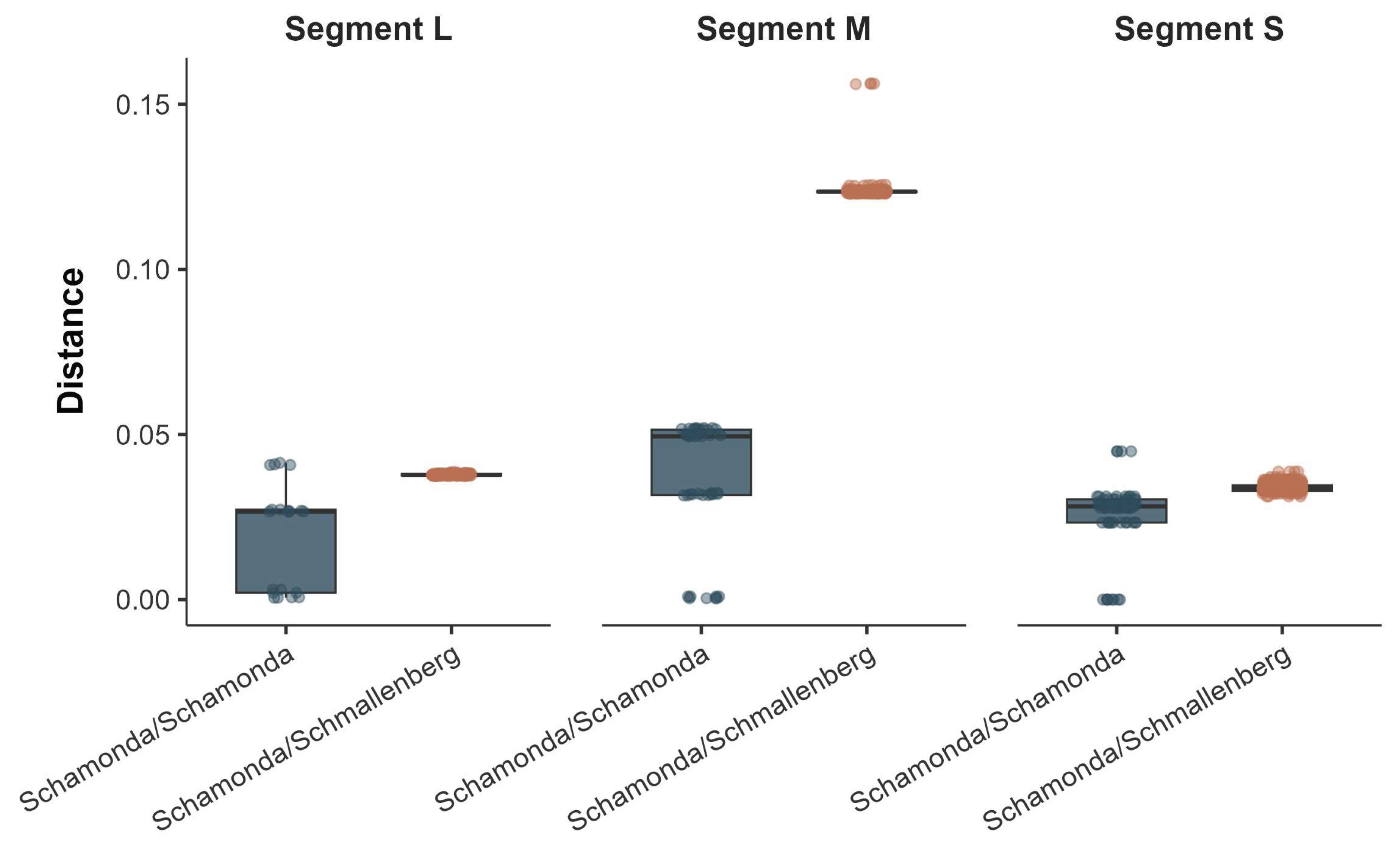
To statistically assess the group assignment of the newly sequenced samples, we compared their genetic distances to publicly available SHAVs and SBVs sequences (Retrieved on the 15th of August). Distances between virus species were significantly different for all segments (pval < 0.01, Figure 1). However, the magnitude of the distance differed between segments with the M segment displaying the greatest difference (0.0873, pval < 0.01). Overall, these results indicate a significantly closer genetic proximity to SHAVs than to SBVs.
Figure 1.
Segment-wise genetic distance of newly sequenced sequences to publicly available known SHAV and SBV sequences available from GenBank. Distances calculated using famsa on trimmed alignments. Boxplots show the median and interquartile range (IQR; 25th - 75th percentiles), with whiskers extending to 1.5 × IQR and individual points representing observations.
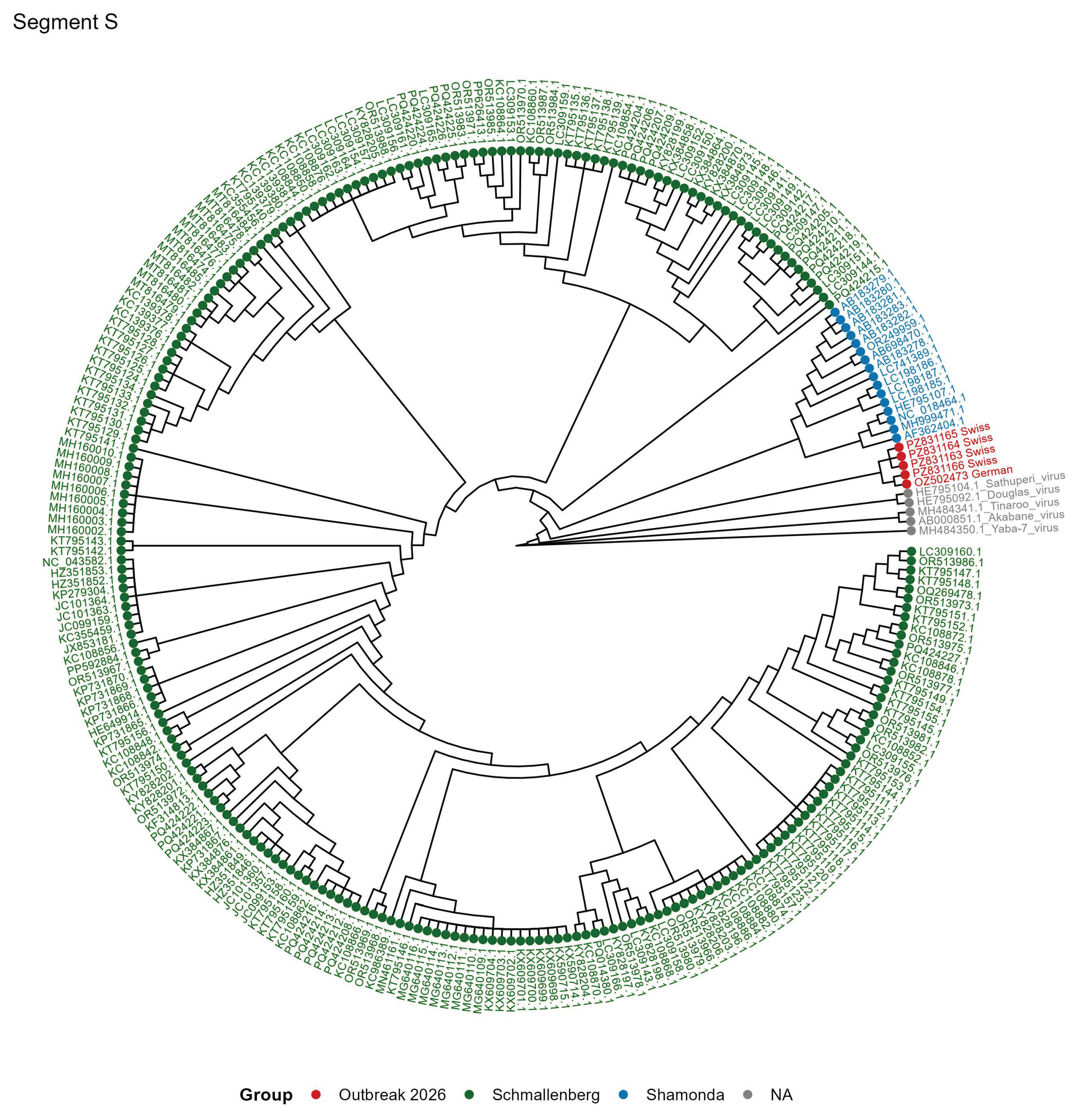
Segment-wise phylogenetic analyses to publicly available sequences revealed a closer relationship between the 2026 outbreak sequences and previously reported SHAVs sequences compared to SBVs (Figure 2 and Figure 3). However, the extent of this relationship is strongly segment specific. Notably, the 2026 outbreak sequences of the S segment formed a distinct subclade within the broader Shamonda virus lineage (Figure 4).
Figure 2.
Phylogenetic tree of the L segment. The tree was constructed with the fasttree approximately-maximum-likelihood method. Branch lengths are not representative of evolutionary distance. Publicly available SBV sequences are indicated in green, whereas publicly available SHAV sequences are shown in blue. The red labels indicate the samples from the 2026 outbreak. Grey labels are representative samples of other Orthobunyavirus viruses from the Simbu serogroup from the ICTV taxonomy used for tree rooting. IDs correspond to GenBank entries.
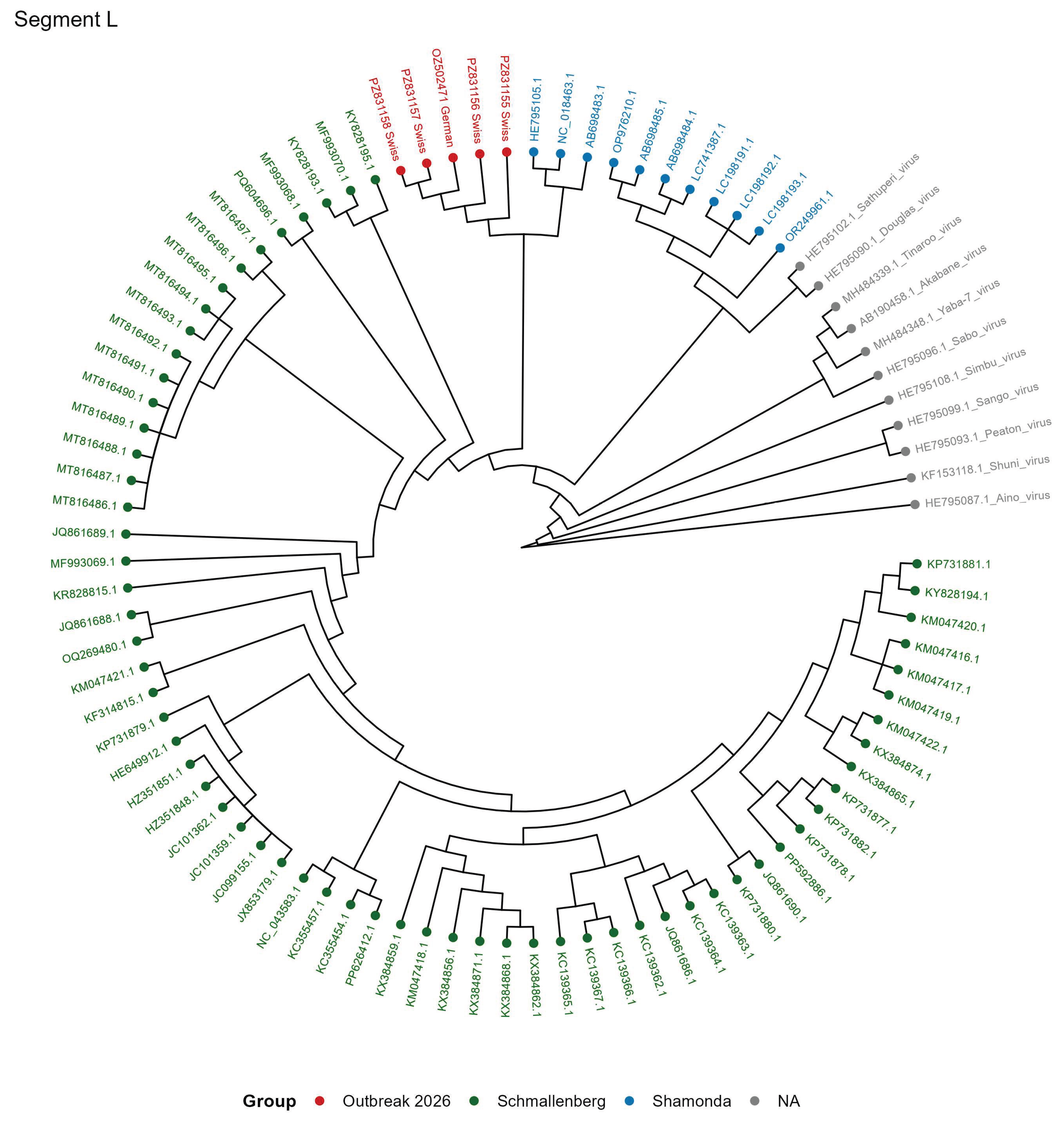
Figure 3.
Phylogenetic tree of the M segment. The tree was constructed with the fasttree approximately-maximum-likelihood method. Branch lengths are not representative of evolutionary distance. Publicly available SBV sequences are indicated in green, whereas publicly available SHAV sequences are shown in blue. The red labels indicate the samples from the 2026 outbreak. Grey labels are representative samples of other Orthobunyavirus viruses from the Simbu serogroup from the ICTV taxonomy used for tree rooting. IDs correspond to GenBank entries.
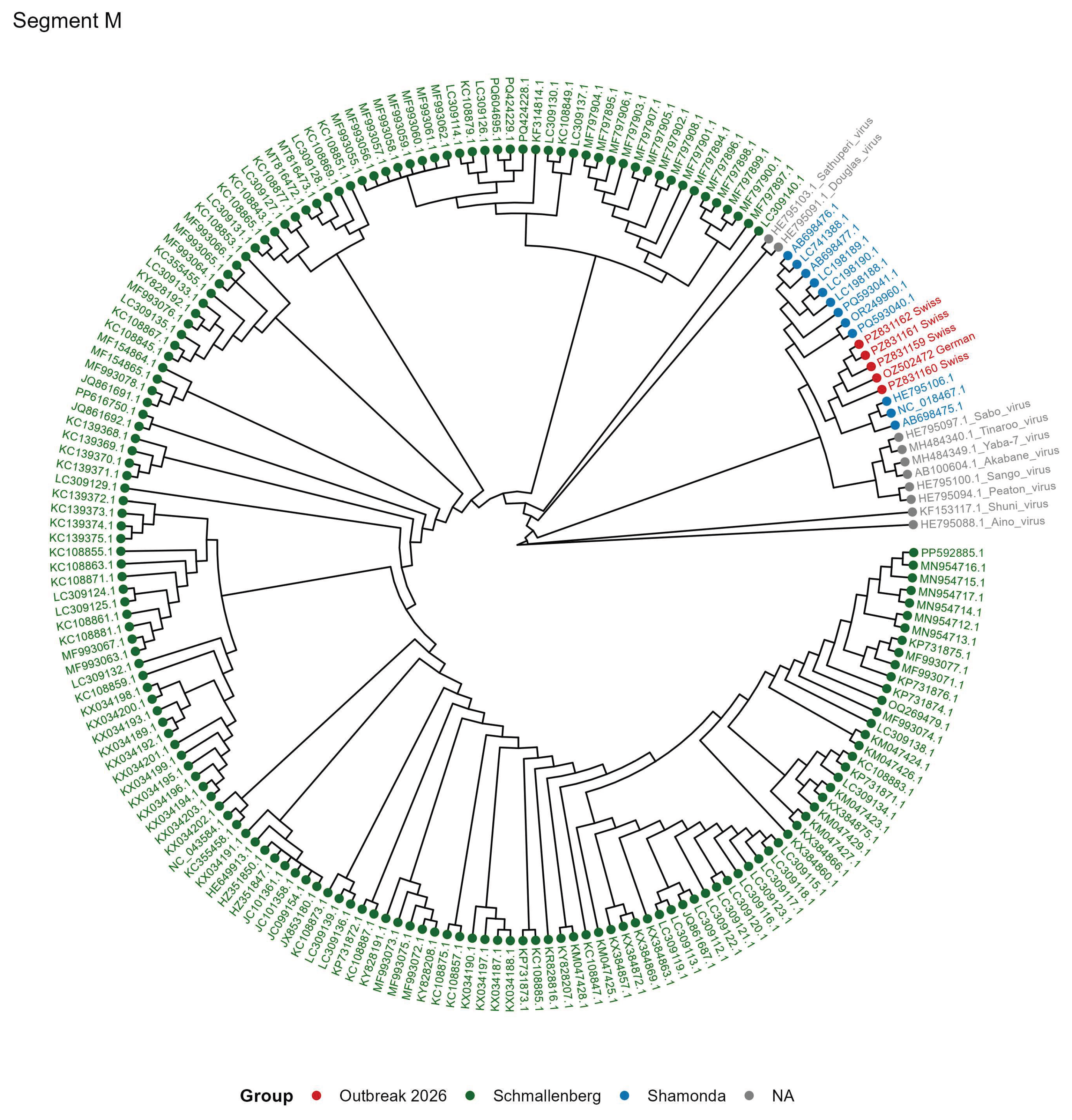
Figure 4.
Phylogenetic tree of the S segment. The tree was constructed with the fasttree approximately-maximum-likelihood method. Branch lengths are not representative of evolutionary distance. Publicly available SBV sequences are indicated in green, whereas publicly available SHAV sequences are shown in blue. The red labels indicate the samples from the 2026 outbreak. Grey labels are representative samples of other Orthobunyavirus viruses from the Simbu serogroup from the ICTV taxonomy used for tree rooting. IDs correspond to GenBank entries.
3.5. Geographic Distribution
As of 25 August 2026, 280 farms had submitted samples for diagnostic testing (Figure 5). The spatial distribution of the submitted samples indicated a west to east pattern. Interestingly, the submitted samples represent a broad altitudinal range from approximately 200 m above sea level (a.s.l). in Sant’Antonino (Ticino) to approximately 1250 m a.s.l. in Frutigen (Bern), encompassing regions characterized by sub-Mediterranean to Alpine climatic conditions.
Figure 5.
Geographic distribution of analyses performed up to 25 August 2026. Red dots indicate municipalities with SHAV-positive farms, green dots indicate municipalities with SHAV-negative farms, and yellow dots indicate municipalities with both SHAV-positive and SHAV-negative farms. Municipalities are indicated with a grey border.
4. Discussion
The serological findings provided the first important indication for a possible widespread outbreak of SBV (or antigenic related viruses) in Switzerland. Across three geographically distinct farms, SBV seroprevalence was very high, reaching 100%, 95% and 90% in Jura, Lucerne and Thurgau, respectively. However, as SBV is not notifiable in Switzerland no data was available on the extend of SBV circulation in previous years. Previous studies have demonstrated that SBV serological assays can detect antibodies induced by related Simbu serogroup viruses, highlighting the limitations of using SBV ELISA results alone to specifically detect SBV circulation or incursions of new but related viruses [25].
The absence of detectable SBV RNA in animals with high levels of SBV antibodies might be time dependent, as viraemia is relatively short, making direct virus genome detection difficult once the acute phase has passed. Consequently, RT-qPCR-negative results cannot exclude recent SBV infection. Nevertheless, the molecular results were particularly informative. Of the 16 animals initially selected because of clinical disease or weak/negative SBV ELISA results, six were positive by both panSimbu RT-qPCR and the SBV L-segment RT-qPCR but negative in RT-qPCRs targeting the SBV M and S segments. This pattern is unlikely to represent a SBV infection in which all three segments would be expected to be RT-qPCR positive. Instead, it suggested that the panSimbu assay and L-segment assay detected a genetically related Orthobunyavirus, while the absence of detection of the SBV M and S segments indicated substantial genomic divergence from SBV in the genomic regions targeted by the primers for these segments.
It is known that isolation and propagation of SBV from viremic serum in cell culture often results in low virus titers during the initial passage [26]. Several additional passages may be required before rapid CPE appears, and virus titers increase to sufficient levels. Based on that we decided to compare virus isolation in parallel using BHK-21 cells, which are commonly used for SBV isolation and SK-6 cells, which have previously been shown to be more efficient for SBV replication and to yield higher virus titter after fewer passages [27]. Interestingly, in our study the isolation of SHAV was more efficient in BHK-21 cells than SK6 cells, showing that the cellular tropism and replication of SHAV may differ from those described for SBV.
The genetic distance provides strong evidence that the novel Orthobunyavirus detected during the 2026 outbreak is genetically more closely related to SHAV than SBV. The segment-wise phylogenetic analyses further support the closer relationship of the 2026 outbreak virus to SHAV. Nevertheless, the phylogenetic topology differed among the three segments, indicating that the evolutionary development of the novel virus may not be identical across the genome. Most notably, the S segment formed a distinct subclade within the broader SHAV lineage. This clustering demonstrates that the novel virus is genetically distinct from previously characterized SHAV sequences, while still remaining in the SHAV lineage. Phylogenetic analyses showed a close evolutionary relationship between SBV and SHAV, although their evolutionary history and the mechanisms underlying their diversification remain incompletely understood. Furthermore, reassortment is an important mechanism of evolution among Orthobunyaviruses, therefore characterization of all three genome segments is essential for determining the genetic relationships and potential reassortment history of newly detected viruses.
Following detection of the initial index case by PCR, the virus has rapidly expanded its geographic distribution, with confirmed infections now reported in 110 farms distributed across 21 Swiss cantons (25th August.2026), including Ticino, south of the Alps. The detection of positive cases on both sides of the Alpine barrier indicates that the virus has successfully crossed this major geographic barrier. Given that such barriers are meaningful for dispersal of insect vectors it is unlikely that vector movement alone accounts for the wide geographical spread of the virus. Instead, animal movement, particularly of infected animals, may have played an important role in facilitating virus introduction and spread across the Alpine region. The broad geographic distribution within a relatively short period is consistent with efficient transmission of an arbovirus within a susceptible ruminant population. The emergence of SBV in Europe in 2011 was associated with acute febrile disease and reduced milk production in adult cattle and with abortions, stillbirths and congenital malformations in ruminants [28]. The current Swiss findings demonstrate that the emergence of another closely related Simbu-group Orthobunyavirus can produce similarly extensive geographic distribution and clinical signs.
Members of the Simbu serogroup are mainly associated with transmission by Culicoides biting midges, however, mosquitoes have also been implicated in the epidemiology of some viruses within this group. Experimental studies of for example, SHUV or SBV has shown significant differences in vector competence between biting midges and mosquitoes [29,30,31]. SHUV RNA has been detected in field-collected mosquitoes, representing several genera, including Mansonia, Culex, Aedes and Anopheles in South Africa between 2014 and 2018 [32]. Detection of viral RNA in field-collected mosquitoes does not by itself confirm vector competence, virus replication within vector or successful transmission. Therefore, to determine the vector species enabling SHAV spread in Europe vector competence studies including both biting midges and mosquitoes will be of critical importance.
Detection of viral RNA in brains of aborted bovine fetuses is of particular significance and provides initial evidence of transplacental transmission and fetal infection, however, further assays like immunohistochemistry or in-situ hybridization demonstrating viral antigen or RNA in affected tissue would be necessary to support the hypothesis that viral replication contributed to the abortion. Nevertheless, fetal infection is a characteristic feature of several Simbu serogroup Orthobunyaviruses. SHAV itself has previously been associated with congenital malformation in cattle, while SBV is well known to cause abortions, stillbirth and congenital malformations following transplacental infection [33].
Finally, the detection of the SHAV raises important questions regarding the epidemiology, host range and evolutionary history of this virus. The extensive geographic distribution within Switzerland, together with reports of positive cases in Germany, France, Netherlands and Austria suggests that transmission was already well established beyond the initially recognized affected farms. For arthropod-borne virus, further investigation of Culicoides midges and Aedes/Culex mosquitoes, including their infection status, seasonal abundance, vector competence, and geographic distribution, will be essential to elucidate the mechanisms underlying virus emergence and spread. Furthermore, demonstrated capacity for genome reassortment among the Simbu serogroup, continued co-circulation of newly identified SHAV and SBV in Europe can create possible genetic diversifications and emergence of novel reassorted viruses. Although direct evidence for reassortment between SBV and SHAV has not been described yet, the close evolutionary relationship between both underscores the importance for continued genomic surveillance to monitor viral evolution and identify potential reassortment.
Author Contributions
Conceptualization, J.K; K.D; C.B, and M.W.; methodology, S.R; R.B; and M.W; software, M.W.; validation, S.R., R.B. and J.K.; formal analysis, S.R; M.W; and R.B.; investigation, S.R.; and R.B; resources, C.B.; data curation, J.K and M.W.; writing—original draft preparation, J.K;. M.W; and K.D; writing—review and editing, J.K; C.B; H.H; J.P.E; J.B; R.B; S.R; K.D; and M.W.X.; visualization, M.W.; supervision, J.K; and C.B;.; project administration, J.K.; and C.B; funding acquisition, C.B; and J.K;. All authors have read and agreed to the published version of the manuscript.”
Funding
Please add: This research received no external funding
Institutional Review Board Statement
This study included exclusively samples collected for diagnostic purposes.
Informed Consent Statement
Not applicable.
Data Availability Statement
The nucleotide sequences of SHAVs from this study have been registered at GenBank under the following accession numbers: PZ831155-PZ831166. Raw sequencing reads are available under project number PRJEB124419 at ENA.
Acknowledgments
We would like to thansk Sabrina Neuhaus and Felix Goldinger for collecting the samples and the whole diagnostic team at IVI. We thank the Next Generation Sequencing Platform of the University of Bern for performing the high-throughput sequencing experiments-
Conflicts of Interest
The authors declare no conflicts of interest
Abbreviations
The following abbreviations are used in this manuscript:
| MDPI | Multidisciplinary Digital Publishing Institute |
| DOAJ | Directory of open access journals |
| TLA | Three-letter acronym |
| LD | Linear dichroism |
References
- Gonzalez-Scarano, F.; Endres, M.J.; Nathanson, N. Bunyaviridae: Pathogenesis. Curr Top Microbiol Immunol 1991, 169, 217–249.
- Coupeau, D.; Bayrou, C.; Baillieux, P.; Marichal, A.; Lenaerts, A.-C.; Caty, C.; Wiggers, L.; Kirschvink, N.; Desmecht, D.; Muylkens, B. Host-Dependence of in Vitro Reassortment Dynamics among the Sathuperi and Shamonda Simbuviruses. Emerg Microbes Infect 2019, 8, 381–395. [CrossRef]
- O’Connor, T.W.; Hick, P.M.; Finlaison, D.S.; Kirkland, P.D.; Toribio, J.-A.L.M.L. Revisiting the Importance of Orthobunyaviruses for Animal Health: A Scoping Review of Livestock Disease, Diagnostic Tests, and Surveillance Strategies for the Simbu Serogroup. Viruses 2024, 16, 294. [CrossRef]
- Wang, J.; Firth, C.; Amos-Ritchie, R.; Davis, S.S.; Yin, H.; Holmes, E.C.; Blasdell, K.R.; Walker, P.J. Evolutionary History of Simbu Serogroup Orthobunyaviruses in the Australian Episystem. Virology 2019, 535, 32–44. [CrossRef]
- Hoffmann, B.; Scheuch, M.; Höper, D.; Jungblut, R.; Holsteg, M.; Schirrmeier, H.; Eschbaumer, M.; Goller, K.V.; Wernike, K.; Fischer, M.; et al. Novel Orthobunyavirus in Cattle, Europe, 2011. Emerg Infect Dis 2012, 18, 469–472. [CrossRef]
- Goller, K.V.; Höper, D.; Schirrmeier, H.; Mettenleiter, T.C.; Beer, M. Schmallenberg Virus as Possible Ancestor of Shamonda Virus. Emerg Infect Dis 2012, 18, 1644–1646. [CrossRef]
- O’Connor, T.W.; Hick, P.M.; Toribio, J.-A.L.M.L.; Peel, A.J.; Kirkland, P.D.; Finlaison, D.S. The Dynamic Transmission of Simbu Group Viruses in New South Wales, Australia. Transboundary and Emerging Diseases 2026, 2026, 9214955. [CrossRef]
- Yanase, T.; Maeda, K.; Kato, T.; Nyuta, S.; Kamata, H.; Yamakawa, M.; Tsuda, T. The Resurgence of Shamonda Virus, an African Simbu Group Virus of the Genus Orthobunyavirus, in Japan. Arch Virol 2005, 150, 361–369. [CrossRef]
- Yanase, T.; Kato, T.; Aizawa, M.; Shuto, Y.; Shirafuji, H.; Yamakawa, M.; Tsuda, T. Genetic Reassortment between Sathuperi and Shamonda Viruses of the Genus Orthobunyavirus in Nature: Implications for Their Genetic Relationship to Schmallenberg Virus. Arch Virol 2012, 157, 1611–1616. [CrossRef]
- Bilk, S.; Schulze, C.; Fischer, M.; Beer, M.; Hlinak, A.; Hoffmann, B. Organ Distribution of Schmallenberg Virus RNA in Malformed Newborns. Veterinary Microbiology 2012, 159, 236–238. [CrossRef]
- Golender, N.; Bumbarov, V.; Assis, I.; Beer, M.; Khinich, Y.; Koren, O.; Edery, N.; Eldar, A.; Wernike, K. Shuni Virus in Israel: Neurological Disease and Fatalities in Cattle. Transboundary and Emerging Diseases 2019, 66, 1126–1131. [CrossRef]
- Kubacki, J.; Fraefel, C.; Bachofen, C. Implementation of Next-Generation Sequencing for Virus Identification in Veterinary Diagnostic Laboratories. J Vet Diagn Invest 2021, 33, 235–247. [CrossRef]
- Li, W.; Godzik, A. Cd-Hit: A Fast Program for Clustering and Comparing Large Sets of Protein or Nucleotide Sequences. Bioinformatics 2006, 22, 1658–1659. [CrossRef]
- Langmead, B.; Salzberg, S.L. Fast Gapped-Read Alignment with Bowtie 2. Nat Methods 2012, 9, 357–359. [CrossRef]
- Danecek, P.; Bonfield, J.K.; Liddle, J.; Marshall, J.; Ohan, V.; Pollard, M.O.; Whitwham, A.; Keane, T.; McCarthy, S.A.; Davies, R.M.; et al. Twelve Years of SAMtools and BCFtools. Gigascience 2021, 10, giab008. [CrossRef]
- Katoh, K.; Toh, H. Recent Developments in the MAFFT Multiple Sequence Alignment Program. Brief Bioinform 2008, 9, 286–298. [CrossRef]
- Price, M.N.; Dehal, P.S.; Arkin, A.P. FastTree 2 – Approximately Maximum-Likelihood Trees for Large Alignments. PLOS ONE 2010, 5, e9490. [CrossRef]
- Deorowicz, S.; Debudaj-Grabysz, A.; Gudyś, A. FAMSA: Fast and Accurate Multiple Sequence Alignment of Huge Protein Families. Sci Rep 2016, 6, 33964. [CrossRef]
- Capella-Gutiérrez, S.; Silla-Martínez, J.M.; Gabaldón, T. trimAl: A Tool for Automated Alignment Trimming in Large-Scale Phylogenetic Analyses. Bioinformatics 2009, 25, 1972–1973. [CrossRef]
- Bates, D.; Mächler, M.; Bolker, B.; Walker, S. Fitting Linear Mixed-Effects Models Using Lme4. Journal of Statistical Software 2015, 67, 1–48. [CrossRef]
- Estimated Marginal Means, Aka Least-Squares Means Available online: https://rvlenth.github.io/emmeans/ (accessed on 27 August 2026).
- Yu, G. Data Integration, Manipulation and Visualization of Phylogenetic Trees; Chapman and Hall/CRC: New York, 2022; ISBN 978-1-003-27924-2.
- Spatial Data Science | With Applications in R | Edzer Pebesma, Roger B Available online: https://www.taylorfrancis.com/books/mono/10.1201/9780429459016/spatial-data-science-edzer-pebesma-roger-bivand (accessed on 27 August 2026).
- Wickham, H. Ggplot2; Use R!; Springer International Publishing: Cham, 2016; ISBN 978-3-319-24275-0.
- Wernike, K.; Aebischer, A.; Sick, F.; Szillat, K.P.; Beer, M. Differentiation of Antibodies against Selected Simbu Serogroup Viruses by a Glycoprotein Gc-Based Triplex ELISA. Veterinary Sciences 2021, 8, 12. [CrossRef]
- Wernike, K.; Eschbaumer, M.; Breithaupt, A.; Hoffmann, B.; Beer, M. Schmallenberg Virus Challenge Models in Cattle: Infectious Serum or Culture-Grown Virus? Vet Res 2012, 43, 84. [CrossRef]
- Hofmann, M.A.; Mader, M.; Flückiger, F.; Renzullo, S. Genetic Stability of Schmallenberg Virus in Vivo during an Epidemic, and in Vitro, When Passaged in the Highly Susceptible Porcine SK-6 Cell Line. Veterinary Microbiology 2015, 176, 97–108. [CrossRef]
- Garigliany, M.-M.; Bayrou, C.; Kleijnen, D.; Cassart, D.; Jolly, S.; Linden, A.; Desmecht, D. Schmallenberg Virus: A New Shamonda/Sathuperi-like Virus on the Rise in Europe. Antiviral Research 2012, 95, 82–87. [CrossRef]
- Möhlmann, T.W.R.; Oymans, J.; Wichgers Schreur, P.J.; Koenraadt, C.J.M.; Kortekaas, J.; Vogels, C.B.F. Vector Competence of Biting Midges and Mosquitoes for Shuni Virus. PLoS Negl Trop Dis 2018, 12, e0006993. [CrossRef]
- Manley, R.; Harrup, L.E.; Veronesi, E.; Stubbins, F.; Stoner, J.; Gubbins, S.; Wilson, A.; Batten, C.; Koenraadt, C.J.M.; Henstock, M.; et al. Testing of UK Populations of Culex Pipiens L. for Schmallenberg Virus Vector Competence and Their Colonization. PLoS One 2015, 10, e0134453. [CrossRef]
- Pagès, N.; Talavera, S.; Verdún, M.; Pujol, N.; Valle, M.; Bensaid, A.; Pujols, J. Schmallenberg Virus Detection in Culicoides Biting Midges in Spain: First Laboratory Evidence for Highly Efficient Infection of Culicoides of the Obsoletus Complex and Culicoides Imicola. Transboundary and Emerging Diseases 2018, 65, e1–e6. [CrossRef]
- Guarido, M.M.; Motlou, T.; Riddin, M.A.; MacIntyre, C.; Manyana, S.C.; Johnson, T.; Schrama, M.; Gorsich, E.E.; Brooke, B.D.; Almeida, A.P.G.; et al. Potential Mosquito Vectors for Shuni Virus, South Africa, 2014–2018. Emerg Infect Dis 2021, 27, 3142–3146. [CrossRef]
- Hirashima, Y.; Kitahara, S.; Kato, T.; Shirafuji, H.; Tanaka, S.; Yanase, T. Congenital Malformations of Calves Infected with Shamonda Virus, Southern Japan. Emerg Infect Dis 2017, 23, 993–996. [CrossRef]
Disclaimer/Publisher’s Note: The statements, opinions and data contained in all publications are solely those of the individual author(s) and contributor(s) and not of MDPI and/or the editor(s). MDPI and/or the editor(s) disclaim responsibility for any injury to people or property resulting from any ideas, methods, instructions or products referred to in the content. |
© 2026 by the authors. Licensee MDPI, Basel, Switzerland. This article is an open access article distributed under the terms and conditions of the Creative Commons Attribution (CC BY) license (http://creativecommons.org/licenses/by/4.0/).
Copyright: This open access article is published under a Creative Commons CC BY 4.0 license, which permit the free download, distribution, and reuse, provided that the author and preprint are cited in any reuse.