

Submitted:
27 August 2026
Posted:
28 August 2026
You are already at the latest version
Abstract
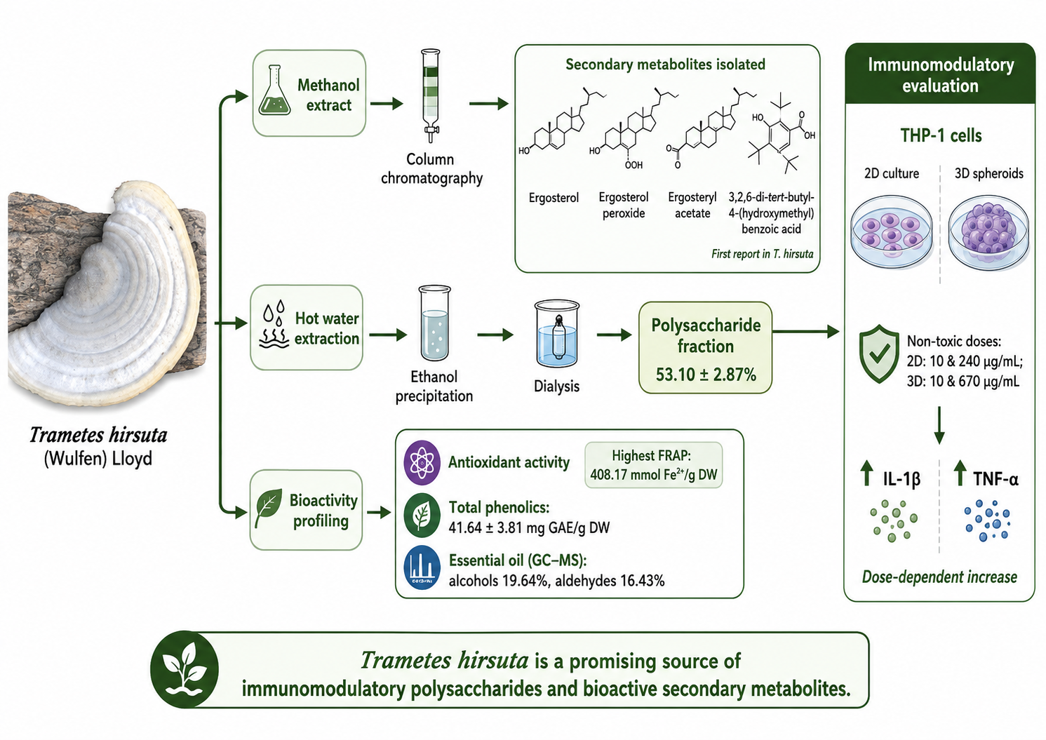
Mushroom-derived products are increasingly recognized as promising immunomodulatory therapeutics. This study aimed to evaluate the immunomodulatory properties of polysaccharides and characterize secondary metabolites from Trametes hirsuta (Wulfen) Lloyd. Sterol derivatives were isolated using column chromatography, while polysaccharides were obtained via hot water extraction, purified by ethanol precipitation, and dialyzed. Polysaccharide content was measured with the phenol-sulfuric acid method. Antioxidant activity was assessed using DPPH and FRAP assays. The volatile fraction was obtained by hydrodistillation and analyzed by GC-MS Spectrometry. The total phenolic content was determined by the Folin-Ciocalteu method. The cytotoxicity of the polysaccharide fraction on THP-1 cells was evaluated using the XTT assay in 2D and 3D cultures and its effects on IL-1β and TNF-α secretion were assessed. Identified compounds included ergosterol, ergosterol peroxide and ergosteryl acetate, and the phenolic compound 3,2,6-di-tert-butyl-4-(hydroxymethyl) benzoic acid, which was identified for the first time in T. hirsuta. The polysaccharide content was quantified at 53.10 ± 2.87%. Cytotoxicity testing showed non-toxic doses of 10 and 240 μg/mL in 2D cultures, and 10 and 670 μg/mL in 3D cultures. Using these concentrations, the extract induced a dose-dependent increase in IL-1β and TNF-α secretion. Total phenolic content of the methanol extract was 41.64 ± 3.81 mg gallic acid equivalent per gram of dry weight. The essential oil was rich in alcohols (19.64%) and aldehydes (16.43%). The methanol extract exhibited the highest antioxidant activity in FRAP assay (408.1667±0.1116 mmol Fe2+/gram dry weight). These findings support the immunomodulatory activity of T. hirsuta polysaccharides and warrant further investigation of their underlying receptor-mediated mechanisms.
Keywords:
Trametes hirsuta
; polysaccharides
; immunomodulation
; molecular pharmacology
; THP-1 cells
; TNF-α
; IL-1β
1. Introduction
Immune deficiencies (IDs) are categorized into primary and secondary immunodeficiency disorders [1]. Both categories are linked or may lead to a range of complications, including autoimmune disorders, infections and various cancers [2], which place a considerable strain on the healthcare system and impact on the lives of those affected [3]. Therefore, many researchers have attempted to enhance immune responses in ID patients using various strategies such as immunostimulant drugs (immune checkpoint inhibitors, cytokines, monoclonal antibodies, vaccines) [4] and immunomodulatory compounds from natural sources (polysaccharides, proteins, lectins, phenolics and sterols) [5].
Among these, mushroom polysaccharides are highly effective with low toxicity and ease of accessibility and cost-effectiveness [6], these compounds can significantly enhance immune cell responses by interacting with certain receptors expressed on immune cells, such as complement receptor 3 (CR3), Toll-like receptors (TLRs), and dectin-1, which are mainly expressed on innate immune cells, with TLRs also present on adaptive immune cells [7]. These receptor-mediated interactions are of particular interest in molecular pharmacology because they connect the structural recognition of fungal polysaccharides with downstream cellular signaling and cytokine responses.
Medicinal mushrooms contain β-glucans polysaccharides, which have a primary role in immune system activation, making them one of the most reliable natural sources for support of immune function [8]. β-Glucans enhance macrophage function and antimicrobial activity in mononuclear cells and neutrophils [9]. β-1,3-Glucans serve as key structural elements in fungal cell walls, playing a crucial role in the activation of macrophages in mammals. Lentinan, a polysaccharide isolated from Lentinola edodes, stands out among various β-1,3-glucans, featuring a structure that includes a backbone of β-1,3-linked glucose residues complemented by side chains of β-1,6-glucose residues [10]. This polysaccharide possesses anti-tumor [11] and anti-inflammatory [12] properties.
Few studies have been done on the immune-modulatory effects of Trametes hirsuta (Wolfen) Lloyd polysaccharides extracts.
Trametes hirsuta exhibits a broad geographical distribution across Europe (e.g. Portugal), Asia and North America, with occurrence records spanning diverse geographical and climatic regions. It is also known as the hairy bracket, is not considered poisonous, but it is also not considered edible due to its tough, corky texture. It has some beneficial applications, including medicinal properties and a role in bioremediation [13]. A 2014 study revealed that glucan polysaccharides derived from T. hirsuta significantly enhance splenocyte proliferation, boost natural killer (NK) cells anti-tumor activity, and increase macrophage phagocytosis in a dose-dependent manner. This effect includes a particularly robust induction of IL-2, IFN-γ and moderate elevation of TNF-α [14]. In 2015, a study on the immunomodulatory activity in mice revealed that T. hirsuta mycelia had the potential to enhance macrophage phagocytic function and increase serum hemolysin concentrations [15].
Overall, in this study, secondary metabolites were isolated and identified using chromatographic and spectroscopic techniques. The immunomodulatory properties of T. hirsuta polysaccharides were evaluated in THP-1 cells by measuring IL-1β and TNF-α secretion, providing a basis for assessing their potential therapeutic effects. Accordingly, the study combines chemical characterization with a human cell-based pharmacological endpoint and provides a foundation for subsequent receptor- and signaling-level mechanistic studies.
2. Materials and Methods
2.1. Materials and Equipment
The filtrate was concentrated using a rotary evaporator (Heidolph, Germany). A Bruker Avance™ 500 DRX spectrometer (Karlsruhe, Germany) was employed to obtain 1H and 13C NMR spectra at frequencies of 500 MHz, with tetramethylsilane (TMS) as the internal standard. Chemical shifts are reported as δ values in parts per million (ppm) and coupling constants (J) are given in Hertz (Hz). The structures of the isolated compounds were elucidated based on their 1H and 13C NMR spectra. Carbon chemical shifts were obtained directly from the 13C NMR spectra, and individual carbon signals were assigned by comparison with previously published spectroscopic data for the corresponding compounds. Potassium bromide pellets were used to record Fourier transform infrared spectra (FT-IR) on a Nicolet 550. Gas Chromatography-Mass (GC-MS) was performed on an Agilent 6890 instrument with a BPX-5 MS capillary column (30 m × 0.25 mm i.d., 0.25 µm film thickness). Polycaprolactone (Mm = 80,000 kDa, Sigma-Aldrich, #cat 440,744) was used to fabricate the 3D scaffold. Electrospinning was performed using an FNM, ESY 20 (Iran) setup an injection. Powder BSA (Merck, Germany) and an ELISA Plate SPL (Jet biofill, China) were utilized to investigate the impact of polysaccharides on cytokines. Cytokines IL-1β and TNF-α concentrations were measured following the manufacturer's guidelines, via specific ELISA kits from Invitrogen, USA. A double-beam Perkin Elmer UV/visible spectrophotometer was applied for this purpose. Dialysis tubing (cut-off 14 kDa), DPPH (2-diphenyl-1-picrylhydrazyl), TPTZ (2,4,6-Tripyridyl-s-triazine) and XTT (2,3-bis(2-methoxy-4-nitro-5-sulfophenyl)-5-[(phenylamino)carbonyl]-2H-tetrazolium hydroxide) were supplied from Sigma Aldrich (USA).
2.2. Sample Collection, Identification and Preparation
T. hirsuta fruiting bodies were collected from the Hezar Jarib forest in Neka (Chalmardi, Hezar-jerib, Neka; 36° 33′ 42″ N 53° 23′ 31″ E), Mazandaran, Iran in November (2022). Microscopic and macroscopic features were employed for mushroom identification. The specimen was deposited in the herbarium of the Faculty of Pharmacy, Department of Pharmacognosy and Biotechnology at Mazandaran University of Medical Sciences. The voucher specimen was assigned the code MAZ-B4-0404-004. The samples underwent a thorough cleaning process to remove all debris, followed by freezing at -20 ℃, for one week to guarantee the deactivation of any insect eggs. Following the freezing process, the items were rinsed with deionized water, dried in an oven at 38 °C, finally ground into coarse powder using a mill [16].
2.3. Physicochemical Properties
The physicochemical properties of T. hirsuta, including ash values and heavy metals, were evaluated separately. For the determination of total ash, a sample of 8.5 g of powdered mushroom was incinerated completely in a weight-determined crucible using an electronic furnace for 6 h at a continuous temperature of 600°C. The final ash weight reflects the total ash content of the mushroom. In the following step, 10 mL of 35% hydrochloric acid (HCl (and 15 mL of deionized water were added to the vessel holding the total ash, followed by boiling for 15 minutes. Next, the last of the mixtures were filtered through ash-less filter paper and rinsed with hot water until achieving neutrality. Residual ashes were subsequently dried and weighed as acid-insoluble ash. To determine the water-soluble ash, 25 mL of deionized water was added to a crucible containing 0.45 g of the total ash, followed by boiling for 10 minutes. The insoluble residue was then collected on ash-less filter paper and incinerated for 6 h at 600°C in an electronic furnace. The final weight represented the water-insoluble ash, while comparison with total ash accounted for the water-soluble ash.
The concentrations of heavy metals, including Copper (Cu), Mercury (Hg), Lead (Pb), and Cadmium (Cd), were evaluated through an acid digestion method, subsequently analyzed via atomic absorption spectroscopy. The procedure involved the measurement and placement of 1 g of mushroom powder in separate silica crucibles. 10 mL of HCl (70%), 5 mL of concentrated sulfuric acid, and 30 mL of nitric acid (70%) were added to each crucible. The mixture was then heated to the boiling point for one hour. Following the heating process, the solutions underwent filtration and were subsequently analyzed via atomic absorption spectroscopy. Standard curves were created using standard solutions of water-soluble salts, specifically CdSO4, HgSO4, PbCl2, and CuSO4, at concentrations of 10, 25, 50, and 100 ppm [8].
2.4. Isolation and Characterization of Volatile Fraction
T. hirsuta fruiting bodies (100 g) were placed in a 1 L round-bottom flask containing 500 mL of deionized water (mushroom-to-water ratio, 1:5, w/v) and subjected to hydrodistillation using a Clevenger-type apparatus for 4 h. The volatile fraction was collected in 2 mL of pentane and subsequently dried over anhydrous sodium sulfate. As the volatile fraction was directly collected in pentane and subsequently concentrated for GC-MS analysis, a gravimetric extraction yield was not determined [17]. GC-MS analysis was performed using an Agilent 6890 instrument equipped with a BPX-5 MS capillary column (30 m × 0.25 mm i.d., 0.25 µm film thickness). The oven temperature was initially maintained at 50 °C for 5 min, increased to 240 °C at 3 °C/min, and then increased to 300 °C at 15 °C/min and held for 3 min. Samples were injected at 250 °C using helium as the carrier gas at a flow rate of 0.5 mL/min in split mode (1:35). The MS interface temperature was set at 220 °C, and mass spectra were acquired in electron-impact (EI) mode at 70 eV over an m/z range of 40-500. Volatile constituents were identified by comparison of their mass spectra with those available in the NIST mass spectral library and by comparison of their Kovats retention indices (KI) with reported literature values. Relative proportions of individual constituents were calculated from their GC peak areas and expressed as percentages of the total identified volatile constituents.
2.5. Isolation and Quantification of Polysaccharides
Mushroom dry powder (245g) was extracted sequentially with n-hexane, chloroform and methanol over three cycles, each lasting 48 h, to eliminate nonpolar compound like small molecules and lipids. Following this, the sample was subjected to cold water extraction for a duration of 6 h, which was repeated three times. Cold water extraction is required to remove non-relevant polar compounds, including phenolic compounds and storage polysaccharides. Lastly, cell wall polysaccharides were isolated using hot water extraction, which was carried out three times at 100°C for 6 h under reflux. The hot aqueous extract was filtered and polysaccharides precipitated by adding ethanol (96%) three times the extract volume, followed by refrigeration of the mixture for 18 h. The precipitate was collected by centrifugation at 5000 rpm and 10 ℃. The sediment was washed several times with methanol for purification and then dialyzed against tap water for 24 h using dialysis tubing with a 14 kDa molecular weight cut-off. The polysaccharide was freeze-dried and stored at -20 ℃ [18].
The total polysaccharide content of the fraction was determined using the phenol-sulfuric acid colorimetric method. The sugar content of the sample was quantified using a standard curve of its constituent monosaccharides. Glucose was selected as the standard sample based on the analysis of monosaccharide components. Half a milliliter of glucose standard solutions and the polysaccharide fraction were separately transferred into test tubes. Then, 500 µL of 4% phenol followed by 2.5 mL of 96% sulfuric acid was added to each tube. The mixture was then placed in a hot water bath for 5 minutes to hydrolyze glycosidic bonds and develop a colored complex. The absorbance was immediately measured at 490 nm and the sugar content in the sample was calculated using the standard curve [19].
2.6. Isolation of the Non-Polar Fractions
T. hirsuta fruiting bodies (624 g) were ground into small fragments using a mill and extracted by maceration with n-hexane (10 L) at room temperature for 48 h. After filtration and evaporation of the solvent to obtain the n-hexane extract, the residual mushroom material was air-dried and subsequently extracted with chloroform under the same conditions. A rotary evaporator was used to concentrate the extract after it had been filtered, yielding 38.8 g of concentrated n-hexane extract. The extracted mixture with n-hexane was placed on a silica gel chromatography column (8.5×25.5 cm, 60 mesh) and separated using a mobile phase containing different ratios of n-hexane and ethyl acetate (EtOAc) (from 10:0 to 0:10), yielding 11 fractions. Fractions 1, 4, and 5 were subsequently loaded onto a silica gel column (1×40 cm, 60 mesh) using n-hexane: EtOAc (10:0 to 7:3) as the mobile phase. Ultimately, Compounds 1, 2, and 3 were isolated in pure form. In the following, the chloroform extract was loaded onto a silica gel chromatography column (5×25.5 cm, 60 mesh) and separated using a mobile phase containing different ratios of n-hexane and EtOAc (from 10:0 to 0:10), yielding 4 fractions. Fractions number 2 were thereafter loaded onto a silica gel column (1×30 cm, 60 mesh) using n-hexane: EtOAc (10:0 to 7:3) as the mobile phase. Ultimately, the pure compounds 4 were separated from the mixture. Each fraction was analyzed using TLC plates pre-coated with silica gel G F254 and a solvent mixture of n-hexane and EtOAc (7:3, v/v) as the mobile phase. Anisaldehyde-H2SO4 was used as spray reagent.
2.7. Antioxidant Activity and Total Phenolic Content Assay
The mushroom methanolic extract was prepared via the maceration method at room temperature for 24 h, repeated three times, followed by concentration in an oven at 50 ℃. The DPPH method is a rapid, easy, and cost-effective approach for assessing the radical scavenging properties of different compounds, especially natural compounds. When antioxidant compounds are present, the purple compound DPPH is reduced and loses its color [20]. Methanol was used to prepare a 0.15 mM DPPH solution. Following this, 1.5 mL of DPPH solution was combined with 1.5 mL of various concentrations of the methanolic extract, and the absorbance of the samples was subsequently measured at 517 nm. The percentage of DPPH inhibition was determined using the following formula:
AC: Control absorption, which contains DPPH solution and methanol. AS: Sample absorption, which contains DPPH solution and the extracts.
Scavenging rate= [(AC- AS)/AC)] ×100
After calculating the inhibition percentage of each concentration, the concentration that was able to restore 50% of DPPH was considered as the IC50.
A spectrophotometric technique described in earlier research was used to measure the FRAP (ferric reducing ability of plasma) [21]. The FRAP reagent was prepared daily with minor adjustments and heated to 37 ℃ before application. Ascorbic acid served as a positive control. The reaction mixture was incubated at 37 ℃ for 4 minutes, after which the absorbance was measured at 593 nm.
The FRAP activities of the methanol extract of T. hirsuta were quantified in terms of µmol FeSO4 per gram of sample. The total phenol content was quantified by employing the Folin-Ciocalteu reagent via standard spectrophotometry, incorporating modifications to the standard curve preparation. A 0.5 mL aliquot of the sample solution at different concentration was mixed with 2.5 mL of 0.2 Folin-Ciocalteu reagent at room temperature. Five minutes later, two mL of a 20% sodium carbonate solution were added, and the mixture was shaken vigorously. The absorbance of the samples was measured at 760 nm following a 15 min incubation at 45 ℃ [22]. Standard concentrations of gallic acid equivalents were used to create a calibration curve (Y=2.2868x + 0.0964, R2 = 0.9992) and results were expressed as gram GAE/g of the dried mushroom methanol extract.
2.8. Cell Viability Assay
THP-1 human monocytic cells were obtained from the National Cell Bank of Iran (NCBI), Pasteur Institute of Iran, Tehran, Iran (Catalog No. C563; corresponding to ATCC TIB-202). To establish the 3D cell culture model, a polycaprolactone (PCL) nanofibrous scaffold was fabricated by electrospinning and used as a three-dimensional support for THP-1 cell culture. PCL was dissolved in acetone at a 9% concentration at room temperature. The solution was stirred on a magnetic stirrer for 1 h at 500 rpm and loaded into a 5 mL syringe fitted with a 22G needle. Electrospinning was performed an injection speed of 1 mL/h, a collector rotation speed of 200 rpm, a tip-to-collector distance of 12 cm, and a total solution volume of 10 mL. Aluminium sheets were coated with the collected electrospun nanofibers. The scaffold surface was subjected to oxygen plasma treatment after electrospinning. After 20 minutes of UV irradiation for sterilization, THP-1 cells were seeded at a density of 4 × 10⁴ cells/well in 96-well plates and incubated for 24 h. Subsequently, the subjects were treated with T. hirsuta polysaccharides at concentrations ranging from 5 to 1600 µg/mL for 48 h. The XTT assay assessed absorbance at 450 nm (with a 620 nm reference) to determine cell viability. Moreover, growth inhibition was calculated in relation to untreated controls. All experiments were performed in triplicate to ensure accuracy and reproducibility [23].
2.9. Assay of TNF-α and IL-1β Level
THP-1 cells were seeded in 24-well plates at a density of 1 × 10⁵ cells/well and treated for 48 h with non-toxic concentrations of the T. hirsuta polysaccharide fraction (10 and 240 μg/mL for 2D cultures and 10 and 670 μg/mL for 3D cultures), selected based on the cell viability results. Supernatants were collected by centrifugation (1000 rpm, 8 min, 4 ℃) and stored at -20 ℃. Levels of TNF-α and IL-1β were measured using commercially available ELISA kits according to the manufacturer’s instructions. Bacterial lipopolysaccharide) LPS, 1 μg/mL (was used to stimulated THP-1 cells as a positive control, and untreated cells served as a negative control to account for non-specific immune responses [24].
2.10. Statistical Analysis
All experiments were repeated three times, and results are reported as mean ± standard deviation (SD). The Kolmogorov-Smirnov test was employed to assess the normality of data distribution. Statistical comparisons were conducted using the non-parametric Mann-Whitney U test for paired parametric t-tests, contingent on data characteristics, with significance established at p < 0.05. The SPSS statistical software (SPSS, Chicago, IL, USA) and GraphPad Prism (San Diego, California, USA) were used for all analysis.
3. Results
3.1. Ash Content Determination
The water-soluble ash, acid-insoluble ash, and total ash percentages in the T. hirsuta sample were quantified (Table 1).
3.2. Heavy Metal Content Analysis
The metal content of T. hirsuta fruiting bodies was determined via acid digestion. Results are presented as milligrams of metal per 100 g of dry mushroom weight. The microelements detected in the analyzed samples included Pb, Fe, Hg, and Cu with concentrations of 3.56, 1.48, 30.15, and 0.0428 mg per 100 g of mushroom, respectively.
3.3. GC-MS Analysis of the Volatile Fraction
The essential oil of T. hirsuta was analyzed for its chemical composition using GC-MS. Fifty-five peaks were detected in the total ion chromatogram. The major compounds identified in the essential oil were 3-octanol (8.57%), decadienol (7.07%), furfural (4.13%), 1-octen-3-ol (7%), 2-pentyl furan (5.61%), tetradecanol (4.42%), and hexadecanoic acid (4.32%) (Table 2).
3.4. Total Polysaccharide Content Determination
The yield of T. hirsuta polysaccharide extract obtained from 244.8 g of T. hirsuta was 19.35 g (dry weight), corresponding to a purified polysaccharide yield of 7.9 %. A glucose standard curve was established to quantify the total polysaccharide content of T. hirsuta, represented by the equation y = 0.0023X + 0.0023, with an R2 value of 0.9983. UV absorption analysis indicated that the total polysaccharide content in the dry polysaccharide fraction of T. hirsuta was 53.103 ± 2.87%.
3.5. FT-IR Analysis of Isolated Polysaccharides
The polysaccharide extract from T. hirsuta exhibited a strong, broad absorption band at 3284.03 cm-1, indicative O-H stretching in amino groups. The band at approximately 1154.20 cm-1 corresponded to C–O stretching, the absorption at 1024.80 cm-1 of C–O–C stretching, and the band between 1000 and 1100 cm-1 to β-glucans. The shoulder observed at 907.83 cm-1 is characteristic of β-glycosidic linkages. Additionally, bands in the 1373.85-1411.26 cm-1 range were assigned to O–H groups of phenolic compounds [25].
3.6. Purification and Isolation of Sterol Derivatives
Chromatographic analysis of the n-hexane extract T. hirsuta led to the isolation of ergosterol, ergosterol peroxide, ergosteryl acetate and di-tert-butyl-4-(hydroxymethyl) benzoic acid.
3.6.1. Compound 1, ergosterol
The compound was obtained as a white crystalline powder, which was not detectable on TLC plates under UV light (λ=254 and 366 nm). Nonetheless, treatment with anisaldehyde-H2SO4 spray reagent allowed easy detection, indicated by a dark purple spot: 1HNMR analysis was performed in CDCl3 (500 MHz), and chemical shifts are reported in ppm :δ 5.187 (1H, dd, H-7), 5.225 (1H, d, H-5), 5.406 (1H, dd, H-22), 5.591 (1H, dd, H-23), 3.7 (1H, H-3), 1.045 (3H, d, J=6.5 Hz, H-21), 0.942 (3H, d, J=6.5 Hz, H-28), 0.865 (3H, d, J=7 Hz, H-26), 0.818 (3H, d, J=7.3 Hz, H-27), 0.651 (3H, s, H-19), 0.565 (3H, s, H-18) and 13C NMR (125 Hz, CDCl3, in ppm). Results are presented in Figure 1(A) and Table 3.
3.6.2. Compound 2, Ergosterol Peroxide
The compound, a white crystalline solid, which was not detectable on TLC under UV light (λ=254 and 366 nm). however, it could be detected as a dark spot after treatment with anisaldehyde-H2SO4 spray reagent. The 1HNMR analysis was performed in CDCl3, and chemical shifts are reported in ppm: δ 6.93 (1H, d, J=5.2 Hz, H-7), 6.5 (1H, d, J=4.8 Hz, H-6), 5.38 (1H, dd, J=6.8 Hz, H22), 5.22 (1H, dd, J=6.4 Hz, H-23), 3.62 (1H, m, H-3), 1.03 (3H, d, J=7.8 Hz, H-21), 0.95 (3H, d, J=7.8 Hz, H-28), 0.907 (3H, s, H-18), 0.914 (3H, s, H-19), 0.792 (3H, d, J=3.4 Hz, H-27), 0.785 (3H, d, J=3.4 Hz, H-26); 13C NMR (125 Hz, CDCl3, in ppm). Results are detailed in Figure 1(B) and Table 3.
3.6.3. Compound 3, Di-Tert-Butyl-4-(Hydroxymethyl)Benzoic Acid
The compound was isolated from the n-hexane extract as a brown solid, which was not detectable on TLC under UV light but could be visualized as a dark spot after treatment with anisaldehyde-H2SO4 spray reagent. The 1H-NMR analysis was performed in CDCl3, and chemical shifts are reported in ppm. 1HNMR (CDCl3, 300.13 MHz): δH (ppm): 1.42 (s, 18H, CH3), 4.94 (s, 2H, CH2), 5.21 (s, 1H, OH), 7.41 (s, 2H, CH Aromatic), 11.01 (s, 1H, OH acid). 13C NMR (CDCl3, 75.65 MHz): δC (ppm): 30.2, 34.2, 46.3, 126.7, 127.3, 135.6, 149.3, 166.6, Figure 1(D).
3.6.4. Compound 4, Ergosteryl Acetate
The compound was isolated from the chloroform extract, as a green solid, which was detectable on TLC under UV light (λ=254 nm) and appeared as a dark spot after treatment with anisaldehyde-H₂SO₄ spray reagent. The 1HNMR analysis was performed in CDCl3, and chemical shifts are reported in ppm. 1HNMR (CDCl3, 300.13 MHz): δH (ppm): 0.88-2.37 (m, 35H, CH2), 2.38 (s, 3H, CH3), 3.83-3.83 (m, 1H, CH), 5.34-5.39 (m, 2H, CH=CH), 6.53 (d, 1H, CH olefin). 13C NMR (CDCl3, 75.65 MHz): δC (ppm): 14.1, 22.5, 22.7, 24.6, 25.6, 27.2, 28.5, 29.0, 29.1, 29.2, 29.3, 29.4, 29.6, 29.6, 31.5, 31.9, 33.3, 34.0, 34.9, 35.3, 36.0, 37.9, 70.9, 128.0, 130.2, 137.1, 147.7, 179.7 Figure 1(C).
3.7. Antioxidant Activity and Total Phenolic Content of the Methanolic Extract
The scavenging activities of methanol and polysaccharide extracts of T. hirsuta against the DPPH free radical were evaluated (Table 4). Both extracts demonstrated the highest activity against the DPPH free radical, as evidenced by the lowest IC50 value. The IC50 value for ascorbic acid, used as a standard, was 5.687±6.7 μg/mL, indicating that the polysaccharides from T. hirsuta have comparatively lower free radical scavenging capacity. Additionally, the antioxidant activity was assessed using the FRAP assay and expressed as µmol Fe (II) equivalent per gram of dry extract. As shown in Table 4, the FRAP value for T. hirsuta polysaccharides was 64.72±0.11 μmol Fe (II)/g. Total phenolic content, measured as mg gallic acid equivalents per gram of dried extract, varied depending on the extraction solvent, with the methanol extract exhibiting higher phenolic content than polysaccharide fraction (Table 4).
3.8. Cytotoxic Effects of T. hirsuta Polysaccharides on THP-1 Cells
Using the XTT assay, the effects of different T. hirsuta polysaccharide concentrations specifically (0, 5, 10, 50, 100, 200, 400, 800, and 1600 μg/mL) on the growth inhibition of the THP-1 cell line were evaluated. As shown in (Figure 2), T. hirsuta polysaccharide exhibited dose-dependent cytotoxic effects on THP-1 cells in both 2D and 3D culture. The results revealed that the direct toxicity of T. hirsuta polysaccharide in 2D culture was greater than in 3D culture. Moreover, IC20 values for T. hirsuta polysaccharide on THP-1 2D and 3D culture were measured based on a dose response curves at 247.3 µg/mL and 671 µg/mL concentrations, respectively. To investigate the alterations in THP-1 cytokine assay, a concentration below IC20 was selected to ensure that at least 80% of cells remained viable, minimizing effects due to cell death. Based on these findings, two non-toxic concentrations were chosen for cytokine assays: 10 and 240 μg/mL for 2D exposure and 10 and 670 μg/mL for 3D exposure of T. hirsuta polysaccharide.
3.9. IL-1β and TNF-α Secretion by THP-1 Cells
The study examined the effect of non-toxic levels of T. hirsuta polysaccharide on TNF- α and IL-1β cytokines production by THP-1 cells cytokines in 2D and 3D as shown in (Figure 3), TNF-α secretion was significantly elevated compared to the untreated control following exposure to T. hirsuta polysaccharide fraction at 10 and 240 μg/mL in 2D cultures and at 10 and 670 μg/mL in 3D cultures (p ˂ 0.05 and p ˂ 0.01, respectively). As shown in (Figure 3), TNF-α secretion was significantly elevated Compared to the untreated control group following exposure to T. hirsuta polysaccharide fraction at 10 and 240 μg/mL in 2D cultures and at 10 and 670 μg/mL in 3D cultures (p ˂ 0.05 and p ˂ 0.01, respectively). The results also indicated that higher non-toxic concentrations of T. hirsuta polysaccharide exhibited greater stimulatory potential than lower concentrations.
4. Discussion
The genus Trametes encompasses a wide range of bioactive compounds, making it an important subject for investigating potential therapeutic properties and conducting pharmacological research. Key constituents identified in Trametes species include steroids, polysaccharides, fatty acids, and phenolic compounds. In this study, spectroscopy and column chromatography techniques were employed to isolate and identify ergosterol and ergosterol peroxide, and one phenolic compound from the n-hexane and chloroform fractions of T. hirsuta fruiting bodies. Notably, in addition to ergosterol and ergosterol peroxide, this study reports for the first time the presence of ergosteryl acetate and di-tert-butyl-4-(hydroxymethyl) benzoic acid in this species. In a related study, Bakhshi et al. isolated various sterols, including ergosterol and ergosterol peroxide, from the n-hexane fraction of T. gibbosa using chromatographic methods [17]. The chemical structures of these compounds were confirmed by 1HNMR and 13CNMR spectroscopy.
Furthermore, the GC-MS analysis revealed the presence of several volatile compounds in T. hirsuta fruiting bodies, including aldehydes, alcohols, ketones, and fatty acids. 1-octene-3-ol, an alcoholic compound, was the predominant compound in the essential oil. This compound has also been reported in T. gibbosa fruiting bodies collected in Göttingen, Germany, as well as in certain oyster mushrooms and other fungal species, including Schizophyllum commune and Phlebia radiata [27]. In a study investigating the water-soluble polysaccharides of Inonotus obliquus at concentrations ranging from 25 to 400 μg/mL, Fan et al. (2012) evaluated their antitumor and immunomodulatory effects against SGC-7901 cells using the MTT assay. In addition, the secretion of cytokine TNF-α from mouse peritoneal macrophages was measured using the ELISA method. The results showed that water-soluble polysaccharides did not exhibit significant direct antitumor activity at concentrations up to 200 μg/mL, However, at concentrations between 50 and 400 μg/mL, they significantly increased TNF-α production. Therefore, polysaccharides of this mushroom play their antitumor and immunomodulatory role indirectly by stimulating macrophage cytokines secretion [28].
Kodama et al. (2002) reported that a branched (1→3) (1→6) β-glucan extracted from Maitake mushroom (Grifola frondosa) increased both the percentage and activity of NK cells in mice infected with H1N1 influenza virus. In evaluating the effect of this polysaccharide on MM-46 carcinoma tumors in mice, they found that, in addition to elevating the proportion of NK cells in the spleen, the cytotoxic activity of these cells against YAC-1 target cells were significantly enhanced. Furthermore, the levels of the cytokines IFN-γ and TNF-α, which are indicative of NK activity, were markedly increased. The authors suggested that NK cells are primarily responsible for the early antitumor effects, whereas both NK and T cells contributed to the long-term antitumor responses induced by this polysaccharide [29].
The current study conducted a spectrophotometric analysis of various extracts from T. hirsuta fruiting bodies, demonstrating a high phenolic content in the methanol extract, quantified at 41.64±3.81 mg GAE/g of dry weight. Both methanol and polysaccharide extracts exhibited notable reducing activity in the FRAP assay, whereas radical scavenging activity assessed by DPPH was higher in the methanol extract. The current research demonstrates that T. hirsuta polysaccharides exhibited lower antioxidant activity compared with ascorbic acid, used as a standard. Gan et al. reported a positive correlation between phenolic content and antioxidant activity in various extracts of Agaricus brasiliensis mushroom. The ethanol extract was exhibited the highest FRAP and DPPH activity and the lowest EC50 value [30].
Bioactive compounds that stimulate or modulate the immune system are referred to as immunomodulators [31]. The total polysaccharide content of T. hirsuta was determined 53.10±2.87%. To evaluate the immune-stimulatory activity of T. hirsuta polysaccharide, cytotoxicity assays were performed on THP-1 cells across a range of concentrations (5-1600 μg/mL). As illustrated in (Figure 2), T. hirsuta polysaccharide exhibited dose-dependent cytotoxic effects in both 2D and 3D cultures. These findings are consistent with a study by Alla (2018), which reported that hot water extracts from various wood-decaying fungi, including T. versicolor, reduced THP-1 cell viability [32].
The cytotoxicity results also indicated that the direct toxicity of T. hirsuta polysaccharide was higher in 2D cultures than in 3D cultures. This difference is attributed to the more direct contact of polysaccharides with cells in 2D models, resulting in stronger effects on immune activity. Li et al. (2018) similarly demonstrated that polysaccharides exert directly influence on immune activities in 2D cultures compared with 3D models, where scaffold structure and the more complex microenvironment limit polysaccharide access to cells. Consistently, Fouad Damiri et al. (2024) showed that higher polysaccharide doses are required in 3D cultures to achieve effects comparable to those observed in 2D models [33]. Overall, reduced direct contact in 3D models leads to lower observed toxicity, necessitating higher polysaccharide concentrations to elicit similar cellular responses.
Non-toxic concentrations, defined as those below IC20, were selected based on cytotoxicity assay data to ensure that the observed effects were not due to cytotoxicity. specifically, two non-toxic concentrations were chosen for 2D exposure (10 and 240 μg/mL) and 3D exposure (10 and 670 μg/mL) of T. hirsuta polysaccharide for subsequent cytokine assays. To evaluate the immunomodulatory potential of the polysaccharide, cytokine secretion by THP-1 cells was measured. As shown in (Figure 3), treatment with non-toxic dose of T. hirsuta polysaccharide elicited inflammatory responses, resulting in increased secretion of pro-inflammatory cytokines TNF-α and IL-1β.
These results align with previous studies. Liping Chen et al. (2024) noted that polysaccharides from Cordyceps sinensis activated dendritic cells and T lymphocytes, increasing the secretion of cytokines such as IL-6, IL-8, and IL-2 [34]. Similarly, Hemmati et al. (2024) revealed that polysaccharides from Trichaptum biforme could activate NK cells and enhance cytokine production [35]. Mushroom polysaccharides are known to exert immunostimulatory effects through receptors such as TLRs, Dectin-1, and CR3.The increase in TNF-α and IL-1β at non-cytotoxic concentrations is consistent with pharmacological modulation of innate immune-cell function rather than solely with a response secondary to overt cytotoxicity.
In this study, the cytokine assay results showed that the stimulatory effect of T. hirsuta polysaccharide was greater in 2D-cultured THP-1 cells compared with 3D culture. Likely due to more direct interactions between the polysaccharide and the cells, which aligns with the cytotoxicity assay results. Limitations of the study include the lack of molecular investigations into the effects of T. hirsuta polysaccharide on gene expression in THP-1 cells, as well as the absence of in vivo animal models. Future studies will address these gaps. Overall, the present study demonstrates that polysaccharides from T. hirsuta can activate the immune system, highlighting their potential for developing therapeutic strategies for immunodeficient patients.
5. Conclusion
This study reports, for the first time, the identification of ergosteryl acetate, and the phenolic compound 3,2,6-di-tert-butyl-4-(hydroxymethyl)benzoic acid in T. hirsuta. The study also investigated the immunostimulatory properties of its polysaccharide fraction on innate immune cells, with a particular focus on monocytes. The results indicate that THP enhances the innate immune response by promoting the secretion of inflammatory cytokines from macrophages, pointing to its potential antitumor effects.
However, this research has limitations. The specific impact of THP on adaptive immune cells, including B and T lymphocytes, were not evaluated. moreover, in vivo studies were not concluded, so the responses of immune cells in animal models remain unassessed, representing a significant area for future research. Future studies could also investigate the combined effects of THP with other immunomodulatory agents or standard chemotherapeutics, which may reveal additional insights. In conclusion, this research highlights the potential of polysaccharides derived from T. hirsuta and emphasizes the importance of mushroom-derived natural products as immunostimulatory agents. Taken together, these findings provide a cellular pharmacology framework for future studies aimed at identifying the molecular targets and signaling events responsible for THP-induced cytokine responses, thereby strengthening the translational evaluation of T. hirsuta polysaccharides as candidate immunomodulatory natural products.
Author Contributions
Conceptualization, S.E.E., H.H. and E.H.; methodology, H.H. and E.H.; software, H.A.K. and H.B.J.; validation, E.H., M.M.; investigation, H.A.K. and S.E.E.; data curation, H.A.K.; writing—original draft preparation, H.A.K. and H.B.J.; writing—review and editing, H.H., M.M. and E.H.; supervision, E.H.; funding acquisition, E.H. All authors have read and agreed to the published version of the manuscript.
Funding
This research was funded by Iranian Vice-Presidency for Science and Technology, Tehran, Iran (grant number: 4022901). Moreover, this work was supported by Mazandaran University of Medical Sciences, School of Pharmacy, Sari, Iran (Ethics approval code: IR.MAZUMS.REC.17225; approval date: October 2022).
Institutional Review Board Statement
The study protocol was approved by the Ethics Committee of Mazandaran University of Medical Sciences (ethics approval code: IR.MAZUMS.REC.17225; approval date: October 2022).
Informed Consent Statement
Not applicable. This study did not involve human participants.
Data Availability Statement
The datasets generated and analyzed during the current study are not publicly available due to privacy concerns and ethical restrictions. However, they are available from the corresponding authors upon reasonable request.
Acknowledgments
The authors would like to thank Mazandaran University of Medical Sciences, School of Pharmacy, for providing laboratory facilities and administrative support and acknowledged to Iranian Vice-Presidency for Science and Technology for their financial support. The authors also appreciate the assistance of colleagues and staff who contributed to the technical aspects of this study. The authors have reviewed and edited the output and take full responsibility for the content of this publication.
Conflicts of Interest
The authors declare no conflicts of interest and that they have no known competing financial interests or personal relationships that could have appeared to influence the work reported in this paper. The funders had no role in the design of the study; in the collection, analyses, or interpretation of data; in the writing of the manuscript; or in the decision to publish the results.
Abbreviations
The following abbreviations are used in this manuscript:
| BSA | Bovine Serum Albumin |
| CDCl3 | Deuterated chloroform |
| CR3 | Complement Receptor 3 |
| DPPH | 2,2-Diphenyl-1-picrylhydrazyl |
| EI | Electron Impact |
| ELISA | Enzyme-Linked Immunosorbent Assay |
| EtOAc | Ethyl acetate |
| FRAP | Ferric Reducing Antioxidant Power |
| FT-IR | Fourier Transform Infrared Spectroscopy |
| GAE | Gallic Acid Equivalents |
| GC-MS | Gas Chromatography-Mass Spectrometry |
| IC50 | Half-maximal Inhibitory Concentration |
| IC20 | 20% Inhibitory Concentration |
| IFN-γ | Interferon-gamma |
| IL-1β | Interleukin-1 beta |
| IL-2 | Interleukin-2 |
| IL-6 | Interleukin-6 |
| IL-8 | Interleukin-8 |
| KI | Kovats Retention Index |
| LPS | Lipopolysaccharide |
| MTT | 3-(4,5-Dimethylthiazol-2-yl)-2,5-diphenyltetrazolium bromide |
| NK | Natural Killer |
| NMR | Nuclear Magnetic Resonance |
| THP | Trametes hirsuta Polysaccharide |
| THP-1 | Human Monocytic Leukemia Cell Line |
| TLC | Thin-Layer Chromatography |
| TLRs | Toll-Like Receptors |
| TNF-α | Tumor Necrosis Factor-alpha |
| TPTZ | 2,4,6-Tripyridyl-s-triazine |
| XTT | 2,3-Bis(2-methoxy-4-nitro-5-sulfophenyl)-5-[(phenylamino)carbonyl]-2H-tetrazolium hydroxide |
| TPC | Total Phenolic Content |
References
- Raje, N.; Dinakar, C. Overview of Immunodeficiency Disorders. Immunol. Allergy Clin. N. Am. 2015, 35, 599–623. [Google Scholar] [CrossRef]
- Sánchez-Ramón, S.; Bermúdez, A.; González-Granado, L.I.; Rodríguez-Gallego, C.; Sastre, A.; Soler-Palacín, P. ID-Signal Onco-Haematology Group. Primary and Secondary Immunodeficiency Diseases in Oncohaematology: Warning Signs, Diagnosis, and Management. Front. Immunol. 2019, 10, 586. [Google Scholar]
- Hsueh, J.C.; Van Hersh, A.T. Immunodeficiency: Burden of Illness. Allergy Asthma Proc. 2024, 45, 1–8. [Google Scholar] [CrossRef]
- Ramamurthy, D.; Nundalall, T.; Cingo, S.; Mungra, N.; Karaan, M.; Naran, K.; Barth, S. Recent advances in immunotherapies against infectious diseases. Immunother. Adv. 2021, 1, ltaa007. [Google Scholar] [CrossRef]
- El Enshasy, H.A.; Hatti-Kaul, R. Mushroom immunomodulators: unique molecules with unlimited applications. Trends Biotechnol. 2013, 31, 668–677. [Google Scholar] [CrossRef]
- Gill, S.K.; Islam, N.; Shaw, I.; Ribeiro, A.; Bradley, B.; Kilcoyne, M.; Ceredig, R.; Joshi, L. Immunomodulatory effects of natural polysaccharides assessed in human whole blood culture and THP-1 cells show greater sensitivity of whole blood culture. Int. Immunopharmacol. 2016, 36, 315–323. [Google Scholar] [CrossRef]
- Liu, C.; Choi, M.W.; Xue, X.; Cheung, P.C.K. Immunomodulatory effect of structurally characterized mushroom sclerotial polysaccharides isolated from Polyporus rhinocerus on bone marrow dendritic cells. J. Agric. Food Chem. 2019, 67, 12137–12143. [Google Scholar] [CrossRef]
- Brown, G.D.; Gordon, S. Fungal β-glucans and mammalian immunity. Immunity 2003, 19, 311–315. [Google Scholar] [CrossRef]
- Del Cornò, M.; Gessani, S.; Conti, L. Shaping the innate immune response by dietary glucans: any role in the control of cancer. Cancers 2020, 12, 155. [Google Scholar] [CrossRef]
- Mirończuk-Chodakowska, I.; Kujawowicz, K.; Witkowska, A.M. Beta-glucans from fungi: Biological and health-promoting potential in the COVID-19 pandemic era. Nutrients 2021, 13, 3960. [Google Scholar] [CrossRef]
- Ohara, Y. Fate of lentinan (antitumor polysaccharide) I: Fate of lentinan in mice, rats and dogs. J. Toxicol. Sci. 1980, 5, 59–72. [Google Scholar] [CrossRef]
- Yin, H.; Xue, G.; Dai, A.; Wu, H. Protective effects of lentinan against lipopolysaccharide-induced mastitis in mice. Front. Pharmacol. 2021, 12, 755768. [Google Scholar] [CrossRef]
- Aranha, G.M.; Contato, A.G.; Salgado, J.C.d.S.; de Oliveira, T.B.; Retamiro, K.M.; Ortolan, G.G.; Crevelin, E.J.; Nakamura, C.V.; de Moraes, L.A.B.; Peralta, R.M. Biochemical characterization and biological properties of mycelium extracts from Lepista sordida GMA-05 and Trametes hirsuta GMA-01: new mushroom strains isolated in Brazil. Braz. J. Microbiol. 2022, 53, 349–358. [Google Scholar] [CrossRef]
- Shenbhagaraman, R.; Premalatha, M.; Jenefar, S.; Jagadish, L.; Saravanamurali, K.; Kaveri, K.; Karthik, S.; Kaviyarasan, V. Immunopotentiating properties of extracellular polysaccharide from Trametes hirsuta strain VKESR. Carbohydr. Polym. 2014, 106, 299–304. [Google Scholar] [CrossRef]
- Ma, R.; Yang, R.; Liu, X.; Chen, Z.; Yang, C.; Wang, S. Chemical composition and immunomodulatory activity of mycelia of the hairy bracket mushroom, Trametes hirsuta (higher basidiomycetes). Int. J. Med. Mushrooms. 2015, 17, 267–276. [Google Scholar] [CrossRef]
- Bari, E.; Daryaei, M.G.; Karim, M.; Bahmani, M.; Schmidt, O.; Woodward, S.; Ghanbary, M.A.T.; Sistani, A. Decay of Carpinus betulus wood by Trametes versicolor—An anatomical and chemical study. Int. Biodeterior. Biodegrad. 2019, 137, 68–77. [Google Scholar] [CrossRef]
- Bakhshi Jouybari, H.; Bekhradnia, A.; Mirzaee, F.; Hosseinzadeh, M.H. Chemical composition of the lumpy bracket mushroom (Trametes gibbosa). Res. J. Pharmacogn. 2022, 9, 19–27. [Google Scholar]
- Agyare, C.; Agana, T.A. Bioactive metabolites from Basidiomycetes. In Advances in Macrofungi; CRC Press, 2019; pp. 230–253. [Google Scholar]
- Jouybari, H.B.; et al. Immunomodulatory activity of polysaccharide from Trametes gibbosa (Pers.) Fr (Basidiomycota, Fungi) mediated by TLR4 signaling pathway. Adv. Biomed. Res. 2023, 12, 127. [Google Scholar] [CrossRef]
- Shokrzadeh, M.; Bakhshi Jouybari, H.; Talebpour Amiri, F.; Ziar, A.; Habibpour, P. Hepatoprotective activity of Caspian saffron (Crocus caspius Fisch. & Mey) flowers against CCl₄-induced acute liver injury in mice. Res. J. Pharmacogn. 2019, 6, 33–39. [Google Scholar] [CrossRef]
- Karaman, M.; Atlagić, K.; Novaković, A.; Šibul, F.; Živić, M.; Stevanović, K.; Pejin, B. Fatty acids predominantly affect anti-hydroxyl radical activity and FRAP value: The case study of two edible mushrooms. Antioxidants 2019, 8, 480. [Google Scholar] [CrossRef]
- Amerifar, M.; Arabnozari, H.; Shokrzadeh, M.; Habibi, E. Evaluation of antioxidant properties and cytotoxicity of brown algae (Nizamuddinia zanardinii) in uterine (Hela) and pancreatic cancer cell lines (Paca-2). Hum. Exp. Toxicol. 2024, 43, 09603271241227228. [Google Scholar] [CrossRef]
- Abdelsalam, M.; Ahmed, M.; Osaid, Z.; Hamoudi, R.; Harati, R. Insights into exosome transport through the blood–brain barrier and the potential therapeutical applications in brain diseases. Pharmaceuticals 2023, 16, 571. [Google Scholar] [CrossRef]
- Taghipour, R.; Hassannia, H.; Arabnozari, H.; Enderami, S.E.; Habibi, E. Natural Polysaccharides in Breast Cancer: Fucoidan’s Role in Enhancing Cisplatin Cytotoxicity and Reducing Chemotherapy Resistance. Latest Res. Breast Cancer-Mol. Insights Diagn. Adv. Ther. Innov. CrossRef. 2025, 4. [Google Scholar]
- Kozarski, M.; Klaus, A.; Nikšić, M.; Vrvić, M.M.; Todorović, N.; Jakovljević, D.; Van Griensven, L.J. Antioxidative activities and chemical characterization of polysaccharide extracts from the widely used mushrooms Ganoderma applanatum, Ganoderma lucidum, Lentinus edodes and Trametes versicolor. J. Food Compos. Anal. 2012, 26, 144–153. [Google Scholar] [CrossRef]
- Habibi, E.; Sadat-Ebrahimi, S. E.; Mousazadeh, S. A.; Amanzadeh, Y. Mycochemical investigation of the turkey tail medicinal mushroom Trametes versicolor (higher basidiomycetes): a potential application of the isolated compounds in documented pharmacological studies. Int. J. Med. Mushrooms. PubMed. 2015, 17, 255–265. [Google Scholar] [CrossRef]
- Thakeow, P.; Angeli, S.; Weißbecker, B.; Schütz, S. Antennal and behavioral responses of Cis boleti to fungal odor of Trametes gibbosa. Chem. Senses 2008, 33, 379–387. [Google Scholar] [CrossRef]
- LiuPing, F.L.; Ding ShaoDong, D.S.; Ai LianZhong, A.L.; Deng KeQuan, D.K. Antitumor and immunomodulatory activity of water-soluble polysaccharide from Inonotus obliquus. Carbohydr. Polym. 2012, 90, 870–874. [Google Scholar] [CrossRef]
- Kodama, N.; Komuta, K.; Sakai, N.; Nanba, H. Effects of D-Fraction, a polysaccharide from Grifola frondosa, on tumor growth involve activation of NK cells. Biol. Pharm. Bull. 2002, 25, 1647–1650. [Google Scholar] [CrossRef]
- Gan, C.; Nurul Amira, B.; Asmah, R. Antioxidant analysis of different types of edible mushrooms (Agaricus bisporus and Agaricus brasiliensis). Int. Food Res. J. 2013, 20, 1095–1102. [Google Scholar]
- Drzewiecka, B.; Wessely-Szponder, J.; Świeca, M.; Espinal, P.; Fusté, E.; Fernández-De La Cruz, E. Bioactive peptides and other immunomodulators of mushroom origin. Biomedicines 2024, 12, 1483. [Google Scholar] [CrossRef]
- Shnyreva, A.V.; Shnyreva, A.; Espinoza, C.; Padrón, J.M.; Trigos, Á. Antiproliferative activity and cytotoxicity of some medicinal wood-destroying mushrooms from Russia. Int. J. Med. Mushrooms 2018, 20, 1–11. [Google Scholar] [CrossRef]
- Damiri, F.; Fatimi, A.; Liu, Y.; Musuc, A.M.; Fajardo, A.R.; Gowda, B.J.; Vora, L.K.; Shavandi, A.; Okoro, O.V. Recent advances in 3D bioprinted polysaccharide hydrogels for biomedical applications: A comprehensive review. Carbohydr. Polym. 2025, 348, 122845. [Google Scholar] [CrossRef]
- Chen, L.; Liu, X.; Zheng, K.; Wang, Y.; Li, M.; Zhang, Y.; Cui, Y.; Deng, S.; Liu, S.; Zhang, G. Cordyceps polysaccharides: A review of their immunomodulatory effects. Molecules 2024, 29, 5107. [Google Scholar] [CrossRef]
- Habibi, E.; Hemmati, P.; Arabnozari, H.; Khalili, H.A.; Sharifianjazi, F.; Enderami, S.E.; Sarker, S.D.; Hassannia, H.; Nahar, L. Phytochemical analysis and immune-modulatory potential of Trichaptum biforme polysaccharides: Implications for cancer. Int. J. Biol. Macromol. 2024, 280, 135691. [Google Scholar] [CrossRef]
Figure 1.
Structures of ergosta and benzoic acid derivatives isolated from T. hirsute.
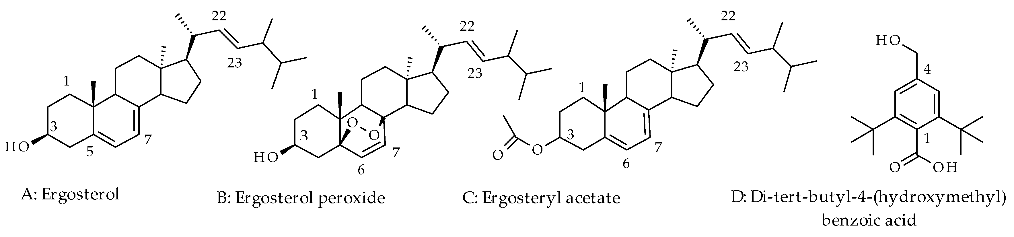
Figure 2.
Cytotoxicity evaluation of T. hirsuta polysaccharides extract on THP-1 cell line using (A) 2D and (B) 3D XTT cytotoxicity assay. inhibitory concentrations (IC20) calculated by constructing a dose-response curve.
Figure 2.
Cytotoxicity evaluation of T. hirsuta polysaccharides extract on THP-1 cell line using (A) 2D and (B) 3D XTT cytotoxicity assay. inhibitory concentrations (IC20) calculated by constructing a dose-response curve.
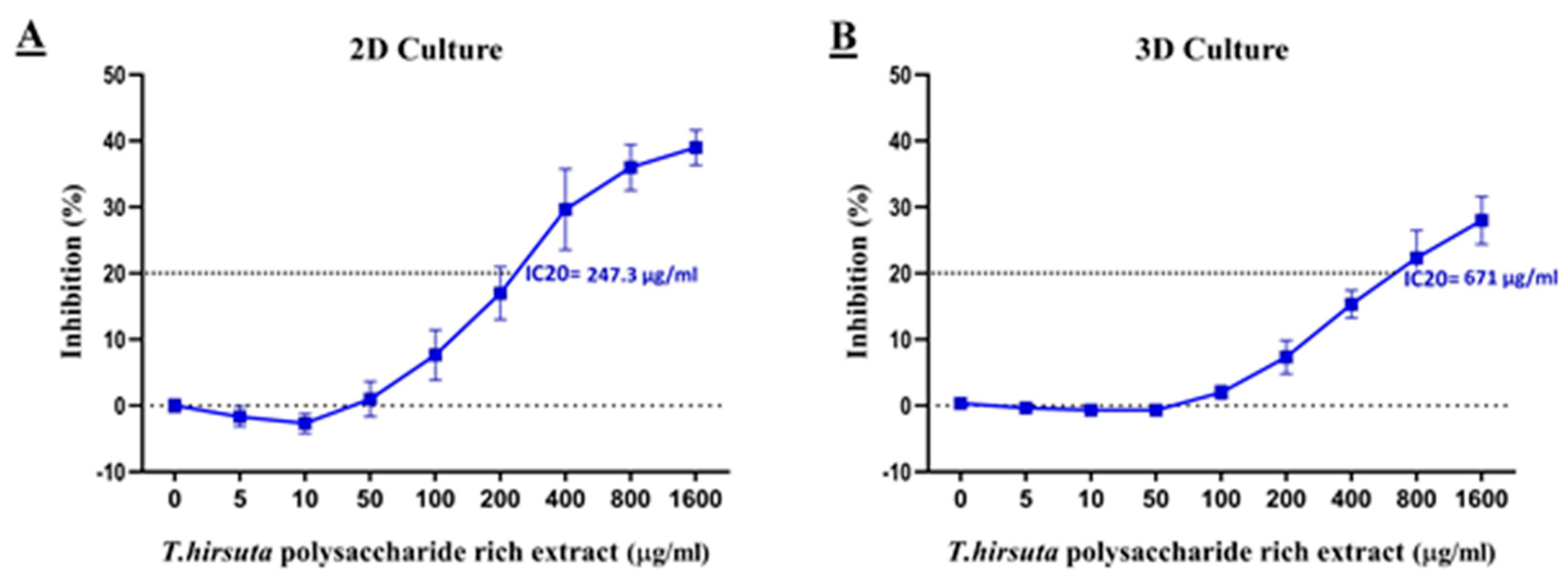
Figure 3.
The impact of non-toxic doses of T. hirsuta polysaccharide extract on cytokine secretion from the THP-1 cell line. Secretion of TNF-α in (A) 2D and (B) 3D culture. Secretion of IL-1 β in (C) 2D and (D) 3D culture. Data represented as a mean ± standard deviation. # control group, * and ** P-value <0.05 relative to the control group.
Figure 3.
The impact of non-toxic doses of T. hirsuta polysaccharide extract on cytokine secretion from the THP-1 cell line. Secretion of TNF-α in (A) 2D and (B) 3D culture. Secretion of IL-1 β in (C) 2D and (D) 3D culture. Data represented as a mean ± standard deviation. # control group, * and ** P-value <0.05 relative to the control group.
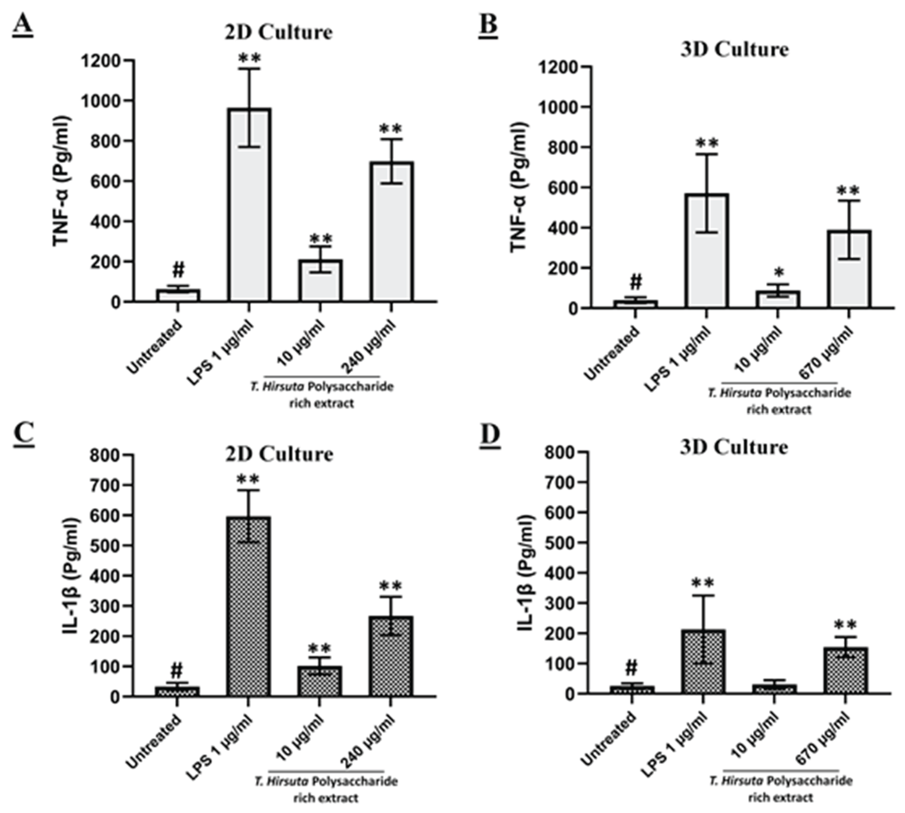

Table 1.
Ash content of T. hirsuta sample.
| Ash | % (w/w) of powdered mushroom1 |
| Total ash | 1.802% |
| Acid-insoluble ash | 0.2 % |
| Water-soluble ash | 50% |
1 Ash contents are expressed as percentage (% w/w) of the powdered mushroom sample on a dry-weight basis.
Table 2.
GC-MS analysis of the volatile fraction of T. hirsuta.
| NO | Retention time | Compounds | Reported KI | *Proportion (%) | Chemical class |
| 1 | 5.92 | Hexanal | 801 | 1.35 | Aldehyde |
| 2 | 7.23 | Furfural | 836 | 4.13 | Heteroaromatic aldehyde |
| 3 | 8.63 | n-hexanol | 870 | 1.07 | Alcohol |
| 4 | 13.53 | Benzaldehyde | 960 | 3.01 | Aldehyde |
| 5 | 14.08 | 1-Octen-3-ol | 979 | 7 | Alcohol |
| 6 | 14.36 | 3-Octanone | 983 | 0.57 | Ketone |
| 7 | 14.48 | Pentylfuran-2 | 988 | 5.61 | Heteroaromatic |
| 8 | 14.94 | Octanol -3 | 991 | 8.57 | Alcohol |
| 9 | 16.89 | 3-Ethyl-2-methyl-1,3-hexadiene | 1030 | 0.33 | Hydrocarbon |
| 10 | 17.79 | Benzene acetaldehyde | 1042 | 1.24 | Aldehyde |
| 11 | 18.3 | Octen-1-al-2E | 1054 | 1.01 | Aldehyde |
| 12 | 18.9 | Acetophenone | 1065 | 1.26 | Ketone |
| 13 | 20.1 | 1-Nonen-4-ol | 1097 | 0.33 | Alcohol |
| 14 | 20.24 | Linalool | 1096 | 0.65 | Terpenoid |
| 15 | 20.58 | Nonanal | 1100 | 1.3 | Aldehyde |
| 16 | 23.45 | 2E-Nonen-1-al | 1161 | 1.22 | Aldehyde |
| 17 | 23.9 | 1-Nonanol | 1169 | 0.3 | Alcohol |
| 18 | 24.95 | Furfuryl acetone | 1183 | 1.65 | Ketone |
| 19 | 26.3 | 2E,4E-Nonadienal | 1212 | 0.65 | Aldehyde |
| 20 | 28.35 | 2E-Decenal | 1263 | 0.33 | Aldehyde |
| 21 | 29.61 | Undecanone | 1294 | 2.1 | Ketone |
| 22 | 31.05 | 2E,4E-Decadienal | 1316 | 7.07 | Aldehyde |
| 23 | 31.8 | Megastigma-4,6E-triene | 1360 | 0.29 | Terpenoid |
| 24 | 33.06 | -Nonalactoneγ | 1363 | 0.58 | Lactone |
| 25 | 36.58 | -Barbatene β | 1442 | 0.57 | Terpenoid |
| 26 | 38.23 | Tridecanal | 1503 | 0.51 | Aldehyde |
| 27 | 40.92 | Nerolidol | 1565 | 1.26 | Terpenoid |
| 28 | 41.84 | Spatulenol | 1582 | 0.46 | Terpenoid |
| 29 | 42.02 | Caryophyllene oxide | 1583 | 0.46 | Terpenoid |
| 30 | 43.02 | Tetradecanal | 1612 | 4.42 | Aldehyde |
| 31 | 46.81 | Tetradecyloxirane | 1708 | 0.96 | Fatty aldehyde |
| 32 | 52.02 | Pentadecanoic acid | 1869 | 3.6 | Fatty acid |
| 33 | 55.32 | Hexadecanoic acid | 1960 | 4.32 | Fatty acid |
*Relative proportions are expressed as percentages of the total identified essential oil constituents. Abbreviations: KI, Kovats retention index; GC–MS, gas chromatography–mass spectrometry.
Table 3.
13C shifts (in ppm) for isolated pure compounds.
| Position of C | 1 (CDCl3) | 2 (CDCl3) |
| 1 | 38.38 | 34.906 |
| 2 | 32.00 | 30.295 |
| 3 | 71.08 | 65.104 |
| 4 | 40.81 | 37.833 |
| 5 | 139.50 | 80.021 |
| 6 | 119.52 | 135.701 |
| 7 | 116.30 | 130.020 |
| 8 | 141.40 | 71.144 |
| 9 | 46.24 | 49.414 |
| 10 | 37.34 | 37.114 |
| 11 | 19.99 | 22.938 |
| 12 | 39.06 | 39.443 |
| 13 | 43.31 | 42.834 |
| 14 | 55.12 | 55.104 |
| 15 | 23.01 | 19.671 |
| 16 | 28.32 | 28.600 |
| 17 | 56.00 | 55.914 |
| 18 | 12.00 | 12.108 |
| 19 | 16.30 | 17.627 |
| 20 | 40.26 | 40.235 |
| 21 | 21.13 | 21.142 |
| 22 | 136 | 136.041 |
| 23 | 131.8 | 131.875 |
| 24 | 42.8 | 43.304 |
| 25 | 33.66 | 33.096 |
| 26 | 21.55 | 19.987 |
| 27 | 19.66 | 21.568 |
| 28 | 17.62 | 17.547 |
Note: Experimental 13C NMR chemical shifts (δ, ppm) of compounds 1 and 2. Carbon assignments were made by comparison with previously reported spectroscopic data for ergosterol and ergosterol peroxide [26]. Abbreviations: CDCl3, deuterated chloroform. Chemical shifts are reported in parts per million (ppm) relative to the residual solvent signal.
Table 4.
The antioxidant activity and total phenolic content of two extracts of T. hirsuta.
|
Sample |
TPC (mg GAE/g of dry weight) |
FRAP (µmol Fe2+ equivalent/g dried extract) |
DPPH (IC50 value, µg/mL) |
| Methanol extract | 41.64±3.81 | 408.1667±0.11 | 488.54±26.67 |
| polysaccharides | - | 432.056±0.12 | 2860.77±25.51 |
| Ascorbic acid | - | 125.063±0.35 | 9.51±0.09 |
Abbreviations: TPC, total phenolic content; GAE, gallic acid equivalents; FRAP, ferric reducing antioxidant power; DPPH, 2,2-diphenyl-1-picrylhydrazyl; IC50, half-maximal inhibitory concentration. Values are presented as mean ± SD.
Disclaimer/Publisher’s Note: The statements, opinions and data contained in all publications are solely those of the individual author(s) and contributor(s) and not of MDPI and/or the editor(s). MDPI and/or the editor(s) disclaim responsibility for any injury to people or property resulting from any ideas, methods, instructions or products referred to in the content. |
© 2026 by the authors. Licensee MDPI, Basel, Switzerland. This article is an open access article distributed under the terms and conditions of the Creative Commons Attribution (CC BY) license (http://creativecommons.org/licenses/by/4.0/).
Copyright: This open access article is published under a Creative Commons CC BY 4.0 license, which permit the free download, distribution, and reuse, provided that the author and preprint are cited in any reuse.